Preprint
Review

This version is not peer-reviewed.

Transcatheter Paravalvular Leak Closure: A Step-by-Step Guide

Submitted:

13 January 2026

Posted:

14 January 2026

You are already at the latest version

Abstract

Paravalvular leak (PVL) remains a clinically important complication after surgical or transcatheter valve implantation, presenting predominantly with heart failure (HF) and/or high-shear hemolysis. While redo surgery can be definitive, contemporary candidates frequently carry prohibitive operative risk, positioning transcatheter PVL closure as a key therapeutic alternative. However, available outcome data are largely derived from observational series and registries with heterogeneity in PVL mechanisms, prosthesis types, imaging protocols, and endpoint definitions. Standardized frameworks—such as those proposed by the PVL Academic Research Consortium—support harmonized PVL grading and clinically meaningful composite endpoints that integrate imaging/hemodynamic results with patient-centered outcomes. Across datasets, the most consistent determinant of benefit is residual PVL severity: procedural efficacy is most commonly defined as achieving ≤mild residual regurgitation without prosthetic leaflet interference, device embolization, or major complications. This review provides a step-by-step, phenotype-driven approach to transcatheter PVL closure, emphasizing multimodality imaging (TEE and cardiac CT, with adjunct CMR and PET when appropriate), access and support planning tailored to valve position, and morphology-matched device selection—often requiring modular multi-device strategies for elongated crescentic channels, particularly in hemolysis-predominant presentations. We also synthesize evidence on complications and bailout management, with a focus on preventable high-severity events (leaflet impingement, embolization, stroke/air, vascular injury, tamponade) and standardized pre-release safety checks. Collectively, contemporary practice supports high implant success in experienced programs, with clinical improvement tightly coupled to procedural endpoint quality and careful Heart Team selection.

Keywords: 
;  ;  ;  ;  ;  ;  ;  ;  ;  ;  

1. Introduction

Paravalvular leak (PVL) is a prototypical example of how a seemingly “small” structural defect can translate into disproportionately large clinical consequences [1,2]. PVL refers to regurgitant flow between a prosthetic valve and surrounding native tissue, typically caused by incomplete apposition of the sewing ring or valve frame to the annulus or by acquired dehiscence [1,2]. PVL is encountered after surgical valve replacement and, through distinct mechanisms, after transcatheter valve implantation. Its clinical spectrum ranges from incidentally detected trivial jets to severe regurgitation with cardiogenic shock, pulmonary edema, or transfusion-dependent hemolysis [1,2,3]. Importantly, PVL severity by color Doppler alone may underestimate clinical impact: narrow, high-velocity turbulent jets can generate substantial shear stress and hemolysis even when global regurgitant volume appears “moderate,” and eccentric jets may be missed or misgraded unless assessed with a rigorous, integrative approach [1,2].
The reported incidence of PVL varies widely depending on (i) valve position (mitral > aortic in surgical series), (ii) valve type (mechanical > bioprosthetic in many datasets), (iii) imaging intensity (routine TEE vs symptom-driven evaluation), and (iv) the definition of “clinically significant PVL.” Contemporary reviews and imaging studies frequently cite mitral PVL after surgical replacement in the range of ~7–17%, while clinically significant PVL is much less common (~1–5%) [1,2,4]. In the aortic position after surgical AVR, rates are generally lower; one contemporary cohort study reported postoperative PVL around ~5% overall (with much lower intraoperative PVL), although incidence estimates differ across eras and techniques [5,6]. Long-term surgical data also emphasize that “major” PVL can occur years after implant, reflecting ongoing tissue remodeling, suture degeneration, infection risk, and annular calcification dynamics rather than purely technical failure [7,8,9].
In TAVI, the phenomenon is related primarily to frame–annulus interaction, calcific asymmetry, underexpansion, malposition, and device-generation characteristics. Moderate or severe paravalvular aortic regurgitation has historically occurred in a meaningful minority of early-generation implants and has been repeatedly associated with worse outcomes; while newer devices have reduced rates, PVL remains a persistent “Achilles heel” because even moderate–severe PVL after TAVI is linked to increased mortality across registries and meta-analyses [10,11,12,13]. A standardized, phenotype-driven operator workflow integrating multimodality imaging, access planning, escalation strategies, and endpoint definition is summarized in the Central Illustration (Figure 1).

2. Pathophysiology

PVL is fundamentally a mechanical failure of the prosthesis–tissue interface, but its clinical expression is governed by the interaction of (i) the structural substrate (defect geometry, channel length, circumferential extent, and dynamic annular motion), (ii) the fluid mechanics of high-velocity regurgitant flow, and (iii) host factors (tissue quality, infection/inflammation, coagulation/anticoagulation, baseline ventricular and pulmonary vascular reserve, and red-cell vulnerability). Contemporary reviews emphasize that PVL should be conceptualized less as a single “hole” and more as a spectrum of irregular, often crescentic, multi-orifice channels whose physiology cannot be reliably inferred from a single 2D color Doppler frame [2,14,15].

2.1. Structural Substrate

2.1.1. Surgical Prostheses

In surgical valves, PVL most commonly arises from separation of the sewing ring (or annuloplasty ring) from adjacent annular tissue. Early PVL (days–weeks) may reflect technical factors, suture disruption, tension on friable tissue, or unrecognized infection; late PVL (months–years) reflects progressive interface degeneration driven by calcific remodeling, chronic inflammatory processes, and mechanical stress concentration at the ring–tissue boundary. Multiple reviews and consensus endpoint documents consistently list predisposing factors such as annular calcification, tissue friability, prior endocarditis, active corticosteroid therapy, and (in many series) mechanical prosthesis type, particularly in redo settings where annular integrity is compromised [2,14,16,17].
A critical nuance—highly relevant to closure strategy—is that many clinically important PVLs are not circular defects but crescentic arcs along the sewing ring, sometimes with multiple exit points (multiple color jets) from a shared channel. This morphology explains why “one large round device” frequently fails to eliminate the high-velocity component of flow and why multi-device or oblong/rectangular device concepts have a physiologic rationale [2,14,15].

2.1.2. Transcatheter Valves (TAVI)

In TAVI, PVL (often termed paravalvular regurgitation, PVR) is driven less by “dehiscence” and more by incomplete sealing between the stent frame and the native annulus/leaflet–calcification complex. Mechanistically, PVR reflects a combination of annular eccentricity, asymmetric or bulky calcification at the landing zone, prosthesis malposition (too high/too deep), and underexpansion. Importantly, PVR after TAVI often consists of multiple, eccentric, irregular jets that follow serpiginous channels along calcific shelves—morphology that differs from classic surgical PVL and complicates both quantification and closure [18,19,20].
Calcification burden and distribution are particularly influential: studies linking device-landing-zone calcification to residual PVL provide a direct structural explanation for why some anatomies remain challenging despite modern device skirts [18,20].

2.2. Fluid Mechanics

PVL flow typically traverses a short, constricted channel with a high-pressure gradient (especially left-sided lesions), producing high-velocity turbulent jets. Turbulence and abrupt changes in flow direction generate regions of elevated shear stress, flow separation, and vortical structures—features that (i) amplify energy loss and hemodynamic burden and (ii) drive hemolysis when shear exposure exceeds red-cell tolerance. Large outcome series and interventional reviews explicitly attribute PVL-related hemolysis to turbulent flow through the defect increasing red blood cell shear stress, causing mechanical trauma and fragmentation [14,16].
Computational fluid dynamics (CFD) work underscores a key clinical observation: hemolysis severity does not scale linearly with a single geometric parameter (e.g., “defect diameter”). Instead, hemolysis risk relates to where shear concentrates, how long erythrocytes are exposed, and what volume of blood experiences suprathreshold shear. In a CFD simulation study of mitral PVL, investigators reported no simple relationship between PVL geometry and hemolysis risk and highlighted the importance of exposure time and the volume of flow exceeding critical shear values [21,22].
This explains why a “moderate” PVL by regurgitant fraction can still cause severe hemolysis (if a narrow, high-velocity component persists), and reducing PVL from moderate to mild may improve HF symptoms, yet hemolysis may persist unless the high-shear micro-jet is eliminated.

2.3. Chamber-Specific Hemodynamic Consequences

2.3.1. Mitral PVL

Mitral PVL behaves physiologically like eccentric MR but with important differences: jets are frequently highly directional, may hug the atrial wall, and can be underestimated by transthoracic imaging. The immediate hemodynamic signature in significant mitral PVL is elevation in left atrial pressure and V-wave, with secondary pulmonary venous hypertension and downstream RV strain. Chronicity drives LA remodeling, atrial arrhythmias, and progressive pulmonary vascular disease in susceptible patients. Reviews emphasize that mitral PVL commonly presents with HF symptoms and may coexist with hemolysis, particularly in mechanical valves where hinge-adjacent jets are especially shear-intensive [2,14,15].

2.3.2. Aortic PVL

Aortic PVL creates diastolic runoff from the aorta into the LV, reducing aortic diastolic pressure and increasing LV end-diastolic volume. Clinically, this translates into exertional dyspnea, reduced forward cardiac output reserve, and in severe cases hypotension or angina (via reduced coronary perfusion pressure). In TAVI specifically, multiple analyses have shown that moderate–severe PVR is prognostically adverse, supporting the concept that even “modest” residual PVL can be physiologically consequential in a population with limited reserve [19,20].

2.3.3. Tricuspid and Pulmonary PVL

Right-sided PVLs are rarer but illustrate an important physiologic contrast: lower pressure gradients generally reduce hemolysis risk, yet volume loading of the RA/RV can produce profound systemic venous congestion (ascites, hepatic congestion, cardiorenal syndrome). The low-pressure environment can also reduce “self-anchoring” forces for occluders, increasing embolization propensity and making stability testing physiologically (not just technically) essential [14,15].

2.4. Hemolysis

Across modern surgical and transcatheter prostheses, “subclinical hemolysis” can be seen, but clinically relevant hemolysis is most strongly linked to high-shear lesions, especially PVL. A contemporary JACC-focused review on mechanical hemolysis in valvular disease synthesizes how shear stress from turbulent jets—before and after valve interventions—drives red-cell deformation, membrane fatigue, and fragmentation, and it emphasizes that anemia severity is modulated by baseline hematologic reserve and iron handling [16,23].
Experimental and modeling literature has proposed shear thresholds above which hemolysis becomes likely. Classic theoretical work discussed hemolysis thresholds in the hundreds of N/m² (Pa) range, and modern CFD-oriented PVL publications frequently reference ~300 Pa as a critical value for erythrocyte destruction in vitro, while also noting that hemolysis depends on both shear magnitude and exposure time [21,24,25].
The practical clinical implication is that hemolysis is often a “micro-jet disease,” not a “regurgitant volume disease.” Thus, in hemolysis-predominant PVL, the procedural endpoint should be framed as elimination of the high-velocity residual component, not merely downgrading severity by an integrative qualitative grade.
From a mechanistic standpoint, hemolysis severity tends to increase when: i) the PVL channel is narrow with a steep pressure gradient (high velocity), ii) there is impingement of the jet on prosthetic struts/hinges (increasing turbulence), iii) the channel has complex curvature producing local shear hot-spots, and iv) host factors reduce red-cell resilience (iron deficiency, renal dysfunction, inflammatory states) [21,22,25].
Outcome registries have shown that patients presenting with hemolysis—especially with mechanical valves—have a lower likelihood of meeting composite “clinical success” definitions after transcatheter PVL closure. The most physiologically plausible explanation is that even mild residual PVL can maintain a high-shear micro-jet sufficient for ongoing hemolysis, whereas HF symptoms may improve with partial reduction [26].

2.5. Infection and Inflammation

PVL can be both a consequence of infective endocarditis (IE) via annular abscess, tissue destruction, and dehiscence; and a substrate for IE by creating areas of abnormal flow, endothelial disruption, and potential microthrombus formation.
Major PVL reviews emphasize the association between clinically significant PVL and IE, particularly in cases of rapid PVL progression, new dehiscence, systemic inflammatory features, or imaging signs of periannular complications [2,15].
Mechanistically, ongoing inflammatory or degenerative processes at the annulus can lead to progressive separation over time, explaining late PVL and the not-uncommon need for repeat intervention when the underlying interface remains biologically unstable.

2.6. Integrative Pathophysiology

Putting the above together, PVL physiology naturally clusters into two dominant, sometimes overlapping phenotypes: i) HF phenotype: driven by regurgitant volume and chamber pressure/volume loading (LA pressure and pulmonary venous hypertension in mitral PVL; diastolic runoff and LV volume load in aortic PVL), and ii) Hemolysis phenotype: driven by high-shear turbulent micro-jets through constricted channels, often amplified by jet impingement on prosthetic structures.
This phenotype axis is not merely descriptive—it dictates which imaging features matter most (circumferential extent/volume load vs micro-jet localization), what procedural endpoint is truly “physiologic success,” and why multi-device strategies are often necessary even when the residual grade appears “acceptable” by conventional echo descriptors.

3. Severity Assessment

Unlike native-valve regurgitation, PVL is typically eccentric, frequently multi-orifice, often crescentic, and commonly affected by acoustic shadowing from the prosthesis and surrounding calcification. Consequently, single-parameter approaches (e.g., jet area, single 2D VC width) are unreliable and can systematically under- or overestimate severity. Contemporary prosthetic valve imaging guidance therefore emphasizes multiview, multiparametric echocardiography complemented by CT for anatomic definition and CMR when quantification remains uncertain or when echo/angiography are discordant [27,28,29,30].
A second, clinically crucial nuance is that PVL “severity” has two partially dissociable dimensions: i) global regurgitant burden (driving HF physiology), and ii) localized high-shear jet physiology (driving hemolysis). This is why “mild residual PVL” may be clinically acceptable in an HF phenotype yet insufficient in hemolysis-dominant PVL, where the procedural target becomes elimination of the high-velocity residual micro-jet rather than merely downgrading an overall grade [17,26].
Historically, many studies reported PVL in 3 classes (mild/moderate/severe). However, intermediate grades are common and difficult to estimate reproducibly—particularly after TAVI, where multiple eccentric jets are typical. Both VARC-3 and the PVL Academic Research Consortium recommend a 5-class scheme (trace; mild; mild–moderate; moderate; moderate–severe; severe) to improve communication, trial standardization, and prognostic discrimination [17,31,32].

3.1. Mitral PVL

For mitral PVL, TEE—particularly 3D TEE—is usually central because of proximity and superior paravalvular delineation compared with TTE. 3D color Doppler enables en-face “surgeon’s view” mapping of PVL location, circumferential extent, and (when feasible) planimetry of the regurgitant orifice/channel entrance, which is often crescentic rather than circular [28,30,33,34].
Core qualitative/semiquantitative markers include i) jet number and distribution: multiple jets may reflect a single crescentic channel with multiple exits, ii) proximal jet width/circumferential extent along the sewing ring (short-axis or en-face 3D), iii) CW Doppler density and contour (supportive only), iv) pulmonary venous flow: systolic blunting or reversal supports significant MR physiology (interpret with AF, elevated LA pressure, and mitral stenosis cautiously), and v) LA pressure surrogates: large V-waves (invasive or echo surrogate) support hemodynamic significance.
Classical PISA/EROA methods are frequently unreliable in PVL (non-hemispheric convergence, multiple orifices, irregular channels). Volumetric regurgitant calculations may be attempted but are error-prone in AF or when coexistent lesions exist. Consequently, guidelines endorse an integrated approach and recommend adjunct imaging when clinical severity and echo grade are discordant [27,28,30].

3.2. Aortic PVL

In the aortic position, TTE is often more informative than TEE for Doppler quantification, but TEE and especially 3D TEE can be valuable for mechanism and jet localization (particularly intraprocedurally) [28,30]. Echo parameters used in an integrative grading strategy include: i) circumferential extent of paravalvular jets in short-axis (key discriminator in many schemas), ii) VC width / multiple VC widths (limited by shadowing and multi-jet anatomy), iii) diastolic flow reversal in descending/abdominal aorta (holodiastolic reversal supports severe AR physiology but depends on HR, compliance, and BP), iv) pressure half-time (supportive but confounded by LV compliance/acute hemodynamics). Several integrative schemas relate circumferential jet extent to severity strata (commonly around the 10–20–30% landmarks) and use these as anchors for intermediate grades in expanded grading systems, recognizing that precision is imperfect and should be corroborated by other hemodynamic markers [31,32,35].

3.3. Multimodality Imaging

3.3.1. Cardiac CT

CT is less about “grading severity” and more about defining the structural severity substrate: channel length, spatial relation to struts/leaflets/calcification, proximity to coronary ostia, and fluoroscopic projection planning for crossing and device orientation. Guidelines increasingly recognize CT’s role in prosthetic valve dysfunction assessment, especially when echo windows are limited [28,30].

3.3.2. CMR

CMR phase-contrast flow imaging can provide robust quantification of aortic regurgitation burden when echo/angiography are discordant, and multiple studies demonstrate that echo can underestimate post-TAVI regurgitation compared with MRI/CMR [36,37,38]. A clinically useful and widely cited post-TAVI CMR approach is regurgitant fraction (RF) grading. In a multicenter study, CMR AR grades were defined as none/trace RF <15%, mild 16–29%, and moderate/severe ≥30%, and RF (per 5% increase) was strongly associated with mortality and HF rehospitalization [38]. This supports a pragmatic paradigm: use echo for mechanism + localization + bedside decisions, and use CMR for definitive regurgitant burden when needed for adjudication, prognostication, or unexplained symptoms.

3.3.3. Angiography

Aortic root angiography (often Sellers’ visual grading) is widely used immediately after TAVI because it is quick and “minimalist,” but it is operator dependent and shows only modest agreement with echo parameters; overlap between grades is substantial [39,40]. Novel quantitative aortography/videodensitometry approaches have been proposed to improve objectivity, but availability and workflow integration vary across centers [41].

4. Procedural Endpoints for PVL Closure

PVL closure is prone to heterogeneity in reporting because success can be expressed as: i) final residual PVL severity (“≤ mild”), ii) relative reduction (“≥1 grade reduction”), or iii) clinical response (NYHA improvement; transfusion independence).
To address this, the PVL Academic Research Consortium proposed standardized clinical trial principles and endpoint definitions, including a consistent severity scheme and discrete procedural success categories [17]. Similarly, post-TAVI device success and PVR reporting are framed within VARC-3 definitions [31].

4.1. Technical Success

Across the PVL literature and consensus documents, “technical success” generally includes i) successful delivery and stable deployment of closure device(s) across the intended PVL channel(s), ii) no interference with prosthetic leaflet/disc motion or prosthesis function, iii) no need for emergency surgery because of device-related complications, and iv) no intraprocedural catastrophic events (often including death/stroke in ARC-style definitions) [17].

4.2. Procedural Success

Many registries define procedural success as technical success plus a meaningful reduction in PVL (commonly ≥1 grade reduction or reduction to no more than mild/moderate, depending on the grading system used) [1,2,42]. Because grading schemas differ (3-class vs 5-class), the manuscript should state the exact scale and the target residual grade a priori.

4.3. Clinical Success

Clinical success should be explicitly tied to the presenting phenotype:
  • HF phenotype: improvement by ≥1 NYHA class, improved functional capacity, and/or reduction in HF hospitalizations;
  • Hemolysis phenotype: improvement in hemolysis markers and, critically, freedom from transfusion/erythropoietin dependence, when hemolysis is the dominant indication.
This definition is consistent with widely cited outcome studies and remains pragmatic for real-world registries. Notably, multicenter data suggest lower clinical success among patients whose index phenotype is hemolysis and/or in mechanical valves—supporting the mechanistic concept that even “small residual PVL” can maintain high-shear hemolysis [17,26,43].
Table 1. Phenotype-driven severity anchors and procedural endpoints for paravalvular leak (PVL) closure.
Predominant clinical phenotype Practical severity anchors (pre-procedure) Minimum imaging endpoints to report Target residual PVL (pragmatic) Clinical success endpoint(s)
Heart failure–predominant PVL (mitral) Symptoms (NYHA); pulmonary venous congestion; LA dilation/V-wave; supportive Doppler signs (e.g., pulmonary venous systolic blunting/reversal when interpretable) 2D/3D TEE mapping (location, circumferential extent, number of jets); pre/post mean mitral gradient; residual PVL grade at exit + predischarge ≤ mild–moderate (5-class) or ≤ mild (3-class), and no clinically relevant iatrogenic mitral stenosis ≥1 NYHA class improvement; fewer HF admissions; QoL improvement
Heart failure–predominant PVL (aortic / post-TAVI) Dyspnea/low output; LV volume load; supportive Doppler (diastolic flow reversal) Echo integrative grade (include circumferential extent); angiography/hemodynamics if used; residual PVL at exit + predischarge Preferably ≤ mild–moderate; consider hemodynamic optimization during procedure (esp. post-TAVI) HF rehospitalization reduction; QoL; survival (longer-term)
Hemolysis-predominant PVL (any position; often mechanical) Hemoglobin trend; LDH↑; haptoglobin↓; indirect bilirubin↑; transfusion/EPO requirement Precise jet localization (often 3D TEE); document elimination of high-velocity residual micro-jet As close to none/trace as achievable (hemolysis is “micro-jet sensitive”) Transfusion independence; improvement/normalization trend of hemolysis labs
Mixed HF + hemolysis Combined anchors above Full integrative imaging set + labs Aim for lowest achievable residual grade without prosthesis compromise NYHA improvement and hemolysis improvement/transfusion-free

5. Devices for Transcatheter PVL Closure

PVL channels are rarely circular; they are frequently crescentic, multi-orifice, and short, irregular tunnels between the sewing ring/stent frame and annular tissue. As a result, the “ideal” PVL occluder should (i) conform to noncylindrical geometry, (ii) provide high sealing efficiency (often with fabric), (iii) remain repositionable/retrievable until final leaflet-motion and residual-jet checks are complete, and (iv) minimize leaflet/disc interference and LVOT/coronary impingement. The limitations of using non-dedicated vascular plugs for PVL—especially the risk of incomplete sealing and leaflet interaction—have been demonstrated in bench and clinical literature and directly motivated dedicated PVL platforms [1,44,45].

5.1. Dedicated PVL Occluders

5.1.1. Occlutech Paravalvular Leak Device (PLD)

The Occlutech PLD is a double-disc nitinol device engineered specifically for PVL anatomy, offered in square and rectangular configurations to better match crescentic/elliptical channels; it incorporates PET patches to enhance sealing and includes radiopaque (gold) markers to facilitate positioning. It is repositionable and fully retrievable and is designed for delivery via antegrade (e.g., transseptal) or retrograde routes depending on anatomy and operator strategy [46,47]. Large multicenter experience has shown high implant and clinical improvement rates with the PLD in both mitral and aortic PVL [46,47]. Longer-term follow-up demonstrated sustained improvement in NYHA class and reduced transfusion dependence in hemolysis-driven cases, with no deaths adjudicated as device-related [48].
Device selection “pearls” (device-specific).
  • Rectangular PLD: best suited for elongated/crescentic defects where sealing is required along an arc;
  • Square PLD: useful for more compact defects;
  • Channel characteristics matter: registry/experience papers stress matching device configuration to channel length and cross-sectional area, avoiding inappropriate oversizing that can increase interference risk [46,47].

5.1.2. Amplatzer Vascular Plug (AVP)

The Amplatzer plug family remains the most widely used platform for transcatheter PVL closure in routine practice, spanning earlier cylindrical vascular plugs (AVP II), low-profile options (AVP IV), and the oblong/rectangular-oval Amplatzer Valvular Plug III (historically reported in several series as “AVP III”), which was developed to better match the common elliptical/crescentic morphology of PVL channels and reduce “round-hole mismatch,” while maintaining a self-expanding nitinol architecture and retrievability/repositionability—features that are particularly valuable when defects are adjacent to mechanical hinges or transcatheter valve frames [49,50]. Among the largest early clinical experiences, mitral/aortic PVL closure with AVP III achieved high implant success and clinically meaningful PVL reduction, while also underscoring a key device-selection constraint: prosthetic leaflet interference, though infrequent, can be catastrophic and should be actively excluded before release [49,51]. Prospective evidence generation is ongoing through the PARADIGM multicenter single-arm study evaluating safety and effectiveness of AVP III in clinically significant aortic/mitral PVL after surgical valve implantation [52]. A single-center “rectangular PVL plug” experience reported favorable mechanical behavior (auto-orientation, minimal leaflet interaction in that cohort) but also highlighted the practical reality that crescentic PVLs frequently require multi-device strategies even with an oblong platform [53]. In contrast, AVP II remains useful primarily for more tubular/round channels where a cylindrical plug can seat securely, but experimental in vitro work demonstrated that vascular plugs may not reliably produce substantial PVL reduction and can interact with prosthetic leaflets—supporting the preference for morphology-matched devices and careful intraprocedural imaging checks [26,44]. Finally, AVP IV has become a practical “workhorse” in anatomies where crossability and deliverability are limiting—particularly post-TAVI PVL constrained by stent-frame geometry—an observation reflected in post-TAVI PVL literature and in the international PLUGinTAVI registry, where AVP III was most frequently used, followed by AVP IV [10].

5.2. Ductal, Septal, and VSD Occluders

These devices predate dedicated PVL platforms and were historically used because PVL-specific devices were not available. Contemporary series still employ them as “niche tools,” particularly when a PVL channel behaves like a short duct/tunnel and disc-based anchoring is advantageous.

5.2.1. Amplatzer Duct Occluder (ADO) and ADO II

Disc-based occluders can be useful when a PVL channel has a tubular segment that can seat the waist while the discs provide stability. ADO II has been reported for mitral PVL closure (including retrograde approaches in select cases) and has also been described in tricuspid PVL closure in high-risk surgical patients [54,55].
Because ADO II is designed with two articulating discs and a conformable mesh architecture, it may be deliverable through relatively low-profile systems (depending on size), which can be a practical advantage in difficult-to-cross channels [55].

5.2.2. Muscular VSD Occluders and ASD Occluders

Muscular VSD occluders offer larger discs and waist configurations that can be helpful for larger PVL channels with adequate landing zones; ASD occluders have also been used historically. However, larger discs can increase the risk of prosthetic leaflet/disc interference—a complication repeatedly observed across PVL closure experience and one of the core reasons dedicated PVL devices with optimized profiles were developed [49,56,57].

5.3. Coils and Adjunctive “Micro-Jet” Solutions

For very small residual PVL jets (particularly when hemolysis persists from a high-shear residual micro-jet), coils have occasionally been used as adjuncts in combination with plugs, but their role is limited by the need for stable anchoring, embolization risk, and less predictable sealing compared with fabric-containing occluders. Most contemporary practice patterns favor plug-based solutions for reproducibility and retrievability, reserving coils for exceptional anatomies or residual pinhole channels once a stable scaffold is established.

5.4. Multi-Device Strategies and Device Combinations

A critical device-related principle is that many clinically significant PVLs—especially crescentic defects spanning a substantial arc—are unlikely to be fully sealed with a single plug. Classic interventional reviews state that crescent-shaped defects extending over a substantial portion of the prosthesis circumference often require two or more devices, deployed sequentially or simultaneously, to achieve near-complete sealing [57,58].
This is particularly relevant to hemolysis phenotypes, where leaving even a small residual high-velocity component can perpetuate hemolysis.
In real-world series, even when a dedicated oblong/rectangular plug is used, additional devices may be required to address multi-orifice exits or to “finish” residual channels—an observation explicitly reported in rectangular PVL plug experience [53].
Table 2. Device platforms for transcatheter paravalvular leak closure: indications, advantages, and limitations.
Device / platform Typical PVL morphology where it fits best Key advantages Key limitations / cautions
Occlutech PLD (square/rectangular; waist/twist) Crescentic/elliptical PVL; irregular channels (mitral & aortic) Purpose-built geometry; PET patches; markers; retrievable/repositionable Requires careful sizing to avoid leaflet interaction; availability varies by region
Amplatzer Valvular Plug III / oblong PVL plug concept Elliptical/crescentic PVL; multi-orifice channels Oblong geometry; broad size range; widely used in PVL practice; under formal evaluation (PARADIGM) Leaflet interference remains a risk; sometimes multiple devices needed
AVP II More tubular/round PVL channels Familiar platform; effective occlusion in suitable geometries Less conformable for crescentic PVL; can interact with leaflets if protruding
AVP IV (low profile) Small channels; post-TAVI PVL where crossability is limiting Low-profile deliverability; common in post-TAVI PVL closure practice Not ideal for large crescentic defects; may require multiple devices
ADO / ADO II Short tunnel-like PVL; selective mitral/tricuspid PVL cases Disc-based stability; can be useful when “duct-like” anatomy exists Off-label in PVL; embolization/interference risk if landing zone is marginal
Muscular VSD / ASD occluders Large defects with adequate landing zone Large discs/waist options Higher interference risk; not designed for PVL geometry

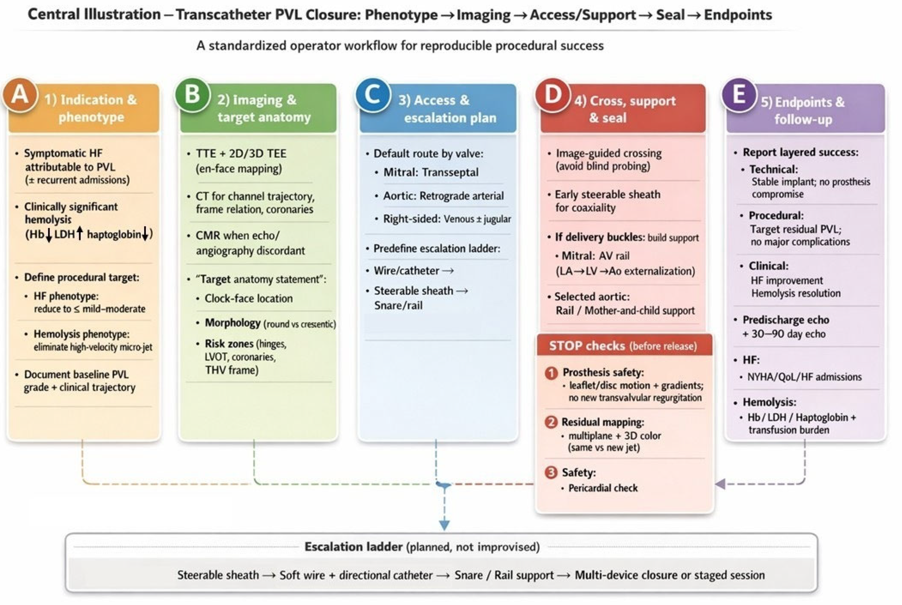
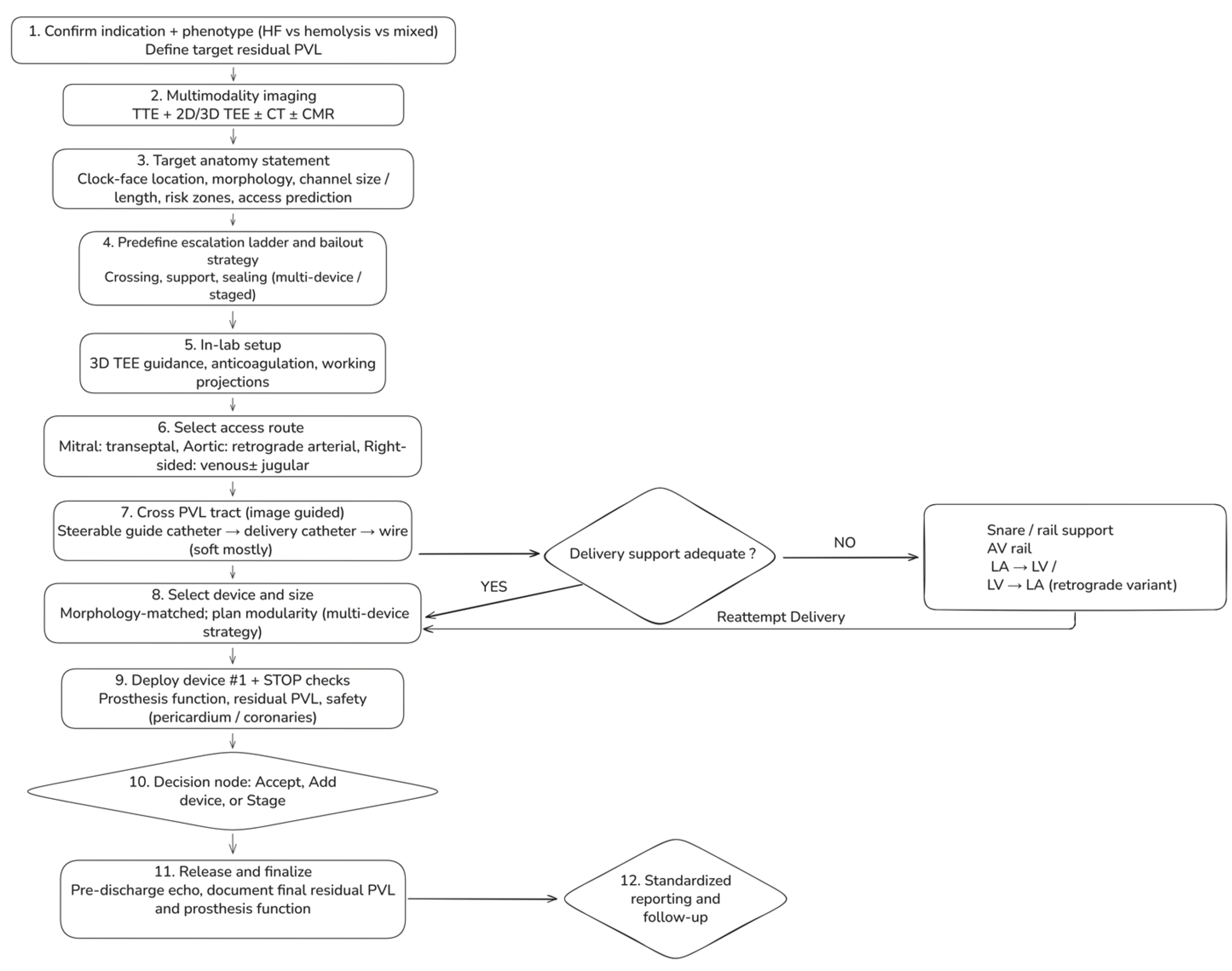
6. Step-By-Step Workflow For Transcatheter PVL Closure

A detailed step-by-step procedural workflow is illustrated in Figure 2.
A standardized, phenotype-driven workflow integrating multimodality imaging, access planning, and planned escalation strategies. The process begins with confirmation of indication and dominant phenotype (heart failure, hemolysis, or mixed), which defines the target residual PVL. Pre-procedural assessment combines transthoracic and 2D/3D transesophageal echocardiography with selective use of cardiac CT and CMR to generate a target anatomy statement. In-lab steps include access selection according to valve position, image-guided PVL crossing, and evaluation of delivery support. When support is inadequate, snare-assisted or arteriovenous rail techniques are used before reattempting delivery. Devices are selected according to defect morphology, with modular multi-device strategies for complex channels. Mandatory STOP checks precede device release, followed by a decision to accept, add devices, or stage the procedure, and standardized post-procedural reporting and follow-up.

6.1. Step 1 — Confirm Indication And Define A Phenotype-Driven Procedural Target (Pre-Procedure clinic/Heart Team)

The procedural plan should begin by documenting three items explicitly: (1) indication, (2) predominant phenotype, and (3) target residual PVL. Indications are typically (i) heart failure attributable to PVL and/or (ii) clinically significant hemolysis (transfusion/iron/EPO requirement with supportive laboratory features). The phenotype should determine the endpoint: HF-predominant patients frequently experience meaningful improvement with reduction to ≤mild–moderate, whereas hemolysis-predominant PVL is “micro-jet sensitive” and often requires near-elimination of the high-velocity residual component—commonly with planned multi-device sealing. This phenotype-to-endpoint logic is aligned with ARC PVL principles and post-TAVI registry experience emphasizing the prognostic importance of achieving mild or less residual regurgitation when safely feasible [10,17].
Minimum labs (hemolysis phenotype): Hb, LDH, haptoglobin, indirect bilirubin, reticulocytes, iron studies, and objective documentation of transfusion burden (e.g., units/month).

6.2. Step 2 — Multimodality Imaging Briefing: A One-Page “Target Anatomy Statement” (TEE + CT ± CMR)

Before entering the lab, the team should share a concise “target anatomy statement” that includes:
  • Valve position and prosthesis type (mechanical/bioprosthetic/transcatheter; ViV/ViR, if applicable).
  • PVL location using a standardized clock-face (mitral surgeon’s view; aortic short-axis orientation).
  • Morphology: round vs crescentic/elliptical; single channel with multiple exits vs multiple discrete channels; estimated channel length, maximum width, and circumferential extent.
  • Risk zones: proximity to mechanical hinges, coronary ostia (aortic), LVOT relevance (mitral), and THV frame constraints (post-TAVI).
  • Access prediction: anticipated approach (transseptal vs retrograde) and likelihood of needing a rail strategy.
CT planning is particularly valuable for: post-TAVI PVL/PVR, redo valves with heavy calcification, discordant echo vs clinical picture, suspected long/tortuous channels, and coronary proximity concerns.

6.3. Step 3 — Predefine an “Escalation Ladder” (Avoid Improvisation Mid-Case)

A predefined escalation ladder should cover:
  • Crossing (steerable sheath → catheter → wire → alternative access)
  • Support (long sheath/mother-and-child → snare-assisted redirection → arteriovenous rail)
  • Sealing (single device → multiple devices → staged closure)

6.4. Step 4 — Room Setup and Intraprocedural Imaging Workflow

General anesthesia is commonly preferred for complex mitral PVL because continuous high-quality 3D TEE guidance is pivotal; selected aortic cases may be feasible under deep sedation if imaging is robust. Imaging roles should be explicit: the echocardiographer provides en-face localization, continuous prosthesis–device interaction assessment, and structured residual mapping after each deployment; the operator calls for mandatory “stop checks” before release.

6.5. Step 5 — Access Strategy (Default and Bailout)

  • Mitral PVL (default: transseptal): Select puncture height and posterior/anterior orientation to optimize catheter coaxiality toward the intended clock-face region. Posterior–inferior puncture often improves reach for posterior leaks; a more superior puncture may improve alignment for anterior leaks, while an overly anterior puncture can promote extreme angulation, prolapse, and early need for rail rescue.
  • Aortic PVL (default: retrograde arterial): Select working projections that “open” the prosthesis and profile the suspected PVL location; when available, CT-derived angulation can reduce trial-and-error. Coronary protection should be considered selectively when anatomy is borderline.
  • Right-sided PVL: Venous access; jugular access often improves coaxiality for tricuspid targets depending on defect location and RA geometry.

6.6. Step 6 — Cross the PVL Tract (Image-Guided, Not “Search-and-Probe”)

Crossing should be performed as a controlled, image-guided maneuver. A pragmatic sequence is: steerable guide catheter (often advantageous for alignment and subsequent multi-device delivery) + directional catheter + soft hydrophilic wire to engage the paravalvular gutter under 3D TEE en-face guidance; once crossed, advance the catheter to stabilize access and reduce wire-induced trauma, then exchange to a supportive wire for delivery as needed. Consider a buddy wire strategy to preserve access after initial device delivery in complex multi-plug closures. If deliverability remains inadequate—or tract engagement is unstable—proceed to snaring/rail escalation.
Safety rule: if intraluminal tract position is not unequivocal (i.e., concern for subannular/extracardiac passage), stop and re-image before any escalation.

6.7. Step 7 — Create support and Apply “Rail Physics” (Snaring + Arteriovenous Rail)

When a delivery system buckles, the dominant limitation is usually support/coaxiality, not device design. Large-bore steerable systems improve support; however, an arteriovenous (AV) rail may be required for either tract stabilization or sheath deliverability.
  • Mitral PVL AV rail (veno–arterial externalization; high-yield rescue): cross PVL from LA → LV, advance into the aorta, snare and externalize via arterial access, then advance the delivery sheath across the tract on stable rail support. This converts unstable pushing into controlled tracking and reduces repeated prolapse.
  • Retrograde variant: in selected anatomies, cross retrogradely from LV → LA and snare/externalize via venous access to establish an AV rail; use snaring also to redirect trajectory and straighten tortuosity before sheath advancement.

6.8. Step 8 — Device selection and Sizing (Morphology-Matched, with Planned Modularity)

Device choice is constrained first by crossability and landing zone, then refined by morphology. Round/tubular channels may be amenable to single-plug closure. Crescentic/elliptical arcs frequently require oblong/rectangular platforms and/or planned multi-device closure; oversizing a single round device risks hinge/leaflet interaction while leaving crescentic gaps. Size devices to the true tract/channel dimensions (3D TEE/CT), not to qualitative color jet appearance. In hemolysis phenotypes, a persistent high-velocity residual micro-jet should trigger escalation to additional sealing rather than acceptance of a “mild” global grade.

6.9. Step 9 — Deployment and Mandatory “Stop Checks” Before Release

A standardized sequence is: position sheath across tract → deploy distal disc/anchor → seat waist with gentle retraction → deploy proximal disc maintaining coaxiality → stop checks → release only when all checks pass. Stop checks after each device should include:
  • Prosthesis function: no mechanical leaflet/disc restriction; no new transvalvular regurgitation; no clinically relevant gradient rise.
  • Residual PVL mapping: multiplane color + 3D color; determine whether residual jets represent remnant vs new channel.
  • Safety: pericardial assessment after extensive manipulation; coronary compromise evaluation in aortic/root-adjacent cases when relevant.

6.10. Step 10 — Decision Node: Accept vs Add Device vs Stage

After Device #1, classify result: (i) accept if the prespecified target is met (HF: ≤mild–moderate; hemolysis: no meaningful residual high-velocity jet), (ii) add device if residual remains clinically relevant and anatomy is favorable, or (iii) stage if procedure time, hemodynamics, contrast burden, or access trauma imply diminishing returns.

6.11. Step 11 — Post-Procedure Assessment And Standardized Reporting

Report outcomes in layers: technical success, procedural success (including prespecified residual target), and phenotype-linked clinical success. Minimum follow-up should include predischarge echo, early follow-up echo (30–90 days), hemolysis laboratory trends when relevant, and HF hospitalization tracking.

7. Valve-Specific Strategy and Operator Pearls

7.1. Mitral PVL

Mitral PVL is often the most technically demanding subtype because transseptal trajectory is frequently non-coaxial, defects are commonly crescentic with multi-orifice exits, and prosthesis-related shadowing may conceal residual jets; thus, contemporary imaging guidance emphasizes 3D TEE en-face mapping for localization and intraprocedural decision-making, with CT used selectively to define tract trajectory and assist transseptal planning in complex redo anatomies.
Transseptal access is the default in most mitral PVLs, but the puncture should be treated as a procedural determinant: posterior–inferior puncture commonly improves reach for posterior leaks and reduces extreme angulation, while a more superior puncture may improve alignment for anterior targets. 3D TEE (bicaval and short-axis) should confirm puncture position relative to the fossa, ensuring adequate height for device delivery while avoiding an excessively superior puncture that limits inferior reach.
In mitral PVL, tract crossing is frequently achievable with hydrophilic wire + directional catheter; the limiting step is often delivery stability. Early use of a steerable sheath (Agilis/Destino/Oscor-class) is frequently first-line in high-volume practice to improve coaxiality, maintain stable engagement, and reduce blind probing in friable planes. When recurrent prolapse or buckling occurs, an arteriovenous rail provides a mechanical solution by converting unstable push into controlled tracking—particularly valuable for medial/posterior crescentic leaks or long/tortuous tracts.
Because mitral PVLs are frequently crescentic, attempting to seal a long arc with a single oversized round device predictably risks incomplete sealing and increases mechanical interference risk. Morphology-matched platforms and planned sequential multi-device closure are often necessary; outcome frameworks and multicenter experiences support the concept that clinical benefit is tightly coupled to the degree of residual PVL reduction, with hemolysis presentations requiring particularly aggressive elimination of high-shear residual jets.
In mechanical mitral prostheses, procedural success is contingent on complete preservation of disc/leaflet excursion. Any suggestion of hinge restriction prior to release should prompt immediate recapture and strategy revision (smaller device, altered orientation, or modular multi-device sealing).
Pearls (mitral):
  • Standardize clock-face mapping (surgeon’s view) between imager and operator before any wire crosses.
  • If you “need a stiffer wire,” pause: most failures are geometry/support failures—change projection, use steerability, or build a rail instead of escalating stiffness blindly.
  • In hemolysis phenotype, do not accept a small, high-velocity residual jet even if global grade looks mild; this is a common reason for “procedural success but clinical failure.”

7.2. Aortic PVL

Surgical aortic PVLs are often more focal than mitral PVL and frequently amenable to a retrograde arterial approach with direct tract engagement. The key intraprocedural hazard is device protrusion into the valve or root with leaflet restriction or coronary compromise; therefore, projection selection should aim to open the prosthesis and separate the PVL channel from struts, ideally supported by CT-derived angulation in redo/root-complex anatomies.
Aortic PVL closure should include explicit assessment of coronary ostia proximity and sinus geometry in selected anatomies (prior root surgery, low coronaries, bulky prosthetic material), with cautious stop checks before release (angiography and/or TEE/hemodynamics if concern exists).
Pearls (surgical aortic):
  • Treat any new transvalvular regurgitation or prosthetic gradient rise as a device–prosthesis interaction problem: recapture immediately.
  • When angiographic grade and echo grade disagree, use hemodynamic adjuncts and consider CMR for definitive regurgitant quantification at follow-up if clinical course is discordant.

7.3. Post-TAVI PVR

Post-TAVI paravalvular regurgitation is mechanistically distinct: multi-jet eccentric flow along calcific shelves and THV frame interfaces produces variable agreement between angiography and echo, and standardized endpoint reporting is therefore essential. Before committing to plug closure, systematically exclude transvalvular regurgitation (leaflet dysfunction/thrombosis), malposition/underexpansion amenable to post-dilation, and anatomies better treated with valve-in-valve strategies; only after optimization should plug closure be pursued for localized channels with suitable landing zones.
In post-TAVI PVR, deliverability frequently dictates device selection: channels may be narrow/angulated and constrained by the frame, making low-profile plug platforms disproportionately valuable. CT-based trajectory planning and (where available) fusion imaging can reduce crossing time and manipulation adjacent to calcific shelves.
Pearls (post-TAVI):
  • Consider invasive hemodynamics as adjuncts, recognizing that vasopressors, pacing, and compliance can confound interpretation.
  • Residual “mild–moderate” (5-class) is not benign in frail, low-reserve patients; aim for reduction to mild or less when safely achievable.

7.4. Tricuspid PVL

Tricuspid PVL is uncommon but increasingly relevant. Hemodynamic gradients are lower, so hemolysis is less common and the dominant phenotype is systemic venous congestion; low-pressure physiology reduces “self-anchoring” forces, increasing embolization risk and making stability testing essential. Venous access is typical; jugular access often improves coaxiality depending on defect location and RA geometry. If TEE windows are limited, ICE may be considered in experienced labs (availability dependent).
Pearls (tricuspid):
  • When stability is uncertain, prefer devices with favorable anchoring geometry and confirm stability across respiratory cycles before release.
  • Maintain snare readiness; right-sided embolization pathways can be rapid.

7.5. Pulmonary PVL

Pulmonary PVL is rare and often occurs in congenital/complex RVOT reconstructions. The rate-limiter is frequently reach and stability; long sheaths and supportive catheter systems are central. CT can be particularly useful when echo windows are limited. Because pulmonary circuits are low pressure, aggressive oversizing may paradoxically destabilize anchoring; prioritize conformability and stability with careful release discipline.
Pearls (pulmonary):
  • Pre-procedure CT is particularly helpful to understand conduit angulation and landing zones when echocardiography is limited.
  • Because pulmonary circuits are low pressure, aggressive oversizing to “force seal” can paradoxically destabilize the system and increase embolization risk; prioritize conformability and stability.

7.6. Universal “Operator Pearls” That Transcend Valve Position

1. Define the endpoint before you start. ARC PVL principles and clinical series consistently show that clinical success tracks with degree of PVL reduction and that hemolysis is uniquely sensitive to small residual micro-jets.
2. Standardize mapping language. A single shared clock-face framework prevents operator–imager miscommunication, a common root cause of prolonged crossing time.
3. Escalate geometry before stiffness. Most difficult cases fail from non-coaxiality and poor support; steerability and rails reduce trauma and time.
4. Release only after “stop checks.” Prosthesis function preservation is non-negotiable; recapture if any interaction is suspected.
Table 3. Valve-specific approach, escalation triggers, and operator pearls for transcatheter PVL closure.
Valve position / scenario Default access Key imaging guidance Escalate early when… Device strategy tendency Unique hazards / hard-stops
Mitral PVL (surgical MVR / ViV / ViR) Transseptal 3D TEE en-face mapping ± CT for tract trajectory & transseptal planning Repeated prolapse/non-coaxiality → steerable sheath; delivery-system buckling → AV rail Crescentic common → oblong/rectangular or multi-device; hemolysis → “micro-jet elimination” Mechanical hinge interference = immediate recapture
Aortic PVL (surgical AVR) Retrograde arterial TTE/TEE integrative + angiography; CT if redo/root complexity Poor support → long sheath/mother-and-child; multiple jets → reassess for multi-channel anatomy Often focal; single device more common Coronary proximity/root anatomy; new transvalvular regurgitation or gradient rise = recapture
Post-TAVI PVR Retrograde arterial (usually) VARC-3-aligned grading; CT for angles/track; hemodynamics as adjunct Cannot cross with standard catheter → low-profile strategy (AVP IV class) Crossability dictates choice; aim reduction to mild or less Frame interaction; multi-jet eccentricity; discordant grading common
Tricuspid PVL Venous; consider jugular TEE/ICE depending on windows Instability/embolization concern → stability testing + snare readiness Often single device; avoid oversizing Embolization risk; confirm stability across respiration
Pulmonary PVL / RVOT Venous CT helpful for conduit geometry Reach/support limitations → long sheaths/rail concepts Stability-focused Low-pressure circuit → confirm anchoring; embolization preparedness
Table 4. Operator troubleshooting guide for PVL closure
Technical challenge Recommended approach Rationale
Cannot engage/cross the PVL channel Switch fluoroscopic projection; use 3D TEE en-face localization; escalate early to a steerable sheath Minimizes blind probing; improves coaxial alignment with the paravalvular gutter
Wire repeatedly prolapses Add a microcatheter for support; use a short-tip hydrophilic wire; reduce LV/LA loop Improves tip control; preserves tract access; reduces traumatic force transmission
Delivery system buckles / cannot advance sheath Build an AV rail (mitral) or selective rail strategy; consider long sheath/mother-and-child support Converts unstable pushing into controlled tracking; stabilizes coaxial delivery
Clinically relevant residual jet persists Prefer a second device (or staged modular sealing) rather than extreme oversizing of a single device Crescentic/multi-orifice channels seal better with modular closure and lower interference risk
Any mechanical leaflet/disc interaction Immediate recapture; downsize and/or reposition; change geometry strategy Prosthesis safety is non-negotiable; subtle restriction can be catastrophic

8. Special Scenarios

8.1. Active or Recent Infective Endocarditis and Perivalvular Extension (Abscess, Pseudoaneurysm, Fistula)

Active infective endocarditis (IE) is a contraindication to transcatheter PVL closure in essentially all contemporary expert frameworks because persistent infection and fragile tissue planes markedly increase the risk of device instability, ongoing dehiscence, embolic events, and failure to control the underlying pathology [14,58]. The 2023 ESC endocarditis guidelines [59] emphasize that prosthetic valve IE frequently manifests with perivalvular complications—abscess, pseudoaneurysm, new partial dehiscence, and intracardiac fistula—which are key determinants of surgical indication and prognosis; imaging aims to detect local extension and guide timing/strategy. In practice, “PVL due to active IE” should trigger a Heart Team surgical discussion as first-line, with transcatheter therapy considered only in exceptional patients at prohibitive surgical risk after infection control and stabilization, and with explicit acknowledgment that evidence is limited mainly to case-based experience.
A distinct subgroup is the patient with healed/treated IE but persistent, anatomically defined complications (e.g., pseudoaneurysm with fistulous communication, or residual PVL causing refractory HF). Here, catheter-based therapy may be considered as palliative or bridge therapy in high-risk patients, provided there is no clinical/microbiological evidence of active infection and imaging demonstrates stable borders and a suitable landing zone. Case literature documents successful closure of complex aortic root pseudoaneurysm and aorta-cavitary fistulas using occluder devices (e.g., duct occluders), illustrating feasibility when the target is a discrete tract and surgical risk is prohibitive—yet these are highly individualized decisions requiring meticulous imaging and follow-up [60,61]. A pragmatic operator pearl is to treat these cases as structural “fistula/aneurysm closure” procedures rather than standard PVL closure: define the chamber-to-chamber communication, ensure device stability across pressure gradients, and anticipate the need for multiple devices or staged closure when the anatomy is complex or multi-channeled [60,62].

8.2. Large Circumferential Dehiscence and The “Rocking Prosthesis” Problem

Classically, significant dehiscence involving a large portion of the sewing ring (commonly cited as > one-third circumference) and/or a visibly “rocking” prosthesis has been considered a contraindication to percutaneous closure because the fundamental problem is valve instability rather than a discrete channel; plugging may not stabilize the prosthesis and can worsen dynamics or embolize [14,57,58]. However, the real-world clinical dilemma is that redo surgery in this population often carries prohibitive risk and may itself be followed by recurrent dehiscence. Consequently, there is emerging case-based experience describing “anchoring-style” percutaneous strategies—using multiple plugs and meticulous multimodality imaging—to both reduce PVL and functionally stabilize a rocking valve in carefully selected, non-operative patients [63].
This extreme scenario includes explicit technical safeguards: (i) pre-procedure CT/TEE definition of the extent of dehiscence and whether any stable segment exists for anchoring, (ii) a plan for modular multi-device closure rather than oversizing a single device, (iii) continuous assessment for worsening rocking or leaflet restriction, and (iv) a low threshold to terminate/stage if device stability is uncertain. Conceptually, these cases behave more like “dynamic annular disruption” than a single PVL channel; the endpoint is not simply PVL reduction but absence of progressive instability after device deployment.

8.3. Multiple Leaks, Crescent-Shaped Channels, and Multi-Orifice Exits

A large proportion of clinically significant PVLs—especially mitral—are crescentic/oblong with serpiginous tracks and may manifest as multiple color jets despite a single underlying channel, making “single device closure” an unreliable default strategy [30,64]. In these anatomies, the procedural failure mode is predictable: one round plug may partially reduce the dominant jet while leaving lateral gaps that sustain clinically meaningful regurgitation or hemolysis. The operator pearl is to adopt a modular sealing strategy: deploy the first device to “frame” or occupy the most stable portion of the tract, then use repeat 3D color mapping to identify residual exits and place additional devices as needed—often with smaller, more conformable devices rather than aggressive oversizing. This logic aligns with ARC endpoint principles emphasizing severity grading and objective residual assessment, and it is consistent with multicenter outcome data showing that clinical success is tied to the degree of residual PVL reduction rather than device implantation alone [17,30].
A particularly important special case is the hemolysis-dominant phenotype, in which even a small residual high-velocity “micro-jet” can perpetuate hemolysis. In this setting, an acceptable angiographic/echo “overall reduction” may still represent clinical failure; the workflow should explicitly allow escalation to a second (or third) device when a focused high-velocity jet persists, provided prosthetic function is preserved and device stability is assured. The ARC PVL framework and contemporary outcome analyses support this outcome-driven approach to residual PVL targets [17,26].

8.4. Mechanical Prostheses (Mitral/Aortic)

Mechanical valves are a special scenario not because PVL closure is impossible, but because the catastrophic failure mode is unique: any disc/leaflet restriction can cause acute hemodynamic collapse and/or thrombosis risk. ASE 2024 emphasizes multiparametric evaluation of prosthetic valve function and regurgitation; in mechanical valves, this translates into a strict intraprocedural rule: if leaflet motion is even subtly altered before release, recapture immediately and revise the plan (downsize, change device geometry, or convert to multi-device closure with lower-profile components) [30]. Operators should predefine “stop checks” that include real-time leaflet excursion, transvalvular gradients, and careful differentiation between residual PVL and new transvalvular regurgitation. Case-based literature illustrates that complex support maneuvers (e.g., arteriovenous rails across mechanical prostheses) can enable successful closure, but only when combined with meticulous imaging and release discipline [14,65].

8.5. PVL in the Setting of Failed Bioprostheses Requiring Transcatheter Valve Therapy (Valve-In-Valve/Valve-In-Ring/TMVR)

A clinically important special scenario is the patient with a failing surgical mitral bioprosthesis or ring who has both intravalvular dysfunction (stenosis/regurgitation) and clinically significant PVL. In selected high-risk patients, the strategy may shift from “PVL closure alone” to combined transcatheter therapy—either same-session or staged—because correcting only one component may not deliver clinical success. Single-center experience and case literature describe simultaneous transcatheter mitral valve implantation with PVL closure to achieve complete percutaneous repair in high-risk anatomies [15,66,67].
In this combined-therapy context, a key operator pearl is to anticipate that a new transcatheter valve (ViV/ViR/TMVR) may alter PVL geometry: it can reduce PVL by improving circularization/coverage, but it can also create or shift paravalvular channels depending on frame interaction and landing zones. Pre-procedure multimodality imaging for TMVR planning (CT-centric) is therefore synergistic with PVL planning and should be described as a unified planning process rather than two separate checklists [30,68,69].
A highly specific sub-phenotype is PVL occurring after TMVR in severe mitral annular calcification (MAC), where rigid calcific shelves and noncircular landing may predispose to paravalvular channels. Case experience demonstrates feasibility of percutaneous closure (often requiring more than one occluder) but underscores the importance of CT-defined frame relation, LVOT considerations, and careful device positioning to avoid interaction with the TMVR frame/leaflets [30,70].

8.6. When Transseptal Is Hostile: Transapical and Alternative Access (And when to Consider Them Early)

A hostile interatrial septum (prior septal closure device, thick septum, unfavorable LA geometry) or an extreme mitral PVL location/trajectory may make transseptal delivery inefficient or unsafe. In that setting, transapical access can provide direct, coaxial access to the mitral annulus and is particularly useful for difficult-to-reach segments that are poorly aligned from the transseptal route [71,72]. Contemporary procedural reviews describe both surgical-assisted and fully percutaneous transapical approaches, emphasizing that the benefit is coaxiality and support at the cost of apical access risk (bleeding, hemothorax, pseudoaneurysm) [71].
For arterial access constraints (severe iliofemoral disease) or challenging aortic PVL trajectories, alternative arterial routes (e.g., brachial) have been reported as feasible in selected cases, again with an emphasis on careful planning and complication awareness [73].

8.7. “Do not Confuse the Lesion”: Transvalvular Regurgitation, Leaflet Perforation, And Mixed Mechanisms

Special scenarios also include patients in whom PVL is not the only—or not the primary—mechanism of regurgitation. ASE 2024 emphasizes the importance of distinguishing paravalvular from transvalvular regurgitation and using multimodality imaging (TEE, CT, CMR) when echo is limited or discordant [30]. This distinction becomes critical post-TAVI (where transvalvular AR may reflect leaflet dysfunction) and in post-IE anatomy (where leaflet perforation or fistula may coexist). In these cases, plugging a PVL may not correct hemodynamics and can delay definitive therapy; hence the procedural workflow should include a specific “mechanism confirmation” checkpoint before committing to device closure. Case-based transcatheter closure of leaflet perforations has been reported, but these should be framed as niche, anatomy-dependent solutions rather than routine PVL closure [59,74].
Table 5. Special scenarios in transcatheter paravalvular leak (PVL) closure: decision points, hazards, and procedural strategy.
Special scenario Why it is different (pathophysiology/anatomy) Best-practice strategy (what we do) Red flags / when not to proceed Imaging focus (must document)
Active infective endocarditis (IE) Ongoing tissue destruction and unstable borders → device instability, recurrent dehiscence, embolic risk Defer transcatheter PVL closure; Heart Team and guideline-directed IE management; consider catheter-based therapy only as exceptional palliative option after infection control Persistent bacteremia/fever; uncontrolled infection; perivalvular extension (abscess, pseudoaneurysm, fistula) → usually surgical indication TEE for dehiscence/abscess; CT/FDG-PET as indicated; document stability and absence of active infection signs
Healed/treated IE with pseudoaneurysm or fistula Lesion behaves as tract communication rather than “simple PVL”; high-pressure gradients and complex geometry Treat as structural tract closure; define entry/exit; consider staged/modular closure; prolonged follow-up Uncertain infection status; fragile expanding pseudoaneurysm; inadequate landing zone TEE + CT to delineate tract, neck, relation to coronaries/valve; document absence of active IE
Large circumferential dehiscence / “rocking” prosthesis Primary issue is valve instability, not a discrete channel; plugs may not stabilize and may embolize Generally surgical evaluation first; if prohibitive risk and selected anatomy, consider exceptional percutaneous modular strategy with strict stop criteria Extensive dehiscence (e.g., >⅓ circumference), severe rocking, unstable borders → high failure/embolization risk 3D TEE to quantify dehiscence extent and motion; CT for annular integrity; document stability after each device before release
Multiple PVLs / crescentic channel with multi-orifice exits “One hole” concept fails; multiple jets may share a single tract; incomplete sealing common Plan multi-device strategy; deploy first device to scaffold stable segment, then remap with 3D color and close residual exits Oversizing one device causing hinge interference; accepting residual high-velocity micro-jet in hemolysis phenotype 3D TEE en-face mapping and post-device residual mapping; CT for channel length/trajectory when complex
Hemolysis-dominant PVL (often mechanical valves) Clinical failure can occur despite “mild” residual if micro-jet persists (high-shear physiology) Endpoint = elimination of high-velocity residual jet; low threshold for second device if safe; track hemolysis labs and transfusion burden Residual high-velocity jet; any leaflet restriction; inability to achieve stable seal 3D TEE for precise micro-jet localization; standardized hemolysis labs pre/post; consider CMR in discordant cases
Mechanical prosthesis (mitral/aortic) Catastrophic failure mode = disc/leaflet restriction “Release discipline”: if any interference → immediate recapture; prefer lower-profile/morphology-matched modular closure Any change in leaflet excursion/gradient; new transvalvular regurgitation → stop and revise Continuous leaflet motion assessment + gradients during positioning; map PVL vs transvalvular jets
Post-TAVI PVL (PVR) Multi-jet eccentric PVR; frame/calcification constraints; crossability often limiting Optimize mechanism first (post-dilation/ViV if indicated); low-profile crossing strategy; aim reduction to mild or less Predominantly transvalvular AR (leaflet dysfunction); unstable malposition requiring ViV; inability to cross safely VARC-aligned grading; CT for trajectory/frame relation/coronaries; echo + hemodynamics as adjunct
Combined therapy (TMVR/ViV/ViR ± PVL closure) Transcatheter valve can change PVL geometry; mixed mechanisms common Unified CT-centric planning; decide staged vs same-session strategy; reassess PVL after valve therapy before committing to plugs LVOT risk dominates strategy; uncertain landing zone; inability to maintain prosthesis function with added devices CT for TMVR planning + PVL relation to frame; 3D TEE for residual mapping after valve implantation
Hostile transseptal route / alternative access needed Septal closure device, thick septum, unfavorable LA geometry, extreme PVL trajectory Define triggers for access change; consider transapical in selected mitral PVLs; consider alternative arterial access for aortic PVL Excessive wire stiffness escalation; repeated uncontrolled prolapse; access trauma CT/TEE to justify access choice and ensure coaxiality; document apical/vascular safety plan
Mixed mechanism regurgitation (PVL + transvalvular AR/MR, leaflet perforation, etc.) Plugging PVL alone may not correct hemodynamics; can delay definitive therapy Mandatory mechanism confirmation checkpoint; treat dominant mechanism first Predominant transvalvular regurgitation; perforation/fistula not amenable to safe occlusion ASE-guided differentiation PVL vs transvalvular; multimodality when discordant

9. Outcome Evidence

The contemporary outcomes literature for transcatheter paravalvular leak (PVL) closure is largely derived from observational cohorts, multicenter registries, and meta-analyses of non-randomized data. Interpretation is limited by substantial heterogeneity in: (i) baseline patient risk (frequently prohibitive risk for redo surgery), (ii) PVL substrate and mechanism (e.g., suture dehiscence, annular calcification–related channels, endocarditis-related pathology), (iii) prosthesis characteristics (mechanical vs bioprosthetic; surgical vs transcatheter), and (iv) endpoint definitions (with variable and sometimes non-comparable definitions of technical, procedural, and clinical success). To address these limitations, the PVL Academic Research Consortium proposed core methodological principles and standardized endpoint definitions for PVL studies, including a structured PVL grading scheme and clinically meaningful composite outcomes integrating hemodynamic/imaging metrics with patient-centered endpoints [17].
Across essentially all available datasets, the most reproducible treatment-response signal is determined by residual PVL severity, rather than device deployment per se. Procedural efficacy is therefore best conceptualized not as “device implanted,” but as achievement of ≤mild residual regurgitation without prosthetic leaflet impairment, device embolization, or major periprocedural complications. Persistent ≥moderate residual PVL is consistently associated with adverse clinical trajectories, including higher mortality, increased rehospitalization and reintervention rates, and persistence of hemolysis and/or heart failure. This concept is embedded in ARC-style endpoint frameworks and is repeatedly corroborated in single-center and multicenter outcomes analyses [17,75].
A contemporary benchmark is provided by a prospective, international European multicenter registry (FFPP; 24 centers, 2017–2019) enrolling 216 symptomatic patients undergoing 238 PVL closure procedures (mitral 64.3%, aortic 34.0%, tricuspid 1.7%), with heart-failure–predominant, hemolysis-predominant, and mixed phenotypes in 48.9%, 7.8%, and 43.3%, respectively. The registry reported successful implantation with PVL reduction to ≤grade 2 in 85.0% of mitral and 91.4% of aortic procedures, with low major periprocedural adverse event rates (3.3% mitral; 1.2% aortic). Importantly, a stringent 1-month clinical success composite (absence of heart-failure readmission, transfusion, surgery, or death) differed by valve position (70.3% for mitral vs 88.0% for aortic PVL closure), underscoring that similar device-implant success does not necessarily translate into equivalent short-term clinical benefit across anatomies and phenotypes [26]. The FFPP registry also provides multivariable data highlighting mechanisms of clinical non-response despite an apparently satisfactory intraprocedural result: independent correlates of failure to achieve clinical success included technical failure (OR 7.7), mechanical prosthesis (OR 3.6), and a hemolytic anemia phenotype (OR 3.7).
In parallel, structured single-center “real-world” series with longitudinal follow-up commonly report technical success near ~90% and procedural success near ~75–80%, reflecting frequent use of multiple devices and staged strategies in complex, crescentic, channel-like defects. For example, one long-term single-center experience of percutaneous mitral PVL closure reported technical success of 89.8% and procedural success of 79.2%, with >1 device used in 32% and planned staged procedures in 16.7%, aligning with expected requirements for modular sealing strategies in elongated crescentic tracks [1].
In the absence of randomized trials, one of the strongest inference signals comes from analyses stratifying outcomes by successful versus unsuccessful PVL reduction. A Bayesian hierarchical meta-analysis (12 studies; 362 patients) reported that successful transcatheter PVL reduction was associated with lower cardiac mortality (OR 0.08, 95% credible interval 0.01–0.90), substantially higher odds of clinical improvement in NYHA class and/or hemolysis parameters (OR 9.95, 95% CrI 2.10–66.73), and fewer repeat surgical interventions (OR 0.08, 95% CrI 0.01–0.40) [76].
Large single-center cohorts further support this relationship using clinically interpretable event rates. In a Mayo Clinic cohort of 231 percutaneous mitral PVL repairs (2006–2017), 70% achieved ≤mild residual PVL. Relative to patients with >mild residual PVL, achievement of ≤mild residual PVL was associated with lower repeat surgical intervention (6% vs 17%), lower 30-day mortality (1% vs 14%), lower 1-year mortality (15% vs 39%), and improved 3-year survival (61% vs 47%) [75]. These findings operationalize procedural endpoint quality into tangible differences in survival and reintervention and support the clinical implication that residual ≥moderate PVL is rarely an acceptable endpoint in high-risk mitral PVL—particularly in heart failure– and hemolysis-driven phenotypes [75].
More recent long-term analyses consistently employ similar definitions—procedural success as ≤mild residual PVL—and evaluate hard endpoints including all-cause mortality, cardiovascular mortality, and heart failure hospitalization, again reinforcing residual PVL grade as the procedural metric with the greatest prognostic value [77].
Comparative evidence consistently indicates a trade-off: surgery typically achieves higher technical success but at the cost of early hazard and perioperative morbidity, whereas transcatheter closure generally offers a lower early risk profile and faster recovery but with a higher probability of residual PVL and potential need for reintervention. A comprehensive meta-analysis (22 studies; 2,373 patients; 63.7% percutaneous) reported higher technical success with surgery (96.7% vs 72.1%, OR 9.7, p<0.001) but higher 30-day mortality (8.6% vs 6.8%, OR 1.90, p<0.001), a signal toward higher stroke (3.3% vs 1.4%), and longer hospitalization. At 1 year, mortality was similar (17.3% vs 17.2%), and symptomatic improvement did not differ significantly, suggesting that beyond early hazard, the principal differentiator is whether the chosen therapy achieves durable elimination of clinically significant PVL in a given anatomy and clinical substrate [78].
Institutional comparative cohorts complement meta-analyses by providing systems-level endpoints. In one cohort of 114 patients (2007–2016) treated by transcatheter intervention (TI, n=56) versus surgery (SI, n=58), TI patients were older and had fewer endocarditis cases; nevertheless, TI was associated with shorter ICU and overall hospital stays and fewer 30-day readmissions, with no significant differences in 1-year survival (83.9% TI vs 75.9% SI) or the 1-year composite endpoint (death, reintervention, or CHF readmission) [79].
Other long-term comparative analyses suggest that surgery may outperform transcatheter closure for composite long-term endpoints (e.g., death and heart failure hospitalization) in selected cohorts, but often at the cost of substantial perioperative mortality and morbidity. This supports a pragmatic Heart Team synthesis: surgery is generally preferred when feasible and expected to be durable (particularly in the setting of infection or major dehiscence), whereas transcatheter closure is favored when surgical risk is prohibitive or when anatomy is conducive to catheter-based elimination of the dominant culprit jet(s) [76,78]. An open-access meta-analysis (13 studies; 2,003 patients) similarly reported lower short-term mortality with transcatheter closure (30-day OR 0.28) while acknowledging the ongoing challenge of residual ≥moderate PVL and persistent symptoms in some datasets—again emphasizing the primacy of endpoint quality (≤mild residual PVL) and careful patient/anatomy selection [80].
Post-TAVI PVL represents a distinct outcomes domain. Anatomy is constrained by the transcatheter frame, calcific shelves, and elliptical/irregular channels; crossing can become the principal rate-limiting step; and procedural success may be constrained by support and deliverability rather than morphology matching alone. The international PLUGinTAVI registry (45 patients across multiple centers) supports feasibility and safety of percutaneous post-TAVI PVL closure and emphasizes that reduction to mild or less is the key determinant of acute and durable clinical improvement in this setting [10].
Overall, contemporary outcomes with modern plug strategies and dedicated PVL devices can be summarized as high implant success in experienced programs, with clinical benefit closely coupled to residual PVL grade and avoidance of device–leaflet interaction. Early foundational experience demonstrated high rates of clinical success among technically successful cases and provided survival benchmarks that established PVL closure as a credible alternative for non-surgical candidates [17]. For the Occlutech Paravalvular Leak Device (PLD), clinical series report high procedural success with signals toward functional improvement and hemoglobin recovery in hemolysis and/or heart failure phenotypes; in one experience (30 patients; 34 PVLs), procedural success was 94.1%, with NYHA class improvement and favorable laboratory trends during follow-up [47].
Quality-of-life (QoL) endpoints remain underreported in structural PVL literature; however, prospective registry data indicate that when procedural success is achieved, QoL improves meaningfully over time across MLHFQ and KCCQ domains, whereas unsuccessful procedures do not demonstrate comparable improvement (noting limited power in failed-procedure subgroups).

10. Complications and Bailout Management in Transcatheter PVL Closure

10.1. How Often Do Major Complications Occur?

Mechanisms:
  • Most commonly due to device protrusion into the prosthetic orifice, unfavorable orientation within an oblique/crescentic track, or deployment too atrial/ventricular relative to the sewing ring.
  • Risk is particularly high in mechanical valves and in anatomies where the PVL channel courses near a hinge point. Leaflet restriction—particularly if intermittent—may precipitate abrupt hemodynamic deterioration, acute pulmonary edema, severe hemolysis, and/or cardiogenic shock. Accordingly, leaflet interaction should be considered a high-severity, preventable complication and should be explicitly surveilled and reported in procedural safety datasets [1].
Prevention:
  • Avoid reliance on a single imaging plane: confirm leaflet excursion using multiple fluoroscopic projections and, for mitral PVL, immediate 3D TEE en-face assessment after partial and full deployment—prior to release.
  • Prefer conformable/oblong occluder platforms for crescentic defects to minimize protrusion; avoid “oversizing for sealing” when it increases risk of orifice intrusion.
  • In borderline cases, confirm hemodynamic and mechanical stability under physiologic stress (e.g., respiratory variation, pacing-induced tachycardia, and/or transient afterload augmentation) while the device remains attached, with the overarching principle of demonstrating stability and safety before release.
Bailout:
  • Immediate recapture is the default strategy. If leaflet restriction resolves with repositioning, redeploy; if persistent, abort deployment and reassess device type, orientation strategy, and/or access route.

10.2.2. Acute or Delayed Device Embolization

Mechanisms:
  • Inadequate device purchase within the track (often due to undersizing), deployment in a short/irregular channel without stable anchoring, strong regurgitant forces, or release prior to definitive stability confirmation.
    Delayed embolization is uncommon but clinically important and underscores the need for systematic intraprocedural stability assessment and longitudinal follow-up [81,82].
Prevention:
  • Treat crescentic PVLs as modular sealing lesions: multiple smaller devices often provide more reliable anchoring than a single oversized device and may reduce protrusion risk.
  • Perform a gentle stability (“tug”) assessment and confirm position in multiple imaging planes prior to release.
  • Establish a predefined retrieval plan (including availability of snare systems and operator familiarity) before device release in cases with marginal anchoring.
Bailout:
  • If attached: recapture and reposition/resize.
  • If embolized: percutaneous retrieval with a snare when feasible; if retrieval is unsafe or risks major embolic injury or valve trauma, urgent surgical management may be required, emphasizing the need for Heart Team readiness in high-risk anatomies.

10.2.3. Stroke, systemic embolism, and air/thrombus events

Mechanisms:
  • Thromboembolism related to wires/catheters, prosthetic surfaces, or device thrombosis (particularly if anticoagulation is interrupted).
  • Air embolism during exchanges, device preparation, or inadequate flushing/de-airing.
  • Debris embolization when crossing calcified shelves or manipulating within a TAVI frame.
    Stroke is a component of major adverse event composites in registry datasets, and air embolism is tracked in structured periprocedural adverse event reporting [81,82].
Prevention:
  • Meticulous de-airing and continuous pressurized flushing of long sheaths and microcatheters; incorporate as a checklist item.
  • Maintain adequate intraprocedural anticoagulation (center-specific ACT targets vary; the key principle is avoidance of subtherapeutic drift), and minimize dwell time of large-bore systems in low-flow chambers.
  • Consider cerebral embolic protection selectively in high-risk anatomies (e.g., complex aortic/post-TAVI manipulation), acknowledging that evidence remains evolving.
Bailout:
  • Immediate neurological assessment and activation of institutional stroke pathways. Avoid procedural escalation to chase residual PVL in the setting of an evolving neurological event unless hemodynamic instability mandates urgent action.

10.2.4. Access-site and vascular complications

Mechanism:
  • Large venous sheaths (transseptal mitral PVL), arterial access (retrograde aortic PVL), repeated exchanges, anticoagulation, and prolonged procedure duration.
  • Vascular complications are routinely captured in periprocedural safety datasets and materially influence recovery and length of stay [81,82].
Prevention:
  • Ultrasound-guided access; consider percutaneous closure/preclose strategies for large-bore femoral access when appropriate.
  • Avoid unnecessary sheath upsizing; use microcatheter-first crossing strategies to preserve the tract and reduce vascular trauma.
Bailout:
  • Immediate angiography for suspected arterial injury; covered stent vs surgical repair guided by lesion type and hemodynamic stability. Reverse anticoagulation only when bleeding risk clearly outweighs device/valve thrombosis risk..

10.2.5. Cardiac structural injury: perforation, tamponade, and coronary complications

10.2.5.1. Pericardial effusion / tamponade

Mechanism:
  • Wire perforation (left atrium, left ventricle, aortic root), aggressive sheath advancement, or apical access injury. These events are included in prespecified adverse event frameworks and remain clinically relevant even in experienced centers [81].
Prevention:
  • Enforce “soft-to-stiff” discipline: use hydrophilic crossing wires only as long as necessary, then exchange for controlled support wires once a stable track is established.
  • Avoid deep sheath seating until rail stability and catheter course are unequivocally confirmed.
Bailout:
  • Maintain pericardiocentesis readiness; adopt a low threshold for surgical escalation when bleeding persists, or hemodynamic compromise is ongoing.

10.2.5.2 Coronary dissection/ischemia

This complication is more relevant to aortic PVL closure and post-TAVI anatomy, where catheter manipulation near the coronaries or within the aortic root can cause iatrogenic injury. Risk mitigation includes careful catheter selection, controlled manipulation, and early recognition of ischemic/hemodynamic signals.

10.2.6. Conduction Disturbances and Arrhythmias

High-grade atrioventricular block is uncommon but biologically plausible, particularly when working near the membranous septum (aortic PVL; some septal mitral locations) or when bulky devices interact with adjacent conduction tissue. Comprehensive safety reporting frameworks appropriately include conduction outcomes in periprocedural event surveillance [81].

10.2.7. Hemolysis Persistence or Worsening After Closure

A clinically important nuance is that partial sealing can, in select cases, exacerbate hemolysis by converting a broad regurgitant jet into a smaller, higher-velocity residual orifice with increased shear stress. Registry data consistently indicate that residual PVL severity is a dominant determinant of outcomes; persistent leak magnitude at follow-up is associated with adverse events and mortality, reinforcing that “near closure” may be insufficient in hemolysis-driven phenotypes [82]. Accordingly, hemolysis-predominant presentations should be approached with an a priori multi-device modular strategy when needed, targeting the lowest safely achievable residual PVL grade while preserving prosthetic valve function.
Table 6. Complication recognition and management during PVL closure.
Complication category Typical mechanism(s) Early warning sign(s) during the case Prevention (operator habits) Immediate bailout
Prosthetic leaflet impingement / prosthesis dysfunction Device protrusion; unfavorable orientation; oversizing; hinge-point proximity New gradient, hypotension, loss of leaflet excursion on fluoroscopy/TEE Multi-view leaflet check before release; conformable devices; avoid “oversize to seal” Recapture immediately; reposition/resize; abort if persistent
Device embolization (acute) Undersizing; short/irregular channel; release without stability testing Sudden loss of device position; hemodynamic change; new regurgitation Tug test; confirm waist purchase; be liberal with multi-device strategies Snare retrieval if safe; surgical escalation if not retrievable
Stroke / systemic embolism / air embolism Thrombus/air; debris mobilization Neuro deficit; sudden desaturation; coronary/cerebral signs De-airing checklist; continuous flush; adequate ACT Activate stroke pathway; stabilize; consider urgent imaging/intervention
Vascular/access complication Large-bore sheath trauma; anticoagulation; prolonged procedure Groin swelling, hypotension; falling Hb Ultrasound guidance; preclose; minimize sheath upsizing Angio/US diagnosis; covered stent/closure; transfuse as needed
Tamponade / perforation Wire/sheath perforation; apical injury Hypotension; echo effusion; rising pericardial pressure signs Soft-to-stiff discipline; controlled sheath advancement Pericardiocentesis; reverse anticoag selectively; surgery if ongoing bleed
Coronary dissection/ischemia (rare) Catheter trauma in root/coronary ostia ST changes; hypotension; angiographic injury Gentle catheter work; avoid aggressive manipulation in root Coronary wiring/stenting; surgical standby
Conduction disturbance Septal tissue interaction; adjacent device pressure New AV block; bradycardia Avoid bulky protrusion near septum; monitor continuously Temporary pacing; permanent device if persistent
Hemolysis persists/worsens Residual high-velocity jet through small gap Rising LDH; falling Hb; ongoing transfusion needs Plan for multi-device sealing; target low residual grade in hemolysis cases Re-intervene if anatomically feasible; reassess mechanism (jet, device position)

11. Conclusions and Future Directions

Transcatheter PVL closure is an imaging-intensive, technically nuanced intervention in which procedural endpoint quality (≤mild residual PVL without prosthetic interaction) is the dominant driver of clinical benefit. A phenotype-informed strategy is essential: HF-predominant cases may improve with meaningful reduction, whereas hemolysis-predominant PVL often requires near-elimination of high-velocity residual jets, frequently via modular multi-device sealing. Optimal outcomes depend on rigorous multimodality characterization of defect geometry and prosthesis relationships, access and support strategies matched to valve position, morphology-adapted device selection, and systematic prevention and management of predictable complications.
Future progress should prioritize: (i) wider adoption of standardized ARC-aligned PVL grading and composite endpoints to improve cross-study comparability; (ii) prospective registries incorporating quality-of-life measures and longer-term durability outcomes; (iii) refinement and broader availability of dedicated PVL devices and delivery platforms; (iv) integration of advanced imaging workflows (CT–TEE fusion, structured 3D quantification, CMR RF-based grading post-TAVI, and selective PET for inflammatory/infective substrates); and (v) pragmatic comparative-effectiveness studies to clarify patient- and anatomy-specific thresholds favoring surgery versus transcatheter therapy. Ultimately, the field’s next step is not simply improving implant success, but reliably achieving durable, hemodynamically meaningful PVL elimination with standardized reporting and patient-centered outcomes.

Funding

This research received no external funding

Conflicts of Interest

The authors declare no conflicts of interest

Abbreviations

The following abbreviations are used in this manuscript:
Abbreviation Definition
2D Two-dimensional
3D Three-dimensional
ACT Activated clotting time
ADO Amplatzer Duct Occluder
AF Atrial fibrillation
AR Aortic regurgitation
ARC Academic Research Consortium
ASD Atrial septal defect
ASE American Society of Echocardiography
AV Arteriovenous (e.g., AV rail)
AVP Amplatzer Vascular Plug
AVR Aortic valve replacement
BP Blood pressure
CFD Computational fluid dynamics
CHF Congestive heart failure
CMR Cardiac magnetic resonance
CT Computed tomography
CW Continuous-wave (Doppler)
EPO Erythropoietin
ESC European Society of Cardiology
FFPP European multicenter PVL registry (Feasibility, First-in-human, and Prospective PVL registry)
HF Heart failure
HOLE Spanish HOLE PVL registry
HR Hazard ratio
ICE Intracardiac echocardiography
ICU Intensive care unit
IE Infective endocarditis
JACC Journal of the American College of Cardiology
KCCQ Kansas City Cardiomyopathy Questionnaire
LA Left atrium
LDH Lactate dehydrogenase
LV Left ventricle
LVOT Left ventricular outflow tract
MAC Mitral annular calcification
MACE Major adverse cardiovascular events
MLHFQ Minnesota Living with Heart Failure Questionnaire
MR Mitral regurgitation
MRI Magnetic resonance imaging
NYHA New York Heart Association (functional class)
OR Odds ratio
PARADIGM Multicenter PVL outcomes study (trial acronym)
PET Positron emission tomography
PISA Proximal isovelocity surface area
EROA Effective regurgitant orifice area
PLD Occlutech Paravalvular Leak Device
PLUGinTAVI International registry of post-TAVI PVL closure (PLUGinTAVI)
PVL Paravalvular leak
PVR Paravalvular regurgitation
QoL Quality of life
RA Right atrium
RF Regurgitant fraction
RV Right ventricle
RVOT Right ventricular outflow tract
SI Surgical intervention
STOP Standardized pre-release safety checklist (“STOP checks”)
TAVI Transcatheter aortic valve implantation
TEE Transesophageal echocardiography
THV Transcatheter heart valve
TI Transcatheter intervention
TMVR Transcatheter mitral valve replacement
TTE Transthoracic echocardiography
UK United Kingdom
VARC-3 Valve Academic Research Consortium-3
VC Vena contracta
VSD Ventricular septal defect

References

  1. Cruz-Gonzalez, I.; Antunez-Muiños, P.; Lopez-Tejero, S.; Sanchez, P.L. Mitral Paravalvular Leak: Clinical Implications, Diagnosis and Management. J. Clin. Med. 2022, 11. [Google Scholar] [CrossRef]
  2. Garcia, E.; Sandoval, J.; Unzue, L.; Hernández-Antolín, R.; Almería, C.; Macaya, C. Paravalvular leaks: Mechanisms, diagnosis and management. EuroIntervention 2012, 8. [Google Scholar] [CrossRef]
  3. Alkhouli, M.; Farooq, A.; Go, R.S.; Balla, S.; Berzingi, C. Cardiac prostheses-related hemolytic anemia. Clin. Cardiol. 2019, 42, 692–700. [Google Scholar] [CrossRef]
  4. Suh, Y.J.; Hong, G.-R.; Han, K.; Im, D.J.; Chang, S.; Hong, Y.J.; et al. Assessment of Mitral Paravalvular Leakage After Mitral Valve Replacement Using Cardiac Computed Tomography. Circ. Cardiovasc. Imaging 2016, 9, e004153. [Google Scholar] [CrossRef]
  5. Matteucci, M.; Ferrarese, S.; Cantore, C.; Massimi, G.; Facetti, S.; Mantovani, V.; et al. Early Aortic Paravalvular Leak After Conventional Cardiac Valve Surgery: A Single-Center Experience. Ann. Thorac. Surg. 2020, 109, 517–525. [Google Scholar] [CrossRef]
  6. Shah, V.N.; Orlov, O.I.; Orlov, C.P.; Buckley, M.; Sicouri, S.; Takebe, M.; et al. Incidence, Natural History, and Factors Associated With Paravalvular Leak Following Surgical Aortic Valve Replacement. Innov. (Phila) 2019, 14, 519–530. [Google Scholar] [CrossRef]
  7. Bouhout, I.; Mazine, A.; Ghoneim, A.; Millàn, X.; El-Hamamsy, I.; Pellerin, M.; et al. Long-term results after surgical treatment of paravalvular leak in the aortic and mitral position. J. Thorac. Cardiovasc. Surg. 2016, 151, 1260–1266.e1. [Google Scholar] [CrossRef]
  8. Genoni, M.; Franzen, D.; Vogt, P.; Seifert, B.; Jenni, R.; Künzli, A.; et al. Paravalvular leakage after mitral valve replacement: Improved long-term survival with aggressive surgery? Eur. J. Cardio-Thorac. Surg. 2000, 17, 14–19. [Google Scholar] [CrossRef]
  9. Hwang, H.Y.; Choi, J.W.; Kim, H.K.; Kim, K.H.; Kim, K.B.; Ahn, H. Paravalvular Leak After Mitral Valve Replacement: 20-Year Follow-Up. Ann Thorac Surg 2015, 100, 1347–1352. [Google Scholar]
  10. Flores-Umanzor, E.; Nogic, J.; Cepas-Guillén, P.; Hascoet, S.; Pysz, P.; Baz, J.A.; et al. Percutaneous paravalvular leak closure after transcatheter aortic valve implantation: The international PLUGinTAVI Registry. EuroIntervention 2023, 19, e442–e449. [Google Scholar] [CrossRef]
  11. Patsalis, P.C.; Konorza, T.F.M.; Al-Rashid, F.; Plicht, B.; Riebisch, M.; Wendt, D.; et al. Incidence, outcome and correlates of residual paravalvular aortic regurgitation after transcatheter aortic valve implantation and importance of haemodynamic assessment. EuroIntervention 2013, 8, 1398–1406. [Google Scholar] [CrossRef]
  12. Ramlawi, B.; Bedeir, K. Overcoming the transcatheter aortic valve replacement Achilles heel: Paravalvular leak. Ann. Cardiothorac. Surg. 2020, 9, 499–501. [Google Scholar] [CrossRef]
  13. Sá, M.P.; Jacquemyn, X.; Van den Eynde, J.; Tasoudis, P.; Erten, O.; Sicouri, S.; et al. Impact of Paravalvular Leak on Outcomes After Transcatheter Aortic Valve Implantation: Meta-Analysis of Kaplan-Meier-derived Individual Patient Data. Struct. Heart 2023, 7, 100118. [Google Scholar] [CrossRef]
  14. Eleid, M.F.; Cabalka, A.K.; Malouf, J.F.; Sanon, S.; Hagler, D.J.; Rihal, CS. Techniques and Outcomes for the Treatment of Paravalvular Leak. Circ. Cardiovasc. Interv. 2015, 8, e001945. [Google Scholar] [CrossRef]
  15. Kliger, C.; Eiros, R.; Isasti, G.; Einhorn, B.; Jelnin, V.; Cohen, H.; et al. Review of surgical prosthetic paravalvular leaks: Diagnosis and catheter-based closure. Eur. Heart J. 2012, 34, 638–649. [Google Scholar] [CrossRef]
  16. Gürsoy, M.O.; Güner, A.; Kalçık, M.; Bayam, E.; Özkan, M. A comprehensive review of the diagnosis and management of mitral paravalvular leakage. Anatol. J. Cardiol. 2020, 24, 350–360. [Google Scholar]
  17. Ruiz, C.E.; Hahn, R.T.; Berrebi, A.; Borer, J.S.; Cutlip, D.E.; Fontana, G.; et al. Clinical Trial Principles and Endpoint Definitions for Paravalvular Leaks in Surgical Prosthesis. Eur. Heart J. 2018, 39, 1224–1245. [Google Scholar] [CrossRef]
  18. Pollari, F.; Dell'Aquila, A.M.; Söhn, C.; Marianowicz, J.; Wiehofsky, P.; Schwab, J.; et al. Risk factors for paravalvular leak after transcatheter aortic valve replacement. J. Thorac. Cardiovasc. Surg. 2019, 157, 1406–1415.e3. [Google Scholar] [CrossRef]
  19. Sinning, J.-M.; Vasa-Nicotera, M.; Chin, D.; Hammerstingl, C.; Ghanem, A.; Bence, J.; et al. Evaluation and Management of Paravalvular Aortic Regurgitation After Transcatheter Aortic Valve Replacement. J. Am. Coll. Cardiol. 2013, 62, 11–20. [Google Scholar] [CrossRef]
  20. van Wely, M.; Rooijakkers, M.; Stens, N.; El Messaoudi, S.; Somers, T.; van Garsse, L.; et al. Paravalvular regurgitation after transcatheter aortic valve replacement: Incidence, quantification, and prognostic impact. Eur. Heart J. - Imaging Methods Pract. 2024, 2, qyae040. [Google Scholar] [CrossRef]
  21. Kozłowski, M.; Wojtas, K.; Orciuch, W.; Jędrzejek, M.; Smolka, G.; Wojakowski, W.; et al. Potential Applications of Computational Fluid Dynamics for Predicting Hemolysis in Mitral Paravalvular Leaks. J. Clin. Med. 2021, 10. [Google Scholar] [CrossRef]
  22. Wojtas, K.; Kozłowski, M.; Orciuch, W.; Makowski, Ł. Computational Fluid Dynamics Simulations of Mitral Paravalvular Leaks in Human Heart. Materials 2021, 14, 7354. [Google Scholar] [CrossRef]
  23. Cannata, A.; Cantoni, S.; Sciortino, A.; Bruschi, G.; Russo, CF. Mechanical Hemolysis Complicating Transcatheter Interventions for Valvular Heart Disease: JACC State-of-the-Art Review. J. Am. Coll. Cardiol. 2021, 77, 2323–2334. [Google Scholar] [CrossRef]
  24. Grigioni, M.; Daniele, C.; D'Avenio, G.; Barbaro, V. A discussion on the threshold limit for hemolysis related to Reynolds shear stress. J. Biomech. 1999, 32, 1107–1112. [Google Scholar]
  25. Jędrzejczak, K.; Antonowicz, A.; Makowski, Ł.; Orciuch, W.; Wojtas, K.; Kozłowski, M. Computational fluid dynamics validated by micro particle image velocimetry to estimate the risk of hemolysis in arteries with atherosclerotic lesions. Chem. Eng. Res. Des. 2023, 196, 342–353. [Google Scholar] [CrossRef]
  26. Hascoët, S.; Smolka, G.; Blanchard, D.; Kloëckner, M.; Brochet, E.; Bouisset, F.; et al. Predictors of Clinical Success After Transcatheter Paravalvular Leak Closure: An International Prospective Multicenter Registry. Circ. Cardiovasc. Interv. 2022, 15, e012193. [Google Scholar] [CrossRef]
  27. Haberka, M.; Malczewska, M.; Pysz, P.; Kozłowski, M.; Wojakowski, W.; Smolka, G. Cardiovascular magnetic resonance and transesophageal echocardiography in patients with prosthetic valve paravalvular leaks: Towards an accurate quantification and stratification. J. Cardiovasc. Magn. Reson. 2021, 23, 31. [Google Scholar] [CrossRef]
  28. Lancellotti, P.; Pibarot, P.; Chambers, J.; Edvardsen, T.; Delgado, V.; Dulgheru, R.; et al. Recommendations for the imaging assessment of prosthetic heart valves: A report from the European Association of Cardiovascular Imaging endorsed by the Chinese Society of Echocardiography, the Inter-American Society of Echocardiography, and the Brazilian Department of Cardiovascular Imaging†. Eur. Heart J. - Cardiovasc. Imaging 2016, 17, 589–590. [Google Scholar]
  29. Lerakis, S.; Hayek, S.; Arepalli, C.D.; Thourani, V.; Babaliaros, V. Cardiac Magnetic Resonance for Paravalvular Leaks in Post-Transcatheter Aortic Valve Replacement. Circulation 2014, 129, e430–e431. [Google Scholar] [CrossRef]
  30. Zoghbi, W.A.; Jone, P.N.; Chamsi-Pasha, M.A.; Chen, T.; Collins, K.A.; Desai, M.Y.; et al. Guidelines for the Evaluation of Prosthetic Valve Function With Cardiovascular Imaging: A Report From the American Society of Echocardiography Developed in Collaboration With the Society for Cardiovascular Magnetic Resonance and the Society of Cardiovascular Computed Tomography. J. Am. Soc. Echocardiogr. 2024, 37, 2–63. [Google Scholar] [CrossRef]
  31. Généreux, P.; Piazza, N.; Alu Maria, C.; Nazif, T.; Hahn Rebecca, T.; et al. Valve Academic Research Consortium 3: Updated Endpoint Definitions for Aortic Valve Clinical Research. JACC 2021, 77, 2717–2746. [Google Scholar] [CrossRef]
  32. Pibarot, P.; Hahn, R.T.; Weissman, N.J.; Monaghan, M.J. Assessment of Paravalvular Regurgitation Following TAVR: A Proposal of Unifying Grading Scheme. JACC Cardiovasc. Imaging 2015, 8, 340–360. [Google Scholar] [CrossRef]
  33. Biner, S.; Kar, S.; Siegel, R.J.; Rafique, A.; Shiota, T. Value of Color Doppler Three-Dimensional Transesophageal Echocardiography in the Percutaneous Closure of Mitral Prosthesis Paravalvular Leak. Am. J. Cardiol. 2010, 105, 984–989. [Google Scholar] [CrossRef]
  34. Gafoor, S.; Steinberg, D.H.; Franke, J.; Bertog, S.C.; Vaskelyte, L.; Hofmann, I.; et al. Tools and Techniques - Clinical: Paravalvular leak closure. EuroIntervention 2014, 9, 1359–1363. [Google Scholar] [CrossRef]
  35. Fanous, E.J.; Mukku, R.B.; Dave, P.; Aksoy, O.; Yang, E.H.; Benharash, P.; et al. Paravalvular Leak Assessment: Challenges in Assessing Severity and Interventional Approaches. Curr. Cardiol. Rep. 2020, 22, 166. [Google Scholar] [CrossRef]
  36. Crouch, G.; Tully, P.J.; Bennetts, J.; Sinhal, A.; Bradbrook, C.; Penhall, A.L.; et al. Quantitative assessment of paravalvular regurgitation following transcatheter aortic valve replacement. J. Cardiovasc. Magn. Reson. 17. [CrossRef]
  37. Frick, M.; Meyer, C.G.; Kirschfink, A.; Altiok, E.; Lehrke, M.; Brehmer, K.; et al. Evaluation of aortic regurgitation after transcatheter aortic valve implantation: Aortic root angiography in comparison to cardiac magnetic resonance. EuroIntervention 2016, 11, 1419–1427. [Google Scholar] [CrossRef]
  38. Ribeiro, H.B.; Orwat, S.; Hayek, S.S.; Larose, É.; Babaliaros, V.; Dahou, A.; et al. Cardiovascular Magnetic Resonance to Evaluate Aortic Regurgitation After Transcatheter Aortic Valve Replacement. J. Am. Coll. Cardiol. 2016, 68, 577–585. [Google Scholar] [CrossRef]
  39. Abdelghani, M.; Tateishi, H.; Spitzer, E.; Tijssen, J.G.; de Winter, R.J.; Soliman, O.I.I.; et al. Echocardiographic and angiographic assessment of paravalvular regurgitation after TAVI: Optimizing inter-technique reproducibility. Eur. Heart J. - Cardiovasc. Imaging 2016, 17, 852–860. [Google Scholar] [CrossRef]
  40. Wang, R.; Kawashima, H.; Mylotte, D.; Rosseel, L.; Gao, C.; Aben, J.P.; et al. Quantitative Angiographic Assessment of Aortic Regurgitation After Transcatheter Implantation of the Venus A-valve: Comparison with Other Self-Expanding Valves and Impact of a Learning Curve in a Single Chinese Center. Glob. Heart 2021, 16, 54. [Google Scholar] [CrossRef]
  41. Abdel-Wahab, M.; Abdelghani, M.; Miyazaki, Y.; Holy Erik, W.; Merten, C.; Zachow, D.; et al. A Novel Angiographic Quantification of Aortic Regurgitation After TAVR Provides an Accurate Estimation of Regurgitation Fraction Derived From Cardiac Magnetic Resonance Imaging. JACC Cardiovasc. Interv. 2018, 11, 287–297. [Google Scholar] [CrossRef] [PubMed]
  42. Sánchez-Recalde, A.; Moreno, R.; Galeote, G.; Jimenez-Valero, S.; Calvo, L.; Sevillano, J.H.; et al. Immediate and mid-term clinical course after percutaneous closure of paravalvular leakage. Rev. Esp. Cardiol. (Engl. Ed.) 2014, 67, 615–623. [Google Scholar] [CrossRef]
  43. Azevedo, A.I.; Braga, P.; Rodrigues, A.; Santos, L.; Melica, B.; Ribeiro, J.; et al. Percutaneous closure of periprosthetic paravalvular leaks: A viable alternative to surgery? Rev. Port. Cardiol. 2017, 36, 489–494. [Google Scholar] [CrossRef]
  44. Burriesci, G.; Peruzzo, P.; Susin, F.M.; Tarantini, G.; Colli, A. In vitro hemodynamic testing of Amplatzer plugs for paravalvular leak occlusion after transcatheter aortic valve implantation. Int J Cardiol 2016, 203, 1093–1099. [Google Scholar]
  45. Nijenhuis, V.J.; Swaans, M.J.; Post, M.C.; Heijmen, R.H.; de Kroon, T.L.; ten Berg, J.M. Open Transapical Approach to Transcatheter Paravalvular Leakage Closure. Circ. Cardiovasc. Interv. 7, 611–620. [CrossRef]
  46. Onorato, E.M.; Muratori, M.; Smolka, G.; Malczewska, M.; Zorinas, A.; Zakarkaite, D.; et al. Midterm procedural and clinical outcomes of percutaneous paravalvular leak closure with the Occlutech Paravalvular Leak Device. EuroIntervention 2020, 15, 1251–1259. [Google Scholar] [CrossRef]
  47. Smolka, G.; Pysz, P.; Kozłowski, M.; Jasiński, M.; Gocoł, R.; Roleder, T.; et al. Transcatheter closure of paravalvular leaks using a paravalvular leak device - a prospective Polish registry. Postep. Kardiol. Interwencyjnej 2016, 12, 128–134. [Google Scholar]
  48. Onorato, E.M.; Alamanni, F.; Muratori, M.; Smolka, G.; Wojakowski, W.; Pysz, P.; et al. Safety, Efficacy and Long-Term Outcomes of Patients Treated with the Occlutech Paravalvular Leak Device for Significant Paravalvular Regurgitation. J. Clin. Med. 2022, 11. [Google Scholar] [CrossRef]
  49. Cruz-Gonzalez, I.; Rama-Merchan, J.C.; Arribas-Jimenez, A.; Rodriguez-Collado, J.; Martin-Moreiras, J.; Cascon-Bueno, M.; et al. Paravalvular leak closure with the Amplatzer Vascular Plug III device: Immediate and short-term results. Rev. Esp. Cardiol. (Engl. Ed.) 2014, 67, 608–614. [Google Scholar]
  50. Hascoët, S.; Smolka, G.; Kilic, T.; Ibrahim, R.; Onorato, E.-M.; Calvert, P.A.; et al. Procedural Tools and Technics for Transcatheter Paravalvular Leak Closure: Lessons from a Decade of Experience. J. Clin. Med. 2023, 12, 119. [Google Scholar] [CrossRef]
  51. Percutaneous closure of paravalvular aortic leaks with the Amplatzer Vascular Plug III ®, late clinical follow-up. EuroIntervention 8(Q).
  52. ClinicalTrials.gov. A Prospective M, Single-Arm Study to Evaluate the Safety and Effectiveness of the Amplatzer Valvular Plug III in Patients With Paravalvular Leak (PARADIGM). NCT04489823. Updated Month Day, Year. Available online: https://clinicaltrials.gov/study/NCT04489823.
  53. Sagar, P.; Pavithran, S.; Rajendran, M.; Sivakumar, K. Single center experience of transcatheter closure of mitral and aortic Paravalvular leaks using the new rechristened rectangular Amplatzer PVL plug. Indian. Heart J. 2022, 74, 239–241. [Google Scholar] [CrossRef] [PubMed]
  54. Iyisoy, A.; Kursaklioglu, H.; Celik, T.; Baysan, O.; Celik, M. Percutaneous closure of a tricuspid paravalvular leak with an Amplatzer duct occluder II via antegrade approach. Cardiovasc. J. Afr. 2011, 22, e7–e9. [Google Scholar] [CrossRef] [PubMed]
  55. Kursaklioglu, H.; Barcin, C.; Iyisoy, A.; Baysan, O.; Celik, T.; Kose, S. Percutaneous closure of mitral paravalvular leak via retrograde approach: With use of the Amplatzer duct occluder II and without a wire loop. Tex. Heart Inst. J. 2010, 37, 461–464. [Google Scholar] [PubMed]
  56. Costa, M. Percutaneous closure of prosthetic paravalvular leaks – Should it be considered the first therapeutic option? Rev. Port. De Cardiol. (Engl. Ed. ) 2017, 36, 495–497. [Google Scholar] [CrossRef]
  57. Cruz-Gonzalez, I.; Rama-Merchan, J.C.; Rodríguez-Collado, J.; Martín-Moreiras, J.; Diego-Nieto, A.; Barreiro-Pérez, M.; et al. Transcatheter closure of paravalvular leaks: State of the art. Neth. Heart J. 2017, 25, 116–124. [Google Scholar] [CrossRef]
  58. Noble, S.; Basmadjian, A.; Ibrahim, R. Transcatheter Prosthetic Paravalvular Leak Closure. Cardiovasc. Med. 2012, 15, 245. [Google Scholar] [CrossRef]
  59. Delgado, V.; Ajmone Marsan, N.; de Waha, S.; Bonaros, N.; Brida, M.; Burri, H. 2023 ESC Guidelines for the management of endocarditis: Developed by the task force on the management of endocarditis of the European Society of Cardiology (ESC) Endorsed by the European Association for Cardio-Thoracic Surgery (EACTS) and the European Association of Nuclear Medicine (EANM). Eur. Heart J. 2023, 44, 3948–4042. [Google Scholar]
  60. Al-Maskari, S.; Panduranga, P.; Al-Farqani, A.; Thomas, E.; Velliath, J. Percutaneous closure of complex paravalvular aortic root pseudoaneurysm and aorta-cavitary fistulas. Indian. Heart J. 2014, 66, 358–362. [Google Scholar] [CrossRef]
  61. Tran, P.; Khan, J.N.; Hildick-Smith, D.; Been, M. Percutaneous device closure of a large complex aortic root pseudoaneurysm. BMJ Case Rep. 2020, 13. [Google Scholar] [CrossRef]
  62. Onorato, E.M.; Vercellino, M.; Masoero, G.; Monizzi, G.; Sanchez, F.; Muratori, M.; et al. Catheter-based Closure of a Post-infective Aortic Paravalvular Pseudoaneurysm Fistula With Severe Regurgitation After Two Valve Replacement Surgeries: A Case Report. Front. Cardiovasc. Med. 2021, 8, 693732. [Google Scholar]
  63. Corrigan, F.E., Iii; Iturbe, J.M.; Lerakis, S.; Kamioka, N.; Babaliaros, V.C.; Clements, S.D. Percutaneous Closure of Paravalvular Leak from a Rocking Mitral Valve in a 74-Year-Old Man at High Surgical Risk. Tex. Heart Inst. J. 2020, 47, 160–162. [Google Scholar] [CrossRef]
  64. Rihal Charanjit, S.; Sorajja, P.; Booker Jeffrey, D.; Hagler Donald, J.; Cabalka, A. l.l.i.s.o.n K. Principles of Percutaneous Paravalvular Leak Closure. JACC: Cardiovasc. Interv. 2012, 5, 121–130. [Google Scholar] [PubMed]
  65. Beneduce, A.; Ancona, M.B.; Russo, F.; Ferri, L.A.; Bellini, B.; Vella, C.; et al. Transcatheter Mitral Paravalvular Leak Closure Using Arteriovenous Rail Across Aortic Bileaflet Mechanical Prosthesis: Multimodality Imaging Approach. Circ. Cardiovasc. Imaging 2023, 16, e014267. [Google Scholar] [CrossRef] [PubMed]
  66. Asami, M.; Pilgrim, T.; Windecker, S.; Praz, F. Case report of simultaneous transcatheter mitral valve-in-valve implantation and percutaneous closure of two paravalvular leaks. Eur. Heart J. Case Rep. 2019, 3, ytz123. [Google Scholar] [CrossRef] [PubMed]
  67. García, E.; Unzué, L.; Almería, C.; Cruz, I.; Nombela, L.; Jiménez-Quevedo, P. Combined Percutaneous Mitral Valve Implantation and Paravalvular Leak Closure in a High-risk Patient With Severe Mitral Regurgitation. Rev. Española De Cardiol. (Engl. Ed. ) 2015, 68, 1053–1192. [Google Scholar] [CrossRef]
  68. Agarwal, V.; Kaple, R.K.; Mehta, H.H.; Singh, P.; Bapat, V.N. Current state of transcatheter mitral valve implantation in bioprosthetic mitral valve and in mitral ring as a treatment approach for failed mitral prosthesis. Ann. Cardiothorac. Surg. 2021, 10, 585–604. [Google Scholar] [CrossRef]
  69. Garcia-Sayan, E.; Chen, T.; Khalique, O.K. Multimodality Cardiac Imaging for Procedural Planning and Guidance of Transcatheter Mitral Valve Replacement and Mitral Paravalvular Leak Closure. Front. Cardiovasc. Med. 2021, 8, 2021. [Google Scholar] [CrossRef]
  70. Leurent, G.; Galli, E.; Le Breton, H.; Auffret, V. Percutaneous closure of paravalvular leak after transcatheter valve implantation in mitral annular calcification. EuroIntervention 2020, 15, 1518–1519. [Google Scholar] [CrossRef]
  71. Celik, M.; Yuksel, U.C. Percutaneous transapical closure of paravalvular leak in bioprosthetic mitral valve without radio-opaque indicators. Cardiovasc. Diagn. Ther. 2019, 9, 60–63. [Google Scholar] [CrossRef]
  72. Venturini, J.M.; Rosenberg, J.; Lang, R.M.; Shah, A.P. Transapical Access for Percutaneous Mitral Paravalvular Leak Repair. Struct. Heart 2017, 1, 121–128. [Google Scholar] [CrossRef]
  73. Zhang, H.; Wang, J.-Y.; Lv, J.-H.; Hu, H.-B.; Xie, R.-G.; Jin, Q.; et al. Transbrachial Access for Transcatheter Closure of Paravalvular Leak Following Prosthetic Valve Replacement. Front. Cardiovasc. Med. 2021, 8, 2021. [Google Scholar] [CrossRef]
  74. Ang Qi, X.; Panama, G.; Salam Mohammad, S.; Siddiqi, Z.; Srivastava, S.; Qintar, M. Transcatheter Repair of Mitral Valve Leaflet Perforation Using Occluder Plug. JACC Case Rep. 2025, 30, 103421. [Google Scholar] [CrossRef]
  75. Alkhouli, M.; Zack, C.J.; Sarraf, M.; Eleid, M.F.; Cabalka, A.K.; Reeder, G.S.; et al. Successful Percutaneous Mitral Paravalvular Leak Closure Is Associated With Improved Midterm Survival. Circ Cardiovasc Interv 10. [CrossRef] [PubMed]
  76. Millán, X.; Skaf, S.; Joseph, L.; Ruiz, C.; García, E.; Smolka, G.; et al. Transcatheter reduction of paravalvular leaks: A systematic review and meta-analysis. Can. J. Cardiol. 2015, 31, 260–269. [Google Scholar] [CrossRef] [PubMed]
  77. Belahnech, Y.; Aguasca, G.M.; García Del Blanco, B.; Ródenas-Alesina, E.; González Alujas, T.; Gutiérrez García-Moreno, L.; et al. Impact of a Successful Percutaneous Mitral Paravalvular Leak Closure on Long-term Major Clinical Outcomes. Can. J. Cardiol. 2024, 40, 1213–1222. [Google Scholar] [CrossRef] [PubMed]
  78. Busu, T.; Alqahtani, F.; Badhwar, V.; Cook, C.C.; Rihal, C.S.; Alkhouli, M. Meta-analysis Comparing Transcatheter and Surgical Treatments of Paravalvular Leaks. Am. J. Cardiol. 2018, 122, 302–309. [Google Scholar] [CrossRef]
  79. Wells, J.; At Condado, J.F.; Kamioka, N.; Dong, A.; Ritter, A.; Lerakis, S.; et al. Outcomes After Paravalvular Leak Closure: Transcatheter Versus Surgical Approaches. JACC Cardiovasc. Interv. 10, 500–507. [CrossRef]
  80. Improta, R.; Di Pietro, G.; Odeh, Y.; Morena, A.; Saade, W.; D'Ascenzo, F.; et al. Transcatheter or surgical treatment of paravalvular leaks: A meta-analysis of 13 studies and 2003 patients. Int. J. Cardiol. Heart Vasc. 2025, 56, 101583. [Google Scholar] [CrossRef]
  81. García, E.; Arzamendi, D.; Jimenez-Quevedo, P.; Sarnago, F.; Martí, G.; Sanchez-Recalde, A.; et al. Outcomes and predictors of success and complications for paravalvular leak closure: An analysis of the SpanisH real-wOrld paravalvular LEaks closure (HOLE) registry. EuroIntervention 2017, 12, 1962–1968. [Google Scholar] [CrossRef]
  82. Calvert, P.A.; Northridge, D.B.; Malik, I.S.; Shapiro, L.; Ludman, P.; Qureshi, S.A.; et al. Percutaneous Device Closure of Paravalvular Leak: Combined Experience From the United Kingdom and Ireland. Circulation 2016, 134, 934–944. [Google Scholar] [CrossRef]
Figure 1. Central illustration: conceptual framework for transcatheter paravalvular leak (PVL) closure. A standardized, phenotype-driven framework integrating multimodality imaging, access planning, and planned escalation to guide transcatheter PVL closure. The workflow progresses from clinical indication and dominant phenotype (heart failure, hemolysis, or mixed), which define the procedural target, through comprehensive imaging to characterize defect location, morphology, and risk zones. Access strategy and escalation pathways are predefined according to valve position and anticipated deliverability. Image-guided crossing, support optimization, and morphology-matched sealing are performed with mandatory safety and efficacy checks prior to device release. Procedural success is assessed using layered endpoints (technical, procedural, and clinical), followed by structured echocardiographic and clinical follow-up.
Preprints 194110 g001
Figure 2. Step-by-step workflow for transcatheter paravalvular leak (PVL) closure.
Preprints 194110 g002
Disclaimer/Publisher’s Note: The statements, opinions and data contained in all publications are solely those of the individual author(s) and contributor(s) and not of MDPI and/or the editor(s). MDPI and/or the editor(s) disclaim responsibility for any injury to people or property resulting from any ideas, methods, instructions or products referred to in the content.
Copyright: This open access article is published under a Creative Commons CC BY 4.0 license, which permit the free download, distribution, and reuse, provided that the author and preprint are cited in any reuse.
Prerpints.org logo

Preprints.org is a free preprint server supported by MDPI in Basel, Switzerland.

Subscribe

Disclaimer

Terms of Use

Privacy Policy

Privacy Settings

© 2026 MDPI (Basel, Switzerland) unless otherwise stated