
Submitted:
13 January 2026
Posted:
14 January 2026
You are already at the latest version
Abstract
Objective: To clarify the pathophysiology of myopic optic neuropathy (MON) and its relationship to glaucomatous optic neuropathy (GON).
Background: MON is presumed to be associated with posterior pole ectasia and de-formation of the lamina cribrosa (LC) and parapapillary region. Its dependance on intra-ocular pressure is expected to be weaker than that of GON, however, the characteristics and clinical behavior of MON remain incompletely understood.
Methods: A PubMed search using the keywords myopia, glaucoma, retinal nerve fiber, optic disc, and axonal transport identified 233 relevant publications, which were analyzed in this narrative review.
Results: In myopic eyes, a large optic disc, thin or defective LC, and parapapillary mi-crovasculature dropout (pMvD) are considered signs of increased vulnerability to glau-comatous injury. Despite these structural risk factors, visual field (VF) progression in myopic patients with glaucoma is often slow. The involvement of MON, which likely de-velops in young adulthood and stabilizes with aging, may explain this discrepancy. MON may substantially contribute to the development of central VF defects in myopic glau-coma, which are associated with elongation of papillomacular bundle, pMvD, and normal tension glaucoma.
Experimental studies demonstrating impaired axonal transport at the optic disc margin provide important insights into the pathogenesis of MON.
Additionally, optic disc deformations in myopia including disc tilting, rotation, and focal thinning or defects of the LC may contribute to atypical VF defects and altered suscep-tibility to glaucomatous damage.
Conclusion:
Interaction between MON and GON may explain atypical VF defects and the relatively slow VF progression observed in myopic patients with glaucoma-like VF defects.
Keywords:
myopia
; glaucoma
; retinal nerve fiver
; optic disc
; axonal transport
; myopic optic neuropathy (MON)
; glaucomatous optic neuropathy (GON)
; lamina cribrosa
; tilting
; central visual field defect
; parapapillary choroidal atrophy
1. Introduction
Myopia, especially high myopia, is a well-established risk factor for the development of open-angle glaucoma (OAG). In typical primary open angle glaucoma (POAG), early visual field defects (VFD) are characterized by Bjerrum scotomas, nasal steps, and paracentral scotomas that usually progress from the midperiphery toward central fixation. In contrast, when glaucoma coexists with high myopia, central VFD, and temporal VFDs, which are unusual in non-myopic glaucoma eyes, may appear at an early stage of the disease [1,2,3,4]. In addition, the distribution of retinal nerve fiber layer defects (RNFLDs) is often atypical compared with that seen in non-myopic glaucomatous eyes [5].
Other than the glaucomatous VFDs, different types of VFDs, named non-glaucomatous VFDs may be observed in highly myopic eyes [6,7,8]. Why such defects occur, and whether they are related to glaucoma, are central questions addressed in this review.
In the 1970s, it was demonstrated that neural damage in glaucomatous eyes occurs predominantly at the superior and inferior regions of the lamina cribrosa (LC) [9,10]. However, impairment of axonal transport is not confined to the LC; it has also been shown to occur at sites where axons bend and are subjected to stretching stress, such as at the edge of Elschnig’s scleral ring.[11,12,13]. In recent years, the concept of myopic optic neuropathy(MON), which is distinct from glaucomatous optic neuropathy (GON), has gained increasing acceptance [7,14,15,16,17,18,19]. The mechanism of neural damage in MON is thought to be associated with stretching tension of the nerve fibers and deformation of the optic disc. Because the underlying pathophysiology differs fundamentally from that of GON, the patterns of VFDs in eyes with high myopia and glaucoma suspect differ from the typical patterns observed in GON [7,8].
When glaucoma occurs in myopic eyes, these eyes may exhibit features of both MON and GON. In addition to MON-related changes, GON in myopic eyes may be further modified by optic disc abnormalities such as optic disc tilting and rotation, reduced peripapillary vessel density, and focal thinning and/or defects of the LC. Nerve fiber damage at the LC may be exacerbated by concomitant stretching injury to the nerve fiber layer (NFL) associated with elongation of the papillo-macular distance, as well as by abnormal elevation or overhanging of the scleral ridge in myopic eyes [20]. Although earlier reports of axonal transport impairment at the optic disc margin were not widely recognized, this concept may provide important insights into the pathophysiology of neural damage in MON, in which elongation of the papillomacular bundle is considered a key risk factor [21].
2. Literature Search Methods
Publications related to MON and myopic glaucoma were searched in PubMed. Using the keywords of myopia, glaucoma, retinal nerve fiber, optic disc, and axonal transport and a total of 512 publications were identified.
The retrieved articles were reviewed after being categorized into three groups:
(1) basic experimental studies;
(2) studies examining the relationship between visual field progression and myopia, and (3) studies investigating the association between optic disc deformation and patterns of visual field impairment.
After screening for relevance, 233 publications were selected and analyzed in this narrative review.
3. Association between MON and GON
Myopic eyes tend to have higher intraocular pressure (IOP) [22,23,24] , larger optic nerve head size [25], a thinner LC [26], and lower corneal hysteresis [27]. These characteristics suggest increased vulnerability of the nerve fibers in myopic eyes. Accordingly, myopia is considered a risk factor for the development of GON [22,28,29,30,31,32,33,34,35,36,37,38,39,40,41,42]. GON may therefore be more prevalent in myopic eyes because retinal NFL damage may occur even at normal IOP in eyes with thin and deformed LC.
Central VFDs are common in eyes with concomitant glaucoma and myopia [1,2,3]. As discussed later in Chapter 13, many studies suggested that the mechanisms underlying central visual field loss include elongation and mechanical stretching of the papillomacular bundle and enlargement of the γ-zone due to posterior pole expansion of the eyeball [43], as well as the development of parapapillary choroidal atrophy (PPA) accompanied by microvascular dropout (MvD) [44]. Li et al. reported that predictors of myopic visual field defects included longer axial length (P=0.026), thinner central corneal thickness (P=0.013), worse baseline VF status (P=0.004), and a larger γ zone (P<0.001), whereas IOP was not a significant risk factor for myopic VF progression (P=0.206) [7]. If elongation of the papillomacular bundle or development of MvD contribute to myopic central VFDs, the underlying pathophysiology may be attributed to IOP independent deformation of the eye. In such case, the onset of myopic VFDs would be expected to coincide with period of progressive axial elongation of the eyeball. In this regard, several interesting studies have been published, reporting rapid progression of VFDs during young adulthood followed by stabilization at older ages [45,46,47]. This pattern contrasts with that observed in typical GON, in which nerve damage is more prevalent and progresses more rapidly in older individuals [48,49,50,51,52,53,54]. Interestingly, parafoveal scotomas have been reported to be associated with lower IOP (≦16mmHg), myopia and Caucasian ethnicity [55], whereas, other studies have demonstrated a positive association between IOP and VF progression in myopic normal tension glaucoma (NTG) eyes [47]. Yoshida et al. further demonstrated that substantial IOP reduction is beneficial for preserving the central visual field [56]. It may be speculated that following IOP reduction, the LC may shift anteriorly, thereby shortening the distance between the Bruch’s membrane opening (BMO) and the LC, as well as between the fovea and the LC. This anterior displacement of the LC may ultimately reduce elongation-related stretching tension on the retinal nerve fibers [57]. If MON and GON act in concert to damage the retinal NFL, IOP reduction may effectively mitigate GON-related injury and thereby contribute to preservation of the central visual field. A randomized control study evaluating the effect of IOP reduction on MON is planned; however, to the best of authors knowledge, no confirmatory reports have been published on this topic to date [58].
From a clinical perspective, it is often difficult to determine the primary cause of visual field abnormalities among potential contributors, including MON, GON, and myopic chorioretinal lesions. Myopic eyes often exhibit optic disc deformation, making it challenging to distinguish myopic optic disc abnormalities from GON [14,59,60,61,62,63,64,65,66,67,68,69,70,71,72,73,74,75]. Although attempts have been made to differentiate GON from myopic optic disc deformation using optical coherence tomography (OCT) and artificial intelligence, this distinction remains challenging [67,74]. Differentiation between MON and GON is clinically more difficult. However, by understanding the characteristic features of MON and GON, it may be possible to identify eyes in which either MON or GON is the dominant mechanism of optic nerve damage. If MON is relatively insensitive to elevated IOP, the therapeutic response to IOP lowering treatment may be limited. The IOP-independent nature of MON may help explain the slow progression of VFDs observed in some eyes with high myopia and coexisting glaucoma [76].
4. Rationale for considering non-glaucomatous nerve damage: evidence from experimental studies.
As was mentioned above, blockage of axonal transport can occur outside the LC (Figure 1). In ocular hypertensive rabbit eye, in which the LC is absent or poorly developed, accumulation of axonally transported materials has been demonstrated at the margin of the optic disc [12]. In another autoradiographic study using monkey eyes, axonal transport blockage was observed not only at the LC but also at the margin of the Elschnig’s scleral ring [11] (Figure 2). These experimental findings provide important insight into the pathogenesis of MON.
In high myopia, the γ zone is widened, and the papillomacular nerve fiber bundle becomes elongated and stretched as a result of posterior globe ectasia. In addition, the parapapillary scleral ridge may be elevated [17,77,78,79]. Localized elevation of ridge-like peripapillary sclera has been reported in highly myopic eyes [79,80], and this may become associated with dome shaped macula [81]. Such focal elevation of collagenous sclera may induce bending and stretching of the overlying retinal NFL, thereby predisposing it to nerve damage.
5. Is myopia a risk factor for visual field progression?
As mentioned above, numerous studied have reported that myopia is a significant risk factor for the development of OAG [22,28,29,30,31,32,33,34,35,36,37,38,39,40,41,42,83,84], POAG [30,31,32,34,35,36,41,42,85,86,87,88], and NTG [29]. Ocular hypertensive patients with myopia also have an increased risk of developing POAG [54,89]. Many reports further indicate that VFDs and RNFL damage tend to progress more readily in myopic glaucoma eyes [4,50,53,90,91,92,93,94,95]. In addition, myopia combined with IOP fluctuation has been identified as a risk factor for NTG [93].
Several investigators have reported that myopia accelerates visual field progression in glaucoma eyes [54,89,90,91] or is associated with more rapid progress of neuronal damage in myopic eyes [4,50,92,94,95,96,97,98]. However, between 2010 and 2020, many studies reported a negative association between myopia and VF progression in NTG and OAG eyes[51,99,100,101,102], as well as POAG eyes[101,103,104,105,106], in both the central and peripheral VFDs [102]. Several investigators even suggested that myopia may be protective against VF progression in OAG [100,106].
This apparent contradiction creates a paradox: VF progression appears to be slow in many myopic eyes despite the presence of thin LC, large optic disc size, and microvascular defects, all of which suggest increased vulnerability of the NFL, as well as the well-known high prevalence of glaucoma in myopic populations.
Lee raised an important question of whether eyes with extremely slow VF progression are truly glaucomatous or not [107]. One paper attempted to explain this paradox by attributing it to defects of the LC that might confer a neuroprotective effect [108]; however, this explanation seems unconvincing, as not all myopic eyes exhibit lamina cribrosa defects (LCDs). As discussed previously, some of these slowly progressive cases may instead reflect features of MON rather than typical GON.
MON may be largely independent of IOP. Interestingly, VF progression in myopic NTG has been reported to occur before the age of 40 [47]_and stabilize after 50 years of age [45,46]. Myopic globe enlargement and progressive optic disc deformation occur predominantly during adolescence. If MON is associated with elongation of papillomacular bundle or deformation of the optic disc and parapapillary structures, it is reasonable to consider that MON-related, non-glaucomatous RNFL defect, which arises outside the LC [11,12], would develop at a younger age and subsequently stabilize once myopic globe ectasia ceases. An illustrative example of such a case is shown in Figure 3, which was reported in 1992 [1], In this study of 117 patients, long axial length (P<0.01), a diagnosis of NTG (P<0.05), and a large optic disc (P<0.05) were identified as significant risk factors for diffuse type papillomacular bundle defects (Figure 3).
This red free light photograph shows diffuse papillomacular bundle defects in a 45-year-old woman with high myopia (-8D) and a large optic disc (disc area 3.13mm2). At the initial visit, her best corrected visual acuity in the left eye was 20/25, and IOP was 19mmHg. Her father and elder brother had OAG. She was diagnosed with bilateral “normal tension glaucoma” with central VFDs (3b). The Octopus G1 program demonstrated a central scotoma with sensitivities reduced by 5-20 decibels (dB). The peripheral visual field was largely preserved. She was sporadically treated with a topical beta blocker for 30 years, during which her IOP ranged from 16 to 19 mmHg.
6. Myopic deformations of the optic disc and its association with visual functions.
In recent years, the concept of MON, which is distinct from GON, has gained increasingly acceptance [7,14,15,16,17,18,19,109]. If retinal nerve fiber damage is predominantly associated with MON, progression of RNFLDs may be slower in older patients. This concept may help explain the paradoxically slow visual field progression observed in eyes with coexisting myopia and glaucoma.
Elongation of the papillomacular bundle in myopic eyes is one factor that may contribute to MON; however, it is not the sole mechanism of nerve damage. Deformation of the optic disc may cause local distortion, kinking, and compression of the nerve fibers, thereby increasing their vulnerability to IOP related stress. This section focusses on the association between optic disc deformation and retinal nerve fiber vulnerability.
6.1. Optic disc morphology
6.1.1. Optic disc size
Optic disc size in myopic eyes shows considerable interindividual variability [110,111], and several studies have reported a positive correlation between optic disc size and axial length [25,112]. However, the two-dimensional disc area tends to be smaller in eyes with optic disc tilting [111]. Both neural rim area and cup area increases with enlargement of the optic disc; however, enlargement of the cup predominates over that of the neural rim. As a result, the cup-to-disc ratio increases as disc size enlarges [113]. In addition, IOP-dependent deformation of the LC is greater in eyes with large optic disc than in those with small discs, which may explain why large discs are more vulnerable to elevated IOP and development of glaucoma.
6.1.2. Effects of optic disc tilting on nerve damage
Optic disc tilting is observed in 0.36% to 3.5% of the general population and is frequently associated with myopia [111,114,115,116]. The presence of optic disc tilting hampers the differential diagnosis of glaucoma [117].
Both tilting and disc torsion are associated with the location of RNFLD and VFDs [118,119,120]. In particular, inferior RNFLDs are commonly observed in eyes with optic disc tilting [121,122].
Association between optic disc tilting and VFDs
VF progression is reported unlikely once VF progression terminated at the region associated with optic disc tilt [123]. Although VF progression in NTG is generally slow, progression appears to be accelerated in eyes with optic disc tilting [46].
Optic disc tilt angle and the presence of β PPA are associated with myopic NTG [124]. Several studies have reported a positive association between optic disc tilting and central VFDs [125,126]. In a cohort of 960 young patients aged 26.6 years old examined for glaucoma in a refractive surgery clinic, 26 eyes were diagnosed with glaucoma. Among them 18 eyes (69.2%) exhibited optic disc tilting [127].
Several studies have reported a positive association between optic disc tilting and wrong VFDs [128,129]. In contrast, other report have suggested a protective effects of optic disc tilting on visual field progression [76] or a slower rate of visual field progression in eyes with tilted optic disc [76,130,131].
Direction of optic disc tilting and VFDs.
Temporal tilting has been identified as a risk factor for VF progression in NTG [46], Vertical and horizontal tilting exert different effects on retinal NFL thickness, and inferior tilting is associated with inferior NFL defects [132], as well as more advanced VF loss [118].
There is general agreement that eyes with optic disc tilting exhibit visual field patterns that differ from those observed in eyes without tilting. However, whether optic disc tilting accelerates visual field progression remains controversial, as published reports are conflicting and the available evidence is inconsistent and inconclusive.
6.1.3. Ovality index
The ovality index is correlated with optic disc tilting and is known to increase in eyes with a longer axial length [133]. It has also been shown that, as children become more myopic, both the development of β-parapapillary atrophy (β-PPA) and an increase in the ovality index occur concurrently [134,135].
6.1.4. Torsion or rotation of the optic disc
Cyclotorsion of the disc is associated with a larger optic disc size, longer axial length, and a shorter disc-foveal distance [111]. Its association with the location of VFDs has been discussed in several studies [136,137]. Both the prevalence and degree of optic disc torsion are significantly greater in eyes with VFDs than in those with normal visual field [138]. In myopic NTG, the direction of ONH tilting and torsion has been shown to be significantly associated with the location of VFDs [139,140].
However, the location of RNFL thinning cannot be adequately explained by disc rotation alone and appears to be more closely related to optic disc tilting [132]. Optic disc rotation not only affects optic disc morphology but is also associated with scleral thinning [141]. Like optic disc tilting, disc rotation has been reported to correlate with the location of central VFDs and RNFLDs [121,126]. In contrast to tilting, many studies have suggested that optic disc rotation is a risk factor for visual field progression [142,143].
6.1.5. Congenital anomalies, hypoplasia and high myopia
The optic disc in myopic eye is generally large; however, hypoplastic optic discs are occasionally observed [144,145,146]. Tilted disc syndrome is considered a congenital anomaly resulting from delayed closure of the embryonic fissure. It is characterized by bilateral inferonasal disc tilting, situs inversus of the retinal vessels, and bitemporal superior VFDs, and is frequently associated with posterior pole anomalies such as inferior crescent and inferior staphyloma. Tilted disc syndrome and chorioretinal coloboma may coexist with high myopia and can produce atypical VFDs [147,148,149]. In some cases, distinguishing MON from tilted disc syndrome may pose a clinical challenge.
7. Elongation of papillomacular distance
8. Abnormal lamina cribrosa (LC) and cup of the disc
8.1. Lamina cribrosa defects (LCDs)
When scleral ectasia occurs, pit-like scleral clefts are observed in 16.2% of eyes with high myopia [153]. This kind of ectasia-related collagenous defect may also be present in the LC. LCDs have been reported to be associated with POAG, vertical optic disc tilt, and peripapillary intrachoroidal cavitation (ICC) [154], as well as with reduced peripapillary vessel density [155]. In addition, LCDs have been linked to visual field abnormalities [156,157,158] and an increased risk of developing glaucoma [159]. Paradoxically, several studies have suggested that LCDs may exert a protective effect against further nerve damage and are not associated with progression of VFDs [108,129,160].
8.2. Thin LC
8.3. Excavation and LC depth.
The depth of the optic cup is generally shallow in non-glaucomatous eyes with high myopia [152,164]. Increased stretching tension associated with axial elongation in high myopia may contribute to this shallower excavation in non-glaucoma eyes. However, thin and structurally weakened LC in highly myopic eyes may lead to pronounced and pit-like posterior deviation in glaucoma eyes. Such increased LC depth has been reported predominantly in eyes with a long axial length subgroup [165].
Anterior LC insertion depth is not related to axial length [166]. Although overall LC depth does not differ significantly between high-myopic and non-high-myopic eyes, LC tilt is negatively associated with high myopia. As a result, the temporal or inferior portion of the LC lies closer to the reference plane [167]. In addition, a more flexible LC in younger patients tends to show greater posterior displacement than the solid LC observed in old patients [168].
9. Parapapillary changes
9.1. Bruch’s membrane opening (BMO)
Enlargement and temporal shift of the BMO are common in highly myopic eyes [77,169,170,171,172,173], particularly when the axial length exceeds 26.0mm [174]. In addition, tilting and rotation of the BMO are characteristic features of myopic eyes [175]. The BMO/LC offset is an important clinical marker for the assessment of glaucomatous damage [176].
9.2. Intrachoroidal cavitation (ICC):
ICC is a large hyporeflective space located beneath the normal plane of the retinal pigment epithelium [177,178], and is associated with both myopia and glaucoma [179]. Peripapillary ICC alone does not cause corresponding VF defects; however, the presence of full-thickness retinal defect and circumpapillary RNFLT thinning at the site of peripapillary ICC is associated with VF defects [180].
9.3. Parapapillary choroidal atrophy (PPA)
The PPA is subclassified into zones α, β, γ, and δ.
The α zone is characterized by irregular hypo- and hyperpigmentation of the Bruch membrane and retinal pigment epithelium. It is present in almost all normal eyes and is preferentially located in the temporal sector of the optic disc [181].
The β zone is characterized by presence of Bruch’s membrane and absence of retinal pigment epithelium, with visible large choroidal vessels and sclera. It is found in approximately 73% of normal eyes and, by itself, is a poor indicator of glaucoma [182]. However, the β zone, which is often referred to as a “glaucomatous halo”, has been reported to be associated with glaucoma [181], myopia [183], drop out of superficial and deep parapapillary vessels [184], and visual field progression [185]. A large β zone has been associated with more rapid visual field progression [186].
The γ zone is characterized by the absence of the Bruch membrane and retinal pigment epithelium and is associated with long axial length and MvD [187]. Several reports suspect increased vulnerability of the retinal NFL in eyes with a wide γ zone [77]; however, others studies have reported that the γ zone is not strongly associated with glaucoma [188]. Formation of the γ zone is associated with temporal shift of the Bruch membrane opening.
The δ zone represent elongation and thinning of the peripapillary scleral flange. It is defined as the area between the dura-mater-sclera merging line and Elschnig’s scleral ring. The dura-mater-sclera merging line is demarcated by the Zinn-Haller arterial circle [181].
9.4. Parapapillary scleral ridge and abnormalities at the optic disc margin that may affect RNFLDs in myopic eyes.
As mentioned previously in chapter 4, a ridge or localized elevation of the peripapillary sclera may be observed in highly myopic eyes [79,80], and some of these cases are associated with dome-shaped macula [81]. Localized elevation of the collagenous sclera can cause bending of the overlying retinal NFL, potentially leading to axonal damage. In addition, overhang of border tissues beyond the clinical optic disc margin may also contribute to RNFL damage in myopic eyes [20].
10. Position of vascular trunk.
Large vessels within the optic disc are accompanied by supporting tissue and may protect adjacent nerve fibers from glaucomatous damage [189]. When the position of the central vascular trunk shifts nasally or inferiorly, the NFL on the opposite side of the optic disc may become more vulnerable to glaucomatous injury [181]. Accordingly, the intradiscal location of large vessels is considered one of the factors influencing regional vulnerability of the retinal NFL to glaucomatous damage [190,191].
11.. Retinal nerve fiber layer in myopic eyes.
11.1. Cleavage of the retinal nerve fiber layer
When two-dimensional ectasia of the posterior pole occurs in myopic eyes, dehiscence of the retinal NFL, referred to as “cleavage” may develop [192,193,194] (Figure 4a). This finding suggests a mismatch between the fixed number of retinal nerve fiber fibers and the expanded surface area of the ocular wall in myopic eyes.
Retinal NFL cleavage may also be observed in eyes with epiretinal membrane [195] or localized vitreo-retinal traction [196]. Such ectasia-associated deformation of ocular tissue may also contribute to the formation of LCDs in highly myopic eyes [153] (Figure 4b). These morphological changes are useful for understanding the mechanical stress imposed on nerve fibers by axial elongation in myopia.
Representative case demonstrating “cleavage” of the retinal NFL in a highly myopic glaucoma eye.
A sharply demarcated margin of the cleaved retinal NFL (arrowhead ) contrast with the blurred margins typically observed in glaucomatous retinal nerve finer layer defects (arrow). (Reproduced with permission from Chihara, E. Myopic Cleavage of Retinal Nerve Fiber Layer Assessed by Split-Spectrum Amplitude-Decorrelation Angiography Optical Coherence Tomography. JAMA Ophthalmol. 2015, 133, e152143. DOI:10.1001/jamaophthalmol.2015.2143) [193].
11.2. Peripapillary hyperreflective ovoid mass-like structures (PHOMS)
PHOMS is a hyperreflective mass-like lesion in the peripapillary region. The pathogenesis of PHOMS is suspected as herniation of distended axons into the peripapillary retina. PHOMS are associated with myopia and optic disc tilting [197] and had been diagnosed as buried optic disc drusen or pseudo-papilledema in old years. Although PHOMS are generally not associated with reduced visual acuity or visual field loss [198], they may lead to retinal NFL thinning [199,200] and enlargement of the blind spot [201]. A wider scleral canal diameter has been reported to be significantly associated with the presence of PHOMS [202].
12. Vascular anomaly and vulnerability of the retinal nerve fiber layer.
12.1. Microvasculature dropout (MvD)
Insufficient blood supply to prelaminar or peripapillary regions of the optic disc had long been suspected as contributing factors of retinal NFL damage in glaucoma. Especially, fluctuations in ocular blood flow may trigger a cascade of events involving liberation of cytokines that ultimately impair the retinal NFL [203,204]. Numerous studies have reported a positive association between reduced vascular supply and the presence of VFDs or retinal NFL thinning [205,206,207]. Choroidal-layer MvD is associated with beta-parapapillary atrophy [184], and its enlargement has been correlated with both the severity and progression of RNFL thinning [208,209,210,211,212,213,214]. Furthermore, the topographic distribution of MvD corresponds closely to the location of VFD [215]. In myopic eyes, parapapillary choroidal MvD is significantly associated with central VFDs [216]. Notably, MvD may demonstrate superior diagnostic performance for detecting glaucoma in highly myopic eyes compared with peripapillary RNFL thickness or macular ganglion cell-inner plexiform layer measurements [217].
12.2. Choroidal thickness
12.3 macular capillary density
13. Special type of VFD (central visual field defects) and associated factors.
Papillo-macular retinal NFL defect were first reported in 1992 in eyes with long axial length, NTG, and eyes with large optic disc [1]. Subsequent studies confirmed an association between central VFD, myopia, and NTG [2,222,223], as well as between central VFD, myopia and POAG [3,224].
In addition to myopia, optic disc rotation and tilting, disc hemorrhage, and nasal displacement of the central retinal vessel trunk have been associated with parafoveal scotomas [55,125,126,225], whereas a large PPA and LCDs have been linked to papillomacular bundle defect [226]. Temporal optic disc tilting also has been associated with central VFDs in POAG; however, the degree of tilting in these cases is generally mild [227].
Eyes with high myopia and OAG have a higher risk of developing central VFDs compared with eyes with low-to moderate myopia [228,229]. Flattening of the optic nerve head, thinning of the prelaminar tissue, and enlargement of the γ-zone suggest mechanical stretching of the optic disc and are associated with central visual field scotoma [43,230].
Several reports further suggest that MvD or reduced vessel density in the deep parapapillary region is associated with central VFD [44,231,232,233].
Moreover, a higher degree of myopia is associated with a faster rate of visual acuity loss [4].
14.. Differentiation of MON and GON
As discussed above, two distinct mechanisms, which were named MON and GON, may underlie glaucoma-like RNFLDs in myopic eyes. The clinical course and response to treatment of these two appear to differ between two entities; therefore, distinguishing MON from GON is clinically important. However, such differentiation is challenging. In highly myopic eyes, the optic disc is frequently enlarged and pale, with a shallow and large cup [164]. Consequently, the spatial contrast between the rim and cup floor is reduced, making delineation of the cup boundary difficult. Moreover, RNFLDs in eyes with high myopia and glaucoma are often diffuse, further complicating discrimination between GON and non-glaucomatous myopic RNFLDs challenging. Although differentiation between MON and GON based on ophthalmoscopic findings or OCT findings may be difficult, the clinical characteristics of these two entities differ. The following table may be helpful in summarizing and understanding the distinct clinical features of MON and GON (Table 1).
15. Conclusion
Growing evidence suggests that both myopic optic neuropathy (MON) and glaucomatous optic neuropathy (GON) contribute to optic nerve damage in eyes with glaucoma like optic nerve atrophy. Experimental studies and clinical observations support the concept that elongation of the papillomacular bundle in highly myopic eyes is associated with the development of central VFDs. In addition, deformation of the optic disc and peripapillary structures influences the distribution and pattern of RNFLDs. MON appears to be less dependent on IOP, may progress predominantly during earlier adulthood, and tends to stabilize with aging. The concept of MON may help explain the relatively stable VFDs observed in older patients with myopia who exhibit features of NTG. Recognition of the overlapping and interacting contributions of MON and GON is essential for understanding the pathophysiology of optic nerve damage and for optimizing the diagnosis and management of myopic eyes with glaucomatous features.
Author Contributions
As the sole author, EC was responsible for conceptualization, methodology, software, validation, formal analysis, investigation, resources, data curation, writing—original draft preparation, writing—review and editing, visualization, project administration, funding acquisition, EC have read and agreed to the published version of the manuscript.
Funding
This research received no external funding.
Institutional Review Board Statement
Not applicable.
Informed Consent Statement
Not applicable.”
Data Availability Statement
This is a review article, and referred publications are available from PubMed Central.
Acknowledgments
During the preparation of this manuscript/study, the author used Chat GPT, version 5 for the purposes of editing English. The authors have reviewed and edited the output and take full responsibility for the content of this publication.”
Conflicts of Interest
The author declare no conflicts of interest.
Abbreviations
The following abbreviations are used in this manuscript:
| MON | Myopic optic neuropathy |
| GON | Glaucomatous optic neuropathy |
| VF | Visual field |
| VFD | Visual field defect |
| BMO | Bruch’s membrane opening |
| POAG | Primary open angle glaucoma |
| OAG | Open angle glaucoma |
| NTG | Normal tension glaucoma |
| LC | Lamina cribrosa |
| LCD | Lamina cribrosa defect |
| ICC | Intrachoroidal cavitation |
| NFL | Nerve fiber layer |
| RNFLD | Retinal nerve fiber layer defect |
| IOP | Intraocular pressure |
| MvD | Microvascular dropout |
| pMvD | Parapapillary microvascular dropout |
| PPA | Parapapillary choroidal atrophy |
| ONH | Optic nerve head |
| PHOMS | Peripapillary hyperreflective ovoid mass-like structures |
| OCT | Optical coherence tomography |
References
- Chihara, E.; Tanihara, H. Parameters associated with papillomacular bundle defects in glaucoma. Graefes Arch Clin Exp Ophthalmol. 1992, 230, 511–517. [Google Scholar] [CrossRef]
- Kimura, Y.; Hangai, M.; Morooka, S.; Takayama, K.; Nakano, N.; Nukada, M.; Ikeda, H.O.; Akagi, T.; Yoshimura, N. Retinal nerve fiber layer defects in highly myopic eyes with early glaucoma. Invest Ophthalmol Vis Sci. 2012, 53, 6472–6478. [Google Scholar] [CrossRef]
- Mayama, C.; Suzuki, Y.; Araie, M.; Ishida, K.; Akira, T.; Yamamoto, T.; Kitazawa, Y.; Funaki, S.; Shirakashi, M.; Abe, H.; et al. Myopia and advanced-stage open-angle glaucoma. Ophthalmology 2002, 109, 2072–2077. [Google Scholar] [CrossRef]
- Huh, M.G.; Shin, Y.I.; Jeong, Y.; Kim, Y.K.; Park, K.H.; Jeoung, J.W. Long-Term Follow-Up of Myopic Glaucoma: Progression Rates and Associated Factors. J Glaucoma 2024, 33, 409–416. [Google Scholar] [CrossRef]
- Chihara, E.; Sawada, A. Atypical nerve fiber layer defects in high myopes with high-tension glaucoma. Arch Ophthalmol. 1990, 108, 228–232. [Google Scholar] [CrossRef]
- Lee, J.; Park, C.K.; Jung, K.I. Characteristics of progressive temporal visual field defects in patients with myopia. Sci Rep. 2021, 11, 9385. [Google Scholar] [CrossRef]
- Li, C.; Chen, Y.; Yang, S.; Xiong, R.; Liu, R.; Zhu, Z.; Chen, S.; He, M.; Wang, W. Long-Term Prediction and Risk Factors for Incident Visual Field Defect in Nonpathologic High Myopia. Invest Ophthalmol Vis Sci. 2024, 65, 43. [Google Scholar] [CrossRef]
- Lin, F.; Chen, S.; Song, Y.; Li, F.; Wang, W.; Zhao, Z.; Gao, X.; Wang, P.; Jin, L.; Liu, Y.; et al. Classification of Visual Field Abnormalities in Highly Myopic Eyes without Pathologic Change. Ophthalmology 2022, 129, 803–812. [Google Scholar] [CrossRef]
- Anderson, D.R.; Hendrickson, A. Effect of intraocular pressure on rapid axoplasmic transport in monkey optic nerve. Invest Ophthalmol. 1974, 13, 771–783. [Google Scholar]
- Quigley, H.A.; Hohman, R.M.; Addicks, E.M.; Massof, R.W.; Green, W.R. Morphologic changes in the lamina cribrosa correlated with neural loss in open-angle glaucoma. Am J Ophthalmol. 1983, 95, 673–691. [Google Scholar] [CrossRef]
- Sakugawa, M.; Chihara, E. Blockage at two points of axonal transport in glaucomatous eyes. Graefes Arch Clin Exp Ophthalmol. 1985, 223, 214–218. [Google Scholar] [CrossRef] [PubMed]
- Chihara, E.; Honda, Y. Analysis of orthograde fast axonal transport and nonaxonal transport along the optic pathway of albino rabbits during increased and decreased intraocular pressure. Exp Eye Res. 1981, 32, 229–239. [Google Scholar] [CrossRef] [PubMed]
- Bunt-Milam, A.H.; Dennis, M.B., Jr.; Bensinger, R.E. Optic nerve head axonal transport in rabbits with hereditary glaucoma. Exp Eye Res. 1987, 44, 537–551. [Google Scholar] [CrossRef] [PubMed]
- Jiang, J.; Song, Y.; Kong, K.; Wang, P.; Lin, F.; Gao, X.; Wang, Z.; Jin, L.; Chen, M.; Lam, D.S.C.; et al. Optic Nerve Head Abnormalities in Nonpathologic High Myopia and the Relationship With Visual Field. Asia Pac J Ophthalmol (Phila) 2023, 12, 460–467. [Google Scholar] [CrossRef]
- Jonas, J.B.; Jonas, R.A.; Panda-Jonas, S. Clinical and histological aspects of the anatomy of myopia, myopic macular degeneration and myopia-associated optic neuropathy. Prog Retin Eye Res. 2025, 109, 101402. [Google Scholar] [CrossRef]
- Jonas, J.B.; Jonas, R.A.; Xu, J.; Wang, Y.X. Prevalence and Cause of Loss of Visual Acuity and Visual Field in Highly Myopic Eyes: The Beijing Eye Study. Ophthalmology 2024, 131, 58–65. [Google Scholar] [CrossRef]
- Jonas, J.B.; Wang, Y.X.; Dong, L.; Panda-Jonas, S. High Myopia and Glaucoma-Like Optic Neuropathy. Asia Pac J Ophthalmol (Phila) 2020, 9, 234–238. [Google Scholar] [CrossRef]
- Doshi, A.; Kreidl, K.O.; Lombardi, L.; Sakamoto, D.K.; Singh, K. Nonprogressive glaucomatous cupping and visual field abnormalities in young Chinese males. Ophthalmology 2007, 114, 472–479. [Google Scholar] [CrossRef]
- Jonas, J.B.; Bikbov, M.M.; Kazakbaeva, G.M.; Wang, Y.X.; Nangia, V.; Milea, D.; Lamirel, C.; Jonas, R.A.; Panda-Jonas, S. Glaucomatous, Glaucoma-Like, and Non-Glaucomatous Optic Neuropathy in High Myopia: The Two-Continent Study. Invest Ophthalmol Vis Sci. 2025, 66, 30. [Google Scholar] [CrossRef]
- Park, H.L.; Kim, Y.C.; Jung, Y.; Park, C.K. Vertical disc tilt and features of the optic nerve head anatomy are related to visual field defect in myopic eyes. Sci Rep. 2019, 9, 3485. [Google Scholar] [CrossRef]
- Jonas, J.B.; Yan, Y.N.; Zhang, Q.; Zhang, Q.; Wei, W.B.; Jonas, R.A.; Wang, Y.X. Retinal nerve fibre layer thickness in association with gamma zone width and disc-fovea distance. Acta Ophthalmol. 2022, 100, 632–639. [Google Scholar] [CrossRef] [PubMed]
- Mitchell, P.; Hourihan, F.; Sandbach, J.; Wang, J.J. The relationship between glaucoma and myopia: the Blue Mountains Eye Study. Ophthalmology 1999, 106, 2010–2015. [Google Scholar] [CrossRef] [PubMed]
- Li, Z.; Li, S.; Liu, R.; Scheetz, J.; Xiao, O.; Zhang, J.; Wang, D.; Guo, X.; Jong, M.; Sankaridurg, P.; et al. Distribution of intraocular pressure and related risk factors in a highly myopic Chinese population: an observational, cross-sectional study. Clin Exp Optom. 2021, 104, 767–772. [Google Scholar] [CrossRef] [PubMed]
- Patel, A.; Patel, D.; Prajapati, V.; Patil, M.S.; Singhal, D. A Study on the Association Between Myopia and Elevated Intraocular Pressure Conducted at a Tertiary Care Teaching Hospital in Gujarat, India. Cureus 2022, 14, e28128. [Google Scholar] [CrossRef]
- Jonas, J.B.; Gusek, G.C.; Naumann, G.O. Optic disk morphometry in high myopia. Graefes Arch Clin Exp Ophthalmol. 1988, 226, 587–590. [Google Scholar] [CrossRef]
- Chen, Y.; Mi, B.; Li, H.; Du, B.; Liu, L.; Xing, X.; Lam, A.K.; To, C.H.; Wei, R. Thinning of the Lamina Cribrosa and Deep Layer Microvascular Dropout in Patients With Open Angle Glaucoma and High Myopia. J Glaucoma 2023, 32, 585–592. [Google Scholar] [CrossRef]
- Wu, W.; Dou, R.; Wang, Y. Comparison of Corneal Biomechanics Between Low and High Myopic Eyes-A Meta-analysis. Am J Ophthalmol. 2019, 207, 419–425. [Google Scholar] [CrossRef]
- Jonas, J.B.; Aung, T.; Bourne, R.R.; Bron, A.M.; Ritch, R.; Panda-Jonas, S. Glaucoma. Lancet 2017, 390, 2183–2193. [Google Scholar] [CrossRef]
- Grødum, K.; Heijl, A.; Bengtsson, B. Refractive error and glaucoma. Acta Ophthalmol Scand. 2001, 79, 560–566. [Google Scholar] [CrossRef]
- Ramakrishnan, R.; Nirmalan, P.K.; Krishnadas, R.; Thulasiraj, R.D.; Tielsch, J.M.; Katz, J.; Friedman, D.S.; Robin, A.L. Glaucoma in a rural population of southern India: the Aravind comprehensive eye survey. Ophthalmology 2003, 110, 1484–1490. [Google Scholar] [CrossRef]
- Suzuki, Y.; Iwase, A.; Araie, M.; Yamamoto, T.; Abe, H.; Shirato, S.; Kuwayama, Y.; Mishima, H.K.; Shimizu, H.; Tomita, G.; et al. Risk factors for open-angle glaucoma in a Japanese population: the Tajimi Study. Ophthalmology 2006, 113, 1613–1617. [Google Scholar] [CrossRef] [PubMed]
- Casson, R.J.; Gupta, A.; Newland, H.S.; McGovern, S.; Muecke, J.; Selva, D.; Aung, T. Risk factors for primary open-angle glaucoma in a Burmese population: the Meiktila Eye Study. Clin Exp Ophthalmol. 2007, 35, 739–744. [Google Scholar] [CrossRef] [PubMed]
- Xu, L.; Wang, Y.; Wang, S.; Wang, Y.; Jonas, J.B. High myopia and glaucoma susceptibility the Beijing Eye Study. Ophthalmology 2007, 114, 216–220. [Google Scholar] [CrossRef] [PubMed]
- Perera, S.A.; Wong, T.Y.; Tay, W.T.; Foster, P.J.; Saw, S.M.; Aung, T. Refractive error, axial dimensions, and primary open-angle glaucoma: the Singapore Malay Eye Study. Arch Ophthalmol. 2010, 128, 900–905. [Google Scholar] [CrossRef]
- Sia, D.I.; Edussuriya, K.; Sennanayake, S.; Senaratne, T.; Selva, D.; Casson, R.J. Prevalence of and risk factors for primary open-angle glaucoma in central Sri Lanka: the Kandy eye study. Ophthalmic Epidemiol. 2010, 17, 211–216. [Google Scholar] [CrossRef]
- Liang, Y.B.; Friedman, D.S.; Zhou, Q.; Yang, X.; Sun, L.P.; Guo, L.X.; Tao, Q.S.; Chang, D.S.; Wang, N.L. Prevalence of primary open angle glaucoma in a rural adult Chinese population: the Handan eye study. Invest Ophthalmol Vis Sci. 2011, 52, 8250–8257. [Google Scholar] [CrossRef]
- Marcus, M.W.; de Vries, M.M.; Junoy Montolio, F.G.; Jansonius, N.M. Myopia as a risk factor for open-angle glaucoma: a systematic review and meta-analysis. Ophthalmology 2011, 118, 1989–1994.e1982. [Google Scholar] [CrossRef]
- Qiu, M.; Wang, S.Y.; Singh, K.; Lin, S.C. Association between myopia and glaucoma in the United States population. Invest Ophthalmol Vis Sci. 2013, 54, 830–835. [Google Scholar] [CrossRef]
- Leske, M.C.; Nemesure, B.; He, Q.; Wu, S.Y.; Fielding Hejtmancik, J.; Hennis, A. Patterns of open-angle glaucoma in the Barbados Family Study. Ophthalmology 2001, 108, 1015–1022. [Google Scholar] [CrossRef]
- Jonas, J.B.; Budde, W.M. Optic nerve damage in highly myopic eyes with chronic open-angle glaucoma. Eur J Ophthalmol. 2005, 15, 41–47. [Google Scholar] [CrossRef]
- Cedrone, C.; Mancino, R.; Ricci, F.; Cerulli, A.; Culasso, F.; Nucci, C. The 12-year incidence of glaucoma and glaucoma-related visual field loss in Italy: the Ponza eye study. J Glaucoma 2012, 21, 1–6. [Google Scholar] [CrossRef] [PubMed]
- Zhou, S.; Burkemper, B.; Pardeshi, A.A.; Apolo, G.; Richter, G.; Jiang, X.; Torres, M.; McKean-Cowdin, R.; Varma, R.; Xu, B.Y. Racial and Ethnic Differences in the Roles of Myopia and Ocular Biometrics as Risk Factors for Primary Open-Angle Glaucoma. Invest Ophthalmol Vis Sci. 2023, 64, 4. [Google Scholar] [CrossRef] [PubMed]
- Zhou, N.; Yoshida, T.; Sugisawa, K.; Yoshimoto, S.; Ohno-Matsui, K. Interplay Between γ-Zone Peripapillary Atrophy and Optic Disc Parameters in Central Visual Field Impairment in Highly Myopic Eyes. Invest Ophthalmol Vis Sci. 2025, 66, 74. [Google Scholar] [CrossRef] [PubMed]
- Jiravarnsirikul, A.; Belghith, A.; Rezapour, J.; Micheletti, E.; Nishida, T.; Moghimi, S.; Suh, M.H.; Jonas, J.B.; Walker, E.; Christopher, M.; et al. Relationship of 24-2C Central Visual Field Damage to Juxtapapillary Choriocapillaris Dropout in Glaucoma Eyes With or Without Axial Myopia. J Glaucoma 2025, 34, 658–668. [Google Scholar] [CrossRef]
- Lee, E.J.; Lee, D.; Kim, M.J.; Kim, K.; Han, J.C.; Kee, C. Different glaucoma progression rates by age groups in young myopic glaucoma patients. Sci Rep. 2024, 14, 2589. [Google Scholar] [CrossRef]
- Han, J.C.; Han, S.H.; Park, D.Y.; Lee, E.J.; Kee, C. Clinical Course and Risk Factors for Visual Field Progression in Normal-Tension Glaucoma With Myopia Without Glaucoma Medications. Am J Ophthalmol. 2020, 209, 77–87. [Google Scholar] [CrossRef]
- Lee, J.; Shin, Y.I.; Huh, M.G.; Jeong, Y.; Kim, Y.K.; Jeoung, J.W.; Park, K.H. Rate of Progression Among Different Age Groups in Glaucoma With High Myopia: A 10-Year Follow-Up Cohort Study. Am J Ophthalmol. 2025, 276, 201–209. [Google Scholar] [CrossRef]
- Jasty, U.; Harris, A.; Siesky, B.; Rowe, L.W.; Verticchio Vercellin, A.C.; Mathew, S.; Pasquale, L.R. Optic disc haemorrhage and primary open-angle glaucoma: a clinical review. Br J Ophthalmol. 2020, 104, 1488–1491. [Google Scholar] [CrossRef]
- Georgopoulos, G.; Andreanos, D.; Liokis, N.; Papakonstantinou, D.; Vergados, J.; Theodossiadis, G. Risk factors in ocular hypertension. Eur J Ophthalmol. 1997, 7, 357–363. [Google Scholar] [CrossRef]
- Kim, S.Y.; Jin, J.J.; Ha, A.; Song, C.H.; Park, S.H.; Kang, K.H.; Lee, J.; Huh, M.G.; Jeoung, J.W.; Park, K.H.; et al. SMOTE-Enhanced Explainable Artificial Intelligence Model for Predicting Visual Field Progression in Myopic Normal Tension Glaucoma. J Glaucoma 2025, 34, 520–527. [Google Scholar] [CrossRef]
- Ernest, P.J.; Schouten, J.S.; Beckers, H.J.; Hendrikse, F.; Prins, M.H.; Webers, C.A. An evidence-based review of prognostic factors for glaucomatous visual field progression. Ophthalmology 2013, 120, 512–519. [Google Scholar] [CrossRef] [PubMed]
- Quigley, H.A.; Enger, C.; Katz, J.; Sommer, A.; Scott, R.; Gilbert, D. Risk factors for the development of glaucomatous visual field loss in ocular hypertension. Arch Ophthalmol. 1994, 112, 644–649. [Google Scholar] [CrossRef] [PubMed]
- Park, H.L.; Hong, K.E.; Park, C.K. Impact of Age and Myopia on the Rate of Visual Field Progression in Glaucoma Patients. Medicine (Baltimore) 2016, 95, e3500. [Google Scholar] [CrossRef] [PubMed]
- Czudowska, M.A.; Ramdas, W.D.; Wolfs, R.C.; Hofman, A.; De Jong, P.T.; Vingerling, J.R.; Jansonius, N.M. Incidence of glaucomatous visual field loss: a ten-year follow-up from the Rotterdam Study. Ophthalmology 2010, 117, 1705–1712. [Google Scholar] [CrossRef]
- Dias, D.T.; Almeida, I.; Sassaki, A.M.; Juncal, V.R.; Ushida, M.; Lopes, F.S.; Alhadeff, P.; Ritch, R.; Prata, T.S. Factors associated with the presence of parafoveal scotoma in glaucomatous eyes with optic disc hemorrhages. Eye (Lond) 2018, 32, 1669–1674. [Google Scholar] [CrossRef]
- Yoshida, T.; Zhou, N.; Yoshimoto, S.; Sugisawa, K.; Ohno, M.; Yasuda, S.; Shiotani, Y.; Teramatsu, R.; Ohno-Matsui, K. Efficacy of Filtration Surgery and Risk Factors for Central Visual Field Deterioration in Highly Myopic Eyes With Open Angle Glaucoma. J Glaucoma 2025, 34, 762–770. [Google Scholar] [CrossRef]
- Jonas, J.B.; Jonas, R.A.; Jonas, S.B.; Panda-Jonas, S. Myopia and Other Refractive Error and Their Relationships to Glaucoma Screening. J Glaucoma 2024, 33, S45–s48. [Google Scholar] [CrossRef]
- Lin, F.B.; Da Chen, S.; Song, Y.H.; Wang, W.; Jin, L.; Liu, B.Q.; Liu, Y.H.; Chen, M.L.; Gao, K.; Friedman, D.S.; et al. Effect of medically lowering intraocular pressure in glaucoma suspects with high myopia (GSHM study): study protocol for a randomized controlled trial. Trials 2020, 21, 813. [Google Scholar] [CrossRef]
- Hsu, C.H.; Chen, R.I.; Lin, S.C. Myopia and glaucoma: sorting out the difference. Curr Opin Ophthalmol. 2015, 26, 90–95. [Google Scholar] [CrossRef]
- Seol, B.R.; Jeoung, J.W.; Park, K.H. Glaucoma Detection Ability of Macular Ganglion Cell-Inner Plexiform Layer Thickness in Myopic Preperimetric Glaucoma. Invest Ophthalmol Vis Sci. 2015, 56, 8306–8313. [Google Scholar] [CrossRef]
- Makashova, N.V.; Eliseeva, E.G. Relationship of changes in visual functions and optic disk in patients with glaucoma concurrent with myopia. Vestn Oftalmol. 2007, 123, 9–12. [Google Scholar] [PubMed]
- Chang, R.T.; Singh, K. Myopia and glaucoma: diagnostic and therapeutic challenges. Curr Opin Ophthalmol. 2013, 24, 96–101. [Google Scholar] [CrossRef] [PubMed]
- Kim, Y.K.; Yoo, B.W.; Jeoung, J.W.; Kim, H.C.; Kim, H.J.; Park, K.H. Glaucoma-Diagnostic Ability of Ganglion Cell-Inner Plexiform Layer Thickness Difference Across Temporal Raphe in Highly Myopic Eyes. Invest Ophthalmol Vis Sci. 2016, 57, 5856–5863. [Google Scholar] [CrossRef] [PubMed]
- Malik, R.; Belliveau, A.C.; Sharpe, G.P.; Shuba, L.M.; Chauhan, B.C.; Nicolela, M.T. Diagnostic Accuracy of Optical Coherence Tomography and Scanning Laser Tomography for Identifying Glaucoma in Myopic Eyes. Ophthalmology 2016, 123, 1181–1189. [Google Scholar] [CrossRef]
- Rao, H.L.; Kumar, A.U.; Bonala, S.R.; Yogesh, K.; Lakshmi, B. Repeatability of Spectral Domain Optical Coherence Tomography Measurements in High Myopia. J Glaucoma 2016, 25, e526–530. [Google Scholar] [CrossRef]
- Kim, Y.W.; Park, K.H. Diagnostic Accuracy of Three-Dimensional Neuroretinal Rim Thickness for Differentiation of Myopic Glaucoma From Myopia. Invest Ophthalmol Vis Sci. 2018, 59, 3655–3666. [Google Scholar] [CrossRef]
- Bowd, C.; Belghith, A.; Rezapour, J.; Christopher, M.; Jonas, J.B.; Hyman, L.; Fazio, M.A.; Weinreb, R.N.; Zangwill, L.M. Multimodal Deep Learning Classifier for Primary Open Angle Glaucoma Diagnosis Using Wide-Field Optic Nerve Head Cube Scans in Eyes With and Without High Myopia. J Glaucoma 2023, 32, 841–847. [Google Scholar] [CrossRef]
- Lin, T.P.H.; Radke, N.V.; Chan, P.P.; Tham, C.C.; Lam, D.S.C. Standardization of High Myopia Optic Nerve Head Abnormalities May Help Diagnose Glaucoma in High Myopia. Asia Pac J Ophthalmol (Phila) 2023, 12, 425–426. [Google Scholar] [CrossRef]
- Poon, L.Y.; Wang, C.H.; Lin, P.W.; Wu, P.C. The Prevalence of Optical Coherence Tomography Artifacts in High Myopia and its Influence on Glaucoma Diagnosis. J Glaucoma 2023, 32, 725–733. [Google Scholar] [CrossRef]
- Sun, M.T.; Tran, M.; Singh, K.; Chang, R.; Wang, H.; Sun, Y. Glaucoma and Myopia: Diagnostic Challenges. Biomolecules 2023, 13. [Google Scholar] [CrossRef]
- Lu, Y.; Ji, Z.; Jia, J.; Shi, R.; Liu, Y.; Shu, Q.; Lu, F.; Ge, T.; He, Y. Progress in clinical characteristics of high myopia with primary open-angle glaucoma. Biotechnol Genet Eng Rev. 2024, 40, 4923–4942. [Google Scholar] [CrossRef] [PubMed]
- Rezapour, J.; Walker, E.; Belghith, A.; Bowd, C.; Fazio, M.A.; Jiravarnsirikul, A.; Hyman, L.; Jonas, J.B.; Weinreb, R.N.; Zangwill, L.M. Diagnostic Accuracy of Optic Nerve Head and Macula OCT Parameters for Detecting Glaucoma in Eyes With and Without High Axial Myopia. Am J Ophthalmol. 2024, 266, 77–91. [Google Scholar] [CrossRef] [PubMed]
- Zhang, X.; Jiang, J.; Kong, K.; Li, F.; Chen, S.; Wang, P.; Song, Y.; Lin, F.; Lin, T.P.H.; Zangwill, L.M.; et al. Optic neuropathy in high myopia: Glaucoma or high myopia or both? Prog Retin Eye Res. 2024, 99, 101246. [Google Scholar] [CrossRef] [PubMed]
- Chen, X.; Zhou, C.; Zhu, Y.; Luo, M.; Hu, L.; Han, W.; Zuo, C.; Li, Z.; Xiao, H.; Huang, S.; et al. Detecting Glaucoma in Highly Myopic Eyes From Fundus Photographs Using Deep Convolutional Neural Networks. Clin Exp Ophthalmol. 2025, 53, 502–515. [Google Scholar] [CrossRef]
- Jiravarnsirikul, A.; Belghith, A.; Rezapour, J.; Bowd, C.; Moghimi, S.; Jonas, J.B.; Christopher, M.; Fazio, M.A.; Yang, H.; Burgoyne, C.F.; et al. Evaluating glaucoma in myopic eyes: Challenges and opportunities. Surv Ophthalmol. 2025, 70, 563–582. [Google Scholar] [CrossRef]
- Lee, J.E.; Sung, K.R.; Lee, J.Y.; Park, J.M. Implications of Optic Disc Tilt in the Progression of Primary Open-Angle Glaucoma. Invest Ophthalmol Vis Sci. 2015, 56, 6925–6931. [Google Scholar] [CrossRef]
- Sawada, Y.; Araie, M.; Shibata, H.; Ishikawa, M.; Iwata, T.; Yoshitomi, T. Optic Disc Margin Anatomic Features in Myopic Eyes with Glaucoma with Spectral-Domain OCT. Ophthalmology 2018, 125, 1886–1897. [Google Scholar] [CrossRef]
- Xue, C.C.; Wang, X.; Han, Y.X.; Zhang, Q.; Zhang, C.; Wang, Y.X.; Jonas, J.B. Parapapillary gamma zone associated with increased peripapillary scleral bowing: the Beijing Eye Study 2011. Br J Ophthalmol. 2023, 107, 1665–1671. [Google Scholar] [CrossRef]
- Jonas, J.B.; Panda-Jonas, S.; Xu, J.; Wei, W.; Wang, Y.X. Prevalence and associations of parapapillary scleral ridges: the Beijing Eye Study. Br J Ophthalmol. 2025, 109, 408–415. [Google Scholar] [CrossRef]
- Wakabayashi, T.; Yonekawa, Y.; Ohno-Matsui, K.; Cohen, S.Y.; Rowland, C.; Pulido, J.S. RIDGE-SHAPED PERIPAPILLA. Retin Cases Brief Rep. 2024, 18, 11–14. [Google Scholar] [CrossRef]
- Liang, I.C.; Shimada, N.; Tanaka, Y.; Nagaoka, N.; Moriyama, M.; Yoshida, T.; Ohno-Matsui, K. Comparison of Clinical Features in Highly Myopic Eyes with and without a Dome-Shaped Macula. Ophthalmology 2015, 122, 1591–1600. [Google Scholar] [CrossRef] [PubMed]
- Yuan, Y.; Li, F.; Ten, W.; Jin, C.; Wu, Y.; Liu, Y.; Ke, B. In vivo assessment of regional scleral stiffness by shear wave elastography and its association with choroid and retinal nerve fiber layer characteristics in high myopia. Graefes Arch Clin Exp Ophthalmol. 2025, 263, 2059–2067. [Google Scholar] [CrossRef] [PubMed]
- Kuzin, A.A.; Varma, R.; Reddy, H.S.; Torres, M.; Azen, S.P. Ocular biometry and open-angle glaucoma: the Los Angeles Latino Eye Study. Ophthalmology 2010, 117, 1713–1719. [Google Scholar] [CrossRef] [PubMed]
- Chon, B.; Qiu, M.; Lin, S.C. Myopia and glaucoma in the South Korean population. Invest Ophthalmol Vis Sci. 2013, 54, 6570–6577. [Google Scholar] [CrossRef]
- Loyo-Berríos, N.I.; Blustein, J.N. Primary-open glaucoma and myopia: a narrative review. Wmj 2007, 106, 85–89, 95. [Google Scholar]
- Chen, S.J.; Lu, P.; Zhang, W.F.; Lu, J.H. High myopia as a risk factor in primary open angle glaucoma. Int J Ophthalmol. 2012, 5, 750–753. [Google Scholar] [CrossRef]
- Vijaya, L.; Rashima, A.; Panday, M.; Choudhari, N.S.; Ramesh, S.V.; Lokapavani, V.; Boddupalli, S.D.; Sunil, G.T.; George, R. Predictors for incidence of primary open-angle glaucoma in a South Indian population: the Chennai eye disease incidence study. Ophthalmology 2014, 121, 1370–1376. [Google Scholar] [CrossRef]
- Chen, L.W.; Lan, Y.W.; Hsieh, J.W. The Optic Nerve Head in Primary Open-Angle Glaucoma Eyes With High Myopia: Characteristics and Association With Visual Field Defects. J Glaucoma 2016, 25, e569–575. [Google Scholar] [CrossRef]
- Landers, J.; Goldberg, I.; Graham, S.L. Analysis of risk factors that may be associated with progression from ocular hypertension to primary open angle glaucoma. Clin Exp Ophthalmol. 2002, 30, 242–247. [Google Scholar] [CrossRef]
- Chihara, E.; Liu, X.; Dong, J.; Takashima, Y.; Akimoto, M.; Hangai, M.; Kuriyama, S.; Tanihara, H.; Hosoda, M.; Tsukahara, S. Severe myopia as a risk factor for progressive visual field loss in primary open-angle glaucoma. Ophthalmologica 1997, 211, 66–71. [Google Scholar] [CrossRef]
- Lee, Y.A.; Shih, Y.F.; Lin, L.L.; Huang, J.Y.; Wang, T.H. Association between high myopia and progression of visual field loss in primary open-angle glaucoma. J Formos Med Assoc. 2008, 107, 952–957. [Google Scholar] [CrossRef] [PubMed]
- Gupta, S.; Singh, A.; Mahalingam, K.; Selvan, H.; Gupta, P.; Pandey, S.; Somarajan, B.I.; Gupta, V. Myopia and glaucoma progression among patients with juvenile onset open angle glaucoma: A retrospective follow up study. Ophthalmic Physiol Opt. 2021, 41, 475–485. [Google Scholar] [CrossRef] [PubMed]
- Lee, J.; Ahn, E.J.; Kim, Y.W.; Ha, A.; Kim, Y.K.; Jeoung, J.W.; Park, K.H. Impact of myopia on the association of long-term intraocular pressure fluctuation with the rate of progression in normal-tension glaucoma. Br J Ophthalmol. 2021, 105, 653–660. [Google Scholar] [CrossRef] [PubMed]
- Asaoka, R.; Sakata, R.; Yoshitomi, T.; Iwase, A.; Matsumoto, C.; Higashide, T.; Shirakashi, M.; Aihara, M.; Sugiyama, K.; Araie, M. Differences in Factors Associated With Glaucoma Progression With Lower Normal Intraocular Pressure in Superior and Inferior Halves of the Optic Nerve Head. Transl Vis Sci Technol. 2023, 12, 19. [Google Scholar] [CrossRef]
- Jiang, J.; Kong, K.; Lin, F.; Zhou, F.; Song, Y.; Liu, X.; Fang, Z.; Xiaokaiti, D.; Jin, L.; Chen, M.; et al. Longitudinal Changes of Retinal Nerve Fiber Layer and Ganglion Cell-Inner Plexiform Layer in Highly Myopic Glaucoma: A 3-Year Cohort Study. Ophthalmology 2025, 132, 644–653. [Google Scholar] [CrossRef]
- Lai, C.; Chuang, L.H.; Lai, C.C.; Liu, C.F.; Yang, J.W.; Chen, H.S.L. Longitudinal changes in optical coherence tomography angiography characteristics in normal-tension glaucoma with or without high myopia. Acta Ophthalmol. 2024, 102, e762–e773. [Google Scholar] [CrossRef]
- Huh, M.G.; Jeong, Y.; Shin, Y.I.; Kim, Y.K.; Jeoung, J.W.; Park, K.H. Assessing Glaucoma Severity and Progression in Individuals with Asymmetric Axial Length: An Intrapatient Comparative Study. Ophthalmology 2025, 132, 39–51. [Google Scholar] [CrossRef]
- Wu, Y.; Ning, K.; He, M.; Huang, W.; Wang, W. Myopia and Rate of Peripapillary Retinal Nerve Fiber Layer Thickness in Diabetic Patients Without Retinopathy: A 2-Year Longitudinal Study. Curr Eye Res. 2024, 49, 742–749. [Google Scholar] [CrossRef]
- Sohn, S.W.; Song, J.S.; Kee, C. Influence of the extent of myopia on the progression of normal-tension glaucoma. Am J Ophthalmol. 2010, 149, 831–838. [Google Scholar] [CrossRef]
- Araie, M.; Shirato, S.; Yamazaki, Y.; Matsumoto, C.; Kitazawa, Y.; Ohashi, Y. Risk factors for progression of normal-tension glaucoma under β-blocker monotherapy. Acta Ophthalmol. 2012, 90, e337–343. [Google Scholar] [CrossRef]
- Springelkamp, H.; Wolfs, R.C.; Ramdas, W.D.; Hofman, A.; Vingerling, J.R.; Klaver, C.C.; Jansonius, N.M. Incidence of glaucomatous visual field loss after two decades of follow-up: the Rotterdam Study. Eur J Epidemiol. 2017, 32, 691–699. [Google Scholar] [CrossRef]
- Lee, J.R.; Kim, S.; Lee, J.Y.; Back, S.; Lee, K.S.; Kook, M.S. Is Myopic Optic Disc Appearance a Risk Factor for Rapid Progression in Medically Treated Glaucomatous Eyes With Confirmed Visual Field Progression? J Glaucoma 2016, 25, 330–337. [Google Scholar] [CrossRef]
- Hung, K.H.; Cheng, C.Y.; Liu, C.J. Risk factors for predicting visual field progression in Chinese patients with primary open-angle glaucoma: A retrospective study. J Chin Med Assoc. 2015, 78, 418–423. [Google Scholar] [CrossRef] [PubMed]
- Naito, T.; Yoshikawa, K.; Mizoue, S.; Nanno, M.; Kimura, T.; Suzumura, H.; Umeda, Y.; Shiraga, F. Relationship between visual field progression and baseline refraction in primary open-angle glaucoma. Clin Ophthalmol. 2016, 10, 1397–1403. [Google Scholar] [CrossRef] [PubMed]
- Yoshino, T.; Fukuchi, T.; Togano, T.; Sakaue, Y.; Seki, M.; Tanaka, T.; Ueda, J. Rate of progression of total, upper, and lower visual field defects in patients with open-angle glaucoma and high myopia. Jpn J Ophthalmol. 2016, 60, 78–85. [Google Scholar] [CrossRef] [PubMed]
- Qiu, C.; Qian, S.; Sun, X.; Zhou, C.; Meng, F. Axial Myopia Is Associated with Visual Field Prognosis of Primary Open-Angle Glaucoma. PLoS One 2015, 10, e0133189. [Google Scholar] [CrossRef]
- Lee, J.Y.; Sung, K.R.; Han, S.; Na, J.H. Effect of myopia on the progression of primary open-angle glaucoma. Invest Ophthalmol Vis Sci. 2015, 56, 1775–1781. [Google Scholar] [CrossRef]
- Wu, J.; Hao, J.; Du, Y.; Cao, K.; Lin, C.; Sun, R.; Xie, Y.; Wang, N. The Association between Myopia and Primary Open-Angle Glaucoma: A Systematic Review and Meta-Analysis. Ophthalmic Res. 2022, 65, 387–397. [Google Scholar] [CrossRef]
- Bikbov, M.M.; Iakupova, E.M.; Gilmanshin, T.R.; Bikbova, G.M.; Kazakbaeva, G.M.; Panda-Jonas, S.; Gilemzianova, L.I.; Jonas, J.B. Prevalence and Associations of Nonglaucomatous Optic Nerve Atrophy in High Myopia: The Ural Eye and Medical Study. Ophthalmology 2023, 130, 1174–1181. [Google Scholar] [CrossRef]
- Jonas, J.B.; Gusek, G.C.; Guggenmoos-Holzmann, I.; Naumann, G.O. Variability of the real dimensions of normal human optic discs. Graefes Arch Clin Exp Ophthalmol. 1988, 226, 332–336. [Google Scholar] [CrossRef]
- Chihara, E.; Chihara, K. Covariation of optic disc measurements and ocular parameters in the healthy eye. Graefes Arch Clin Exp Ophthalmol. 1994, 232, 265–271. [Google Scholar] [CrossRef]
- Tsai, C.S.; Ritch, R.; Shin, D.H.; Wan, J.Y.; Chi, T. Age-related decline of disc rim area in visually normal subjects. Ophthalmology 1992, 99, 29–35. [Google Scholar] [CrossRef] [PubMed]
- Caprioli, J.; Miller, J.M. Optic disc rim area is related to disc size in normal subjects. Arch Ophthalmol. 1987, 105, 1683–1685. [Google Scholar] [CrossRef] [PubMed]
- You, Q.S.; Xu, L.; Jonas, J.B. Tilted optic discs: The Beijing Eye Study. Eye (Lond) 2008, 22, 728–729. [Google Scholar] [CrossRef] [PubMed]
- How, A.C.; Tan, G.S.; Chan, Y.H.; Wong, T.T.; Seah, S.K.; Foster, P.J.; Aung, T. Population prevalence of tilted and torted optic discs among an adult Chinese population in Singapore: the Tanjong Pagar Study. Arch Ophthalmol. 2009, 127, 894–899. [Google Scholar] [CrossRef]
- Hwang, Y.H.; Yoo, C.; Kim, Y.Y. Myopic optic disc tilt and the characteristics of peripapillary retinal nerve fiber layer thickness measured by spectral-domain optical coherence tomography. J Glaucoma 2012, 21, 260–265. [Google Scholar] [CrossRef]
- Shin, H.Y.; Park, H.Y.; Park, C.K. The effect of myopic optic disc tilt on measurement of spectral-domain optical coherence tomography parameters. Br J Ophthalmol. 2015, 99, 69–74. [Google Scholar] [CrossRef]
- Lee, J.E.; Lee, J.; Lee, J.Y.; Kook, M.S. Patterns of Damage in Young Myopic Glaucomatous-appearing Patients With Different Optic Disc Tilt Direction. J Glaucoma 2017, 26, 144–152. [Google Scholar] [CrossRef]
- Choi, J.H.; Han, J.C.; Kee, C. The Effects of Optic Nerve Head Tilt on Visual Field Defects in Myopic Normal Tension Glaucoma: The Intereye Comparison Study. J Glaucoma 2019, 28, 341–346. [Google Scholar] [CrossRef]
- Han, S.; Sung, K.R.; Park, J.; Yoon, J.Y.; Shin, J.W. Sub-classification of myopic glaucomatous eyes according to optic disc and peripapillary features. PLoS One 2017, 12, e0181841. [Google Scholar] [CrossRef]
- Lan, Y.W.; Chang, S.Y.; Sun, F.J.; Hsieh, J.W. Different Disc Characteristics Associated With High Myopia and the Location of Glaucomatous Damage in Primary Open-Angle Glaucoma and Normal-Tension Glaucoma. J Glaucoma 2019, 28, 519–528. [Google Scholar] [CrossRef]
- Cakir, I.; Altan, C.; Yalcinkaya, G.; Tellioglu, A.; Yilmaz, E.; Alagoz, N.; Taskapili, M. Optic disc tilt and rotation effects on positions of superotemporal and inferotemporal retinal nerve fibre layer peaks in myopic Caucasians. Clin Exp Optom. 2023, 106, 845–851. [Google Scholar] [CrossRef]
- Han, J.C.; Lee, E.J.; Kim, S.H.; Kee, C. Visual Field Progression Pattern Associated With Optic Disc Tilt Morphology in Myopic Open-Angle Glaucoma. Am J Ophthalmol. 2016, 169, 33–45. [Google Scholar] [CrossRef]
- Han, J.C.; Lee, E.J.; Kim, S.B.; Kee, C. The Characteristics of Deep Optic Nerve Head Morphology in Myopic Normal Tension Glaucoma. Invest Ophthalmol Vis Sci. 2017, 58, 2695–2704. [Google Scholar] [CrossRef]
- Kim, M.; Hong, E.; Lee, E.J. Optic Disc Morphology and Paracentral Scotoma in Patients with Open-Angle Glaucoma and Myopia. J Clin Med. 2023, 12. [Google Scholar] [CrossRef] [PubMed]
- Sung, M.S.; Heo, H.; Ji, Y.S.; Park, S.W. Predicting the risk of parafoveal scotoma in myopic normal tension glaucoma: role of optic disc tilt and rotation. Eye (Lond) 2017, 31, 1051–1059. [Google Scholar] [CrossRef] [PubMed]
- Lee, J.E.; Sung, K.R.; Park, J.M.; Yoon, J.Y.; Kang, S.Y.; Park, S.B.; Koo, H.J. Optic disc and peripapillary retinal nerve fiber layer characteristics associated with glaucomatous optic disc in young myopia. Graefes Arch Clin Exp Ophthalmol. 2017, 255, 591–598. [Google Scholar] [CrossRef] [PubMed]
- Sawada, Y.; Hangai, M.; Ishikawa, M.; Yoshitomi, T. Association of Myopic Deformation of Optic Disc with Visual Field Progression in Paired Eyes with Open-Angle Glaucoma. PLoS One 2017, 12, e0170733. [Google Scholar] [CrossRef]
- Sun, Y.; Guo, Y.; Xie, Y.; Cao, K.; Liu, X.; Yang, Y.; Shi, Y.; Fan, S.; Wang, H.; Wang, N. Intereye Comparison of Focal Lamina Cribrosa Defect in Normal-Tension Glaucoma Patients with Asymmetric Visual Field Loss. Ophthalmic Res. 2021, 64, 447–457. [Google Scholar] [CrossRef]
- Kwon, J.; Sung, K.R.; Park, J.M. Myopic glaucomatous eyes with or without optic disc shape alteration: a longitudinal study. Br J Ophthalmol. 2017, 101, 1618–1622. [Google Scholar] [CrossRef]
- Seol, B.R.; Park, K.H.; Jeoung, J.W. Optic Disc Tilt and Glaucoma Progression in Myopic Glaucoma: A Longitudinal Match-Pair Case-Control Study. Invest Ophthalmol Vis Sci. 2019, 60, 2127–2133. [Google Scholar] [CrossRef]
- Lee, K.M.; Lee, E.J.; Kim, T.W. Lamina cribrosa configuration in tilted optic discs with different tilt axes: a new hypothesis regarding optic disc tilt and torsion. Invest Ophthalmol Vis Sci. 2015, 56, 2958–2967. [Google Scholar] [CrossRef] [PubMed]
- Tay, E.; Seah, S.K.; Chan, S.P.; Lim, A.T.; Chew, S.J.; Foster, P.J.; Aung, T. Optic disk ovality as an index of tilt and its relationship to myopia and perimetry. Am J Ophthalmol. 2005, 139, 247–252. [Google Scholar] [CrossRef] [PubMed]
- Zhang, J.S.; Li, J.; Wang, J.D.; Xiong, Y.; Cao, K.; Hou, S.M.; Yusufu, M.; Wang, K.J.; Li, M.; Mao, Y.Y.; et al. The association of myopia progression with the morphological changes of optic disc and β-peripapillary atrophy in primary school students. Graefes Arch Clin Exp Ophthalmol. 2022, 260, 677–687. [Google Scholar] [CrossRef] [PubMed]
- Zhang, X.J.; Li, Y.; Zhang, Y.; Li, X.; Kam, K.W.; Young, A.L.; Ip, P.; Zhang, W.; Tham, C.C.; Chen, L.J.; et al. Association of Optic Nerve Head Metrics and Parapapillary Gamma Zone With Myopia Onset and Progression in Children: The Hong Kong Children Eye Study. Invest Ophthalmol Vis Sci. 2025, 66, 1. [Google Scholar] [CrossRef]
- Chihara E., T.S. Positive correlation between rotation of the optic disc and location of glaucomatous scotomata; Kugler Publications Amsterdam/New York, 1992; pp. 199–205. [Google Scholar]
- Park, H.Y.; Lee, K.; Park, C.K. Optic disc torsion direction predicts the location of glaucomatous damage in normal-tension glaucoma patients with myopia. Ophthalmology 2012, 119, 1844–1851. [Google Scholar] [CrossRef]
- Lee, K.S.; Lee, J.R.; Kook, M.S. Optic disc torsion presenting as unilateral glaucomatous-appearing visual field defect in young myopic Korean eyes. Ophthalmology 2014, 121, 1013–1019. [Google Scholar] [CrossRef]
- Park, H.Y.; Lee, K.I.; Lee, K.; Shin, H.Y.; Park, C.K. Torsion of the optic nerve head is a prominent feature of normal-tension glaucoma. Invest Ophthalmol Vis Sci. 2014, 56, 156–163. [Google Scholar] [CrossRef]
- Lee, J.E.; Lee, J.Y.; Kook, M.S. Retinal Nerve Fiber Layer Damage in Young Myopic Eyes With Optic Disc Torsion and Glaucomatous Hemifield Defect. J Glaucoma 2017, 26, 77–86. [Google Scholar] [CrossRef]
- Park, H.Y.; Choi, S.I.; Choi, J.A.; Park, C.K. Disc Torsion and Vertical Disc Tilt Are Related to Subfoveal Scleral Thickness in Open-Angle Glaucoma Patients With Myopia. Invest Ophthalmol Vis Sci. 2015, 56, 4927–4935. [Google Scholar] [CrossRef]
- Sung, M.S.; Kang, Y.S.; Heo, H.; Park, S.W. Optic Disc Rotation as a Clue for Predicting Visual Field Progression in Myopic Normal-Tension Glaucoma. Ophthalmology 2016, 123, 1484–1493. [Google Scholar] [CrossRef]
- Tu, P.N.; Hung, C.H.; Chen, Y.C. Implications of optic disc rotation in the visual field progression of myopic open-angle glaucoma. Graefes Arch Clin Exp Ophthalmol. 2025, 263, 1405–1415. [Google Scholar] [CrossRef] [PubMed]
- Weiss, A.H.; Ross, E.A. Axial myopia in eyes with optic nerve hypoplasia. Graefes Arch Clin Exp Ophthalmol. 1992, 230, 372–377. [Google Scholar] [CrossRef] [PubMed]
- Ohguro, H.; Ohguro, I.; Tsuruta, M.; Katai, M.; Tanaka, S. Clinical distinction between nasal optic disc hypoplasia (NOH) and glaucoma with NOH-like temporal visual field defects. Clin Ophthalmol. 2010, 4, 547–555. [Google Scholar] [CrossRef] [PubMed]
- Pang, Y.; Frantz, K.A.; Roberts, D.K. Association of refractive error with optic nerve hypoplasia. Ophthalmic Physiol Opt. 2015, 35, 570–576. [Google Scholar] [CrossRef]
- Apple, D.J.; Rabb, M.F.; Walsh, P.M. Congenital anomalies of the optic disc. Surv Ophthalmol. 1982, 27, 3–41. [Google Scholar] [CrossRef]
- Shinohara, K.; Moriyama, M.; Shimada, N.; Nagaoka, N.; Ishibashi, T.; Tokoro, T.; Ohno-Matsui, K. Analyses of shape of eyes and structure of optic nerves in eyes with tilted disc syndrome by swept-source optical coherence tomography and three-dimensional magnetic resonance imaging. Eye (Lond) 2013, 27, 1233–1241; quiz 1242. [Google Scholar] [CrossRef]
- Wang, J.; Zhang, S.; Jiang, H.; Duan, J.; Xi, R.; Wang, S.; Wang, J.; Chai, S. Association between myopia and relative peripheral refraction in children with monocular Tilted disc syndrome. BMC Ophthalmol. 2025, 25, 275. [Google Scholar] [CrossRef]
- Jonas, R.A.; Wang, Y.X.; Yang, H.; Li, J.J.; Xu, L.; Panda-Jonas, S.; Jonas, J.B. Optic Disc-Fovea Distance, Axial Length and Parapapillary Zones. The Beijing Eye Study 2011. PLoS One 2015, 10, e0138701. [Google Scholar] [CrossRef]
- Jonas, J.B.; Jonas, R.A.; Bikbov, M.M.; Wang, Y.X.; Panda-Jonas, S. Myopia: Histology, clinical features, and potential implications for the etiology of axial elongation. Prog Retin Eye Res. 2023, 96, 101156. [Google Scholar] [CrossRef]
- Wong, M.W.; Chong, C.; Sharma, S.; Phang, L.C.; Lor, S.; Hoang, Q.V.; Girard, M.; Cheng, C.Y.; Schmetterer, L.; Jonas, J.B.; et al. Independent Effects of Axial Length and Intraocular Pressure on the Highly Myopic Optic Nerve Head. Invest Ophthalmol Vis Sci. 2025, 66, 49. [Google Scholar] [CrossRef] [PubMed]
- Ohno-Matsui, K.; Akiba, M.; Moriyama, M.; Shimada, N.; Ishibashi, T.; Tokoro, T.; Spaide, R.F. Acquired optic nerve and peripapillary pits in pathologic myopia. Ophthalmology 2012, 119, 1685–1692. [Google Scholar] [CrossRef] [PubMed]
- Kimura, Y.; Akagi, T.; Hangai, M.; Takayama, K.; Hasegawa, T.; Suda, K.; Yoshikawa, M.; Yamada, H.; Nakanishi, H.; Unoki, N.; et al. Lamina cribrosa defects and optic disc morphology in primary open angle glaucoma with high myopia. PLoS One 2014, 9, e115313. [Google Scholar] [CrossRef] [PubMed]
- Choe, S.; Kim, Y.W.; Lim, H.B.; Park, K.H.; Jeoung, J.W. Effects of Beta-zone Peripapillary Atrophy and Focal Lamina Cribrosa Defects on Peripapillary Vessel Parameters in Young Myopic Eyes. J Glaucoma 2021, 30, 703–710. [Google Scholar] [CrossRef]
- Mochida, S.; Yoshida, T.; Nomura, T.; Hatake, R.; Ohno-Matsui, K. Association between peripheral visual field defects and focal lamina cribrosa defects in highly myopic eyes. Jpn J Ophthalmol. 2022, 66, 285–295. [Google Scholar] [CrossRef]
- Han, J.C.; Cho, S.H.; Sohn, D.Y.; Kee, C. The Characteristics of Lamina Cribrosa Defects in Myopic Eyes With and Without Open-Angle Glaucoma. Invest Ophthalmol Vis Sci. 2016, 57, 486–494. [Google Scholar] [CrossRef]
- Sawada, Y.; Araie, M.; Ishikawa, M.; Yoshitomi, T. Multiple Temporal Lamina Cribrosa Defects in Myopic Eyes with Glaucoma and Their Association with Visual Field Defects. Ophthalmology 2017, 124, 1600–1611. [Google Scholar] [CrossRef]
- Miki, A.; Ikuno, Y.; Asai, T.; Usui, S.; Nishida, K. Defects of the Lamina Cribrosa in High Myopia and Glaucoma. PLoS One 2015, 10, e0137909. [Google Scholar] [CrossRef]
- Sawada, Y.; Araie, M.; Kasuga, H.; Ishikawa, M.; Iwata, T.; Murata, K.; Yoshitomi, T. Focal Lamina Cribrosa Defect in Myopic Eyes With Nonprogressive Glaucomatous Visual Field Defect. Am J Ophthalmol. 2018, 190, 34–49. [Google Scholar] [CrossRef]
- Jonas, J.B.; Berenshtein, E.; Holbach, L. Lamina cribrosa thickness and spatial relationships between intraocular space and cerebrospinal fluid space in highly myopic eyes. Invest Ophthalmol Vis Sci. 2004, 45, 2660–2665. [Google Scholar] [CrossRef]
- Jonas, J.B.; Wang, Y.X.; Dong, L.; Guo, Y.; Panda-Jonas, S. Advances in myopia research anatomical findings in highly myopic eyes. Eye Vis (Lond) 2020, 7, 45. [Google Scholar] [CrossRef]
- Jonas, J.B.; Kutscher, J.N.; Panda-Jonas, S.; Hayreh, S.S. Lamina cribrosa thickness correlated with posterior scleral thickness and axial length in monkeys. Acta Ophthalmol. 2016, 94, e693–e696. [Google Scholar] [CrossRef] [PubMed]
- Jonas, J.B.; Dichtl, A. Optic disc morphology in myopic primary open-angle glaucoma. Graefes Arch Clin Exp Ophthalmol. 1997, 235, 627–633. [Google Scholar] [CrossRef] [PubMed]
- Yun, S.C.; Hahn, I.K.; Sung, K.R.; Yoon, J.Y.; Jeong, D.; Chung, H.S. Lamina cribrosa depth according to the level of axial length in normal and glaucomatous eyes. Graefes Arch Clin Exp Ophthalmol. 2015, 253, 2247–2253. [Google Scholar] [CrossRef] [PubMed]
- Park, D.Y.; Noh, H.; Kee, C.; Han, J.C. Topographic Relationships among Deep Optic Nerve Head Parameters in Patients with Primary Open-Angle Glaucoma. J Clin Med. 2022, 11. [Google Scholar] [CrossRef]
- Han, Y.; Wang, X.; Xue, C.C.; Jonas, J.B.; Wang, Y.X. Lamina Cribrosa Configurations in Highly Myopic and Non-Highly Myopic Eyes: The Beijing Eye Study. Invest Ophthalmol Vis Sci. 2024, 65, 28. [Google Scholar] [CrossRef]
- Sawada, Y.; Hangai, M.; Murata, K.; Ishikawa, M.; Yoshitomi, T. Lamina Cribrosa Depth Variation Measured by Spectral-Domain Optical Coherence Tomography Within and Between Four Glaucomatous Optic Disc Phenotypes. Invest Ophthalmol Vis Sci. 2015, 56, 5777–5784. [Google Scholar] [CrossRef]
- Saito, H.; Kambayashi, M.; Araie, M.; Murata, H.; Enomoto, N.; Kikawa, T.; Sugiyama, K.; Higashide, T.; Miki, A.; Iwase, A.; et al. Deep Optic Nerve Head Structures Associated With Increasing Axial Length in Healthy Myopic Eyes of Moderate Axial Length. Am J Ophthalmol. 2023, 249, 156–166. [Google Scholar] [CrossRef]
- Jonas, J.B.; Ohno-Matsui, K.; Panda-Jonas, S. Optic Nerve Head Histopathology in High Axial Myopia. J Glaucoma 2017, 26, 187–193. [Google Scholar] [CrossRef]
- Hu, G.; Chen, Q.; Xu, X.; Lv, H.; Du, Y.; Wang, L.; Yin, Y.; Fan, Y.; Zou, H.; He, J.; et al. Morphological Characteristics of the Optic Nerve Head and Choroidal Thickness in High Myopia. Invest Ophthalmol Vis Sci. 2020, 61, 46. [Google Scholar] [CrossRef]
- Wu, L.; Foo, L.L.; Hu, Z.; Pan, W.; Jiang, Y.; Saw, S.M.; Hoang, Q.V.; Lan, W. Bruch's Membrane Opening Changes in Eyes With Myopic Macular Degeneration: AIER-SERI Adult High Myopia Study. Invest Ophthalmol Vis Sci. 2024, 65, 36. [Google Scholar] [CrossRef] [PubMed]
- Dai, Y.; Wang, L.; Hong, J.; Sun, X. Eight Years and Beyond Longitudinal Changes of Peripapillary Structures on OCT in Adult Myopia. Am J Ophthalmol. 2024, 264, 178–186. [Google Scholar] [CrossRef] [PubMed]
- Zhang, Q.; Xu, L.; Wei, W.B.; Wang, Y.X.; Jonas, J.B. Size and Shape of Bruch's Membrane Opening in Relationship to Axial Length, Gamma Zone, and Macular Bruch's Membrane Defects. Invest Ophthalmol Vis Sci. 2019, 60, 2591–2598. [Google Scholar] [CrossRef] [PubMed]
- Rezapour, J.; Bowd, C.; Dohleman, J.; Belghith, A.; Proudfoot, J.A.; Christopher, M.; Hyman, L.; Jonas, J.B.; Fazio, M.A.; Weinreb, R.N.; et al. The influence of axial myopia on optic disc characteristics of glaucoma eyes. Sci Rep. 2021, 11, 8854. [Google Scholar] [CrossRef]
- Kim, S.H.; Kim, M.; Lee, K.M. How is eyeball growth associated with optic nerve head shape and glaucoma? The Lamina cribrosa/Bruch's membrane opening offset theory. Exp Eye Res. 2024, 245, 109975. [Google Scholar] [CrossRef]
- Toranzo, J.; Cohen, S.Y.; Erginay, A.; Gaudric, A. Peripapillary intrachoroidal cavitation in myopia. Am J Ophthalmol. 2005, 140, 731–732. [Google Scholar] [CrossRef]
- Spaide, R.F.; Akiba, M.; Ohno-Matsui, K. Evaluation of peripapillary intrachoroidal cavitation with swept source and enhanced depth imaging optical coherence tomography. Retina 2012, 32, 1037–1044. [Google Scholar] [CrossRef]
- Huang, J.; Luo, N.; Ye, L.; Cheng, L.; Xiang, Y.; Yang, Y.; Lu, H.; Huang, J. Peripapillary intrachoroidal cavitation in myopic eyes with open-angle glaucoma: association with myopic fundus changes. Graefes Arch Clin Exp Ophthalmol. 2025, 263, 2619–2629. [Google Scholar] [CrossRef]
- Akiyama, K.; Aoki, S.; Shirato, S.; Sakata, R.; Honjo, M.; Aihara, M.; Saito, H. Visual Field of Eyes with Peripapillary Intrachoroidal Cavitation and Its Association with Deep Optic Nerve Head Structural Changes. Ophthalmol Glaucoma 2025, 8, 414–423. [Google Scholar] [CrossRef]
- Wang, Y.X.; Panda-Jonas, S.; Jonas, J.B. Optic nerve head anatomy in myopia and glaucoma, including parapapillary zones alpha, beta, gamma and delta: Histology and clinical features. Prog Retin Eye Res. 2021, 83, 100933. [Google Scholar] [CrossRef]
- Zhang, Q.; Wang, Y.X.; Wei, W.B.; Xu, L.; Jonas, J.B. Parapapillary Beta Zone and Gamma Zone in a Healthy Population: The Beijing Eye Study 2011. Invest Ophthalmol Vis Sci. 2018, 59, 3320–3329. [Google Scholar] [CrossRef] [PubMed]
- Dai, Y.; Jonas, J.B.; Huang, H.; Wang, M.; Sun, X. Microstructure of parapapillary atrophy: beta zone and gamma zone. Invest Ophthalmol Vis Sci. 2013, 54, 2013–2018. [Google Scholar] [CrossRef] [PubMed]
- Sung, M.S.; Heo, H.; Park, S.W. Microstructure of Parapapillary Atrophy Is Associated With Parapapillary Microvasculature in Myopic Eyes. Am J Ophthalmol. 2018, 192, 157–168. [Google Scholar] [CrossRef] [PubMed]
- Park, H.Y.; Shin, D.Y.; Jeon, S.J.; Park, C.K. Association Between Parapapillary Choroidal Vessel Density Measured With Optical Coherence Tomography Angiography and Future Visual Field Progression in Patients With Glaucoma. JAMA Ophthalmol. 2019, 137, 681–688. [Google Scholar] [CrossRef]
- Teng, C.C.; De Moraes, C.G.; Prata, T.S.; Tello, C.; Ritch, R.; Liebmann, J.M. Beta-Zone parapapillary atrophy and the velocity of glaucoma progression. Ophthalmology 2010, 117, 909–915. [Google Scholar] [CrossRef]
- Suh, M.H.; Zangwill, L.M.; Manalastas, P.I.C.; Belghith, A.; Yarmohammadi, A.; Akagi, T.; Diniz-Filho, A.; Saunders, L.; Weinreb, R.N. Deep-Layer Microvasculature Dropout by Optical Coherence Tomography Angiography and Microstructure of Parapapillary Atrophy. Invest Ophthalmol Vis Sci. 2018, 59, 1995–2004. [Google Scholar] [CrossRef]
- Jonas, J.B.; Wang, Y.X.; Zhang, Q.; Fan, Y.Y.; Xu, L.; Wei, W.B.; Jonas, R.A. Parapapillary Gamma Zone and Axial Elongation-Associated Optic Disc Rotation: The Beijing Eye Study. Invest Ophthalmol Vis Sci. 2016, 57, 396–402. [Google Scholar] [CrossRef]
- Chihara, E.; Honda, Y. Preservation of nerve fiber layer by retinal vessels in glaucoma. Ophthalmology 1992, 99, 208–214. [Google Scholar] [CrossRef]
- Lee, K.M.; Choung, H.K.; Kim, M.; Oh, S.; Kim, S.H. Positional Change of Optic Nerve Head Vasculature during Axial Elongation as Evidence of Lamina Cribrosa Shifting: Boramae Myopia Cohort Study Report 2. Ophthalmology 2018, 125, 1224–1233. [Google Scholar] [CrossRef]
- Jonas, J.B.; Fernández, M.C. Shape of the neuroretinal rim and position of the central retinal vessels in glaucoma. Br J Ophthalmol. 1994, 78, 99–102. [Google Scholar] [CrossRef]
- Chihara, E.; Chihara, K. Apparent cleavage of the retinal nerve fiber layer in asymptomatic eyes with high myopia. Graefes Arch Clin Exp Ophthalmol. 1992, 230, 416–420. [Google Scholar] [CrossRef]
- Chihara, E. Myopic Cleavage of Retinal Nerve Fiber Layer Assessed by Split-Spectrum Amplitude-Decorrelation Angiography Optical Coherence Tomography. JAMA Ophthalmol. 2015, 133, e152143. [Google Scholar] [CrossRef]
- Song, J.E.; Wy, S.; Kim, J.A.; Lee, E.J. Retinal Nerve Fiber Layer Defect Associated With Progressive Myopia. J Glaucoma 2023, 32, e103–e105. [Google Scholar] [CrossRef] [PubMed]
- Hwang, Y.H.; Kim, Y.Y.; Kim, H.K.; Sohn, Y.H. Characteristics of eyes with inner retinal cleavage. Graefes Arch Clin Exp Ophthalmol. 2015, 253, 215–220. [Google Scholar] [CrossRef] [PubMed]
- Muraoka, Y.; Tsujikawa, A.; Hata, M.; Yamashiro, K.; Ellabban, A.A.; Takahashi, A.; Nakanishi, H.; Ooto, S.; Tanabe, T.; Yoshimura, N. Paravascular inner retinal defect associated with high myopia or epiretinal membrane. JAMA Ophthalmol. 2015, 133, 413–420. [Google Scholar] [CrossRef] [PubMed]
- Lyu, I.J.; Park, K.A.; Oh, S.Y. Association between myopia and peripapillary hyperreflective ovoid mass-like structures in children. Sci Rep. 2020, 10, 2238. [Google Scholar] [CrossRef]
- Jeon-Chapman, J.; Estrela, T.; Heidary, G.; Gise, R. Prevalence, time course, and visual impact of peripapillary hyperreflective ovoid mass-like structures (PHOMS) in pediatric patients with optic nerve pathologies. J aapos 2024, 28, 103966. [Google Scholar] [CrossRef]
- Rabinovich, M.; Seray, S.; Amoroso, F.; Madar, C.; Souied, E.H. Peripapillary hyperreflective ovoid mass-like structures and the retinal nerve fiber layer thinning. Eur J Ophthalmol. 2024, 34, Np126–np130. [Google Scholar] [CrossRef]
- Maalej, R.; Bouassida, M.; Picard, H.; Clermont, C.V.; Hage, R. Are Peripapillary Hyperreflective Ovoid Mass-like Structures with an Elevated Optic Disc Still a Diagnosis Dilemma? Ophthalmology 2025, 132, 309–316. [Google Scholar] [CrossRef]
- Wu, Q.; Hu, R.; Liu, Q.; Li, F.; Wang, Y.; Yi, Z.; Yuan, J.; Shao, Y.; Shen, M.; Zheng, H.; et al. Enlarged Blind Spot Linked to Gamma Zone and Peripapillary Hyperreflective Ovoid Mass-Like Structures in Non-Pathological Highly Myopic Eyes. Invest Ophthalmol Vis Sci. 2025, 66, 5. [Google Scholar] [CrossRef]
- Pratt, L.; Rehan, S.; West, J.; Watts, P. Is the presence of peripapillary hyperreflective ovoid mass-like structures (PHOMS) in children related to the optic disc area, scleral canal diameter and refractive status? Eye (Lond) 2025, 39, 296–299. [Google Scholar] [CrossRef]
- Flammer, J.; Mozaffarieh, M. What is the present pathogenetic concept of glaucomatous optic neuropathy? Surv Ophthalmol. 2007, 52 Suppl 2, S162–173. [Google Scholar] [CrossRef] [PubMed]
- Yokoyama, Y.; Aizawa, N.; Chiba, N.; Omodaka, K.; Nakamura, M.; Otomo, T.; Yokokura, S.; Fuse, N.; Nakazawa, T. Significant correlations between optic nerve head microcirculation and visual field defects and nerve fiber layer loss in glaucoma patients with myopic glaucomatous disk. Clin Ophthalmol. 2011, 5, 1721–1727. [Google Scholar] [CrossRef] [PubMed]
- Akagi, T.; Iida, Y.; Nakanishi, H.; Terada, N.; Morooka, S.; Yamada, H.; Hasegawa, T.; Yokota, S.; Yoshikawa, M.; Yoshimura, N. Microvascular Density in Glaucomatous Eyes With Hemifield Visual Field Defects: An Optical Coherence Tomography Angiography Study. Am J Ophthalmol. 2016, 168, 237–249. [Google Scholar] [CrossRef] [PubMed]
- Chihara, E.; Dimitrova, G.; Amano, H.; Chihara, T. Discriminatory Power of Superficial Vessel Density and Prelaminar Vascular Flow Index in Eyes With Glaucoma and Ocular Hypertension and Normal Eyes. Invest Ophthalmol Vis Sci. 2017, 58, 690–697. [Google Scholar] [CrossRef]
- Shin, J.W.; Kwon, J.; Lee, J.; Kook, M.S. Relationship between vessel density and visual field sensitivity in glaucomatous eyes with high myopia. Br J Ophthalmol 2018. [Google Scholar] [CrossRef]
- Kim, J.A.; Lee, E.J.; Kim, T.W. Evaluation of Parapapillary Choroidal Microvasculature Dropout and Progressive Retinal Nerve Fiber Layer Thinning in Patients With Glaucoma. JAMA Ophthalmol. 2019, 137, 810–816. [Google Scholar] [CrossRef]
- Igarashi, R.; Ochiai, S.; Akagi, T.; Miyamoto, D.; Sakaue, Y.; Iikawa, R.; Fukuchi, T. Parapapillary choroidal microvasculature dropout in eyes with primary open-angle glaucoma. Sci Rep. 2023, 13, 20601. [Google Scholar] [CrossRef]
- Lee, E.J.; Song, J.E.; Hwang, H.S.; Kim, J.A.; Lee, S.H.; Kim, T.W. Choroidal Microvasculature Dropout in the Absence of Parapapillary Atrophy in POAG. Invest Ophthalmol Vis Sci. 2023, 64, 21. [Google Scholar] [CrossRef]
- Hwang, H.S.; Lee, E.J.; Kim, J.A.; Lee, S.H.; Kim, T.W. Influence of choroidal microvasculature dropout on progressive retinal nerve fibre layer thinning in primary open-angle glaucoma: comparison of parapapillary β-zones and γ-zones. Br J Ophthalmol. 2024, 108, 357–365. [Google Scholar] [CrossRef] [PubMed]
- Latif, K.; Nishida, T.; Moghimi, S.; Micheletti, E.; Du, K.; Weinreb, R.N. Relationship of Choroidal Microvasculature Dropout and Beta Zone Parapapillary Area With Visual Field Changes in Glaucoma. Am J Ophthalmol. 2024, 257, 16–24. [Google Scholar] [CrossRef] [PubMed]
- Cuba-Sulluchuco, F.; Mendez-Hernandez, C. Relationship Between Parapapillary Microvasculature Dropout and Visual Field Defect in Glaucoma: A Cross-Sectional OCTA Analysis. J Clin Med. 2025, 14. [Google Scholar] [CrossRef] [PubMed]
- Suwan, Y.; Chansangpetch, S.; Fard, M.A.; Pooprasert, P.; Chalardsakul, K.; Threetong, T.; Tipparut, S.; Saaensupho, T.; Tantraworasin, A.; Hojati, S.; et al. Association of myopia and parapapillary choroidal microvascular density in primary open-angle glaucoma. PLoS One 2025, 20, e0317881. [Google Scholar] [CrossRef]
- Na, H.M.; Lee, E.J.; Lee, S.H.; Kim, T.W. Evaluation of Peripapillary Choroidal Microvasculature to Detect Glaucomatous Damage in Eyes With High Myopia. J Glaucoma 2020, 29, 39–45. [Google Scholar] [CrossRef]
- Park, H.L.; Jeon, S.J.; Park, C.K. Features of the Choroidal Microvasculature in Peripapillary Atrophy Are Associated With Visual Field Damage in Myopic Patients. Am J Ophthalmol. 2018, 192, 206–216. [Google Scholar] [CrossRef]
- Ruan, K.; Cheng, D.; Wang, Y.; Ni, W.; Chen, H.; Chen, H.; Sun, S.; Lin, S.; Tao, A.; Shao, Y.; et al. Diagnostic accuracy of optic disc microvasculature dropout for detecting glaucoma in eyes with high myopia. Br J Ophthalmol. 2025, 109, 1259–1265. [Google Scholar] [CrossRef]
- Hu, G.; Xie, J.; Shi, Y.; Li, M.; Ye, L.; Chen, Q.; Lv, H.; Yin, Y.; Zou, H.; He, J.; et al. Morphological characteristics of the optic nerve head and impacts on longitudinal change in macular choroidal thickness during myopia progression. Acta Ophthalmologica 2022, 100. [Google Scholar] [CrossRef]
- Lin, F.; Qiu, Z.; Li, F.; Chen, Y.; Peng, Y.; Chen, M.; Song, Y.; Xiong, J.; Cheng, W.; Liu, Y.; et al. Macular and submacular choroidal microvasculature in patients with primary open-angle glaucoma and high myopia. Br J Ophthalmol. 2023, 107, 650–656. [Google Scholar] [CrossRef]
- Kwon, J.; Choi, J.; Shin, J.W.; Lee, J.; Kook, M.S. Alterations of the Foveal Avascular Zone Measured by Optical Coherence Tomography Angiography in Glaucoma Patients With Central Visual Field Defects. Invest Ophthalmol Vis Sci. 2017, 58, 1637–1645. [Google Scholar] [CrossRef]
- Shoji, T.; Kanno, J.; Weinreb, R.N.; Yoshikawa, Y.; Mine, I.; Ishii, H.; Ibuki, H.; Shinoda, K. OCT angiography measured changes in the foveal avascular zone area after glaucoma surgery. Br J Ophthalmol. 2022, 106, 80–86. [Google Scholar] [CrossRef] [PubMed]
- Araie, M.; Arai, M.; Koseki, N.; Suzuki, Y. Influence of myopic refraction on visual field defects in normal tension and primary open angle glaucoma. Jpn J Ophthalmol. 1995, 39, 60–64. [Google Scholar] [PubMed]
- Sakata, R.; Aihara, M.; Murata, H.; Mayama, C.; Tomidokoro, A.; Iwase, A.; Araie, M. Contributing factors for progression of visual field loss in normal-tension glaucoma patients with medical treatment. J Glaucoma 2013, 22, 250–254. [Google Scholar] [CrossRef] [PubMed]
- Cao, Q.; Li, L.; Zhong, H.; Wei, T.; Yuan, Y.S.; Li, Y. Changes to central visual fields in cases of severe myopia in a Chinese population. Ann Palliat Med. 2020, 9, 2616–2622. [Google Scholar] [CrossRef]
- Jeon, S.J.; Park, H.L.; Kim, Y.C.; Kim, E.K.; Park, C.K. Association of Scleral Deformation Around the Optic Nerve Head With Central Visual Function in Normal-Tension Glaucoma and Myopia. Am J Ophthalmol. 2020, 217, 287–296. [Google Scholar] [CrossRef]
- Huh, M.G.; Shin, Y.I.; Jeong, Y.; Kim, Y.K.; Jeoung, J.W.; Park, K.H. Papillomacular bundle defect (PMBD) in glaucoma patients with high myopia: frequency and risk factors. Sci Rep. 2023, 13, 21958. [Google Scholar] [CrossRef]
- Usui, S.; Ikuno, Y.; Asai, T.; Kikawa, T.; Akiba, M.; Miki, A.; Matsushita, K.; Kawasaki, R.; Nishida, K. Effect of peripapillary tilt direction and magnitude on central visual field defects in primary open-angle glaucoma with high myopia. Jpn J Ophthalmol. 2020, 64, 414–422. [Google Scholar] [CrossRef]
- Huang, J.; Ye, L.; Luo, N.; Cheng, L.; Xiang, Y.; Han, Y.; Huang, J. Relationships of Paracentral Scotoma With Structural and Vascular Parameters in Highly Myopic Eyes With Early Open-angle Glaucoma. J Glaucoma 2025. [Google Scholar] [CrossRef]
- Suda, K.; Miyake, M.; Akagi, T.; Ikeda, H.O.; Kameda, T.; Hasegawa, T.; Numa, S.; Tsujikawa, A. Exploration of the cutoff values of axial length that is susceptible to develop advanced primary open angle glaucoma in patients aged less than 50 years. Graefes Arch Clin Exp Ophthalmol. 2025, 263, 2631–2639. [Google Scholar] [CrossRef]
- Park, K.; Kim, J.; Lee, J. Optic Nerve Head Curvature Flattening Is Associated with Central Visual Field Scotoma. J Clin Med. 2024, 13. [Google Scholar] [CrossRef]
- Hong, K.E.; Kim, S.A.; Shin, D.Y.; Park, C.K.; Park, H.L. Ocular and Hemodynamic Factors Contributing to the Central Visual Function in Glaucoma Patients With Myopia. Invest Ophthalmol Vis Sci. 2022, 63, 26. [Google Scholar] [CrossRef]
- Kim, S.A.; Park, C.K.; Park, H.L. Factors Affecting Visual Acuity and Central Visual Function in Glaucoma Patients With Myopia. Am J Ophthalmol. 2023, 253, 106–118. [Google Scholar] [CrossRef]
- Micheletti, E.; El-Nimri, N.; Nishida, T.; Moghimi, S.; Rezapour, J.; Fazio, M.A.; Suh, M.H.; Bowd, C.; Belghith, A.; Christopher, M.; et al. Central visual field damage in glaucoma eyes with choroidal microvasculature dropout with and without high axial myopia. Br J Ophthalmol. 2024, 108, 372–379. [Google Scholar] [CrossRef]
Figure 1.
Autoradiographic evidence of axonal transport blockade at the scleral margin (arrow) in an ocular hypertensive rabbit eye. The LC is absent or poorly developed in the rabbit optic nerve head. In this autoradiography, only 3 H-leucine radiolabeled proteins are visualized as black signals. In ocular hypertensive rabbits, accumulation of axonally transported radiolabeled proteins is observed at the margin of the optic disc (arrow), where nerve fibers are bent, stretched and compressed against the sclera. Radiolabeled proteins are abundant at the surface of the disc but markedly decrease in its deeper portions, indicating a blockade of axonal transport at the disc margin. (Reproduced with permission from; Chihara E. , Honda Y. Analysis of orthograde fast axonal transport along the optic pathway of albino rabbits during increased and decreased intraocular pressure. Experimental Eye Research, 32: 229-239, 1981).
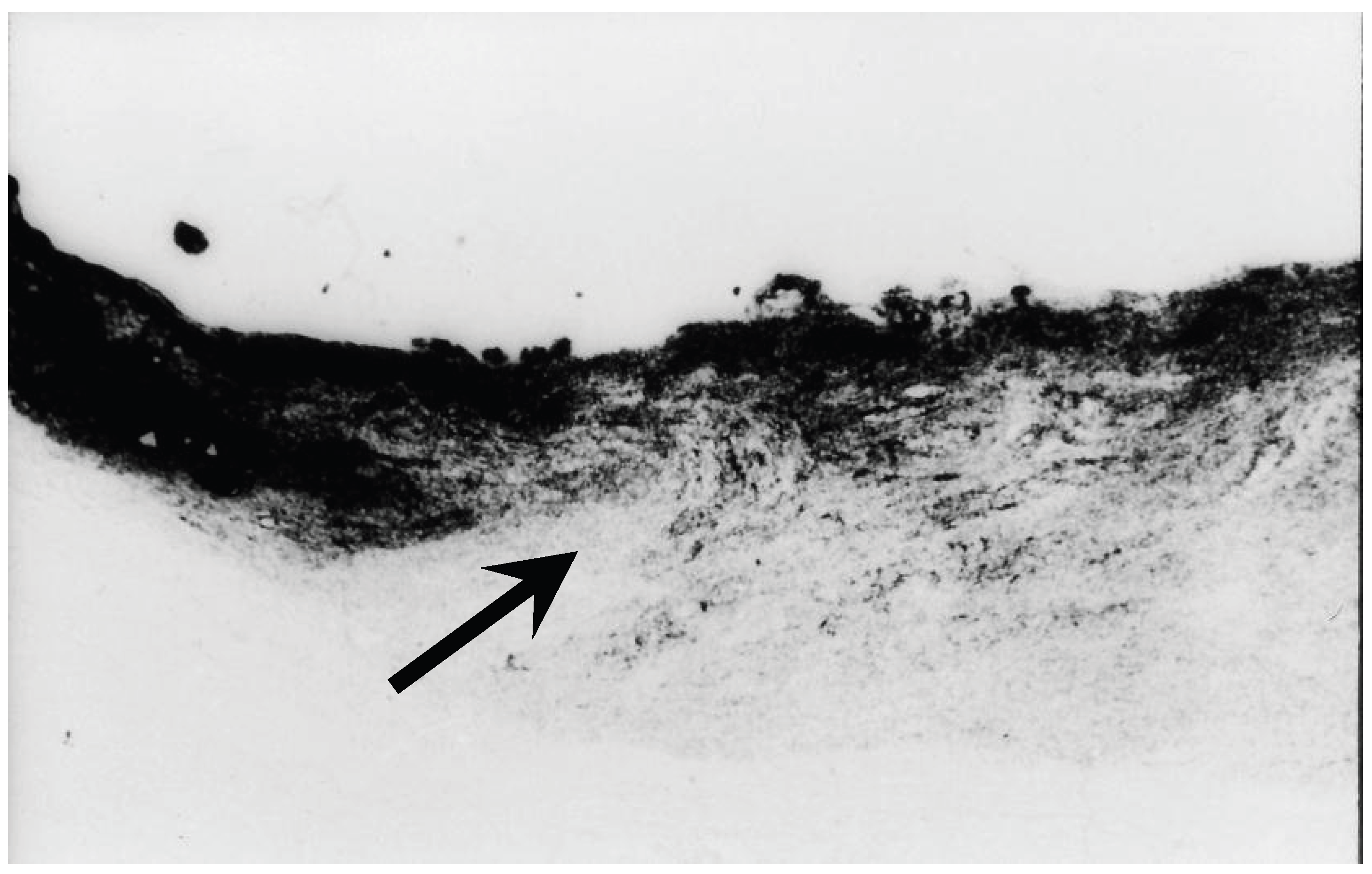
Figure 2.
: Dual blockage of axonal transport in monkey eye. In addition to the blockage of axonal transport at the LC (white arrow), axonal transport is also interrupted at the margin of the optic disc near Elschnig’s scleral ring (arrow head). This dual-site blockage is of particular interest for explaining damage to the papillmacular bundle and central VFDs in eyes with an elongated papillomacular distance, as is observed in high myopia eyes. (Reproduced with permission from Sakugawa M., Chihara E. Blockage at two points of axonal flow in glaucomatous eyes. Graefe’s Arch. Klin. Exp. Ophthalmol. 223: 214-218, 1985).
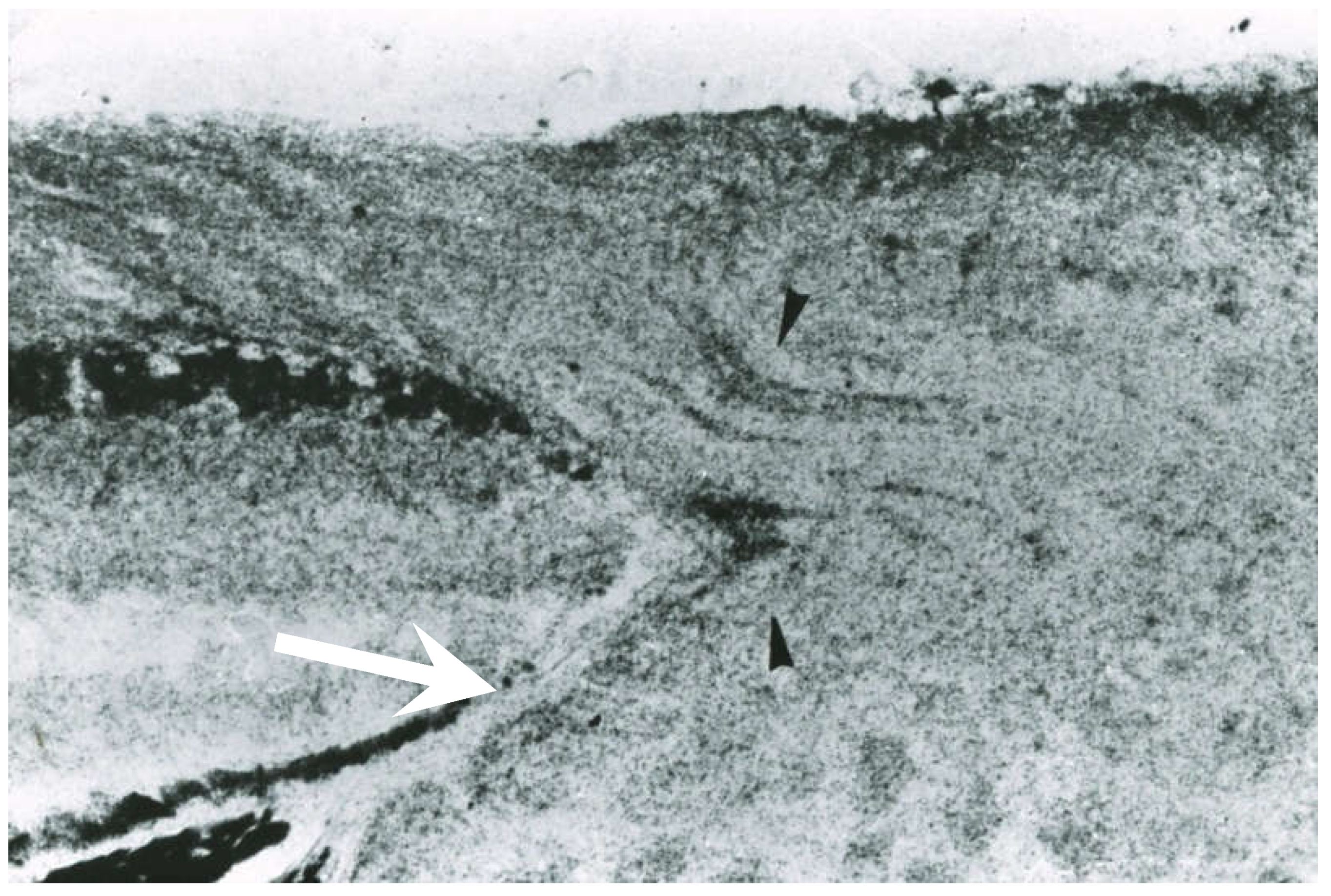
Figure 3.
a:Red free light image of papillomacular bundle defects in eyes diagnosed as so-called “normal tension glaucoma” [1], originally published in 1992 (Reproduction with permission from Chihara E., Tanihara H. Parameters associated with Papillo-macular bundle defects in glaucoma. Graefes Arch for Clinical and Experimental Ophthalmology 1992;230:511-517). Figure 3c,d: Thirty-eight years after the initial examination, she was re-examined in 2025 at the age of 85 years. The papillomacular bundle defects examined using en face OCT imaging and the central visual field loss were remarkably similar to those observed in 1987 (Fig. 3c). She had undergone phacoemulsification with intraocular lens implantation in 2009, after which her IOP remained in the high-teen range. In 2025, Only mild VF progression was detected, characterized by an increase in the superior nasal step to -18 dB on Humphrey Field Analyzer testing; the calculated mean deviation slope was -0.11dB/year (Fig. 3d). The yellow circle indicates the margin of the BMO. Based on these findings, this eye was diagnosed as having a combination of MON and GON, with MON-dominant RNFLDs.
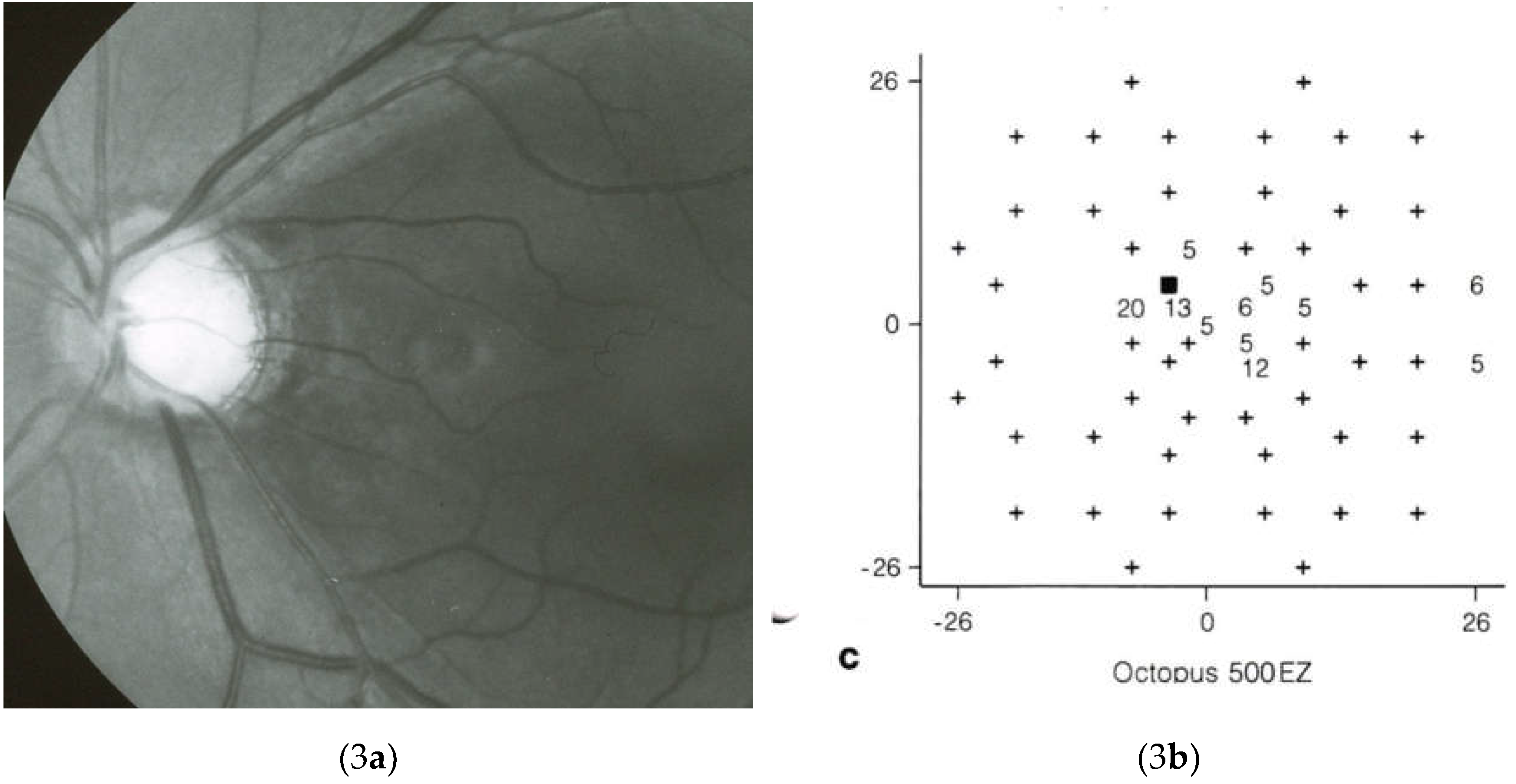
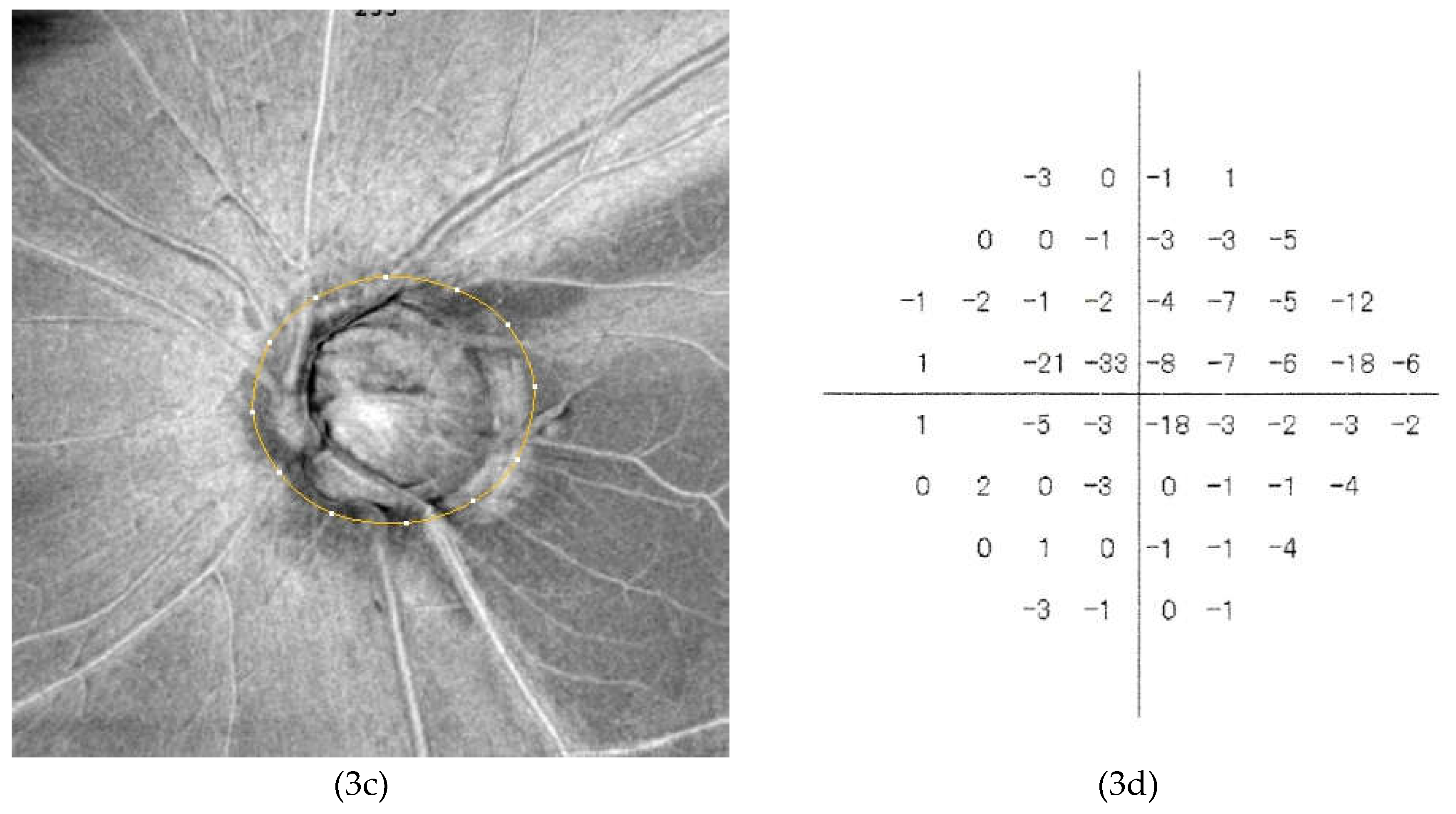
Figure 4.
a: Cleavage of RNFL in eyes with high myopia. Figure 4b: Lamina cribrosa (LC) defects in eyes with high myopia. Ectasia of the LC may result in LC defects (arrows) in eyes with high myopia. Reproduced with permission from : Ohno-Matsui, K.; et al. Acquired optic nerve and peripapillary pits in pathologic myopia. Ophthalmology. 2012, 119, 1685–1692. DOI:10.1016/j.ophtha.2012.01.047. l [153]
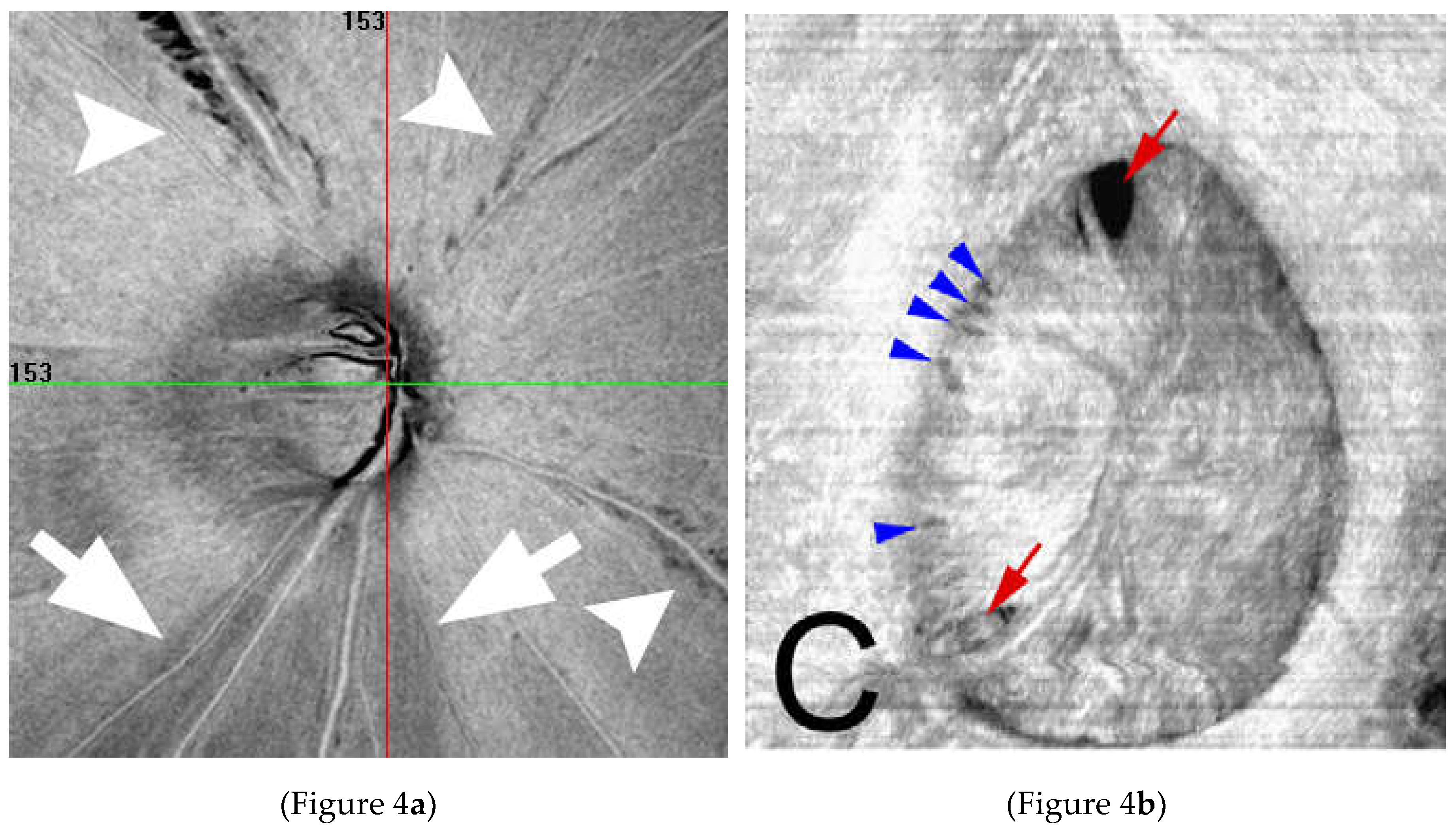

Table 1.
Comparison of Clinical Characteristics between MON and GON.
| MON | GON | |
| Dependence on IOP | Less dependent on IOP [18] | Apparent IOP dependent |
| Speed of progression | Slow [18] | Fast |
| VF Progression associated with age | Progress mainly before age 50 and tends not to progress after 50 [45,46,47]. | Older age is a risk factor for progression [48,49,51] |
| Association with myopia | High [109] | Moderate |
| Pattern of visual field defects | Atypical patterns, enlarged blind spot, central visual field defects [7] | Bjerrum scotoma, nasal step, paracentral scotoma, and diffuse loss |
| Papillo-macular bundle length | Long [15] | Variable |
| Deformation of disc | γ-zone commonly present [7,109] | Variable |
MON: myopic optic neuropathy; GON: glaucomatous optic neuropathy, IOP: intraocular pressure; VF: visual field
Disclaimer/Publisher’s Note: The statements, opinions and data contained in all publications are solely those of the individual author(s) and contributor(s) and not of MDPI and/or the editor(s). MDPI and/or the editor(s) disclaim responsibility for any injury to people or property resulting from any ideas, methods, instructions or products referred to in the content. |
© 2026 by the authors. Licensee MDPI, Basel, Switzerland. This article is an open access article distributed under the terms and conditions of the Creative Commons Attribution (CC BY) license (http://creativecommons.org/licenses/by/4.0/).
Copyright: This open access article is published under a Creative Commons CC BY 4.0 license, which permit the free download, distribution, and reuse, provided that the author and preprint are cited in any reuse.


