
Submitted:
09 January 2026
Posted:
09 January 2026
You are already at the latest version
Abstract
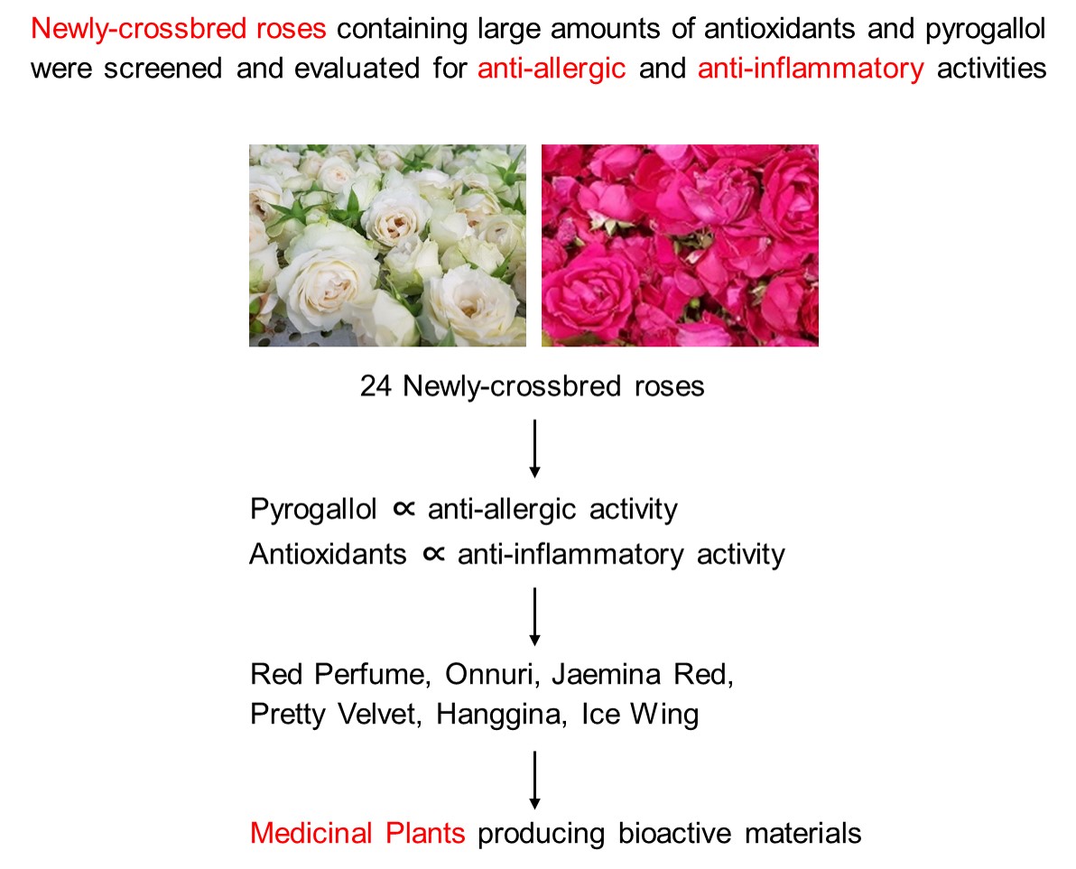
Anti-allergic and anti-inflammatory activities of the extracts of rosebuds newly-crossbred in Korea were investigated in vitro and in vivo. Twenty-four candidate rosebuds were extracted with 80% ethanol, and analyzed for polyphenols, flavonoids, tannins, proanthocyanidins, and pyrogallol (1,2,3-benzenetriol). The extracts’ in vitro anti-allergic and anti-inflammatory activities were analyzed through inhibitory effects on the β-hexosaminidase release from Compound 48/80-stimulated RBL-2H3 cells and nitric oxide production from lipopolysacchrade-activated RAW 264.7 macrophages, respectively. The in vivo activity was assessed via protection against lethality and itching (scratching) symptoms in mice challenged with Compound 48/80. Among candidates, Lover Shy, Pretty Velvet, Ice Wing, Red Perfume, Onnuri, Jaemina Red, and Hanggina were found to possess high concentrations of antioxidative components. By comparison, Pretty Velvet, Red Perfume, Jaemina Red, Hanggina, Onnuri, and Ice Wing were highly effective in anti-allergic and anti-inflammatory activities in vitro, in parallel with their concentrations of pyrogallol. Their anti-allergic effects were confirmed in mice: The 6 extracts protected against Compound 48/80-induced mortality and scratching behaviors in a dose-dependent manner. The allergen-induced increases in serum IgE and histamine as well as inflammatory cytokines, tumor-necrosis factor-α and interleukin-1β, were remarkably attenuated following treatment with the rosebud extracts. Therefore, it is suggested that the extracts and active ingredients from cross-bred Korean rosebuds exert anti-allergic and anti-inflammatory activities through their high levels of antioxidants and pyrogallol, and that could be promising candidates to overcome allergic responses such as atopic dermatitis.
Keywords:
rosebud extract
; antioxidant
; pyrogallol (1
; 2
; 3-benzenetriol)
; anti-allergic activity
; antiinflammatory activity
1. Introduction
As an excessive immune function of the body, hypersensitivity reactions are classified into four types [1]. Among them, type 1 hypersensitivity reactions occur when the immunoglobulin E (IgE), which has been produced excessively through repeated exposure to allergens, binds to mast cells, and cause systemic anaphylactic shock or local itching and inflammation as granules containing histamines and chemotactic factors are released [2]. Representative type 1 hypersensitivity reactions include atopic dermatitis and respiratory asthma, which are mediated by Th2 cytokines. Atopic dermatitis is a chronic relapsing inflammatory skin disease, showing the highest prevalence in childhood. The number of atopic dermatitis patients in Korea was over 1 million in 2022 (Korea Health Insurance Review and Assessment Service) and has been increasing sharply every year. And the worldwide incidence of atopic dermatitis has increased by 2-3 times over the last 30 years [3].
The cause of atopic dermatitis is not clear, but genetic and environmental factors are involved. For example, atopic dermatitis is often caused by hypersensitivity reactions to dust, mites, bacteria, and fungi in the house, pollens, environmental chemicals, and chemicals added to food [4]. According to a review [5], allergic inflammations occur when external triggers, called allergens, penetrate the impaired skin barriers and reach subcutaneous mast cells. Many inflammatory mediators, including histamine and chemotactic factors released from mast cells, cause itching symptoms. Itching symptoms lead to scratching behaviors and tissue damage, exacerbating skin inflammation so that an ‘itching-scratching’ vicious cycle is formed. In addition, diverse factors aggravate atopic dermatitis: dry skin, ambient temperature and humidity, strenuous exercise and sweat, strong skin scrubbing, skin irritation by wool and fibers, food, drugs, house dust, mites, animal hair, irritant chemicals, infections, and mental stress. In addition to the above external factors, internal factors such as immune dysfunction or hypersensitivity to injured tissues can also cause atopic dermatitis [4].
In clinical practice, antihistamines and steroids are commonly prescribed as therapeutic agents. Although antihistamines suppress itching symptoms caused by urticarial [6], they play only a limited role in the treatment of atopic dermatitis. Since long-term use of antihistamines can cause adverse effects, interest in natural products with high therapeutic effects and low side effects is increasing. The search for new functional substances from natural products has been accelerating after the enactment of the Korea Food and Drug Administration’s notification on safety and efficacy evaluation standards and approval procedures for functional foods and functional cosmetics [7].
Roses with various colors and shapes are a dicotyledonous plant belonging to the genus Rosa in the Rosaceae family. Rose flowers have been widely used for ornamental purposes and rose oil has been widely used as a raw material for perfumes and basic cosmetics. By comparison with scent and oils, the nutritional and pharmacological activities of roses were not studied enough. Roses were suggested as a therapeutic agent, because they contain various phytochemicals including antioxidants [8]. In fact, it was found that rose petals contain vitamins, malic acid, cratgolic acid, flavonoids, and lactones, so have a high antioxidative potential [9]. In addition, anthocyanins possessing anti-aging, cardioprotective, anti-cancer, and anti-diabetes activities were rich in rose extracts [10,11,12]. As additional advantages, roses are caffeine-free [13], and exhibit various antibacterial activities [14].
Recently, we reported strong antioxidative and anti-inflammatory effects of rosebud extracts from new 24 rose cultivars [15]. In addition, we demonstrated that they exerted neuroprotective, anti-microbial, skin-whitening, wrinkle-improving, and anti-allergic properties [16,17]. Notably, it was found out that pyrogallol is a major antioxidant displaying neuroprotection and anti-allergic effects [18,19,20,21]. In this context, we extended analyses of antioxidants and pyrogallol in 24 rosebud extracts, and assessed their anti-allergic and anti-inflammatory activities in vitro and in vivo, demonstrating protective potentials against systemic and local skin anaphylactic responses.
2. Materials and Methods
2.1. Preparation of Rosebud Extracts
Dried rosebuds from 24 new cultivars (Rosa hybrida) crossbred in Gumi Floriculture Research Institute (Gumi, Republic of Korea) in 2021 were used: i.e., (1) Lover Shy, (2) Lovely Scarlet, (3) Loving Heart, (4) Red Perfume, (5) Luminus, (6) Mirinae Gold, (7) Betty, (8) Bichina, (9) Aileen, (10) Onnuri, (11) Yunina, (12) Jaemina Red, (13) Jinseonmi, (14) Chilbaegri, (15) Tamina, (16) Tamnari, (17) Pretty Velvet, (18) Peach Grace, (19) Pink Love, (20) Pink Perfume, (21) Hanaro, (22) Hanaram, (23) Hanggina, and (24) Ice Wing (Figure 1).
Based on our previous studies, the rosebuds were extracted with 80% ethanol to achieve high antioxidant contents [15,22]. The dried rosebuds were pulverized in a rotor mill (Laval Lab Inc., Laval, QC, Canada), and immersed in 80% ethanol in an ultrasonic water bath, in which the extraction solvent/solid ratio was set to 49: 1 (980 mL 80% ethanol/20 g dried rosebuds). The bath was heated at 60~70 °C for 2 hours, and then ultrasonic-extracted for 1 hour. After extraction, the mixture was cooled at room temperature, filtered, and then concentrated to 60 brix under a reduced pressure using a vacuum evaporator (Rotary Vacuum Evaporator N-N series; Eyela, Tokyo, Japan).
2.2. Analysis of Antioxidative Components
2.2.1. Analysis of Total Polyphenols
The total polyphenol content of the rosebud extracts was measured according to the method of Dewanto et al. [15,23]. That is, the total polyphenol content was measured according to the principle that Folin-Ciocalteu’s phenol reagent is reduced by polyphenolic compounds in the extracts to develop a molybdenum color. The concentration of the samples was adjusted to 1 mg/mL, 2 mL of 2% Na2CO3 was added for reaction for 3 minutes, 100 µL of 50% Folin-Ciocalteu’s phenol reagent was added, the mixture was left alone for 30 minutes, and the absorbance of the reaction solution was measured at 750 nm. A standard calibration curve was prepared using gallic acid (Sigma-Aldrich, St. Louis, MO, USA) diluted to 10, 20, 30, 40 and 50 times as standard materials, and the polyphenol content was expressed as mg of gallic acid in 1 g of the sample.
2.2.2. Analysis of Flavonoids
The total flavonoid content of the rosebud extracts was analyzed using the method of Zhishen et al. [24] with some modification. The concentration of the samples was adjusted to 1 mg/mL, 1 mL of distilled water and 75 µL of 5% sodium nitrite were added to 250 µL of the sample solution, 150 µL of 10% aluminum chloride hydrate was added 5 minutes later, 500 µL of 1 M NaOH was added 6 minutes later, the mixture was left alone for 11 minutes, and the absorbance was measured at 510 nm. A standard calibration curve was prepared with (+)-catechin hydrate (Sigma-Aldrich), and the flavonoid content was expressed as mg of catechin in 1 g of the sample.
2.2.3. Analysis of Tannins
The total tannin content of the rosebud extracts was measured according to the method of Duval and Shetty [25]. The concentration of the sample was adjusted to 1 mg/mL, 1 mL of 95% ethanol and 1 mL of distilled water were added to 1 mL of the sample solution, the mixture was shaken well, 1 mL of 5% Na2CO3 solution and 0.5 mL of 1 N Folin-Ciocalteu’s reagent (Sigma-Aldrich) were added to the mixture, the color was developed at room temperature for 60 minutes, and the absorbance was measured at 725 nm. A standard calibration curve was prepared with tannic acid (Sigma-Aldrich), and the tannin content was expressed as mg of tannic acid in 1 g of the sample.
2.2.4. Analysis of Proanthocyanidins
The total proanthocyanidin content of the rosebud extracts was measured using the vanillin-sulfuric acid method modified according to the method of Takahama et al. [26]. The concentration of the sample was adjusted to 1 mg/mL, 0.5 mL of 1.2% vanillin solution and 0.5 mL of 20% sulfuric acid were added to 0.2 mL of the sample solution, the mixture was left alone for 20 minutes, and the absorbance was measured at 500 nm using an ELISA reader (UV-1650PC; Shimadzu, Kyoto, Japan). A standard calibration curve was prepared with (+)-catechin (Sigma-Aldrich), and the proanthocyanidin content was expressed as mg of catechin in 1 g of the sample.
2.2.5. Analysis of Pyrogallol (1,2,3-Benzenetriol)
The pyrogallol content of the rosebud extracts was analyzed by HPLC (ACME 9000 system; Younglin, Anyang, Korea) [18]. Separation was performed on a YMC-Triart C18 column (4.6 × 250 mm; YMC, Kyoto, Japan). The mobile phase consisted of acetonitrile containing 0.1% acetic acid (A) and water (B), delivered under gradient elution conditions. The flow rate was set at 1.0 mL/min, and a 20 μL aliquot of each sample was injected. Detection was carried out using a UV detector at a wavelength of 280 nm. Pyrogallol (extra-pure grade) was obtained from Daejung Chemicals & Metals (Siheung, Korea). Quantification was performed by comparison with a calibration curve constructed using a pyrogallol standard.
2.3. Measurement of Mast Cell Degranulation
The cell line RBL-2H3 (KCLB No. 22256, Lot No. 43077) was from the Korean Collection for Type Cultures of the Korea Research Institute of Bioscience and Biotechnology (Jeongeup, Korea). The cells were cultivated in a 5% CO2 incubator (37 °C), using a minimal essential medium (MEM) supplemented with 10% fetal bovine serum (FBS) and antibiotic-antimycotic (Gibco, Grand Island, NE, USA).
Mast cell degranulation was measured with the release of β-hexosaminidase, which is a granule marker [16,27]. The cells were separated from the culture flask by treatment with an enzyme (trypsin-EDTA), and the cell suspension was centrifuged at 500 g for 5 min. Cell suspension was prepared at 2 × 10⁶ cells/mL, and aliquoted (100 μL) into a 96-well cell culture plate, and stabilized for at least 3 hours.
After discarding culture medium, the wells were treated with the new culture medium containing anti-dinitrophenyl (DNP)-IgE (10 ng/mL), and cultivated overnight. The cells were treated with rosebud extracts (final concentrations of 10, 32 or 100 µg/mL), and incubated for 30 min. Thereafter, the cells were stimulated with DNP-human serum albumin (HAS; 10 µg/mL) to induce mast cell degranulation. After 1-hour incubation, 60 µL of the supernatant was taken, mixed with 15 µL of substrate buffer, and made to react at 37 °C for 2 hours. The stop solution (150 µL) was added, and the absorbance was measured at 405 nm.
2.4. Measurement of Macrophage Nitric Oxide (NO) Production
The inflammatory response of macrophages was measured with NO production according to cell activation [15]. The cell line RAW 264.7 (ATCC TIB-71, KCLB No. 40071) used in the test was bought from the Korean Collection for Type Cultures of the Korea Research Institute of Bioscience and Biotechnology (Jeongeup, Korea). The cells were cultivated overnight in a 96-well plate at a concentration of 1 × 10⁷ cells/mL. In order to activate cells, the cells were treated with lipopolysaccharide (LPS; 1 μg/mL). Rosebud extracts were added at concentrations of 10, 32 or 100 μg/mL, and incubated for 24 hours. An equal volume of Griess reagent (Promega, Madison, WI, USA) was mixed, and the absorbance was measured at 540 nm 10 min later for NO measurement. A standard calibration curve was prepared with sodium nitrite, and the NO concentration in the culture medium was calculated.
2.5. Measurement of Mouse Allergic Reaction
2.5.1. Animals
Male ICR mice (6 weeks old, 25-28 g) for the induction of systemic and local hypersensitivity reactions were procured from Daehan Biolink (Eumseong, Korea) and used after 1-week acclimation to the laboratory environment. The environment of the animal laboratory was adjusted to a temperature of 23 ± 2 °C, relative humidity of 55 ± 10%, ventilation frequency of 12 times/hour, lighting cycle of 12 hours (07:00~19:00), and illumination of 150~300 lux. They were fed standard rodent chow and purified water ad libitum. The animal experiments were conducted under the approval of the Institutional Animal Care and Use Committee (IACUC) of Chungbuk National University, and according to the Standard Operation Procedures (SOP) of the same institution.
2.5.2. Measurement of Systemic Allergic Reaction (Death from Shock)
The anti-allergic activity of rosebud extracts on systemic anaphylactic shock was assessed in mice challenged Compound-48/80, a mast cell degranulator. Rose extracts were diluted in saline and intraperitoneally administered to ICR mice at doses of 10, 30 or 100 mg/kg. Thirty min later, a lethal dose (8 mg/kg) of Compound 48/80 was intraperitoneally injected to induce anaphylactic reaction, and thereafter the mortality rate of the mice was recorded for 60 min [16,28].
2.5.3. Measurement of Local Allergic Reaction (Skin Itching)
In order to investigate the anti-allergic activity of rosebud extracts on atopic dermatitis, the extracts were diluted in purified water, orally administered to ICR mice at doses of 10, 30 or 100 mg/kg. Thirty min later, Compound 48/80 (50 μg/site, 50 μL at 1 mg/mL) was subcutaneously injected between the two shoulder blades [16,28,29]. Thereafter, the number of scratching behaviors was recorded to 60 min.
2.5.4. Measurement of Blood IgE and Histamine
Blood was collected from the abdominal vena cava, into a Vacutainer (BD 367986; BD, Seoul, Korea), and centrifuged at 3000 rpm for 15 min to obtain serum. Serum IgE and histamine were determined via ELISA according to the manufacturer’s instructions. Briefly, serum was loaded into ELISA wells together with antibodies specific for IgE (K3231081; Komabiotech, Seoul, Korea) or histamine (ab213975; Abcam, Cambridge, UK), and incubated for 0.5–1 hour at room temperature. After washing, the secondary antibody was treated and incubated for 30 min. Following treatment with a substrate for color development, absorbance was measured at 450 nm [30].
2.5.5. Measurement of Blood Cytokines
2.6. Statistical Analysis
Data are presented as mean ± standard deviation. Statistical analysis was performed with SPSS version 26.0 program (SPSS Inc., Chicago, IL, USA). Differences among groups were analyzed with one-way ANOVA, followed by Tukey’s HSD at a level of P < 0.05.
3. Results
3.1. Antioxidant Contents in Rosebud Extracts
We analyzed extensive antioxidants including polyphenols, flavonoids, tannins, and proanthocyanidins from 24 rose cultivars. There were big differences in antioxidant contents ranging from 814.0 ± 22.3 mg/g (Lover Shy) to 159.9 ± 5.6 mg/g (Chilbaegri) (Figure 1). Among 24 rosebud extracts, Lover Shy (814.0 ± 22.3 mg/g), Pretty Velvet (758.6 ± 42.0 mg/g), Ice Wing (723.1 ± 26.2 mg/g), Red Perfume (698.2 ± 31.4 mg/g), Onnuri (678.5 ± 15.8 mg/g), Jaemina Red (618.4 ± 34.0 mg/g), and Hanggina (568.3 ± 16.2 mg/g) were found to contain high concentrations of the antioxidants (Figure 1; Supplementary Table S1).
3.2. 1,2,3-Benzenetriol Content in Rosebud Extracts
Next, we analyzed 1,2,3,-benzenetriol (pyrogallol), as an anti-allergic ingredient, in 24 rosebud extracts. As a result of HPLC analysis (Figure 2). Pyrogallol standard was detected at a retention time of 8.5450 min, and pyrogallol was detected at the same time in a rosebud extract. There was a difference in pyrogallol concentration ranging from 1.77 ± 0.14 mg/g (Red Perfume) to 0.21 ± 0.03 mg/g (Betty) (Figure 2). Among 24 rose cultivars, the concentration of pyrogallol was highest in Red Perfume (1.77 ± 0.14 mg/g), Pretty Velvet (1.34 ± 0.10 mg/g), Hanggina (1.26 ± 0.14 mg/g), Jaemina Red (1.22 ± 0.06 mg/g), Ice Wing (1.14 ± 0.07 mg/g), Onnuri (1.10 ± 0.13 mg/g), and Lover Shy (1.02 ± 0.07 mg/g) (Figure 3). Notably, the order of pyrogallol concentration was similar with the antioxidant content, although Lover Shy showed relatively-low pyrogallol level compared with high antioxidants.
3.3. Inhibition of Mast Cell Degranulation
In order to assess allergic reaction, we analyzed β-hexosaminidase, instead of histamine, released from mast cells sensitized with IgE, since both of them are secreted by allergic reactions such as asthma, rhinitis, and atopic dermatitis (Park et al., 2011). β-Hexosaminidase release markedly increased RBL-2H3 cells following sensitization with anti-DNP IgE (Figure 4A). However, such an IgE-mediated increase in β-hexosaminidase release was attenuated by rosebud extracts (100 μg/mL), in which Red Perfume, Onnuri, Jaemina Red, Pretty Velvet, Hanggina, and Ice Wing were the most effective, reducing to a normal level. Notably, the anti-allergic activity was in parallel with the concentration of pyrogallol in the rosebud extracts (Figure 3). Further, we assessed the concentration-effectiveness at 10, 30, and 100 μg/mL. As results, the selected 6 extracts, Red Perfume, Onnuri, Jaemina Red, Pretty Velvet, Hanggina, and Ice Wing, displayed concentration-dependent anti-allergic activities (Figure 4B).
3.4. Inhibition of Macrophage NO Production
As an inflammatory parameter, we analyzed NO production from macrophages activated with LPS. Treatment of RAW 264.7 cells with LPS greatly increased NO release (Figure 5A). However, all 24 rosebud extracts inhibited the NO production, wherein Red Perfume, Onnuri, Jaemina Red, Pretty Velvet, Hanggina, and Ice Wing were highly effective. Interestingly, the anti-inflammatory activity was in parallel with the concentrations of antioxidants and pyrogallol in the rosebud extracts (Figure 1 and Figure 3). Furthermore, the selected 6 extracts, Red Perfume, Onnuri, Jaemina Red, Pretty Velvet, Hanggina, and Ice Wing, exhibited concentration-dependent anti-inflammatory activities at 10, 30, and 100 μg/mL (Figure 5B).
3.5. Inhibition of Systemic Allergic Reaction
All the mice (n = 6/group) intraperitoneally challenged with a lethal dose (8 mg/kg) of Compound-48/80 died within 60 min due to a systemic shock reaction (Figure 6). Notably, 30-min intraperitoneal pretreatment with 6 rosebud extracts (10, 30 or 100 mg/kg) protected against the Compound-48/80-induced mortality in a dose-dependent manner. In a relative potency, Pretty Velvet, Onnuri, and Ice Wing were somewhat more effective than Jaemina Red, Hanggina, and Red Perfume, although at the high dose (100 mg/kg) of each rosebud extract all the mice survived.
3.6. Inhibition of Skin Allergic Reaction
Based on the systemic anti-allergic activity of rosebud extracts, we tried to confirm their effects on skin allergic reaction. The mice subcutaneously injected with Compound-48/80 (50 μg/site) showed severe itching symptom, scratching 68.0 ± 5.5 times for 60 min (Figure 7). However, 30-min oral pretreatment with the 6 rosebud extracts, Red Perfume, Onnuri, Jaemina Red, Pretty Velvet, Hanggina, and Ice Wing, significantly attenuated the scratching behavior in a dose-dependent manner.
3.7. Inhibition of Blood IgE and Histamine
According to the anti-allergic activity of the rosebud extracts, we analyzed blood IgE and histamine to clarify underlying mechanisms. The serum IgE level of the mice markedly increased after challenge with Compound-48/80 (Figure 8A). As a related mediator, serum histamine also significantly increased as an allergic response (Figure 8B). It is interest to note that the 6 rosebud extracts inhibited both the IgE and histamine levels, especially to lower blood concentrations than normal levels at high doses.
3.8. Inhibition of Blood Inflammatory Cytokines
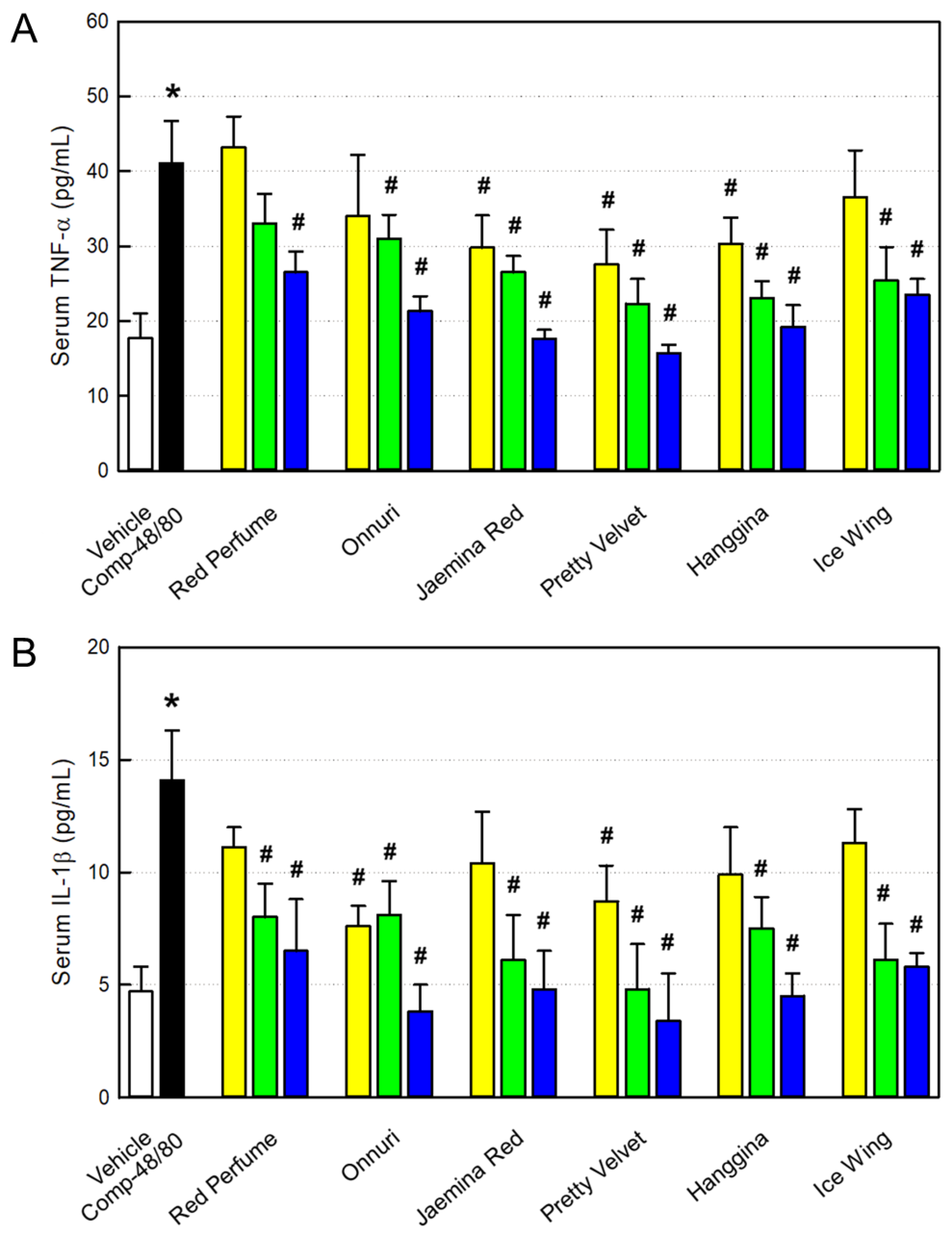
As markers of inflammation, we analyzed blood TNF-α and IL-1β, the major inflammatory cytokines. As expected, both the cytokines were greatly enhanced by challenge with Compound-48/80 (Figure 9). However, the 6 rosebud extracts were found to lower both the blood TNF-α and IL-1β levels, to normal levels at high doses, as inferred from the inhibitory effects on the NO production from RAW 264.7 macrophages (Figure 4).
4. Discussion
Most animals consuming oxygen in metabolism inevitably suffer from tissue injury and aging due to the generation of reactive oxygen species (ROS), called oxidative stress [32]. In order to counteract ROS toxicity, living organisms synthesize or ingest antioxidant molecules. When oxidative stress exceeds the antioxidant defense system, theoretically, the oxidative damage can be attenuated by ingesting antioxidants.
Antioxidative compounds present in plants, that is, phytochemicals, are largely divided into phenolics, carotenoids, alkaloids, and organosulfur- or nitrogen-containing compounds. Among them, polyphenols, that is, phenolics account for the largest part. In particular, among phenolics, flavonoids that have strong antioxidant activity are well known to prevent and delay aging by playing a role in preventing oxidative stress through the inhibition of lipid peroxidation [3]. Flavonoids are mainly composed of anthocyanidins, flavonols, flavones, catechins, and flavanones, and it has been demonstrated that most of the flavonoids have strong antioxidative and anti-bacterial activities depending on their structures [33]. Tannins are a sort of polyphenols that have an astringent taste. Originally, tannin was a word referring to substances used as preservatives when skins of animals are made into leather, but it is now a general term for large polyphenolic compounds that have sufficient hydroxyl groups and bind strongly to proteins or other macromolecules [34]. Proanthocyanidins and anthocyanins, well known as red pigments in plants, reduce oxidative stress, aging, and heart dysfunction with their free radical-scavenging abilities [35].
In our previous study, Colorado rose petal extract was found to contain high concentrations of polyphenols and flavonoids, so exhibited potential antioxidative and anti-inflammatory activities [18,22]. As expected, the extract protected against epileptic brain damage of mice challenged with excitotoxic kainic acid. Thereafter, we prepared rosebud extracts from 24 newly-crossbred roses, and showed good correlations between polyphenol content and antioxidative potency as well as between polyphenol concentration and anti-inflammatory activity in vitro [15]. Especially, an extract from Pretty Velvet rose exhibited strong in vivo anti-inflammatory effects in a subcutaneous air-pouch inflammation model through steroid- and NSAID-like activities. In the present study, we extended analyses to polyphenols, flavonoids, tannins, and proanthocyanidins, and demonstrated their anti-inflammatory effects, showing good relationship with the antioxidant content in vitro and in vivo.
Tissue damage caused by ROS is involved in most inflammatory reactions, and also promotes immune hypersensitivity reactions, that is, systemic and local allergic reactions. Nowadays, opportunities to be exposed to various allergens are rapidly increasing, for example, fine dust from reckless development and combustion of petrochemicals, house dust mites due to carpet culture, animal hair due to the increase in companion animal population, spring pollen, and new foreign fruits and foods. [1,36,37,38,39]. Among them, asthma and atopic dermatitis, which are type 1 hypersensitivity reactions, are emerging as social problems. Mast cells are distributed in most organs and tissues, and are important triggering cells for immune reactions such as allergy and anaphylaxis. In particular, the anaphylaxis reaction is induced by histamine and chemotactic factors released from mast cells as well as various inflammatory cytokines from other inflammatory cells [38,40,41].
Steroids are widely prescribed to treat atopic dermatitis, which is emerging as a major disease of modern people. But, they should be used in an appropriate dose and in good balance because of their wide adverse effects such as osteoporosis, osteonecrosis, myopathy, peptic ulcer, arteriosclerosis, dyslipidemia, acne, and skin atrophy [42]. In addition, the US Food and Drug Administration (FDA) required carcinogenicity warnings to be attached to Elidel® and Protopic®, which are non-steroidal atopy treatments, and the Ministry of Food and Drug Safety (MFDS) of Korea restricts the use of them in young patients. Therefore, social demand for the development of safe therapeutic agents is rapidly increasing, and natural products may fulfill this requirement.
Earlier, we demonstrated that a rose petal extract has a strong anti-allergic property, inhibiting β-hexosaminidase release from RBL-2H3 cells as well as systemic and cutaneous anaphylaxtic reactions in animals [16]. Notably, we found out that pyrogallol is a major antioxidant displaying neuroprotection in an animal model of ischemic stroke [18,19]. Actually, pyrogallol was found to have anti-allergic activity, alleviating nasal symptoms including sneezing in toluene-2,4-diisocyanate (TDI)-sensitized rats by suppressing Th2 cytokine expression calcineurin/nuclear factor of activated T-cells (NFAT) signaling [20,21].
In the present study, therefore, we analyzed pyrogallol contents in 24 rosebud extracts, and assessed their anti-allergic activities. There was a good correlation between pyrogallol concentration and β-hexosaminidase-blocking activity. Next, we selected 6 extracts (Red Perfume, Onnuri, Jaemina Red, Pretty Velvet, Hanggina, and Icw Wing) showing strong anti-allergic and anti-inflammatory activities in vitro, and investigated their effects on systemic and local (skin) anaphylactic reactions in vivo. As inferred from the blocking activity on β-hexosaminidase release, the 6 rosebud extracts protected against Compound-48/80 lethality in a dose-dependent manner. Furthermore, the extracts markedly attenuated skin scratching behaviors in mice challenged with Compound-48/80, as shown in a previous study [16]. Such anti-allergic effects of rosebud extracts were supported by blood IgE, histamine and inflammatory cytokines. Indeed, serum IgE, histamine, TNF-α, and IL-1β were reduced following the rosebud extracts in parallel with the dose-dependent patterns in itching symptoms.
Roses have been widely used for decoration or as a component of perfumes because of their beautiful appearance and strong scent. In particular, Turkey, Bulgaria, Morocco, and China produce rose oil in large quantities and supply it as a raw material for cosmetics all over the world, and more recently, petals are used as jam or dessert [43]. However, the pharmacological activities of rose oil are not well demonstrated. Rather, we have reported various effects such as antioxidation, anti-allergy, skin whitening, wrinkle improvement, anti-bacterial, anti-inflammatory, and neuroprotection of non-oil solvent extracts [15,16,17,18,22,44,45]. In the present study, we extended insights into the major component (pyrogallol) as an anti-allergic molecule of 24 new cultivars manufactured in Korea Floriculture Research Institute. Based on the research results, it is expected that we can develop more effective medicinal plants possessing higher pyrogallol content for the management of allergic diseases such as atopic dermatitis.
We demonstrated that 24 rosebud extracts displayed anti-allergic and anti-inflammatory effects in parallel with their ingredients including pyrogallol and/or antioxidant phytochemicals. Especially, pyrogallol was found to play a central role for anti-anaphylactic activity. Therefore, it is proposed that the extracts and active ingredients from cross-bred Korean rosebuds exert anti-allergic and anti-inflammatory activities through their high levels of pyrogallol and antioxidants, and that could be promising candidates to overcome allergic responses such as atopic dermatitis.
Author Contributions
Material preparation, S.T.K; cell and animal experiments and data analysis, I.-J.K., K.-E.T. and Z.B.; project administration, S.S.K.; ingredient analysis, H.-S.J.; conceptualization, writing, and editing, H.-S.J. and Y.-B.K. All authors have read and agreed to the published version of the manuscript.
Funding
This research was supported by the Regional Innovation System & Education (RISE) Glocal University 30 program through the Chungbuk Regional Innovation System & Education Center, funded by the Ministry of Education (MOE) and the Chungcheongbuk-do, Republic of Korea (2025-RISE-11-014).
Institutional Review Board Statement
All animal experimental procedures were approved and carried out in accordance with the Institutional Animal Care and Use Committee (IACUC) of Laboratory Animal Research Center at Chungbuk National University, Korea (Approval No. CBNUA-1544-21-01).
Conflicts of Interest
The authors declare no conflicts of interest.
References
- Burns-Naas, L.A.; Meade, B.J.; Munson, A.E. Toxic response of the immune system. In The Basic Science of Poisons, 6th ed.; Klaassen, C.D., Ed.; McGraw Hill/Medical: Columbus, OH, USA, 2001; pp. 419–470. [Google Scholar]
- Hossen, M.A.; Fujii, Y.; Ogawa, M.; Takubo, M.; Tsumuro, T.; Kamei, C. Effect of loratadine on mouse models of atopic dermatitis associated pruritus. Int. Immunopharmacol. 2005, 5, 1331–1336. [Google Scholar] [CrossRef] [PubMed]
- Park, S.G.; Noh, H.M.; Kweon, S.H.; Jo, E.H.; Jang, H.C.; Kim, H.K.; Park, H.J.; Kim, W.J.; Park, M.C. The study on the environmental factors of atopic dermatitis in oriental-western medicine. J. Korean Med. Ophthalmol. Otolaryngol. Dermatol. 2018, 31, 52–70. [Google Scholar]
- Jeon, J.H.; Kwon, S.C.; Park, D.; Shin, S.; Jang, M.J.; Joo, S.S.; Kang, H.; Kim, S.H.; Oh, J.Y.; Jeong, J.H.; et al. Effects of red and white rose petal extracts and Ganoderma lucidum culture on ovalbumin-induced atopic dermatitis. Lab. Anim. Res. 2008, 24, 347–354. [Google Scholar]
- Li, K. Itch in atopic dermatitis: From pathogenesis to treatment. Allergy Asthma Respir. Dis. 2014, 2, 8–15. [Google Scholar] [CrossRef]
- Trautmann, A.; Anders, D.; Stoevesandt, J. H1-Antihistamine premedication in NSAID-associated urticaria. J. Allergy Clin. Immunol. Pract. 2016, 4, 1205–1212. [Google Scholar] [CrossRef] [PubMed]
- Korea Ministry of Food and Drug Safety. Regulation on the Evaluation of Functional Cosmetics. 2020. Available online: https://www.law.go.kr.
- Gorji, A. Pharmacological treatment of headache using tranditional Persian medicine. Trends Pharmacol. Sci. 2003, 24, 331–334. [Google Scholar] [CrossRef]
- Pellegrini, N.; Serafini, M.; Colombi, B.; Del Rio, D.; Salvatore, S.; Bianchi, M. Total antioxidant capacity of plant foods, beverages and oils consumed in Italy assessed by three different in vitro assays. J. Nutr. 2003, 133, 2812–2819. [Google Scholar] [CrossRef]
- Lee, H.R.; Lee, J.M.; Choi, N.S.; Lee, J.M. The anti-oxidative and anti-microbial ability of ethanol extracts from Rosa hybrida. Korean J. Food Sci. Technol. 2003, 35, 373–378. [Google Scholar]
- Konczak, I.; Zhang, W. Anthocyanins—more than nature’s colours. J. Biomed. Biotechnol. 2004, 2004, 239–240. [Google Scholar] [CrossRef]
- Cho, E.K.; Son, J.-Y.; Kang, K.O. Antioxidant activities of rose, camellia and cockscomb flower extracts. Food Serv. Indust. J. 2015, 11, 21–33. [Google Scholar]
- Vinokur, Y.; Rodov, V.; Reznick, N.; Goldman, G.; Horev, B.; Umiel, N. Rose petal tea as an anti-oxidant rich beverage: Cultivar effects. J. Food Sci. 2006, 71, S42–S47. [Google Scholar] [CrossRef]
- Özkan, G.; Sağdiç, O.; Baydar, N.G.; Baydar, H. Anti-oxidant and anti-bacterial activities of Rosa damascena flower extracts. Food Sci. Technol. Int. 2004, 10, 277–281. [Google Scholar] [CrossRef]
- Wang, C.; Kim, I.J.; Seong, H.R.; Noh, C.H.; Park, S.; Kim, T.M.; Jeong, H.-S.; Kim, K.Y.; Kim, S.T.; Yuk, H.G.; et al. Antioxidative and anti-inflammatory activities of rosebud extracts of newly crossbred roses. Nutrients 2023, 15, 2376. [Google Scholar] [CrossRef] [PubMed]
- Jeon, J.H.; Kwon, S.C.; Park, D.; Shin, S.; Jeong, J.H.; Hwang, S.Y.; Kim, Y.B.; Joo, S.S. Anti-allergic effects of white rose petal extract and anti-atopic properties of its n-hexane fraction. Arch. Pharm. Res. 2009, 32, 823–830. [Google Scholar] [CrossRef]
- Choi, E.K.; Guo, H.; Choi, J.K.; Jang, S.K.; Shin, K.; Cha, Y.S.; Choi, Y.; Seo, D.W.; Lee, Y.B.; Joo, S.S.; et al. Extraction conditions of white rose petals for the inhibition of enzymes related to skin aging. Lab. Anim. Res. 2015, 31, 148–152. [Google Scholar] [CrossRef]
- Yang, G.; Park, D.; Lee, S.H.; Bae, D.-K.; Yang, Y.-H.; Kyung, J.; Kim, D.; Choi, E.-K.; Hong, J.T.; Jeong, H.-S.; et al. Neuroprotective effects of a butanol fraction of Rosa hybrida petals in an ischemia-reperfusion stroke model. Biomol. Ther. 2013, 21, 454–461. [Google Scholar] [CrossRef]
- Moon, J.; Gwak, H.; Lee, T.-H.; An, E.S.; Kim, Y.B.; Park, D. Antioxidant effects of pyrogallol in neural cells and mice brain. Brain Dig. Learn. 2019, 9, 113–124. [Google Scholar] [CrossRef]
- Nakano, T.; Ikeda, M.; Wakugawa, T.; Kashiwada, Y.; Kaminuma, O.; Kitamura, N.; Yabumoto, M.; Fujino, H.; Kitamura, Y.; Fukui, H.; et al. Identification of pyrogallol from Awa-tea as an anti-allergic compound that suppresses nasal symptoms and IL-9 gene expression. J. Med. Investig. 2020, 67, 289–297. [Google Scholar] [CrossRef]
- Mizuguchi, H.; Ito, T.; Nishida, K.; Wakugawa, T.; Nakano, T.; Tanabe, A.; Watano, T.; Kitamura, N.; Kaminuma, O.; Kimura, K.; et al. Structure-activity relationship studies of pyrogallol as a calcineurin/NFAT signaling suppressor. J. Pharmacol. Sci. 2024, 155, 140–147. [Google Scholar] [CrossRef]
- Yon, J.M.; Kim, Y.B.; Park, D. The ethanol fraction of white rose petal extract abrogates excitotoxicity-induced neuronal damage in vivo and in vitro through inhibition of oxidative stress and proinflammation. Nutrients 2018, 10, 1375. [Google Scholar] [CrossRef]
- Dewanto, V.; Wu, X.; Liu, R.H. Processed sweet corn has higher antioxidant activity. J. Agric. Food Chem. 2002, 50, 4959–4964. [Google Scholar] [CrossRef]
- Zhishen, J.; Mengcheng, T.; Jianming, W. The determination of flavonoid contents in mulberry and their scavenging effects on superoxide radicals. Food Chem. 1999, 64, 555–559. [Google Scholar] [CrossRef]
- Duval, B.; Shetty, K. The stimulation of phenolics and antioxidative activity in pea (Pisum sativam) elicited by genetically transformed anise root extract. J. Food Biochem. 2001, 25, 361–377. [Google Scholar] [CrossRef]
- Takahama, U.; Tanaka, M.; Hirota, S. Proanthocyanidins in buckwheat flour can reduce salivary nitrite to nitric oxide in the stomach. Plant Foods Hum. Nutr. 2010, 65, 1–7. [Google Scholar] [CrossRef]
- Ban, H.J.; Park, D.I.; Kang, K.H. Inhibitory effect of Artemisiae asiaticae Herba on degranulation, production of cytokine, and FcεRI expression in RBL-2H3 cells. Korean J. Orient. Physiol. Pathol. 2012, 26, 915–921. [Google Scholar]
- Kim, S.H.; Choi, C.H.; Kim, S.Y.; Eun, J.S.; Shin, T.Y. Anti-allergic effects of Artemisia iwayomogi on mast cell-mediated allergy model. Exp. Biol. 2005, 230, 82–88. [Google Scholar] [CrossRef]
- Inagaki, N.; Igeta, K.; Kim, J.F.; Nagao, M.; Shiraishi, N.; Nakamura, N.; Nagai, H. Involvement of unique mechanisms in the induction of scratching behavior in BALB/c mice by compound 48/80. Eur. J. Pharmacol. 2002, 448, 175–183. [Google Scholar] [CrossRef]
- Joo, S.S.; Park, D.; Shin, S.; Jeon, J.H.; Kim, T.K.; Kim, J.S.; Park, S.K.; Hwang, B.Y.; Lee, D.I.; Kim, Y.B. Anti-allergic effects and mechanisms of the ethanolic extract of Angelica gigas in dinitrofluorobenzene-induced inflammation models. Environ. Toxicol. Pharmacol. 2010, 30, 127–133. [Google Scholar] [CrossRef]
- Shin, S.; Joo, S.S.; Park, D.; Jeon, J.H.; Kim, T.K.; Kim, J.S.; Park, S.K.; Hwang, B.Y.; Kim, Y.B. Ethanol extract of Angelica gigas inhibits croton oil-induced inflammation by suppressing the cyclooxygenase—Prostaglandin pathway. J. Vet. Sci. 2010, 11, 43–50. [Google Scholar] [CrossRef]
- Hwang, M.R.; Kim, H.E.; Park, D.K.; Heu, Y.C.; Lee, H.J.; Kang, N.J. Induction of oxidative stress and activation of antioxidant enzymes by infection of powdery mildew in cucurbita plants. J. Agric. Life Sci. 2013, 47, 75–81. [Google Scholar]
- Middleton, E.; Kandaswami, C. Potential health-promoting properties of citrus flavonoids. Food Technol. 1994, 48, 115–119. [Google Scholar]
- Schofield, P.; Mbugua, D.M.; Pell, A.N. Analysis of condensed tannins: A review. Anim. Feed Sci. Technol. 2001, 91, 21–40. [Google Scholar] [CrossRef]
- Moyer, R.A.; Hummer, K.E.; Finn, C.E.; Frei, B.; Wrolstad, R.E. Anthocyanins, phenolics, and antioxidant capacity in diverse small fruits: Vaccinium, Rubus, and Ribes. J. Agric. Food Chem. 2002, 50, 519–525. [Google Scholar] [CrossRef]
- Charlesworth, E.N.; Beltrani, V.S. Pruritic dermatoses: Overview of etiology and therapy. Am. J. Med. 2002, 113, 25S–33S. [Google Scholar] [CrossRef] [PubMed]
- Novak, N.; Bieber, T. Allergic and nonallergic forms of atopic diseases. J. Allergy Clin. Immunol. 2003, 112, 252–262. [Google Scholar] [CrossRef] [PubMed]
- Shin, T.Y.; Kim, S.H.; Suk, K.; Ha, J.H.; Kim, I.; Lee, M.G.; Jun, C.D.; Kim, S.Y.; Lim, J.P.; Eun, J.S.; et al. Anti-allergic effects of Lycopus lucidus on mast cell-mediated allergy model. Toxicol. Appl. Pharmacol. 2005, 209, 255–262. [Google Scholar] [CrossRef]
- Swierczyniska-Machura, D.; Krakowiak, A.; Palczynski, C. Occupational allergy caused by ornamental plants. Med. Pr. 2006, 57, 359–364. [Google Scholar]
- Church, M.K.; Levi-Schaffer, F. The human mast cell. J. Allergy Clin. Immunol. 1997, 99, 155–160. [Google Scholar] [CrossRef]
- Miyajima, I.; Dombrowicz, D.; Martin, T.R.; Ravetch, J.V.; Kinet, J.P.; Galli, S.J. Systemic anaphylaxis in the mouse can be mediated largely through IgG1 and Fc gammaRIII. Assessment of the cardiopulmonary changes, mast cell degranulation, and death associated with active or IgE- or IgG1-dependent passive anaphylaxis. J. Clin. Investig. 1997, 99, 901–914. [Google Scholar] [CrossRef] [PubMed]
- Kim, J.M.; Park, S.H. Risk and benefit of steroid therapy. J. Korean Soc. Intern. Med. 2009, 77, 298–303. [Google Scholar]
- Demir, A.U.; Karakaya, G.; Kalyoncu, A.F. Allergy symptoms and IgE immune response to rose: An occupational and an environmental disease. Allergy 2002, 57, 936–939. [Google Scholar] [CrossRef] [PubMed]
- Park, D.; Jeon, J.H.; Kwon, S.C.; Shin, S.; Jang, J.Y.; Jeong, J.H.; Lee, H.S.; Kim, D.I.; Kim, Y.B.; Joo, S.S. Antioxidative activities of white rose flower extract and pharmaceutical advantages of its hexane fraction via free radical scavenging effects. Biochem. Cell Biol. 2009, 87, 943–952. [Google Scholar] [CrossRef] [PubMed]
- Lee, H.J.; Kim, H.S.; Kim, S.T.; Park, D.; Hong, J.T.; Kim, Y.B.; Joo, S.S. Anti-inflammatory effects of hexane fraction from white rose flower extracts via inhibition of inflammatory repertoires. Biomol. Ther. 2011, 19, 331–335. [Google Scholar] [CrossRef]
Figure 1.
Antioxidant contents in 24 rosebud extracts. The data are sum of polyphanols, flavonoids, tannins, and proanthocyanidins.
Figure 1.
Antioxidant contents in 24 rosebud extracts. The data are sum of polyphanols, flavonoids, tannins, and proanthocyanidins.
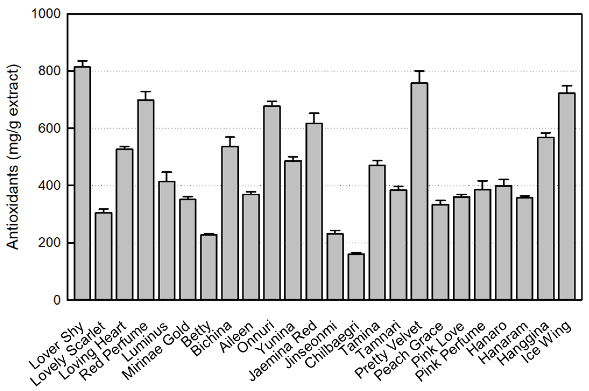
Figure 2.
HPLC chromatogram of pyrogallol standard (A) and a rosebud extract (B).
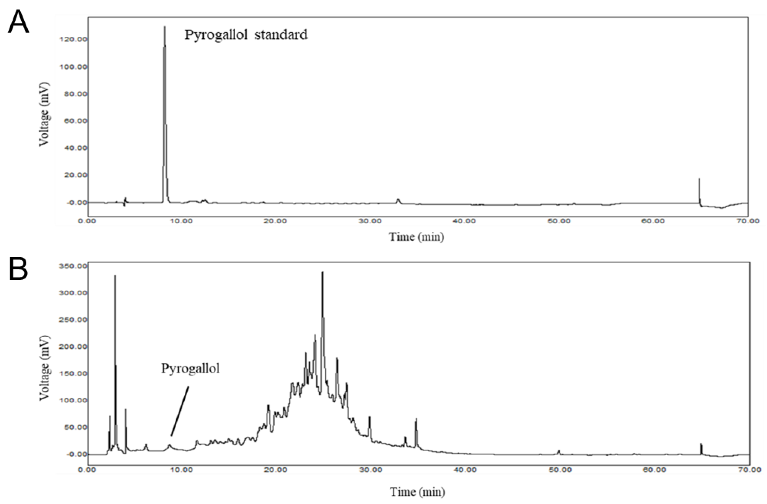
Figure 3.
1,2,3-Benzenetriol (pyrogallol) content in 24 rosebud extracts.
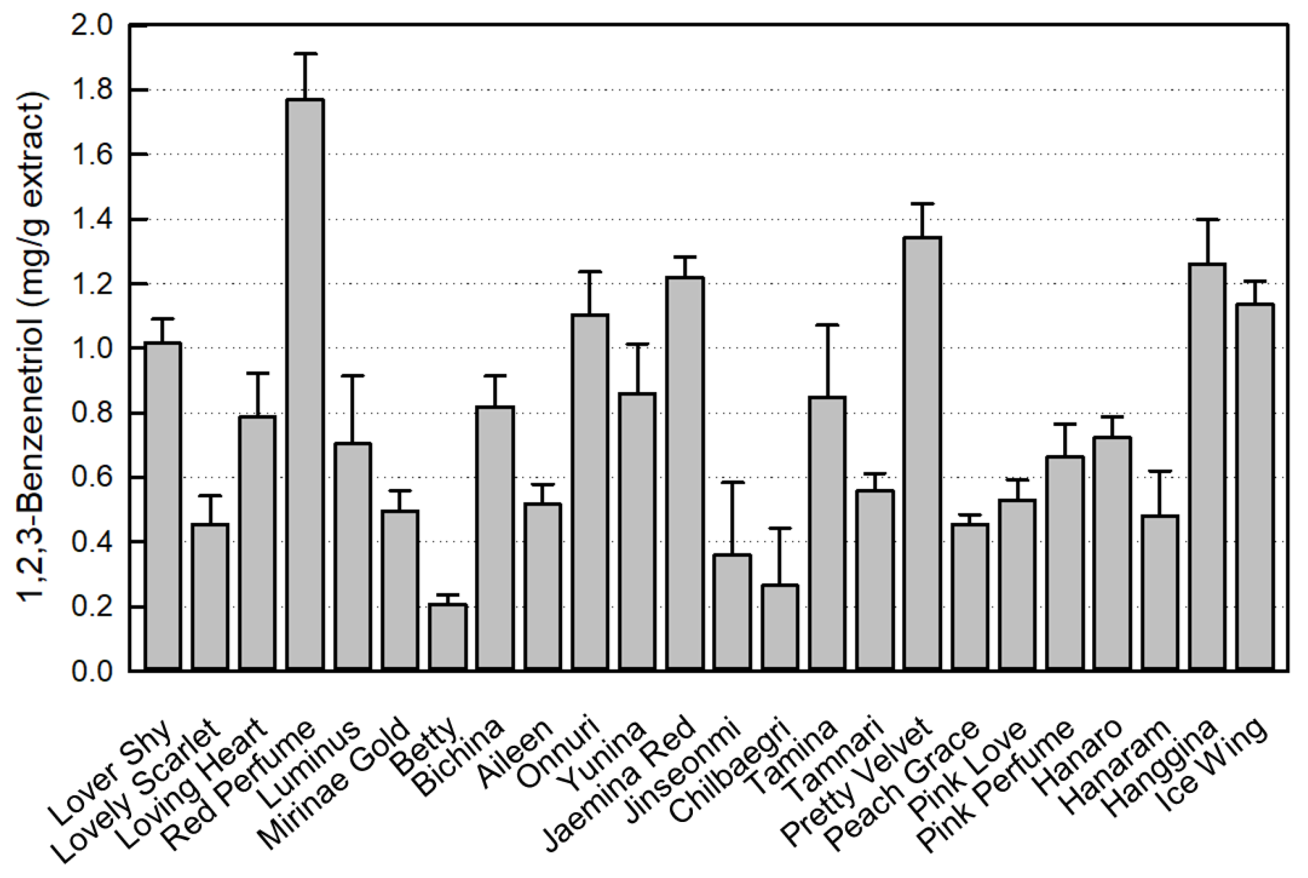
Figure 4.
Inhibition by rosebud extracts of β-hexosaminidase release from RBL-2H3 mast cells. (A) Anti-allergic activities of 24 rosebud extracts (100 μg/mL). (B) Concentration-dependent activities of selected rosebud extracts at concentrations of 10 (yellow), 32 (green) or 100 (blue) μg/mL. * Significantly different from Vehicle control (P < 0.05). # Significantly different from DNP-HSA alone (P < 0.05).
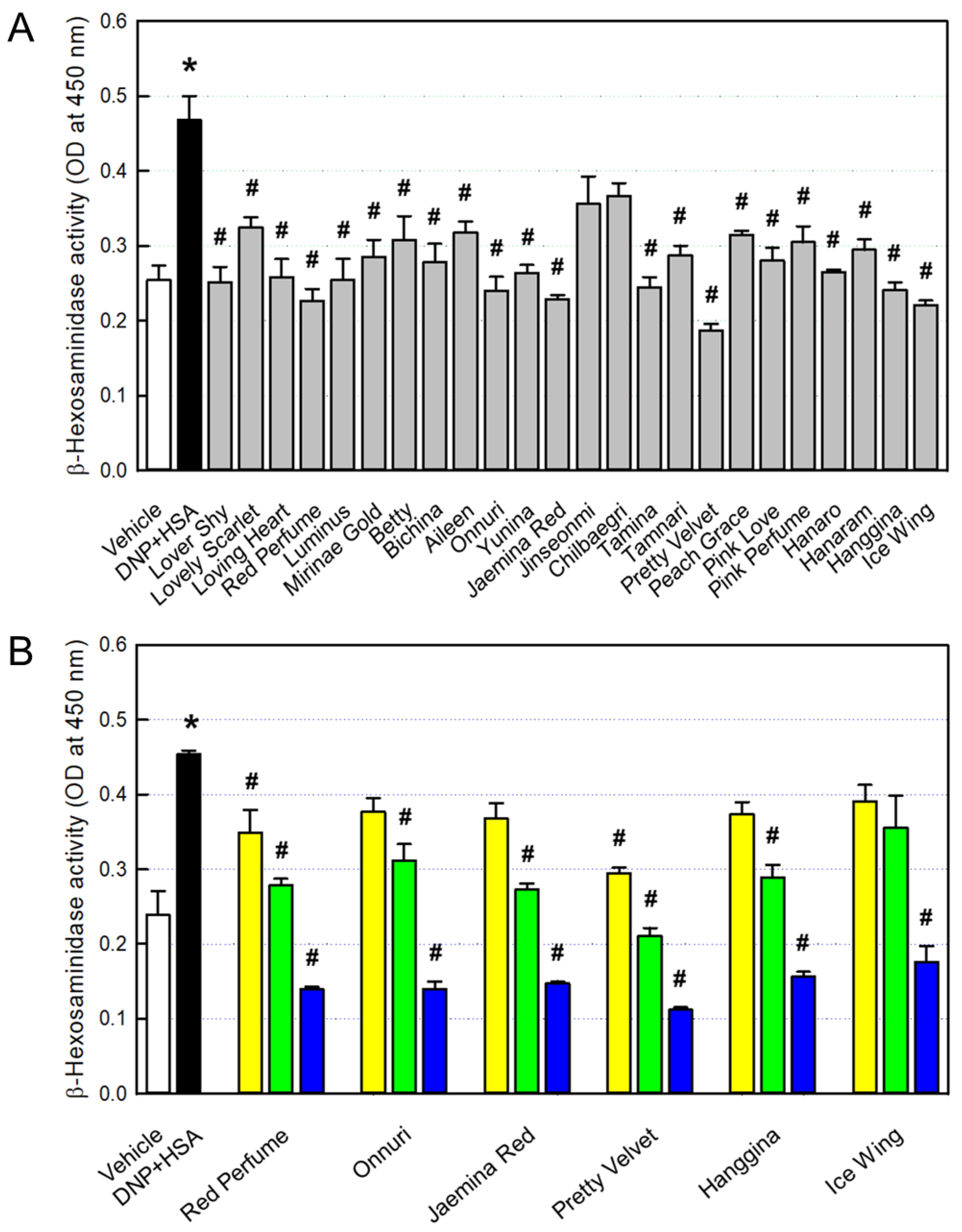
Figure 5.
Inhibition by rosebud extracts of nitric oxide production from RAW 264.7 macrophages. (A) Anti-inflammatory activities of 24 rosebud extracts (100 μg/mL). (B) Concentration-dependent activities of selected rosebud extracts at concentrations of 10 (yellow), 32 (green) or 100 (blue) μg/mL. * Significantly different from Vehicle control (P < 0.05). # Significantly different from LPS alone (P < 0.05).
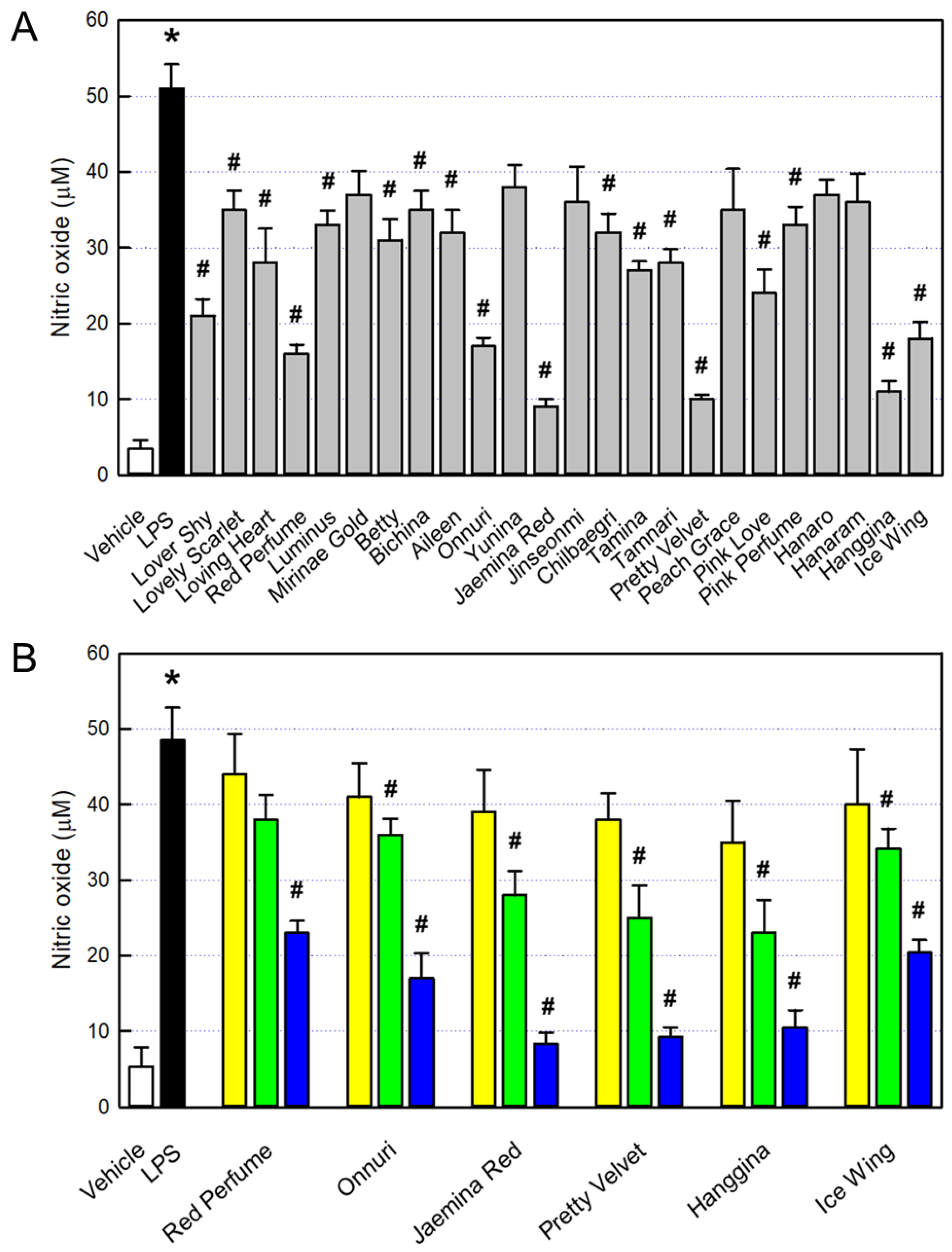
Figure 6.
Protection by rosebud extracts against systemic anaphylactic shock (mortality) of Compoud-48/80-challenged mice (died/challenged). Yellow: 10 mg/kg, green: 30 mg/kg, blue: 100 mg/kg.
Figure 6.
Protection by rosebud extracts against systemic anaphylactic shock (mortality) of Compoud-48/80-challenged mice (died/challenged). Yellow: 10 mg/kg, green: 30 mg/kg, blue: 100 mg/kg.
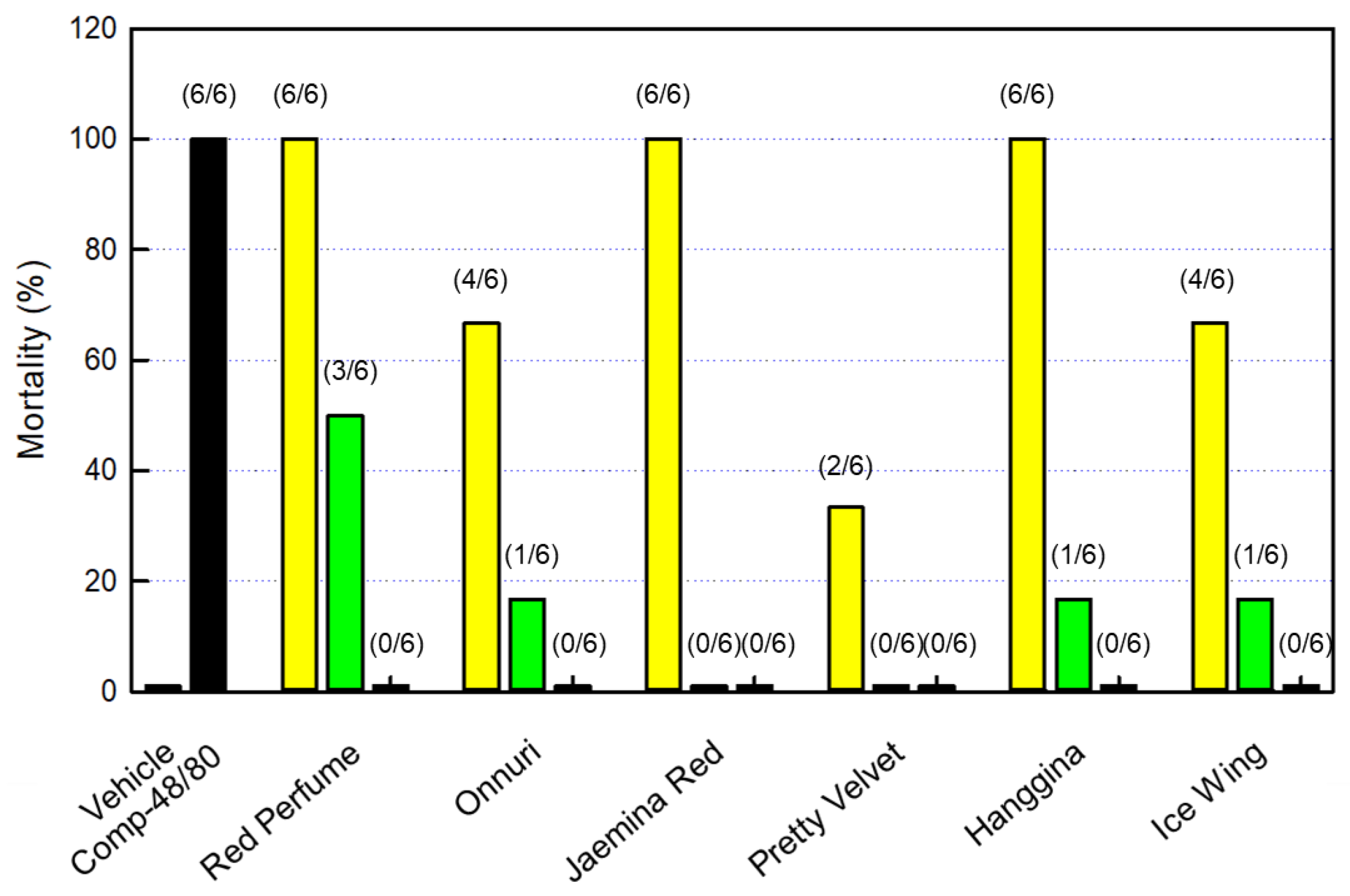
Figure 7.
Inhibition by rosebud extracts of skin allergic reaction (scratching behaviors) in Compoud-48/80-challenged mice. Yellow: 10 mg/kg, green: 30 mg/kg, blue: 100 mg/kg. * Significantly different from Vehicle control (P < 0.05). # Significantly different from Compound-48/80 alone (P < 0.05).
Figure 7.
Inhibition by rosebud extracts of skin allergic reaction (scratching behaviors) in Compoud-48/80-challenged mice. Yellow: 10 mg/kg, green: 30 mg/kg, blue: 100 mg/kg. * Significantly different from Vehicle control (P < 0.05). # Significantly different from Compound-48/80 alone (P < 0.05).
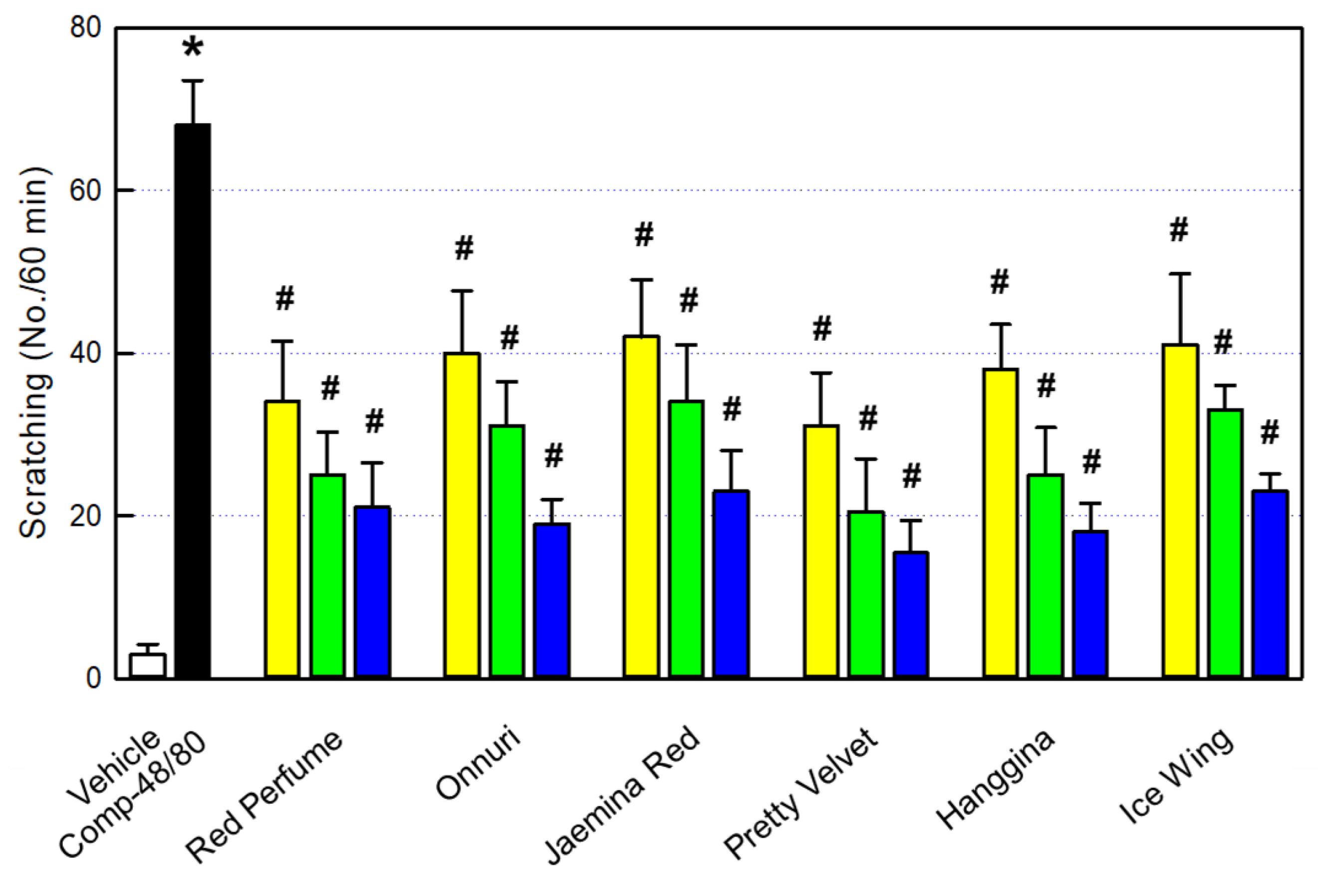
Figure 8.
Inhibition by rosebud extracts of blood IgE (A) and histamine (B) in Compoud-48/80-challenged mice. Yellow: 10 mg/kg, green: 30 mg/kg, blue: 100 mg/kg. * Significantly different from Vehicle control (P < 0.05). # Significantly different from Compound-48/80 alone (P < 0.05).
Figure 8.
Inhibition by rosebud extracts of blood IgE (A) and histamine (B) in Compoud-48/80-challenged mice. Yellow: 10 mg/kg, green: 30 mg/kg, blue: 100 mg/kg. * Significantly different from Vehicle control (P < 0.05). # Significantly different from Compound-48/80 alone (P < 0.05).
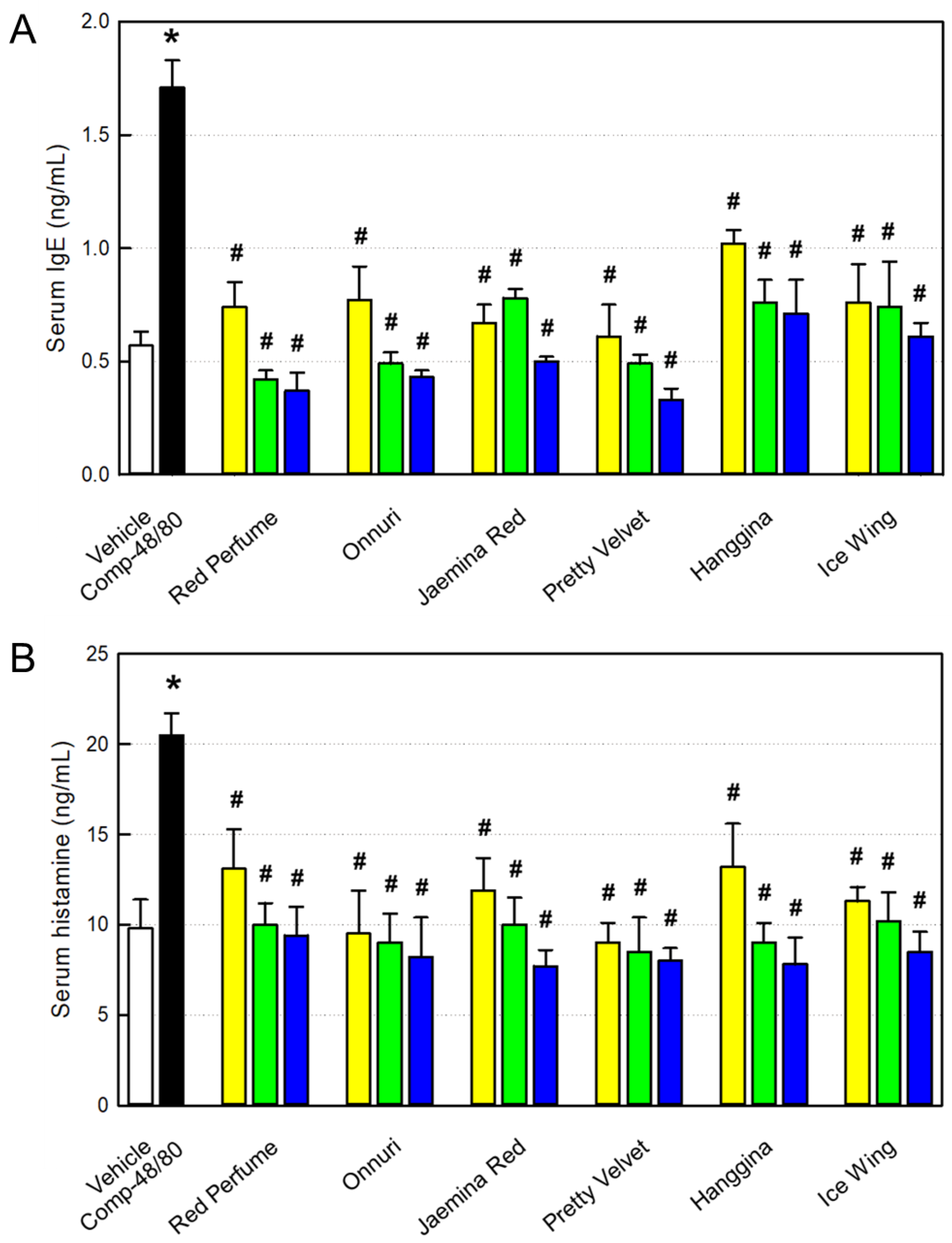
Figure 9.
Inhibition by rosebud extracts of blood tumor-necrosis factor-α (TNF-α; A) and interleukin-1β (IL-1β; B) in Compoud-48/80-challenged mice. Yellow: 10 mg/kg, green: 30 mg/kg, blue: 100 mg/kg. * Significantly different from Vehicle control (P < 0.05). # Significantly different from Compound-48/80 alone (P < 0.05).
Disclaimer/Publisher’s Note: The statements, opinions and data contained in all publications are solely those of the individual author(s) and contributor(s) and not of MDPI and/or the editor(s). MDPI and/or the editor(s) disclaim responsibility for any injury to people or property resulting from any ideas, methods, instructions or products referred to in the content. |
© 2026 by the authors. Licensee MDPI, Basel, Switzerland. This article is an open access article distributed under the terms and conditions of the Creative Commons Attribution (CC BY) license (http://creativecommons.org/licenses/by/4.0/).
Copyright: This open access article is published under a Creative Commons CC BY 4.0 license, which permit the free download, distribution, and reuse, provided that the author and preprint are cited in any reuse.


