Submitted:
08 January 2026
Posted:
08 January 2026
You are already at the latest version
Abstract
Core body temperature (CBT) is a fundamental physiological parameter tightly regulated by thermoregulatory mechanisms and is critically important for heat stress assessment, clinical management, and circadian rhythm research. Although invasive measurements such as pulmonary artery, esophageal, and rectal temperatures provide high accuracy, their practical use is limited by invasiveness, discomfort, and restricted feasibility for continuous monitoring in daily-life or field environments. Consequently, extensive efforts have been devoted to developing non-invasive CBT measurement and estimation techniques. This review provides an application-oriented synthesis of invasive reference methods and representative non-invasive approaches, including in-ear sensors, infrared thermography, ingestible telemetric sensors, heat-flux-based techniques, and model-based estimation using wearable physiological signals. For each approach, measurement principles, accuracy, invasiveness, usability, and application domains are comparatively examined, with particular emphasis on trade-offs between measurement fidelity and real-world implementability. Rather than ranking methods by absolute performance, this review highlights their relative positioning across clinical, occupational, and daily-life contexts. While no single non-invasive technique can universally replace invasive gold standards, recent advances in wearable sensing, heat-flux modeling, and multimodal estimation demonstrate growing potential for practical CBT monitoring. Overall, the findings suggest that future CBT assessment will increasingly rely on hybrid and context-aware systems that integrate complementary methods to enable reliable monitoring under real-world conditions.
Keywords:
core body temperature
; non-invasive measurement
; wearable sensors
; in-ear thermometry
; infrared thermography
; ingestible sensors
; heat flux sensing
1. Introduction
Core body temperature (CBT) is a fundamental physiological parameter that represents the temperature of deep tissues and organs within the body, including the viscera, blood, and central nervous system, and is distinct from surface skin temperature. CBT is tightly regulated by the thermoregulatory system centered in the hypothalamus and is typically maintained within a narrow range of approximately 36.5–37.5 °C at rest, while dynamically changing in response to physical activity, environmental temperature, and circadian rhythms [1,2]. In contrast to skin temperature, which is strongly influenced by ambient conditions, clothing, and peripheral blood flow and does not necessarily reflect internal physiological states, CBT directly represents the internal thermal environment resulting from metabolic heat production, circulatory dynamics, and sweating responses. Consequently, CBT plays a critical role in heat stress assessment, maintenance of organ function, perioperative hypothermia management during anesthesia, diagnosis of infectious and febrile diseases, as well as sleep and circadian rhythm research [3].
Despite its physiological importance, accurate measurement of CBT remains challenging. According to its strict definition, reliable assessment requires temperature measurements at sites close to the central circulation, such as the pulmonary artery, esophagus, rectum, or urinary bladder. However, these approaches are invasive or semi-invasive and impose substantial limitations in terms of wearability, user comfort, and feasibility for long-term or free-living measurements. In clinical and experimental settings, esophageal and rectal temperatures have long been regarded as gold standards for CBT measurement; nevertheless, issues such as discomfort during probe insertion, risk of infection, and practical difficulties during exercise or occupational activities persist [4]. These limitations have driven growing global interest in the development of non-invasive techniques for daily and continuous CBT monitoring.
Representative non-invasive approaches include in-ear devices that measure temperature near the external auditory canal [5], infrared thermography for acquiring and analyzing body surface temperature distributions [6], ingestible telemetric pill-type sensors [7], heat-flux-based methods relying on skin temperature and heat flux measurements [8], and algorithmic estimation techniques that infer CBT from other physiological signals such as heart rate, skin temperature, heat flux, ambient temperature, and humidity [9]. Each of these methods exhibits distinct advantages and limitations with respect to accuracy, temporal responsiveness, wearability, and robustness to environmental influences. Therefore, a comprehensive understanding of their characteristics is essential to select appropriate techniques for specific users and application scenarios.
As summarized in Table 1, most previous review papers have focused on a limited subset of core body temperature sensing techniques rather than providing a comprehensive overview [10,11,12,13,14,15,16]. In contrast, this review covers a wide range of core body temperature measurement and estimation approaches, including the latest studies, and offers an application-oriented comparison by explicitly addressing the trade-offs among accuracy, invasiveness, and real-world usability. Specifically, both invasive and non-invasive techniques—such as in-ear sensors, infrared thermography, ingestible sensors, heat-flux-based methods, and model-based estimation approaches—are reviewed, and their applicability in clinical, occupational, and daily-life contexts is discussed. This review is intended for researchers and practitioners seeking to select or design CBT monitoring systems tailored to specific application requirements.
2. Core Body Temperature Measurement Techniques
2.1. Invasive and Semi-Invasive Reference Measurements
The most reliable approaches for evaluating core body temperature (CBT) involve invasive measurements in which temperature probes are inserted into anatomical sites located close to the central circulation, enabling direct assessment of internal temperature. Representative measurement sites include the esophagus, rectum, urinary bladder, and pulmonary artery. Because these sites are adjacent to deep trunk blood flow and are relatively insulated from ambient temperature and fluctuations in skin blood perfusion, they are considered to provide measurements closest to the “true” CBT [17]. Among them, pulmonary artery blood temperature directly reflects the temperature of the central circulation and has long been regarded as the reference standard in intensive care units and operating rooms [18]. However, pulmonary artery catheterization is a highly invasive medical procedure associated with serious risks, including vascular injury, arrhythmias, and infection, making it impractical for routine physiological research or use in daily-life settings [19].
Esophageal temperature is measured by inserting a temperature probe into the thoracic esophagus, which lies in close proximity to the heart and major blood vessels, and is widely used in anesthetized surgical procedures and exercise physiology studies [20]. Esophageal temperature exhibits relatively rapid responsiveness to thermal perturbations and can sensitively capture CBT changes during exercise or conditions involving pronounced thermoregulatory responses [21]. Nevertheless, probe insertion causes discomfort to participants, and measurements are susceptible to artifacts arising from swallowing and body movement, rendering this method unsuitable for continuous monitoring in daily life [22].
Rectal temperature has been the most widely used surrogate of CBT and has served as a reference measure across clinical medicine, exercise science, and environmental physiology [23]. The rectum is well perfused and exhibits relatively small temperature fluctuations, resulting in high measurement stability. However, rectal temperature responds more slowly to changes in CBT than esophageal temperature, and during intense exercise it may lag behind increases in CBT by several minutes [24]. In addition, issues related to privacy, hygiene, and user acceptance substantially limit its wearability, making long-term or field-based measurements impractical [25,26].
Bladder temperature is measured using a temperature sensor integrated into a urinary catheter. Because urine in the bladder is thermally coupled to deep abdominal blood flow, bladder temperature is often used as an indicator of CBT in postoperative care and intensive care settings [27]. However, its accuracy may be influenced by urine volume, and catheter insertion carries an inherent risk of infection, imposing clinical constraints that limit its general applicability [28].
In summary, invasive and semi-invasive measurement techniques—including pulmonary artery, esophageal, rectal, and bladder temperatures—provide the most accurate assessment of CBT and are widely regarded as reference standards. Nevertheless, due to discomfort during probe insertion, restrictions on movement, hygiene requirements, and infection risks, these methods are unsuitable for continuous, long-term, or everyday monitoring. Consequently, their use is largely confined to controlled environments such as operating rooms, intensive care units, laboratories, or rigorously supervised exercise physiology studies. At the same time, these reference measurements play a central role in the validation and benchmarking of non-invasive CBT estimation methods, including wearable in-ear devices, heat-flux-based techniques, and machine-learning-based models, and remain indispensable for the development and evaluation of emerging CBT monitoring technologies.
2.2. Non-Invasive and Minimally Burdensome Measurement Approaches
Non-invasive and minimally burdensome approaches for core body temperature monitoring have been actively investigated to overcome the practical limitations of invasive reference measurements. In this review, such approaches discussed in this section include in-ear temperature sensors, infrared thermography, ingestible telemetric sensors, heat-flux-based methods, and model-based estimation techniques using wearable physiological and environmental signals. These methods differ substantially in their measurement principles, achievable accuracy, wearability, and robustness to environmental and physiological variability.
In this review, ingestible sensors are classified as non-invasive approaches because they do not require probe insertion through the skin or body cavities. Nevertheless, they involve oral administration and are therefore not entirely intervention-free, which should be considered when interpreting their applicability.
2.2.1. In-Ear Temperature Sensors for Core Body Temperature Monitoring
The ear is located close to the brain and major blood vessels such as the carotid arteries, and tympanic membrane temperature has been shown to closely reflect changes in deep brain temperature [29]. For this reason, infrared tympanic thermometers that measure eardrum temperature have long been widely used in clinical and non-clinical settings [30]. However, infrared tympanic thermometers are designed for brief, spot measurements and are therefore unsuitable for continuous monitoring [31]. Moreover, maintaining long-term contact between a sensor and the tympanic membrane is impractical and uncomfortable for users [32].
To address these limitations, wearable in-ear devices that measure temperature near the entrance of the ear canal have been actively investigated [5,33,34,35,36,37,38]. For example, Ota et al. developed a wearable device equipped with an infrared sensor for measuring ear canal temperature with the aim of enabling daily CBT monitoring, and demonstrated good agreement between the developed device and a commercially available ear thermometer under hot and exercise conditions [5]. Chaglla et al. reported improved temperature measurement accuracy by coating the surface of a commercial infrared sensor with graphene ink [33]. However, these studies were conducted with a limited number of participants under relatively idealized exercise conditions, and they did not employ invasive gold-standard reference measurements, as described in Section 2.1, but instead relied on commercial ear thermometers. Therefore, caution is required when interpreting the agreement between CBT and temperatures measured by the proposed devices.
Ko et al. investigated the relationship between ear canal temperature measured using an infrared sensor and rectal temperature during sleep in female participants [34]. Their results showed that a sufficient insertion depth (approximately 14 mm) was required to achieve a strong correlation between ear canal and rectal temperatures. This finding reflects the presence of a temperature gradient from the tympanic membrane to the ear canal entrance, caused by environmental influences, resulting in position-dependent temperature differences. Roossien et al. evaluated the feasibility of using a commercial wearable ear-mounted sensor for heat stress management in workers exposed to high thermal loads [35]. Although a high correlation was observed between the wearable sensor readings and hospital-grade infrared thermometers, the authors reported that environmental changes and personal protective equipment adversely affected measurement accuracy and occasionally caused abnormal readings, highlighting challenges in highly variable environments.
Kato et al. reported a reliability assessment of a wearable infrared ear canal thermometer (VTB01, Vitarate) designed for sports and occupational applications [36]. They evaluated measurement accuracy under various hot and exercise conditions while periodically introducing airflow toward the participants. Although systematic errors attributable to environmental changes were observed, the accuracy remained within acceptable limits under the tested conditions. The authors noted, however, that more severe heat stress, exercise intensity, or environmental variability could further increase measurement errors. In addition, the use of medical film to reduce heat exchange between the ear canal and the external environment likely mitigated environmental effects, and performance without such insulation may be lower. To improve robustness against environmental variability, Olson et al. developed a device incorporating two temperature sensors positioned at different locations within the ear canal [37]. Their results demonstrated improved accuracy compared with a single-sensor configuration when evaluating errors associated with ambient temperature differences and inter-individual CBT variation. Nevertheless, this study did not consider time-varying ambient temperature or airflow effects, which are commonly encountered in real-world use scenarios. Zhang et al. reported an in-ear device featuring a longer insertion depth (27 mm) designed to measure temperature closer to the tympanic membrane for heat-stroke prevention applications [38]. When properly positioned, the device showed good agreement with rectal temperature during exercise in hot environments, except immediately after the onset and cessation of exercise. However, the study involved a small sample size (six participants) and was conducted under controlled laboratory conditions.
Overall, in-ear temperature sensors offer reduced user burden compared with gold-standard invasive CBT measurements, as they require only device placement in the ear. However, because the ear canal is inherently susceptible to external environmental influences, achieving robust measurements under dynamic conditions remains a key challenge. Although potential applications include sports, occupational settings, and fever screening—where substantial environmental variation and CBT elevation are expected [5,35,36,37,38]—current evidence of effectiveness is largely limited to sleep monitoring [27], intraoperative settings [39], and light activities performed under relatively stable environmental conditions [35]. While several strategies to enhance robustness against environmental disturbances have been proposed [37,38], validation has so far been confined to idealized conditions, underscoring the need for reliability assessments that better reflect real-world usage scenarios.
2.2.2. Infrared Thermography for Non-Contact Temperature Assessment
Infrared thermography (IRT) is a non-contact and non-invasive temperature measurement technique that estimates surface temperature by detecting infrared radiation emitted from the human body [6]. All objects with temperatures above absolute zero emit infrared energy as a function of their temperature. IRT passively detects mid- to long-wavelength infrared radiation emitted from the skin and converts the measured radiative intensity into an apparent temperature using the Stefan–Boltzmann law, enabling acquisition of high-spatial-resolution maps of skin temperature distribution [6]. Unlike conventional contact-based thermometers, IRT does not require physical contact between the subject and the measurement device. As a result, it has been widely applied and investigated in a broad range of contexts, including fever screening during infectious disease outbreaks [6], as well as medical, industrial, and animal health management applications [40,41].
IRT offers several notable advantages for body temperature assessment. The primary advantage is its non-invasive nature, as temperature can be measured without direct contact, thereby minimizing discomfort and reducing the risk of cross-contamination [42]. This characteristic is particularly valuable in situations where conventional temperature measurement methods are difficult to apply, such as mass screening of large populations or monitoring of vulnerable groups. During the COVID-19 pandemic, non-contact infrared cameras and so-called “forehead thermometers” were widely deployed at various checkpoints to rapidly identify individuals with suspected fever while maintaining physical distancing [43]. In addition, the ability to measure temperature remotely is important for minimizing stress on subjects [42,44]. In veterinary medicine and pediatric care, rectal thermometry has long been considered the standard for core temperature assessment; however, it imposes substantial stress on animals and infants and is often impractical for repeated measurements in large populations. In this context, thermography has attracted attention as a simple and animal-welfare-conscious approach, enabling frequent or continuous temperature monitoring without physical restraint [42,44]. For example, in livestock health management, measuring temperatures around the eye region or across the body surface using IRT has been explored as a means to detect signs of fever or inflammation without excessive handling of animals [44].
Another important advantage of IRT is its rapid measurement capability and broad spatial coverage [45]. Modern infrared cameras can scan an individual almost instantaneously and can continuously screen multiple individuals per minute, making them particularly useful in high-traffic environments such as airports, hospitals, and disaster triage centers [45]. In emergency and disaster response scenarios, thermal imaging cameras enable rapid detection of human heat signatures even in darkness or smoke, and are routinely used by firefighters and rescue teams to locate survivors and identify thermal hotspots [47]. Furthermore, thermographic images provide full-body surface temperature maps, allowing spatial analysis of temperature distributions [45]. This capability enables visualization of localized thermal abnormalities, such as regions of inflammation or infection and heat distribution during physical activity, which may be missed by single-point temperature measurements [47]. In occupational health and sports medicine, for example, visualizing how different body regions heat up during work or exercise can help identify areas exposed to excessive thermal load and support the development of effective cooling strategies [46,47]. Overall, owing to its non-contact and real-time characteristics, IRT represents a highly convenient and versatile technology for temperature assessment in both humans and animals.
Despite these advantages, important limitations exist when IRT is used for estimating core body temperature (CBT), particularly with respect to accuracy and reliability [14,16,48]. Skin temperature serves only as an indirect surrogate of CBT, and the relationship between the two can vary substantially depending on both physiological and environmental factors [16,48,49]. Under stable indoor conditions and with appropriate measurement protocols, thermographic temperatures measured at specific anatomical sites—such as the inner canthus of the eye—have been shown to reasonably track internal temperature [50,51,52]. However, in practice, the sensitivity and specificity of IRT for fever screening are often insufficient. Numerous studies have reported high false-negative rates, in which non-contact infrared devices fail to detect individuals who are truly febrile [52]. For example, historical reports have documented extremely low detection rates of infected individuals during large-scale airport screening using thermal imaging systems [52]. During the 2014 Ebola virus disease outbreak and the SARS epidemic, airport-based IRT screening failed to identify any confirmed cases, corresponding to a sensitivity of 0% in some instances [53]. Similarly, during the early phase of the COVID-19 pandemic, only a few percent of cases worldwide were detected through airport temperature screening [54,55]. While some of these failures can be attributed to improper deployment or operational issues, they also stem from the inherent physiological variability of skin temperature [14,48,56,57]. Factors such as ambient temperature, airflow, recent physical activity, sweating state, and inter-individual metabolic differences can influence skin temperature independently of CBT [14,48]. The forehead is a common measurement site for handheld infrared thermometers; however, it is highly sensitive to environmental cooling and heating, and immediately after exposure to hot or cold environments, forehead temperature often fails to accurately reflect internal thermal status [16,57,58]. Indeed, simple forehead scanning has been shown to be unable to track CBT changes during exercise or under heat stress [59,60,61,62,63,64]. For instance, studies have demonstrated that standard infrared thermometers failed to detect clinically elevated CBT in marathon runners or firefighters wearing protective clothing [59,63].
In response to these challenges, research efforts have focused on improving the robustness of IRT-based temperature assessment against environmental variability and inter-individual differences. Wang et al. proposed a sensing system combining IRT with auxiliary sensors, including an anemometer and temperature–humidity sensors, for applications in cattle temperature management, and reported that the combined system achieved improved CBT estimation accuracy compared with IRT alone in field experiments [65]. However, their validation was limited to indoor livestock facilities, and the effectiveness of the system in open or free-ranging environments remains unverified. Shan et al. developed a CBT prediction model for human temperature management in medical, sports, and elderly care contexts by integrating personal characteristics (e.g., sex, age, and ethnicity) with skin temperatures measured at multiple facial regions using IRT, reporting superior accuracy and consistency compared with conventional linear regression and machine-learning models [66]. Nevertheless, this model was validated using publicly available datasets collected under constrained indoor conditions, and its performance under extreme thermal environments or in the presence of physiological responses such as sweating has not yet been examined.
In summary, although IRT-based CBT monitoring has been explored across a wide range of applications owing to its high convenience and non-contact nature, raw infrared measurements alone do not constitute a complete substitute for direct CBT assessment. Environmental sensitivity, physiological variability, and user- and device-related errors collectively limit the ability of IRT to provide accurate internal temperature estimates under diverse conditions. At present, IRT does not deliver precise internal temperature measurements across all scenarios and, at best, functions as a screening tool for identifying individuals with suspected fever. Nevertheless, ongoing efforts to integrate IRT with other sensors and machine-learning techniques may help overcome these limitations, potentially enabling broader and more reliable applications in the future.
2.2.3. Ingestible Telemetric Sensors for Core Body Temperature Measurement
Ingestible sensors, also referred to as smart pills, are non-invasive monitoring technologies that are orally administered, introduced into the gastrointestinal (GI) tract, and capable of measuring physiological signals from within the body and wirelessly transmitting the data to an external receiver [67,68,69]. At present, GI temperature measurement using ingestible sensors is widely employed as an alternative approach for core body temperature (CBT) assessment, replacing invasive measurements such as rectal or esophageal temperature monitoring [67]. The operating principle of ingestible temperature sensors is relatively straightforward: a miniature capsule containing a temperature-dependent sensing element, such as a thermistor or quartz crystal oscillator, is swallowed, and temperature data measured within the GI tract are transmitted wirelessly to an external receiver [67]. Early concepts of ingestible temperature sensing were proposed in the 1980s [70,71], and since the 2000s, advances in wireless communication and miniaturization of electronic components have led to the commercialization of devices such as VitalSense® [72], CorTemp® [73], myTemp® [74], and e-Celsius® [75]. These technologies have been increasingly applied in exercise physiology, clinical medicine, and occupational safety research [7].
The primary advantage of ingestible sensors for CBT monitoring lies in their ability to provide non-invasive and continuous measurements under free-living conditions [7]. Although rectal and esophageal temperatures are regarded as gold-standard indicators of CBT and offer high measurement reliability, they are invasive and often associated with substantial discomfort, limiting their practicality for long-term monitoring and field applications [7]. In contrast, once ingested, telemetric sensors can continuously record CBT for several hours to several tens of hours with minimal interference to daily activities or physical exercise [76]. This capability has made ingestible sensors a standard measurement tool in thermal and exercise physiology for recording CBT responses to heat stress and physical exertion, as well as for assessing the risk of heat-related illnesses such as heat stroke [73,77,78,79]. Successful applications have also been reported in demanding environments where conventional methods are impractical, including among divers, long-distance runners, military training scenarios, soccer players, and triathletes [74]. In clinical contexts, ingestible sensors have been investigated for fever monitoring, with studies reporting that GI temperature measurements provide higher reliability than axillary or non-contact forehead temperature measurements. Such approaches may facilitate improved characterization of fever patterns, earlier detection of conditions such as sepsis, and reduced workload for nursing staff by enabling automated and continuous temperature monitoring [79]. In veterinary and animal research, studies involving dogs have demonstrated that ingestible sensors can safely and reliably measure CBT, offering a temperature monitoring method that aligns with animal welfare considerations [81].
Despite these advantages, ingestible sensors present several important limitations. First, sensor movement within the GI tract over time and the influence of food and fluid intake can induce transient temperature drops or measurement bias, particularly during the early post-ingestion period or while the sensor remains in the stomach [82,83]. Consequently, many studies recommend initiating data analysis only after several hours have elapsed following ingestion to ensure measurement stability [83,84]. However, excessive delay increases the risk of sensor excretion, necessitating careful timing in both research and practical applications [85]. Second, systematic sensor-to-sensor bias and inter-device variability have been reported, requiring individual calibration or correction for high-precision research applications [7]. For example, Hunt et al. evaluated 119 commercially available ingestible sensors (myTemp) in a temperature-controlled water bath and found that 59.7% exhibited bias exceeding specified accuracy thresholds, underscoring the need for pre-calibration within physiologically relevant temperature ranges [86]. Product-dependent differences have also been documented. Bongers et al. tested four commercial ingestible sensors (CorTemp, e-Celsius, myTemp, and VitalSense) in a water bath and reported that although all devices demonstrated high overall accuracy, notable differences in bias and response time existed among products [87]. Third, because ingestible sensors are excreted from the body within several tens of hours [7], long-term CBT monitoring over weeks or longer requires repeated ingestion. Excretion time varies considerably among individuals; for instance, Monnard et al. reported a mean excretion time of 31 hours, ranging from 13 to 82 hours after ingestion [76]. Therefore, prolonged monitoring necessitates individualized planning of ingestion schedules. Additional practical considerations include the predominantly disposable nature of current capsules, associated costs, and restrictions on use during magnetic resonance imaging (MRI) examinations [80].
Recent research trends have focused on further miniaturization and functional enhancement of ingestible sensors, alongside increased emphasis on power supply and safety considerations. Alternative energy strategies, such as energy harvesting from the GI environment and the use of biodegradable power sources, have been proposed to replace conventional button batteries, enabling safer and more sustainable designs [67,88]. Moreover, efforts toward multifunctional sensing capabilities and the development of mechanisms that allow prolonged residence within the GI tract are ongoing [68]. These advances are expected to further enhance the functionality, usability, and application scope of ingestible telemetric sensors for CBT monitoring.
2.2.4. Heat-Flux-Based Methods for Core Body Temperature Estimation
Methods that estimate core body temperature (CBT) by measuring temperature and heat flux in the vicinity of the skin have been investigated for several decades as non-invasive alternatives to invasive reference measurements such as rectal and esophageal temperatures [8,89,90,91]. Representative approaches within this framework include the zero-heat-flux (ZHF) method [89,90,92,93,94,95,96,97], the single-heat-flux (SHF) method [8,98,99,100,101,102,103,104,105,106,107,108,109,110,111,112,113,114,115], and the dual-heat-flux (DHF) method [91,116,117,118,119,120,121,122,123,124,125]. All of these techniques share the common principle of estimating CBT based on thermal transport information obtained from sensors attached to the skin surface.
The ZHF method aims to actively eliminate heat flux between the skin and the environment, thereby driving the skin surface temperature toward equivalence with CBT [90]. This is achieved by placing a temperature sensor and a heating element on the skin surface and controlling the heating so that no net heat flow occurs across the skin boundary, establishing thermal equilibrium between the skin surface and deeper tissues. Based on this principle, ZHF-based devices have been explored for applications such as temperature management during cardiac and vascular surgery, as well as in hospital wards and intensive care units [92,93,94,95,96]. More recently, Lu et al. investigated the feasibility of ZHF methods for heat stress management under high-temperature environments and reported that an improved ZHF probe incorporating a Peltier element enabled stable CBT measurements even under hot conditions [97], extending potential applications beyond clinical settings. However, due to the need for active thermal control to maintain zero heat flux, ZHF systems typically require high power consumption and relatively thick probe structures, resulting in limited flexibility and wearability. These characteristics restrict their suitability for free movement and long-term portable monitoring, posing significant challenges for wearable applications [91,121]. In addition, reduced accuracy has been reported at CBT values below 32 °C [95], indicating limitations for hypothermic conditions.
The SHF method estimates CBT by simultaneously measuring skin temperature and heat flux and approximating the human body as a one-dimensional heat conduction model [8]. Unlike ZHF methods, SHF does not require active heating, enabling low-power operation and simpler device structures [121]. Consequently, SHF-based approaches have been widely investigated for applications such as circadian rhythm monitoring [98], exercise physiology, and heat stress monitoring in occupational safety contexts [8,98,99,106], making them attractive candidates for wearable CBT estimation during daily activities. In recent years, commercial devices such as CORE® and Calera Research® those developed by greenteg AG have become available, and studies have reported validation results for their use during exercise in hot environments and in bedside clinical applications [107,108,109,110,111,112,113,114,115]. However, several practical challenges remain. SHF methods typically require prior calibration against a reference measurement to account for inter-individual differences in thermal resistance of the skin and subcutaneous tissues [121]. Moreover, estimation accuracy is sensitive to environmental factors such as ambient temperature and airflow [96], and even slight changes in sensor–skin contact can disrupt heat flux measurements and destabilize CBT estimates [117,118]. To address these limitations, recent studies have proposed self-calibration techniques that eliminate the need for reference measurements [102,104], as well as probe designs that enhance robustness against environmental disturbances [99,101,103,105]. Leveraging their inherent advantages of low power consumption and structural simplicity, SHF-based methods are expected to advance toward practical wearable devices as these challenges are progressively mitigated.
The DHF method extends the SHF approach by utilizing two heat flux measurement paths with different thermal resistances, enabling more precise estimation of the temperature gradient between the skin surface and deeper tissues [91]. By simultaneously incorporating multiple heat flux signals, DHF methods reduce uncertainty associated with skin-side thermal properties and have been reported to improve robustness against inter-individual variability [125,126]. Applications investigated to date include temperature management in clinical settings [120], circadian rhythm monitoring [120], and physiological condition assessment during work under hot environments [123,124]. Nevertheless, several fundamental challenges must be addressed for wearable implementation. First, because DHF systems rely on multi-layer or multi-channel structures, changes in skin contact can disrupt the ratio between heat flux channels, leading to unstable estimates. Motion artifacts arising from walking, arm movement, and sweat-induced interface changes are difficult to avoid in real-world conditions and represent major sources of error [103,106,127]. Second, variations in skin blood flow and sweating during exercise alter local thermal resistance, violating the steady-state assumptions of the underlying thermal model and resulting in response delays and estimation errors [123,126]. Third, the additional thermal resistance layers, thermocouples, and wiring required for dual-channel configurations tend to increase sensor thickness and stiffness, reducing conformity to curved skin surfaces and compromising wearing comfort [125]. Furthermore, achieving complete robustness against ambient temperature fluctuations and airflow disturbances remains challenging, as small variations in heat flux can significantly affect estimation accuracy [106,127]. Addressing these issues will require advances in flexible and thin material design, interface stabilization mechanisms, compensation for environmental disturbances, and algorithmic corrections capable of handling dynamic physiological conditions.
A common advantage of heat-flux-based methods is their ability to provide continuous CBT monitoring through simple attachment to the skin surface without the need for invasive procedures. Unlike ingestible sensors, these methods are not constrained by limited usage duration or sensor migration within the body, enabling long-term and repeated measurements. Compared with infrared thermography, heat-flux-based approaches are not limited by field-of-view constraints and can provide stable data acquisition during physical activity. However, each method exhibits distinct trade-offs. ZHF methods offer high accuracy and are well established in clinical practice but are constrained by power consumption and bulky probe structures that limit wearability. SHF methods are structurally simple and energy efficient, making them well suited for wearable applications, yet they remain sensitive to variations in tissue properties and environmental disturbances, which pose challenges for stable CBT estimation in daily-life settings. DHF methods improve robustness against tissue property uncertainty and external disturbances compared with SHF approaches but introduce increased structural complexity and reduced wearing comfort due to thicker and less flexible sensor designs. Overcoming these practical challenges in each method is expected to play a key role in expanding their applicable use scenarios in the future.
2.2.5. Model-Based and Multimodal Estimation of Core Body Temperature
Core body temperature (CBT) is a critical indicator directly linked to the assessment of heat stress and excessive thermal load. However, direct measurements such as rectal or esophageal temperatures are invasive and operationally restrictive, making them unsuitable for continuous monitoring in occupational settings, sports, military operations, and daily-life environments [128,129,130]. To bridge this gap, a broad range of studies has explored the estimation of CBT from physiological and environmental signals that can be readily acquired using wearable devices, including heart rate (HR), skin temperature, heat flux, acceleration (activity level), and ambient conditions [9,128,129,130,131,132,133,134,135,136,137,138,139,140,141,142,143,144,145,146,147,148,149,150]. Among these inputs, HR has been widely adopted as a primary variable because it is closely linked to metabolic heat production and heat dissipation through circulatory regulation. Early frameworks proposed the use of Kalman filters and extended Kalman filters to perform sequential state estimation of CBT based on time-series observations [128,131]. More recently, multimodal estimation approaches incorporating skin temperature, heat flux, and ambient temperature and humidity—signals commonly used in heat-flux-based methods—have been investigated, demonstrating improved robustness and estimation performance compared with HR-only models [9,133,134,137,138,141,146,150].
The underlying principles of these approaches can be broadly categorized into physiology- and physics-based models and data-driven (statistical or machine-learning-based) models. In the former category, simplified compartmental or thermophysiological models that approximate human heat balance and heat transfer are employed to estimate CBT using wearable-derived inputs such as HR, skin temperature, activity level, and environmental conditions [133,140]. For example, Hamatani et al. combined wearable sensing with the Gagge two-node thermophysiological model, using HR, skin temperature, and ambient temperature and humidity acquired from wearable or portable sensors as inputs to compute the temporal evolution of CBT and skin temperature [132]. While such approaches offer strong physiological interpretability, they are constrained by parameter sensitivity and the inherent limitations of model simplification [140,150]. More recently, Hirata et al. developed a simplified compartment model derived from their previously reported high-resolution anatomical human model and combined it with wearable measurements to enable real-time CBT estimation, reflecting ongoing efforts to adapt physiologically grounded models for practical deployment [140].
In contrast, data-driven approaches learn a direct mapping from observed signals to CBT (or changes in CBT) using regression, state-space models, or machine-learning techniques, thereby improving adaptability to inter-individual and contextual variability [9,128,129,130,131,133,135,136,137,138,139,140,141,142,143,144,145,146,147,148,149]. In practice, hybrid approaches combining model-based and data-driven elements have also been reported. Kalman-filter-based frameworks, in particular, are well suited for real-time estimation under noisy field conditions because they allow recursive state updates with explicit handling of measurement uncertainty [134,150]. However, recent studies have highlighted that thermophysiological states during exercise (heating phase) and recovery (cooling phase) can differ substantially even at similar HR levels, leading to increased estimation errors when a single model is used. To address this issue, biphasic or multi-model approaches have been proposed to improve robustness while retaining HR-only estimation. For example, Rizvi and Falcone et al. introduced methods that switch between observation models for exercise and recovery phases based on HR [142,148]. Subsequently, Zhao et al. extended these approaches by dynamically updating the contribution of exercise and recovery observation models using weighted transitions or long short-term memory (LSTM) networks, enabling smoother and more adaptive model switching [149,150]. As a result, current research in this domain increasingly focuses on achieving a balance between computational efficiency suitable for wearable implementation and generalization performance across diverse conditions [130,142,148,149].
The advantages of model-based and multimodal CBT estimation approaches are threefold. First, they are entirely non-invasive and enable continuous data acquisition using existing wearable devices such as heart rate monitors, skin temperature sensors [9,128,145]. Second, they are well suited for environments where direct CBT measurement is impractical, including occupational heat safety management, physical activity under personal protective equipment (PPE), and heat risk monitoring in military, firefighting, and sports field settings [133]. In the context of occupational and industrial hygiene, CBT estimation models have been continuously evaluated and validated for assessing thermal load and managing heat stress risk in real-world workplaces [129,138,139,141,143,145,147]. Third, advances in signal processing and estimation algorithms have demonstrated that acceptable accuracy may be achieved even with single-input models based solely on HR, offering design flexibility to minimize sensor count and wearing burden [128,148].
At the same time, several limitations must be acknowledged. These approaches do not directly measure CBT; instead, they infer it from proxy signals that can be influenced by non-thermal factors such as psychological stress, dehydration, cardiovascular adaptation, physical fitness, and circadian variation, making estimation robustness highly condition dependent [9,144,146]. For instance, studies estimating CBT from ECG-derived metrics (e.g., R–R intervals) have shown that input features are susceptible to diverse physiological fluctuations, highlighting the difficulty of absolute CBT estimation when inter-individual differences and diurnal variation are present [134]. Moreover, model validity may be constrained by environmental conditions (e.g., outdoor exposure, airflow, solar radiation) and subject characteristics, and caution is required when extrapolating laboratory-based validation results to real-world field settings [129,133,141,143]. Overall, rather than aiming to fully replace invasive CBT measurements, model-based and multimodal estimation approaches are increasingly evolving toward integrated frameworks that balance computational simplicity, robustness, and generalizability, combining model structure (e.g., biphasic or multi-model representations) with learning and adaptation mechanisms to enable practically useful CBT and heat risk monitoring in real-world scenarios.
3. Discussion and Future Perspectives
Table 2 summarizes the advantages, limitations, and representative application scenarios of the various core body temperature (CBT) measurement techniques reviewed in this paper. In addition, Figure 1 and Table 3 compile previously reported validation results regarding the accuracy of each method. Collectively, these comparisons clearly demonstrate that no single technique simultaneously satisfies high accuracy, low invasiveness, and high usability. Instead, an inherent trade-off exists between measurement accuracy and practical implementability. Consequently, CBT measurement technologies must be selected and designed according to the specific requirements of the intended application.
CBT is a physiologically well-defined parameter, and in situations where accurate determination of its absolute value is essential, invasive measurement methods remain indispensable. Temperatures measured at sites close to the central circulation, such as the pulmonary artery, esophagus, and rectum, have long been used as reference values (gold standards) for CBT assessment. However, these approaches impose substantial invasiveness and subject burden, which fundamentally limit their suitability for long-term or continuous monitoring in daily-life or field environments. Accordingly, invasive methods should be regarded as tools selectively employed in applications where maximum accuracy is prioritized, such as clinical medicine and fundamental physiological research.
Ingestible sensors occupy a unique position among CBT monitoring technologies, as they offer near–gold-standard accuracy while remaining non-invasive. Their ability to provide stable CBT measurements during exercise and under high-temperature conditions has led to widespread adoption in sports science, military research, and firefighting studies. Nevertheless, practical constraints—including single-use operation, limited measurement duration, cost, and irretrievability—restrict their applicability for routine, long-term daily monitoring. At present, ingestible sensors are therefore best positioned as reference tools for research and evaluation purposes. Looking ahead, advances that enable prolonged gastrointestinal residence or controlled sensor positioning may substantially expand their potential use cases by facilitating longer-term monitoring.
In-ear sensors represent an intermediate solution in terms of CBT measurement accuracy among non-invasive, surface-based approaches. Their favorable wearability and capability for continuous monitoring make them well suited for use in daily life and occupational settings. However, measurement accuracy is limited by factors such as variability in sensor placement, inter-individual differences in ear canal anatomy, and susceptibility to ambient environmental conditions. As a result, in-ear sensors are more appropriate for tracking relative changes or identifying trends and risk levels, rather than for determining absolute CBT values with high precision.
Infrared thermography (IRT) offers fully non-contact and rapid measurement and is among the most practical approaches from an implementation standpoint. It has therefore been widely deployed for fever screening during infectious disease outbreaks and for population-level assessment in public health contexts. However, its accuracy for estimating CBT at the individual level is limited and strongly influenced by environmental conditions and measurement sites. Accordingly, IRT should be positioned not as a quantitative CBT measurement technique, but rather as a screening tool specialized for identifying individuals with suspected fever.
Heat-flux-based sensors offer greater physiological relevance than methods relying solely on skin temperature, as they directly capture heat transfer near the skin. The zero-heat-flux (ZHF) method provides high accuracy and has a strong track record in clinical applications, but its reliance on active heating mechanisms results in higher power consumption and thicker sensor structures, limiting its suitability for wearable deployment. The single-heat-flux (SHF) method enables simpler structures and low-power operation, making it attractive for wearables; however, it remains sensitive to variations in skin thermal properties and environmental disturbances. Recent advances in sensor design and signal processing have begun to mitigate these limitations. The dual-heat-flux (DHF) method further reduces uncertainty related to skin-side thermal properties by leveraging multiple heat flux measurements, achieving a favorable balance between accuracy and practicality. Although system complexity remains a challenge, DHF currently appears to be one of the most promising approaches for next-generation wearable CBT sensors.
Heart-rate-based CBT estimation represents an approach with high practicality and moderate accuracy. While it does not directly measure CBT, its compatibility with existing wearable devices provides excellent scalability and extensibility. This makes it particularly suitable for early detection of increasing thermal load or transitions into hazardous heat stress zones. As such, HR-based estimation methods constitute practical options for occupational safety management, sports science, and daily health monitoring. Moreover, recent studies have demonstrated that fusing HR with additional signals such as skin temperature and heat flux can improve robustness against inter-individual variability and environmental changes, suggesting strong potential for further development as next-generation wearable CBT monitoring technologies.
In summary, CBT monitoring methods exhibit a clear trade-off between accuracy and usability, and no single optimal solution exists. Instead, effective deployment requires selecting and, where appropriate, combining methods according to application-specific requirements. Beyond improving individual techniques, future progress is likely to be driven by hybrid approaches that integrate complementary methods and by adaptive system designs that dynamically switch estimation strategies based on context and usage scenarios. Such developments are expected to further accelerate the real-world implementation and societal impact of CBT monitoring technologies.
4. Limitation of This Review
This review has several limitations. This review was not conducted as a formal systematic review; rather, it aimed to provide a conceptual and application-driven synthesis of representative methods. Accordingly, the scope and coverage of the literature were guided by relevance to measurement principles, accuracy, invasiveness, and practical usability, rather than by predefined systematic inclusion criteria.
First, the present work primarily organized and compared core body temperature (CBT) monitoring methods from the perspectives of measurement principles, accuracy, invasiveness, and practical implementability. However, in real-world applications, additional factors such as cost, maintainability, regulatory and certification requirements (e.g., medical device approval), and data privacy are also critical decision-making considerations. These aspects were not addressed in sufficient depth and remain important topics for future investigation.
Second, the discussion of accuracy and performance for each method relies on experimental conditions and evaluation metrics reported in the existing literature. The accuracy of CBT measurement and estimation can vary substantially depending on subject characteristics, environmental conditions, exercise modalities, and the definition of the reference temperature. Consequently, direct numerical comparison across different studies has inherent limitations. In this review, we therefore avoided making definitive claims regarding absolute superiority and instead focused on relative positioning and qualitative comparison among methods.
Third, many emerging approaches based on machine learning and sensor fusion are still at an early research stage, and their long-term stability and generalization performance in real-world environments have not yet been sufficiently validated. As a result, the future perspectives presented in this review reflect potential extensions of current technologies and should be re-evaluated as further empirical evidence becomes available through large-scale and long-term validation studies.
5. Conclusions
This review summarized representative measurement and estimation approaches for core body temperature (CBT) monitoring and compared their underlying principles, accuracy, invasiveness, usability, and application potential. Invasive reference techniques, non-invasive sensing approaches—including in-ear sensors, infrared thermography, ingestible sensors, and heat-flux-based methods—as well as model-based estimation using heart rate and other wearable physiological signals were reviewed from the perspective of the trade-offs between measurement fidelity and practical implementability.
The comparison demonstrates that no single optimal solution exists for CBT monitoring across all application scenarios. Invasive techniques remain indispensable for providing accurate reference values but are inherently unsuitable for continuous monitoring in daily-life or field environments. Ingestible sensors enable non-invasive measurements with accuracy close to invasive standards; however, their use is constrained by limited measurement duration and operational considerations. In contrast, in-ear sensors, heat-flux-based sensors, and model-based estimation approaches offer superior wearability and deployability, albeit with inherent limitations in absolute accuracy, positioning them as practical solutions for real-world CBT assessment rather than strict replacements for gold-standard methods.
Among non-invasive techniques, heat-flux-based approaches provide relatively high physiological relevance by directly capturing thermal transport near the skin. In particular, dual-heat-flux methods show promise in mitigating inter-individual variability and uncertainty in skin thermal properties. Meanwhile, CBT estimation based on heart rate and multimodal wearable signals offers high scalability by leveraging existing wearable platforms and continues to evolve as a practical tool for heat-risk detection and safety management, in addition to absolute CBT estimation.
Future progress in CBT monitoring will require not only incremental improvements in individual techniques but also the development of hybrid and context-aware systems that integrate complementary methods and adapt estimation strategies to specific applications. Advances in sensor miniaturization and flexibility, stabilization of the sensor–skin interface, and the integration of advanced signal processing and machine-learning techniques will be essential to improve robustness and generalization under real-world conditions.
CBT remains a key physiological indicator across diverse domains, including heat illness prevention, occupational safety, sports science, and public health. The systematic organization and application-oriented perspective presented in this review are expected to provide a useful framework for future research, technological development, and the practical deployment of CBT monitoring systems.
Funding
This research received no external funding.
Data Availability Statement
The author confirm that the data supporting the findings of this study are available within the article.
Acknowledgments
The authors are grateful to the colleagues for their essential contributions to the work.
Conflicts of Interest
The authors declare no conflicts of interest.
References
- K. J. Collins, “Regulation of Body Temperature. In: J. Tinker, W. M. Zapol (eds), Care of the Critically III Patient.,” Springer, London. [CrossRef]
- C. L. Lin, “Fundamental Concepts of Human Thermoregulation and Adaptation to Heat: A Review in the Context of Global Warming,” Int. J. Environ. Res. Public Health, vol. 17, no. 21, 7795, 2020. [CrossRef]
- M. N. Cramer, D. Gagnon, O. Laitano, and C. G. Crandall, “Human temperature regulation under heat stress in health, disease, and injury,” Physiol. Rev., vol. 102, no. 4, pp. 1907-1989, 2022. [CrossRef]
- T. Tamura, M. Huang, and T. Togawa, “Current Developments in Wearable Thermometer,” Adv. Biomed. Eng., vol. 7, pp. 88-99, 2018. [CrossRef]
- H. Ota, M. Chao, Y. Gao, E. Wu, L.-C. Tai, K. Chen, Y. Matsuoka, K. Iwai, H. M. Fahad, W. Gao, H. Y. Y. Nyein, L. Lin, and A. Javey, “3D Printed “Earable” Smart Devices for Real-Time Detection of Core Body Temperature,” ACS Sensors, vol. 2, no. 7, pp. 990-997, 2017. [CrossRef]
- Q. Wang, Y. Zhou, P. Ghassemi, D. McBride, J. P. Casamento, and T. Pfefer, “Infrared Thermography for Measuring Elevated Body Temperature: Clinical Accuracy, Calibration, and Evaluation,” Sensors, vol. 22, no. 1, 215, 2021. [CrossRef]
- C. Byrne and C. L. Lim, “The ingestible telemetric body core temperature sensor: a review of validity and exercise applications,” Br. J. Sports Med., vol. 41, no. 3, pp. 126-133, 2007. [CrossRef]
- H. C. Gunga, M. Sandsund, R. E. Reinertsen, F. Sattler, and J. Koch, “A non-invasive device to continuously determine heat strain in humans,” J. Therm. Biol., vol. 33, no. 5, pp. 297-307, 2008. [CrossRef]
- R. Niedermann, E. Wyss, S, Annaheim, A, Psikuta, S, Davey, and R. M. Rossi, “Prediction of human core body temperature using non-invasive measurement methods,” Int. J. Biometeorol., vol. 58, pp. 7-15, 2013. [CrossRef]
- D. S. Moran and L. Mendal, “Core Temperature Measurement: Methods and Current Insights,” Sports Med., vol. 32, no. 14, pp. 879-885, 2002. [CrossRef]
- A. Conway, M. Bittner, D. Phan, K. Chang, N. Kamboj, E, Tipton, and M. Parotto, “Accuracy and precision of zero-heat-flux temperature measurements with the 3M™ Bair Hugger™ Temperature Monitoring System: a systematic review and meta-analysis, J. Clin. Monit. Comput., vol. 35, pp. 39-49, 2021. [CrossRef]
- S. L. Cutuli, E. J. See, E. A. Osawa, P. Ancona, D. Marshall, G. M. Eastwood, N. J. Glassford, R. Bellomo, “Accuracy of non-invasive body temperature measurement methods in adult patients admitted to the intensive care unit: a systematic review and meta-analysis,” Crit. Care Resusc., vol. 1, pp. 6-13, 2023. [CrossRef]
- T. Falcone, F. Cordella, V. Molinaro, L. Zollo, and S. D. Ferraro, “Real-time human core temperature estimation methods and their application in the occupational field: A systematic review,” Measurement, vol. 183, 109776, 2021. [CrossRef]
- J. Foster, A. B. Lloyd, and G. Havenith, “Non-contact infrared assessment of human body temperature: The journal Temperature toolbox,” Temperature, vol. 8, no. 4, pp. 306-319, 2021. [CrossRef]
- C. M. Dolson, E. R. Harlow, D. M. Phelan, T. J. Gabbett, B. Gaal, C. McMellen, B. J. Geletka, J. G. Calcei, J. E. Voos, and D. R. Seshadri, “Wearable Sensor Technology to Predict Core Body Temperature: A Systematic Review,” Sensors, vol. 22, 7639, 2022. [CrossRef]
- Y. Zhao and H. M. Bergmann, “Non-Contact Infrared Thermometers and Thermal Scanners for Human Body Temperature Monitoring: A Systematic Review,” Sensors, vol. 23, no. 17, 7439, 2023. [CrossRef]
- D. I. Sessler, D. S. Warner, and M. A. Mark, “Temperature Monitoring and Perioperative Thermoregulation,” Anesthesiology, vol. 109, no. 2, pp. 318-338, 2008. [CrossRef]
- J.-Y. Lefrant, L. Muller, J. E. de La Coussaye, M. Benbabaali, C. Lebris, N. Zeitoun, C. Mari, G. Saïssi, J. Ripart, and J.-J. Eledjam, “Temperature measurement in intensive care patients: comparison of urinary bladder, oesophageal, rectal, axillary, and inguinal methods versus pulmonary artery core method,” Intensive Care Med., vol. 29, pp. 414-418, 2003. [CrossRef]
- D. C. Evans, V. A. Doraiswamy, M. P. Prosciak, M. Silviera, M. J. Seamon, V. Rodriguez Funes, J. Clipolla, C. F. Wang, S. Kavuturu, D. A. Torigian, C. H. Cook, D. E. Lindsey, S. M. Steinberg, and S. P. Stawicki, “Complications Associated with Pulmonary Artery Catheters: A Comprehensive Clinical Review,” Scand. J. Surg., vol. 98, no. 4, pp. 199-208, 2009. [CrossRef]
- H. Hymczak, A. Golab, K. Mendrala, D. Plicner, T. Darocha, P. Podsiadlo, D. Hudziak, R. Gocol, and S. Kosinski, “Core Temperature Measurement—Principles of Correct Measurement, Problems, and Complications,” Int. J. Environ. Public Health, vol. 18, 10606, 2021. [CrossRef]
- S. M. C. Lee, W. J. Williams, and S. M. F. Schneider, “Core Temperature Measurement During Supine Exercise: Esophageal, Rectal, and Intestinal Temperatures,” Aviation, Space, and Environmental Medicine, vol. 71, no. 9, pp. 939-945, 2000.
- Y. Sekiguchi, L. N. Belval, L. Stearns, and J. Casa, “Monitoring Internal Body Temperature,” Sports Science Exchange, vol. 29, No. 192, pp. 1-5, 2019.
- K. C. Miller and W. M. Adams, “Common Body Temperature Sites Provide Invalid Measures of Body Core Temperature in Hyperthermic Humans Wearing American Football Uniforms,” Temperature, vol. 8, no. 2, pp. 166-175, 2020. [CrossRef]
- M. J. Crowe, M. T. Meehan, and R. E. Jones, “Comparison of Rectal and Gastrointestinal Core Temperatures During Heat Tolerance Testing,” Medicina, vol. 61, no. 6, 1111, 2025. [CrossRef]
- P. Eggenberger, B. A. MacRae, S. Kemp, M. Bürgisser, R. M. Rossi, and S. Annaheim, “Prediction of Core Body Temperature Based on Skin Temperature, Heat Flux, and Heart Rate Under Different Exercise and Clothing Conditions in the Heat in Young Adult Males,” Front. Physiol., vol. 9, 1780, 2018. [CrossRef]
- R. W. Scott, and K. Fredriksen, “Barriers to body temperature monitoring among prehospital personnel: a qualitative study using the modified nominal group technique,” BMJ Open, vol. 12, no. 6, e058910, 2022. [CrossRef]
- J. K. Lilly, J. P. Boland, and S. Zekan, “Urinary Bladder Temperature Monitoring: A New Index of Body Core Temperature,” Crit. Care Med., vol. 8, no. 12, pp. 742-745, 1980.
- A. Bräuer, A. Fazliu, T. Perl, D. Heise, K. Meissner, and I. F. Brandes, “Accuracy of zero-heat-flux thermometry and bladder temperature measurement in critically ill patients,” Sci. Rep., vol. 10, 21746, 2020. [CrossRef]
- J. Langenhorst, A. Benkert, S. Peterss, M. Feuerecker, T. Scheiermann, P. Scheiermann, M. Witte, A. Benkert, A. Bayer, S. Prueckner, M. Pichlmaier, and R. Schniepp, “Agreement of in-ear temperature to core body temperature measures during invasive whole-body cooling for hypothermic circulatory arrest in aortic arch surgery,” Sci. Rep., vol. 14, 27607, 2024. [CrossRef]
- F. Pompei, M. Pompei, “Noninvasive temporal artery thermometry: physics, physiology, and clinical accuracy,” Thermosense XXVI, vol. 5405, pp. 61-67, 2004. [CrossRef]
- K. D. Olson, P. O’Brien, A. S. Lin, D. A. Fabry, S. Hanke, and M. J. Schroeder, “A Continuously Worn Dual Temperature Sensor System for Accurate Monitoring of Core Body Temperature from the Ear Canal,” Sensors, vol. 23, no. 17, 7323, 2023. [CrossRef]
- S. C. C. Tan, T. C. K. Tan, C. Y. N. Chiang, J. Pan, and I. C. C. Low, “External auricle temperature enhances ear-based wearable accuracy during physiological strain monitoring in the heat,” Sci. Rep., vol. 14, 12418, 2024. [CrossRef]
- J. S. E. Chaglla, N. Celik, and W. Balachandran, “Measurement of Core Body Temperature Using Graphene-Inked Infrared Thermopile Sensor,” Sensors, vol. 18, no. 10, 3315, 2018. [CrossRef]
- Y. Ko, J. Y. Jung, H.-T. Kim, and J.-Y. Lee, “Auditory canal temperature measurement using a wearable device during sleep: Comparisons with rectal temperatures at 6, 10, and 14 cm depths,” J. Therm. Biol., vol. 85, 102410, 2019. [CrossRef]
- C. C. Roossien, A. P. Hodselmans, R. Heus, M. F. Reneman, and G. J. Verkerke, “Evaluation of a Wearable Non-Invasive Thermometer for Monitoring Ear Canal Temperature during Physically Demanding (Outdoor) Work, Int. J. Environ. Res. Public Health, vol. 18, no. 9, 4896, 2021. [CrossRef]
- I. Kato, H. Watanabe, K. Nagashima, “Evaluation of newly developed wearable ear canal thermometer, mimicking the application to activities on sports and labor fields,” J. Physiol. Sci., vol. 73, 15, 2023. [CrossRef]
- K. D. Olson, P. O’Brien, A. S. Lin, D. A. Fabry, S. Hanke, and M. J. Schroeder, “A Continuously Worn Dual Temperature Sensor System for Accurate Monitoring of Core Body Temperature from the Ear Canal,” Sensors, vol. 23, no. 17, 7323, 2023. [CrossRef]
- W. Zhang, L. Li, Y. Wang, X. Dong, C. Liu, L. Sun, Q. Guan, F. Zhang, and S. Xu, “Continuous Core Body Temperature Monitoring for Heatstroke Alert via a Wearable In-Ear Thermometer,” ACS Sens., vol. 10, no. 2, 1440-1449, 2025. [CrossRef]
- D. B. Ellebrecht, D. Gola, and M. Kaschwich, “Evaluation of a Wearable in-Ear Sensor for Temperature and Heart Rate Monitoring: A Pilot Study,” J. Med. Syst., vol. 46, 91, 2022. [CrossRef]
- S. Hasting, S. W. Kim, and R. D. Brown, “Face Temperature as an Indicator of Thermal Stress in Outdoor Work Environments,” Atmosphere, vol. 11, no. 6, 627, 2020. [CrossRef]
- U. Riaz, M. Idris, M. Ahmed, F. Ali, and L. Yang, “Infrared Thermography as a Potential Non-Invasive Tool for Estrus Detection in Cattle and Buffaloes,” Animals, vol. 13, no. 8, 1425, 2023. [CrossRef]
- G. Hayward, J. Y. Verbkel, F. A. Ismail, G. Edwards, K. Wang, S. Fleming, G. A. Holtman, M. Glogowska, E. Morris, K. Curtis, and A. V. D. Bruel, “Non-contact infrared versus axillary and tympanic thermometers in children attending primary care: a mixed-methods study of accuracy and acceptability,” Br. J. Gen. Pract., vol. 70, no. 693, pp. e236-e244, 2020. [CrossRef]
- D. Perpetuini, C. Filippini, D. Cardone, and A. Merla, “An Overview of Thermal Infrared Imaging-Based Screenings during Pandemic Emergencies,” Int. J. Environ. Res. Public Health, vol. 18, no. 6, 3286, 2021. [CrossRef]
- Z. Cai, J. Cui, H. Yuan, and M. Cheng, “Application and research progress of infrared thermography in temperature measurement of livestock and poultry animals: A review,” Comput. Electron. Agric., vol. 205, 107586, 2023. [CrossRef]
- C. Hildebrandt, C. Raschner, and K. Ammer, “An Overview of Recent Application of Medical Infrared Thermography in Sports Medicine in Austria,” Sensors, vol. 10, no. 5, pp. 4700-4715, 2010. [CrossRef]
- C. J. Coehoom, P. S. Martin, J. Teran, H. Cowart, L. Waite, and S. Newman, “Firefighter uncompensable heat stress results in excessive upper body temperatures measured by infrared thermography: Implications for cooling strategies,” Appl. Ergon., vol. 120, 104342, 2024. [CrossRef]
- A. Cabizosu, C. M. Pagan, P. E. Alcaraz, and F. J. M. Noguera, “Infrared Thermography Sensor in the Analysis of Acute Metabolic Stress Response during Race Walking Competition,” Biosensors, vol. 14, no. 10, 478, 2024. [CrossRef]
- E. F. J. Ring and K. Ammer, “Infrared thermal imaging in medicine,” Physiol. Meas., vol. 33, no. 3, pp. R33-R46, 2012. [CrossRef]
- K. C. Miller and W. M. Adams, “Common body temperature sites provide invalid measures of body core temperature in hyperthermic humans wearing American football uniforms,” Temperature, vol. 8, no. 2, pp. 166-175, 2020. [CrossRef]
- L. P. J. Teunissen and H. A. M. Daanen, “Infrared thermal imaging of the inner canthus of the eye as an estimator of body core temperature,” J. Med. Eng. Technol., vol. 35, no. 3-4, pp. 134-138, 2011. [CrossRef]
- A. A. Fernandes, D. G. Moreira, C. J. Brito, C. D. da Silva, M. Sillero-Quintana, E. M. Pimenta, A. J. E. Bach, E. S. Garcia, and J. C. B. Marins, “Validity of inner canthus temperature recorded by infrared thermography as a non-invasive surrogate measure for core temperature at rest, during exercise and recovery,” J. Therm. Biol., vol. 62, part A, pp. 50-55, 2016. [CrossRef]
- H. Nishimura and K. Kamiya, “Fever screening during the influenza (H1N1-2009) pandemic at Narita International Airport, Japan,” BMC Infect Dis., vol. 11, 111, 2011. [CrossRef]
- K. Khaksari, T. Nguyen, B. Hill, T. Quang, J. Perreault, V. Gorti, R. Malpani, E. Blick, T. G. Cano, B. Shadgan, and A. H. Grandjbakhche,” J. Med. Imaging, vol. 8, no. S1, 010901, 2021. [CrossRef]
- P. Dollard, I. Griffin, A. Berro, N. J. Cohen, K. Singler, Y. Hader, C. de la Motte Hurst, A. Stolp, S. Atti, and L. Hausman, C. E. Shockey, S. Roohi, C. M. Brown, L. D. Rotz, M. S. Cetron, and F. Alvarado-Ramy, “Risk Assessment and Management of COVID-19 Among Travelers Arriving at Designated U.S. Airports, January 17–September 13, 2020, MMWR. Morb. Mortal. Wkly. Rep., vol. 69, pp. 1681-1685, 2020.
- R. K. Kakulu, E. G. Kimaro, and E. A. Mpolya, “Effectiveness of Point of Entry Health Screening Measures among Travelers in the Detection and Containment of the International Spread of COVID-19: A Review of the Evidence,” Int. J. Environ. Res. Public Health, vol. 21, no. 4, 410, 2024. [CrossRef]
- A. S. Hussain, H. S. Hussain, N. Betcher, R. Behm, and B. Cagir, “Proper use of noncontact infrared thermometry for temperature screening during COVID-19,” Sci. Rep., vol. 11, 11832, 2021. [CrossRef]
- S. J. L. Sullivan, J. E. Rinaldi, P. Hariharan, J. P. Casamento, S. Baek, N. Seay, O. Vesnovsky, and L. D. T. Topoleski, “Clinical evaluation of non-contact infrared thermometers,” Sci. Rep., vol. 11, no. 1, 22079, 2021. [CrossRef]
- C.-C. Liu, R.-E. Chang, and W.-C. Chang, “Limitations of Forehead Infrared Body Temperature Detection for Fever Screening for Severe Acute Respiratory Syndrome,” Infect. Control Hosp. Epidemiol., vol. 25, no. 12, pp. 1109-1111, 2004. [CrossRef]
- K. Ronneberg, W. O. Roberts, A. D. McBean, and B. A. Center, “Temporal artery temperature measurements do not detect hyperthermic marathon runners, Med. Sci. Sport Exerc., vol. 40, no. 8, pp. 1373-1375, 2008. [CrossRef]
- D. J. Casa, S. M. Becker, M. S. Ganio, C. M. Brown, S. W. Yeargin, M. W. Roti, J. Siegler, J. A. Blower, N. R. Glaviano, R. A. Huggins, L. E. Armstrong, and C. M. Maresh, “Validity of Devices That Assess Body Temperature During Outdoor Exercise in the Heat,” J. Athl. Train., vol. 42, no. 3, pp. 333-342, 2007.
- K. C. Miller and W. M. Adams, “Common body temperature sites provide invalid measures of body core temperature in hyperthermic humans wearing American football uniforms,” Temperature, vol. 8, no. 2, pp. 166-175, 2021. [CrossRef]
- T. C. Hower and K. D. Blehm, “Infrared Thermometry in the Measurement of Heat Stress in Firefighters Wearing Protective Clothing,” Appl. Occup. Environ. Hyg., vol. 5, no. 11, pp. 782-786, 1990. [CrossRef]
- M. S. Ganio, C. M. Brown, D. J. Casa, S. M. Becker, S. W. Yeargin, B. P. Mcdermott, L. M. Boots, P. W. Boyd, L. E. Armstrong, and C. M. Maresh, “Validity and Reliability of Devices That Assess Body Temperature During Indoor Exercise in the Heat,” J. Athl. Train., vol. 44, no. 2, pp. 124-135, 2009. [CrossRef]
- D. A. Low, A. Vu, M. Brown, S. L. Davis, D. M. Keller, B. D. Levine, amd C. G. Crandall, “Temporal Thermometry Fails to Track Body Core Temperature during Heat Stress”, Med. Sci. Sport Exerc., vol. 39, no. 7, pp. 1029-1035, 2007.
- F.-K. Wang, J.-Y. Shih, P.-H. Juan, Y.-C. Su, and Y. C. Wang, “Non-Invasive Cattle Body Temperature Measurement Using Infrared Thermography and Auxiliary Sensors,” Sensors, vol. 21, no. 7, 425, 2021. [CrossRef]
- C. Shan, J. Hu, T. Zhou, and J. Li, “Accurate Core Body Temperature Prediction for Infrared Thermography Considering Ambient Temperature and Personal Features,” IEEE J. Biomed. Health Inform., vol. 29, no. 7, pp. 5016-5027, 2025.
- A. Abdigazy, M. Arfan, G. Lazzi, C. Sideris, A. Abramson, and Y. Khan, “End-to-end design of ingestible electronics,” Nature Electronics, vol. 7, pp. 102–118, 2024. [CrossRef]
- M. M. Mau, S. Sarker, and B. S. Terry, “Ingestible devices for long-term gastrointestinal residency: a review,” Progress in Biomedical Engineering, vol. 3, no. 4, p. 042001, 2021. . [CrossRef]
- K. Kalantar-zadeh, N. Ha, J. Z. Ou, and K. J. Berean, “Ingestible sensors,” ACS Sensors, vol. 2, no. 4, pp. 468–483, 2017. [CrossRef]
- R. C. Eberhart and A. F. Hogrefe, “A commandable ingestible temperature monitor,” Proc. IEEE EMBS, 1988. [CrossRef]
- B. B. Mittal, V. Sathiaseelan, A. W. Rademaker, M. C. Pierce, P. M. Johnson, and W. N. Brand, “Evaluation of an ingestible telemetric temperature sensor for deep hyperthermia applications,” Int. J. Radiat. Oncol. Biol. Phys., vol. 21, no. 5, pp. 1353-1361, 1991. [CrossRef]
- H.-J. Engels, H. N. Yarandi, and J. E. Davis, “Utility of an ingestible capsule for core temperature measurements during body warming,” J. Exxerc. Physiol. Online, vol. 12, no. 1, pp. 1–9, 2009.
- A. D. Ruddock, G. A. Tew, and A. J. Purvis, “Reliability of intestinal temperature using an ingestible telemetry pill system during exercise in a hot environment,” J. Strength Cond. Res., vol. 28, no. 3, pp. 861–869, 2014. [CrossRef]
- C. C. W. G. Bongers, M. T. E. Hopman, and T. M. H. Eijsvogels, “Validity and reliability of the myTemp ingestible temperature capsule,” J. Sci. Med. Sport, vol. 21, no. 3, pp. 322-326, 2018. [CrossRef]
- O. C. Koumar, R. Beaufils, C. Chesneau, H. Normand, and N. Bessot, “Validation of e-Celsius gastrointestinal telemetry system as measure of core temperature,” J. Therm. Biol., vol. 112, 103471, 2023. [CrossRef]
- C. R. Monnard, E.-J. Fares, J. Calonne, J. L. Miles-Chan, J.-P. Montani, D. Durrer, Y. Schutz, and A. G. Dulloo, “Issues in continuous 24-h core body temperature monitoring in humans using an ingestible capsule telemetric sensor,” Front. Endocrinol., vol. 8, 130, 2017. [CrossRef]
- M. A. Kolka, L. Levine, and L. A. Stephenson, “Use of an ingestible telemetry sensor to measure core temperature under chemical protective clothing,” J. Therm. Biol., vol. 22, nos. 4–5, pp. 343–349, 1997. [CrossRef]
- D. A. Goodman, R. W. Kenefick, B. S. Cadarette, and S. N. Cheuvront, ”Influence of sensor ingestion timing on consistency of temperature measures,” Med. Sci. Sports Exerc., vol. 41, no. 3, pp. 597–602, 2009. [CrossRef]
- G. J. S. Travers, D. S. Nichols, A. Farooq, S. Racinais, and J. D. Periard, “Validation of an ingestible temperature data logging and telemetry system during exercise in the heat,” Temperature, vol. 3, no. 2, pp. 208-219, 2016. [CrossRef]
- H.Fanyu, C. Magnin, and P. Brouqui, “Ingestible sensors correlate closely with peripheral temperature measurements in febrile patients,” J. Infect., vol. 80, no. 2, pp. 161-166, 2020. [CrossRef]
- S. Osinchuk, S. M. Taylor, C. L. Shmon, J. Pharr, and J. Campbell, “Comparison between core temperatures measured telemetrically using the CorTemp® ingestible temperature sensor and rectal temperature in healthy Labrador retrievers,” Can, Vet. J., vol. 55, no. 10, pp. 939-945, 2014.
- D. Darwent, X. Zhou, C. van den Heuvel, C. Sargent, and G. D. Roach, “The Validity of Temperature-Sensitive Ingestible Capsules for Measuring Core Body Temperature in Laboratory Protocols,” vol. 28, no. 8, pp. 719-726, 2011. [CrossRef]
- C. W. G. Bongers, M. T. E. Hopman, and T. M. H. Eijsvogels, “Using an ingestible telemetric temperature pill to assess gastrointestinal temperature during exercise,” J. Vis. Exp., no. 104, 53258, 2015. [CrossRef]
- J. W. Domitrovich, J. S. Cuddy, and B. C. Ruby, “Core-temperature sensor ingestion timing and measurement variability,” J. Athl. Train., vol. 45, no. 6, pp. 594–600, 2010. [CrossRef]
- D. A. Goodman, R. W. Kenefick, B. S. Cadarette, and S. N. Cheuvront, ”Influence of sensor ingestion timing on consistency of temperature measures,” Med. Sci. Sports Exerc., vol. 41, no. 3, pp. 597–602, 2009. [CrossRef]
- A. P. Hunt, A. J. E. Bach, D. N. Borg, J. T. Costello, and I. B. Stewart, “The Systematic Bias of Ingestible Core Temperature Sensors Requires a Correction by Linear Regression,” Front. Physiol., vol. 8, 260, 2017. [CrossRef]
- C. C. W. G. Bongers, H. A. M. Daanen, C. P. Bogerd, M. T. E. Hopman, and T. M. H. Eijsvogels, “Validity, reliability, and inertia of four different temperature capsule systems,” Med. Sci. Sports Exerc., vol. 50, no. 1, pp. 169–175, 2018. . [CrossRef]
- S. Yoshida, H. Miyaguchi, T. Nakamura, “Development of Ingestible Thermometer With Built-in Coil Antenna Charged by Gastric Acid Battery and Demonstration of Long-Time in Vivo Telemetry,” IEEE Access, vol. 9, pp. 102368-102377, 2021. [CrossRef]
- R. H. Fox and A. J. Solman, “A new technique for monitoring the deep body temperature in man from the intact skin surface.”, J. Physiol., vol. 212, no. 2, pp. 8-10, 1971.
- R.H. Fox, A.J. Solman, R. Issacs, A.J. Fry, I.C. MacDonald, “A new method for monitoring deep body temperature from the skin surface,” Clin. Sci., vol. 44, pp. 81-86, 1973. [CrossRef]
- K.-I. Kitamura, X. Zhu, W. Chen, and T. Nemoto, “Development of a new method for the noninvasive measurement of deep body temperature without a heater,” Medical Engineering & Physics, vol. 32, no. 1, pp. 1–6, 2010. [CrossRef]
- T. Togawa, T. Nemoto, T. Tsuji, and K. Suma, “Deep temperature monitoring in intensive care,” Resuscitation, vol. 7, no. 1, pp. 53-57, 1979. [CrossRef]
- T. Tsuji, “Patient monitoring during and after open heart surgery by an improved deep body thermometer,” In: Atsumi, K., Kajiya, F., Tsuji, T., Tsujioka, K. (eds) Medical Progress through Technology. Springer, Dordrecht, 1987. [CrossRef]
- L. P. J. Teunissen, J. Klewer, A. de Haan, J. J. de Koning, and H. A. M. Daanen, “Non-invasive continuous core temperature measurement by zero heat flux,” Physiological Measurement, vol. 32, no. 5, pp. 559–570, 2011. [CrossRef]
- M.-T. Mäkinen, A. Pesonen, I. Jousela, J. Päivärinta, S. Poikajärvi, A. Albäck, U.-S. Salminen, and E. Pesonen, “Novel Zero-Heat-Flux Deep Body Temperature Measurement in Lower Extremity Vascular and Cardiac Surgery,” Journal of Cardiothoracic and Vascular Anesthesia, vol. 30, no. 4, pp. 973–978, 2016. [CrossRef]
- C. Dahyot-Fizelier, S. Lamarche, T. Kerforne, T. Bénard, B. Giraud, R. Bellier, E. Carise, D. Frasca, and O. Mimoz, “Accuracy of Zero-Heat-Flux Cutaneous Temperature in Intensive Care Adults,” Critical Care Medicine, vol. 45, no. 7, pp. e715–e717, 2017. [CrossRef]
- H. Lu, S. Aratake, H. Naito, M. Nogawa, T. Nemoto, T. Togawa, and S. Tanaka, “Development of a Core Body Thermometer Applicable for High-Temperature Environment Based on the Zero-Heat-Flux Method,” Sensors, vol. 23, no. 4, 1970, 2023. [CrossRef]
- H.-C. Gunga, A. Werner, A. Stahn, M. Steinach, T. Schlabs, E. Koralewski, D. Kunz, D. L. Belavý, D. Felsenberg, F. Sattler, and J. Koch, “The Double Sensor—A non-invasive device to continuously monitor core temperature in humans on earth and in space,” Respiratory Physiology & Neurobiology, vol. 169, Supplement, pp. S63–S68, 2009. [CrossRef]
- O. Opatz, A. Stahn, A. Werner, and H.-C. Gunga, “Determining core body temperature via heat flux - a new promising approach,” Resuscitation, vol. 81, no. 11, pp. 1588–1589, 2010. [CrossRef]
- D. Matsunaga, Y. Tanaka, M. Seyama, and K. Nagashima, “Non-invasive and wearable thermometer for continuous monitoring of core body temperature under various convective conditions,” in Proc. IEEE Engineering in Medicine and Biology Society (EMBC), 2020, pp. 4377–4380, 2020. [CrossRef]
- Y. Hashimoto, S. Tada, and Y. Nishida, “Improvement of environmental robustness in non-invasive core body temperature sensor studied numerically and experimentally,” Sensors and Actuators A: Physical, vol. 368, 115136, 2024. [CrossRef]
- Y. Hashimoto, S. Tada, and Y. Nishida, “Reference-Free Calibration for Wearable Core Body Temperature Sensor Based on Single-Heat-Flux Method,” IEEE Sensors Letters, vol. 8, no. 9, pp. 1–4, 2024. [CrossRef]
- Y. Tanaka, D. Matsunaga, T. Tajima, M. Seyama, I. Kato, and K. Nagashima, “Skin-Attachable Sensor for Continuous Core Body Temperature Monitoring,” IEEE Sensors Journal, vol. 24, no. 23, pp. 38708–38714, 2024. [CrossRef]
- Y. Hashimoto, S. Tada, and Y. Nishida, “Rapid Dataset-Free Self-Calibration for Heat Flux-Based Core Body Temperature Sensors,” IEEE Access, vol. 13, pp. 61048–61055, 2025. [CrossRef]
- Y. Hashimoto, S. Tada, and Y. Nishida, “Lightweight Probe Cover for Wearable Thermal Device Designed Through Topology Optimization,” IEEE Access, vol. 13, pp. 69081–69089, 2025. [CrossRef]
- Y. Hashimoto, K. Noto, S. Tada, and Y. Nishida, “Wearable Multimodal Sensor Probe for Monitoring Core Body Temperature, Electrocardiogram, Heart Rate, and Sweat Rate,” IEEE Access, vol. 13, pp. 70769–70778, 2025. [CrossRef]
- P. S. R. Goods, P. Maloney, J. Miller, D. Jennings, J. Fahey-Gilmour, P. Peeling, and B. Galna, “Concurrent validity of the CORE wearable sensor with BodyCap temperature pill to assess core body temperature during an elite women’s field hockey heat training camp,” Eur. J. Sport Sci., vol. 23, no. 8, pp. 1509–1517, 2023. [CrossRef]
- H. A. M. Daanen, V. Kohlen, and L. P. J. Teunissen, “Heat flux systems for body core temperature assessment during exercise,” J. Therm. Biol., vol. 112, p. 103480, 2023. [CrossRef]
- X. Xu, G. Wu, Z. Lian, and H. Xu, “Feasibility analysis of applying non-invasive core body temperature measurement in sleep research,” Energy Build., vol. 303, p. 113827, 2024. [CrossRef]
- W. M. Januário, N. F. Lessa, A. J. O. Schittine, E. R. B. A. Prata, J. C. B. Marins, A. J. Natali, S. P. Wanner, and T. N. Prímola-Gomes, “Validity and reproducibility of the CALERA Research Sensor to estimate core temperature at different intensities of a cycling exercise in the heat,” J. Therm. Biol., vol. 123, 103907, 2024. [CrossRef]
- A. Kaltsatou, M. Anifanti, A. D. Flouris, G. Xiromerisiou, and E. Kouidi, “Validity of the CALERA research sensor to assess body core temperature during maximum exercise in patients with heart failure,” Sensors, vol. 24, no. 3, 807, 2024. [CrossRef]
- U. E. Ehlers, J. Ulmer, M. Keller, C. Klein, and U. Pietsch, “Comparison of continuous temperature measurement methods in the intensive care unit: standard bladder catheter measurements versus non-invasive transcutaneous sensors,” J. Clin. Monit. Comput., vol. 39, pp. 193–203, 2025. [CrossRef]
- J. I. Priego-Quesada, N. MacKay, D. C. Adejuwon, and D. A. Keir, “Effect of aerobic fitness on the validity of the Calera Research™ sensor to estimate core temperature during exercise,” J. Therm. Biol., vol. 127, p. 104067, 2025. [CrossRef]
- B. C. McLaughlin, J. T. Aguilera, and A. C. D’Lugos, “Validity of the CORE wearable sensor during constant-load cycling exercise in the heat,” J. Therm. Biol., vol. 132, 104241, 2025. [CrossRef]
- H. Xu, Y. Xu, H. Wang, T.-H. Lei, and F. Wang, “Validity of the wearable CORE temperature sensor during 8-hour indoor heat exposure with and without electric fan use,” Build. Environ., vol. 282, p. 113288, 2025. [CrossRef]
- M. Huang and W. Chen, “Theoretical simulation of the dual-heat-flux method in deep body temperature measurements,” 2010 Annual International Conference of the IEEE Engineering in Medicine and Biology, Buenos Aires, Argentina, 2010, pp. 1–4. [CrossRef]
- M. Huang, W. Chen, K. Kitamura, T. Nemoto, and T. Tamura, “Improvement of the dual-heat-flux method for deep body temperature measurement based on a finite element model,” 2013 35th Annual International Conference of the IEEE Engineering in Medicine and Biology Society (EMBC), Osaka, Japan, 2013, pp. 1–4. [CrossRef]
- M. Huang, T. Tamura, W. Chen, and S. Kanaya, “Evaluation of structural and thermophysical effects on the measurement accuracy of deep body thermometers based on dual-heat-flux method,” Journal of Thermal Biology, vol. 47, pp. 26–31, 2015. [CrossRef]
- J. Feng, C. Zhou, C. He, Y. Li, and X. Ye, “Development of an improved wearable device for core body temperature monitoring based on the dual heat flux principle,” Physiological Measurement, vol. 38, no. 4, pp. 652–668, 2017. [CrossRef]
- M. Huang, T. Tamura, Z. Tang, W. Chen, and S. Kanaya, “A wearable thermometry for core body temperature measurement and its experimental verification,” IEEE Journal of Biomedical and Health Informatics, vol. 21, no. 3, pp. 708–714, 2017. [CrossRef]
- J. Fang, C. Zhou, and X. Ye, “Optimization of a Wearable Device for Core Body Temperature Monitoring Based on the Dual-Heat-Flux Model,” IOP Conference Series: Materials Science and Engineering, vol. 677, no. 3, p. 032006, 2019. [CrossRef]
- A. Yoshida, R. Kamon, T. Naka, N. Chigusa, S. Kinoshita, and T. Kawabata, “Evaluation of conventional invasive measurements and examination of non-invasive measurement technique on human body core temperature,” IOP Conference Series: Materials Science and Engineering, vol. 1137, no. 1, 012038, 2021. [CrossRef]
- K. Tokizawa, T. Shimuta, and H. Tsuchimoto, “Validity of a wearable core temperature estimation system in heat using patch-type sensors on the chest,” Journal of Thermal Biology, vol. 108, 103294, 2022. [CrossRef]
- T. Tamura, M. Huang, T. Yoshimura, S. Umezu, and T. Ogata, “An Advanced Internet of Things System for Heatstroke Prevention with a Noninvasive Dual-Heat-Flux Thermometer,” Sensors, vol. 22, no. 24, 9985, 2022. [CrossRef]
- J. Żmigrodzki, S. Cygan, J. Łusakowski, and P. Lamprecht, “Analytical Analysis of Factors Affecting the Accuracy of a Dual-Heat Flux Core Body Temperature Sensor,” Sensors, vol. 24, no. 6, 1887, 2024. [CrossRef]
- X. Xu, A. J. Karis, M. J. Buller, and W. R. Santee, “Relationship between core temperature, skin temperature, and heat flux during exercise in heat,” European Journal of Applied Physiology, vol. 113, no. 9, pp. 2381–2389, 2013. [CrossRef]
- C. Shan, J. Hu, J. Zou, and A. Zhang, “Wearable Personal Core Body Temperature Measurement Considering Individual Differences and Dynamic Tissue Blood Perfusion,” IEEE Journal of Biomedical and Health Informatics, vol. 26, no. 5, pp. 2158–2168, 2022. [CrossRef]
- Mark J. Buller, William J. Tharion, Reed W. Hoyt and Odest C. Jenkins, Estimation of human internal temperature from wearable physiological sensors, Proceedings of the AAAI Conference on Artificial Intelligence, vol. 24, no. 2, pp. 1763-1768, 2010. [CrossRef]
- A. P. Hunt, M. J. Buller, M. J. Maley, J. T. Costello and I. B. Stewart, Validity of a noninvasive estimation of deep body temperature when wearing personal protective equipment during exercise and recovery, Mil. Med. Res., vol. 6, 20, 2019. [CrossRef]
- Y. Zhao and J. H. M. Bergmann, Core body temperature estimation from heart rate via multi-model Kalman filtering and variance-based fusion, Physiol. Meas., vol. 46, no. 10, article 105002, 2025. [CrossRef]
- M. J. Buller, W. J. Tharion, S. N. Cheuvront, S. J. Montain, R. W. Kenefick, J. Castellani, W. A. Latzka, W. S. Roberts, M. Richter, O. C. Jenkins and R. W. Hoyt, Estimation of human core temperature from sequential heart rate observations, Physiological Measurement, vol. 34, no. 7, pp. 781-798, 2013. [CrossRef]
- T. Hamatani, A. Uchiyama and T. Higashino, Estimating core body temperature based on human thermal model using wearable sensors, Proceedings of the 30th Annual ACM Symposium on Applied Computing, pp. 521-526, 2015. [CrossRef]
- Victoria L. Richmond, Sarah Davey, Katy Griggs and George Havenith, Prediction of core body temperature from multiple variables, Annals of Work Exposures and Health, vol. 59, no. 9, pp. 1168-1178, 2015. [CrossRef]
- Srinivas Laxminarayan, Vineet Rakesh, Tatsuya Oyama, Josh B Kazman, Ran Yanovich, Itay Ketko, Yoram Epstein, Shawnda Morrison and Jaques Reifman, Individualized estimation of human core body temperature using noninvasive measurements, Journal of Applied Physiology, vol. 124, no. 6, pp. 1387-1402, 2017. [CrossRef]
- S. Y. Sim, K. M. Joo, H. B. Kim, S. Jang, B. Kim, S. Hong, S. Kim and K. S. Park, Estimation of circadian body temperature rhythm based on heart rate in healthy, ambulatory subjects, IEEE J. Biomed. Health Inform., vol. 21, no. 2, pp. 407-415, 2017. [CrossRef]
- D. P. Looney, M. J. Buller, A. V. Gribok, J. L. Leger, A. W. Potter, W. V. Rumpler, W. J. Tharion, A. P. Welles, K. E. Friedl and R. W. Hoyt, Estimating resting core temperature using heart rate, Journal for the Measurement of Physical Behaviour, vol. 1, no. 2, pp. 79-85, 2018. [CrossRef]
- Alexander P. Welles, Xiaojiang Xu, William R. Santee, David P. Looney, Mark J. Buller, Adam W. Potter and Reed W. Hoyt, Estimation of core body temperature from skin temperature, heat flux, and heart rate using a Kalman filter, Computers in Biology and Medicine, vol. 99, pp. 1-6, 2018. [CrossRef]
- P. Eggenberger, B. A. MacRae, S. Kemp, M. Burgisser, R. M. Rossi and S. Annaheim, Prediction of core body temperature based on skin temperature, heat flux, and heart rate under different exercise and clothing conditions in the heat in young adult males, Frontiers in Physiology, vol. 9, article 1780, 2018. [CrossRef]
- Joshua Hagen, Aaron Himmler, Joseph Clark, Jad Ramadan, Jason Stone, Jon Divine and Robert Mangine, Test and evaluation of heart rate derived core temperature algorithms for use in NCAA Division I football athletes, Journal of Functional Morphology and Kinesiology, vol. 5, no. 3, 46, 2020. [CrossRef]
- Akimasa Hirata, Taiki Miyazawa, Ryota Uematsu, Sachiko Kodera, Yuki Hashimoto, Kazuhiko Takagahara, Yuichi Higuchi, Hiroyoshi Togo, Takashi Kawahara and Hideto Tanaka, Body Core Temperature Estimation Using New Compartment Model With Vital Data From Wearable Devices, IEEE Access, vol. 9, pp. 124452-124462, 2021. [CrossRef]
- N. E. Moyen, R. C. Bapat, B. Tan, L. A. Hunt, O. Jay and T. Mundel, Accuracy of algorithm to non-invasively predict core body temperature using the Kenzen wearable device, Int. J. Environ. Res. Public Health, vol. 18, no. 24 13126, 2021. [CrossRef]
- I. H. Rizvi and U. Raj, A modified Kalman filter based model for core temperature estimation during exercise and recovery with/without personal cooling interventions, Journal of Thermal Biology, vol. 109, 103307, 2022. [CrossRef]
- S. J. Pearson, B. Highlands, R. Jones and M. J. Matthews, Comparisons of core temperature between a telemetric pill and heart rate estimated core temperature in firefighters, Safety and Health at Work, vol. 13, no. 1, pp. 99-103, 2022. [CrossRef]
- C. Kurosaka, T. Maruyama, S. Yamada, Y. Hachiya, Y. Ueta and T. Higashi, “Estimating core body temperature using electrocardiogram signals,” PLoS ONE, vol. 17, no. 6, p. e0270626, 2022. [CrossRef]
- J. Q. de Korte, B. J. Veenstra, M. van Rijswick, E. J. K. Derksen, M. T. E. Hopman, C. C. W. G. Bongers and T. M. H. Eijsvogels, A heart rate based algorithm to estimate core temperature responses in elite athletes exercising in the heat, Front. Sports Act. Living, vol. 4, 882254, 2022. [CrossRef]
- S. Etienne, R. Oliveras, G. Schiboni, L. Durrer, F. Rochat, P. Eib, M. Zahner, M. Osthoff, S. Bassetti and J. Eckstein, Free-living core body temperature monitoring using a wrist-worn sensor after COVID-19 booster vaccination a pilot study, Biomed. Eng. Online, vol. 22, 25, 2023. [CrossRef]
- Mandy A. G. Peggen, Coen C. W. G. Bongers, Johannus Q. de Korte, Bertil J. Veenstra, Koen Levels, Maria T. E. Hopman and Thijs M. H. Eijsvogels, Validity of the estimated core temperature algorithm during real-world prolonged walking exercise under warm ambient conditions, Journal of Thermal Biology, vol. 125, article 103982, 2024. [CrossRef]
- T. Falcone, S. Del Ferraro, V. Molinaro, L. Zollo and P. Lenzuni, A real-time biphasic Kalman filter based model for estimating human core temperature from heart rate measurements for application in the occupational field, Front. Public Health, vol. 12, 1219595, 2024.
- Y. Zhao and J. H. M. Bergmann, “Estimating core body temperature from heart rate using a residual-compensated adaptive Kalman filter,” Biocybernetics and Biomedical Engineering, vol. 45, no. 4, pp. 617–629, 2025. [CrossRef]
- Y. Zhao and J. Bergmann, “A Hybrid Core Temperature Estimation Method Integrating Physiological and Environmental Factors,” IEEE Sensors Letters, vol. 9, no. 7, pp. 1-4, 2025. [CrossRef]
- A. Mah, L. G. Zadeh, M. K. Tehrani, S. Askari, and B. Shadgan, “Studying the accuracy of infrared thermography for measuring core body temperature,” Proc. SPIE 11956, Biophotonics in Exercise Science, Sports Medicine, Health Monitoring Technologies, and Wearables III, 119560E, 2022. [CrossRef]
Figure 1.
Summary of validation accuracy (Bias ± SD) reported in previous studies for core body temperature monitoring methods.
Figure 1.
Summary of validation accuracy (Bias ± SD) reported in previous studies for core body temperature monitoring methods.
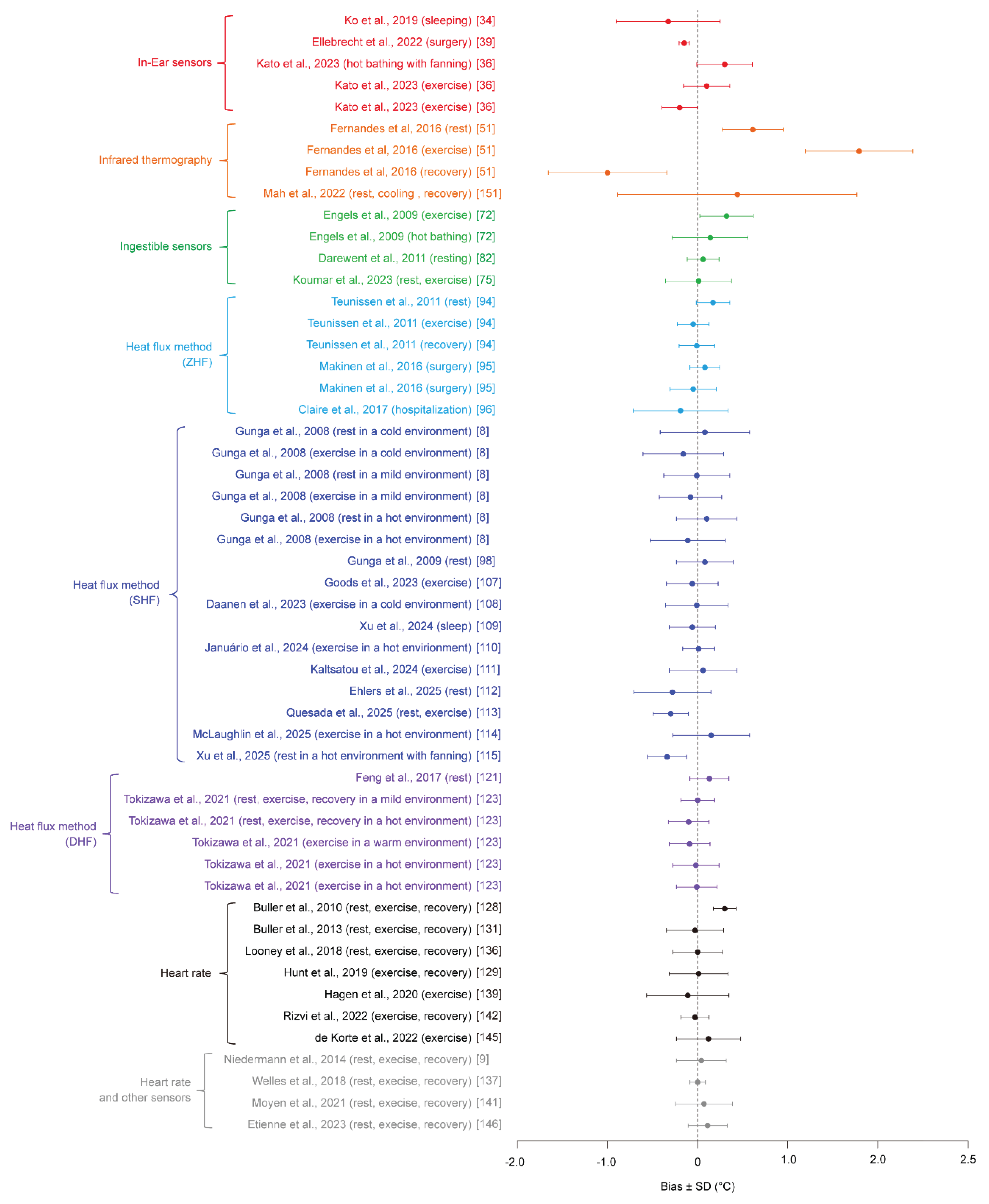

Table 1.
Coverage of methods in each eview.
| Review (Year) |
In-ear sensors | Ingestible sensors | Infrared thermography | Heat flux based merhod | HR-based method | Mult-sensor fusion | ||
|---|---|---|---|---|---|---|---|---|
| Zero-heat-flux method | Single-heat-flux method | Dual-heat-flux method | ||||||
| Moran et al. (2002) [10] | ✓ | — | — | — | — | — | — | — |
| Byrne et al. (2007) [7] | — | ✓ | — | — | — | — | — | — |
| Conway et al. (2020) [11] | — | — | — | ✓ | — | — | — | — |
| Cutuli et al. (2021) [12] | ✓ | — | — | ✓ | — | — | — | — |
| Falcone et al. (2021) [13] | — | — | — | ✓ | ✓ | — | ✓ | ✓ |
| Foster et al. (2021) [14] | — | — | ✓ | — | — | — | — | — |
| Dolson et al. (2022) [15] | — | — | — | — | — | — | ✓ | ✓ |
| Zhao et al. (2023) [16] | — | — | ✓ | — | — | — | — | — |
|
This study |
✓ | ✓ | ✓ | ✓ | ✓ | ✓ | ✓ | ✓ |
Legend: ✓ = covered; — = not covered.
Table 2.
Summary of core body temperature (CBT) monitoring methods, including their advantages, limitations, and representative applications.
Table 2.
Summary of core body temperature (CBT) monitoring methods, including their advantages, limitations, and representative applications.
| Method | Advantages | Limitations | Representative application scenarios |
|---|---|---|---|
| Invasive measurement (pulmonary artery, esophageal, rectal) |
Highest accuracy Closest to physiological definition Gold standard |
Highly invasive Low usability Unsuitable for long-term or field use |
Surgery and anesthesia management Intensive care Physiological reference measurements |
| In-ear sensors | Non-invasive Good wearability Suitable for continuous monitoring |
Sensitive to placement and ambient conditions Limited accuracy |
Daily health monitoring Sports and occupational safety Wearable healthcare |
| Infrared thermography | Non-contact Rapid measurement Suitable for mass screening |
Low individual accuracy Strongly affected by environment Indirect estimation |
Fever screening during pandemics Public health surveillance Disaster response |
| Ingestible sensors | High accuracy close to invasive methods | Single-use Limited monitoring duration Cost Recovery issues |
Sports science Military and firefighting training Validation studies |
| Zero-heat-flux (ZHF) method | High accuracy Clinically validated Continuous monitoring |
Requires active heating High power consumption Bulky sensor |
Perioperative monitoring Intensive care Clinical research |
| Single-heat-flux (SHF) method | Simple structure Low power consumption Wearable potential |
Sensitive to skin thermal properties and environment Limited robustness |
Wearable prototypes Moderate-accuracy field monitoring |
| Dual-heat-flux (DHF) method | Improved robustness to individual variability Better balance of accuracy and wearability |
Increased complexity Calibration and motion artifacts |
Advanced wearable core temperature sensors Field and occupational monitoring |
| Heart rate–based estimation | Extremely high usability Uses existing wearables Low cost |
Model dependency Training data required Generalization issues Strong individual dependence |
Heat strain screening Large-scale population monitoring |
| Heart rate + other sensors (skin temperature, acceleration, etc.) |
Higher accuracy than single-sensor methods Scalable Adaptive models |
Model dependency Training data required Generalization issues |
Industrial safety Sports monitoring Continuous daily-life assessment |
Table 3.
Summary of reported accuracy metrics for core body temperature (CBT) monitoring methods.
| Measurement technique |
Paper | Subject types | Number of Validation subjects, n | Environmental conditions | Exercise types | Reference for core body temperature | Bias ± SD [°C] |
|---|---|---|---|---|---|---|---|
| In-Ear sensors | Ko et al. 2019 [34] | Healthy females | 9 | 27 °C, 50%RH | Sleep | Rectal | -0.32 ± 0.58 |
| Ellebrecht et al. 2022 [39] | Patients | 10 | N/A | Surgery | Bladder | -0.15 ± 0.06 | |
| Kato et al. 2023 [36] | Healthy adults (23 ± 2 years) | 9 (6 males, 3 females) | 28 °C, 50%RH | Lower-limb warm-water immersion with 2 m/s airflow | Rectal | 0.30 ± 0.31 | |
| Kato et al. 2023 [36] | Healthy adult males (22 ± 1 years) | 11 | 35 °C, 50%RH | Walking at 4 km/h | Rectal | 0.10 ± 0.26 | |
| Kato et al. 2023 [36] | Healthy adults (23 ± 3 years) | 9 | 35 °C, 65%RH | Walking at 4 km/h with a 5% incline | Rectal | -0.20 ± 0.20 | |
| Infrared thermography | Fernandes et al. 2016 [51] | Healthy, physically active adult males | 12 | 24.9 ± 0.6 °C, 62.3 ± 5.7%RH | Rest | Gastrointestinal | 0.61 ± 0.31 |
| Fernandes et al. 2016 [51] | Healthy, physically active adult males | 12 | 24.9 ± 0.6 °C, 62.3 ± 5.7%RH | Exercise (60%VO2max) | Gastrointestinal | 1.79 ± 0.60 | |
| Fernandes et al. 2016 [51] | Healthy, physically active adult males | 12 | 24.9 ± 0.6 °C, 62.3 ± 5.7%RH | Recovery | Gastrointestinal | -1.00 ± 0.66 | |
| Mah et al. 2022 [151] | Healthy adults (28.3 ± 9.4 years) | 30 (14 males, 16 females) | 23 °C, 55%RH | Rest, Lower-limb cold-water immersion, recovery | Oral | 0.44 ± 1.33 | |
| Ingestible sensors | Engels et al. 2009 [72] | Healthy adult females (55.3 ± 5.9 years) | 8 | N/A | 70% HRmax exercise | Rectal | 0.32 ± 0.30 |
| Engels et al. 2009 [72] | Healthy adult females (55.3 ± 5.9 years) | 8 | N/A | 40 °C whole-body warm-water immersion | Rectal | 0.14 ± 0.42 | |
| Darewent et al. 2011 [82] | Healthy adult males (22.4 ± 2.4 years) | 11 | 21.0 ± 1.0 °C | Rest | Rectal | 0.06 ± 0.18 | |
| Koumar et al. 2023 [77] | Healthy adults (18-59 years) | 23 (13 males, 10 females) | N/A | In hospital (24 h), fasting, light activity, normal sleep | Rectal | 0.01 ± 0.37 | |
| Heat flux method (ZHF) | Teunissen et al. 2011 [94] | Healthy adults (28.3 ± 5.3 years) | 7 (4 males, 3 females) | 35 °C, 50%RH | Rest | Esophageal | 0.17 ± 0.19 |
| Teunissen et al. 2011 [94] | Healthy adults (28.3 ± 5.3 years) | 7 (4 males, 3 females) | 35 °C, 50%RH | Exercise | Esophageal | -0.05 ± 0.18 | |
| Teunissen et al. 2011 [94] | Healthy adults (28.3 ± 5.3 years) | 7 (4 males, 3 females) | 35 °C, 50%RH | Recovery | Esophageal | -0.01 ± 0.20 | |
| Makinen et al. 2016 [95] | Vascular surgery patients | 15 (11 males, 4 females) | N/A | Vascular surgery | Esophageal | 0.08 ± 0.17 | |
| Makinen et al. 2016 [95] | Cardiac surgery patients | 15 (11 males, 4 females) | N/A | Cardiac surgery | Pulmonary artery | -0.05 ± 0.26 | |
| Clare et al. 2017 [96] | ICU patients | 52 | N/A | Rest | Esophageal | -0.19 ± 0.53 | |
| Heat flux method (SHF) | Gunga et al. 2008 [8] | Healthy adult males (39.5 ± 10.2 years) | 20 | 10 °C | Rest | Rectal | 0.08 ± 0.50 |
| Gunga et al. 2008 [8] | Healthy adult males (39.5 ± 10.2 years) | 20 | 10 °C | Exercise at 35%, 45%, and 55% VO2max) |
Rectal | -0.16 ± 0.45 | |
| Gunga et al. 2008 [8] | Healthy adult males (39.5 ± 10.2 years) | 20 | 25 °C | Rest | Rectal | -0.01 ± 0.37 | |
| Gunga et al. 2008 [8] | Healthy adult males (39.5 ± 10.2 years) | 20 | 25 °C | Exercise at 25%, 45%, and 55% VO2max) |
Rectal | -0.08 ± 0.35 | |
| Gunga et al. 2008 [8] | Healthy adult males (39.5 ± 10.2 years) | 20 | 40 °C | Rest | Rectal | 0.10 ± 0.34 | |
| Gunga et al. 2008 [8] | Healthy adult males (39.5 ± 10.2 years) | 20 | 40 °C | Exercise at 25%, 35%, and 45% VO2max) |
Rectal | -0.11 ± 0.42 | |
| Gunga et al. 2009 [98] | Healthy adult males (31.9 ± 8.0 years) | 7 | N/A | Rest | Rectal | 0.08 ± 0.32 | |
| Goods et al. 2023 [107] | Athlete females (26 ± 4 years) | 19 | 31.0 °C, 38.5%RH | Exercise (Training) | Gastrointestinal | -0.06 ± 0.29 | |
| Goods et al. 2023 [107] | Athlete females (26 ± 4 years) | 19 | 32.2 °C, 50%RH | Exercise (Match) | Gastrointestinal | -0.10 ± 0.29 | |
| Goods et al. 2023 [107] | Athlete females (26 ± 4 years) | 19 | 27.6 °C, 80%RH | Exercise (Match) | Gastrointestinal | -0.17 ± 0.27 | |
| Goods et al. 2023 [107] | Athlete females (26 ± 4 years) | 19 | 27.4 °C, 74%RH | Exercise (Match) | Gastrointestinal | -0.34 ± 0.20 | |
| Daanen et al. 2023 [108] | Healthy adults (24.3 ± 1.2 years) | 9 (4 males, 5 females) | 18 °C, 50%RH | Rest, exercise (cycling), recovery | Rectal | -0.01 ± 0.35 | |
| Xu et al. 2024 [109] | Healthy adults (20-25 years) | 14 (7 males, 7 females) | 16, 20, 24 °C | Rest, sleep | Gastrointestinal | -0.06 ± 0.26 | |
| Januario et al. 2024 [110] | Healthy adults (33.4 ± 8.2 years) | 15 (7 males, 8 females) | 32 °C, 60%RH | Rest, exercise (cycling), recovery | Gastrointestinal | 0.01 ± 0.18 | |
| Kaltsatou et al. 2024 [111] | Chronic heart failure patients (53.5 ± 8.5 years) | 12 (8 males, 4 females) | 28 °C, 39%RH | Rest | Gastrointestinal | -0.14 ± 0.80 | |
| Kaltsatou et al. 2024 [111] | Chronic heart failure patients (53.5 ± 8.5 years) | 12 (8 males, 4 females) | 28 °C, 39%RH | Exercise (Bruce protocol) | Gastrointestinal | 0.06 ± 0.38 | |
| Kaltsatou et al. 2024 [111] | Chronic heart failure patients (53.5 ± 8.5 years) | 12 (8 males, 4 females) | 28 °C, 39%RH | Recovery | Gastrointestinal | 0.11 ± 0.28 | |
| Ehler et al. 2025 [111] | ICU patients (63.3 ± 15.1 years) | 112 (64 males, 48 females) | N/A | N/A | Bladder | -0.38 ± 0.43 | |
| Quesada et al. 2025 [112] | Healthy adults (23 ± 4 years) | 20 (10 males, 10 females) | 22.2 ± 0.5 °C, 37 ± 10%RH | Rest, exercise (cycling), recovery | Gastrointestinal | -0.3 ± 0.2 | |
| McLaughlin et al. 2025 [113] | Healthy adults | 24 (13 males, 11 females) | 35.9 ± 0.3 °C, 20.7 ± 3.3%RH | Exercise (cycling) | Rectal | 0.15 ± 0.43 | |
| Xu et al. 2025 [1014] | Healthy males | 24 | 40 °C, 57-58%RH, 0.15±0.05 m/s | Free-living conditions ranging from rest to low-intensity daily activities (8 h) | Rectal | -0.19 ± 0.52 | |
| Xu et al. 2025 [114] | Healthy females | 14 | 40 °C, 57-58%RH, 0.15±0.05 m/s | Free-living conditions ranging from rest to low-intensity daily activities (8 h) | Rectal | 0.06 ± 0.50 | |
| Xu et al. 2025 [114] | Healthy males | 24 | 40 °C, 57-58%RH, 0.15±3.2±0.4 m/s | Free-living conditions ranging from rest to low-intensity daily activities (8 h) | Rectal | -0.34 ± 0.22 | |
| Xu et al. 2025 [114] | Healthy females | 14 | 40 °C, 57-58%RH, 0.15±3.2±0.4 m/s | Free-living conditions ranging from rest to low-intensity daily activities (8 h) | Rectal | -0.27 ± 0.19 | |
| Heat flux method (DHF) | Feng et al. 2017 [121] | Healthy adults (26.8 ± 2.1 years) | 34 (30 males, 4 females) | 26 °C, 50-60%RH | Rest | Sublingual | 0.13 ± 0.22 |
| Tokizawa et al. 2021 [123] | Healthy adults (37 ± 7 years) | 21 (15 males, 6 females) | 25 °C, 35 °C | Rest, exercise, recovery | Esophageal | 0.00 ± 0.19 | |
| Tokizawa et al. 2021 [123] | Healthy adult males (36 ± 8 years) | 9 | 35 °C | Rest, exercise, recovery | Esophageal | -0.10 ± 0.23 | |
| Tokizawa et al. 2021 [123] | Healthy adult males (36 ± 11 years) | 11 | 30 °C | Exercise | Esophageal | -0.09 ± 0.23 | |
| Tokizawa et al. 2021 [123] | Healthy adult males (36 ± 11 years) | 11 | 40 °C | Exercise | Esophageal | -0.02 ± 0.26 | |
| Tokizawa et al. 2021 [123] | Healthy adult males (30 ± 6 years) | 8 | 35 °C | Exercise | Esophageal | -0.01 ± 0.23 | |
| Heart rate (Kalman filter) | Buller et al. 2010 [128] | Soldiers, runners | 25 | 20-40 °C | Exercise (Low-to-moderate-intensity exercise, 2–8 h, intermittent) | Rectal, esophageal | 0.30 ± 0.13 |
| Heart rate (Extended Kalman filter) | Buller et al. 2013 [131] | Healthy adults (20-30 years) | 83 (82 males, 1 females) | 9-45 °C, 9-97%RH, 0-4 m/s | Treadmill walking/running, cycling, long-distance marching/patrolling; ~1–24 h | Rectal, esophageal, gastrointestinal | -0.03 ± 0.32 |
| Looney et al. 2018 [136] | Healthy adults (24 ± 3 years) | 8 (6 males, 2 females) | 18-22 °C | Sleep and light seated activities (≈16 h) | Gastrointestinal | 0.00 ± 0.28 | |
| Hunt et al. 2019 [129] | Healthy adult males (26.4 ± 6.0 years) | 8 | 24 °C, 50%RH; 32 °C, 60%RH | Treadmill walking at 2.5–5.5 km/h with recovery periods | Gastrointestinal | 0.01 ± 0.33 | |
| Hagen et al. 2020 [139] | Healthy adult male athletes (American football) | 13 | N/A | Sports training (5-min × 18–22 sets) | Gastrointestinal | -0.11 ± 0.46 | |
| Rizvi et al. 2022 [142] | Healthy adults (21-23 years) | 16 | 36±0.5 °C, 59±5%RH, 0.17±0.05 m/s | Treadmill walking at 4.5 km/h with recovery periods | Gastrointestinal | -0.03 ± 0.16 | |
| de Korte et al. 2022 [145] | Elite athletes (26 ± 5 years) | 101 (49 males, 52 females) | 31.6 ± 1.0 °C, 74 ± 5%RH | Graded exercise | Gastrointestinal | 0.15 ± 0.36 | |
| Paggen et al. 2024 [147] | General adults (some on cardiovascular/psychotropic medications) | 18 | N/A | Free-living conditions ranging from rest to low-intensity daily activities (≈26 h) | Gastrointestinal | -0.03 ± 0.16 | |
| Heart rate and other sensors | Niedermann et al. 2014 [9] | Healthy adult males (23.0 ± 3.9 years) | 10 | 30±0.2 °C, 42.9±1.1%RH, <0.3 m/s | Rest, exercise at 40% and 60% VO2max, recovery | Gastrointestinal | -0.17 ± 0.14 |
| Niedermann et al. 2014 [9] | Healthy adult males (24.6 ± 2.0 years) | 10 | 10.1±0.2 °C, 49.5±4.9%RH, 0.5±0.1 m/s | Rest, exercise at 60% VO2max, recovery | Gastrointestinal | 0.04 ± 0.28 | |
| Welles et al. 2018 [137] | Healthy adult soldiers (22 ± 4 years) | 8 | 25 °C, 50%RH; 35 °C, 70%RH; 40 °C, 20%RH | Rest, exercise, recovery | Gastrointestinal | 0.00 ± 0.09 | |
| Moyen et al. 2021 [141] | Healthy adults (28.9 ± 7.8 years) | 27 (19 males, 8 females) | 13.4-43.2 °C, 11-75%RH | Rest, exercise, recovery | Rectal, gastrointestinal | 0.07 ± 0.32 | |
| Etienne et al. 2023 [146] | Post-vaccination healthy adults (35.8 ± 8.2 years) | 17 | N/A | Free-living conditions ranging from rest to low-intensity daily activities (≈26 h) | Gastrointestinal | 0.09 ± 0.22 |
Disclaimer/Publisher’s Note: The statements, opinions and data contained in all publications are solely those of the individual author(s) and contributor(s) and not of MDPI and/or the editor(s). MDPI and/or the editor(s) disclaim responsibility for any injury to people or property resulting from any ideas, methods, instructions or products referred to in the content. |
© 2026 by the authors. Licensee MDPI, Basel, Switzerland. This article is an open access article distributed under the terms and conditions of the Creative Commons Attribution (CC BY) license (http://creativecommons.org/licenses/by/4.0/).
Copyright: This open access article is published under a Creative Commons CC BY 4.0 license, which permit the free download, distribution, and reuse, provided that the author and preprint are cited in any reuse.


