
Submitted:
06 January 2026
Posted:
07 January 2026
You are already at the latest version
Abstract
Potyviruses express their genome as a single large open reading frame, translated into a polyprotein that is post-translationally cleaved by three virus-encoded proteases into 10 functional proteins. Several of these potyviral proteins, including nuclear inclusion protein b (NIb), are multifunctional. Here, using the classic GFP silencing in Nicotiana benthamiana gfp-transgenic plants model, we show that NIb, in addition to its canonical role as the viral RNA-dependent RNA polymerase (RdRP), functions as a suppressor of RNA silencing. Mutational analyses revealed a previously unreported NIb nuclear localization signal (NLS) consisting of a triple-lysine motif, and NIb suppression of RNA silencing activity was lost when this novel NLSs were mutated, suggesting that nuclear localization is required for NIb suppression of RNA silencing activity. Analysis of sequenced GFP siRNAs revealed three prominent siRNA hotspots at ≈nt 175, ≈320–330, and 560–700 nt. These data showed that there were differences in the positional distribution of the siRNAs between samples expressing NIb and those expressing NIbDel3x2, the NIb null mutant that does not suppress RNA silencing. However, there was no increase in the transcript‑wide siRNA burden between the two treatments. Furthermore, NIb was found to interact with four key RNA silencing pathway proteins—AGO4, HSP70, HSP90, and SGS3. Except for HSP90, each of these proteins showed degradation products that were absent in NIb mutants that did not suppress RNA silence. These findings support a role for NIb in countering host defense during virus infection.
Keywords:
AGO4
; HSP70
; HSP90
; nuclear inclusion protein b (NIb)
; RdRP
; SGS3
; siRNA
; suppressor of RNA silencing
1. Introduction
Diseases caused by potato virus Y (PVY) are a serious challenge facing potato (Solanum tuberosum L.) production worldwide. To date, at least nine recombination patterns of PVYO and PVYN sequences have been identified in potato-infecting PVY isolates, the three most common recombinant patterns being found in strains PVYNTN, PVYN:O, and PVYN-Wi [1,2]. PVY has a positive single-stranded RNA [(+)ssRNA)] genome that has a single open reading frame (ORF), which upon translation undergoes proteolytic processing, producing 10 functional proteins: P1, HcPro, P3, 6K1, CI, 6K2, VPg, NIa, NIb, and CP [3]. Proteolytic processing is carried out by three virus-encoded proteins, proteinases P1 and HcPro, and the NIa protease [4,5,6]. During replication, an additional polyprotein, P3N-PIPO, is produced due to RdRP slippage, resulting in frameshift mutation at the P3 cistron [7,8,9].
The catalytic subunit of the NIb (nuclear inclusion protein b) functions as an RNA-dependent RNA polymerase (RdRP) and therefore is responsible for viral genome replication and mRNA synthesis. During genome replication, NIb localizes to endoplasmic reticulum (ER) membranes by interacting with the 6K2–VPg–NIaPro polyprotein, leading to the assembly of the viral replication complex (VRC), where replication takes place [10,11,12]. NIb has equally been found to recruit important VRC components, including poly(A)-binding protein (PABP) [13], PABP2, PABP8, eukaryotic elongation factor 1A (eEF1A), and heat shock cognate protein (HSC70) [12,14,15,16]. The polymerase activity of NIb is regulated by the autophagy protein, Beclin1 (or ATG6), which binds NIb at its conserved GDD motif and targets NIb for autophagic degradation [17].
NIb engages in multiple immune response interactions—for instance, it can suppress NPR1-mediated immune signaling, and activate effector-triggered immunity [10,17,18,19,20]. Due to this broad interactome, NIb has been dubbed the potyvirus ‘most sticky’ protein [12]. Except for influenza virus that replicates in the nucleus, all known RNA viruses, including potyviruses, replicate in the cytoplasm. However, the NIb protein contains nuclear localization signals (NLSs) and is found in both the cytoplasm and nucleus, where together with NIa, it forms amorphous or crystalline nuclear inclusions (NIs) in infected cells [12,21,22,23]. Hence, nuclear localization likely enables post translational modification, including sumoylation [18], as well as provides a favorable cellular environment for interaction with host proteins.
Plants use the RNA silencing mechanism to counter invading viruses. To establish a successful infection, therefore, the virus must overcome this host antiviral defense. Viruses achieve this by encoding suppressors of RNA silencing, and some viruses have been shown to encode more than one suppressor of RNA silencing, including East African cassava mosaic Cameroon virus (EACMCV), which encodes three suppressors, namely AC4, AV2, and TrAP [24,25,26]; citrus tristeza virus that encodes p20 p23, and CP [27], and beet necrotic yellow vein virus encoded p14 and p31 [28,29]. To date, two potyviral silencing suppressors have been reported, the extensively characterized HcPro [30,31,32,33,34,35,36,37], and turnip mosaic virus (TuMV) VPg [38]. Here, we show that PVY NIb suppresses RNA silencing, and that this function is dependent on NIb nuclear localization. We also show that NIb suppression of RNA silencing is accounted for by its interaction with several protein components of the RNA silencing pathway.
2. Results
2.1. The PVY NIb Is a Suppressor of RNA Silencing
The potyvirus NIb is a multifunctional protein with distinct functional domains [7,12,39]. To test whether NIb suppresses the host antiviral defense, we used the classic N. benthamiana line 16c plants model [40] . The line 16c plants constitutively express the GFP transgene and enables testing of putative viral suppressors of gene silencing. NIb cistrons from four PVY strains (PVYNTN, PVYO, PVYN:O, and PVYN-Wi) were each cloned under the control of cauliflower mosaic virus (CaMV) 35S promoter in pEarleyGate101 binary vector [41]. Tomato bushy stunt virus p19 silencing suppressor was used as the RNA silencing suppressor positive control. For the negative control, we generated a null mutant of PVYNTN NIb, NIbDel3x2. These constructs were introduced to Agrobacterium tumefaciens strain GV3101, and used for co-infiltration with 35s::GFP into N. benthamiana 16c plants as described previously [26]. GFP fluorescence in infiltrated leaves was monitored using a handheld longwave UV lamp. By 2 days post-inoculation (dpi), all infiltrated leaf patches displayed intense green fluorescence (Figure 1A). However, by 7 dpi, the level of green fluorescence had started declining and was replaced by the red autofluorescence from chlorophyll molecules in patches co-infiltrated with NIbDel3x2/35S::GFP, while leaf patches co-infiltrated with each NIb/35s::GFP continued to display intense green fluorescence. At 10 dpi, tissues co-infiltrated with NIbDel3x2/35S::GFP displayed intense red fluorescence while the wild-type tissues continued to display intense green fluorescence (Figure 1B). Thus, in NIb expressing tissues, a functional NIb maintains GFP expression by countering gfp silencing, while silencing is established in tissues expressing NIbDel3x2 null mutant.
2.2. Characterization of NIb Suppression of RNA Silencing
To characterize NIb suppression of RNA silencing, we produced mutations of PVYNTN HcPro, including NIbΔ1/17, NIbΔ491/519, NIbKKK135/137AAA, NIbKK303/304AA, NIbGDD351/353AAA, NIbDEEE491/494AAA, and NIBDEL3X2 as described above. These constructs and wild-type HcPro cistrons were each co-infiltrated with 35s::GFP into N. benthamiana line 16c plant leaves. Examination of co-infiltrated leaf patches showed that at 2 dpi, all co-infiltrated tissues displayed green fluorescence, similar to Figure 1A. However, by 10 dpi, tissues expressing NIbDEEE491/494AAA, NIbGDD351/353AAA, NIbΔ1/17, NIbKKK135/137AAA, NIbΔ491/519, and NIbDel3x2 had lost much of the green fluorescence, which was progressively replaced by red chlorophyll fluorescence, while tissues expressing NIbKK303/304AA and the wild-type cistrons continued to display green fluorescence (Figure 2).
The fact that at 2 dpi, there were similar levels of GFP fluorescence in all constructs tested indicates that there were similar GFP expression levels. To confirm that loss of GFP fluorescence over time was due to gene silencing, we carried out a Northern blot analysis of GFP transcripts at 7 dpi. Results generally reflected loss of GFP fluorescence; for example, very low levels of GFP transcripts were observed in tissues co-infiltrated with an empty vector and 35s::GFP, while very high levels of GFP transcripts were observed in tissues containing PVYNTN NIb and p19 (Figure 3A).
We further compared GFP levels in a Western blot analysis using anti-GFP antibodies over time. At 7 dpi, the levels of GFP were similar in all treatments (Figure 3B), however, at 10 dpi, GFP was barely detectable in samples expressing NIb mutants, while leaves expressing wild-type NIb maintained strong GFP protein levels through 14 dpi. These findings confirm that loss of green fluorescence in tissues co-infiltrated with NIb mutants and 35s::GFP was due to PTGS, and that NIb suppresses PTGS.
2.3. Nuclear Localization Is Required for NIb Suppression of RNA Silencing
Subcellular localization is critical for protein function, as it determines its access to interaction partners, as well as providing the cellular environment needed for interaction. Thus, to determine whether subcellular localization influenced NIb RNA silencing suppression, we investigated NIb constructs described above, which contain a C-terminal fused YFP in plasmid pEarleygate101. N. benthamiana plant leaves were agroinfiltrated with these constructs and examined with a confocal microscope within 48 hours of infiltration. The wild-type NIb was observed to localize in the nucleus, as well as in the cytoplasm, where it was found associated with cytoplasmic membranes where virus replication occurs, consistent with its role of RdRP (Figure 4A). Mutants NIbDEEE491/494AAA, NIbKK303/304AA, and NIbGDD351/353AAA also localized in the nucleus and cytoplasm. In contrast, NIbΔ1/17, which was shown to contain a NLS [12], NIbΔ491/519, and NIbKKK135/137AAA localized only in the cytoplasm (Figure 4) (Supplementary Figure S1). This suggests that 135KKK137 motif is in a previously unreported NIb NLS. The findings also indicate that nuclear localization is required for NIb suppression of RNA silencing. Additionally, the NIb C-terminal 18 amino acid truncation mutant, NIbΔ491/519, which does not suppress RNA silencing was found not to localize in the nucleus, suggesting that the C-terminal truncation contains another NLS or regulates NIb nuclear import.
2.4. Small RNA Analysis
We profiled small RNAs mapping to GFP in 16c leaves co-infiltrated with PVYNTN NIb/35s::GFP, as well as with NIbDel3x2/35s::GFP at 14 dpi (n = 18 libraries). Position-wise coverage in counts per million (CPM) on a linear scale showed three reproducible siRNA hotspots across the GFP sequence at approximately nt 175, nt 320–330, and nt 560–700 (Figure 5A). Peak heights differed modestly between treatments: early hotspots tended to be somewhat higher in samples expressing NIbDel3x2, whereas in the 560–700 nt they were similar or slightly higher in samples expressing NIb. This pattern indicates a modest shift where GFP siRNAs accumulate along the transcript rather than a uniform gain or loss at all sites. We therefore interpret the NIb versus NIbDel3x2 contrast as a positional redistribution of GFP-derived siRNAs, rather than a pronounced change in any single peak.
To assess aggregate output, we integrated CPM along the transcript as a cumulative running sum. The two groups reached similar end values, which indicates no large difference in transcript-wide GFP siRNA burden in this dataset (Figure 5B; median NIbDel3x2/NIb ≈ 1.0; Wilcoxon p ≈ 0.1). Consistent with previous studies of the 16c GFP transgene, GFP-mapping siRNAs in both treatments were strongly antisense-biased [42] (Figure 6). In this system, abundant antisense siRNAs are expected when a constitutively expressed transgene becomes a target of RNA silencing, because antisense strands are preferentially loaded into AGO-containing effector complexes.
We also quantified siRNAs mapping to the transiently expressed NIb transcripts. NIb-derived siRNAs were readily detectable and broadly distributed across the NIb coding sequence (Figure 7). In contrast to GFP, strand polarity for NIb was not strongly antisense-dominant: NIb samples showed a modest excess of sense-strand siRNAs, whereas NIbDel3x2 samples had a more balanced sense/antisense profile (Figure 8). NIb and NIbDel3x2 displayed broadly similar positional patterns along the NIb transcript, with only modest differences in local peak heights. Thus, while NIb does not prevent production of siRNAs from its own transcript, the strand composition of these siRNAs differs from that of GFP-derived species, consistent with differences in how the two RNAs enter the RNA silencing pathway (transgene-derived vs. transiently expressed coding transcript).
2.5. NIb Forms a Complex with Four RNA Silencing Pathway Proteins
To identify host protein(s) in the RNA silencing pathway that likely interact with NIb, leading to suppression of RNA silencing, we used an in planta protein-protein interaction assay. The pEarleygate101 plasmid used to express NIb, as described above, contains YFP and HA-tag as the C-terminal fusion, and the nine host genes investigated for interaction with NIb, contain a C-terminal 6xHIS tag as described above. This allowed us to detect interacting complexes using HIS-tag purification beads followed by immunoblotting with HA-tag antibodies. NIb-HA fusion was therefore co-agroinfiltrated into N. benthamiana plants with each host protein. At 2 to 5 dpi, infiltrated leaves were collected, and protein complexes purified using Ni-NTA (nickel-nitrilotriacetic acid) magnetic bead kit (ThermoFisher Scientific, USA) under native conditions. After native PAGE gel electrophoresis separation of protein complexes, the presence of NIb-HA fusions was tested using HA antibodies in a Western blot assay. Of the nine co-infiltrations, protein bands were observed in AGO4, HSP70, HSP90, and SGS3 samples, indicating interaction between these proteins and NIb (Figure 9). These proteins play key roles in RNA silencing. For example, AGO4 is a key effector of the RNA-directed DNA methylation antiviral pathway [43]; HSP70 and HSP90 are molecular chaperones required for loading siRNAs into RISC [44]; and SGS3 works with RDR6 to convert ssRNAs into dsRNA, which DCL proteins process into siRNA [45]. Therefore, these results suggest that NIb likely suppresses RNA silencing by interacting with at least one of these proteins. Interestingly, NIb/HSP70, NIb/AGO4, and NSP/SGS3 complexes showed smaller molecular weight proteins, possibly indicating that interaction between NIb and these RNA silencing proteins leads to degradation of the interacting complex.
2.6. RNA Silencing Suppression Deficient NIb Mutants Do Not Interact with AGO4, HSP70, and SGS3
To identify the specific partner(s) with which NIb interacts to suppress RNA silencing, we reasoned that such a protein would not interact with NIbΔ1/17, NIbKKK135/137AAA, NIbΔ491/519, and/or NIbDel3x2, all of which do not suppress RNA silencing. First, NIb constructs, as well as that of AGO4, HSP70, HSP90, and SGS3 were assessed for expression. Interestingly, whereas NIbΔ1/17, NIbΔ491/519, and NIbKKK135/137AAA were detectable from total plant protein at 2 dpi, only NIb could be detected at 10 dpi (Figure 10A). This result is consistent with our observation from siRNA analysis (Figure 8) that transiently expressed NIb is a target of RNA silencing. Thus, detection of NIb at 10 dpi is due to its suppression of RNA silencing. As for the RNA silencing proteins, unlike NIb, AGO4, HSP70, HSP90, and SGS3 were only detectable when isolated with Ni-NTA magnetic bead kit (Figure 10B). HSP70 and HSP90 were observed to dimers given that this analysis was under native conditions.
Assessment of NIb and mutants was carried out by co-expressed with AGO4, HSP70, HSP90, and SGS3 in N. benthamiana plants and protein complexes isolated with Ni-NTA followed by Western blot analysis using HA, as described above. Results confirmed interaction between NIb and AGO4, HSP70, and SGS3, respectively leading to apparent degradation (Figure 11). In contrast, no protein complexes were observed in tissues co-infiltrated with mutants NIbΔ1/17, NIbKKK135/137AAA, NIbΔ491/519, and NIbDel3x2 indicating that interaction between NIb and AGO4, HSP70, and SGS3 likely plays a key role in NIb suppression of RNA silencing. Similar to NIb wild-type, NIbΔ1/17, NIbKKK135/137AAA, and NIbΔ491/519 were observed to interact with HSP90. These results therefore suggest that interaction between NIb and HSP90 is likely not the determinant in NIb suppression of RNA silencing. Given that NIbΔ1/17, NIbKKK135/137AAA, and NIbΔ491/519 are excluded from the nucleus (Figure 4), these results confirm that nuclear localization is likely required for NIb suppression of RNA silencing.
3. Discussion
Here, we demonstrate that NIb suppresses host antiviral RNA silencing, yet this is only one out of its several other actions against host immune responses. For example, TuMV NIb has been found to be sumoylated by small ubiquitin-like modifier 3 (SUMO3) and to also bind to SUMO-conjugating enzyme 1 (SCE1), and thus making SUMO3 unavailable to activate the NPR1-mediated immune response to viral infection [12,18,46]. NPR1 is a master regulator of salicylic acid-mediated immunity. TuMV NIb SUMOylation also reduces SUMOylation of pelota, which is involved in the degradation of potyviral genomic RNA [47]. NIb suppression of antiviral RNA silencing, therefore, adds to the repertoire of NIb functions that counter host immunity.
This study shows that NIb suppression of RNA silencing depends on nuclear localization, and we identified the 135KKK137 motif as a previously unreported NIb NLS. This motif is also found in the NIb of several potyviruses, including TuMV [48], tomato necrotic stunt virus [49], sorghum mosaic virus [50], and pepper yellow mosaic virus [51]. A search of the NIb NLS using an online NLS prediction tool [52] identified 133GGKKKD138 as the NIb NLS, and therefore in agreement with our results. Also playing a role in NIb suppression of RNA silencing is the C-terminal region, where the construct NIbΔ491/519 with a deleted C-terminal 28 amino acids, as well as the construct with a mutated acidic motif 491DEEE494 (NIbDEEE491/494AAA) were found to lose much of the RNA silencing suppression capability.
Here, we found that NIb interacts with at least four proteins in the RNA silencing pathway, namely, HSP70, HSP90, AGO4, and SGS3, and except for HSP90, these interactions lead to degradation of the interacting complexes. These proteins play key roles in host antiviral RNA silencing. For example, HSP70 recruits HSP90 to the RISC in Drosophila and human [44], as well as plant [53] systems. Mechanistically, in the nucleus, HSP70 binds and transiently pries open the otherwise closed AGO, and HSP90 is recruited into the complex, a process that stabilizes the open and active AGO, which then receives a sRNA duplex [54,55]. By interacting with HSP70/HSP90, NIb likely inhibits siRNA recruitment into the RISC, resulting in suppression of RNA silencing.
Failure of constructs with a mutated NLS to suppress RNA silencing can be explained by the fact that loading of siRNAs to AGO4 by HSP70/HSP90 occurs in the nucleus [44]. Additionally, although the SGS3/SDE5/RDR6 complex is formed in the cytoplasm where synthesis of ssRNA to dsRNA occurs, SGS3 primarily localizes in the nucleus [56,57]. Hence, interaction between NIb and SGS3 likely occurs in the nucleus, and blocks SGS3 nuclear export to the cytoplasm to form the SGS3/SDE5/RDR6 complex. However, the fact that there were similar levels of siRNAs in silenced tissues and non-silenced tissues suggest that the interaction between NIb and SGS3 is likely not critical in NIb suppression of RNA silencing. Interestingly, the VPg of another potyvirus, turnip mosaic virus (TuMV), was previously reported to suppress RNA silencing by degrading SGS3 [38].
Canonically, AGO4 is known to associate with repeat-associated siRNAs (rasiRNAs) to control transcriptional gene silencing through methylation [58]. In this process, 23-nt/24-nt or 24-nt/24-nt siRNA duplexes are loaded into AGO4 in the cytoplasm, with the 24-nt siRNA strand acting as the guide that directs AGO4-siRNA complex entry into the nucleus where the 24-nt siRNA strand binds to the cognate nascent RNA transcript at the locus where DNA methylation occurs [59,60]. Interestingly, AGO4 has also been found to interact with RNA silencing suppressors of several RNA genome viruses, including cucumber mosaic virus 2b [61], alfalfa dwarf virus P [62], and tobacco rattle virus 16K [63] proteins. Thus, interaction between NIb and AGO4, leading to degradation of the complex, adds to the ability of plant RNA viruses to counter host defense strategies through interaction with AGO4, hence creating a favorable cellular environment for viral proliferation. This is a new role for AGO4, in addition to its well documented role of regulation of gene expression through DNA methylation.
Interestingly, HSP70 also targets and degrades the CP to promote genome RNA replication [64,65]. Moreover, consistent with observations made in this study, HSP70 was reported to cause the degradation of turnip mosaic potyvirus RdRP [14]. It is important to note that NIb RdRP activity is regulated by Beclin1 (ATG6), which interacts with NIb, and the interaction complex is targeted for autophagic degradation [17]. How interaction between NIb and HSP70/HSP90, leading to degradation of the NIb/HSP70 complex, as shown in this study, and involvement of these proteins in the VRC formation contributes to the overall virus infection process will need to be addressed.
5. Model for NIb Suppression of RNA Silencing and Conclusion
GFP silencing in GFP-transgenic plants is induced by 35S::GFP, where ssRNA GFP transcripts are converted to dsRNAs by RDR6 in cooperation with SGS3. The dsRNA is then processed by DCL proteins into double stranded siRNAs, which are loaded into AGO proteins to form RISC [66] by HSP70/HSP90 in an energy dependent process [45]. Once in AGO proteins, the double stranded siRNAs are separated, and one strand guides the RISC to trigger global silencing of GFP transcripts in GFP-transgenic plants. Accordingly, antiviral silencing of RNA viruses is initiated when DCL proteins process viral dsRNA into siRNA. For (+)ssRNA genome viruses, including PVY, viral dsRNA is either a replication intermediate from annealed viral transcripts or synthesized from the replicated genome by RDR6 of the SGS3/SDE5/RDR6 complex. The dsRNA is then processed into siRNAs, which are loaded to AGO4 by HSP70/HSP90 to form a mature RISC. Since there were no major differences between GFP siRNA accumulation in leaf tissues exhibiting NIb-induced suppression of RNA silencing and those with no silencing (Figure 5), it is likely that NIb suppression of RNA silencing occurs downstream of siRNA production. The strong antisense bias of GFP-derived siRNAs in both treatments (Figure 6) indicates that GFP continues to be processed. From these observations, therefore, a model for NIb suppression of RNA silencing is proposed in Figure 12. Hence, NIb likely suppresses antiviral RNA silencing by targeting AGO4, HSP70/HSP90, and/or SGS3, leading to complex degradation. Thus, NIb interactions with AGO4 and HSP70 (and SGS3) are most parsimoniously interpreted as disrupting the use of these siRNAs in effector complexes, rather than blocking their biogenesis. Finally, NIb-derived siRNAs were observed to be more balanced between sense and antisense strands (Figure 8), consistent with their origin from a transiently expressed coding transcript, compared to a stably integrated GFP-transgene.
6. Materials and Methods
6.1. Plasmid Construction and Agrobacterium Inoculation
NIb proteins investigated in this study were from PVY strains PVYNTN, PVYO, PVYN:O, and PVYN-Wi found in North America [67]. RT-PCR amplification of each cistron was carried out using primers listed in Table 1A. For each cistron, ATGGCC that code for methionine and alanine, were added at the 5′ ends of the forward primer for translation initiation. Amplification products were introduced into the GatewayTM donor vector, pDONR/Zeo (Invitrogen; Thermo Fisher Scientific, USA) obtaining EntryTM clones. These EntryTM clones were shuttled into GatewayTM binary vectors pEarleygate100 for RNA silencing suppression analysis, and pEarleygate101 for subcellular localization and protein-protein interaction analysis. The binary plasmids obtained were introduced into A. tumefaciens strain GV3101 using the freeze-thaw method [68], and cultures were grown overnight at 30°C for infiltration.

Table 1.
Primers used in characterizing potato virus Y NIb suppression of RNA silencing.
| A/ Wild-type NIb primers | ||
| ID | Sequence | Construct |
| NTN_NIb-F NTN_NIb-R |
ggggacaagtttgtacaaaaaagcaggcttaATGGCCGCTAAACATTCTGCG |
PVYNTN |
| ggggaccactttgtacaagaaagctgggttTTGATGGTGCACTTCATAAGTATCG | ||
| NIb. NO.Nwi.O-F NIb.NO.Nwi.O-R |
ggggacaagtttgtacaaaaaagcaggcttaATGGCCGCTAAGCACTCTG |
PVYO, PVYN:O, PVYN-Wi |
| ggggaccactttgtacaagaaagctgggttTTGATGGTGTACTTCATAAGAGTCAAATTC | ||
The lowercase sequences are attB1 and attB2 adaptor sequences for Gateway recombination; the ATGGCC at the 5′ ends of forward primers, encoding methionine and alanine were added for translation initiation and in frame with the cistron.
| B/ Primers used to introduce mutations | ||
| ID | Sequence | Mutation |
| NIbΔKKK-F NIbΔKKK-R |
AGCTGACTACTTCGAGCATTTTAC | NIb KKK135/137AAA |
| GCTGCGCCTCCATACATAGCTCC | ||
| NIbΔKK-F NIbΔKK-R |
AACAATTGTCGCTGCTTTTAGAGGTAATAATAGCGGTC | NIbKK303/304AA |
| CCATCTGGAGTTGAGATTG | ||
| NIbΔGDD-F NIbΔGDD-R |
TGCATTATTGATTGCTGTGAATCC | NIbGDD351/353AAA |
| GCAGCATTAACAAAGAATACACACG | ||
| NIbΔDEEE-F NIbΔDEEE-R |
GCAGCTCTGAAGGCTTTCACTGAAATG | NIbDEEE491/494AAA |
| TGCAGCTACTGTCCTATTCATGTACAAC | ||
| NIbΔ1/17-F NIbΔ1/17-R |
GTGGCGACAATGAAGAGTC | NIbΔ1/17 |
| GGCCATTAAGCCTGCTTT | ||
| NIbΔ491/519-F NIbΔ491/519-R |
AACCCAGCTTTCTTGTAC | NIbΔ491/519 |
| TACTGTCCTATTCATGTAC | ||
| NIbΔGT855-856-F NIbΔGT855-856-R |
ACACAGAAATAATTTACACAC | NIBDEL3X2 |
| AAATTGCGCAACATTTGC | ||
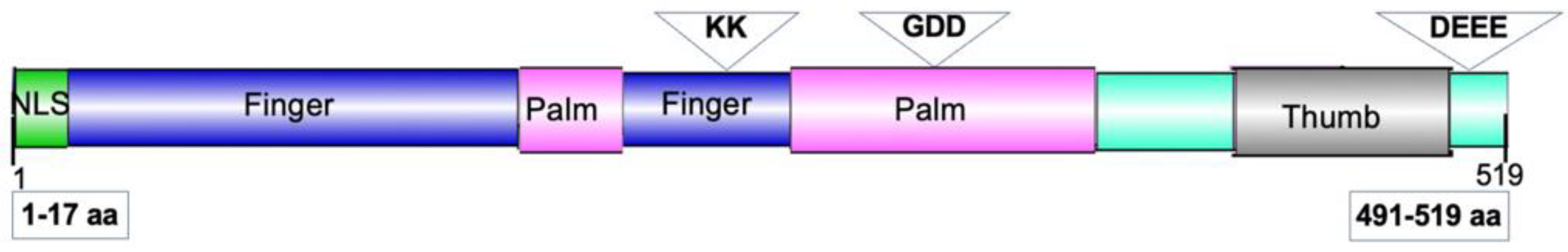
To identify amino acid residues involved in NIb suppression of RNA silencing, we introduced mutations in the NIb of PVYNTN, which is spreading increasingly in North America while the incidence of PVYO, the ordinary strain, has been decreasing [69,70,71,72,73]. These mutations were introduced using the Q5 Site-Directed Mutagenesis Kit (New England Biolabs, USA) with primers listed in Table 1B. These mutations and NIb functional domains are illustrated in Figure 1. First, the N-terminal 17 amino acid residues that contain the recently reported NIb nuclear localization signal (NLS) [12] was deleted, obtaining the mutant designated NIbΔ1/17. A second truncation was produced by deleting the last 29 amino acids of PVYNTN NIb, obtaining NIbΔ491/519. To identify additional NLSs, we searched for lysine rich motifs, which are typically found in NLSs and identified 135KKK137 and 303KK304. These lysine residues were replaced with alanine obtaining NIbKKK135/137AAA and NIbKK303/304AA, respectively. We investigated other conserved motifs, including the acidic motif 491DEEE494 and 351GDD353 that were substituted with alanine obtaining NIbDEEE491/494AAA and NIbGDD351/353AAA, respectively. The 351GDD353 motif is a critical component of the catalytic site within the viral RdRP protein [74]. An NIb null mutant, NIbDel3x2 (negative control), was generated from PVYNTN NIb by deleting nucleotides GT at position bps 855-856 and truncating 50 bps from the 3′ end. These PVYNTN NIb mutants were cloned into pEarleygate100 and pEarleygate101 binary vectors as described above, and the recombinant plasmids introduced into A. tumefaciens strain GV3101.
Figure 12.
Representative diagram of the potyvirus NIb protein highlighting its functional domains. The conserved fingers, palm, and thumb subdomains of the RdRP are shown. Open inverted triangles mark amino acid residues that were substituted with alanine in this study, and open rectangles indicate truncations (deletions) introduced in NIb.
6.2. Determination of NIb Suppression of RNA Silencing
Assessment of RNA silencing suppression was carried out using the GFP-transgenic N. benthamiana line 16c plants [40]. Plants were grown in a growth room at 16 h light and 22 ± 3 °C temperature. To induce gfp silencing in these experiments, we cloned the GFP ORF into pEarleygate100, obtaining 35S::GFP, which was introduced into Agrobacterium as described above. Overnight Agrobacterium cultures were pelleted and resuspended in an agroinfiltration buffer (10 mM MgCl2, 10 mM MES, pH 5.6) at an OD60 of 0.8. Cultures of each construct were then mixed with those of 35s::GFP and the mixture co-infiltrated into N. benthamiana at the four-leaf stage using a 1-ml, needleless syringe. GFP fluorescence was monitored using a handheld longwave UV lamp.
6.3. Northern Blot Analysis
To confirm that gfp is transcribed, we carried out a Northern blot analysis of infiltrated leaves of N. benthamiana at 7 dpi. Total RNA was extracted using the TRIzol™ Reagent (Invitrogen; Thermo Fisher Scientific, USA) and DNA traces removed with the RNase-free DNase I kit (Thermo Fisher Scientific, USA) as described by the manufacturer. The purity of the RNA was checked by NanoDrop spectrophotometer (NanoDrop Technologies, Wilmington, DE). Total RNA (20 μg) was heated at 55 °C for 5 min prior to separation in a 1.2 % agarose/MOPS/ formaldehyde gel in 1X MOPS buffer. The RNA was transferred overnight to a Nylon membrane (Schleicher and Schuell, Keene, NH) and hybridized with gene specific probes using the North2South Chemiluminescent Hybridization and Detection Kit Inc (Thermo Fisher Scientific, USA). Images were captured using a G:Box and GeneSys software version: 1.8.11 (Syngene, Iselin, NJ, USA).
5.4. Determination of Subcellular Localization of NIb and NIb Mutants
For subcellular localization analysis, Agrobacterium cultures of NIb constructs were infiltrated into the abaxial surface of the first two true leaves of N. benthamiana and subcellular localization determined at 48 hours post infiltration. Here, infiltrated leaf pieces were excised, gently vacuum infiltrated with water and placed on a slide and covered with a coverslip for confocal imaging. Images were acquired on an upright Zeiss LSM 780 laser-scanning microscope (Carl Zeiss, Inc., Oberkochen, Germany) using a Zeiss 40X C-Apochromat (NA 1.2) objective lens. Multichannel images of yellow fluorescent protein (YFP) and chloroplast autofluorescence were acquired using the 488 nm line of an Argon/Krypton laser with the 500 to 550 band pass and 650 long-pas 390/ emission filters, respectively. Images were captured as single optical sections or as a z-series of optical sections, and z-series data sets were displayed as single maximum intensity projection generated with Zeiss Zen Black software vSP2.
6.5. siRNA Analysis
siRNAs direct the RISC to target RNAs for degradation. In this study, we compared levels of siRNA between N. benthamiana line 16c plant leaves infiltrated with NIb and those infiltrated with NIbDel3x2, the null mutant of NIb that does not suppress RNA silencing. Samples were collected from six biological replicates and three technical replicates at 14 dpi, and total RNA was extracted and then libraries prepared following standard plant sRNA workflows (5′-phosphate-dependent 5′ adapter ligation; 3′ adapter ligation; reverse transcription; PCR). Samples were sequenced as single-end reads long enough to fully cover 20–24-nt inserts. After sequencing, raw FASTQs reads were trimmed with Cutadapt v4.6 using error-tolerant 3′-adapter matching and reads outside 20–24 nt were discarded. All downstream analyses used only the 20–24-nt subsets.
Trimmed reads were aligned end-to-end with no mismatches (Bowtie v1.3.1, -v 0) to GFP and NIb sequences, respectively. Alignments were sorted and indexed with SAMtools v1.22. To standardize coordinates and overlay sequence differences on depth tracks, we called variants directly from the sRNA alignments using SAMtools/BCFtools (samtools mpileup -Ou | bcftools call -mv, haploid). Sites used for figure overlays met depth ≥10 and QUAL ≥ 30. GFP and NIb, respective consensus FASTA sequences were built with BCFtools software package and used as targets for coverage and window-based tests. Per-base coverage was computed with deepTools v3.5.1 bamCoverage at binSize = 1 nt and normalized to counts per million (CPM). Strand-resolved profiles were produced from strand-split BAMs/bedGraphs (see scripts/make_strand_bedgraphs.sh). Group summaries show the library-wise mean (line) and 95% CI (ribbon).
Per-nucleotide coverage was computed in 1-nt bins and normalized as CPM (reads per million mapped reads). For aggregate comparisons, CPM was integrated along the transcript (cumulative running sum across nt; for non-1-nt bins, CPM was multiplied by bin width before summation) to obtain the transcript-wide siRNA burden. Group differences in per-library totals were evaluated by Wilcoxon rank-sum tests. Positional line plots show group means with 95% CIs; strand-resolved profiles were generated from strand-split alignments.
6.6. Identification of NIb Interacting Host Proteins
To suppress RNA silencing many silencing suppressors interact with proteins in the plant RNA silencing pathway. Here, to identify the host protein with which NIb likely interacts to suppress RNA silencing, we carried out an in planta interaction assay with nine genes in the RNA silencing pathway: AGO1, AGO4, DCL4, HSP70, HSP90, HYL, RDR6, SGS1, and SGS3. These genes were fused to 6xHIS tag in plasmid pYL436 [75] and each was co-expressed with HA tag fused NIb constructs in N. benthamiana leaves. At 2 to 5 dpi, total protein was extracted from infiltrated tissues in a native extraction buffer (50 mM Tris, pH 7.4, 150 mM NaCl, 1 mM EDTA, 0.1% Nonidet P-40, and proteinase inhibitor mix), and centrifuged at 14,000 rpm for 25 min. Total plant protein (8 to 10 mg) was used to purify protein complexes by immobilized metal affinity chromatography (IMAC) with Ni-NTA (nickel-nitrilotriacetic acid) magnetic bead kit (ThermoFisher Scientific, USA) under native conditions. Ni–NTA agarose bead bound proteins were eluted and separated on ExpressPlus™ PAGE (4–20%) (GenScript, Piscataway, NJ, USA) native gel, followed by electroblotting onto a PVDF membrane. The membrane was incubated with a primary rabbit HA-tag antibody or HIS-tag antibody (Proteintech, Rosemont, IL) at a dilution of 1:5,000, and proteins detected using HRP-conjugated goat anti-rabbit secondary IgG (H+L) (ThermoFisher Scientific, USA), also at a dilution of 1:5,000.
Supplementary Materials
The following supporting information can be downloaded at the website of this paper posted on Preprints.org.
Author Contributions
Conceptualization, V.N.F.; methodology, V.N.F.; C.A.S.; M.A.G.; investigation, V.N.F; P.M.N.; S.H.; M.B.D.; M.I.A.; writing—original draft preparation, V.N.F.; writing—review and editing, V.N.Y., P.M.N.; C.A.S.; project administration, V.N.F.; funding acquisition, V.N.F. All authors have read and agreed to the published version of the manuscript.
Institutional Review Board Statement
Not applicable.
Informed Consent Statement
Not applicable.
Data Availability Statement
The raw data supporting the conclusions of this article will be made available by the authors on request.
Conflicts of Interest
The authors declare no conflicts of interest. The funders had no role in the design of the study; in the collection, analyses, or interpretation of data; in the writing of the manuscript; or in the decision to publish the results.
References
- Hu, X.; Karasev, A.V.; Brown, C.J.; Lorenzen, J.H. Sequence Characteristics of Potato Virus Y Recombinants. J. Gen. Virol. 2009, 90, 3033–3041. [Google Scholar] [CrossRef]
- Rowley, J.S.; Gray, S.M.; Karasev, A. V. Screening Potato Cultivars for New Sources of Resistance to Potato Virus Y. Am. J. Potato Res. 2014 921 2015, 92, 38–48. [Google Scholar] [CrossRef]
- Merits, A.; Rajamäki, M.-L.; Lindholm, P.; Runeberg-Roos, P.; Kekarainen, T.; Puustinen, P.; Mäkeläinen, K.; Valkonen, J.P.T.; Saarma, M. Proteolytic Processing of Potyviral Proteins and Polyprotein Processing Intermediates in Insect and Plant Cells. J. Gen. Virol. 2002, 83, 1211–1221. [Google Scholar] [CrossRef]
- Chung, B.Y.-W.; Miller, W.A.; Atkins, J.F.; Firth, A.E. An Overlapping Essential Gene in the Potyviridae. Proc. Natl. Acad. Sci. 2008, 105, 5897–5902. [Google Scholar] [CrossRef]
- Revers, F.; García, J.A. Molecular Biology of Potyviruses. Adv. Virus Res. 2015, 92, 101–199. [Google Scholar] [CrossRef] [PubMed]
- Urcuqui-Inchima, S.; Haenni, A.-L.; Bernardi, F. Potyvirus Proteins: A Wealth of Functions. Virus Res. 2001, 74, 157–175. [Google Scholar] [CrossRef] [PubMed]
- Kozieł, E.; Surowiecki, P.; Przewodowska, A.; Bujarski, J.J.; Otulak-Kozieł, K. Modulation of Expression of PVYNTN RNA-Dependent RNA Polymerase (NIb) and Heat Shock Cognate Host Protein HSC70 in Susceptible and Hypersensitive Potato Cultivars. Vaccines 2021, 9, 1254. [Google Scholar] [CrossRef] [PubMed]
- Olspert, A.; Chung, B.Y.-W.; Atkins, J.F.; Carr, J.P.; Firth, A.E. Transcriptional Slippage in the Positive-Sense RNA Virus Family Potyviridae. EMBO Rep. 2015, 16, 995–1004. [Google Scholar] [CrossRef]
- Rodamilans, B.; Valli, A.; Mingot, A.; San León, D.; Baulcombe, D.; López-Moya, J.J.; García, J.A. RNA Polymerase Slippage as a Mechanism for the Production of Frameshift Gene Products in Plant Viruses of the Potyviridae Family. J. Virol. 2015, 89, 6965–6967. [Google Scholar] [CrossRef]
- Hong, Y.; Hunt, A.G. RNA Polymerase Activity Catalyzed by a Potyvirus-Encoded RNA-Dependent RNA Polymerase. Virology 1996, 226, 146–151. [Google Scholar] [CrossRef]
- Li, X.H.; Valdez, P.; Olvera, R.E.; Carrington, J.C. Functions of the Tobacco Etch Virus RNA Polymerase (NIb): Subcellular Transport and Protein-Protein Interaction with VPg/Proteinase (NIa). J. Virol. 1997, 71, 1598–1607. [Google Scholar] [CrossRef]
- Shen, W.; Shi, Y.; Dai, Z.; Wang, A. The RNA-Dependent RNA Polymerase NIb of Potyviruses Plays Multifunctional, Contrasting Roles during Viral Infection. Viruses 2020, 12, E77. [Google Scholar] [CrossRef]
- Wang, X.; Ullah, Z.; Grumet, R. Interaction between Zucchini Yellow Mosaic Potyvirus RNA-Dependent RNA Polymerase and Host Poly-(A) Binding Protein. Virology 2000, 275, 433–443. [Google Scholar] [CrossRef] [PubMed]
- Dufresne, P.J.; Thivierge, K.; Cotton, S.; Beauchemin, C.; Ide, C.; Ubalijoro, E.; Laliberté, J.-F.; Fortin, M.G. Heat Shock 70 Protein Interaction with Turnip Mosaic Virus RNA-Dependent RNA Polymerase within Virus-Induced Membrane Vesicles. Virology 2008, 374, 217–227. [Google Scholar] [CrossRef]
- Huang, T.-S.; Wei, T.; Laliberteݩ, J.-F.; Wang, A. A Host RNA Helicase-Like Protein, AtRH8, Interacts with the Potyviral Genome-Linked Protein, VPg, Associates with the Virus Accumulation Complex, and Is Essential for Infection. Plant Physiol. 2010, 152, 255–266. [Google Scholar] [CrossRef]
- Thivierge, K.; Cotton, S.; Dufresne, P.J.; Mathieu, I.; Beauchemin, C.; Ide, C.; Fortin, M.G.; Laliberté, J.-F. Eukaryotic Elongation Factor 1A Interacts with Turnip Mosaic Virus RNA-Dependent RNA Polymerase and VPg-Pro in Virus-Induced Vesicles. Virology 2008, 377, 216–225. [Google Scholar] [CrossRef]
- Li, F.; Zhang, C.; Li, Y.; Wu, G.; Hou, X.; Zhou, X.; Wang, A. Beclin1 Restricts RNA Virus Infection in Plants through Suppression and Degradation of the Viral Polymerase. Nat. Commun. 2018, 9, 1268. [Google Scholar] [CrossRef]
- Cheng, X.; Xiong, R.; Li, Y.; Li, F.; Zhou, X.; Wang, A. Sumoylation of Turnip Mosaic Virus RNA Polymerase Promotes Viral Infection by Counteracting the Host NPR1-Mediated Immune Response. Plant Cell 2017, 29, 508–525. [Google Scholar] [CrossRef]
- Haldeman-Cahill, R.; Daròs, J.A.; Carrington, J.C. Secondary Structures in the Capsid Protein Coding Sequence and 3’ Nontranslated Region Involved in Amplification of the Tobacco Etch Virus Genome. J. Virol. 1998, 72, 4072–4079. [Google Scholar] [CrossRef] [PubMed]
- Mäkinen, K.; Hafrén, A. Intracellular Coordination of Potyviral RNA Functions in Infection. Front. Plant Sci. 2014, 5, 110. [Google Scholar] [CrossRef] [PubMed]
- Ivanov, K.I.; Eskelin, K.; Lõhmus, A.; Mäkinen, K. Molecular and Cellular Mechanisms Underlying Potyvirus Infection. J. Gen. Virol. 2014, 95, 1415–1429. [Google Scholar] [CrossRef] [PubMed]
- Restrepo, M.A.; Freed, D.D.; Carrington, J.C. Nuclear Transport of Plant Potyviral Proteins. Plant Cell 1990, 2, 987–998. [Google Scholar] [CrossRef] [PubMed]
- Schaad, M.C.; Jensen, P.E.; Carrington, J.C. Formation of Plant RNA Virus Replication Complexes on Membranes: Role of an Endoplasmic Reticulum-Targeted Viral Protein. EMBO J. 1997, 16, 4049–4059. [Google Scholar] [CrossRef]
- Chowda-Reddy, R.V.; Dong, W.; Felton, C.; Ryman, D.; Ballard, K.; Fondong, V.N. Characterization of the Cassava Geminivirus Transcription Activation Protein Putative Nuclear Localization Signal. Virus Res. 2009, 145, 270–278. [Google Scholar] [CrossRef]
- Chowda-Reddy, R.V.; Achenjang, F.; Felton, C.; Etarock, M.T.; Anangfac, M.-T.; Nugent, P.; Fondong, V.N. Role of a Geminivirus AV2 Protein Putative Protein Kinase C Motif on Subcellular Localization and Pathogenicity. Virus Res. 2008, 135, 115–124. [Google Scholar] [CrossRef] [PubMed]
- Fondong, V.N.; Reddy, R.V.C.; Lu, C.; Hankoua, B.; Felton, C.; Czymmek, K.; Achenjang, F. The Consensus N-Myristoylation Motif of a Geminivirus AC4 Protein Is Required for Membrane Binding and Pathogenicity. Mol. Plant-Microbe Interact. MPMI 2007, 20, 380–391. [Google Scholar] [CrossRef]
- Lu, R.; Folimonov, A.; Shintaku, M.; Li, W.-X.; Falk, B.W.; Dawson, W.O.; Ding, S.-W. Three Distinct Suppressors of RNA Silencing Encoded by a 20-Kb Viral RNA Genome. Proc. Natl. Acad. Sci. U. S. A. 2004, 101, 15742–15747. [Google Scholar] [CrossRef]
- Jiang, N.; Zhang, C.; Liu, J.; Guo, Z.; Zhang, Z.; Han, C.; Wang, Y. Development of Beet Necrotic Yellow Vein Virus-based Vectors for Multiple-gene Expression and Guide RNA Delivery in Plant Genome Editing. Plant Biotechnol. J. 2019, 17, 1302–1315. [Google Scholar] [CrossRef]
- Zhang, L.; Wang, Z.; Wang, X.; Li, D.; Han, C.; Zhai, Y.; Yu, J. Two Virus-Encoded RNA Silencing Suppressors, P14 ofBeet Necrotic Yellow Vein Virus and S6 ofRice Black Streak Dwarf Virus. Chin. Sci. Bull. 2005, 50, 305–310. [Google Scholar] [CrossRef]
- Anandalakshmi, R.; Pruss, G.J.; Ge, X.; Marathe, R.; Mallory, A.C.; Smith, T.H.; Vance, V.B. A Viral Suppressor of Gene Silencing in Plants. Proc. Natl. Acad. Sci. U. S. A. 1998, 95, 13079–13084. [Google Scholar] [CrossRef]
- Kasschau, K.D.; Carrington, J.C. A Counterdefensive Strategy of Plant Viruses: Suppression of Posttranscriptional Gene Silencing. Cell 1998, 95, 461–470. [Google Scholar] [CrossRef]
- Mallory, A.C.; Ely, L.; Smith, T.H.; Marathe, R.; Anandalakshmi, R.; Fagard, M.; Vaucheret, H.; Pruss, G.; Bowman, L.; Vance, V.B. HC-Pro Suppression of Transgene Silencing Eliminates the Small RNAs but Not Transgene Methylation or the Mobile Signal. Plant Cell 2001, 13, 571–583. [Google Scholar] [CrossRef] [PubMed]
- Pruss, G.J.; Lawrence, C.B.; Bass, T.; Li, Q.Q.; Bowman, L.H.; Vance, V. The Potyviral Suppressor of RNA Silencing Confers Enhanced Resistance to Multiple Pathogens. Virology 2004, 320, 107–120. [Google Scholar] [CrossRef] [PubMed]
- Wu, H.-W.; Lin, S.-S.; Chen, K.-C.; Yeh, S.-D.; Chua, N.-H. Discriminating Mutations of HC-Pro of Zucchini Yellow Mosaic Virus with Differential Effects on Small RNA Pathways Involved in Viral Pathogenicity and Symptom Development. Mol. Plant-Microbe Interact. MPMI 2010, 23, 17–28. [Google Scholar] [CrossRef] [PubMed]
- Valli, A.A.; Gallo, A.; Rodamilans, B.; López-Moya, J.J.; García, J.A. The HCPro from the Potyviridae Family: An Enviable Multitasking Helper Component That Every Virus Would like to Have. Mol. Plant Pathol. 2018, 19, 744–763. [Google Scholar] [CrossRef]
- De, S.; Pollari, M.; Varjosalo, M.; Mäkinen, K. Association of Host Protein VARICOSE with HCPro within a Multiprotein Complex Is Crucial for RNA Silencing Suppression, Translation, Encapsidation and Systemic Spread of Potato Virus A Infection. PLOS Pathog. 2020, 16, e1008956. [Google Scholar] [CrossRef]
- Fondong, V.N.; Niraula, P.M. Potyvirus HcPro Suppressor of RNA Silencing Induces PVY Superinfection Exclusion in a Strain-Specific Manner. Int. J. Mol. Sci. 2025, 26, 11644. [Google Scholar] [CrossRef]
- Cheng, X.; Wang, A. The Potyvirus Silencing Suppressor Protein VPg Mediates Degradation of SGS3 via Ubiquitination and Autophagy Pathways. J. Virol. 2016, 91, e01478-16. [Google Scholar] [CrossRef]
- Petrov, N.M.; Stoyanova, M.I.; Stoev, A.V.; Gaur, R.K. Induction of Gene Silencing of NIb Gene Region of Potato Virus Y by dsRNAs and siRNAs and Reduction of Infection in Potato Plants Cultivar Djeli. Biotechnol. Biotechnol. Equip. 2022, 36, 159–164. [Google Scholar] [CrossRef]
- Voinnet, O.; Vain, P.; Angell, S.; Baulcombe, D.C. Systemic Spread of Sequence-Specific Transgene RNA Degradation in Plants Is Initiated by Localized Introduction of Ectopic Promoterless DNA. Cell 1998, 95, 177–187. [Google Scholar] [CrossRef]
- Earley, K.W.; Haag, J.R.; Pontes, O.; Opper, K.; Juehne, T.; Song, K.; Pikaard, C.S. Gateway-Compatible Vectors for Plant Functional Genomics and Proteomics. Plant J. Cell Mol. Biol. 2006, 45, 616–629. [Google Scholar] [CrossRef]
- Uslu, V.V.; Dalakouras, A.; Steffens, V.A.; Krczal, G.; Wassenegger, M. High-Pressure Sprayed siRNAs Influence the Efficiency but Not the Profile of Transitive Silencing. Plant J. Cell Mol. Biol. 2022, 109, 1199–1212. [Google Scholar] [CrossRef]
- Zilberman, D.; Cao, X.; Johansen, L.K.; Xie, Z.; Carrington, J.C.; Jacobsen, S.E. Role of Arabidopsis ARGONAUTE4 in RNA-Directed DNA Methylation Triggered by Inverted Repeats. Curr. Biol. CB 2004, 14, 1214–1220. [Google Scholar] [CrossRef]
- Iwasaki, S.; Kobayashi, M.; Yoda, M.; Sakaguchi, Y.; Katsuma, S.; Suzuki, T.; Tomari, Y. Hsc70/Hsp90 Chaperone Machinery Mediates ATP-Dependent RISC Loading of Small RNA Duplexes. Mol. Cell 2010, 39, 292–299. [Google Scholar] [CrossRef]
- Li, F.; Wang, Y.; Zhou, X. SGS3 Cooperates with RDR6 in Triggering Geminivirus-Induced Gene Silencing and in Suppressing Geminivirus Infection in Nicotiana Benthamiana. Viruses 2017, 9, 247. [Google Scholar] [CrossRef]
- Xiong, R.; Wang, A. SCE1, the SUMO-Conjugating Enzyme in Plants That Interacts with NIb, the RNA-Dependent RNA Polymerase of Turnip Mosaic Virus, Is Required for Viral Infection. J. Virol. 2013, 87, 4704–4715. [Google Scholar] [CrossRef]
- Ge, L.; Jia, M.; Shan, H.; Gao, W.; Jiang, L.; Cui, H.; Cheng, X.; Uzest, M.; Zhou, X.; Wang, A.; et al. Viral RNA Polymerase as a SUMOylation Decoy Inhibits RNA Quality Control to Promote Potyvirus Infection. Nat. Commun. 2025, 16, 157. [Google Scholar] [CrossRef]
- Wang, H.-Y.; Liu, J.-L.; Gao, R.; Chen, J.; Shao, Y.-H.; Li, X.-D. Complete Genomic Sequence Analyses of Turnip Mosaic Virus Basal-BR Isolates from China. Virus Genes 2009, 38, 421–428. [Google Scholar] [CrossRef] [PubMed]
- Li, R.; Gao, S.; Hernandez, A.G.; Wechter, W.P.; Fei, Z.; Ling, K.-S. Deep Sequencing of Small RNAs in Tomato for Virus and Viroid Identification and Strain Differentiation. PloS One 2012, 7, e37127. [Google Scholar] [CrossRef] [PubMed]
- Chen, J.; Chen, J.; Adams, M.J. Characterisation of Potyviruses from Sugarcane and Maize in China. Arch. Virol. 2002, 147, 1237–1246. [Google Scholar] [CrossRef] [PubMed]
- Lucinda, N.; da Rocha, W.B.; Inoue-Nagata, A.K.; Nagata, T. Complete Genome Sequence of Pepper Yellow Mosaic Virus, a Potyvirus, Occurring in Brazil. Arch. Virol. 2012, 157, 1397–1401. [Google Scholar] [CrossRef]
- NLSExplorer Nuclear Localization Signal Exploration. Available online: http://www.csbio.sjtu.edu.cn/bioinf/NLSExplorer/index.html (accessed on 28 November 2025).
- Iki, T.; Yoshikawa, M.; Nishikiori, M.; Jaudal, M.C.; Matsumoto-Yokoyama, E.; Mitsuhara, I.; Meshi, T.; Ishikawa, M. In Vitro Assembly of Plant RNA-Induced Silencing Complexes Facilitated by Molecular Chaperone HSP90. Mol. Cell 2010, 39, 282–291. [Google Scholar] [CrossRef] [PubMed]
- Iwakawa, H.-O.; Tomari, Y. Life of RISC: Formation, Action, and Degradation of RNA-Induced Silencing Complex. Mol. Cell 2022, 82, 30–43. [Google Scholar] [CrossRef]
- Tsuboyama, K.; Tadakuma, H.; Tomari, Y. Conformational Activation of Argonaute by Distinct yet Coordinated Actions of the Hsp70 and Hsp90 Chaperone Systems. Mol. Cell 2018, 70, 722–729.e4. [Google Scholar] [CrossRef] [PubMed]
- Elmayan, T.; Blein, T.; Elvira-Matelot, E.; Le Masson, I.; Christ, A.; Bouteiller, N.; Crespi, M.D.; Vaucheret, H. Arabidopsis SGS3 Is Recruited to Chromatin by CHR11 to Select RNA That Initiate siRNA Production. Nat. Commun. 2025, 16, 2978. [Google Scholar] [CrossRef] [PubMed]
- Pontes, O.; Vitins, A.; Ream, T.S.; Hong, E.; Pikaard, C.S.; Costa-Nunes, P. Intersection of Small RNA Pathways in Arabidopsis Thaliana Sub-Nuclear Domains. PloS One 2013, 8, e65652. [Google Scholar] [CrossRef]
- Chinnusamy, V.; Zhu, J.-K. Epigenetic Regulation of Stress Responses in Plants. Curr. Opin. Plant Biol. 2009, 12, 133–139. [Google Scholar] [CrossRef]
- Edwards, S.A.; Slotkin, R.K. Broken up but Still Living Together: How ARGONAUTE’s Retention of Cleaved Fragments Explains Its Role during Chromatin Modification. Genes Dev. 2023, 37, 69–71. [Google Scholar] [CrossRef]
- Wang, F.; Huang, H.-Y.; Huang, J.; Singh, J.; Pikaard, C.S. Enzymatic Reactions of AGO4 in RNA-Directed DNA Methylation: siRNA Duplex Loading, Passenger Strand Elimination, Target RNA Slicing, and Sliced Target Retention. Genes Dev. 2023, 37, 103–118. [Google Scholar] [CrossRef]
- Hamera, S.; Song, X.; Su, L.; Chen, X.; Fang, R. Cucumber Mosaic Virus Suppressor 2b Binds to AGO4-Related Small RNAs and Impairs AGO4 Activities. Plant J. 2012, 69, 104–115. [Google Scholar] [CrossRef]
- Bejerman, N.; Mann, K.S.; Dietzgen, R.G. Alfalfa Dwarf Cytorhabdovirus P Protein Is a Local and Systemic RNA Silencing Supressor Which Inhibits Programmed RISC Activity and Prevents Transitive Amplification of RNA Silencing. Virus Res. 2016, 224, 19–28. [Google Scholar] [CrossRef]
- Fernández-Calvino, L.; Martínez-Priego, L.; Szabo, E.Z.; Guzmán-Benito, I.; González, I.; Canto, T.; Lakatos, L.; Llave, C. Tobacco Rattle Virus 16K Silencing Suppressor Binds ARGONAUTE 4 and Inhibits Formation of RNA Silencing Complexes. J. Gen. Virol. 2016, 97, 246–257. [Google Scholar] [CrossRef]
- Lõhmus, A.; Hafrén, A.; Mäkinen, K. Coat Protein Regulation by CK2, CPIP, HSP70, and CHIP Is Required for Potato Virus A Replication and Coat Protein Accumulation. J. Virol. 2017, 91, e01316-16. [Google Scholar] [CrossRef]
- Hafrén, A.; Hofius, D.; Rönnholm, G.; Sonnewald, U.; Mäkinen, K. HSP70 and Its Cochaperone CPIP Promote Potyvirus Infection in Nicotiana Benthamiana by Regulating Viral Coat Protein Functions. Plant Cell 2010, 22, 523–535. [Google Scholar] [CrossRef]
- Himber, C.; Dunoyer, P.; Moissiard, G.; Ritzenthaler, C.; Voinnet, O. Transitivity-Dependent and -Independent Cell-to-Cell Movement of RNA Silencing. EMBO J. 2003, 22, 4523–4533. [Google Scholar] [CrossRef] [PubMed]
- Niraula, P.M.; Baldrich, P.; Cheema, J.A.; Cheema, H.A.; Gaiter, D.S.; Meyers, B.C.; Fondong, V.N. Antagonism and Synergism Characterize the Interactions between Four North American Potato Virus Y Strains. Int. J. Plant Biol. 2024, 15, 412–428. [Google Scholar] [CrossRef]
- Weigel, D.; Glazebrook, J. Transformation of Agrobacterium Using the Freeze-Thaw Method. CSH Protoc. 2006, 2006, pdb.prot4666. [Google Scholar] [CrossRef]
- Carroll, J.E.; Smith, D.M.; Gray, S.M. Preferential Acquisition and Inoculation of PVYNTN over PVYO in Potato by the Green Peach Aphid Myzus Persicae (Sulzer). J. Gen. Virol. 2016, 97, 797–802. [Google Scholar] [CrossRef]
- Funke, C.N.; Nikolaeva, O.V.; Green, K.J.; Tran, L.T.; Chikh-Ali, M.; Quintero-Ferrer, A.; Cating, R.A.; Frost, K.E.; Hamm, P.B.; Olsen, N.; et al. Strain-Specific Resistance to Potato Virus Y (PVY) in Potato and Its Effect on the Relative Abundance of PVY Strains in Commercial Potato Fields. Plant Dis. 2017, 101, 20–28. [Google Scholar] [CrossRef]
- Gray, S.; De Boer, S.; Lorenzen, J.; Karasev, A.; Whitworth, J.; Nolte, P.; Singh, R.; Boucher, A.; Xu, H. Potato Virus Y: An Evolving Concern for Potato Crops in the United States and Canada. Plant Dis. 2010, 94, 1384–1397. [Google Scholar] [CrossRef] [PubMed]
- Karasev, A. V.; Gray, S.M. Continuous and Emerging Challenges of Potato Virus Y in Potato. Annu. Rev. Phytopathol. 2013, 51, 571–586. [Google Scholar] [CrossRef]
- MacKenzie, T.D.B.; Nie, X.; Bisht, V.; Singh, M. Proliferation of Recombinant PVY Strains in Two Potato-Producing Regions of Canada, and Symptom Expression in 30 Important Potato Varieties with Different PVY Strains. Plant Dis. 2019, 103, 2221–2230. [Google Scholar] [CrossRef] [PubMed]
- Vázquez, A.L.; Alonso, J.M.M.; Parra, F. Mutation Analysis of the GDD Sequence Motif of a Calicivirus RNA-Dependent RNA Polymerase. J. Virol. 2000, 74, 3888–3891. [Google Scholar] [CrossRef] [PubMed]
- Rubio, V.; Shen, Y.; Saijo, Y.; Liu, Y.; Gusmaroli, G.; Dinesh-Kumar, S.P.; Deng, X.W. An Alternative Tandem Affinity Purification Strategy Applied to Arabidopsis Protein Complex Isolation. Plant J. Cell Mol. Biol. 2005, 41, 767–778. [Google Scholar] [CrossRef] [PubMed]
Figure 1.
PVY NIb suppresses GFP silencing in N. benthamiana 16c gfp-transgenic plants. Leaves were co-infiltrated with 35s::GFP and wild-type NIb constructs or tomato bushy stunt virus p19, then imaged under UV light at (A) 2- and at (B) 10 days post-infiltration (dpi). Patches co-infiltrated with 35s::GFP and NIb of PVYNTN, PVYO, PVYN:O, and PVYN-Wi, as well as patches containing p19 continued to display GFP green fluorescence. In contrast, the patch co-infiltrated with 35s::GFP and NIb null mutant, NIbDel3x2, had lost green fluorescence and showed red chlorophyll autofluorescence, indicating GFP silencing.
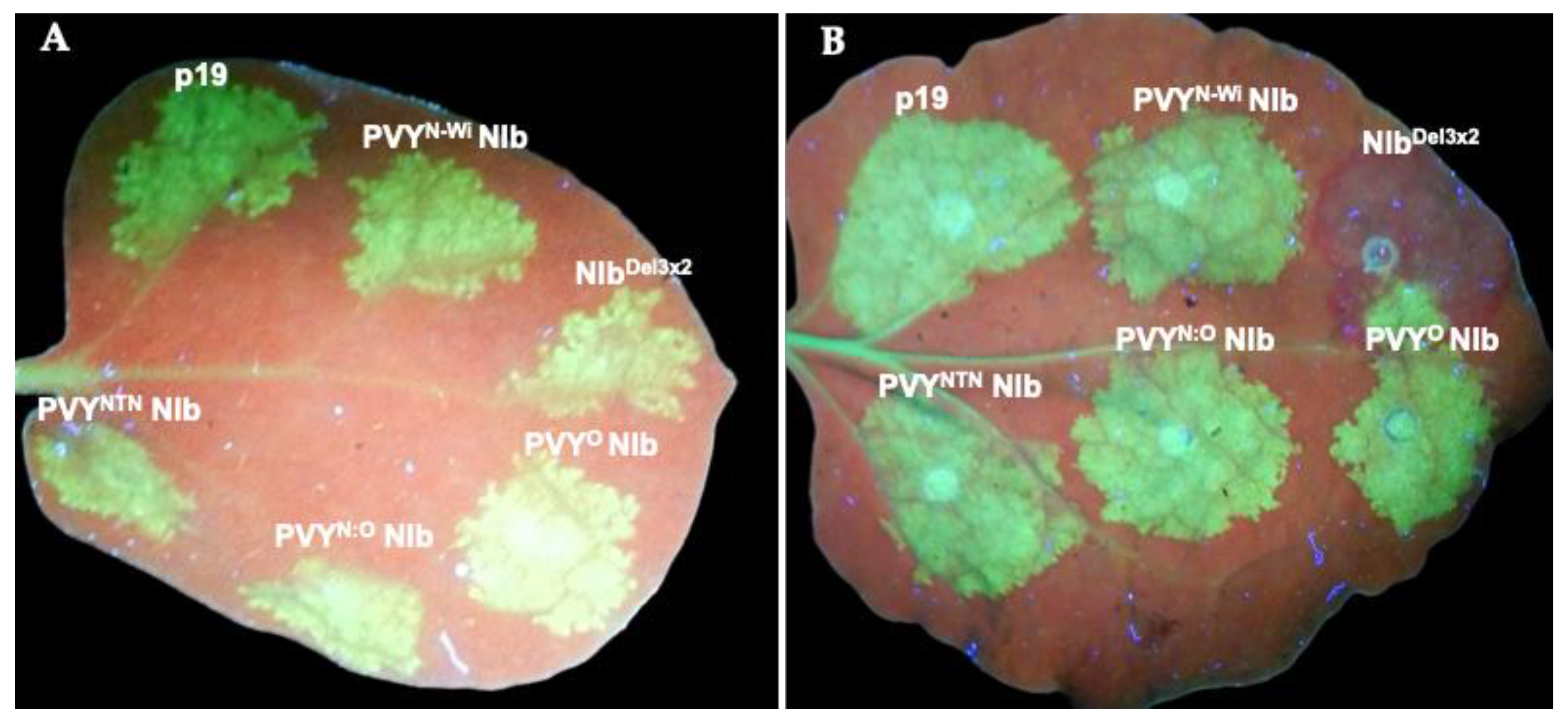
Figure 2.
NIb domains and motifs required for RNA silencing suppression. N. benthamiana 16c plant leaves were co-infiltrated with 35s::GFPand NIb four PVY strains (PVYNTN, PVYO, PVYN:O, PVYN-Wi), as well as with p19, and seven PVYNTN NIb mutants (NIbΔ1/17, NIbKKK135/137AAA, NIbKK303/304AA, NIbDEEE491/494AAA, NIbGDD351/353AAA, NIbΔ4491/519, and NIbDel3x2), respectively. By 10 dpi, NIbΔ1/17, NIbKKK135/137AAA, NIbDEEE491/494AAA, NIbΔ4491/519, and NIbDel3x2 had lost much of the GFP green fluorescence. In contrast, NIb and NIbKK303/304AA continued to display green fluorescence, while NIbGDD351/353AAA showed only weak green fluorescence.
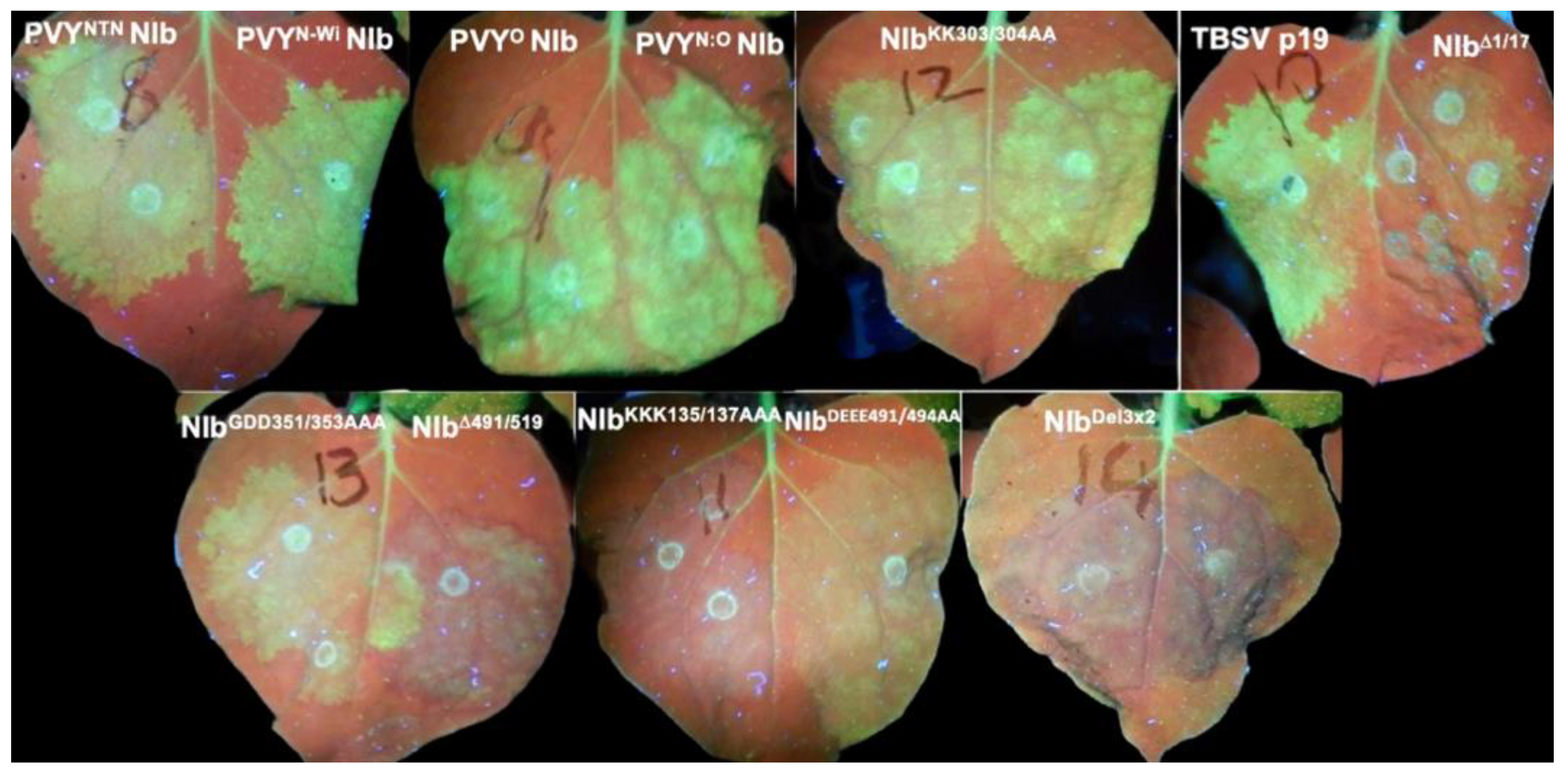
Figure 3.
NIb suppresses RNA silencing. (A) Northern blot detection of GFP RNA of total RNA at 7 dpi. Total RNA was isolated from N. benthamiana line 16c plant leaves co-infiltrated with 35s::GFPand each of PVYNTN NIb, NIbΔ1/17, NIbΔ4491/519, NIbDel3x2, NIbDEEE491/494AAA, NIbGDD351/353AAA, NIbKKK135/137AAA, NIbKK303/304AAA, TBSV p19, and an empty vector, and then probed with a biotin-labeled GFP probe. Very low levels of GFP transcripts were observed in tissues co-infiltrated with an empty vector and GFP, while high levels of GFP transcripts were observed in tissues containing PVYNTN NIb and p19. (B) Western blot analysis of GFP protein in infiltrated 16c leaves at 7, 10, and 14 dpi. At 7 dpi, leaves co-expressing GFP with wild-type NIb showed high levels of GFP protein compared to much lower GFP levels in leaves expressing NIb mutants NIbΔ1/17, NIbKKK135/137AAA, NIbΔ4491/519, and NIbDel3x2. By 10 dpi, GFP was barely detectable in mutant NIb samples, and by 14 dpi GFP was almost completely undetectable in those samples, while leaves expressing wild-type NIb maintained a high GFP protein accumulation.
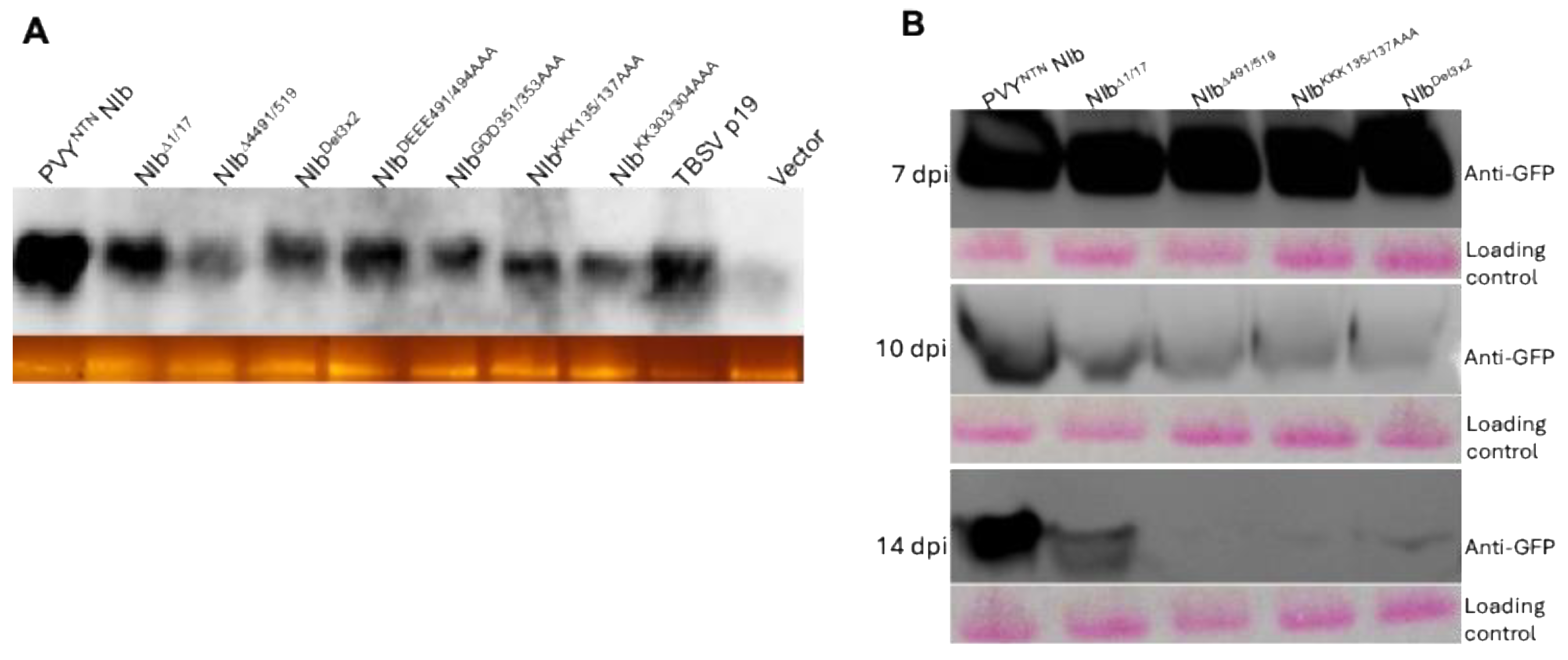
Figure 4.
Subcellular localization of NIb and mutants using confocal microscopy. z-stack imaging shows that in the cytoplasm NIb localizes in cytoplasmic membranes (5A). Wild-type NIb (5B), NIbΔ1/17 (5C), NIbDEEE491/494AAA (5I), NIbKK303/304AA (5H), and NIbGDD351/353AAA (5G) localized to both the nucleus (indicated by arrows) and the cytoplasm. In contrast, NIbKKK135/137AAA (5D, 5E) and NIbΔ491/519 (5F) localized in the cytoplasm, and nuclear membrane with no detectable nuclear localization. Images were acquired on an upright Zeiss LSM 780 laser-scanning microscope (using a Zeiss 40x C-Apochromat (NA 1.2) objective lens. Additional confocal images of NIbKKK135/137AAA subcellular localization are in Supplementary Figure S1.
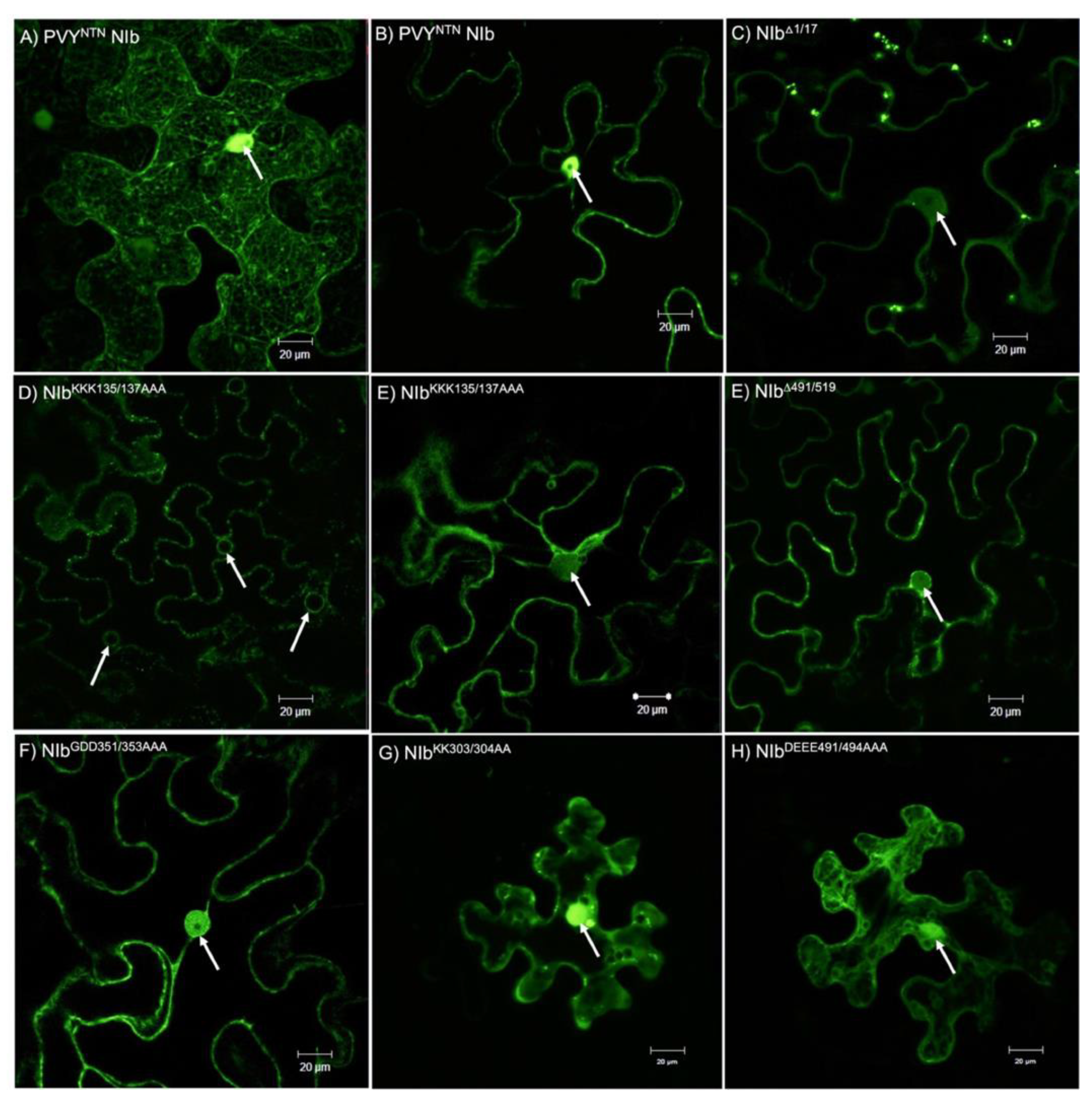
Figure 5.
Distribution and aggregate burden of GFP-derived siRNAs. (A) Per-nucleotide coverage of small RNAs mapping to GFP in 16c leaves at 14 dpi (CPM; linear scale). Solid lines show the group mean; ribbons are 95% CIs (n = 18 libraries). Three reproducible hotspots occur at ~175 nt, ~320–330 nt, and 560–700 nt. Peak heights differ modestly between treatments, indicating positional redistribution rather than uniform gain/loss. (B) Cumulative (running) sum of CPM along GFP. The curve endpoint equals the transcript-wide total. In this dataset, totals are similar between NIb and NIbDel3x2 (median Del/Nib ≈ 1.0; Wilcoxon rank-sum p ≈ 0.10).
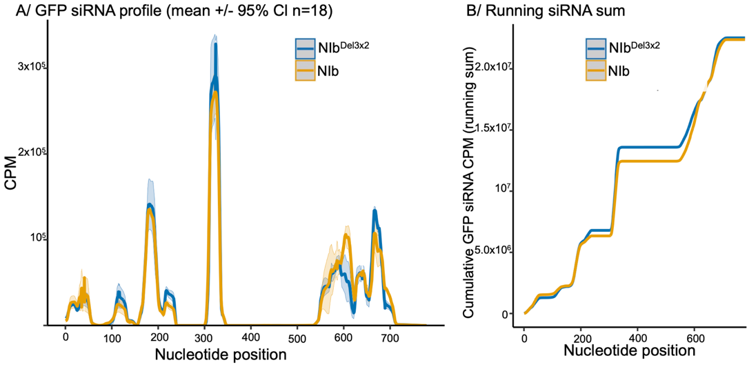
Figure 6.
Strand bias of GFP-derived siRNAs. Sense (+) and antisense (−) siRNA coverage along the GFP sequence in line 16c leaves at 14 dpi. Panels show NIb (top) and NIbDel3x2 (bottom) samples separately. Colors indicate strand polarity [(+)siRNA vs (−)siRNA]. In both treatments, GFP siRNAs are strongly antisense-biased, with (−)siRNAs greatly outnumbering (+)siRNAs.
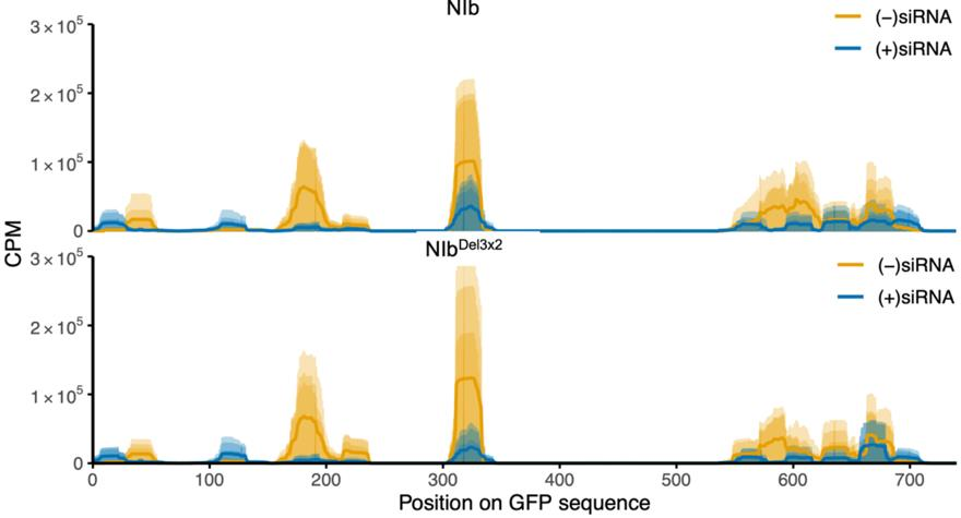
Figure 7.
Profiles of small RNAs mapping to the NIb transcript in GFP line 16c plants co-infiltrated with wild-type NIb vs NIbDel3x2 at 14 dpi. Lines indicate mean NIb-siRNA coverage along the NIb coding sequence (reads per million, 1-nt bins; shaded 95% CI, n = 18). Overall, NIb-derived siRNAs were present at relatively low levels (note the much lower coverage compared to GFP siRNAs in Figure 6). Notably, leaves expressing wild-type NIb produced slightly more NIb-derived siRNAs than those expressing NIbDel3x2. The NIb and NIbDel3x2 samples showed broadly similar siRNA distribution profiles along the length of the NIb sequence, suggesting that NIb’s suppressor activity does not eliminate processing of its own transcripts.
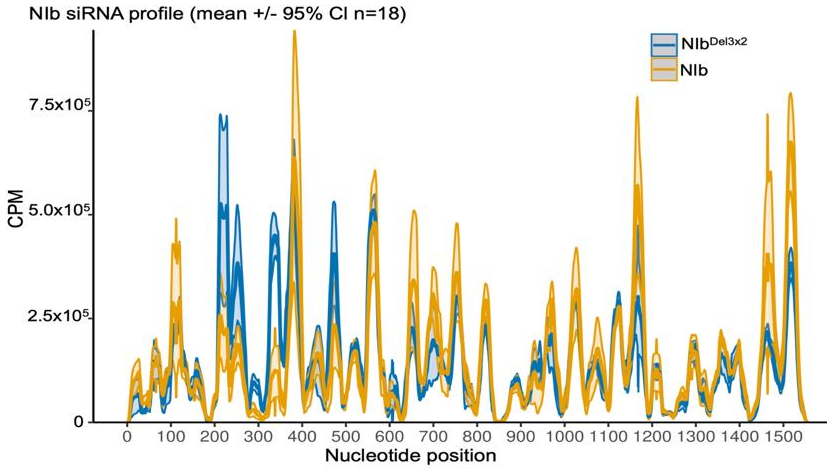
Figure 8.
Strand profiles of NIb-derived siRNAs. Sense (+) and antisense (−) siRNA coverage along the NIb coding sequence in N. benthamiana line 16c leaves at 14 dpi. Panels show NIb and NIbDel3x2 samples separately; colors indicate strand polarity [(+)siRNA vs (−)siRNA]. In contrast to the GFP-derived siRNA pattern (Fig. 7), NIb-derived siRNAs do not show a strong antisense excess: wild-type NIb samples display a modest surplus of (+)siRNAs over (−)siRNAs, whereas NIbDel3x2 samples exhibit a more balanced sense/antisense profile.
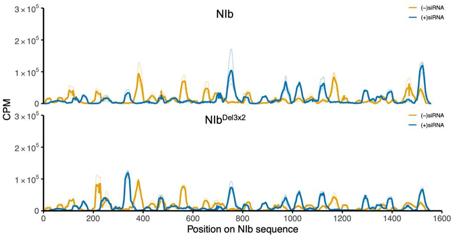
Figure 9.
In planta interaction between NIb and host RNA silencing proteins. NIb–HA was co-expressed in N. benthamiana leaves with each of AGO1, AGO4, DCL4, HSP70, HSP90, HYL1, RDR6, SGS1, SGS3 proteins tagged with 6xHIS. At 5 dpi, total protein was extracted and subjected to Ni–NTA affinity purification of protein complexes followed by immunoblotting using anti-HA antibodies. NIb was found to co-purify with AGO4, HSP70, HSP90, and SGS3. No NIb signal was detected in samples with the other candidate proteins, indicating no stable interaction. Samples containing AGO4, HSP70, and SGS3 showed degraded protein products. The SDS-PAGE coomassie blue stain of total protein lysate for Ni–NTA affinity purification can be found in Supplementary Figure S2.
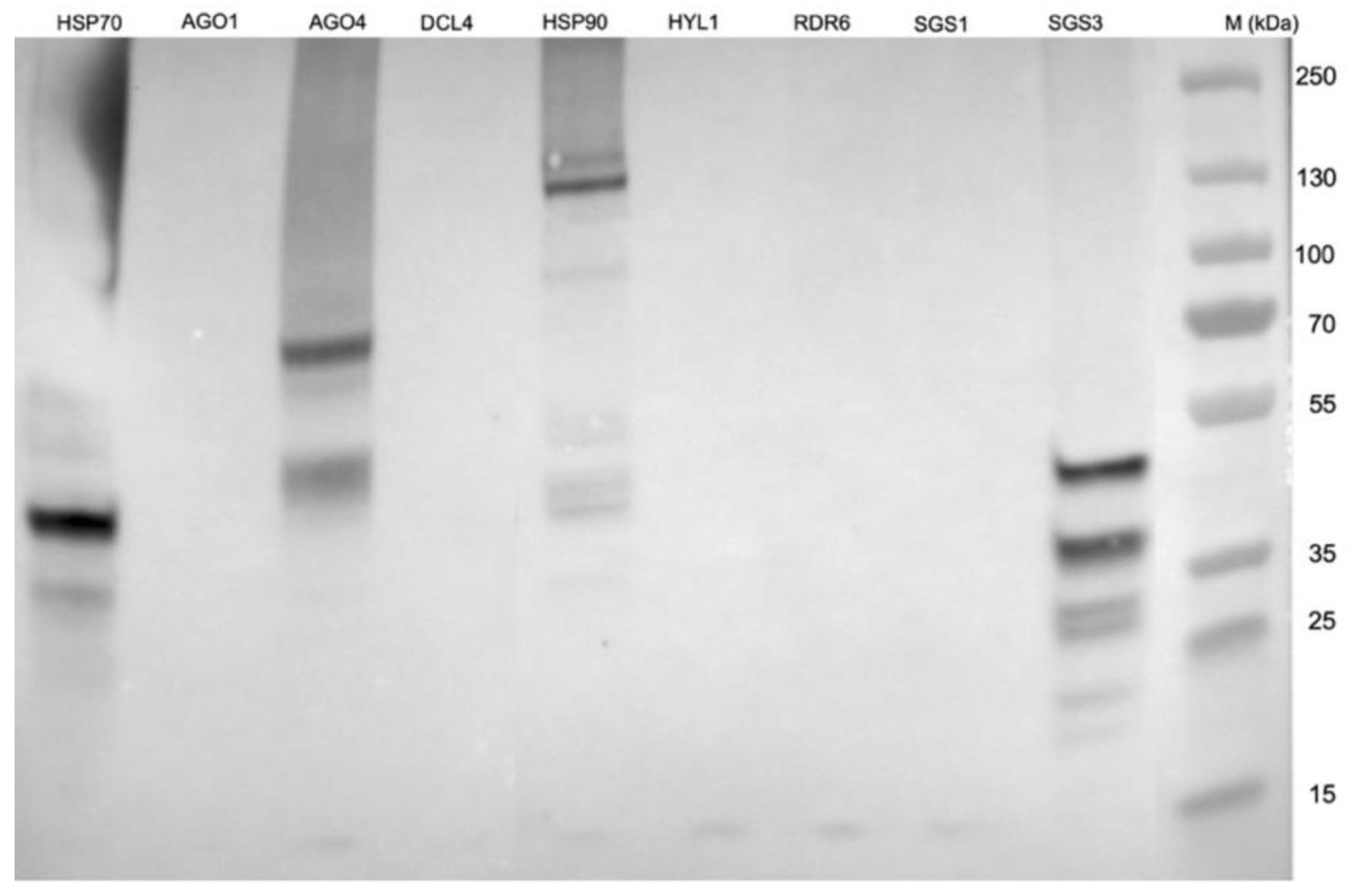
Figure 10.
Expression of PVYNTN NIb, NIbΔ1/17, NIbΔ491/519, NIbKKK135/137AAA, and NIbΔ491/519 tested (A), and of AGO4, HSP70, HSP90, and SGS3. (A) Total plant proteins were extracted from infiltrated N. benthamiana leaves followed by western blot analysis using anti-HA tag antibodies at 2 dpi, as well as at 10 dpi when only NIb was detectable. (B) To analyze RNA silencing pathway proteins, total protein was first isolated with Ni-NTA magnetic bead kit prior to detection with anti-HIS-tag antibodies. TP: total protein from control plants inoculated with an empty vector; M1: PageRuler™ Plus Prestained Protein Ladder; M2: precision plus protein standard kaleidoscope.
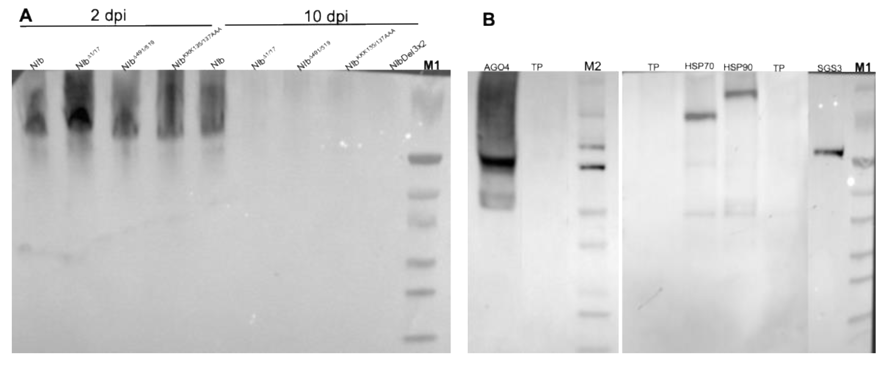
Figure 11.
Figure 11. NIb mutants do not interact with AGO4, HSP70, and SGS3 as detected with anti-HA tag (left panel) and with anti-HIS tag right panel) antibodies. Analysis of samples co-expressing NIb (1) and AGO4 showed cleaved protein complexes. In contrast, no such complexes were found in samples expressing NIbΔ1/17 (2); NIbΔ491/519 (3); NIbKKK135/137AAA (4) and NIbDel3x2 (5). Similar to AGO4, co-expression of NIb constructs and (B) HSP70, and (C) SGS3 showed no accumulation of protein complexes. In contrast no cleaved products were observed in co-expressions with (D) HSP90. The SDS-PAGE coomassie blue stain of total protein lysate for Ni–NTA affinity purification can be found in Supplementary Figure S3. M: Novus Biologicals Blu11 Prestained Protein Ladder; X: artifacts.
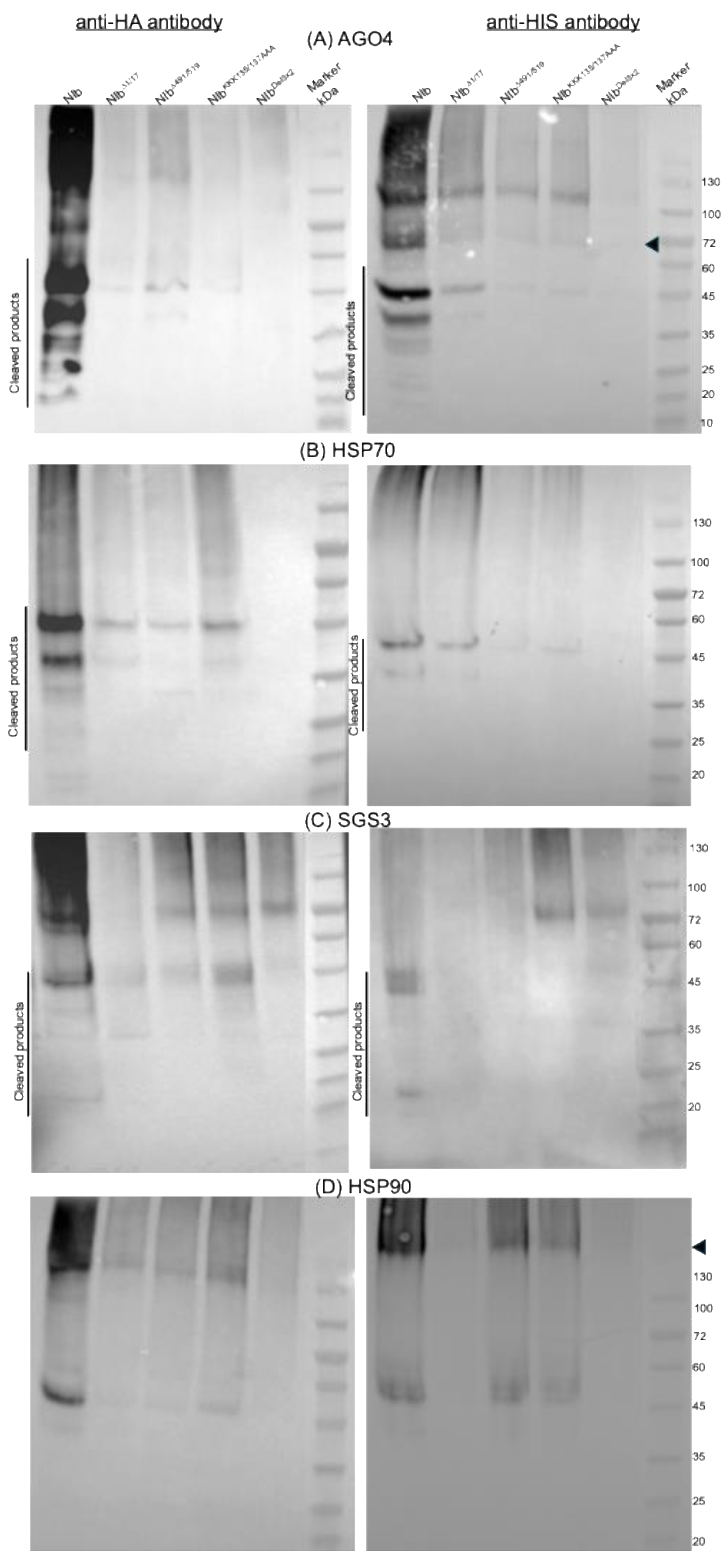
Figure 12.
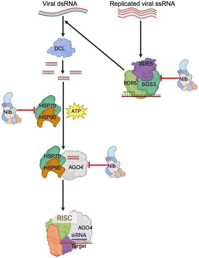
Antiviral silencing of RNA viruses. Viral dsRNA is processed into siRNA by DCL proteins. HSP70 then loads the siRNA into AGO proteins cooperatively with HSP90, which provides the energy needed, forming a mature RISC. The dsRNA is either from the replication intermediate or synthesized from the replicated genome by the RDR6/SGS3/SDE5 complex. This study shows that NIb counters this antiviral RNA silencing by targeting AGO4, HSP70/HSP90, and SGS3.
Figure 12.
Antiviral silencing of RNA viruses. Viral dsRNA is processed into siRNA by DCL proteins. HSP70 then loads the siRNA into AGO proteins cooperatively with HSP90, which provides the energy needed, forming a mature RISC. The dsRNA is either from the replication intermediate or synthesized from the replicated genome by the RDR6/SGS3/SDE5 complex. This study shows that NIb counters this antiviral RNA silencing by targeting AGO4, HSP70/HSP90, and SGS3.
Disclaimer/Publisher’s Note: The statements, opinions and data contained in all publications are solely those of the individual author(s) and contributor(s) and not of MDPI and/or the editor(s). MDPI and/or the editor(s) disclaim responsibility for any injury to people or property resulting from any ideas, methods, instructions or products referred to in the content. |
© 2026 by the authors. Licensee MDPI, Basel, Switzerland. This article is an open access article distributed under the terms and conditions of the Creative Commons Attribution (CC BY) license (http://creativecommons.org/licenses/by/4.0/).
Copyright: This open access article is published under a Creative Commons CC BY 4.0 license, which permit the free download, distribution, and reuse, provided that the author and preprint are cited in any reuse.


