Preprint
Article

This version is not peer-reviewed.

Raw Milk Cheese Microbiomes: A Paradigm for Interactions of Lactic Acid Bacteria in Food Ecosystems

Submitted:

06 January 2026

Posted:

08 January 2026

You are already at the latest version

Abstract

While industrial scale dairy fermentations often employ pasteurized milk as the substrate, many farmhouse and traditional production practices apply raw milk derived from a variety of mammals. Certain artisanal production systems rely on the autochthonous microbiota of the milk, fermentation vessels, equipment and/or environment to initiate milk coagulation. While the technological properties of lactic acid bacteria associated with dairy fermentations are well described, their interactions with other organisms during fermentation and cheese ripening are poorly investigated. This study presents an overview of the microbial ecology of raw and pasteurized milk used in the production of cheeses. Furthermore, we report on the motility phenotype, lactose utilization ability and metabolic products of isolates of Hafnia paralvei and Hafnia alvei, and determine that these strains could grow in a non-antagonistic manner on plates with strains of Lactococcus lactis and Streptococcus thermophilus. As artisanal and farmhouse production systems are often associated with protected or regionally significant products, it is essential to develop a clear understanding of the microbial communities within and the complex relationships between the community members.

Keywords: 
;  ;  ;  ;  ;  ;  ;  

1. Introduction

The dairy sector is a major contributor to the Irish economy [1,2]. 94% of Irish dairy products are exported, contributing over €6.5 billion annually in exports to over 140 international markets [3]. Ireland ranks as the third highest producer of cheese per capita globally (56.7 kg per person) with almost 300,000 metric tonnes of cheese produced each year [4]. While Ireland is renowned for its production of Cheddar-style cheeses using so-called “defined” bacterial starter cultures [5], there is considerable growth in farmhouse cheese manufacture [6] which uses artisanal production processes and undefined starter cultures [7]. Artisanal production systems often involve the use of raw, unpasteurised bovine or ovine milk as substrates for fermentations. While these foods are considered safe for human consumption, detailed knowledge of the microbiota of such products is limited. Furthermore, while the majority of bacteria that contribute to these fermentations are likely to be members of the heterogenous group of lactic acid bacteria (LAB) including Lactococcus, Streptococcus and Lactobacillus spp., the microbiota of raw milk cheeses often contain enteric organisms including members of the Enterobacteriaceae and non-starter lactic acid bacteria, such as Enterococcus spp. [5]. These species may contribute to the flavour profile of the product, yet may simultaneously contribute to the dissemination of antibiotic-resistance associated genes within the human gastrointestinal microbiota, which is a major public health concern [8,9].
Certain LAB have long been applied deliberately in the production of fermented dairy foods [10,11]. Furthermore, species such as S. thermophilus and L. lactis have been granted the generally regarded safe (GRAS) status owing to their long and safe history of use in foods and, as such, are commercially exploited in food production [12].
Sustainability in dairy production systems is beneficial for the continued success and further growth of the agri-food sector. With growth in farmhouse cheese production sites and volumes in Ireland, it is becoming increasingly important to define the microbial communities that underpin such artisanal fermentations. The role of LAB in acidifying milk, ripening and flavour formation and rapid completion of the fermentation is essential in raw milk-based fermentations since slow or incomplete fermentations result in nutrients becoming available to non-LAB spoilage or pathogenic organisms [12]. Strains of the Gram-negative bacterium Hafnia are commonly isolated from raw milk and (particularly) soft cheeses [13,14,15,16]. However, while this bacterium has been reported as a contributor to good quality cheese in the context of ripening and flavour development [11,12], it may also be associated with negative outcomes particularly if the overall abundance of Hafnia is high [13].
While several studies have evaluated the presence of specific bacterial species [5,12,17,18] and while there are emerging reports of the metagenomic evaluation of fermented foods [19,20,21,22], there are limited reports that provide a holistic understanding of the microbiota of artisanally-produced cheeses and particularly those produced in an Irish context [6,17,23,24]. Therefore, the current study aimed to define the microbiota of Irish farmhouse, artisanally-produced cheeses using culture-dependent and independent approaches. Finally, we sought to explore the interactions between lactic acid starter bacterial isolates and non-LAB Hafnia isolates from these cheeses.

2. Materials and Methods

2.1. Cheeses Evaluated in This Study

Twelve cheeses were analyzed in this study including Brie (n=2), Camembert (n=2) and Pecorino (n=2) cheese samples as well as one Reblochon, Smoked Drumlin, Caciocavallo, Mozzarella, Saint Felicien and Fleur du Maquis cheese samples. All twelve cheeses were sourced locally and produced in Ireland in 2024. Six of the cheeses were produced using pasteurized milk and six cheeses were produced from raw milk (Table 1).

2.2. Metagenomic DNA Extraction

The DNA of four of the above listed raw milk cheeses including a Brie, Camembert, Smoked Drumlin and Reblochon sample was extracted as follows. 2 g samples from the core and rind/surface of each cheese wheel were aseptically collected using a pre-sterilised knife for DNA extraction. Each cheese samples was mixed with 25 ml of 2.2% sodium citrate (heated to 45ºC; Sigma-Aldrich Germany) and homogenized in a stomacher 400 (Lab-Blender) for 5 minutes at 230 rpm.
The homogenized samples were centrifuged at room temperature (18ºC) 13,800 x g for 10 minutes after which the fat layers from the samples were removed. The supernatant from each sample was discarded, and the cells/cheese debris resuspended and washed in 1 ml of 2% sodium citrate (heated to 45ºC; Sigma-Aldrich). The sample was then centrifuged at 10,800 x g for 5 minutes. The washing step was repeated three times. The washed cells were resuspended in 1 ml of lysis buffer (20 mM Tris HCl – pH 8, 2 mM EDTA- pH 8, 2% polyethylene glycol), 50 µg/ml lysozyme solution and 100 U mutanolysin (Sigma-Aldrich; Fisher.CO.UK).
The solution was incubated at 37ºC for 3 hours. After incubation, 250 µg/ml of Proteinase K was added to the solution and incubated again at 56ºC for 1 hour. After incubation, each solution was precipitated using 1 ml 96% ice cold ethanol. Using GenElute™ Bacterial Genomic DNA kit (Sigma-Aldrich, NA2120-1KT), washing and elution of DNA was carried out according to manufacturer’s instructions, but applying 40 µl of elution buffer instead of 200 µl. A second elution was performed for each sample. The extracted DNA was quantified using Qubit™ dsDNA HS assay kit (Invitrogen by Thermo Fischer Scientific) and a Qubit® 2.0 Fluorometer.

2.3. Metagenome Sequencing & Analysis

Using the extracted DNA, partial 16S rRNA gene sequences were amplified. Primer pairs Probio_Uni/Probio_Rev (CCTACGGGRSGCAGCAG/ATTACCGCGGCTGCT) were used, targeting the V3 region of the 16S rRNA gene sequence [25]. Overhang sequences by Illumina adapter were added, and library preparation was performed according to the 16S rRNA Metagenomic Sequencing Library Preparation Protocol (Part #15044223 Rev. B – Illumina).
Amplicon quality was assessed by electrophoresis, and purification was performed using a magnetic bead-based clean-up to remove primer dimers. DNA concentrations were quantified fluorometrically and normalized to 4 nM prior to pooling. Sequencing was performed using the Illumina NextSeq 2000 platform. Raw paired-end reads were processed with a custom QIIME2-based pipeline [19,26]. Reads were filtered to retain sequences between 140 and 400 bp in length, with an average quality score >20. Sequences containing homopolymers >7 bp or primer mismatches were excluded. Amplicon Sequence Variants (ASVs) were inferred using DADA2 [27] with 100% sequence homology. ASVs not observed at least twice in the same sample were removed. Taxonomic classification was performed with QIIME2 [19,26] using the SILVA reference database [28].
Alpha diversity was calculated using Observed ASVs, Chao1, and Shannon indices; beta diversity was assessed using weighted UniFrac distances and visualized via Principal Coordinates Analysis (PCoA).

2.4. Culture-Based Analysis

Culture-based analysis of the four cheese samples for which metagenomic profiling was also performed (Brie, Camembert, Reblochon, Smoked Drumlin) was undertaken by establishing the viable plate counts of total bacteria on tryptic soy agar (TSA; Sigma-Aldrich, Germany); lactobacilli, lactococci and Leuconostoc spp. on de Man-Rogosa-Sharpe agar (MRS – Oxoid Ltd., UK); thermophilic coccoid lactic acid bacteria on S. thermophilus isolation agar (HiMedia Ltd., India). For the remaining eight cheese samples, the culture-based analysis was confined to presumptive LAB counts on S. thermophilus agar and M17 agar supplemented with 0.5% lactose [29]. (Sigma-Aldrich)
5 g of each cheese was aseptically transferred into a sterile stomacher bag using a sterile spatula. 45 ml of ¼ Ringer’s solution (Merck, Germany) was added to the cheese and homogenized for one minute using a stomacher (Stomacher Circular 400; Seward, UK). Serial dilutions (10ˉ¹ to 10ˉ4) of the cheese homogenates were then prepared in ¼ strength Ringer’s solution. 100 µl of each dilution sample was spread plated on respective agar plates with different media per selection and incubated overnight anaerobically or aerobically at 30 °C, 37 °C and 42 °C for the selection of bacterial isolates.
Viable counts were recorded after 24 hours except for counts of isolates on MRS agar plates which were recorded after 48 hours incubation. Single representative colony isolates from each cheese product were streaked on LM17 agar (or other media on which the organisms were originally isolated) to purify the isolates prior to glycerol stock preparation (20% glycerol (Fisher.CO.UK) and storage at -70 °C.

2.5. Species Identification of Bacterial Isolates Using 16S rRNA Gene Sequencing

Bacterial isolates were maintained using the same medium and incubation conditions used for their isolation. Colonies of the isolates were used as template for 16S rRNA gene amplification. For the Polymerase Chain Reaction (PCR), Lucfw (tgcctaatacatgcaagt) and LucRv (cttgttacgacttcaccc) primers (which amplifies the entire gene), one taq polymerase master mix (BioLabs) and nuclease-free water (Thermo Scientific) were mixed and amplified under conditions of 94 °C for 3 minutes, followed by 30 cycles of 94 °C for 30 seconds, 50 °C for 30 seconds and 68 °C for 1 minute and 30 seconds and a final extension of 68 °C for 7 minutes. The amplicons were purified using GenElute PCR Clean-up Kit (Sigma Aldrich) according to manufacturer’s instructions.
Gel electrophoresis was carried out on a 1% agarose gel diluted in Tris-Acetate-EDTA (TAE) buffer with 2.5µl SYBR Safe DNA gel stain (RayBiotech) added [23]. The amplicons were visualized using UV transillumination. PCR products were purified according to the PureLink® PCR Purification Kit (Invitrogen by Life technologies) manufacture’s instructions. Sanger sequencing of PCR products was performed by Genewiz Inc. (Germany). Results from the generated sequences were analyzed using BLASTN analysis against available sequence data on National Centre for Biotechnology Information (NCBI) database (https://blast.ncbi.nlm.nih.gov/Blast.cgi).

2.7. Characterisation of Hafnia Isolates

2.7.1. Gas production Evaluation

Twelve 1 ml sterile durum tubes were each placed in sterile test tubes containing 10 ml LM17 broth. These were then inoculated with 100 µl of fresh overnight cultures of each of the isolated Hafnia strains (strains were named CO1 to CO12) and incubated overnight at different temperatures of 4 °C, room temperature (RT), 30 °C, 37 °C and 42 °C to establish if the strains produce gas. All tests were performed in triplicate.

2.7.2. Organic Acid Production

Hafnia isolates were grown overnight in LM17 broth and the culture was then centrifuged at 1409 x g for 10 mins. The resulting supernatant was filter-sterilized twice using 0.22 µm filters (Sarstedt, Germany). 750 µl of each filtered supernatant was transferred in triplicate into pre-labeled HPLC vials. 750 µl of uninoculated LM17 broth was used as a negative control for the assay. The supernatants of three independent biological replicates of each strain were analyzed.

2.7.3. Growth Temperature Range Evaluation

Overnight cultures of Hafnia strains were prepared in both LM17 and LB broth and incubated at 4 °C, room temperature (RT ~20 °C), 30 °C, 37 °C and 42 °C. Fresh overnight cultures were diluted in Ringer’s solution and plated on LM17 and LB agar for each of the following temperatures: 42 °C, 37 °C, 30 °C 4 °C and at Room Temperature (RT ~20 °C). Plates were incubated overnight at the above-mentioned temperatures. Viable counts and colony morphologies were recorded after 24 hours incubation. All assays were performed in triplicate.
Simultaneously, 100 µl of a fresh culture of the twelve Hafnia strains were inoculated into 10 ml LM17 broth and incubated over a 24-hour period at 4 °C, RT, 30 °C, 37 °C and 42 °C. OD600 nm readings at each temperature were recorded every two hours for the first 8 hours and then a final reading was taken at 24 hours. All assays were performed in triplicate.

2.7.4. Salt Tolerance

LB broth was prepared with a final concentration of 3, 4 and 5% NaCl, respectively. Tubes containing 10 ml of each of the salt-containing LB or LM17 broth were inoculated with 100 µl of fresh overnight cultures of the twelve Hafnia isolates and were then incubated for 24 hours at 37 °C, after which the optical density at 600 nm (OD600) was recorded. The salt tolerance tests were performed in triplicate.

2.7.5. Microscopic Evaluation

The Gram staining technique using crystal violet, Gram’s iodine, alcohol and safranin was performed to evaluate the morphology of the Hafnia cells to evaluate aggregation activity and any differences imposed by growth in LM17 and LB growth media. A single colony from each strain grown in LM17 or LB agar was picked with a loop and placed on a glass slide with a drop of water prior to staining. Samples were imaged on a light microscope at 40X magnification (Figure S1).

2.7.6. Motility Assays

Motility assays for the Hafnia strains were performed using both LB and LM17 media. 15 ml of 1.2% technical agar was used as base agar for all motility assay plates. The agar base was overlaid with 0.3% LB or LM17 soft agar, respectively. Using filter tips, 2 µl a fresh culture of each Hafnia isolate (emanating from an overnight culture grown on the same medium) was carefully applied to the center of the soft agar. The plates were then incubated upright at 37 °C, and the plates were visually inspected, and phenotypes recorded at 24 and 48 hours.
Eiken agar motility assays were also performed to assess swarming and swimming phenotypes for each of the twelve Hafnia isolates. A solution of 0.8% Eiken broth and 0.6% Eiken agar (Eiken Chemical Ltd. E-MC35) with 0.5% glucose was prepared to which NaCl was added at a final concentration of 3, 4 and 5% to establish the impact of the added salt/osmotic pressure on motility in comparison to a negative control without added salt. Using sterile 1 µl loops, a single colony of each freshly grown Hafnia strain was transferred from overnight LM17 agar plates. A single colony of each strain was tapped in the center onto the top layer of the Eiken agar. The plates were incubated upright on a flat tray without agitation at 37ºC. The strains were visually inspected after 24 hours, and the phenotypes were recorded (Gutiérrez-Barranquero, at el., 2019). All assays were performed as independent biological triplicate assays.

2.7.7. Hafnia and LAB Interaction Assays

10 µl of fresh overnight cultures of either S. thermophilus CO-St16 or L. lactis LL1 and H. paralvei CO12 was spotted on two LM17 agar plates respectively. The LAB cultures were each spotted 0.5 cm apart from the Hafnia cultures and incubated overnight for 24 hours. One agar plate spotted with L. lactis, and H. paralvei was incubated aerobically at 30 °C for one day to enable the L. lactis strain to grow optimally. S. thermophilus and the H. paralvei strains were incubated anaerobically at 42 °C to accommodate CO-St16. Plates were removed and incubated at RT (~20 °C) for five days. Interactions between the two LAB isolates and the Hafnia isolates were observed for growth, compatibility and/or antagonism over a week period.

3. Results

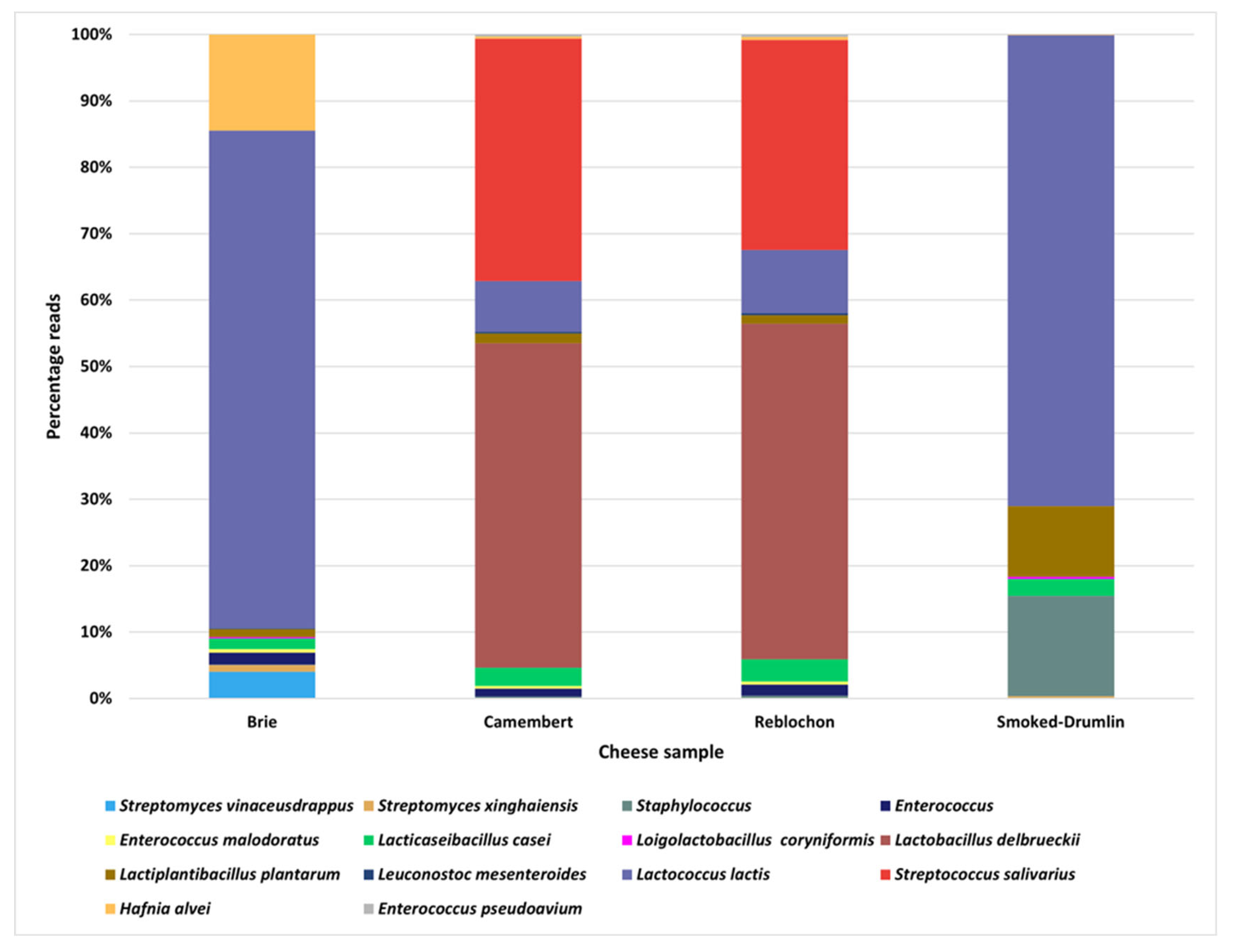
3.1. Metagenomic and Culture-Based Analysis of Four Raw-Milk Derived Cheeses

To establish the total viable bacterial counts (on TSA) and the subpopulations of LAB (LM17 & MRS agar) and coliforms (MacConkey agar), four raw milk cheese samples were evaluated by cultivation at 30 °C and 37 °C. Total counts ranged between 106-107 cfu/ml (Table 2). Presumed LAB counts were observed to constitute the majority of the total population with high counts (~105-106 cfu/ml) being observed for three of the four cheese samples (the exception being Smoked Drumlin where there were no detectable coliforms). The numbers of culturable LAB were similar across all four samples (~106-107 cfu/ml) (Table 2).
To establish the dominant culturable species across the four assessed cheeses, 16S rRNA gene sequencing was performed for 17 presumptive LAB (Brie n=5; Smoked Drumlin n=4; Camembert n=4; Reblochon n=4) and six isolates from the total counts on TSA (Brie n=2; Smoked Drumlin n=2; Reblochon n=2).
Among the presumptive LAB isolates derived from counts on LM17 agar, only three isolates were validated as LAB (one Enterococcus spp., one Leuconostoc (Lc)mesenteroides and one Lactococcus (L.) lactis isolate), while the remaining five isolates were represented by H. alvei (n= 2), H. paralvei (n= 1), Staphylococcus equorum (n= 2). With the exception of one isolate, the selected isolates from MRS agar (n=9 total) were identified as LAB members, i.e., five L. lactis isolates (from Brie n=3, Camembert n=1 and Reblochon n=1), one Lactiplantibacillus plantarum isolate (from Smoked Drumlin), one Levilactobacillus brevis (from Camembert) and one Lc. mesenteroides (from Reblochon).
The sole non-LAB isolate was identified as Staphylococcus casei. Among the six isolates derived from counts on TSA, H. alvei and H. paralvei were dominant (n=2 and n=3, respectively) while one Enterobacter spp. isolate was also identified.
Based on 16S rRNA based metagenomic analysis, the microbiota of the analyzed Camembert and Reblochon cheeses were dominated by Streptococcus salivarius and Lactobacillus delbrueckii, while those of the Brie and Smoked Drumlin cheeses were dominated by L. lactis (Figure 1). The mesophilic production system applied to Brie and Smoked Drumlin is consistent with the finding of mesophilic lactococci and Leuconostoc spp. as major components of their microbiota. Similarly, since Reblochon is typically produced using a mixture of mesophilic and thermophilic production steps, the high abundance of S. salivarius (which may likely be represented by S. thermophilus) is consistent with the production regime. Conversely, since Camembert is usually produced under mesophilic conditions, the microbiota composition was noteworthy (Figure 1). Furthermore, Lb. plantarum and Lacticaseibacillus casei were present in low abundance in all four cheeses and may comprise part of the non-starter LAB microbiota. Notably, there appeared to be an abundance of Hafnia reads in the Brie sample when compared to those representing the other cheeses, being consistent with the isolation of Hafnia strains from this cheese sample (Figure 1 & Table S1).

3.2. Thermophilic Culture-Based Analysis Increases Selection for Lactic Acid Bacteria

The culture-based analysis of the four raw milk cheeses described above revealed a diversity of non-LAB isolates, particularly on LM17 agar at 30 °C. Therefore, to establish if higher temperatures would yield a more selective LAB composition on media that is designed to enrich for LAB, we analyzed the LAB culturable counts of eight additional raw milk and pasteurized milk cheeses on LM17 agar at 42 °C. Thermophilic counts across all eight cheeses were approximately 105 cfu/ml (Table S2). Subsequently, 22 representative colonies were picked and purified from the eight additional cheese samples for speciation using 16S rRNA gene sequencing revealing a more selective enrichment for LAB species including S. thermophilus, L. lactis, E. faecalis, E. faecium, Pediococcus acidilactici, Lacticaseibacillus paracasei (19 of 22 isolates were identified as LAB; Figure S2). Interestingly, three isolates were identified as H. alvei/paralvei from the second Brie and Camembert samples being consistent with the cultivation-based analysis of the first four raw milk samples. The isolates derived specifically from the pasteurized milk cheeses were all identified as LAB (Table 1 and Figure S3).

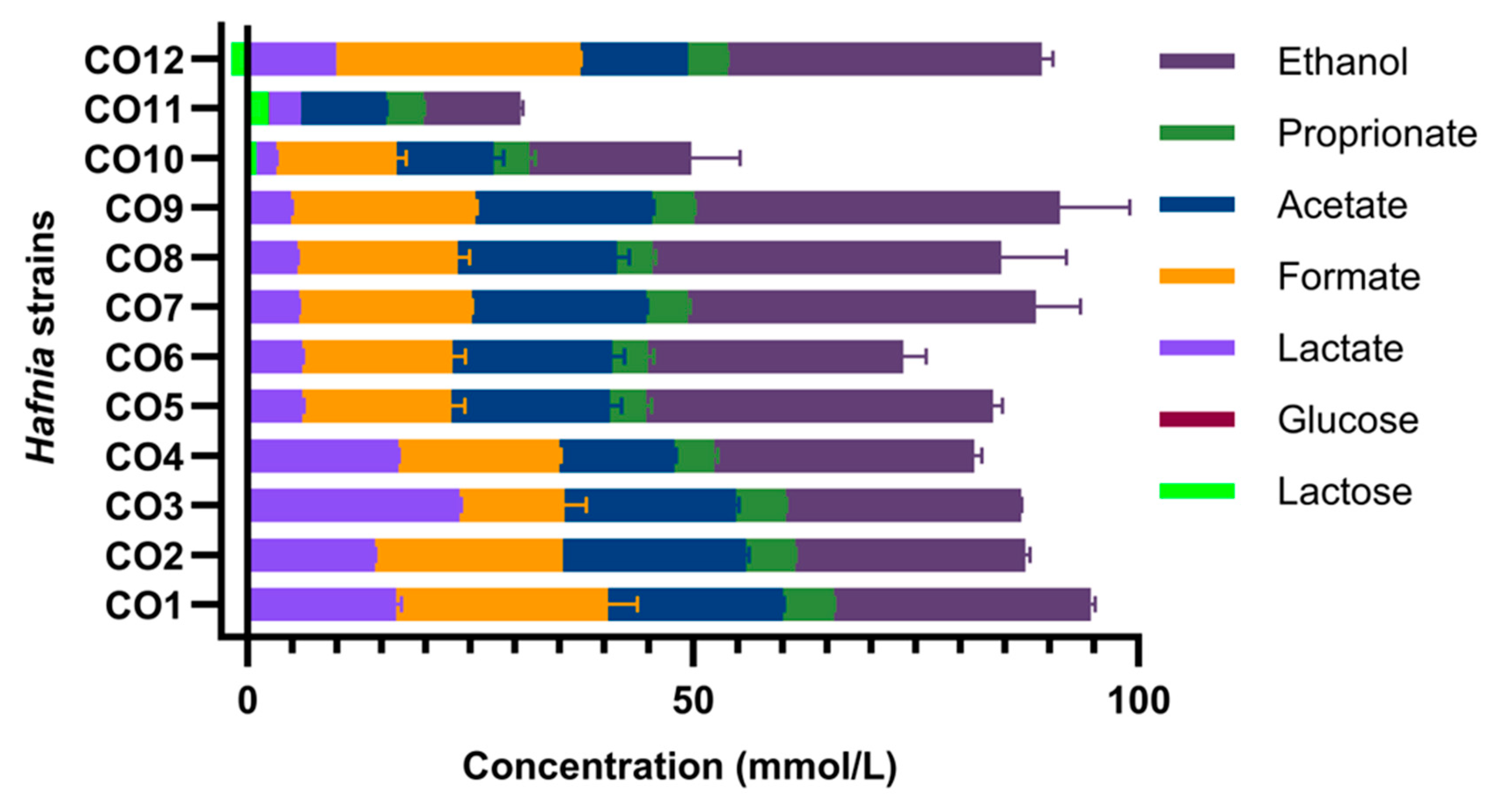
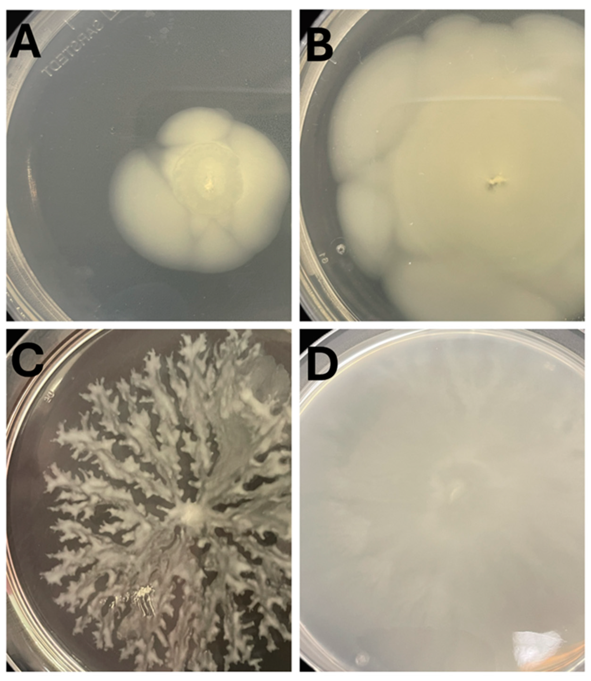
3.3. Hafnia Are Prevalent in Raw Milk Cheeses & Produce Gas from Lactose Metabolism

Hafnia strains were isolated exclusively from raw milk cheeses evaluated in this study i.e., Brie, Camembert and Reblochon cheese samples i.e., Hafnia was absent in pasteurised milk-derived cheeses evaluated in this study. In total, twelve H. alvei/paralvei strains were isolated and 16S rRNA gene sequence analysis established that among these were ten H. paralvei strains (five of which were isolated from the two Brie samples, three from the two Camembert sample and two from Reblochon and named CO1 through to CO9 and CO12) and two H. alvei strains (both isolated from the Reblochon sample and named CO10, CO11). The Hafnia isolates were evaluated for their growth capability in different media and were shown to reach higher optical densities in LM17 broth than in LB broth after 24 hours incubation at 37 °C (Figure 4 and Figure S4).
The colony morphologies of all strains at 30 °C and 37 °C on LM17 agar were observed to be large, glossy and creamy white in appearance (Figure S5). Hafnia strains produce gas in LM17 broth (Figure S6) due to the metabolism of the available lactose, while in LB broth, gas production is not observed. Furthermore, the ability to utilize lactose and subsequent conversion into organic acids was evaluated using high-performance liquid chromatography (HPLC) (Figure 3) and phenotypic evaluation on MacConkey agar (Figure S7). H. alvei strains CO10 and CO11 did not appear to metabolize lactose on MacConkey agar (this being consistent with a lack of gas production in broth assays), whereas the H. paralvei isolates produced pink colonies indicating that lactose utilization had occurred (Figure S7).
Ethanol was produced by all strains in addition to formate, acetate and lactate (Figure 3). Beyond lactose utilization, it was unclear if the Hafnia isolates could grow in milk through the metabolism of milk proteins, for example. Therefore, we evaluated the ability of the twelve Hafnia strains to grow in milk and their acidification capacity. The initial pH of the milk was 6.5 while the pH of the milk inoculated with Hafnia isolates dropped by at least one pH unit after 24 hours (Table 3). Two LAB strains isolated in this study (S. thermophilus COSt11 and L. lactis L.L1) were used as positive controls for the assay and were also shown to fully coagulate and acidify the milk within 24 hours of the assay as expected.

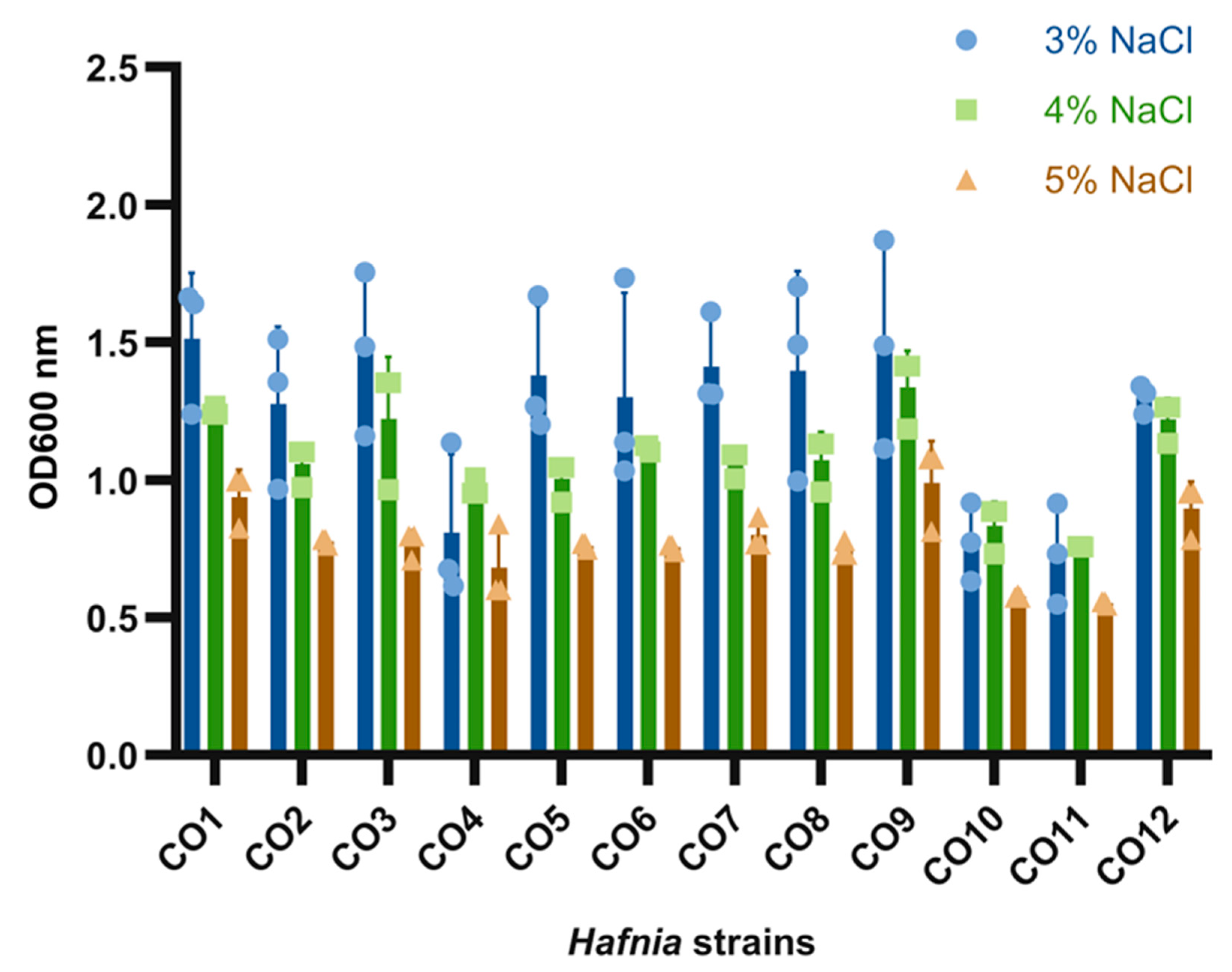
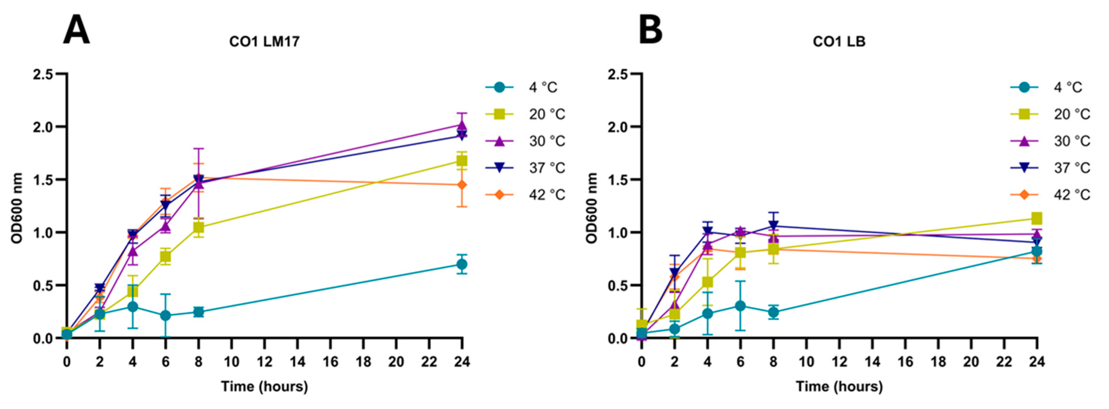
3.4. Hafnia Strains Are Tolerant to a Wide Range of Growth Conditions

All Hafnia strains were capable of growth across the range of temperatures evaluated in this study (4 - 42ºC) in both LM17 and LB broth (Figure 4&S4). H. alvei strains, however, grew relatively slower than the ten H. paralvei strains while they are also capable of growth in both LB and LM17 broth. Notably, the strains were observed to achieve higher optical densities in LM17 than in LB broth, which may be reflective of the apparent exopolysaccharide production in LM17 broth. This higher optical density in LM17 broth coincided with the presence of a thick pellicle on the surface of the LM17 broth that was not observed in LB broth. It is noteworthy that the extent and timing of observed pellicle formation is strain-dependent.
Figure 5. Graph depicting the optical density after 24 hours incubation of the twelve H. paralvei and H. alvei strains in LB broth supplemented with 3-5% salt. All strains were observed to tolerate all salt concentrations although with reduced optical density with increasing salt concentrations.
Preprints 193229 g004

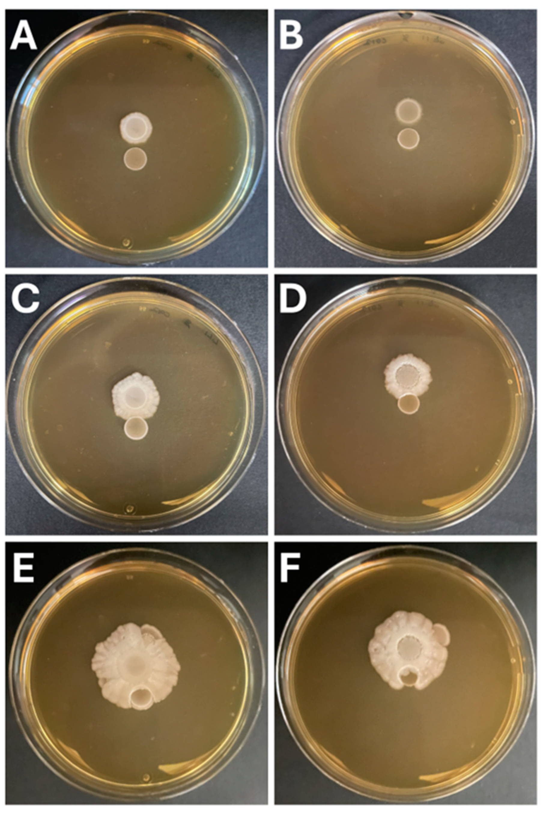
3.5. Hafnia Strains Do Not Exhibit Antagonistic Interactions with Lactic Acid Bacteria

Hafnia strains are known to be motile. In the present study, the Hafnia isolates displayed different extents of motility on LB and LM17 agar, respectively (Figure 6A&B). Greater motility was observed on LM17 (Figure 6B) relative to LB (Figure 6A). Eiken agar motility assays were also conducted on all 12 H. paralvei/alvei strains to define the type of motility. Three H. paralvei strains display swarming motility type (Figure 6C), while seven H. paralvei and H. alvei strains display swimming motility type (Figure 6D), and two H. paralvei strains display both swarming and swimming motility capabilities, respectively.
The ability of the Hafnia isolates to grow on LM17 agar and their motility phenotype on this medium prompted us to evaluate possible interaction (antagonistic or symbiotic) with lactic acid bacteria derived from the raw milk cheeses. To explore this phenomenon, we co-inoculated (by spot plating) the individual Hafnia isolates with two individual LAB isolates emanating from this study, i.e., a S. thermophilus and a L. lactis isolate. Using H. paralvei CO12 as a representative, it was evident that the LAB isolates did not antagonize the Hafnia isolate as the motile direction was such that the S. thermophilus and L. lactis spot cultures were engulfed (Figure 7A-F). Therefore, it appears that there is possible cooperation or at the least no obvious antagonism between the evaluated LAB and Hafnia isolates.

4. Discussion

The functional role of LAB and their contributions to dairy fermentations are well studied [30,31]. In the context of raw milk-derived cheeses, the diversity and contributions of organisms beyond the LAB are less well interrogated [11,20,21,32]. H. alvei has recently been linked to cheese ripening and likely beneficial interaction with Debaromyces hansenii and Brevibacterium aurantiacum with the three organisms described as a “ripening culture” in smear-ripened cheeses [33]. H. alvei and B. aurantiacum were positively associated to the production of volatile sulfur compounds being desirable for use in commercial cultures for aroma development in cheeses [33]. H. alvei was also observed to vigorously stimulate growth of B. aurantiacum by eight to 10-fold within 28 days of cheese manufacturing and was also shown to provide iron to B. aurantiacum (possibly by siderophore production) [33]. However, further investigation is needed to validate the interaction of these two species in cheese ripening. Conversely, strain H. alvei H4 (a wild-type species) has been associated with spoilage of chilled aquatic foods [34,35]. It is mainly isolated from spoiled foods such as fish, raw milk, chicken, ground beef with its spoilage properties being linked to quorum sensing (QS) through the production of N-(3-oxohexanoyl)homoserine lactone, N-butyryl-homoserine lactone and N-hexanoyl-dl-homoserine lactone (types of acyl-homoserine lactone signaling molecules) that stimulate biofilm formation [36]. Quorum sensing in H. alvei has also been linked to the regulation of proteolytic pathways as well as the production of both acidic and alkaline metabolites in this species [35], thus enabling it to better survive in stressful conditions [35]. In the current study, we present a detailed analysis of the microbiota in raw and pasteurized milk cheeses (Table 1) using both culture-dependent and independent approaches. We characterized Hafnia isolates and their seemingly non-antagonistic relationship with LAB (L. lactis and S. thermophilus) and the precise nature and extent of co-operation between strains of these organisms will be the subject of ongoing investigation (and is therefore beyond the scope of this manuscript).
The microbiota of a handful of raw milk-derived cheeses has been analyzed using metagenomics approaches including Pecorino, Caciocavallo and Mozzarella [11]. From these studies the dominant identified species include S. thermophilus, L. lactis, Lb. plantarum and L. mesenteroides [20,37]. Culture-dependent approaches have been applied in other studies to evaluate raw milk and pasteurized cheese isolates, e.g., gouda, grana-like cheese, soft cheeses from bovine and ovine raw milk, identifying LAB such as Lactococcus spp. and Leuconostoc spp. as the most dominant species [38,39,40]. Here, we have taken a combined approach to view the microbial complexity of four raw milk-derived cheeses and established that in addition to the core LAB component, multiple other species were present that may contribute to the organolepsis of the individual cheeses.
As the numbers of artisanal raw milk cheese producers appear to be increasing, in-depth understanding of microbiota present in raw milk used to produce cheeses is needed. Only a small number of studies have applied the combination of culture-dependent and independent approaches to study these complex bacteria environments [41,42,43]. We propose that partnering metagenomics with culture-dependent analysis in identifying species in raw milk is beneficial to define the true complexity of these products and to understand how we may apply more deliberately strains of non-LAB species for their functional properties in the future in precision fermentation approaches.
Hafnia spp. have been isolated from different environments including soil, food, human and animal feces, and water [44]. They have been described as commensal organisms [45], while they have also been reported to exhibit opportunistic pathogenic potential in some cases [46,47,48,49,50,51]. Several studies [47,52,53,54,55,56] have identified infections in humans that have been associated with H. alvei, including urinary tract infections (pyelonephritis); sinus tract infection in open fractures; hospital acquired pneumonia; possible osteomyelitis; reactive arthritis; cholangitis; cholecystitis; appendicitis; septicemia and three deaths reported as having been caused by H. alvei infections [47]. Patients were mostly those receiving care in hospitals with underlying illnesses including diabetes, malignancy or recently undergone surgery [52,55]. H. alvei species have also been associated with cheese spoilage at ripening stages [14,34,57]. H. alvei species have been reported to have the ability to decarboxylate lysine and ornithine in cheese [34], which is associated with the production of unpleasant odors and undesirable flavors.
Two other studies have reported the isolation of Hafnia psychrotolerans from marine environments and fish products where it is deemed a foodborne pathogen [58,59]. Conversely, H. paralvei has not been well characterized in terms of its contribution to aroma, texture, ripening or improving quality of cheese and other dairy products nor its impact on food deterioration/spoilage. H. paralvei strains were shown to be the most frequently isolated bacteria in all cheese samples, with a total of ten H. paralvei isolates compared to two H. alvei isolates in this study. Since H. paralvei species have been less studied than H. alvei species, it is important to understand how they behave in food matrices and most importantly how they interact with other microbes, especially LAB. Based on evidence in the present study, H. paralvei strains have a broader temperature and salt tolerance than H. alvei species. It would be beneficial to fully understand the mechanisms of each Hafnia species in cheese productions.
If lactose has not been fully exploited by LAB in the initial fermentation, we propose that H. paralvei may have the opportunity to “bloom” in the storage and ripening phases, although it should also be considered that they may produce gas that may be associated with product defects, particularly in hard or semi-hard cheeses. Recently, H. alvei was identified as a dominant component of the spoilage microbiota (72.1%) of a cheese derived from raw goat’s milk where early blowing was observed [14]. Conversely, gas production (carbon dioxide) could have bio-preservative effects by reducing the oxidation-reduction potential of the product that would impact the growth of aerobic spoilage and pathogenic bacteria. Furthermore, H. paralvei isolates were observed to acidify milk (Table 3), reducing the pH thereby contributing to the exclusion of spoilage/pathogenic organisms. Future investigations will seek to understand the possible mutualism that exists between Hafnia and LAB in the dairy niche and to define the specific role of Hafnia in cheese ripening and/or spoilage.

Supplementary Materials

The following supporting information can be downloaded at: Raw milk cheese microbiomes: a paradigm for interactions of lactic acid bacteria in food ecosystems Table S1: Culture-dependent isolates of the four raw milk cheeses in comparison to culture-independent analysis.; Table S2: Viable counts on LM17 agar from the eight additional cheeses for LAB species analysis.; Figure S1: Microscopic evaluation of Hafnia cells, A-strain CO3 grown on LM17 agar, B-strain CO1 grown on LB agar.; Figure S2: Species identification of the 22 isolates from eight cheeses based on 16S rRNA gene sequencing.; Figure S3: Comparison based on culture-dependent and culture-independent analysis of the 12 analyzed cheeses.; Figure S4: Hafnia temperature growth profiles.; Figure S5: Hafnia colony morphologies.; Figure S6: Hafnia gas production in LM17 broth after 24 hours of incubation.; Figure S7: Hafnia strains lactose metabolism.

Author Contributions

Conceptualization, J.M., F.J.R.; methodology, C.K.O., O.S., Z.K. and F.J.R.; software, G.A.L., B.M. and M.V.; technical, D.F.W.; validation, C.K.O.; formal analysis, C.K.O., G.A.L., B.M. and Z.K.; investigation, C.K.O., O.S.; resources, J.M., D.v.S., F.J.R. and M.V.; data curation, C.K.O., O.S. and Z.K; writing— original draft preparation, J.M. and C.K.O.; writing—review and editing, all authors; supervision, J.M., D.v.S.; project administration, J.M. and C.K.O.; funding acquisition, J.M. All authors have read and agreed to the published version of the manuscript.

Funding

This publication has emanated from research conducted with the financial support of Research Ireland under Grant no. 20/FFP-P/8664.

Data Availability Statement

Data Availability Statement: The metagenomics data of four raw milk cheeses; Brie, Camembert, Reblochon and Smoked Drumlin have been deposited in the GenBank database. The raw sequences of the 16S rRNA microbial profiling of each of the four cheese samples are available through the Bioproject accession number PRJNA1345581.

Conflicts of Interest

The authors declare no conflicts of interest.

References

  1. Kitto, C. Q2 2025 Irish & European Cheese Market Report | Ingredient Solutions 2025.
  2. Department of Agriculture, Food & the Marine Market Access: Dairy. Available online: http://www.marketaccess.agriculture.gov.ie/dairy/ (accessed on 13 October 2025).
  3. Economics and Social - Dairy Industry Ireland. Available online: https://sitecore-prd-ne-cd01.azurewebsites.net/dairyindustryireland/our-dairy-story/economics-and-social (accessed on 13 October 2025).
  4. Ireland Ranked Third for World’s Most Cheese Production per Capita. Available online: https://www.irishcentral.com/culture/food-drink/ireland-world-cheese-production (accessed on 13 October 2025).
  5. García-Díez, J.; Saraiva, C. Use of Starter Cultures in Foods from Animal Origin to Improve Their Safety. Int. J. Environ. Res. Public. Health 2021, 18, 2544. [Google Scholar] [CrossRef]
  6. Production of Farmhouse Cheese. Teagasc Agric. Food Dev. Auth.
  7. Camargo, A.C.; De Araújo, J.P.A.; Fusieger, A.; De Carvalho, A.F.; Nero, L.A. Microbiological Quality and Safety of Brazilian Artisanal Cheeses. Braz. J. Microbiol. 2021, 52, 393–409. [Google Scholar] [CrossRef] [PubMed]
  8. Legese, M.H.; Asrat, D.; Mihret, A.; Hasan, B.; Mekasha, A.; Aseffa, A.; Swedberg, G. Genomic Epidemiology of Carbapenemase-Producing and Colistin-Resistant Enterobacteriaceae among Sepsis Patients in Ethiopia: A Whole-Genome Analysis. Antimicrob. Agents Chemother. 2022, 66, e00534-22. [Google Scholar] [CrossRef]
  9. Carvalho, M.J.; Sands, K.; Thomson, K.; Portal, E.; Mathias, J.; Milton, R.; Gillespie, D.; Dyer, C.; Akpulu, C.; Boostrom, I.; et al. Antibiotic Resistance Genes in the Gut Microbiota of Mothers and Linked Neonates with or without Sepsis from Low- and Middle-Income Countries. Nat. Microbiol. 2022, 7, 1337–1347. [Google Scholar] [CrossRef]
  10. Hayek, S.A.; Gyawali, R.; Aljaloud, S.O.; Krastanov, A.; Ibrahim, S.A. Cultivation Media for Lactic Acid Bacteria Used in Dairy Products. J. Dairy Res. 2019, 86, 490–502. [Google Scholar] [CrossRef]
  11. Milani, C.; Fontana, F.; Alessandri, G.; Mancabelli, L.; Lugli, G.A.; Longhi, G.; Anzalone, R.; Viappiani, A.; Duranti, S.; Turroni, F.; et al. Ecology of Lactobacilli Present in Italian Cheeses Produced from Raw Milk. Appl. Environ. Microbiol. 2020, 86, e00139-20. [Google Scholar] [CrossRef]
  12. Coelho, M.C.; Malcata, F.X.; Silva, C.C.G. Lactic Acid Bacteria in Raw-Milk Cheeses: From Starter Cultures to Probiotic Functions. Foods 2022, 11, 2276. [Google Scholar] [CrossRef]
  13. Merchán, A.V.; Ruiz-Moyano, S.; Hernández, M.V.; Martín, A.; Lorenzo, M.J.; Benito, M.J. Characterization of Autochthonal Hafnia Spp. Strains Isolated from Spanish Soft Raw Ewe’s Milk PDO Cheeses to Be Used as Adjunct Culture. Int. J. Food Microbiol. 2022, 373, 109703. [Google Scholar] [CrossRef]
  14. Early Blowing in Raw Goats’ Milk Cheese: Gas Production Capacity of Enterobacteriaceae Species Present during Manufacturing and Ripening.
  15. Trmčić, A.; Chauhan, K.; Kent, D.J.; Ralyea, R.D.; Martin, N.H.; Boor, K.J.; Wiedmann, M. Coliform Detection in Cheese Is Associated with Specific Cheese Characteristics, but No Association Was Found with Pathogen Detection. J. Dairy Sci. 2016, 99, 6105–6120. [Google Scholar] [CrossRef]
  16. Morales, P.; Fernandez-Garcia, E.; Nunez, M. Caseinolysis in Cheese by Enterobacteriaceae Strains of Dairy Origin. Lett. Appl. Microbiol. 2003, 37, 410–414. [Google Scholar] [CrossRef]
  17. Bettera, L.; Dreier, M.; Schmidt, R.S.; Gatti, M.; Berthoud, H.; Bachmann, H.-P. Selective Enrichment of the Raw Milk Microbiota in Cheese Production: Concept of a Natural Adjunct Milk Culture. Front. Microbiol. 2023, 14, 1154508. [Google Scholar] [CrossRef] [PubMed]
  18. Unno, R.; Suzuki, T.; Osaki, Y.; Matsutani, M.; Ishikawa, M. Causality Verification for the Correlation between the Presence of Nonstarter Bacteria and Flavor Characteristics in Soft-Type Ripened Cheeses. Microbiol. Spectr. 2022, 10, e02894-22. [Google Scholar] [CrossRef]
  19. Bokulich, N.A.; Kaehler, B.D.; Rideout, J.R.; Dillon, M.; Bolyen, E.; Knight, R.; Huttley, G.A.; Gregory Caporaso, J. Optimizing Taxonomic Classification of Marker-Gene Amplicon Sequences with QIIME 2’s Q2-Feature-Classifier Plugin. Microbiome 2018, 6, 90. [Google Scholar] [CrossRef] [PubMed]
  20. Fontana, F.; Longhi, G.; Alessandri, G.; Lugli, G.A.; Mancabelli, L.; Tarracchini, C.; Viappiani, A.; Anzalone, R.; Ventura, M.; Turroni, F.; et al. Multifactorial Microvariability of the Italian Raw Milk Cheese Microbiota and Implication for Current Regulatory Scheme. mSystems 2023, 8, e01068-22. [Google Scholar] [CrossRef]
  21. Giraffa, G. The Microbiota of Grana Padano Cheese. A Review. Foods 2021, 10, 2632. [Google Scholar] [CrossRef] [PubMed]
  22. Tenorio-Salgado, S.; Castelán-Sánchez, H.G.; Dávila-Ramos, S.; Huerta-Saquero, A.; Rodríguez-Morales, S.; Merino-Pérez, E.; Roa De La Fuente, L.F.; Solis-Pereira, S.E.; Pérez-Rueda, E.; Lizama-Uc, G. Metagenomic Analysis and Antimicrobial Activity of Two Fermented Milk Kefir Samples. MicrobiologyOpen 2021, 10, e1183. [Google Scholar] [CrossRef]
  23. Tsigkrimani, M.; Bakogianni, M.; Paramithiotis, S.; Bosnea, L.; Pappa, E.; Drosinos, E.H.; Skandamis, P.N.; Mataragas, M. Microbial Ecology of Artisanal Feta and Kefalograviera Cheeses, Part I: Bacterial Community and Its Functional Characteristics with Focus on Lactic Acid Bacteria as Determined by Culture-Dependent Methods and Phenotype Microarrays. Microorganisms 2022, 10, 161. [Google Scholar] [CrossRef]
  24. Frantzen, C.A.; Kleppen, H.P.; Holo, H. Lactococcus Lactis Diversity in Undefined Mixed Dairy Starter Cultures as Revealed by Comparative Genome Analyses and Targeted Amplicon Sequencing of epsD. Appl. Environ. Microbiol. 2018, 84, e02199-17. [Google Scholar] [CrossRef]
  25. Milani, C.; Hevia, A.; Foroni, E.; Duranti, S.; Turroni, F.; Lugli, G.A.; Sanchez, B.; Martín, R.; Gueimonde, M.; Van Sinderen, D.; et al. Assessing the Fecal Microbiota: An Optimized Ion Torrent 16S rRNA Gene-Based Analysis Protocol. PLoS ONE 2013, 8, e68739. [Google Scholar] [CrossRef]
  26. Caporaso, J.G.; Kuczynski, J.; Stombaugh, J.; Bittinger, K.; Bushman, F.D.; Costello, E.K.; Fierer, N.; Peña, A.G.; Goodrich, J.K.; Gordon, J.I.; et al. QIIME Allows Analysis of High-Throughput Community Sequencing Data. Nat. Methods 2010, 7, 335–336. [Google Scholar] [CrossRef]
  27. Callahan, B.J.; McMurdie, P.J.; Rosen, M.J.; Han, A.W.; Johnson, A.J.A.; Holmes, S.P. DADA2: High-Resolution Sample Inference from Illumina Amplicon Data. Nat. Methods 2016, 13, 581–583. [Google Scholar] [CrossRef]
  28. Quast, C.; Pruesse, E.; Yilmaz, P.; Gerken, J.; Schweer, T.; Yarza, P.; Peplies, J.; Glöckner, F.O. The SILVA Ribosomal RNA Gene Database Project: Improved Data Processing and Web-Based Tools. Nucleic Acids Res. 2012, 41, D590–D596. [Google Scholar] [CrossRef] [PubMed]
  29. Parlindungan, E.; McDonnell, B.; Lugli, G.A.; Ventura, M.; Van Sinderen, D.; Mahony, J. Dairy Streptococcal Cell Wall and Exopolysaccharide Genome Diversity. Microb. Genomics 2022, 8. [Google Scholar] [CrossRef]
  30. Pérez-Alvarado, O.; Zepeda-Hernández, A.; Garcia-Amezquita, L.E.; Requena, T.; Vinderola, G.; García-Cayuela, T. Role of Lactic Acid Bacteria and Yeasts in Sourdough Fermentation during Breadmaking: Evaluation of Postbiotic-like Components and Health Benefits. Front. Microbiol. 2022, 13, 969460. [Google Scholar] [CrossRef] [PubMed]
  31. De Vuyst, L.; Leroy, F. Functional Role of Yeasts, Lactic Acid Bacteria and Acetic Acid Bacteria in Cocoa Fermentation Processes. FEMS Microbiol. Rev. 2020, 44, 432–453. [Google Scholar] [CrossRef] [PubMed]
  32. Gatti, M.; Bottari, B.; Lazzi, C.; Neviani, E.; Mucchetti, G. Invited Review: Microbial Evolution in Raw-Milk, Long-Ripened Cheeses Produced Using Undefined Natural Whey Starters. J. Dairy Sci. 2014, 97, 573–591. [Google Scholar] [CrossRef]
  33. Pham, N.-P.; Landaud, S.; Lieben, P.; Bonnarme, P.; Monnet, C. Transcription Profiling Reveals Cooperative Metabolic Interactions in a Microbial Cheese-Ripening Community Composed of Debaryomyces Hansenii, Brevibacterium Aurantiacum, and Hafnia alvei. Front. Microbiol. 2019, 10, 1901. [Google Scholar] [CrossRef]
  34. Sameli, N.; Sioziou, E.; Bosnea, L.; Kakouri, A.; Samelis, J. Assessment of the Spoilage Microbiota during Refrigerated (4 °C) Vacuum-Packed Storage of Fresh Greek Anthotyros Whey Cheese without or with a Crude Enterocin A-B-P-Containing Extract. Foods 2021, 10, 2946. [Google Scholar] [CrossRef]
  35. Li, X.; Hou, H. Redefining LuxI as a Metabolic Gatekeeper in Bacterial Spoilage of Refrigerated Turbot by Hafnia alvei H4. Food Microbiol. 2026, 134, 104949. [Google Scholar] [CrossRef]
  36. Hou, H.M.; Jiang, F.; Zhang, G.L.; Wang, J.Y.; Zhu, Y.H.; Liu, X.Y. Inhibition of Hafnia alvei H4 Biofilm Formation by the Food Additive Dihydrocoumarin. J. Food Prot. 2017, 80, 842–847. [Google Scholar] [CrossRef]
  37. White, K.; Eraclio, G.; Lugli, G.A.; Ventura, M.; Mahony, J.; Bello, F.D.; Van Sinderen, D. A Metagenomics Approach to Enumerate Bacteriophages in a Food Niche. In Bacteriophages; Tumban, E., Ed.; Methods in Molecular Biology; Springer US: New York, NY, 2024; Vol. 2738, pp. 185–199 ISBN 978-1-0716-3548-3. ISBN 978-1-0716-3548-3.
  38. Park, W.; Yoo, J.; Oh, S.; Ham, J.; Jeong, S.; Kim, Y. Microbiological Characteristics of Gouda Cheese Manufactured with Pasteurized and Raw Milk during Ripening Using Next Generation Sequencing. Food Sci. Anim. Resour. 2019, 39, 585–600. [Google Scholar] [CrossRef] [PubMed]
  39. Alessandria, V.; Ferrocino, I.; De Filippis, F.; Fontana, M.; Rantsiou, K.; Ercolini, D.; Cocolin, L. Microbiota of an Italian Grana-Like Cheese during Manufacture and Ripening, Unraveled by 16S rRNA-Based Approaches. Appl. Environ. Microbiol. 2016, 82, 3988–3995. [Google Scholar] [CrossRef]
  40. Ibraheim, H.K.; Madhi, K.S.; Baqer, G.K.; Gharban, H.A.J. Effectiveness of Raw Bacteriocin Produced from Lactic Acid Bacteria on Biofilm of Methicillin-Resistant Staphylococcus aureus. Vet. World 2023, 491–499. [Google Scholar] [CrossRef] [PubMed]
  41. Ruta, S.; Murray, M.; Kampff, Z.; McDonnell, B.; Lugli, G.A.; Ventura, M.; Todaro, M.; Settanni, L.; Van Sinderen, D.; Mahony, J. Microbial Ecology of Pecorino Siciliano PDO Cheese Production Systems. Fermentation 2023, 9, 620. [Google Scholar] [CrossRef]
  42. Klištincová, N.; Koreňová, J.; Rešková, Z.; Čaplová, Z.; Burdová, A.; Farkas, Z.; Polovka, M.; Drahovská, H.; Pangallo, D.; Kuchta, T. Bacterial Consortia of Ewes’ Whey in the Production of Bryndza Cheese in Slovakia. Lett. Appl. Microbiol. 2025, 78, ovaf047. [Google Scholar] [CrossRef] [PubMed]
  43. Frantzen, C.A.; Kot, W.; Pedersen, T.B.; Ardö, Y.M.; Broadbent, J.R.; Neve, H.; Hansen, L.H.; Dal Bello, F.; Østlie, H.M.; Kleppen, H.P.; et al. Genomic Characterization of Dairy Associated Leuconostoc Species and Diversity of Leuconostocs in Undefined Mixed Mesophilic Starter Cultures. Front. Microbiol. 2017, 8. [Google Scholar] [CrossRef]
  44. Song, H.S.; Kim, J.Y.; Kim, Y.B.; Jeong, M.S.; Kang, J.; Rhee, J.-K.; Kwon, J.; Kim, J.S.; Choi, J.-S.; Choi, H.-J.; et al. Complete Genome Sequence of a Commensal Bacterium, Hafnia alvei CBA7124, Isolated from Human Feces. Gut Pathog. 2017, 9, 41. [Google Scholar] [CrossRef]
  45. Irlinger, F.; In Yung, S.A.Y.; Sarthou, A.-S.; Delbès-Paus, C.; Montel, M.-C.; Coton, E.; Coton, M.; Helinck, S. Ecological and Aromatic Impact of Two Gram-Negative Bacteria (Psychrobacter celer and Hafnia alvei) Inoculated as Part of the Whole Microbial Community of an Experimental Smear Soft Cheese. Int. J. Food Microbiol. 2012, 153, 332–338. [Google Scholar] [CrossRef]
  46. Ramos-Vivas, J.; Tapia, O.; Elexpuru-Zabaleta, M.; Pifarre, K.T.; Armas Diaz, Y.; Battino, M.; Giampieri, F. The Molecular Weaponry Produced by the Bacterium Hafnia alvei in Foods. Molecules 2022, 27, 5585. [Google Scholar] [CrossRef]
  47. Ionescu, M.I.; Neagoe, D. Ștefan; Crăciun, A.M.; Moldovan, O.T. The Gram-Negative Bacilli Isolated from Caves—Sphingomonas Paucimobilis and Hafnia alvei and a Review of Their Involvement in Human Infections. Int. J. Environ. Res. Public. Health 2022, 19, 2324. [Google Scholar] [CrossRef]
  48. Dragacevic, L.; Tsibulskaya, D.; Kojic, M.; Rajic, N.; Niksic, A.; Popovic, M. Identification and Characterization of New Hafnia Strains from Common Carp (Cyprinus Carpio), Potentially Possessing Probiotic Properties and Plastic Biodegradation Capabilities. Int. J. Mol. Sci. 2025, 26, 1119. [Google Scholar] [CrossRef]
  49. Microbiology of Hafnia alvei Microbiology of Hafnia alvei.
  50. Qin, M.; Han, S.; Chen, M.; Li, P.; Wang, Y.; Niu, W.; Gao, C.; Wang, H.; Li, Y. Biofilm Formation of Hafnia paralvei Induced by C-Di-GMP through Facilitating bcsB Gene Expression Promotes Spoilage of Yellow River Carp (Cyprinus Carpio). Food Microbiol. 2024, 120, 104482. [Google Scholar] [CrossRef] [PubMed]
  51. Yin, Z.; Yuan, C.; Du, Y.; Yang, P.; Qian, C.; Wei, Y.; Zhang, S.; Huang, D.; Liu, B. Comparative Genomic Analysis of the Hafnia Genus Reveals an Explicit Evolutionary Relationship between the Species alvei and paralvei and Provides Insights into Pathogenicity. BMC Genomics 2019, 20, 768. [Google Scholar] [CrossRef] [PubMed]
  52. Padilla, D.; Acosta, F.; Bravo, J.; Grasso, V.; Real, F.; Vivas, J. Invasion and Intracellular Survival of Hafnia alvei Strains in Human Epithelial Cells. J. Appl. Microbiol. 2008, 105, 1614–1622. [Google Scholar] [CrossRef] [PubMed]
  53. Litrenta, J.; Oetgen, M. Hafnia alvei: A New Pathogen in Open Fractures. Trauma Case Rep. 2017, 8, 41–45. [Google Scholar] [CrossRef]
  54. Ramos, A.; Dámaso, D. Extraintestinal Infection Due to Hafnia alvei. Eur. J. Clin. Microbiol. Infect. Dis. 2000, 19, 708–710. [Google Scholar] [CrossRef]
  55. Gunthard, H.; Pennekamp, A. Clinical Significance of Extraintestinal Hafnia alvei Isolates from 61 Patients and Review of the Literature. Clin. Infect. Dis. 1996, 22, 1040–1045. [Google Scholar] [CrossRef] [PubMed]
  56. Zeidler, H.; Hudson, A.P. Reactive Arthritis Update: Spotlight on New and Rare Infectious Agents Implicated as Pathogens. Curr. Rheumatol. Rep. 2021, 23, 53. [Google Scholar] [CrossRef] [PubMed]
  57. Sameli, N.; Samelis, J. Growth and Biocontrol of Listeria monocytogenes in Greek Anthotyros Whey Cheese without or with a Crude Enterocin A-B-P Extract: Interactive Effects of the Native Spoilage Microbiota during Vacuum-Packed Storage at 4 °C. Foods 2022, 11, 334. [Google Scholar] [CrossRef] [PubMed]
  58. Gu, Z.; Liu, Y.; Shen, L.; Liu, X.; Xiao, N.; Jiao, N.; Liu, H.; Zhou, Y.; Zhang, S. Hafnia psychrotolerans Sp. Nov., Isolated from Lake Water. Int. J. Syst. Evol. Microbiol. 2015, 65, 971–974. [Google Scholar] [CrossRef]
  59. Trevisani, M.; Cecchini, M.; Fedrizzi, G.; Corradini, A.; Mancusi, R.; Tothill, I.E. Biosensing the Histamine Producing Potential of Bacteria in Tuna. Front. Microbiol. 2019, 10, 1844. [Google Scholar] [CrossRef]
Figure 1. Microbiota composition representation at species level for the four raw milk cheeses (Brie, Camembert, Reblochon, Smoked Drumlin) based on 16S rRNA metagenome sequencing outputs. Each species is colour-coded according to the legend below the chart.
Preprints 193229 g001
Figure 3. The concentration of organic acids (mmol/L) produced by Hafnia spp. isolates grown in LM17 broth (relative to a media control) revealed the production of lactic acid, acetic acid, formic acid as well as a low concentration of propionic acid by all strains apart from CO11 in addition to ethanol. CO11 was not observed to produce formate.
Preprints 193229 g002
Figure 4. Representative growth profile of H. paralvei CO1 at different temperatures (4, 20, 30, 37 and 42ºC) in both LM17 (A) and LB (B) broth. Notably, higher optical densities are achieved in LM17 broth than in LB broth, which is observed for all evaluated Hafnia strains (Figure S4).
Preprints 193229 g003
Figure 6. Representative images of three H. paralvei strains (CO1, CO2 , CO3) following incubation in motility assays. Panels A and B (48 hours) display strain CO3 motility on LB (panel A) and LM17 (panel B) agar. Panels C (swarming motility) and D (swimming motility) display the motility profiles of strain CO2 (panel C) and strain CO1 (panel D) after 24 hours on Eiken agar. The type of motility displayed on eiken agar; swimming (panel D) or swarming (panel C) is strain dependent irrespective of species.
Preprints 193229 g005
Figure 7. Representative images of motility assays of H. paralvei C012 in proximity to L. lactis and S. thermophilus isolates grown on LM17 agar. Panels (A&B) show isolates of CO12 (H. paralvei – top isolates) and LL1 (L. lactis – bottom isolate fig. A) and COSt11 (S. thermophilus – bottom fig. B) after 24 hours of incubation. Panels C&D show the same isolates in the same order following 72 hours of incubation at room temperature with CO12 growing towards the LAB isolates and panels E&F show figures of the isolates after six days of incubation at room temperature and where the Hafnia isolates engulf the LAB isolates.
Preprints 193229 g006
Table 1. Details of the 12 cheeses selected for the study including their animal origin, texture and maturity level.
Product Origin of animal milk Texture Milk state Time post-production
Fleur du Maquis Sheep Soft Pasteurized 3 – 6 weeks
Brie Cow Soft Raw milk 1 week– 3 months
Saint Felicien Cow Soft Pasteurized 9 days
Mozzarella Buffalo Semi-soft Pasteurized 0 days (fresh)
Caciocavallo Cow Hard Pasteurized 3 – 6 months
Camembert Cow Soft Raw milk 1 week–3 months
Pecorino Sheep Semi-hard & hard Pasteurized 1 – 6 months
Smoked Drumlin Cow Hard Raw milk 1 – 3 weeks
Reblochon Cow Semi-soft Raw milk 1 – 3 weeks
Table 2. Viable counts of the four raw-milk cheeses selected for metagenomic and culture-dependent analysis.
Medium TSA (cfu/ml) LM17 (cfu/ml) MRS (cfu/ml) MacConkey (cfu/ml)
Temperature 30 °C 37 °C 30 °C 37 °C 30 °C 37 °C 37 °C
Cheese (cfu/ml) (cfu/ml) (cfu/ml) (cfu/ml)
Brie 2.2×10⁷ 2×10⁷ 2.32×10⁷ 2.07×10⁷ 1.45×10⁶ 6.5×10⁶ 7.3×10⁶
Camembert 2.3×10⁶ 3.9×10⁶ 5.9×10⁶ 4.9×10⁶ 6.1×10⁶ 5.8×10⁶ 4×10⁵
Smoked Drumlin 5.4×10⁶ 4.4×10⁶ 2.95×10⁷ 1.53×10⁷ 4.5×10⁶ 2.8×10⁶ 0
Reblochon 3.7×10⁷ 1.4×10⁷ 3.13×10⁷ 2.9×10⁷ 1.73×10⁷ 1.14×10⁷ 8.4×10⁵
Table 3. Hafnia acidification of milk after 24 hours.
Hafnia strain pH (24 hours)
CO1 5.3 ± 0.1
CO2 5.2 ± 0.23
CO3 5.4 ± 0.17
CO4 5.4 ± 0.12
CO5 5.3 ± 0.20
CO6 5.3 ± 0.15
CO7 5.4 ± 0.12
CO8 5.3 ± 0.10
CO9 5.2 ± 0.23
CO12 5.2 ± 0.15
S. thermophilus pH (24 hours)
COSt11 4 ± 0
L. lactis pH (24 hours)
L.L1 3.8 ± 0.12
Disclaimer/Publisher’s Note: The statements, opinions and data contained in all publications are solely those of the individual author(s) and contributor(s) and not of MDPI and/or the editor(s). MDPI and/or the editor(s) disclaim responsibility for any injury to people or property resulting from any ideas, methods, instructions or products referred to in the content.
Copyright: This open access article is published under a Creative Commons CC BY 4.0 license, which permit the free download, distribution, and reuse, provided that the author and preprint are cited in any reuse.
Prerpints.org logo

Preprints.org is a free preprint server supported by MDPI in Basel, Switzerland.

Subscribe

Disclaimer

Terms of Use

Privacy Policy

Privacy Settings

© 2026 MDPI (Basel, Switzerland) unless otherwise stated