
Submitted:
02 January 2026
Posted:
05 January 2026
You are already at the latest version
Abstract
Transcranial magnetic stimulation (TMS) is a widely used non-invasive brain stimulation techniques that gained growing interest as a diagnostic and therapeutic tool for Parkinson’s disease (PD), as well as for some atypical parkinsonisms and secondary parkinsonian syndromes. Briefly, TMS enables targeted stimulation of specific cortical regions through externally applied magnetic pulses, avoiding surgical intervention (as occurs in deep brain stimulation) and making it a safe, repeatable, and well-tolerated approach. Over the past two decades, extensive research has explored the clinical utility of TMS in PD, with particular emphasis on motor cortex excitability, synaptic plasticity, and functional connectivity, which are central contributors to both motor and non-motor symptoms in PD patients. In addition, TMS has been shown to modulate cortical plasticity, i.e., the brain’s capacity to reorganize neural circuits, suggesting potential benefits for longer-term non-pharmacological management and rehabilitation protocols. More recently, studies have also investigated the role of TMS in atypical and secondary parkinsonisms, indicating that it may help characterize distinct neurophysiological abnormalities and provide symptomatic improvement in selected patients. This updated literature review critically synthesizes current evidence on the application of different TMS protocols across the spectrum of parkinsonian disorders, highlighting clinical potential, methodological limitations, and future research directions.
Keywords:
cortical excitability
; neural plasticity
; neuromodulation
; movement disorders
; Parkinson’s disease
; parkinsonism
; neurorehabilitation
1. Introduction and Basic Principles
Over the past years, there has been an increased interest in exploring the pathophysiology of movement disorders with neurophysiology studies, using a variety of techniques such as electroencephalography, electromyography, and non-invasive brain stimulation (NIBS). Among the NIBS techniques, transcranial magnetic stimulation (TMS) revealed its utility as a safe and painless method able to explore and monitor the brain excitability and functioning.
TMS was developed in 1985 by Barker and colleagues [1] and through the evaluation of the primary motor cortex (M1), it has given important pathophysiological and clinical insights into the integrity and function of the cortico-spinal tract in healthy subjects and in patients with neurological disorders or systemic diseases also affecting the nervous system [2,3,4]. A comprehensive review on the diagnostic utility of TMS has been recently provided [5].
TMS operates on the principles of Faraday’s law of electromagnetic induction. A transducing coil, connected to a high-voltage, high-current discharge system, generates a strong, rapidly changing magnetic field. When this coil is positioned tangentially to the scalp, the magnetic field penetrates the skull with minimal attenuation and induces a secondary electrical current in the conductive intracranial tissues. This induced electric field is oriented perpendicular to the magnetic field and flows in the opposite direction to the current in the stimulation coil [6].
1.1. Single Pulse Measures
A single TMS pulse delivered over the primary motor cortex (M1) at the optimal scalp location evokes a motor evoked potential (MEP) in muscles on the opposite side of the body. The operator can adjust the intensity of the current passing through the coil [6], as well as modify the frequency and interstimulus interval (ISI) of the pulses-parameters that play a critical role in determining the physiological effects of TMS [7].
The resting motor threshold (rMT), i.e., the minimal stimulation intensity required to elicit MEPs at rest, is considered as a global measure of cortical excitability, since it is an aggregate index of the excitability of the motoneurons membranes, neuronal inputs in cortical pyramidal neurons, spinal motoneurons, neuromuscular junctions, and muscle cells [8]. While the rMT is most often defined as the minimum intensity to elicit a MEP of ≥50 μV in at least half of consecutive trials in the relaxed target muscle, the active MT (aMT) is defined as lowest intensity required to elicit an MEP amplitude ≥ 200 μV during slight isometric tonic muscle contraction.
The MEP amplitude, defined as the peak-to-peak magnitude of the motor response, primarily reflects the excitability of motor output neurons in the cortex, as well as the functional state of nerve roots and peripheral motor pathways, all the way to the target muscle [7]. The amplitude of MEP expresses a compound index of the output cells excitability within the M1, motor axons, nerve roots, peripheral motor nerves, and muscles [7]. The magnetic input–output (IO) curve and MEP amplitude assess neurons that are less excitable or spatially distant from the center of target muscle representation in the M1 and assess the strength of corticospinal projections [9]. Conversely, MEP latency represents the time between the TMS pulse over M1 and the onset of the muscle response, encompassing conduction through the central and peripheral nervous systems, the neuromuscular junction, and the muscle itself.
CMCT, on the other hand, is calculated by subtracting the latency of MEPs elicited via spinal (motor root) stimulation from those evoked by cortical stimulation. This measure isolates the conduction time along the corticospinal tract, specifically from upper to lower motor neurons [10]. The peripheral motor conduction time can be estimated using also the F-wave method as reflected by the following formula (F+M1)/2,where F represents the shortest F-wave latency, M is the distal motor latency and 1ms represents the turnaround time for spinal motor neurons activated antidromically [11]. The latency of the MEP and the central motor conduction time (CMCT) are both indicators of the integrity of the corticospinal tract.
The cortical silent period (cSP), which refers to the electromyographic inhibition of the motor responses occurring after a suprathreshold stimulation of the M1 during a tonic voluntary contraction of contralateral muscles, is an index of the inhibitory intracortical circuit [12]. This parameter, which normally lasts a few hundred msec, is considered as a dynamic metric of a specific inhibitory intracortical circuitry [13], largely mediated by the gamma-aminobutyric acid (GABA)-B activity [14]. Muscle activation and hemisphere stimulation of the same side evokes the ipsilateral SP (iSP), which it is thought to be generated by transcallosal fibers projecting to contralateral GABAergic interneurons [15].
1.2. Paired Pulse Measures
With the advance of novel paradigms such as paired-pulse, TMS becomes an intriguing research tool to evaluate different aspects of cortex functionality as intracortical circuits, cortical connectivity, and neural plasticity [16]. Paired-pulse TMS paradigms allow the assessment of the short-interval intracortical inhibition (SICI) and the intracortical facilitation (ICF) phenomena [17]. This standard protocol involves delivering a conditioning stimulus (CS) at a subthreshold intensity, followed by a test stimulus (TS) at a suprathreshold level. By adjusting the intensity of the CS and the interstimulus interval (ISI) between the two pulses, researchers can assess various aspects of intracortical interneuronal function and interaction. When the ISI is between 1 and 5 milliseconds, the CS reduces the amplitude of the MEP, a phenomenon known as short-latency intracortical inhibition (SICI) [17]. At longer ISIs, from 7 to 20 milliseconds, the CS enhances the MEP response, referred to as intracortical facilitation (ICF) [18].
The activity of GABA-A interneurons is the most likely mediator of SICI [19], whereas the neurophysiology of ICF is more complex. It probably relates to the activation of a cortical circuit projecting upon cortico-spinal cells different from that preferentially activated by single-pulse TMS. ICF seems to be greatly mediated by excitatory glutamatergic interneurons [20]. Long-interval intracortical inhibition (LICI) is typically elicited by a suprathreshold CS followed by a suprathreshold TS at 50-200 ms ISI [21,22]. LICI appears to be mediated by GABAB post-synaptic receptors [23].
Short-latency afferent inhibition (SAI) is a TMS protocol used to assess sensorimotor integration by measuring the inhibitory effects of peripheral afferent input on motor cortex excitability. First described by Tokimura and colleagues [24], the technique involves delivering an electrical stimulus to a peripheral nerve (typically the median nerve at the wrist), followed approximately 20 milliseconds later by a TMS pulse over the contralateral primary motor cortex. Under normal conditions, this results in a significant suppression of the MEP amplitude, reflecting an inhibitory process believed to be mediated by central cholinergic circuits. Accordingly, in normal conditions, scopolamine (a muscarinic antagonistic) abolishes or reduces SAI [25], whereas acetylcholine positively stimulates it [26]. Previous evidence suggests that SAI might reflect the integrity of intracortical circuitries underlying sensory-motor integration [27,28,29,30]. As such, SAI provides insights in disorders affecting cognition and movement [31]
1.3. TMS Neuroplasticity-Related Measures
Distinctive rTMS protocols, such as the paired associative stimulation (PAS) [32], the theta burst stimulation (TBS) [33], and quadripulse stimulation (QBS) paradigms [34], can aid toward a further understanding of the complex phenomena of neural plasticity including the so-called “metaplasticity” [35]. These methods reproduce the findings of in vitro and in vivo long-term potentiation (LTP) and long-term depression (LTD) protocols used in animal models and are commonly used to assess or induce LTP/LTD-like plasticity in the healthy human M1 and in patients with movement disorders.
The PAS protocol implies an application of magnetic stimuli after a short time interval of exercise or by means of low-frequency repetitive electric stimulation of median nerve coupled with TMS of the contralateral M1. TBS is a type of patterned rTMS with several advantages, such as short time of single treatment and low stimulation intensity compared with traditional rTMS. However TBS applied to the prefrontal cortex produced more variable responses, with no consistent patterns maybe due to the individual participant and methodological factors [36].
1.4. Repetitive TMS Paradigms
The rTMS, in which trains of magnetic impulses are delivered in rapid succession on the same cortical target, allows to transiently modulate the functioning of the stimulated cortical area, also through the induction/release of neurotrophins and angiogenic factors, the change of electrocortical excitability, and the variation of cerebral blood flow (CBF), thus inducing short- and long-term neuroplastic phenomena. This mainly depends on the stimulation frequency, being high-frequency stimulations (>1 Hz) usually excitatory towards the stimulated cortical target, while low-frequency stimulations (≤1 Hz) are often inhibitory.
Although the mechanisms behind these neurobiological effects are not yet fully understood, they are thought to be related to processes such as LTD and LTP in the brain. rTMS has a broad spectrum of therapeutic and rehabilitative applications, and in 2008 it was approved by the Food and Drug Administration as an adjunct treatment for adults with drug-resistant major depression (MD) [37]. Subsequently rTMS was applied to in other psychiatric and neurological conditions, including movement and motor disorders, as a possible therapy [38,39,40]. In this context, rTMS guidelines and their update were published [41,42].
1.5. TBS Paradigms
Theta-burst stimulation (TBS) is a form of rTMS that delivers patterned bursts of stimulation at a frequency mimicking the natural theta rhythm of the brain (5 Hz). TBS typically involves three pulses delivered at 50 Hz, repeated at intervals of 200 msec. It has been shown to induce LTP-like or LTD-like effects on cortical excitability depending on the frequency and the parameters used.
There are two commonly used patterns of TBS, continuous (cTBS) and intermittent (iTBS). In iTBS, 20 2s periods (10 bursts) of TBS are applied at a rate of 0.1 Hz. The iTBS, which enhances cortical excitability, has been shown to be beneficial in several animal models of neurodegeneration, including PD [43,44]. In cTBS, bursts of 3 pulses at 50 Hz are applied at a frequency of 5 Hz for either 20 seconds (100 bursts) or 40 seconds (200 bursts). cTBS reduces cortical excitability and it has been used to modulate overactive brain areas. TBS offers the advantage of a shorter stimulation duration compared to conventional rTMS protocols, making it more practical for clinical applications. Moreover, TBS has been shown to influence network connectivity and functional reorganization in the brain, further enhancing its potential as a therapeutic tool. Given its promising results, TBS is increasingly being explored as an effective treatment modality for various neurological disorders.
1.6. QPS Paradigm
Quadripulse stimulation (QPS) is a patterned form of repetitive transcranial magnetic stimulation (rTMS) designed to induce long-lasting changes in cortical excitability. Introduced by Hamada et al. [45], QPS delivers bursts of four monophasic magnetic pulses at fixed interpulse intervals, typically repeated every 5 seconds for a total duration of 30 minutes. The direction and magnitude of the induced plasticity, i.e., LTP-like or LTD-like—depend on the specific interpulse interval used: shorter intervals (e.g., 5 msec, QPS5) tend to facilitate cortical excitability, while longer intervals (e.g., 50 msec, QPS50) are associated with inhibitory effects. QPS has demonstrated superior reliability and interindividual consistency in modulating motor cortex excitability compared to conventional rTMS protocols, making it a valuable tool for probing synaptic plasticity in both healthy individuals and clinical populations. Moreover, due to its precise temporal structure and robust aftereffects, QPS has been increasingly employed in the study of neuroplastic alterations in movement disorders, including PD and atypical parkinsonian syndromes.
2. Parkinson’s Disease Through the “Looking Glass” of TMS
PD is a progressive neurodegenerative movement disorder affecting ~1% of the population aged > 65 years [46]. Although the classical histological lesion in PD is the degeneration of the substantia nigra pars compacta, the neuropathology seems to spare across several brain region. In particular the thinning of the medial frontal (premotor and supplementary) motor cortex, posterior cingulate cortex, precuneus, lateral occipital and temporal cortex, as well as the dorsolateral prefrontal cortex, has been also described in the disease [47]. Motor features such as tremor, bradykinesia, rigidity, and postural instability are the hallmarks for its clinical assessment and diagnosis [48].
Traditionally, motor symptoms are used for PD subtyping, with tremor-dominant and postural instability and gait difficulty as the most consistently identified motor subtypes [49]. However, several evidences highlighted that PD is heterogeneous and include complex motor and non-motor features [50]. More recently a “mild motor-predominant” subtype (mild motor and non-motor symptoms) or cluster I and a “diffuse malignant” subtype (combination of severe motor and non-motor manifestations) or cluster II were identified [51].
Given this heterogeneous nature, one of the most pressing clinical challenges is to discriminate across the different symptoms of PD, especially in the earliest stages. In line with these complex pathological and clinical features of PD, TMS can be useful as it gives the opportunity to evaluate the direct and indirect effects on neurodegeneration on cortical functionality and circuits. Moreover, given the induction of neuroplastic effects a plethora of studies have emerged in the attempt to investigate plasticity or introduce a novel therapeutical approach in PD.
2.1. Motor Threshold
Several changes in cortical excitability patterns have been reported in PD patients. Although most studies showed normal rMT [52,53,54,55,56], other studies have found reduced rMT [57,58] more prominent on the side exhibiting greater rigidity [59,60,61], indicating enhanced excitability.
Similarly aMT has been reported to be normal [62] although increased aMT may correlate with bradykinesia in PD patients, which could be related to difficulties in volitional contraction [63].
A recent study showed that PD patients with tremor-dominant subtype had both rMT and aMT reduced compared to controls and akinetic-rigid patients [64]. Another recent pilot study reported a reduction in M1 excitability, reflected by an increased resting motor threshold (rMT), both during freezing episodes and in the transitional period preceding them, suggesting that altered M1 excitability may play a critical role in the pathophysiology of freezing in PD.[65]
2.2. MEP and Recruitment Curve
The MEP amplitude and the IO curve steepness were found to be increased at rest but reduced with muscle contraction in PD [58]. The slope of the IO curve correlated with disease stage and severity of bradykinesia [58,62]. A compensatory increase in cortical excitability in response to bradykinesia may account for these TMS findings. Interestingly, the achievement of a sustained long-duration response (LDR) to levodopa may act synergistically with motor learning to induce adaptive changes in neuroplasticity in basal ganglia and cortical networks measured by MEPs [66]
2.3. Central Motor Conduction Time
Most studies indicated that the CMCT was shortened in PD patients, especially in the late stages, and was normal in drug naïve or early stage patients [52,53,67,68]. CMCT latency might be prolonged in levodopa-PD patients [68,69,70]. However, prolonged CMCT was consistently found in early onset PD patients with Parkin mutation (PARK2) but not in those without the mutation [67,71,72].
2.4. Contralateral and Ipsilateral Silent Period
cSP is reduced in patients with PD [73] and is normalized with dopaminergic mediations [74] indicating a dysfunction of GABAergic circuits in PD. However, high doses of dopaminergic medications can lengthen the SP in PD which became normalized with internal globus pallidus deep brain stimulation [75]. Of further relevance, a significant reduction of cSP duration has been reported in PD patients that were in the “OFF” compared to “ON” state, although in both states the cSP duration was not significantly different when compared to healthy controls [56]. Consequently, monitoring cSP duration could serve as a therapeutic biomarker in PD.
Studies revealed that the function of the corpus callosum is affected in PD patients. The iSP was shorter and smaller with stimulation of the more affected hemisphere but could be restored by levodopa [61]. Moreover PD patients with tremor-dominant subtype have shorter iSP duration compared to akinetic-rigid patients, while iSP latency tended to be longer in akinetic-rigid patients compared to healthy controls [64].
2.5. Intracortical Inhibition
SICI is reduced in PD indicated and confirming dysfunction of GABAergic circuits [54,55,56,62,76,77] and it is normalized by dopaminergic medications [55,56] as well as with subthalamic nucleus deep brain stimulation [78]. Importantly, SICI was also reduced on the less affected side, even in drug-naïve patients whose less affected side was minimally symptomatic [76]. With disease progression, there is a further reduction in SICI [79].
In PD patients, variable LICI findings were described. Reduced LICI at ISIs 100 to 150 msec was reported at rest [80,81], although normal or increased LICI have also been reported [30,82]. Increased LICI at ISIs 150–200 msec was also reported in PD with minimal muscle contraction [83]. The reason of this heterogeneity could be related to different measurement conditions such as conditioning stimulus intensity, ISI, or target muscle status.
2.6. Intracortical Facilitation
The findings about ICF in PD are variable with some studies showing reduced ICF [84,85] and others finding normal ICF [56,86]. The increase in SICF was observed in de novo PD patients [87] and is further enhanced in PD patients with dyskinesia [88]. In another study, the combined effect of SICI and SICF-1 (ISI 1.5 msec) was comparable between drug naïve PD patients and healthy controls [87]. iRBD and PD with RBD shared a reduced ICF, thus suggesting the involvement of glutamatergic transmission both in subjects at risk for degeneration and in those with an overt α-synucleinopathy [53].
Taken together, these paired-pulse findings suggest that abnormal interactions between cortical circuits may be a feature of PD, and the effects may depend on disease stage.
2.7. Short-Latency Afferent Inhibition
Reduced SAI, especially on the more affected side [30], is a stable finding in PD with cognitive deficits [28,89,90,91,92], confirming the involvement of cholinergic dysfunction in the development of cognitive impairment in PD. Reduction of SAI in PD patients seems associated with a higher risk of developing cognitive decline and other associated symptoms, including visual hallucinations [93], dysphagia [94], olfactory dysfunction [95,96] and REM-sleep behavior disorder (RBD) [90]. These findings suggest that SAI could potentially serve as a biomarker for PD cognitive decline. Interestingly, these results strongly implicate SAI abnormalities and visual hallucinations as two epiphenomena of the cholinergic system dysfunction in PD patients who probably will develop dementia.
Neurophysiological studies revealed stronger that SAI is associated with higher gait speed and longer step length in patients receiving dopaminergic medications [91], while reduction of SAI was also evident in PD patients prone to falling, even after adjusting for cognitive function [97], suggesting a role for SAI as a predictive biomarker for gait, posture, and balance impairment. Neuroimaging studies has documented structural and functional abnormalities in a number of cortical and subcortical brain regions including cholinergic deficits in PD patients with freezing of gait (FOG) [98]. Despite the presence of cognitive deficits (poorer executive and visuospatial performances), a study failed to detect any significant decrease of cholinergic activity evaluated by SAI in FOG+ compared to FOG- [99]. However, in a more recent study reduced SAI was associated with severe FOG manifestations, impaired gait characteristics and variability in PD patients with ONOFF-FOG, suggesting impaired thalamocortical cholinergic-GABAergic SAI pathways underlying ONOFF-FOG [100].
2.8. TMS-Derived Neural Plasticity in PD
The majority of the studies revealed reduced PAS-induced plasticity in PD patients off medication [101,102,103] and reduced LTP/LTD-like plasticity following TBS in PD patients [104,105], however a study revealed that iTBS produced similar effects on cortical excitability in PD patients and in controls [106]. In studies designed to investigate LTP/LTD-like plasticity in de novo PD patients, reduced responses to PAS and TBS in the more affected as well as in the less affected arm were described [101,107]. Interestingly the decreased responses to PAS was asymmetric as observed in the more affected side, whereas the less affected side was characterized by exaggerated responses interpreted as compensation [108] and such asymmetry progressively decreased over time [79]. A study has also suggested a correlation between MEP changes elicited by PAS and the likelihood of developing early motor complications [109].
In the study of Belvisi and colleagues [110] comparing the “mild motor-predominant” subtype to the “diffuse malignant” subtype, reduced responses to iTBS was observed in the “diffuse malignant” subtype. These data may indicate that the subtyping of PD patients is not a mere clinical classification but reflects different pathophysiological mechanisms and could represent promising biomarkers to evaluate PD subtypes.
In conclusion, data from single-pulse, paired-pulse-TMS and plasticity investigation protocols seem to converge on the concept that different variables influence cortical excitability and plasticity in PD such as the clinical variables and the phenotype, the stage of the disease, the therapeutic state, the presence of complication as motor fluctuations or dyskinesia.
3. Experimental Role of rTMS, TBS, and QPS Protocols in PD
3.1. Repetitive TMS
More recently, TMS has received attention as a possible non-invasive treatment for PD and an increasing number of studies explored the therapeutic effect of other protocol of stimulation as rTMS and TBS on the motor and non-motor symptoms. Although the effects of TMS are restricted to the cortex, stimulating the appropriate cortical region within the basal ganglia circuitry can modulate activity within these loops, potentially offering clinical benefits [111].
Following the pioneering work of Strafella and colleagues [112] and Kim and colleagues [113], numerous subsequent studies have supported the idea that modulating cortical activity directly through rTMS can produce secondary effects on interconnected structures within the basal ganglia–cortex loops. Based on this relevant finding, promising results have been reported regarding the therapeutic effects of rTMS on both motor and cognitive symptoms in PD. However, these studies often vary in terms of stimulation protocols and the targeted cortical regions, such as the M1 and the prefrontal cortex.
The motor part of the Unified Parkinson’s Disease Rating Scale (UPDRS-III) is typically used as the primary outcome measure. While one study found no significant difference in the therapeutic effects on motor function between sham and real rTMS in PD patients [114], several studies suggest a consensus about the efficacy of HF-rTMS on M1 on motor symptoms and freezing of gait that increased with the bilateral stimulation or potentiation with electrical stimulation of the leg cortical representation [41,113,115,116,117,118,119,120] or with the stimulation of the hand representation [121,122]. The studies reported an improvement in the UPDRS-III motor scores ranging from 15% to 26%, with effects persisting for several weeks up to one month following the completion of the rTMS protocol. [113,120,121,122] and were confirmed in some meta-analysis [123,124,125].
A systematic review highlighted the beneficial effects of rTMS on FOG and cognitive dysfunction in PD [126]. Another clinically relevant finding concerns the role of rTMS in enhancing the effects of treadmill training in PD patients. Evidence indicates that both 1 Hz and 25 Hz rTMS outperformed sham stimulation in reinforcing activity-dependent plasticity, leading to sustained improvements in motor symptoms and dual-task walking performance [127]. Additionally, the positive impact of HF bilateral M1 rTMS on depressive symptoms and health-related quality of life in PD was observed at an early stage [122].
The supplementary motor area (SMA) plays a critical role in motor planning and execution in PD. Studies have highlighted that dysfunction in the SMA contributes to motor symptoms such as bradykinesia, freezing of gait, and difficulty in sequential movement. During the years, several functional imaging studies showed reduced supplementary motor area (SMA) excitability in patients with PD, probably secondary to basal ganglia dysfunction.
The application of 5-Hz rTMS is thought to normalize the excitability of the supplementary motor area (SMA) and, secondarily, modulate basal ganglia function. However, studies have yielded mixed results regarding the effects of SMA stimulation. While some research reported positive outcomes [128,129] others observed less favorable results [130]. Specifically, improvements in the UPDRS-III score were modest, ranging from a 4.5% to 6.8% reduction [128,129]. Conversely, in certain studies, the SMA was identified as a more suitable target for brain stimulation in patients with freezing of gait (FOG) [131]. Recently, a study employing 5-Hz TMS targeting the left SMA once per week for 8 consecutive weeks demonstrated significant motor improvements, as evidenced by enhancements in clinical scales, dexterity performance, and alterations in connectivity with remote brain regions such as the right precentral area, superior frontal gyrus, middle frontal gyrus, thalamus, and cerebellum [132]. Chi et al. corroborated these findings [133] showing that PD with lower sensorimotor connectivity, assessed by resting-state fMRI, experienced greater motor improvement following high-frequency rTMS.
Other brain regions have also been investigated as potential targets for rTMS in PD. Two randomized controlled trials have evaluated the therapeutic potential of cerebellar rTMS in PD patients. All studies employed inhibitory TMS protocols, with two utilizing low-frequency rTMS (1 Hz). Low-frequency rTMS targeting the medial or lateral cerebellum has shown promise as an alternative treatment for tremor in PD, a symptom that is often resistant to dopaminergic therapy [134]. Minks and colleagues demonstrated that 1-Hz cerebellar rTMS applied to the right hemisphere influenced upper limb motor performance in early-stage PD patients in a differential manner, improving gross motor skills while impairing fine motor skills [135].
The efficacy of HF-rTMS on depression in PD emerged from some studies. A study tested the efficacy of 15 Hz rTMS in treating depression in patients with PD showing its non-inferiority compared to fluoxetine [136]. Pal and colleagues demonstrated beneficial effects of rTMS on left dorsal lateral prefrontal cortex (DLPFC) in depressed PD patients that lasted at least 30 days after treatment [137]. Meta-analysis provides evidence that rTMS over the DLPFC can improve depression similar to SSRI treatment, with no effect on the motor function and cognition of PD patients with depression [124,138,139]. These findings are supported by a recent systematic review, which reported that NIBS, particularly rTMS, effectively alleviated depression and depressive symptoms in PD patients when compared to sham stimulation or placebo. However, no improvement was observed in anxiety or apathy; also, no significant difference was found between NIBS and antidepressant therapy alone [140].
In the attempt to determine whether which of the three consecutive days of HF-rTMS over the M1, the SMA, and the DLPFC were the best treatment targets, Yokoe and colleagues conducted a randomized, double-blind crossover design study. The application of HF-rTMS over the M1 and SMA significantly improved the motor symptoms in the PD patients but did not alter the mood disturbances [141].
In conclusion, high-frequency rTMS applied to the M1 and SMA has demonstrated significant improvements in motor symptoms and FOG in PD. Additionally, rTMS has been shown to improve cognitive dysfunction, depression, and health-related quality of life in PD patients. Further studies are needed to refine treatment protocols and explore the long-term effects of these interventions.
Notably, iTBS has been shown to increase striatal dopamine levels in hemiparkinsonian rats and reduce astrocytic and microglial activation [142]. In animal models of PD, iTBS has demonstrated improvements in both motor and behavioral functions by modulating the expression of NMDAR subunits [44], reducing oxidative stress markers, and increasing antioxidant levels [143]. Additionally, iTBS has been found to restore the balance between dopamine and adenosine signaling [144]. These findings have led to the initiation of double-blind randomized clinical trials aimed at investigating the plasticity induced by iTBS in PD patients and its potential rehabilitative applications [33,145,146].
3.2. Theta Burst Stimulation
When evaluating the therapeutic effects of TBS in PD, recent evidence highlights a limited number of studies investigating its impact in relation to dopaminergic medication status [147]. Specifically, only two studies assessed TBS in the “on” medication state [148,149], while another two examined its effects during the “off” state [104,150]. Four additional studies evaluated outcomes in both medication states [151,152,153,154]. Although most studies reported encouraging results, the findings remain inconclusive, potentially due to factors such as an insufficient number of stimulation sessions or the dampening effect of dopaminergic drugs on motor cortex plasticity.
To better determine the therapeutic efficacy of TBS, particularly for upper limb motor impairments in PD, future research should focus on optimizing stimulation targets and employing cumulative, multi-session protocols. Promisingly, bilateral iTBS over the M1 has been associated with significant improvements in postural stability, gait disturbances, and limb bradykinesia, as well as enhancements in UPDRS part III scores. These clinical benefits were accompanied by biological and neuroimaging changes, including reduced GFAP levels, increased BDNF expression, decreased left caudate gray matter volume, and enhanced functional connectivity in the putamen–parietal–cerebellar and SMA–prefrontal networks [155]. A meta-analysis of eight studies reported that cTBS applied over the SMA significantly improved UPDRS-III scores in PD patients in the “off” medication state, but not in the “on” state or in relation to upper limb motor symptoms [147]. In contrast, iTBS targeting both the M1 and the dorsolateral prefrontal cortex (DLPFC) was found to significantly reduce gait slowing in the “off” medication condition and also exerted antidepressant effects within two weeks [147]. Additionally, a study combining iTBS over the SMA with video game-based motor training demonstrated improved manual dexterity in PD patients [156].
Currently, the NET-PD study a two-year, prospective, single-center, double-blind, multi-arm, delayed-start, sham-controlled clinical trial, is investigating the long-term neuroprotective potential of iTBS in patients with PD [157]. A study showed that cerebellar cTBS had an anti-dyskinetic effect in PD patients with levodopa-induced dyskinesia, possibly due to modulation of cerebello-thalamo-cortical pathways [158]. A subsequent study confirmed the efficacy of cerebellar cTBS in reducing dyskinesia in PD and show that the clinical effect was accompanied by a decrease in serum BDNF [159]. A neuronavigated study investigated the effects of cTBS and iTBS on dorsal pre-motor cortex (PMd) for its implication in the fine motor control and dexterity, demonstrating that TBS over PMd does not significantly interfere with dexterity in PD [149].
3.3. Quadripulse Stimulation
Hamada et al. [45] demonstrated that QPS-induced plasticity is significantly attenuated in patients with PD compared to healthy controls, suggesting a disruption in synaptic plasticity mechanisms. This diminished response was particularly evident with QPS5, a facilitatory protocol, pointing toward a reduced capacity for LTP-like effects. Subsequent works [160,161] showed that dopaminergic medication partially restores QPS-induced plasticity, supporting the role of dopamine in modulating motor cortical responsiveness. While evidence remains limited, these findings underscore the potential of QPS as a biomarker of cortical dysfunction and a possible neuromodulatory intervention in PD, although further large-scale and longitudinal studies are warranted.
4. TMS and Drugs in PD
Long-term therapy with levodopa and dopamine agonists in PD patients is complicated by the development of fluctuations in motor response, such as levo-dopa induced dyskinesia (LID). One Hz rTMS over the SMA reduced drug-induced dyskinesia by continuous apomorphine infusion in a group of patients with advanced PD [162].
Chronic levodopa seems to improve the abnormal PAS-induced plasticity in PD [103,163] but not the iTBS-induced LTP plasticity [105,107] with similar no effect of acute L-DOPA administration in de novo PD patients [107]. Different plasticity responses to levodopa according to the patients’ clinical features (stable responders to levodopa, motor fluctuations without LID and motor-fluctuations with LID) have been demonstrated by several studies [107,164,165]. Of note, a previous work on healthy subject demonstrated that L-Dopa enhanced both LTP- and LTD-like plasticity as compared to placebo. In contrast, neither an LTP-like effect nor an LTD-like effect was modulated by pramipexole highlighted that both D1 and D2 coactivation is required for LTD [166]. These data should be taken into consideration when enrolling patients with complex therapies.
Most studies have demonstrated reduced LTP/LTD-like plasticity in patients with PD with possible beneficial effect of levodopa in modulating the response to TBS and PAS protocols [167]. Recently a study described that QPS-induced LTP-like effect was reduced in PD and restored by L-DOPA. The degree of the LTP was negatively correlated with MDS-UPDRS I and III scores, but not with MMSE and MoCA-J. Interestingly the upper limb bradykinesia and rigidity showed a negative correlation with the LTP-like effect whereas the tremor had no correlation [168]. In patients with advanced PD, switching from intermittent to continuous levodopa delivery increased corticospinal excitability and improved deficient intracortical inhibition and abnormal motor cortex plasticity, along with amelioration of motor fluctuations and dyskinesia [169].
So far, data on the utility and efficacy of TMS seem to provide encouraging perspectives on the diagnostic and therapeutic applications of NIBS techniques. Some TMS parameters in PD showed differences in laterality or are more affected in phenotypes with a more aggressive course. Thus, it would be useful and desirable to identify stable and reproducible profile that allow the neurophysiological characterization of clinical phenotypes. This, moreover, could allow expanding the therapeutic role of NIBS in PD and other movement disorders.
As rTMS technologies advance, navigated rTMS for NIBS of motor and non-motor areas emerged. Using MRI scans, this technology allows greater accuracy and precision in the stimulation site. Moreover, the same area can be stimulated in each consecutive session over the entire treatment course that reduces variability of responses. These features make the navigated systems much more reliable and user-friendly, compared with regular hand-held coils equipment. Despite the potential advantages of navigated rTMS, its efficacy has not been tested extensively yet.
5. TMS and Sleep Disorders in PD
Recently, some studies explored the use of TMS to address diagnostic and therapeutic issues related to sleep disorders in PD patients. This is of particular clinical and research interest, given the common prevalence (e.g., insomnia) and the strong predictive role (i.e., RBD) of some sleep disorders in PD patients, even before the onset of typical motor symptoms. Indeed, TMS alterations commonly found in these patients may contribute not only to motor but also to non-motor symptoms, including sleep disturbances. Interestingly, these might correlate with the severity of motor symptoms that can disrupt sleep, such as rigidity and bradykinesia, thus hypothesizing a complex bidirectional intersection between motor and sleep disorders in PD [170]. Although the direct application of TMS in PD-related sleep disorders is less common, it can help to understand the pathophysiology underlying both clinical manifestations and can be integrated with other techniques, such as advanced neuroimaging [171].
In this scenario, RBD, i.e., a loss of physiological inhibition of muscle tone during REM sleep characterized by dream-enacting behaviors, is widely recognized as a prodromal manifestation of alpha-synucleinopathies. Patients with isolated RBD (iRBD) have an extremely high estimated risk to develop a neurodegenerative disease, even after a long follow up [172]. Moreover, in comparison with PD patients without RBD, the occurrence of RBD seems to identify a unique, more malignant PD phenotype, characterized by more severe motor and non-motor symptoms and increased risk for cognitive decline [173,174]. While some medications (e.g., melatonin, clonazepam, etc.) may have some therapeutic benefits on RBD, there is no available treatment able to modify the disease course or, at least, slow down the neurodegenerative phenoconversion [175]. Nevertheless, the long prodromal phase may allow an early diagnostic and therapeutic window and, therefore, the identification of any TMS marker of disease onset and progression is crucial [176].
To date, however, few studies only have explored the TMS correlates of RBD and its progression. The first study in RBD found an impaired SAI, which supported the cholinergic dysfunction in those patients developing cognitive decline [177]. This was further supported in RBD patients in the context of PD [90]. As such, cholinergic dysfunction may substantially contribute to non-motor aspects of PD as well, raising also the hypothesis that RBD increased the risk of cognitive changes in PD. A more recent study found that iRBD patients exhibited an electrocortical profile similar to that observed in PD [178]. Moreover, intracortical disinhibition was related to muscular tone change, which supports the model of retrograde influence of the brainstem to the cortex [179]. Therefore, an altered control in RBD, which would arise from the brainstem and ascend to the cortex, may determine both a reduced atonia during REM sleep and an imbalanced cortical disinhibition and hypofacilitation, favoring the former. Recently, a direct comparison between iRBD and PD with RBD revealed that both patient groups had a significantly decreased intracortical facilitation compared to healthy controls, thus sharing the involvement of glutamatergic transmission [53]. Finally, another report has recently compared PD without and with RBD: an enhancement of GABA-mediated and a reduction of glutamine-mediated activity was found in PD+RBD only, thus suggesting a distinctive pathophysiological processes in these subjects [180].
Several studies have investigated the therapeutic potential of TMS for sleep disorders in PD. For instance, very recently, Khedr et al. found that rTMS applied over the parietal cortex improved sleep quality in PD patients, reducing sleep fragmentation and nocturnal awakenings [181]. Another recent study explored the effects of rTMS on excessive daytime sleepiness (EDS) in PD, demonstrating that low-frequency rTMS over the right dorsolateral prefrontal cortex (DLPFC) may alleviate EDS symptoms [182]. Furthermore, it has been found that high-frequency rTMS over the parietal cortex improved sleep quality; in particular, rTMS improved sleep fragmentation and sleep efficiency, and reduced nocturnal awakenings [183]. Lastly, to date, no study has applied rTMS in iRBD or in PD patients with RBD, as recently reviewed [184]. Overall, studies suggest that TMS can be a potential therapeutic intervention for sleep disorders in PD. Stimulation parameters and target areas need to be carefully used to address various sleep-related symptoms; similarly, adequate monitoring is needed for adequate follow up.
6. TMS in PD-Associated Dementia
Although PD was historically regarded primarily as a motor disorder, it is now well recognized that behavioral, personality, and cognitive changes, culminating in dementia, are major non-motor features of the late stages of the disease [185]. Since most TMS studies in PD have excluded patients with overt dementia, available insights on Parkinson’s disease dementia (PDD) must often be inferred from subgroup analyses involving individuals with mild cognitive impairment (MCI) or early cognitive symptoms. Among the neurophysiological measures, short-latency afferent inhibition (SAI)-which is thought to reflect central cholinergic function-has emerged as particularly relevant. SAI is significantly reduced on the more affected side in PD patients overall [30], and is markedly diminished in those with dementia [89,186] or MCI [92,93,187]. These findings suggest that abnormal SAI may represent an early biomarker of cholinergic dysfunction and may be associated with the development of visual hallucinations both of which are considered clinical harbingers of cognitive decline and eventual dementia in PD, as previously introduced.
A recent pilot study explored the effects of high-frequency rTMS over the M1 in patients with PD and comorbid dementia. The authors reported improvements in motor performance and modest gains in cognitive scores (MMSE, MoCA), suggesting a potential role for M1-targeted stimulation even in cognitively impaired patients [188].
Another study evaluated the effects of intermittent theta burst stimulation (iTBS) over the left dorsolateral prefrontal cortex (DLPFC) in patients with PD-MCI. Significant improvements were observed in visuospatial and executive functions, with effects persisting up to one month post-treatment [189]. In contrast, another randomized controlled trial failed to find significant cognitive improvements following bilateral high-frequency rTMS over the DLPFC in a similar population, highlighting the variability in responsiveness and the potential dependence on baseline cognitive network integrity [190].
7. TMS in Atypical Parkinsonian Syndromes
While TMS has been extensively studied in idiopathic PD, its application in atypical parkinsonian syndromes, such as PSP, MSA, CBD, and dementia with Lewy bodies (DLB), remains relatively underexplored. These disorders, characterized by distinct pathophysiological mechanisms and clinical manifestations, often exhibit limited and transient responsiveness to conventional dopaminergic therapies, highlighting the need for alternative therapeutic approaches.
7.1. Progressive Supranuclear Palsy
PSP is a neurodegenerative disorder clinically characterized by akinetic rigidity, early-onset gait instability, axial dystonia, and impaired voluntary vertical gaze. In rare cases, PSP may initially present with features resembling primary lateral sclerosis, without typical parkinsonian signs, gaze palsy, aphasia, or dementia. In such cases, post-mortem analysis has revealed classical PSP-related neuropathology involving motor regions. Specifically, tau-positive, argyrophilic tufted astrocytes, neurofibrillary tangles, coiled bodies, and thread-like processes have been observed in M1 and superior frontal gyrus, with lesser involvement of the basal ganglia and brainstem nuclei. Marked fibrillary gliosis has also been documented in the precentral gyrus and corticospinal tract [191]. Based on these considerations, a TMS-based exploration of PSP may disclose valuable diagnostic and prognostic hints, along with some experimental treatment approaches.
Patients with PSP included in TMS studies were diagnosed according to the NINDS criteria, although the samples varied in terms of disease stage and pharmacological treatment. A consistent finding across studies is impairment of interhemispheric callosal connectivity, assessed via the ipsilateral silent period (iSP), which may aid in the differential diagnosis of parkinsonian syndromes [192,193,194]. In addition to prolonged iSP, PSP patients have shown abnormally increased MEP amplitudes at rest [192] and a significant reduction in SICI, suggestive of reduced intracortical inhibition and M1 disinhibition [192,195,196]. Reduction in the cortical silent period (CSP) has been reported in a subset of 18 patients [192,196], further supporting cortical disinhibition in PSP. Moreover, cTBS, which typically suppresses cortical excitability, paradoxically facilitated MEPs in PSP patients [196], again indicating altered plasticity or homeostatic regulation in M1. Lastly, cerebellar brain inhibition (CBI), a measure of functional cerebellar-cortical connectivity mediated by inhibitory projections, was found to be significantly reduced, reflecting impaired cerebellar modulation of cortical output in PSP [195,197], suggesting the involvement of Purkinje cells or the dentato-thalamo-cortical pathway. Interestingly, cerebellar iTBS produced improvements in dysarthria and enhanced cerebellar interhemispheric functional connectivity [195].
In PSP, preliminary studies have investigated the potential of cerebellar high-frequency rTMS to enhance CBI, aiming to improve postural stability and speech functions. A pilot study demonstrated that 10 Hz cerebellar rTMS increased CBI by 32–50% and yielded improvements in postural control and speech in two patients with PSP, suggesting a promising avenue for intervention [198]. Additionally, a double blind, placebo-controlled, crossover study involving 20 PSP patients applied high-frequency rTMS over the primary motor cortex and prefrontal areas during passive leg cycling. The study reported improvements in motor performance and apathy, indicating potential benefits of rTMS in managing PSP symptoms [199]
7.2. Multiple System Atrophy
Multiple system atrophy (MSA) is a progressive neurodegenerative disorder characterized by parkinsonism, autonomic dysfunction, cerebellar ataxia, and corticospinal tract involvement. Clinical subtypes of MSA are classified based on the predominance of symptoms: MSA with cerebellar features (MSA-C), MSA with predominant parkinsonism (MSA-P), or MSA with prominent autonomic failure. A pathological hallmark of MSA is the presence of oligodendroglial cytoplasmic inclusions, which are frequently distributed across the primary and higher-order motor cortices, as well as the pyramidal, extrapyramidal, and cortico-cerebellar systems [200].
In patients with sporadic olivopontocerebellar atrophy, often associated with MSA, elevated MTs and reduced MEP amplitudes have been correlated with cerebral hemispheric atrophy (Martinez et al., 1995). CMCT to the lower limbs has been found to be prolonged, particularly in patients with longer disease duration [201,202]. However, the degree of corticospinal involvement remains somewhat controversial. In a study of 45 patients with either MSA-C or MSA-P, MEPs were found to be within normal limits [203], suggesting preserved corticospinal conduction. In contrast, another study employing the triple stimulation technique, a more sensitive method for assessing corticospinal dysfunction, revealed corticospinal impairment in six MSA patients [204].
TMS investigations also point to a reduction in intracortical inhibition in MSA [192,205,206]. Specifically, SICI was significantly reduced at an interstimulus interval (ISI) of 3 msec in 10 MSA-P patients, but not in 4 MSA-C patients, while ICF at ISI 12 msec was not significantly altered [205,206]. Interestingly, acute L-DOPA administration enhanced SICI in MSA-P patients without producing concurrent clinical improvement [205]. Additionally, SAI, a marker of cholinergic and sensorimotor integration, was abnormally reduced in 8 out of 10 MSA-P patients, indicating a disruption in sensory-motor cortical processing [207]. Cortical plasticity mechanisms also appear to be impaired in MSA-P: in contrast to the facilitatory effect observed in healthy individuals, 5-Hz repetitive TMS produced inhibitory effects on M1 excitability [208]. Furthermore, TBS, both intermittent and continuous, failed to modulate MEP amplitude in MSA-P and MSA-C, suggesting a strong deficit in M1 LTP/LTD-like plasticity [206].
Regarding MSA, the literature on rTMS application is scarce. Pan et al. [209] conducted a randomized, double-blind, sham-controlled trial to evaluate the effects of high-frequency rTMS on fatigue in patients with MSA. The study involved 22 patients who received either active rTMS (10 Hz, 10 sessions) over the left dorsolateral prefrontal cortex (DLPFC) or sham stimulation. The primary outcome was fatigue severity, assessed using the Fatigue Severity Scale (FSS). Results showed a significant short-term improvement in fatigue levels in the active rTMS group compared to the sham group. Additionally, improvements in depression and sleep quality were observed. The authors concluded that high-frequency rTMS over the left DLPFC might offer a safe and effective short-term intervention for fatigue in MSA, potentially via modulation of non-motor circuits.
7.3. Corticobasal Degeneration
Corticobasal degeneration (CBD) is a rare neurodegenerative disorder that typically presents with asymmetric akinetic-rigid syndrome, which is poorly responsive to L-dopa, along with upper limb apraxia often associated with dystonia, commonly referred to as “alien limb” phenomenon. As the disease progresses, additional features such as progressive myoclonus, gait disturbances, ocular motility abnormalities, and a frontal-type dementia, may emerge. Structural imaging studies have revealed atrophy and gray matter loss primarily affecting the premotor cortices and SMA [210,211].
Converging neurophysiological evidence suggests a functional impairment of motor areas in CBD. Resting MT is significantly elevated in CBD patients compared to healthy controls [192], indicating decreased cortical excitability. This hypoexcitability may be associated with the focal degeneration of cortico-cortical inhibitory circuits, potentially contributing to the characteristic motor impairments seen on the affected side.
Some TMS evidences have reported shortening of the cSP [212,213,214], along with disrupted interhemispheric (iSP) inhibition, reflecting structural and/or functional callosal damage [194,215] Wolters et al., 2004). These alterations have also been correlated with cognitive deficits [216] and appear to worsen in parallel with the progression of motor symptoms [212].
Paired-pulse TMS studies targeting short ISIs between 1 and 5 msec have shown a reduction in SICI in the M1 contralateral to the clinically affected side, indicating further impairment of inhibitory interneuronal circuits [215,217,218]. Overall, CBD is characterized by increased MT, shortened cSP, reduced SICI, and impaired transcallosal inhibition. These findings point toward a widespread breakdown of inhibitory mechanisms within the motor cortex, likely attributable to progressive neuronal degeneration [219]. This global disinhibition may underlie the observed corticospinal and interhemispheric hypoexcitability, a hallmark of this atypical parkinsonian syndrome [192].
Interestingly, motor mapping studies using TMS have demonstrated an expanded cortical representation of the hand area in CBD patients with alien-hand syndrome, which may represent a compensatory reorganization of motor networks in response to ongoing neuronal loss [220]. To date, no study has explored rTMS/TBS in patients with CBD.
7.4. Dementia with Lewy Bodies
DLB is a neurodegenerative synucleinopathy characterized by fluctuating cognition, attentional and visuospatial deficits, recurrent complex visual hallucinations, and parkinsonian features typically occurring in the absence of resting tremor [221]. In patients exhibiting parkinsonism, reduced regional cerebral blood flow has been observed in the primary motor cortex (M1) and left supplementary motor area (SMA) [222].
Despite the clinical relevance of DLB, few TMS studies have investigated patients in moderate to advanced disease stages. Single-pulse TMS has demonstrated that rMT in DLB patients is similar to that of healthy controls [223]. However, in line with neurochemical studies demonstrating significant cholinergic deficits [224], SAI is significantly reduced in these patients [223,225].
Recent investigations aimed to clarify the neurophysiological basis of some hallmark symptoms, such as complex visual hallucinations. Notably, reduced SAI has been shown to correlate with the presence of hallucinations [225], highlighting the role of impaired cholinergic function in their pathogenesis. In addition, enhanced visual cortical excitability, reflected by lower phosphene thresholds, has been associated with greater hallucination severity [226], suggesting that both cholinergic dysfunction and altered occipital excitability may contribute to the clinical phenotype of DLB. In the context of DLB, rTMS has been primarily investigated for its effects on cognitive and neuropsychiatric symptoms. While some preliminary studies suggest potential benefits, the evidence remains limited, and larger, controlled trials are needed.
Overall, while initial studies indicate that rTMS may offer therapeutic benefits for certain symptoms associated with atypical parkinsonian disorders, the current evidence base is limited by small sample sizes and methodological heterogeneity. Future research should focus on large-scale, randomized controlled trials to determine the efficacy, safety, and optimal protocols for rTMS in these complex and diverse conditions [227]
8. TMS in Secondary Parkinsonisms
While TMS and rTMS have been extensively studied for idiopathic PD, their application in various forms of secondary parkinsonism is significantly less documented and, therefore, direct evidence remains scarce. Secondary parkinsonisms, which can arise from different causes, such as vascular injury (vascular parkinsonism), drug exposure (drug-induced parkinsonism), head trauma (post-traumatic parkinsonism), infections, inflammation, toxins, or other underlying neurological disorders, often present different pathophysiological mechanisms and clinical features compared to idiopathic PD and atypical parkinsonisms [228]. In some cases, secondary parkinsonisms may recover, at least partially, as soon as the underlying cause is removed or alleviated. This distinction is crucial, as the therapeutic effects of rTMS observed in idiopathic PD, often targeting motor cortex excitability and related neural circuits, may not be directly translatable in these cases.
8.1. Vascular Parkinsonism
Vascular parkinsonism (VP) is a characterized by parkinsonian motor features resulting primarily from cerebrovascular lesions rather than neurodegenerative processes. VP often presents with lower body predominance, gait impairment, cognitive dysfunction, and poor response to dopaminergic therapy [229,230]. Given its distinct pathophysiology, TMS has been employed to investigate cortical excitability and motor system integrity in VP, aiming to differentiate it from idiopathic PD and to understand its neurophysiology.
Several TMS studies have examined cortical excitability parameters in VP patients. Variable results in rMT in VP reflected a variable integrity of corticospinal pathways depending on the extent and location of vascular lesions [231,232]. Notably, cSP duration, an index of GABA-B mediated intracortical inhibition, has been found to be shortened in some VP cohorts, suggesting reduced intracortical inhibitory control possibly due to subcortical ischemic damage affecting motor networks [231].
SICI, reflecting GABA-A receptor-mediated inhibition, tends to be variably affected in VP. Some studies report a reduction in SICI, indicating impaired inhibitory [232], whereas others observe preserved SICI, likely due to heterogeneity in lesion distribution and disease severity (Rossi et al., 2014). ICF findings are similarly inconsistent, with reports ranging from normal to mildly altered facilitation profiles [233]. Conversely, LICI appeared to be enhanced in a patient with VP [234]. Furthermore, studies using paired-pulse TMS protocols have suggested altered sensorimotor integration in VP patients, as assessed by SAI. Reduced SAI in VP compared to controls indicates cholinergic dysfunction or impaired thalamocortical connectivity, which might contribute to gait and postural abnormalities characteristic of this condition [235].
Overall, TMS findings in VP support the notion of a disrupted balance between excitatory and inhibitory cortical circuits, secondary to subcortical ischemic injury. This neurophysiological profile contrasts with the more prominent cortical disinhibition seen in idiopathic PD, potentially offering a useful biomarker for differential diagnosis.
Therapeutically, VP is often refractory to standard dopaminergic therapy, prompting exploration of alternative treatments including neuromodulation techniques such as rTMS. Emerging evidence suggests that rTMS may improve motor symptoms and gait disturbances in VP patients by modulating cortical excitability and enhancing motor network function [236]. A recent systematic review assessed various interventions for VP, including rTMS. While levodopa response was generally modest in VP compared to idiopathic Parkinson’s disease, the review highlighted that rTMS and other novel therapies like vitamin D supplementation and photobiomodulation showed encouraging preliminary results [237]. However, these findings are limited by small sample sizes and risk of bias, underscoring the need for more rigorous trials to confirm efficacy and establish standardized treatment protocols.
In conclusion, although data remain limited, rTMS emerges as a promising non-invasive approach to modulate motor circuits in VP. However, future well-designed, large-scale studies are required to clarify its role and optimize stimulation parameters.
8.2. Other Secondary Parkinsonisms
While some research has explored rTMS for levodopa-induced dyskinesia in PD, which is a drug-induced movement disorder, this is distinct from drug-induced parkinsonism itself, and specific studies on rTMS for the latter are not prominent in current literature. The duration of the post-excitatory inhibition after TMS was investigated in 16 patients with drug-induced parkinsonism and in 20 healthy control individuals. Notably, group comparison revealed a significant shorter post-excitatory inhibition in patients than in control individuals and regression analyses showed a negative correlation between the severity of the drug-induced parkinsonism and the duration of the post-excitatory inhibition [238]. Also, cSP was found to be abnormal both in patients with PD and in those with drug-induced parkinsonism. Namely, dopaminergic drugs modulated the cSP duration in patients and in normal subjects, through mechanisms acting mainly at basal ganglia and possibly also directly at cortical level [239].
Similarly, while TMS has been investigated in the context of traumatic brain injury (TBI), its specific efficacy in mitigating post-traumatic parkinsonian symptoms is not well-established. For other forms of secondary parkinsonism, such as those induced by toxins, infections, or as a manifestation of other neurological conditions (excluding vascular causes), there is a general lack of dedicated studies assessing the diagnostic and therapeutic potential of TMS. Therefore, although the principles of neuromodulation via TMS/rTMS offer a theoretical basis for early diagnosis and potential intervention, the current body of research does not provide robust evidence or specific guidelines for its application across the diverse spectrum of non-vascular secondary parkinsonisms. Clinical trials specifically targeting these other forms of parkinsonism are needed to determine the efficacy, optimal parameters, and specific indications for TMS and rTMS in these patient populations.
8.3. Functional Movement Disorders
TMS has been investigated also in functional movement disorders (FMD), primarily as a tool to explore cortical excitability and understand pathophysiology, as well as a potential therapeutic modality. Studies have used TMS to assess motor cortical physiology in FMD, often revealing abnormalities in cortical inhibition and plasticity, though findings can be variable and sometimes differ from those in organic movement disorders [240]. For instance, some research suggests altered intracortical inhibition and facilitatory processes in patients with FMD, which might contribute to their symptoms. Repetitive TMS has also been explored, with some studies reporting symptomatic improvement in FMD, although the mechanisms remain under discussion, including the potential for placebo or non-specific effects alongside a “true” neuromodulation [241,242,243].
Overall, diagnostic and therapeutic utility of TMS in FMD is less established, though it can help in differentiating FMD from other movement disorders by demonstrating inconsistencies or normal physiological parameters where abnormalities would be expected in organic conditions.
8. Conclusive Remarks and Future Outlooks
TMS has emerged as a promising non-invasive neuromodulatory technique in the context of PD, with growing evidence supporting its potential to alleviate both motor and non-motor symptoms. rTMS, particularly when applied to motor and prefrontal cortices, has demonstrated modest yet significant improvements in symptoms such as bradykinesia, gait disturbances, depression, and cognitive dysfunction. However, heterogeneity in stimulation protocols regarding frequency, intensity, target area, and number of sessions continues to limit comparability across studies and hinders the establishment of standardized therapeutic guidelines.
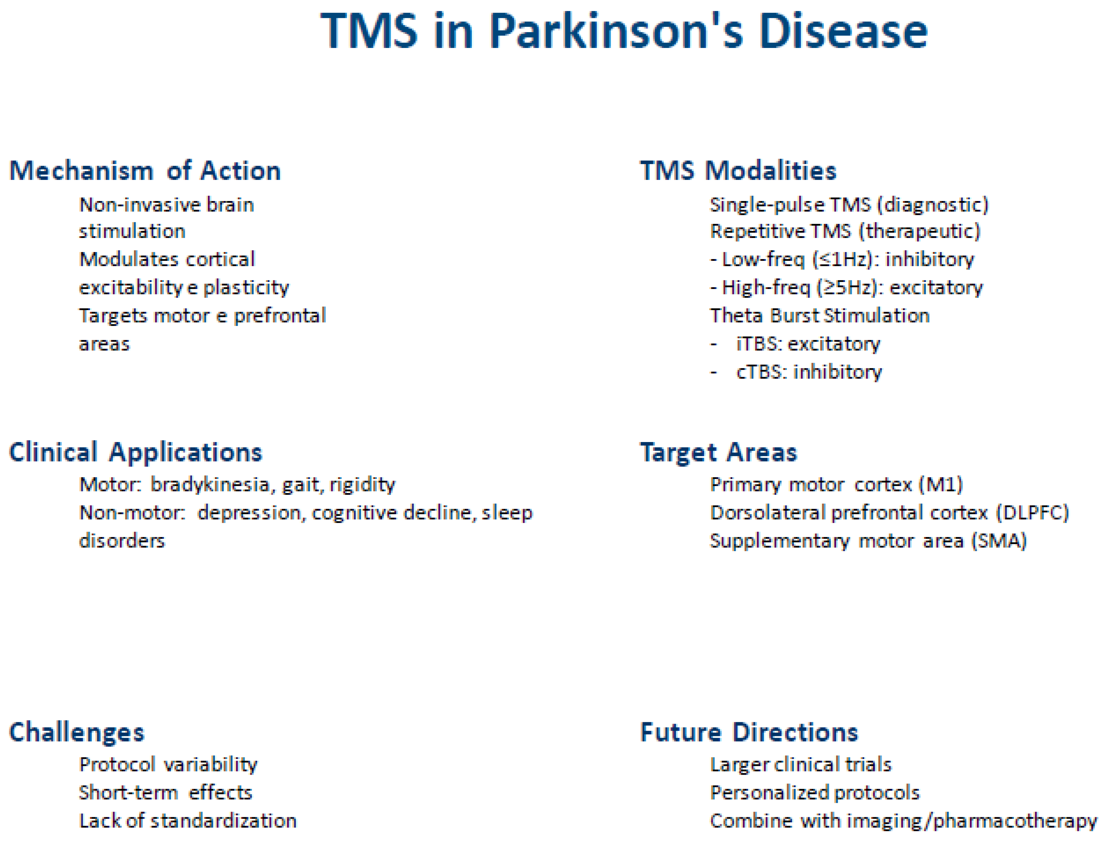
While TMS appears generally safe and well-tolerated, further large-scale randomized controlled trials are needed to confirm its clinical efficacy, optimize stimulation parameters, and assess long-term effects. Moreover, combining TMS with neuroimaging, electrophysiological monitoring, and pharmacological therapies may enhance our understanding of its mechanisms and support the development of individualized treatment strategies. Figure 1 provides a schematic summary of the existing evidence concerning the diagnostic and therapeutic application of different TMS modalities in PD.
Figure 1.
In conclusion, TMS represents a valuable investigational and therapeutic tool in PD management, but its routine clinical application requires more robust and consistent evidence. Continued interdisciplinary research is essential to unlock its full potential in improving the quality of life for patients with PD and other movement disorders.
Author Contributions
Conceptualization, M.C. and G.L.; methodology, R.F. and F.F.; software, A.Q.; validation, R.F., and M.Z.; formal analysis, F.F.; investigation, M.P.M. and A.P.; resources, M.P.; data curation, R.B.; writing—original draft preparation, M.C., M.P., and G.L.; writing—review and editing, M.P.M. and A.P.; visualization, A.Q.; supervision, R.B.; project administration, M.Z.; funding acquisition: N/A. All authors have read and agreed to the published version of the manuscript.
Funding
This research received no external funding.
Institutional Review Board Statement
Not applicable.
Informed Consent Statement
Not applicable.
Data Availability Statement
No new data were created or analyzed in this study. Data sharing is not applicable to this article.
Conflicts of Interest
The authors declare no conflicts of interest.
References
- Barker, A.T.; Jalinous, R.; Freeston, I.L. Non-Invasive Magnetic Stimulation of Human Motor Cortex. Lancet Lond. Engl. 1985, 1, 1106–1107. [Google Scholar] [CrossRef]
- Bella, R.; Lanza, G.; Cantone, M.; Giuffrida, S.; Puglisi, V.; Vinciguerra, L.; Pennisi, M.; Ricceri, R.; D’Agate, C.C.; Malaguarnera, G.; et al. Effect of a Gluten-Free Diet on Cortical Excitability in Adults with Celiac Disease. PloS One 2015, 10, e0129218. [Google Scholar] [CrossRef]
- Lanza, G.; Fisicaro, F.; Dubbioso, R.; Ranieri, F.; Chistyakov, A.V.; Cantone, M.; Pennisi, M.; Grasso, A.A.; Bella, R.; Di Lazzaro, V. A Comprehensive Review of Transcranial Magnetic Stimulation in Secondary Dementia. Front. Aging Neurosci. 2022, 14, 995000. [Google Scholar] [CrossRef] [PubMed]
- Pennisi, M.; Lanza, G.; Cantone, M.; Ricceri, R.; Ferri, R.; D’Agate, C.C.; Pennisi, G.; Di Lazzaro, V.; Bella, R. Cortical Involvement in Celiac Disease before and after Long-Term Gluten-Free Diet: A Transcranial Magnetic Stimulation Study. PloS One 2017, 12, e0177560. [Google Scholar] [CrossRef]
- Vucic, S.; Stanley Chen, K.-H.; Kiernan, M.C.; Hallett, M.; Benninger, D.H.; Di Lazzaro, V.; Rossini, P.M.; Benussi, A.; Berardelli, A.; Currà, A.; et al. Clinical Diagnostic Utility of Transcranial Magnetic Stimulation in Neurological Disorders. Updated Report of an IFCN Committee. Clin. Neurophysiol. Off. J. Int. Fed. Clin. Neurophysiol. 2023, 150, 131–175. [Google Scholar] [CrossRef]
- Lefaucheur, J.-P. Transcranial Magnetic Stimulation. Handb. Clin. Neurol. 2019, 160, 559–580. [Google Scholar] [CrossRef]
- Rossini, P.M.; Burke, D.; Chen, R.; Cohen, L.G.; Daskalakis, Z.; Di Iorio, R.; Di Lazzaro, V.; Ferreri, F.; Fitzgerald, P.B.; George, M.S.; et al. Non-Invasive Electrical and Magnetic Stimulation of the Brain, Spinal Cord, Roots and Peripheral Nerves: Basic Principles and Procedures for Routine Clinical and Research Application. An Updated Report from an I.F.C.N. Committee. Clin. Neurophysiol. Off. J. Int. Fed. Clin. Neurophysiol. 2015, 126, 1071–1107. [Google Scholar] [CrossRef] [PubMed]
- Rossini, P.M.; Rossi, S. Transcranial Magnetic Stimulation: Diagnostic, Therapeutic, and Research Potential. Neurology 2007, 68, 484–488. [Google Scholar] [CrossRef]
- Chen, R. Studies of Human Motor Physiology with Transcranial Magnetic Stimulation. Muscle Nerve. Suppl. 2000, 9, S26–32. [Google Scholar] [CrossRef] [PubMed]
- Udupa, K.; Chen, R. Central Motor Conduction Time. Handb. Clin. Neurol. 2013, 116, 375–386. [Google Scholar] [CrossRef]
- Mills, K.R. Magnetic Brain Stimulation: A Review after 10 Years Experience. Electroencephalogr. Clin. Neurophysiol. Suppl. 1999, 49, 239–244. [Google Scholar]
- Chen, R.; Lozano, A.M.; Ashby, P. Mechanism of the Silent Period Following Transcranial Magnetic Stimulation. Evidence from Epidural Recordings. Exp. Brain Res. 1999, 128, 539–542. [Google Scholar] [CrossRef] [PubMed]
- Cantello, R.; Gianelli, M.; Civardi, C.; Mutani, R. Magnetic Brain Stimulation: The Silent Period after the Motor Evoked Potential. Neurology 1992, 42, 1951–1959. [Google Scholar] [CrossRef]
- Siebner, H.R.; Dressnandt, J.; Auer, C.; Conrad, B. Continuous Intrathecal Baclofen Infusions Induced a Marked Increase of the Transcranially Evoked Silent Period in a Patient with Generalized Dystonia. Muscle Nerve 1998, 21, 1209–1212. [Google Scholar] [CrossRef]
- Kobayashi, M.; Pascual-Leone, A. Transcranial Magnetic Stimulation in Neurology. Lancet Neurol. 2003, 2, 145–156. [Google Scholar] [CrossRef] [PubMed]
- Pennisi, G.; Bella, R.; Lanza, G. Motor Cortex Plasticity in Subcortical Ischemic Vascular Dementia: What Can TMS Say? Clin. Neurophysiol. Off. J. Int. Fed. Clin. Neurophysiol. 2015, 126, 851–852. [Google Scholar] [CrossRef]
- Kujirai, T.; Caramia, M.D.; Rothwell, J.C.; Day, B.L.; Thompson, P.D.; Ferbert, A.; Wroe, S.; Asselman, P.; Marsden, C.D. Corticocortical Inhibition in Human Motor Cortex. J. Physiol. 1993, 471, 501–519. [Google Scholar] [CrossRef]
- Ziemann, U.; Rothwell, J.C.; Ridding, M.C. Interaction between Intracortical Inhibition and Facilitation in Human Motor Cortex. J. Physiol. 1996, 496 Pt 3, 873–881. [Google Scholar] [CrossRef]
- Di Lazzaro, V.; Pilato, F.; Dileone, M.; Ranieri, F.; Ricci, V.; Profice, P.; Bria, P.; Tonali, P.A.; Ziemann, U. GABAA Receptor Subtype Specific Enhancement of Inhibition in Human Motor Cortex. J. Physiol. 2006, 575, 721–726. [Google Scholar] [CrossRef]
- Ziemann, U. TMS and Drugs. Clin. Neurophysiol. Off. J. Int. Fed. Clin. Neurophysiol. 2004, 115, 1717–1729. [Google Scholar] [CrossRef]
- Valls-Solé, J.; Pascual-Leone, A.; Wassermann, E.M.; Hallett, M. Human Motor Evoked Responses to Paired Transcranial Magnetic Stimuli. Electroencephalogr. Clin. Neurophysiol. 1992, 85, 355–364. [Google Scholar] [CrossRef] [PubMed]
- Wassermann, E.M.; Samii, A.; Mercuri, B.; Ikoma, K.; Oddo, D.; Grill, S.E.; Hallett, M. Responses to Paired Transcranial Magnetic Stimuli in Resting, Active, and Recently Activated Muscles. Exp. Brain Res. 1996, 109, 158–163. [Google Scholar] [CrossRef]
- McDonnell, M.N.; Orekhov, Y.; Ziemann, U. The Role of GABA(B) Receptors in Intracortical Inhibition in the Human Motor Cortex. Exp. Brain Res. 2006, 173, 86–93. [Google Scholar] [CrossRef]
- Tokimura, H.; Di Lazzaro, V.; Tokimura, Y.; Oliviero, A.; Profice, P.; Insola, A.; Mazzone, P.; Tonali, P.; Rothwell, J.C. Short Latency Inhibition of Human Hand Motor Cortex by Somatosensory Input from the Hand. J. Physiol. 2000, 523, 503–513. [Google Scholar] [CrossRef] [PubMed]
- Di Lazzaro, V.; Oliviero, A.; Profice, P.; Pennisi, M.A.; Di Giovanni, S.; Zito, G.; Tonali, P.; Rothwell, J.C. Muscarinic Receptor Blockade Has Differential Effects on the Excitability of Intracortical Circuits in the Human Motor Cortex. Exp. Brain Res. 2000, 135, 455–461. [Google Scholar] [CrossRef] [PubMed]
- Di Lazzaro, V.; Oliviero, A.; Pilato, F.; Saturno, E.; Dileone, M.; Marra, C.; Ghirlanda, S.; Ranieri, F.; Gainotti, G.; Tonali, P. Neurophysiological Predictors of Long Term Response to AChE Inhibitors in AD Patients. J. Neurol. Neurosurg. Psychiatry 2005, 76, 1064–1069. [Google Scholar] [CrossRef]
- Dubbioso, R.; Esposito, M.; Peluso, S.; Iodice, R.; De Michele, G.; Santoro, L.; Manganelli, F. Disruption of GABA(A)-Mediated Intracortical Inhibition in Patients with Chorea-Acanthocytosis. Neurosci. Lett. 2017, 654, 107–110. [Google Scholar] [CrossRef]
- R, D.; F, M.; Hr, S.; D.L., V. Fast Intracortical Sensory-Motor Integration: A Window Into the Pathophysiology of Parkinson’s Disease. Front. Hum. Neurosci. 2019, 13. [Google Scholar] [CrossRef]
- Martorana, A.; Mori, F.; Esposito, Z.; Kusayanagi, H.; Monteleone, F.; Codecà, C.; Sancesario, G.; Bernardi, G.; Koch, G. Dopamine Modulates Cholinergic Cortical Excitability in Alzheimer’s Disease Patients. Neuropsychopharmacol. Off. Publ. Am. Coll. Neuropsychopharmacol. 2009, 34, 2323–2328. [Google Scholar] [CrossRef]
- Sailer, A.; Molnar, G.F.; Paradiso, G.; Gunraj, C.A.; Lang, A.E.; Chen, R. Short and Long Latency Afferent Inhibition in Parkinson’s Disease. Brain J. Neurol. 2003, 126, 1883–1894. [Google Scholar] [CrossRef]
- Dubbioso, R.; Manganelli, F.; Siebner, H.R.; Di Lazzaro, V. Fast Intracortical Sensory-Motor Integration: A Window Into the Pathophysiology of Parkinson’s Disease. Front. Hum. Neurosci. 2019, 13, 111. [Google Scholar] [CrossRef]
- Carson, R.G.; Kennedy, N.C. Modulation of Human Corticospinal Excitability by Paired Associative Stimulation. Front. Hum. Neurosci. 2013, 7, 823. [Google Scholar] [CrossRef] [PubMed]
- Li, C.; Huang, Y.; Bai, Y.; Tsai, S.; Su, T.; Cheng, C. Critical Role of Glutamatergic and GABAergic Neurotransmission in the Central Mechanisms of Theta-burst Stimulation. Hum. Brain Mapp. 2019, 40, 2001–2009. [Google Scholar] [CrossRef]
- Matsumoto, H.; Ugawa, Y. Quadripulse Stimulation (QPS). Exp. Brain Res. 2020, 238, 1619–1625. [Google Scholar] [CrossRef]
- Yger, P.; Gilson, M. Models of Metaplasticity: A Review of Concepts. Front. Comput. Neurosci. 2015, 9, 138. [Google Scholar] [CrossRef]
- Kirkovski, M.; Donaldson, P.H.; Do, M.; Speranza, B.E.; Albein-Urios, N.; Oberman, L.M.; Enticott, P.G. A Systematic Review of the Neurobiological Effects of Theta-Burst Stimulation (TBS) as Measured Using Functional Magnetic Resonance Imaging (fMRI). Brain Struct. Funct. 2023, 228, 717–749. [Google Scholar] [CrossRef] [PubMed]
- George, M.S.; Lisanby, S.H.; Avery, D.; McDonald, W.M.; Durkalski, V.; Pavlicova, M.; Anderson, B.; Nahas, Z.; Bulow, P.; Zarkowski, P.; et al. Daily Left Prefrontal Transcranial Magnetic Stimulation Therapy for Major Depressive Disorder: A Sham-Controlled Randomized Trial. Arch. Gen. Psychiatry 2010, 67, 507–516. [Google Scholar] [CrossRef]
- Bella, R.; Ferri, R.; Cantone, M.; Pennisi, M.; Lanza, G.; Malaguarnera, G.; Spampinato, C.; Giordano, D.; Raggi, A.; Pennisi, G. Motor Cortex Excitability in Vascular Depression. Int. J. Psychophysiol. Off. J. Int. Organ. Psychophysiol. 2011, 82, 248–253. [Google Scholar] [CrossRef] [PubMed]
- Bella, R.; Ferri, R.; Lanza, G.; Cantone, M.; Pennisi, M.; Puglisi, V.; Vinciguerra, L.; Spampinato, C.; Mazza, T.; Malaguarnera, G.; et al. TMS Follow-up Study in Patients with Vascular Cognitive Impairment-No Dementia. Neurosci. Lett. 2013, 534, 155–159. [Google Scholar] [CrossRef]
- Nicoletti, V.G.; Fisicaro, F.; Aguglia, E.; Bella, R.; Calcagno, D.; Cantone, M.; Concerto, C.; Ferri, R.; Mineo, L.; Pennisi, G.; et al. Challenging the Pleiotropic Effects of Repetitive Transcranial Magnetic Stimulation in Geriatric Depression: A Multimodal Case Series Study. Biomedicines 2023, 11, 958. [Google Scholar] [CrossRef]
- Lefaucheur, J.-P.; André-Obadia, N.; Antal, A.; Ayache, S.S.; Baeken, C.; Benninger, D.H.; Cantello, R.M.; Cincotta, M.; de Carvalho, M.; De Ridder, D.; et al. Evidence-Based Guidelines on the Therapeutic Use of Repetitive Transcranial Magnetic Stimulation (rTMS). Clin. Neurophysiol. Off. J. Int. Fed. Clin. Neurophysiol. 2014, 125, 2150–2206. [Google Scholar] [CrossRef]
- Lefaucheur, J.-P.; Aleman, A.; Baeken, C.; Benninger, D.H.; Brunelin, J.; Di Lazzaro, V.; Filipović, S.R.; Grefkes, C.; Hasan, A.; Hummel, F.C.; et al. Evidence-Based Guidelines on the Therapeutic Use of Repetitive Transcranial Magnetic Stimulation (rTMS): An Update (2014–2018). Clin. Neurophysiol. 2020, 131, 474–528. [Google Scholar] [CrossRef]
- Wang, Y.; Liu, J.; Hui, Y.; Wu, Z.; Wang, L.; Wu, X.; Bai, Y.; Zhang, Q.; Li, L. Dose and Time-Dependence of Acute Intermittent Theta-Burst Stimulation on Hippocampus-Dependent Memory in Parkinsonian Rats. Front. Neurosci. 2023, 17, 1124819. [Google Scholar] [CrossRef]
- Zeljkovic Jovanovic, M.; Stanojevic, J.; Stevanovic, I.; Stekic, A.; Bolland, S.J.; Jasnic, N.; Ninkovic, M.; Zaric Kontic, M.; Ilic, T.V.; Rodger, J.; et al. Intermittent Theta Burst Stimulation Improves Motor and Behavioral Dysfunction through Modulation of NMDA Receptor Subunit Composition in Experimental Model of Parkinson’s Disease. Cells 2023, 12, 1525. [Google Scholar] [CrossRef]
- Hamada, M.; Terao, Y.; Hanajima, R.; Shirota, Y.; Nakatani-Enomoto, S.; Furubayashi, T.; Matsumoto, H.; Ugawa, Y. Bidirectional Long-Term Motor Cortical Plasticity and Metaplasticity Induced by Quadripulse Transcranial Magnetic Stimulation. J. Physiol. 2008, 586, 3927–3947. [Google Scholar] [CrossRef] [PubMed]
- Bloem, B.R.; Okun, M.S.; Klein, C. Parkinson’s Disease. The Lancet 2021, 397, 2284–2303. [Google Scholar] [CrossRef]
- Dickson, D.W. Neuropathology of Parkinson Disease. Parkinsonism Relat. Disord. 2018, 46 Suppl 1, S30–S33. [Google Scholar] [CrossRef]
- Marsili, L.; Rizzo, G.; Colosimo, C. Diagnostic Criteria for Parkinson’s Disease: From James Parkinson to the Concept of Prodromal Disease. Front. Neurol. 2018, 9, 156. [Google Scholar] [CrossRef]
- Jankovic, J.; McDermott, M.; Carter, J.; Gauthier, S.; Goetz, C.; Golbe, L.; Huber, S.; Koller, W.; Olanow, C.; Shoulson, I. Variable Expression of Parkinson’s Disease: A Base-Line Analysis of the DATATOP Cohort. The Parkinson Study Group. Neurology 1990, 40, 1529–1534. [Google Scholar] [CrossRef] [PubMed]
- Sauerbier, A.; Jenner, P.; Todorova, A.; Chaudhuri, K.R. Non Motor Subtypes and Parkinson’s Disease. Parkinsonism Relat. Disord. 2016, 22 Suppl 1, S41–46. [Google Scholar] [CrossRef] [PubMed]
- Shakya, S.; Prevett, J.; Hu, X.; Xiao, R. Characterization of Parkinson’s Disease Subtypes and Related Attributes. Front. Neurol. 2022, 13. [Google Scholar] [CrossRef]
- Fisicaro, F.; Lanza, G.; Cantone, M.; Ferri, R.; Pennisi, G.; Nicoletti, A.; Zappia, M.; Bella, R.; Pennisi, M. Clinical and Electrophysiological Hints to TMS in De Novo Patients with Parkinson’s Disease and Progressive Supranuclear Palsy. J. Pers. Med. 2020, 10, 274. [Google Scholar] [CrossRef] [PubMed]
- Lanza, G.; Cosentino, F.I.I.; Lanuzza, B.; Tripodi, M.; Aricò, D.; Figorilli, M.; Puligheddu, M.; Fisicaro, F.; Bella, R.; Ferri, R.; et al. Reduced Intracortical Facilitation to TMS in Both Isolated REM Sleep Behavior Disorder (RBD) and Early Parkinson’s Disease with RBD. J. Clin. Med. 2022, 11, 2291. [Google Scholar] [CrossRef] [PubMed]
- MacKinnon, C.D.; Gilley, E.A.; Weis-McNulty, A.; Simuni, T. Pathways Mediating Abnormal Intracortical Inhibition in Parkinson’s Disease. Ann. Neurol. 2005, 58, 516–524. [Google Scholar] [CrossRef]
- Ni, Z.; Bahl, N.; Gunraj, C.A.; Mazzella, F.; Chen, R. Increased Motor Cortical Facilitation and Decreased Inhibition in Parkinson Disease. Neurology 2013, 80, 1746–1753. [Google Scholar] [CrossRef]
- Ridding, M.C.; Inzelberg, R.; Rothwell, J.C. Changes in Excitability of Motor Cortical Circuitry in Patients with Parkinson’s Disease. Ann. Neurol. 1995, 37, 181–188. [Google Scholar] [CrossRef] [PubMed]
- Tremblay, F.; Tremblay, L.E. Cortico-Motor Excitability of the Lower Limb Motor Representation: A Comparative Study in Parkinson’s Disease and Healthy Controls. Clin. Neurophysiol. Off. J. Int. Fed. Clin. Neurophysiol. 2002, 113, 2006–2012. [Google Scholar] [CrossRef]
- Valls-Solé, J.; Pascual-Leone, A.; Brasil-Neto, J.P.; Cammarota, A.; McShane, L.; Hallett, M. Abnormal Facilitation of the Response to Transcranial Magnetic Stimulation in Patients with Parkinson’s Disease. Neurology 1994, 44, 735–741. [Google Scholar] [CrossRef]
- Cantello, R.; Gianelli, M.; Bettucci, D.; Civardi, C.; De Angelis, M.S.; Mutani, R. Parkinson’s Disease Rigidity: Magnetic Motor Evoked Potentials in a Small Hand Muscle. Neurology 1991, 41, 1449–1456. [Google Scholar] [CrossRef]
- Kolmancic, K.; Perellón-Alfonso, R.; Pirtosek, Z.; Rothwell, J.C.; Bhatia, K.; Kojovic, M. Sex Differences in Parkinson’s Disease: A Transcranial Magnetic Stimulation Study. Mov. Disord. Off. J. Mov. Disord. Soc. 2019, 34, 1873–1881. [Google Scholar] [CrossRef]
- Spagnolo, F.; Coppi, E.; Chieffo, R.; Straffi, L.; Fichera, M.; Nuara, A.; Gonzalez-Rosa, J.; Martinelli, V.; Comi, G.; Volontè, M.A.; et al. Interhemispheric Balance in Parkinson’s Disease: A Transcranial Magnetic Stimulation Study. Brain Stimulat. 2013, 6, 892–897. [Google Scholar] [CrossRef]
- Bologna, M.; Guerra, A.; Paparella, G.; Giordo, L.; Alunni Fegatelli, D.; Vestri, A.R.; Rothwell, J.C.; Berardelli, A. Neurophysiological Correlates of Bradykinesia in Parkinson’s Disease. Brain J. Neurol. 2018, 141, 2432–2444. [Google Scholar] [CrossRef] [PubMed]
- Ellaway, P.H.; Davey, N.J.; Maskill, D.W.; Dick, J.P. The Relation between Bradykinesia and Excitability of the Motor Cortex Assessed Using Transcranial Magnetic Stimulation in Normal and Parkinsonian Subjects. Electroencephalogr. Clin. Neurophysiol. 1995, 97, 169–178. [Google Scholar] [CrossRef] [PubMed]
- Khedr, E.M.; Lefaucheur, J.-P.; Hasan, A.M.; Osama, K. Are There Differences in Cortical Excitability between Akinetic-Rigid and Tremor-Dominant Subtypes of Parkinson’s Disease? Neurophysiol. Clin. Clin. Neurophysiol. 2021, 51, 443–453. [Google Scholar] [CrossRef]
- Topka, M.; Schneider, M.; Zrenner, C.; Belardinelli, P.; Ziemann, U.; Weiss, D. Motor Cortex Excitability Is Reduced during Freezing of Upper Limb Movement in Parkinson’s Disease. NPJ Park. Dis. 2022, 8, 161. [Google Scholar] [CrossRef]
- Sciacca, G.; Mostile, G.; Disilvestro, I.; Donzuso, G.; Nicoletti, A.; Zappia, M. Long-Duration Response to Levodopa, Motor Learning, and Neuroplasticity in Early Parkinson’s Disease. Mov. Disord. Off. J. Mov. Disord. Soc. 2023, 38, 626–635. [Google Scholar] [CrossRef] [PubMed]
- Perretti, A.; De Rosa, A.; Marcantonio, L.; Iodice, V.; Estraneo, A.; Manganelli, F.; Garavaglia, B.; Filla, A.; Santoro, L.; De Michele, G. Neurophysiological Evaluation of Motor Corticospinal Pathways by TMS in Idiopathic Early-Onset Parkinson’s Disease. Clin. Neurophysiol. Off. J. Int. Fed. Clin. Neurophysiol. 2011, 122, 546–549. [Google Scholar] [CrossRef]
- Soysal, A.; Sobe, I.; Atay, T.; Sen, A.; Arpaci, B. Effect of Therapy on Motor Cortical Excitability in Parkinson’s Disease. Can. J. Neurol. Sci. J. Can. Sci. Neurol. 2008, 35, 166–172. [Google Scholar] [CrossRef] [PubMed]
- Diószeghy, P.; Hidasi, E.; Mechler, F. Study of Central Motor Functions Using Magnetic Stimulation in Parkinson’s Disease. Electromyogr. Clin. Neurophysiol. 1999, 39, 101–105. [Google Scholar]
- Mochizuki, Y.; Oishi, M.; Hara, M.; Takasu, T. Central Motor Conduction Time in Parkinson’s Disease. J. Clin. Neurosci. Off. J. Neurosurg. Soc. Australas. 1999, 6, 17–19. [Google Scholar] [CrossRef]
- De Rosa, A.; Volpe, G.; Marcantonio, L.; Santoro, L.; Brice, A.; Filla, A.; Perretti, A.; De Michele, G. Neurophysiological Evidence of Corticospinal Tract Abnormality in Patients with Parkin Mutations. J. Neurol. 2006, 253, 275–279. [Google Scholar] [CrossRef] [PubMed]
- Schneider, S.A.; Talelli, P.; Cheeran, B.J.; Khan, N.L.; Wood, N.W.; Rothwell, J.C.; Bhatia, K.P. Motor Cortical Physiology in Patients and Asymptomatic Carriers of Parkin Gene Mutations. Mov. Disord. Off. J. Mov. Disord. Soc. 2008, 23, 1812–1819. [Google Scholar] [CrossRef] [PubMed]
- Cantello, R.; Tarletti, R.; Civardi, C. Transcranial Magnetic Stimulation and Parkinson’s Disease. Brain Res. Brain Res. Rev. 2002, 38, 309–327. [Google Scholar] [CrossRef]
- Priori, A.; Berardelli, A.; Inghilleri, M.; Accornero, N.; Manfredi, M. Motor Cortical Inhibition and the Dopaminergic System. Pharmacological Changes in the Silent Period after Transcranial Brain Stimulation in Normal Subjects, Patients with Parkinson’s Disease and Drug-Induced Parkinsonism. Brain J. Neurol. 1994, 117 Pt 2, 317–323. [Google Scholar] [CrossRef]
- Chen, R.; Garg, R.R.; Lozano, A.M.; Lang, A.E. Effects of Internal Globus Pallidus Stimulation on Motor Cortex Excitability. Neurology 2001, 56, 716–723. [Google Scholar] [CrossRef]
- Ammann, C.; Dileone, M.; Pagge, C.; Catanzaro, V.; Mata-Marín, D.; Hernández-Fernández, F.; Monje, M.H.G.; Sánchez-Ferro, Á.; Fernández-Rodríguez, B.; Gasca-Salas, C.; et al. Cortical Disinhibition in Parkinson’s Disease. Brain J. Neurol. 2020, 143, 3408–3421. [Google Scholar] [CrossRef] [PubMed]
- Guerra, A.; Colella, D.; Giangrosso, M.; Cannavacciuolo, A.; Paparella, G.; Fabbrini, G.; Suppa, A.; Berardelli, A.; Bologna, M. Driving Motor Cortex Oscillations Modulates Bradykinesia in Parkinson’s Disease. Brain J. Neurol. 2022, 145, 224–236. [Google Scholar] [CrossRef]
- Cunic, D.; Roshan, L.; Khan, F.I.; Lozano, A.M.; Lang, A.E.; Chen, R. Effects of Subthalamic Nucleus Stimulation on Motor Cortex Excitability in Parkinson’s Disease. Neurology 2002, 58, 1665–1672. [Google Scholar] [CrossRef]
- Kojovic, M.; Kassavetis, P.; Bologna, M.; Pareés, I.; Rubio-Agusti, I.; Berardelli, A.; Edwards, M.J.; Rothwell, J.C.; Bhatia, K.P. Transcranial Magnetic Stimulation Follow-up Study in Early Parkinson’s Disease: A Decline in Compensation with Disease Progression? Mov. Disord. Off. J. Mov. Disord. Soc. 2015, 30, 1098–1106. [Google Scholar] [CrossRef]
- Chu, J.; Wagle-Shukla, A.; Gunraj, C.; Lang, A.E.; Chen, R. Impaired Presynaptic Inhibition in the Motor Cortex in Parkinson Disease. Neurology 2009, 72, 842–849. [Google Scholar] [CrossRef]
- Pierantozzi, M.; Palmieri, M.G.; Marciani, M.G.; Bernardi, G.; Giacomini, P.; Stanzione, P. Effect of Apomorphine on Cortical Inhibition in Parkinson’s Disease Patients: A Transcranial Magnetic Stimulation Study. Exp. Brain Res. 2001, 141, 52–62. [Google Scholar] [CrossRef]
- Valzania, F.; Strafella, A.P.; Quatrale, R.; Santangelo, M.; Tropeani, A.; Lucchi, D.; Tassinari, C.A.; De Grandis, D. Motor Evoked Responses to Paired Cortical Magnetic Stimulation in Parkinson’s Disease. Electroencephalogr. Clin. Neurophysiol. 1997, 105, 37–43. [Google Scholar] [CrossRef]
- Berardelli, A.; Inghilleri, M.; Priori, A.; Marchetti, P.; Curra, A.; Rona, S.; Manfredi, M. Inhibitory Cortical Phenomena Studied with the Technique of Transcranial Stimulation. Electroencephalogr. Clin. Neurophysiol. Suppl. 1996, 46, 343–349. [Google Scholar]
- Bares, M.; Kanovský, P.; Klajblová, H.; Rektor, I. Intracortical Inhibition and Facilitation Are Impaired in Patients with Early Parkinson’s Disease: A Paired TMS Study. Eur. J. Neurol. 2003, 10, 385–389. [Google Scholar] [CrossRef] [PubMed]
- Strafella, A.P.; Valzania, F.; Nassetti, S.A.; Tropeani, A.; Bisulli, A.; Santangelo, M.; Tassinari, C.A. Effects of Chronic Levodopa and Pergolide Treatment on Cortical Excitability in Patients with Parkinson’s Disease: A Transcranial Magnetic Stimulation Study. Clin. Neurophysiol. Off. J. Int. Fed. Clin. Neurophysiol. 2000, 111, 1198–1202. [Google Scholar] [CrossRef] [PubMed]
- Berardelli, A.; Rona, S.; Inghilleri, M.; Manfredi, M. Cortical Inhibition in Parkinson’s Disease. A Study with Paired Magnetic Stimulation. Brain J. Neurol. 1996, 119 Pt 1, 71–77. [Google Scholar] [CrossRef] [PubMed]
- Shirota, Y.; Ohminami, S.; Tsutsumi, R.; Terao, Y.; Ugawa, Y.; Tsuji, S.; Hanajima, R. Increased Facilitation of the Primary Motor Cortex in de Novo Parkinson’s Disease. Parkinsonism Relat. Disord. 2019, 66, 125–129. [Google Scholar] [CrossRef]
- Guerra, A.; Suppa, A.; D’Onofrio, V.; Di Stasio, F.; Asci, F.; Fabbrini, G.; Berardelli, A. Abnormal Cortical Facilitation and L-Dopa-Induced Dyskinesia in Parkinson’s Disease. Brain Stimulat. 2019, 12, 1517–1525. [Google Scholar] [CrossRef]
- Celebi, O.; Temuçin, C.M.; Elibol, B.; Saka, E. Short Latency Afferent Inhibition in Parkinson’s Disease Patients with Dementia. Mov. Disord. Off. J. Mov. Disord. Soc. 2012, 27, 1052–1055. [Google Scholar] [CrossRef]
- Nardone, R.; Bergmann, J.; Brigo, F.; Christova, M.; Kunz, A.; Seidl, M.; Tezzon, F.; Trinka, E.; Golaszewski, S. Functional Evaluation of Central Cholinergic Circuits in Patients with Parkinson’s Disease and REM Sleep Behavior Disorder: A TMS Study. J. Neural Transm. Vienna Austria 1996 2013, 120, 413–422. [Google Scholar] [CrossRef]
- Rochester, L.; Yarnall, A.J.; Baker, M.R.; David, R.V.; Lord, S.; Galna, B.; Burn, D.J. Cholinergic Dysfunction Contributes to Gait Disturbance in Early Parkinson’s Disease. Brain J. Neurol. 2012, 135, 2779–2788. [Google Scholar] [CrossRef]
- Yarnall, A.J.; Rochester, L.; Baker, M.R.; David, R.; Khoo, T.K.; Duncan, G.W.; Galna, B.; Burn, D.J. Short Latency Afferent Inhibition: A Biomarker for Mild Cognitive Impairment in Parkinson’s Disease? Mov. Disord. Off. J. Mov. Disord. Soc. 2013. [Google Scholar] [CrossRef]
- Manganelli, F.; Vitale, C.; Santangelo, G.; Pisciotta, C.; Iodice, R.; Cozzolino, A.; Dubbioso, R.; Picillo, M.; Barone, P.; Santoro, L. Functional Involvement of Central Cholinergic Circuits and Visual Hallucinations in Parkinson’s Disease. Brain J. Neurol. 2009, 132, 2350–2355. [Google Scholar] [CrossRef]
- Lee, K.D.; Koo, J.H.; Song, S.H.; Jo, K.D.; Lee, M.K.; Jang, W. Central Cholinergic Dysfunction Could Be Associated with Oropharyngeal Dysphagia in Early Parkinson’s Disease. J. Neural Transm. Vienna Austria 1996 2015, 122, 1553–1561. [Google Scholar] [CrossRef] [PubMed]
- Oh, E.; Park, J.; Youn, J.; Kim, J.S.; Park, S.; Jang, W. Olfactory Dysfunction in Early Parkinson’s Disease Is Associated with Short Latency Afferent Inhibition Reflecting Central Cholinergic Dysfunction. Clin. Neurophysiol. Off. J. Int. Fed. Clin. Neurophysiol. 2017, 128, 1061–1068. [Google Scholar] [CrossRef]
- Versace, V.; Langthaler, P.B.; Sebastianelli, L.; Höller, Y.; Brigo, F.; Orioli, A.; Saltuari, L.; Nardone, R. Impaired Cholinergic Transmission in Patients with Parkinson’s Disease and Olfactory Dysfunction. J. Neurol. Sci. 2017, 377, 55–61. [Google Scholar] [CrossRef] [PubMed]
- Pelosin, E.; Ogliastro, C.; Lagravinese, G.; Bonassi, G.; Mirelman, A.; Hausdorff, J.M.; Abbruzzese, G.; Avanzino, L. Attentional Control of Gait and Falls: Is Cholinergic Dysfunction a Common Substrate in the Elderly and Parkinson’s Disease? Front. Aging Neurosci. 2016, 8, 104. [Google Scholar] [CrossRef] [PubMed]
- Bharti, K.; Suppa, A.; Tommasin, S.; Zampogna, A.; Pietracupa, S.; Berardelli, A.; Pantano, P. Neuroimaging Advances in Parkinson’s Disease with Freezing of Gait: A Systematic Review. NeuroImage Clin. 2019, 24, 102059. [Google Scholar] [CrossRef]
- Picillo, M.; Dubbioso, R.; Iodice, R.; Iavarone, A.; Pisciotta, C.; Spina, E.; Santoro, L.; Barone, P.; Amboni, M.; Manganelli, F. Short-Latency Afferent Inhibition in Patients with Parkinson’s Disease and Freezing of Gait. J. Neural Transm. Vienna Austria 1996 2015, 122, 1533–1540. [Google Scholar] [CrossRef]
- Wang, L.; Ji, M.; Sun, H.; Gan, C.; Zhang, H.; Cao, X.; Yuan, Y.; Zhang, K. Reduced Short-Latency Afferent Inhibition in Parkinson’s Disease Patients with L-Dopa-Unresponsive Freezing of Gait. J. Park. Dis. 2022, 12, 2507–2518. [Google Scholar] [CrossRef]
- Kačar, A.; Filipović, S.R.; Kresojević, N.; Milanović, S.D.; Ljubisavljević, M.; Kostić, V.S.; Rothwell, J.C. History of Exposure to Dopaminergic Medication Does Not Affect Motor Cortex Plasticity and Excitability in Parkinson’s Disease. Clin. Neurophysiol. Off. J. Int. Fed. Clin. Neurophysiol. 2013, 124, 697–707. [Google Scholar] [CrossRef]
- Morgante, F.; Espay, A.J.; Gunraj, C.; Lang, A.E.; Chen, R. Motor Cortex Plasticity in Parkinson’s Disease and Levodopa-Induced Dyskinesias. Brain J. Neurol. 2006, 129, 1059–1069. [Google Scholar] [CrossRef]
- Ueki, Y.; Mima, T.; Kotb, M.A.; Sawada, H.; Saiki, H.; Ikeda, A.; Begum, T.; Reza, F.; Nagamine, T.; Fukuyama, H. Altered Plasticity of the Human Motor Cortex in Parkinson’s Disease. Ann. Neurol. 2006, 59, 60–71. [Google Scholar] [CrossRef]
- Eggers, C.; Fink, G.R.; Nowak, D.A. Theta Burst Stimulation over the Primary Motor Cortex Does Not Induce Cortical Plasticity in Parkinson’s Disease. J. Neurol. 2010, 257, 1669–1674. [Google Scholar] [CrossRef]
- Suppa, A.; Marsili, L.; Belvisi, D.; Conte, A.; Iezzi, E.; Modugno, N.; Fabbrini, G.; Berardelli, A. Lack of LTP-like Plasticity in Primary Motor Cortex in Parkinson’s Disease. Exp. Neurol. 2011, 227, 296–301. [Google Scholar] [CrossRef]
- Zamir, O.; Gunraj, C.; Ni, Z.; Mazzella, F.; Chen, R. Effects of Theta Burst Stimulation on Motor Cortex Excitability in Parkinson’s Disease. Clin. Neurophysiol. Off. J. Int. Fed. Clin. Neurophysiol. 2012, 123, 815–821. [Google Scholar] [CrossRef]
- Kishore, A.; Joseph, T.; Velayudhan, B.; Popa, T.; Meunier, S. Early, Severe and Bilateral Loss of LTP and LTD-like Plasticity in Motor Cortex (M1) in de Novo Parkinson’s Disease. Clin. Neurophysiol. Off. J. Int. Fed. Clin. Neurophysiol. 2012, 123, 822–828. [Google Scholar] [CrossRef] [PubMed]
- Kojovic, M.; Bologna, M.; Kassavetis, P.; Murase, N.; Palomar, F.J.; Berardelli, A.; Rothwell, J.C.; Edwards, M.J.; Bhatia, K.P. Functional Reorganization of Sensorimotor Cortex in Early Parkinson Disease. Neurology 2012, 78, 1441–1448. [Google Scholar] [CrossRef] [PubMed]
- Kishore, A.; James, P.; Krishnan, S.; Yahia-Cherif, L.; Meunier, S.; Popa, T. Motor Cortex Plasticity Can Indicate Vulnerability to Motor Fluctuation and High L-DOPA Need in Drug-Naïve Parkinson’s Disease. Parkinsonism Relat. Disord. 2017, 35, 55–62. [Google Scholar] [CrossRef] [PubMed]
- Belvisi, D.; Fabbrini, A.; De Bartolo, M.I.; Costanzo, M.; Manzo, N.; Fabbrini, G.; Defazio, G.; Conte, A.; Berardelli, A. The Pathophysiological Correlates of Parkinson’s Disease Clinical Subtypes. Mov. Disord. Off. J. Mov. Disord. Soc. 2021, 36, 370–379. [Google Scholar] [CrossRef]
- Latorre, A.; Rocchi, L.; Berardelli, A.; Bhatia, K.P.; Rothwell, J.C. The Use of Transcranial Magnetic Stimulation as a Treatment for Movement Disorders: A Critical Review. Mov. Disord. Off. J. Mov. Disord. Soc. 2019, 34, 769–782. [Google Scholar] [CrossRef]
- Strafella, A.P.; Paus, T.; Fraraccio, M.; Dagher, A. Striatal Dopamine Release Induced by Repetitive Transcranial Magnetic Stimulation of the Human Motor Cortex. Brain J. Neurol. 2003, 126, 2609–2615. [Google Scholar] [CrossRef]
- Kim, J.Y.; Chung, E.J.; Lee, W.Y.; Shin, H.Y.; Lee, G.H.; Choe, Y.-S.; Choi, Y.; Kim, B.J. Therapeutic Effect of Repetitive Transcranial Magnetic Stimulation in Parkinson’s Disease: Analysis of [11C] Raclopride PET Study. Mov. Disord. Off. J. Mov. Disord. Soc. 2008, 23, 207–211. [Google Scholar] [CrossRef] [PubMed]
- Filipović, S.R.; Rothwell, J.C.; van de Warrenburg, B.P.; Bhatia, K. Repetitive Transcranial Magnetic Stimulation for Levodopa-Induced Dyskinesias in Parkinson’s Disease. Mov. Disord. Off. J. Mov. Disord. Soc. 2009, 24, 246–253. [Google Scholar] [CrossRef] [PubMed]
- Chang, W.H.; Kim, M.S.; Park, E.; Cho, J.W.; Youn, J.; Kim, Y.K.; Kim, Y.-H. Effect of Dual-Mode and Dual-Site Noninvasive Brain Stimulation on Freezing of Gait in Patients With Parkinson Disease. Arch. Phys. Med. Rehabil. 2017, 98, 1283–1290. [Google Scholar] [CrossRef]
- González-García, N.; Armony, J.L.; Soto, J.; Trejo, D.; Alegría, M.A.; Drucker-Colín, R. Effects of rTMS on Parkinson’s Disease: A Longitudinal fMRI Study. J. Neurol. 2011, 258, 1268–1280. [Google Scholar] [CrossRef] [PubMed]
- Maruo, T.; Hosomi, K.; Shimokawa, T.; Kishima, H.; Oshino, S.; Morris, S.; Kageyama, Y.; Yokoe, M.; Yoshimine, T.; Saitoh, Y. High-Frequency Repetitive Transcranial Magnetic Stimulation over the Primary Foot Motor Area in Parkinson’s Disease. Brain Stimulat. 2013, 6, 884–891. [Google Scholar] [CrossRef]
- Potvin-Desrochers, A.; Paquette, C. Potential Non-Invasive Brain Stimulation Targets to Alleviate Freezing of Gait in Parkinson’s Disease. Neuroscience 2021, 468, 366–376. [Google Scholar] [CrossRef]
- Rektorova, I.; Sedlackova, S.; Telecka, S.; Hlubocky, A.; Rektor, I. Repetitive Transcranial Stimulation for Freezing of Gait in Parkinson’s Disease. Mov. Disord. Off. J. Mov. Disord. Soc. 2007, 22, 1518–1519. [Google Scholar] [CrossRef]
- Rossi, S.; Ferro, M.; Cincotta, M.; Ulivelli, M.; Bartalini, S.; Miniussi, C.; Giovannelli, F.; Passero, S. A Real Electro-Magnetic Placebo (REMP) Device for Sham Transcranial Magnetic Stimulation (TMS). Clin. Neurophysiol. Off. J. Int. Fed. Clin. Neurophysiol. 2007, 118, 709–716. [Google Scholar] [CrossRef]
- Brys, M.; Fox, M.D.; Agarwal, S.; Biagioni, M.; Dacpano, G.; Kumar, P.; Pirraglia, E.; Chen, R.; Wu, A.; Fernandez, H.; et al. Multifocal Repetitive TMS for Motor and Mood Symptoms of Parkinson Disease: A Randomized Trial. Neurology 2016, 87, 1907–1915. [Google Scholar] [CrossRef]
- Makkos, A.; Pál, E.; Aschermann, Z.; Janszky, J.; Balázs, É.; Takács, K.; Karádi, K.; Komoly, S.; Kovács, N. High-Frequency Repetitive Transcranial Magnetic Stimulation Can Improve Depression in Parkinson’s Disease: A Randomized, Double-Blind, Placebo-Controlled Study. Neuropsychobiology 2016, 73, 169–177. [Google Scholar] [CrossRef]
- Chou, Y.; Hickey, P.T.; Sundman, M.; Song, A.W.; Chen, N. Effects of Repetitive Transcranial Magnetic Stimulation on Motor Symptoms in Parkinson Disease: A Systematic Review and Meta-Analysis. JAMA Neurol. 2015, 72, 432–440. [Google Scholar] [CrossRef]
- Li, S.; Jiao, R.; Zhou, X.; Chen, S. Motor Recovery and Antidepressant Effects of Repetitive Transcranial Magnetic Stimulation on Parkinson Disease. Medicine (Baltimore) 2020, 99, e19642. [Google Scholar] [CrossRef]
- Zanjani, A.; Zakzanis, K.K.; Daskalakis, Z.J.; Chen, R. Repetitive Transcranial Magnetic Stimulation of the Primary Motor Cortex in the Treatment of Motor Signs in Parkinson’s Disease: A Quantitative Review of the Literature. Mov. Disord. Off. J. Mov. Disord. Soc. 2015, 30, 750–758. [Google Scholar] [CrossRef]
- Deng, S.; Dong, Z.; Pan, L.; Liu, Y.; Ye, Z.; Qin, L.; Liu, Q.; Qin, C. Effects of Repetitive Transcranial Magnetic Stimulation on Gait Disorders and Cognitive Dysfunction in Parkinson’s Disease: A Systematic Review with Meta-Analysis. Brain Behav. 2022, 12, e2697. [Google Scholar] [CrossRef] [PubMed]
- Chung, C.L.-H.; Mak, M.K.-Y.; Hallett, M. Transcranial Magnetic Stimulation Promotes Gait Training in Parkinson Disease. Ann. Neurol. 2020, 88, 933–945. [Google Scholar] [CrossRef]
- Hamada, M.; Ugawa, Y.; Tsuji, S. Effectiveness of rTMS on Parkinson’s Disease Study Group, Japan High-Frequency rTMS over the Supplementary Motor Area for Treatment of Parkinson’s Disease. Mov. Disord. Off. J. Mov. Disord. Soc. 2008, 23, 1524–1531. [Google Scholar] [CrossRef] [PubMed]
- Shirota, Y.; Ohtsu, H.; Hamada, M.; Enomoto, H.; Ugawa, Y. Research Committee on rTMS Treatment of Parkinson’s Disease Supplementary Motor Area Stimulation for Parkinson Disease: A Randomized Controlled Study. Neurology 2013, 80, 1400–1405. [Google Scholar] [CrossRef]
- Sayın, S.; Cakmur, R.; Yener, G.G.; Yaka, E.; Uğurel, B.; Uzunel, F. Low-Frequency Repetitive Transcranial Magnetic Stimulation for Dyskinesia and Motor Performance in Parkinson’s Disease. J. Clin. Neurosci. Off. J. Neurosurg. Soc. Australas. 2014, 21, 1373–1376. [Google Scholar] [CrossRef] [PubMed]
- Kim, S.J.; Paeng, S.H.; Kang, S.Y. Stimulation in Supplementary Motor Area Versus Motor Cortex for Freezing of Gait in Parkinson’s Disease. J. Clin. Neurol. Seoul Korea 2018, 14, 320–326. [Google Scholar] [CrossRef] [PubMed]
- Bhat, P.; Kumaran, S.S.; Goyal, V.; Srivastava, A.K.; Behari, M. Effect of rTMS at SMA on Task-Based Connectivity in PD. Behav. Brain Res. 2023, 452, 114602. [Google Scholar] [CrossRef]
- Chi, S.; Wen, X.; Yu, Y.; Wang, G.; Zhang, J.; Xue, C.; Zhang, X.; Wang, Z.; Gesang, M.; Chen, J.; et al. Sensorimotor Network Connectivity Correlates with Motor Improvement after Repetitive Transcranial Magnetic Stimulation in Patients with Parkinson’s Disease. Parkinsonism Relat. Disord. 2023, 106, 105218. [Google Scholar] [CrossRef]
- Lefaivre, S.C.; Brown, M.J.N.; Almeida, Q.J. Cerebellar Involvement in Parkinson’s Disease Resting Tremor. Cerebellum Ataxias 2016, 3, 13. [Google Scholar] [CrossRef]
- Minks, E.; Mareček, R.; Pavlík, T.; Ovesná, P.; Bareš, M. Is the Cerebellum a Potential Target for Stimulation in Parkinson’s Disease? Results of 1-Hz rTMS on Upper Limb Motor Tasks. Cerebellum Lond. Engl. 2011, 10, 804–811. [Google Scholar] [CrossRef]
- Fregni, F.; Santos, C.M.; Myczkowski, M.L.; Rigolino, R.; Gallucci-Neto, J.; Barbosa, E.R.; Valente, K.D.; Pascual-Leone, A.; Marcolin, M.A. Repetitive Transcranial Magnetic Stimulation Is as Effective as Fluoxetine in the Treatment of Depression in Patients with Parkinson’s Disease. J. Neurol. Neurosurg. Psychiatry 2004, 75, 1171–1174. [Google Scholar] [CrossRef]
- Pal, E.; Nagy, F.; Aschermann, Z.; Balazs, E.; Kovacs, N. The Impact of Left Prefrontal Repetitive Transcranial Magnetic Stimulation on Depression in Parkinson’s Disease: A Randomized, Double-Blind, Placebo-Controlled Study. Mov. Disord. Off. J. Mov. Disord. Soc. 2010, 25, 2311–2317. [Google Scholar] [CrossRef] [PubMed]
- Hai-Jiao, W.; Ge, T.; Li-Na, Z.; Deng, C.; Da, X.; Shan-Shan, C.; Liu, L. The Efficacy of Repetitive Transcranial Magnetic Stimulation for Parkinson Disease Patients with Depression. Int. J. Neurosci. 2020, 130, 19–27. [Google Scholar] [CrossRef]
- Randver, R. Repetitive Transcranial Magnetic Stimulation of the Dorsolateral Prefrontal Cortex to Alleviate Depression and Cognitive Impairment Associated with Parkinson’s Disease: A Review and Clinical Implications. J. Neurol. Sci. 2018, 393, 88–99. [Google Scholar] [CrossRef]
- Zheng, H.-B.; Liu, B.; Shen, J.; Xie, F.; Ji, Q.-M.; Zhu, X.-Y. Non-Invasive Brain Stimulation for Treating Psychiatric Symptoms in Parkinson’s Disease: A Systematic Review and Meta-Analysis. J. Clin. Neurosci. Off. J. Neurosurg. Soc. Australas. 2022, 106, 83–90. [Google Scholar] [CrossRef] [PubMed]
- Yokoe, M.; Mano, T.; Maruo, T.; Hosomi, K.; Shimokawa, T.; Kishima, H.; Oshino, S.; Morris, S.; Kageyama, Y.; Goto, Y.; et al. The Optimal Stimulation Site for High-Frequency Repetitive Transcranial Magnetic Stimulation in Parkinson’s Disease: A Double-Blind Crossover Pilot Study. J. Clin. Neurosci. Off. J. Neurosurg. Soc. Australas. 2018, 47, 72–78. [Google Scholar] [CrossRef]
- Cacace, F.; Mineo, D.; Viscomi, M.T.; Latagliata, E.C.; Mancini, M.; Sasso, V.; Vannelli, A.; Pascucci, T.; Pendolino, V.; Marcello, E.; et al. Intermittent Theta-Burst Stimulation Rescues Dopamine-Dependent Corticostriatal Synaptic Plasticity and Motor Behavior in Experimental Parkinsonism: Possible Role of Glial Activity. Mov. Disord. Off. J. Mov. Disord. Soc. 2017, 32, 1035–1046. [Google Scholar] [CrossRef]
- Zeljkovic Jovanovic, M.; Stanojevic, J.; Stevanovic, I.; Ninkovic, M.; Nedeljkovic, N.; Dragic, M. Sustained Systemic Antioxidative Effects of Intermittent Theta Burst Stimulation beyond Neurodegeneration: Implications in Therapy in 6-Hydroxydopamine Model of Parkinson’s Disease. Antioxid. Basel Switz. 2024, 13, 218. [Google Scholar] [CrossRef] [PubMed]
- Jovanovic, M.Z.; Stanojevic, J.; Stevanovic, I.; Ninkovic, M.; Ilic, T.V.; Nedeljkovic, N.; Dragic, M. Prolonged Intermittent Theta Burst Stimulation Restores the Balance between A2AR- and A1R-Mediated Adenosine Signaling in the 6-Hydroxidopamine Model of Parkinson’s Disease. Neural Regen. Res. 2025, 20, 2053–2067. [Google Scholar] [CrossRef]
- Jin, Z.-H.; Wang, Y.-X.; Meng, D.-T.; Qin, Y.; Duan, Y.-N.; Fang, J.-P.; Wang, R.-D.; Liu, Y.-J.; Liu, C.; Wang, P.; et al. Intermittent Theta-Burst Stimulation Combined with Physical Therapy as an Optimal Rehabilitation in Parkinson’s Disease: Study Protocol for a Randomised, Double-Blind, Controlled Trial. Trials 2023, 24, 410. [Google Scholar] [CrossRef] [PubMed]
- Rashid-López, R.; Macías-García, P.; Sánchez-Fernández, F.L.; Cano-Cano, F.; Sarrias-Arrabal, E.; Sanmartino, F.; Méndez-Bértolo, C.; Lozano-Soto, E.; Gutiérrez-Cortés, R.; González-Moraleda, Á.; et al. Neuroimaging and Serum Biomarkers of Neurodegeneration and Neuroplasticity in Parkinson’s Disease Patients Treated by Intermittent Theta-Burst Stimulation over the Bilateral Primary Motor Area: A Randomized, Double-Blind, Sham-Controlled, Crossover Trial Study. Front. Aging Neurosci. 2023, 15, 1258315. [Google Scholar] [CrossRef]
- Cheng, B.; Zhu, T.; Zhao, W.; Sun, L.; Shen, Y.; Xiao, W.; Zhang, S. Effect of Theta Burst Stimulation-Patterned rTMS on Motor and Nonmotor Dysfunction of Parkinson’s Disease: A Systematic Review and Metaanalysis. Front. Neurol. 2021, 12, 762100. [Google Scholar] [CrossRef]
- Chung, S.W.; Hill, A.T.; Rogasch, N.C.; Hoy, K.E.; Fitzgerald, P.B. Use of Theta-Burst Stimulation in Changing Excitability of Motor Cortex: A Systematic Review and Meta-Analysis. Neurosci. Biobehav. Rev. 2016, 63, 43–64. [Google Scholar] [CrossRef]
- Vanbellingen, T.; Wapp, M.; Stegmayer, K.; Bertschi, M.; Abela, E.; Kübel, S.; Nyffeler, T.; Müri, R.; Walther, S.; Nef, T.; et al. Theta Burst Stimulation over Premotor Cortex in Parkinson’s Disease: An Explorative Study on Manual Dexterity. J. Neural Transm. Vienna Austria 1996 2016, 123, 1387–1393. [Google Scholar] [CrossRef]
- Brugger, F.; Wegener, R.; Baty, F.; Walch, J.; Krüger, M.T.; Hägele-Link, S.; Bohlhalter, S.; Kägi, G. Facilitatory rTMS over the Supplementary Motor Cortex Impedes Gait Performance in Parkinson Patients with Freezing of Gait. Brain Sci. 2021, 11, 321. [Google Scholar] [CrossRef]
- Benninger, D.H.; Berman, B.D.; Houdayer, E.; Pal, N.; Luckenbaugh, D.A.; Schneider, L.; Miranda, S.; Hallett, M. Intermittent Theta-Burst Transcranial Magnetic Stimulation for Treatment of Parkinson Disease. Neurology 2011, 76, 601–609. [Google Scholar] [CrossRef]
- Degardin, A.; Devos, D.; Defebvre, L.; Destée, A.; Plomhause, L.; Derambure, P.; Devanne, H. Effect of Intermittent Theta-Burst Stimulation on Akinesia and Sensorimotor Integration in Patients with Parkinson’s Disease. Eur. J. Neurosci. 2012, 36, 2669–2678. [Google Scholar] [CrossRef]
- Eggers, C.; Günther, M.; Rothwell, J.; Timmermann, L.; Ruge, D. Theta Burst Stimulation over the Supplementary Motor Area in Parkinson’s Disease. J. Neurol. 2015, 262, 357–364. [Google Scholar] [CrossRef]
- Ji, G.-J.; Liu, T.; Li, Y.; Liu, P.; Sun, J.; Chen, X.; Tian, Y.; Chen, X.; Dahmani, L.; Liu, H.; et al. Structural Correlates Underlying Accelerated Magnetic Stimulation in Parkinson’s Disease. Hum. Brain Mapp. 2021, 42, 1670–1681. [Google Scholar] [CrossRef]
- Rashid-López, R.; Macías-García, P.; Cruz-Gómez, Á.J.; Sánchez-Fernández, F.L.; Cano-Cano, F.; Sanmartino, F.; Sarrias-Arrabal, E.; Lozano-Soto, E.; Méndez-Bértolo, C.; López-Sosa, F.; et al. Bilateral Primary Motor Area Intermittent Theta-Burst Stimulation May Alleviate Gait and Postural Disturbances in Parkinson’s Disease Patients by Astrocytic Modulation, Caudate Volume Changes, and Increased Functional Neuroplasticity. Parkinsonism Relat. Disord. 2024, 123, 106074. [Google Scholar] [CrossRef]
- Pastore-Wapp, M.; Kaufmann, B.C.; Nyffeler, T.; Wapp, S.; Bohlhalter, S.; Vanbellingen, T. Feasibility of a Combined Intermittent Theta-Burst Stimulation and Video Game-Based Dexterity Training in Parkinson’s Disease. J. Neuroengineering Rehabil. 2023, 20, 2. [Google Scholar] [CrossRef]
- Li, P.; Luo, N.; Sun, S.; Li, Y.; Shen, D.; Zhu, X.; Zhou, L.; Zhou, H.; Liu, J. Neuroprotective Effects of Intermittent Theta Burst Stimulation in Parkinson’s Disease (NET-PD): A Study Protocol for a Delayed-Start Randomized Double-Blind Sham-Controlled Trial. J. Clin. Med. 2022, 11, 4972. [Google Scholar] [CrossRef] [PubMed]
- Koch, G.; Brusa, L.; Carrillo, F.; Lo Gerfo, E.; Torriero, S.; Oliveri, M.; Mir, P.; Caltagirone, C.; Stanzione, P. Cerebellar Magnetic Stimulation Decreases Levodopa-Induced Dyskinesias in Parkinson Disease. Neurology 2009, 73, 113–119. [Google Scholar] [CrossRef] [PubMed]
- Sanna, A.; Follesa, P.; Puligheddu, M.; Cannas, A.; Serra, M.; Pisu, M.G.; Dagostino, S.; Solla, P.; Tacconi, P.; Marrosu, F. Cerebellar Continuous Theta Burst Stimulation Reduces Levodopa-Induced Dyskinesias and Decreases Serum BDNF Levels. Neurosci. Lett. 2020, 716, 134653. [Google Scholar] [CrossRef] [PubMed]
- Enomoto, H.; Terao, Y.; Kadowaki, S.; Nakamura, K.; Moriya, A.; Nakatani-Enomoto, S.; Kobayashi, S.; Yoshihara, A.; Hanajima, R.; Ugawa, Y. Effects of L-Dopa and Pramipexole on Plasticity Induced by QPS in Human Motor Cortex. J. Neural Transm. Vienna Austria 1996 2015, 122, 1253–1261. [Google Scholar] [CrossRef]
- Enomoto, H.; Ugawa, Y. Abnormal plasticity and drug effect in Parkinson’s disease. Rinsho Shinkeigaku 2012, 52, 1204–1206. [Google Scholar] [CrossRef]
- Koch, G.; Brusa, L.; Caltagirone, C.; Peppe, A.; Oliveri, M.; Stanzione, P.; Centonze, D. rTMS of Supplementary Motor Area Modulates Therapy-Induced Dyskinesias in Parkinson Disease. Neurology 2005, 65, 623–625. [Google Scholar] [CrossRef]
- Bagnato, S.; Agostino, R.; Modugno, N.; Quartarone, A.; Berardelli, A. Plasticity of the Motor Cortex in Parkinson’s Disease Patients on and off Therapy. Mov. Disord. Off. J. Mov. Disord. Soc. 2006, 21, 639–645. [Google Scholar] [CrossRef]
- Kishore, A.; Popa, T.; Balachandran, A.; Chandran, S.; Pradeep, S.; Backer, F.; Krishnan, S.; Meunier, S. Cerebellar Sensory Processing Alterations Impact Motor Cortical Plasticity in Parkinson’s Disease: Clues from Dyskinetic Patients. Cereb. Cortex N. Y. N 1991 2014, 24, 2055–2067. [Google Scholar] [CrossRef]
- Kishore, A.; Popa, T.; James, P.; Yahia-Cherif, L.; Backer, F.; Varughese Chacko, L.; Govind, P.; Pradeep, S.; Meunier, S. Age-Related Decline in the Responsiveness of Motor Cortex to Plastic Forces Reverses with Levodopa or Cerebellar Stimulation. Neurobiol. Aging 2014, 35, 2541–2551. [Google Scholar] [CrossRef]
- Enomoto, H.; Terao, Y.; Kadowaki, S.; Nakamura, K.; Moriya, A.; Nakatani-Enomoto, S.; Kobayashi, S.; Yoshihara, A.; Hanajima, R.; Ugawa, Y. Effects of L-Dopa and Pramipexole on Plasticity Induced by QPS in Human Motor Cortex. J. Neural Transm. Vienna Austria 1996 2015, 122, 1253–1261. [Google Scholar] [CrossRef]
- Suppa, A.; Bologna, M.; Conte, A.; Berardelli, A.; Fabbrini, G. The Effect of L-Dopa in Parkinson’s Disease as Revealed by Neurophysiological Studies of Motor and Sensory Functions. Expert Rev. Neurother. 2017, 17, 181–192. [Google Scholar] [CrossRef] [PubMed]
- Moriyasu, S.; Shimizu, T.; Honda, M.; Ugawa, Y.; Hanajima, R. Motor Cortical Plasticity and Its Correlation with Motor Symptoms in Parkinson’s Disease. eNeurologicalSci 2022, 29, 100422. [Google Scholar] [CrossRef] [PubMed]
- Kolmančič, K.; Zupančič, N.K.; Trošt, M.; Flisar, D.; Kramberger, M.G.; Pirtošek, Z.; Kojović, M. Continuous Dopaminergic Stimulation Improves Cortical Maladaptive Changes in Advanced Parkinson’s Disease. Mov. Disord. Off. J. Mov. Disord. Soc. 2022, 37, 1465–1473. [Google Scholar] [CrossRef]
- Antelmi, E.; Lanza, G.; Mogavero, M.P.; Mingolla, G.P.; Plazzi, G.; Ferini-Strambi, L.; Ferri, R.; Tinazzi, M. Intersection of Sleep Disorders and Parkinson Disease: Unveiling the Bidirectional Relationship. Mov. Disord. Clin. Pract. 2025, 12, 11–20. [Google Scholar] [CrossRef] [PubMed]
- Donzuso, G.; Cicero, C.E.; Giuliano, L.; Squillaci, R.; Luca, A.; Palmucci, S.; Basile, A.; Lanza, G.; Ferri, R.; Zappia, M.; et al. Neuroanatomical Findings in Isolated REM Sleep Behavior Disorder and Early Parkinson’s Disease: A Voxel-Based Morphometry Study. Brain Imaging Behav. 2024, 18, 83–91. [Google Scholar] [CrossRef]
- Joza, S.; Hu, M.T.; Jung, K.-Y.; Kunz, D.; Stefani, A.; Dušek, P.; Terzaghi, M.; Arnaldi, D.; Videnovic, A.; Schiess, M.C.; et al. Progression of Clinical Markers in Prodromal Parkinson’s Disease and Dementia with Lewy Bodies: A Multicentre Study. Brain J. Neurol. 2023, 146, 3258–3272. [Google Scholar] [CrossRef]
- Figorilli, M.; Meloni, M.; Lanza, G.; Casaglia, E.; Lecca, R.; Saibene, F.L.; Congiu, P.; Puligheddu, M. Considering REM Sleep Behavior Disorder in the Management of Parkinson’s Disease. Nat. Sci. Sleep 2023, 15, 333–352. [Google Scholar] [CrossRef]
- Figorilli, M.; Meloni, F.; Lecca, R.; Tamburrino, L.; Mascia, M.G.; Cocco, V.; Meloni, M.; Marques, A.R.; Vidal, T.; Congiu, P.; et al. Severity of REM Sleep without Atonia Correlates with Measures of Cognitive Impairment and Depressive Symptoms in REM Sleep Behaviour Disorder. J. Sleep Res. 2023, 32, e13880. [Google Scholar] [CrossRef] [PubMed]
- Mogavero, M.P.; Ferri, R.; Marelli, S.; Lanza, G.; Terzaghi, M.; Castelnuovo, A.; DelRosso, L.M.; Schenck, C.H.; Ferini-Strambi, L. Polysomnographic Features Associated with Clonazepam and Melatonin Treatment in Isolated REM Sleep Behavior Disorder: Time for New Therapeutic Approaches? CNS Neurosci. Ther. 2024, 30, e14569. [Google Scholar] [CrossRef] [PubMed]
- Figorilli, M.; Lanza, G.; Congiu, P.; Lecca, R.; Casaglia, E.; Mogavero, M.P.; Puligheddu, M.; Ferri, R. Neurophysiological Aspects of REM Sleep Behavior Disorder (RBD): A Narrative Review. Brain Sci. 2021, 11, 1588. [Google Scholar] [CrossRef] [PubMed]
- Nardone, R.; Bergmann, J.; Kunz, A.; Christova, M.; Brigo, F.; Tezzon, F.; Trinka, E.; Golaszewski, S. Cortical Afferent Inhibition Is Reduced in Patients with Idiopathic REM Sleep Behavior Disorder and Cognitive Impairment: A TMS Study. Sleep Med. 2012, 13, 919–925. [Google Scholar] [CrossRef]
- Lanza, G.; Aricò, D.; Lanuzza, B.; Cosentino, F.I.I.; Tripodi, M.; Giardina, F.; Bella, R.; Puligheddu, M.; Pennisi, G.; Ferri, R.; et al. Facilitatory/Inhibitory Intracortical Imbalance in REM Sleep Behavior Disorder: Early Electrophysiological Marker of Neurodegeneration? Sleep 2020, 43, zsz242. [Google Scholar] [CrossRef]
- Peever, J.; Luppi, P.-H.; Montplaisir, J. Breakdown in REM Sleep Circuitry Underlies REM Sleep Behavior Disorder. Trends Neurosci. 2014, 37, 279–288. [Google Scholar] [CrossRef]
- Bhattacharya, A.; Kamble, N.; Yadav, R.; Stezin, A.; Pal, P.K. Abnormal Intracortical Functions in Parkinson’s Disease with Rapid Eye Movement Sleep Behaviour Disorder. Can. J. Neurol. Sci. J. Can. Sci. Neurol. 2022, 49, 672–677. [Google Scholar] [CrossRef]
- Khedr, E.M.; Ahmed, G.K.; Korayem, M.A.; Elamary, S.A.S.H.; El-Kholy, M.M.; Haridy, N.A. Short-Term Therapeutic Effect of Repetitive Transcranial Magnetic Stimulations of Sleep Disorders in Parkinson’s Disease: A Randomized Clinical Trial (Pilot Study). Brain Sci. 2024, 14, 556. [Google Scholar] [CrossRef]
- Arias-Carrion, O.; Ortega-Robles, E.; Ortuno-Sahagun, D.; Ramirez-Bermudez, J.; Hamid, A.; Shalash, A. Sleep-Related Disorders in Parkinson’s Disease: Mechanisms, Diagnosis, and Therapeutic Approaches. CNS Neurol. Disord. Drug Targets 2025, 24, 132–143. [Google Scholar] [CrossRef]
- van Dijk, K.D.; Møst, E.I.S.; Van Someren, E.J.W.; Berendse, H.W.; van der Werf, Y.D. Beneficial Effect of Transcranial Magnetic Stimulation on Sleep in Parkinson’s Disease. Mov. Disord. Off. J. Mov. Disord. Soc. 2009, 24, 878–884. [Google Scholar] [CrossRef] [PubMed]
- Lanza, G.; Fisicaro, F.; Cantone, M.; Pennisi, M.; Cosentino, F.I.I.; Lanuzza, B.; Tripodi, M.; Bella, R.; Paulus, W.; Ferri, R. Repetitive Transcranial Magnetic Stimulation in Primary Sleep Disorders. Sleep Med. Rev. 2023, 67, 101735. [Google Scholar] [CrossRef]
- Jellinger, K.A. Pathobiology of Cognitive Impairment in Parkinson Disease: Challenges and Outlooks. Int. J. Mol. Sci. 2023, 25, 498. [Google Scholar] [CrossRef]
- Nardone, R.; Florio, I.; Lochner, P.; Tezzon, F. Cholinergic Cortical Circuits in Parkinson’s Disease and in Progressive Supranuclear Palsy: A Transcranial Magnetic Stimulation Study. Exp. Brain Res. Exp. Hirnforsch. Expérimentation Cérébrale 2005, 163, 128–131. [Google Scholar] [CrossRef] [PubMed]
- Nardone, R.; Bergmann, J.; Brigo, F.; Christova, M.; Kunz, A.; Seidl, M.; Tezzon, F.; Trinka, E.; Golaszewski, S. Functional Evaluation of Central Cholinergic Circuits in Patients with Parkinson’s Disease and REM Sleep Behavior Disorder: A TMS Study. J. Neural Transm. Vienna Austria 1996 2013, 120, 413–422. [Google Scholar] [CrossRef]
- Khedr, E.M.; Mohamed, K.O.; Ali, A.M.; Hasan, A.M. The Effect of Repetitive Transcranial Magnetic Stimulation on Cognitive Impairment in Parkinson’s Disease with Dementia: Pilot Study. Restor. Neurol. Neurosci. 2020, 38, 55–66. [Google Scholar] [CrossRef]
- Trung, J.; Hanganu, A.; Jobert, S.; Degroot, C.; Mejia-Constain, B.; Kibreab, M.; Bruneau, M.-A.; Lafontaine, A.-L.; Strafella, A.; Monchi, O. Transcranial Magnetic Stimulation Improves Cognition over Time in Parkinson’s Disease. Parkinsonism Relat. Disord. 2019, 66, 3–8. [Google Scholar] [CrossRef] [PubMed]
- Buard, I.; Sciacca, D.M.; Martin, C.S.; Rogers, S.; Sillau, S.H.; Greher, M.R.; Chen, R.; Kluger, B.M. Transcranial Magnetic Stimulation Does Not Improve Mild Cognitive Impairment in Parkinson’s Disease. Mov. Disord. Off. J. Mov. Disord. Soc. 2018, 33, 489–491. [Google Scholar] [CrossRef]
- Nagao, S.; Yokota, O.; Nanba, R.; Takata, H.; Haraguchi, T.; Ishizu, H.; Ikeda, C.; Takeda, N.; Oshima, E.; Sakane, K.; et al. Progressive Supranuclear Palsy Presenting as Primary Lateral Sclerosis but Lacking Parkinsonism, Gaze Palsy, Aphasia, or Dementia. J. Neurol. Sci. 2012, 323, 147–153. [Google Scholar] [CrossRef] [PubMed]
- Kühn, A.A.; Grosse, P.; Holtz, K.; Brown, P.; Meyer, B.-U.; Kupsch, A. Patterns of Abnormal Motor Cortex Excitability in Atypical Parkinsonian Syndromes. Clin. Neurophysiol. Off. J. Int. Fed. Clin. Neurophysiol. 2004, 115, 1786–1795. [Google Scholar] [CrossRef]
- Wittstock, M.; Pohley, I.; Walter, U.; Grossmann, A.; Benecke, R.; Wolters, A. Interhemispheric Inhibition in Different Phenotypes of Progressive Supranuclear Palsy. J. Neural Transm. Vienna Austria 1996 2013, 120, 453–461. [Google Scholar] [CrossRef] [PubMed]
- Wolters, A.; Classen, J.; Kunesch, E.; Grossmann, A.; Benecke, R. Measurements of Transcallosally Mediated Cortical Inhibition for Differentiating Parkinsonian Syndromes. Mov. Disord. Off. J. Mov. Disord. Soc. 2004, 19, 518–528. [Google Scholar] [CrossRef]
- Brusa, L.; Ponzo, V.; Mastropasqua, C.; Picazio, S.; Bonnì, S.; Di Lorenzo, F.; Iani, C.; Stefani, A.; Stanzione, P.; Caltagirone, C.; et al. Theta Burst Stimulation Modulates Cerebellar-Cortical Connectivity in Patients with Progressive Supranuclear Palsy. Brain Stimulat. 2014, 7, 29–35. [Google Scholar] [CrossRef]
- Conte, A.; Belvisi, D.; Bologna, M.; Ottaviani, D.; Fabbrini, G.; Colosimo, C.; Williams, D.R.; Berardelli, A. Abnormal Cortical Synaptic Plasticity in Primary Motor Area in Progressive Supranuclear Palsy. Cereb. Cortex N. Y. N 1991 2012, 22, 693–700. [Google Scholar] [CrossRef]
- Shirota, Y; M, H.; R, H.; Y, T.; H, M.; S, O.; S, T.; Y, U. Cerebellar Dysfunction in Progressive Supranuclear Palsy: A Transcranial Magnetic Stimulation Study. Mov. Disord. Off. J. Mov. Disord. Soc. 2010, 25. [Google Scholar] [CrossRef]
- Dale, M.L.; DeVries, W.H.; Mancini, M.; George, M.S. Cerebellar rTMS for Motor Control in Progressive Supranuclear Palsy. Brain Stimulat. 2019, 12, 1588–1591. [Google Scholar] [CrossRef] [PubMed]
- Fichera, M.; Volonté, M.A.; Comi, G.; Leocani, L. Repetitive Transcranial Magnetic Stimulation with H Coil in Progressive Supranuclear Palsy: A Double-Blind, Placebo-Controlled, Crossover Study (P3.8-001). Neurology 2019, 92, P3.8–001. [Google Scholar] [CrossRef]
- Papp, M.I.; Lantos, P.L. The Distribution of Oligodendroglial Inclusions in Multiple System Atrophy and Its Relevance to Clinical Symptomatology. Brain J. Neurol. 1994, 117 Pt 2, 235–243. [Google Scholar] [CrossRef]
- Abbruzzese, G.; Marchese, R.; Trompetto, C. Sensory and Motor Evoked Potentials in Multiple System Atrophy: A Comparative Study with Parkinson’s Disease. Mov. Disord. Off. J. Mov. Disord. Soc. 1997, 12, 315–321. [Google Scholar] [CrossRef]
- Tabaraud, F.; Hugon, J.; Salle, J.Y.; Boulesteix, J.M.; Rigaud, M.; Vallat, J.M.; Dumas, M. Central Motor Conduction and Glutamate Deshydrogenase: Activity in Olivo-Ponto-Cerebellar Atrophy. Neurophysiol. Clin. Clin. Neurophysiol. 1989, 19, 433–441. [Google Scholar] [CrossRef]
- Abele, M.; Schulz, J.B.; Bürk, K.; Topka, H.; Dichgans, J.; Klockgether, T. Evoked Potentials in Multiple System Atrophy (MSA). Acta Neurol. Scand. 2000, 101, 111–115. [Google Scholar] [CrossRef]
- Eusebio, A.; Azulay, J.-P.; Witjas, T.; Rico, A.; Attarian, S. Assessment of Cortico-Spinal Tract Impairment in Multiple System Atrophy Using Transcranial Magnetic Stimulation. Clin. Neurophysiol. Off. J. Int. Fed. Clin. Neurophysiol. 2007, 118, 815–823. [Google Scholar] [CrossRef] [PubMed]
- Marchese, R.; Trompetto, C.; Buccolieri, A.; Abbruzzese, G. Abnormalities of Motor Cortical Excitability Are Not Correlated with Clinical Features in Atypical Parkinsonism. Mov. Disord. Off. J. Mov. Disord. Soc. 2000, 15, 1210–1214. [Google Scholar] [CrossRef]
- Suppa, A.; Marsili, L.; Di Stasio, F.; Latorre, A.; Parvez, A.K.; Colosimo, C.; Berardelli, A. Primary Motor Cortex Long-Term Plasticity in Multiple System Atrophy. Mov. Disord. Off. J. Mov. Disord. Soc. 2014, 29, 97–104. [Google Scholar] [CrossRef] [PubMed]
- Mascia, M.M.; Valls-Solé, J.; Martí, M.J.; Salazar, G. Sensorimotor Integration in Patients with Parkinsonian Type Multisystem Atrophy. J. Neurol. 2005, 252, 473–481. [Google Scholar] [CrossRef]
- Löscher, W.N.; Stampfer-Kountchev, M.; Sawires, M.; Seppi, K.; Mueller, J.; Szubski, C.; Hirnsperger, K.; Brenneis, C.; Poewe, W.; Wenning, G.K. Abnormal Responses to Repetitive Transcranial Magnetic Stimulation in Multiple System Atrophy. Mov. Disord. Off. J. Mov. Disord. Soc. 2007, 22, 174–178. [Google Scholar] [CrossRef]
- Pan, J.; Mi, T.-M.; Ma, J.-H.; Sun, H.; Chan, P. High-Frequency Repetitive Transcranial Magnetic Stimulation Over the Left Dorsolateral Prefrontal Cortex Shortly Alleviates Fatigue in Patients With Multiple System Atrophy: A Randomized Controlled Trial. Front. Neurol. 2021, 12, 755352. [Google Scholar] [CrossRef]
- Lee, S.E.; Rabinovici, G.D.; Mayo, M.C.; Wilson, S.M.; Seeley, W.W.; DeArmond, S.J.; Huang, E.J.; Trojanowski, J.Q.; Growdon, M.E.; Jang, J.Y.; et al. Clinicopathological Correlations in Corticobasal Degeneration. Ann. Neurol. 2011, 70, 327–340. [Google Scholar] [CrossRef] [PubMed]
- Rankin, K.P.; Mayo, M.C.; Seeley, W.W.; Lee, S.; Rabinovici, G.; Gorno-Tempini, M.L.; Boxer, A.L.; Weiner, M.W.; Trojanowski, J.Q.; DeArmond, S.J.; et al. Behavioral Variant Frontotemporal Dementia with Corticobasal Degeneration Pathology: Phenotypic Comparison to bvFTD with Pick’s Disease. J. Mol. Neurosci. MN 2011, 45, 594–608. [Google Scholar] [CrossRef]
- Fujimoto, K.; Sayama, S.; Shizuma, N.; Kano, M.; Nakano, I. Intracranial inhibitory mechanisms in clinically diagnosed corticobasal degeneration: a study of a silent period followed by transcranial magnetic stimulation. Rinsho Shinkeigaku 2000, 40, 701–706. [Google Scholar]
- Leiguarda, R.C.; Merello, M.; Nouzeilles, M.I.; Balej, J.; Rivero, A.; Nogués, M. Limb-Kinetic Apraxia in Corticobasal Degeneration: Clinical and Kinematic Features. Mov. Disord. Off. J. Mov. Disord. Soc. 2003, 18, 49–59. [Google Scholar] [CrossRef]
- Lu, C.S.; Ikeda, A.; Terada, K.; Mima, T.; Nagamine, T.; Fukuyama, H.; Kohara, N.; Kojima, Y.; Yonekura, Y.; Chen, R.S.; et al. Electrophysiological Studies of Early Stage Corticobasal Degeneration. Mov. Disord. Off. J. Mov. Disord. Soc. 1998, 13, 140–146. [Google Scholar] [CrossRef] [PubMed]
- Alberici, A.; Bonato, C.; Calabria, M.; Agosti, C.; Zanetti, O.; Miniussi, C.; Padovani, A.; Rossini, P.M.; Borroni, B. The Contribution of TMS to Frontotemporal Dementia Variants. Acta Neurol. Scand. 2008, 118, 275–280. [Google Scholar] [CrossRef] [PubMed]
- Trompetto, C.; Buccolieri, A.; Marchese, R.; Marinelli, L.; Michelozzi, G.; Abbruzzese, G. Impairment of Transcallosal Inhibition in Patients with Corticobasal Degeneration. Clin. Neurophysiol. Off. J. Int. Fed. Clin. Neurophysiol. 2003, 114, 2181–2187. [Google Scholar] [CrossRef]
- Frasson, E.; Bertolasi, L.; Bertasi, V.; Fusina, S.; Bartolomei, L.; Vicentini, S.; Rizzuto, N.; Priori, A. Paired Transcranial Magnetic Stimulation for the Early Diagnosis of Corticobasal Degeneration. Clin. Neurophysiol. Off. J. Int. Fed. Clin. Neurophysiol. 2003, 114, 272–278. [Google Scholar] [CrossRef]
- Okuma, Y.; Urabe, T.; Mochizuki, H.; Miwa, H.; Shimo, Y.; Mori, H.; Mizuno, Y. Asymmetric Cortico-Cortical Inhibition in Patients with Progressive Limb-Kinetic Apraxia. Acta Neurol. Scand. 2000, 102, 244–248. [Google Scholar] [CrossRef]
- Lanza, G.; Bella, R.; Giuffrida, S.; Cantone, M.; Pennisi, G.; Spampinato, C.; Giordano, D.; Malaguarnera, G.; Raggi, A.; Pennisi, M. Preserved Transcallosal Inhibition to Transcranial Magnetic Stimulation in Nondemented Elderly Patients with Leukoaraiosis. BioMed Res. Int. 2013, 2013, 351680. [Google Scholar] [CrossRef]
- Valls-Solé, J. Neurophysiological Characterization of Parkinsonian Syndromes. Neurophysiol. Clin. Clin. Neurophysiol. 2000, 30, 352–367. [Google Scholar] [CrossRef] [PubMed]
- McKeith, I.; Cummings, J. Behavioural Changes and Psychological Symptoms in Dementia Disorders. Lancet Neurol. 2005, 4, 735–742. [Google Scholar] [CrossRef]
- Takahashi, R.; Ishii, K.; Shimada, K.; Ohkawa, S.; Nishimura, Y. Hypoperfusion of the Motor Cortex Associated with Parkinsonism in Dementia with Lewy Bodies. J. Neurol. Sci. 2010, 288, 88–91. [Google Scholar] [CrossRef] [PubMed]
- Di Lazzaro, V.; Pilato, F.; Dileone, M.; Saturno, E.; Profice, P.; Marra, C.; Daniele, A.; Ranieri, F.; Quaranta, D.; Gainotti, G.; et al. Functional Evaluation of Cerebral Cortex in Dementia with Lewy Bodies. NeuroImage 2007, 37, 422–429. [Google Scholar] [CrossRef]
- Tiraboschi, P.; Hansen, L.A.; Alford, M.; Sabbagh, M.N.; Schoos, B.; Masliah, E.; Thal, L.J.; Corey-Bloom, J. Cholinergic Dysfunction in Diseases with Lewy Bodies. Neurology 2000, 54, 407–411. [Google Scholar] [CrossRef]
- Marra, C.; Quaranta, D.; Profice, P.; Pilato, F.; Capone, F.; Iodice, F.; Di Lazzaro, V.; Gainotti, G. Central Cholinergic Dysfunction Measured “in Vivo” Correlates with Different Behavioral Disorders in Alzheimer’s Disease and Dementia with Lewy Body. Brain Stimulat. 2012, 5, 533–538. [Google Scholar] [CrossRef]
- Taylor, J.-P.; Firbank, M.; Barnett, N.; Pearce, S.; Livingstone, A.; Mosimann, U.; Eyre, J.; McKeith, I.G.; O’Brien, J.T. Visual Hallucinations in Dementia with Lewy Bodies: Transcranial Magnetic Stimulation Study. Br. J. Psychiatry J. Ment. Sci. 2011, 199, 492–500. [Google Scholar] [CrossRef] [PubMed]
- Petsani, C.; Aloizou, A.-M.; Siokas, V.; Messinis, L.; Peristeri, E.; Bakirtzis, C.; Nasios, G.; Dardiotis, E. Therapeutic Application of rTMS in Atypical Parkinsonian Disorders. Behav. Neurol. 2021, 2021, 3419907. [Google Scholar] [CrossRef]
- Lanza, G.; Papotto, M.; Pennisi, G.; Bella, R.; Ferri, R. Epileptic Seizure as a Precipitating Factor of Vascular Progressive Supranuclear Palsy: A Case Report. J. Stroke Cerebrovasc. Dis. Off. J. Natl. Stroke Assoc. 2014, 23, e379-381. [Google Scholar] [CrossRef] [PubMed]
- Lanza, G.; Bramanti, P.; Cantone, M.; Pennisi, M.; Pennisi, G.; Bella, R. Vascular Cognitive Impairment through the Looking Glass of Transcranial Magnetic Stimulation. Behav. Neurol. 2017, 2017, 1421326. [Google Scholar] [CrossRef]
- Zijlmans, J.C.M.; Katzenschlager, R.; Daniel, S.E.; Lees, A.J.L. The L-Dopa Response in Vascular Parkinsonism. J. Neurol. Neurosurg. Psychiatry 2004, 75, 545–547. [Google Scholar] [CrossRef]
- Liepert, J.; Bär, K.J.; Meske, U.; Weiller, C. Motor Cortex Disinhibition in Alzheimer’s Disease. Clin. Neurophysiol. Off. J. Int. Fed. Clin. Neurophysiol. 2001, 112, 1436–1441. [Google Scholar] [CrossRef]
- Agrawal, A.; Bhattacharya, A.; Kamble, N.; Mailankody, P.; Yadav, R.; Pal, P.K. Cortical Excitability Changes in Patients of Vascular Parkinsonism with Cognitive Impairment. Parkinsonism Relat. Disord. 2023, 116, 105869. [Google Scholar] [CrossRef] [PubMed]
- Agrawal, A.; Bhattacharya, A.; Kamble, N.; Mailankody, P.; Yadav, R.; Pal, P.K. Cortical Excitability Changes in Patients of Vascular Parkinsonism with Cognitive Impairment. Parkinsonism Relat. Disord. 2023, 116, 105869. [Google Scholar] [CrossRef]
- Di Lazzaro, V.; Oliviero, A.; Mazzone, P.; Pilato, F.; Saturno, E.; Insola, A.; Visocchi, M.; Colosimo, C.; Tonali, P.A.; Rothwell, J.C. Direct Demonstration of Long Latency Cortico-Cortical Inhibition in Normal Subjects and in a Patient with Vascular Parkinsonism. Clin. Neurophysiol. Off. J. Int. Fed. Clin. Neurophysiol. 2002, 113, 1673–1679. [Google Scholar] [CrossRef]
- Antczak, J.; Rusin, G.; Słowik, A. Transcranial Magnetic Stimulation as a Diagnostic and Therapeutic Tool in Various Types of Dementia. J. Clin. Med. 2021, 10, 2875. [Google Scholar] [CrossRef] [PubMed]
- Yip, C.W.; Cheong, P.W.T.; Green, A.; Prakash, P.K.; Fook-Cheong, S.K.; Tan, E.K.; Lo, Y.L. A Prospective Pilot Study of Repetitive Transcranial Magnetic Stimulation for Gait Dysfunction in Vascular Parkinsonism. Clin. Neurol. Neurosurg. 2013, 115, 887–891. [Google Scholar] [CrossRef]
- del Toro-Pérez, C.; Guevara-Sánchez, E.; Martínez-Sánchez, P. Treatment of Vascular Parkinsonism: A Systematic Review. Brain Sci. 2023, 13, 489. [Google Scholar] [CrossRef]
- Masur, H.; Althoff, S.; Erim, Y.; Oberwittler, C.; Hornung, W.P. Postexcitatory Inhibition after Transcranial Magnetic Stimulation of the Motor Cortex in Patients with Drug-Induced Parkinsonism and in Healthy Individuals. Int. Clin. Psychopharmacol. 1998, 13, 79–82. [Google Scholar] [CrossRef]
- Priori, A.; Berardelli, A.; Inghilleri, M.; Accornero, N.; Manfredi, M. Motor Cortical Inhibition and the Dopaminergic System. Pharmacological Changes in the Silent Period after Transcranial Brain Stimulation in Normal Subjects, Patients with Parkinson’s Disease and Drug-Induced Parkinsonism. Brain J. Neurol. 1994, 117 Pt 2, 317–323. [Google Scholar] [CrossRef] [PubMed]
- Liepert, J.; Hassa, T.; Tüscher, O.; Schmidt, R. Electrophysiological Correlates of Motor Conversion Disorder. Mov. Disord. Off. J. Mov. Disord. Soc. 2008, 23, 2171–2176. [Google Scholar] [CrossRef]
- Garcin, B.; Mesrati, F.; Hubsch, C.; Mauras, T.; Iliescu, I.; Naccache, L.; Vidailhet, M.; Roze, E.; Degos, B. Impact of Transcranial Magnetic Stimulation on Functional Movement Disorders: Cortical Modulation or a Behavioral Effect? Front. Neurol. 2017, 8. [Google Scholar] [CrossRef] [PubMed]
- Gonsalvez, I.; Spagnolo, P.; Dworetzky, B.; Baslet, G. Neurostimulation for the Treatment of Functional Neurological Disorder: A Systematic Review. Epilepsy Behav. Rep. 2021, 16, 100501. [Google Scholar] [CrossRef] [PubMed]
- Oriuwa, C.; Mollica, A.; Feinstein, A.; Giacobbe, P.; Lipsman, N.; Perez, D.L.; Burke, M.J. Neuromodulation for the Treatment of Functional Neurological Disorder and Somatic Symptom Disorder: A Systematic Review. J. Neurol. Neurosurg. Psychiatry 2022, 93, 280–290. [Google Scholar] [CrossRef] [PubMed]
Disclaimer/Publisher’s Note: The statements, opinions and data contained in all publications are solely those of the individual author(s) and contributor(s) and not of MDPI and/or the editor(s). MDPI and/or the editor(s) disclaim responsibility for any injury to people or property resulting from any ideas, methods, instructions or products referred to in the content. |
© 2026 by the authors. Licensee MDPI, Basel, Switzerland. This article is an open access article distributed under the terms and conditions of the Creative Commons Attribution (CC BY) license (http://creativecommons.org/licenses/by/4.0/).
Copyright: This open access article is published under a Creative Commons CC BY 4.0 license, which permit the free download, distribution, and reuse, provided that the author and preprint are cited in any reuse.


