
Submitted:
30 December 2025
Posted:
31 December 2025
You are already at the latest version
Abstract
Cardio-renal syndrome (CRS) is a term referring to a bidirectional group of disorders in which there is a concomitant compromise of both organs, the heart and the kidney, leading to a significant increase in morbidity and mortality. In recent years, numerous publications have addressed this complex entity from different points of view.For better understanding, five subtypes have been established: depending on its form of presentation, acute or chronic; the organ initially affected and whether there is another responsible systemic disease.CRS represents a complex interaction between both organs with several neurohormonal, inflammatory and hemodynamic pathophysiological mechanisms involved. Its different forms of presentation and the difficulty of its management requires a multidisciplinary and comprehensive therapeutic approach targeting all the mechanisms involved in its pathogenesis.
Throughout this review we will analyze all relevant aspects of CRS from its classification to current diagnosis and treatment.
Keywords:
cardio-renal syndrome
; heart failure
; kidney disease
1. Introduction
A substantial proportion of patients exhibit varying degrees of cardiac and renal dysfunction, where a primary injury in one of these organs may lead to secondary disorder in the other [1,3]. Cardio-renal syndrome (CRS) is characterized by the coexistence of cardiac and renal impairment within a bidirectional and dynamic interaction, in which heart failure (HF) precipitates or exacerbates renal functional decline and vice versa [3,5]. In this way, the direct or indirect effect of each affected organ can initiate or worsen the combined disorder of both through a complex interplay of neurohormonal feedback mechanisms [2,6]. The lack of a clear definition has contributed to uncertainty in the diagnosis and management of this syndrome for a long time. For this reason, a subdivision of CRS into five subtypes has been proposed to allow a more specific approach for each case[2]. The first attempt came from the National Heart, Lung and Blood Institute in 2004, considering this syndrome as the result of an interaction between kidneys and other organs, leading to an increased circulating volume and secondarily aggravating HF symptoms. In 2008 the Acute Dialysis Quality Initiative (ADQI) proposed a broader approach, dividing CRS into two major phenotypes, cardio-renal and reno-cardiac, which have been further subdivided into five subtypes[7]. However, identifying the initial injury and the secondary events can be challenging in clinical practice, as an overlap between the different phenotypes commonly manifests during the disease progression [7]
2. Epidemiology: A Syndrome with High Incidence and Prevalence
The estimated prevalence of chronic kidney disease (CKD) is around 10-20% in many countries. It is well established that these patients carry a high cardiovascular (CV) risk, resulting in an increased incidence of CV events, including CV mortality [2,5]. Data from the United States show that over 60% of patients with CKD have CV disease, and the degree of CV disease is closely correlated with the severity of CKD. Moreover, the impact of CKD on CV disease becomes even more pronounced in patients requiring dialysis [3].
Additionaly, it is estimated that the prevalence of HF reaches approximately 64 million adults worldwide, among whom CKD is present in more than 50% [1,5].Furthermore, the National Registry of Acute Descompensated Heart Failure has shown that 30% of patients hospitalized because of HF descompensation exhibit impaired renal function [3,4,6].
Acute kidney injury (AKI) is an ominous predictor of mortality in cardiogenic shock and have been associated with in-hospital mortality rates of approximately 50% across several observational studies[8].
Regarding each subtype of CRS, type 1 precipitates the need for hospitalization in over one million people per year in the United States, with an estimated incidence between 27% and 45%. Besides, the prevalence of type 2 CRS is approximately 25% [2].
Type 3 CRS appears to be less frequent than type 1, probably be due to limited research on this subtype and, as referred in the consensus document, the challenge in understanding the epidemiology of type 3 CRS is that its incidence and associated risk factors fail to consider the inciting event for CSA-AKI as either primarily AKI-related or heart-related [2].About chronic reno-cardiac syndrome (type 4) as we have previously mentioned, cardiac disease is common in patients with CKD and this is associated with a high increase in both cardiovascular morbidity and mortality. Finally, epidemiological data on type 5 CRS, secondary to other diseases, are difficult to obtain precisely due to the wide range of systemic illnesses involved.
3. Classification of Cardio-Renal Syndrome and phenotypes: What are we talking about.
First description of CRS is attributed to the English physician Robert Bright, who in 1836 described the relationship between the kidney and the heart in his classic paper published in Volume 1 Guy's Hospital Reports entitled “Cases and observations illustrative of renal disease accompanied by the secretion of albuminous urine”, in which the significant cardiac structural changes seen in patients with advanced kidney disease were revealed [9].
Years later and, as we have previously mentioned, the pioneering definition of CRS was established by the Working Group of the National Heart, Lung, and Blood Institute in 2004, defining the CRS as the result of interactions between the kidneys and other circulatory compartments that increase circulating volume, which exacerbates the symptoms of heart failure (HF) and disease progression [10].
Therefore, it represents the confluence of heart-kidney interactions across several interfaces. Later, in September 2008, a consensus conference was held in Venice, sponsored by the Acute Dialysis Quality Initiative (ADQI), with experts and leaders in the fields of nephrology, critical care, cardiac surgery, and cardiology. This meeting resulted in a consensus document that established the definition and classification of CRS into five phenotypes as we know it today, depending on the organ initially injured and the acute or chronic nature of the damage [11]. In Table 1 we summarizes the current 5 CRS subtypes.

Table 1.
Subtypes of Cardio Renal Syndrome [11].
Table 1.
Subtypes of Cardio Renal Syndrome [11].
| Nomenclature | Phenotype/Syndrome | Primary Event | Clinical Example |
|---|---|---|---|
| Acute cardio renal syndrome | Type 1 | Worsening Heart function | Acuter heart failre. Cardiogenic Shock |
| Chronic cardio renal syndrome | Type 2 | Chronic heart abnormalities | Chronic HeartFailure |
|
Acute reno cardiac syndrome |
Type 3 | Rapid worsening of kidney function | Acute kidney injury: uremia, hiperKalemia , volume overload |
| Chronic reno cardiac syndrome | Type 4 | Chronic Kidney disease | Chronic kidney disease:left ventricular hypertrophy, diastolic dysfunction |
| Secondary cardio renal syndrome | Type 5 | Acute or chronic systemic illnes | Diabetes mellitus, amyloidosis, sepsis, vasculitis, noncardiogenic shock |
The relevance of CRS lies in the fact that the coexistence of heart and kidney disease has been linked to a worse medium and long-term prognosis, due to a higher risk of readmissions and mortality in patients [12]. Understanding the symbiosis between both organs and the early recognition of damage to one of them can help to stop the deterioration in the other one earlier[11].
According to the proposed classification, the five types of CRS are defined as follows
3.1. Acute Cardio-Renal Syndrome (CRS1)
Acute cardio-renal syndrome, also known as CRS syndrome type 1, is defined as an abrupt worsening of cardiac function that occurs in at least 30 % of patients with acute decompensated heart failure and can lead to the development of acute kidney injury.Traditionally, acute kidney injury (AKI) in acute heart failure (AHF) patients is defined by worsening renal function (WRF) during hospitalization, which has been broadly defined as serum creatinine changes ranging from 0.3 to 0.5 mg/dL. However there is a great disagreement between heart failure and nephrology guidelines on the best criteria for WRF [13].
3.2. Chronic Cardio-Renal Syndrome (CRS2)
Cardio-Renal syndrome type 2 is described by chronic cardiac abnormalities resulting in renal damage and or dysfunction. This syndrome is common and has been reported in 63% of patients hospitalized with congestive heart failure (CHF)[17,18].
Two conditions have been proposed to make a diagnosis of CRS type 2: that CHF and chronic kidney disease (CKD) coexist in the patient and that CHF causally provokes the CKD. Sometimes it results difficult to determine which of the two pathologies is primary versus secondary, for this reason it has been suggested the term CRS type 2/4 [19].
3.3. Acute Reno-Cardiac syndrome (CRS3)
This subtype refers to heart injury and/or dysfunction secondary to acute worsening of kidney function. There are a number of potential contributing causes for AKI that may predispose to the development of CRS type 3, such as fluid overload, metabolic acidosis, retention of uremic toxins, electrolyte disturbances, accumulation of myocardial depressant factors, neurohormonal activation, and systemic inflammation. All of them have been postulated to lead to cardiac dysfunction [13]. Also the development of AKI may also adversely impact cardiac function, by contributing to alternations in drug pharmacokinetics and pharmacodynamics [20,21].
3.4. Chronic Reno-Cardiac syndrome (CRS4)
It is the result of Chronic kidney disease (CKD) leading to heart injury, disease, and/or dysfunction. This subtype refers to disease or dysfunction of the heart occurring secondary to CKD. Different examples of this subtype are: diabetic nephropathy, chronic glomerular disease or autosomal dominant polycystic kidney disease. They all can promote the progression of chronic HF (with preserved or reduced ejection fraction [EF]) [22], ventricular hypertrophy, diastolic dysfunction, and increased risk of adverse cardiovascular events [23,24].
Type 4 of CRS is associated with high morbidity and mortality and its incidence is increasing with population aging.
3.5. Secondary Cardio-Renal Syndrome (CRS5):
It refers to systemic conditions leading to simultaneous injury and/or dysfunction of heart and kidney for example sepsis, lupus, amyloidosis or inflammatory conditions [23].
4. Pathophisiologycal Mechanism in Cardio- Renal Syndrome: Complex and Multifactorial
Pathophisiology of CRS is complex and multifactorial; different processes are involved and each of them contributes to a different extent depending on the patient's clinical situation [25]. We briefly review these mechanisms:
4.1. Cardiac Output and Cardiac Index: Low Cardiac Output-Renal Hypoperfusion
Among hospitalized patients, worsening renal failure (WRF) is common and it is associated with increased risk of death and prolonged hospitalization as proved in different studies [22]. Hemodynamic factors like hypotension play a role but it is not the only one. Low cardiac output and central fluid redistribution with renal hypoperfusion activate compensatory mechanisms such as estimulation of the sympathetic nervous system, renin-angiotensin-aldosterone system (RAAS), and vasopressin secretion which lead to enhanced water and sodium reabsorption. As a result of the renal afferent arteriolar vasoconstriction and decresase of glomerular perfusion, renal function finally worsens[26]. This response induces also fibrosis, apoptosis and ventricular remodeling [25]. In this context, one might think that increasing cardiac output and blood pressure with inotropes would be sufficient to improve renal and other organ perfusion. However, the results of the ESCAPE study (Evaluation Study of Congestive Heart Failure and Pulmonary Artery Catheterization Effectiveness) found no correlation between renal function and cardiac output, and improvements in cardiac output did not translate into better renal function, reduced mortality, or fewer hospitalizations. Therefore, these results suggest that decreased cardiac output and hemodynamic failure are not solely responsible for the progressive decline in renal function in patients with heart failure [27].As a conclusion, pathophysiology and management is far more complicated than previously thought.
4.2. Increase in Central Venous Pressure
Carl Ludwig the first scientific in describing the process of glomerular filtration and F. R. Winton in 1931 reached the same conclusion: the increase in venous pressure was responsible for a reduction in urine flow. Although the mechanisms are not clearly established, the increase in central venous pressure affects the kidney by increasing its afterload and intra-renal pressure [28,29]. This increase leads to a decrease in glomerular filtration rate (GFR) and an increase in water and sodium retention; therefore, relieving decongestion is clearly relevant in the management of patients with heart failure and CRS [26].Otherwise, and also related to venous congestion, the study published by Maeder et al showed that patients with at least a moderate tricuspid regurgitation had lower estimated GFR and that a linear relationship existed between severity of tricuspid regurgitation and degree of GFR impairment probably secondary to elevation of central and renal venous pressure [30].
4.3. Neurohormonal Dysregulation and Sympathetic Activity
The sympathetic nervous system (SNS) is activated in both acute kidney injury and heart failure. The direct effects of catecholamines on cardiac myocytes can contribute to both normal physiological adaptation and pathological remodeling, and may be associated with cellular hypertrophy, apoptosis, and alterations in contractile function. Furthermore, stimulation of the SNS involves stimulation of the RAAS. Angiotensin II (AT II) induces apoptosis and fibrosis of myocytes and is also involved in oxidative stress, inflammation, and extracellular matrix regulation, which ultimately lead to renal parenchymal damage [31,32].RAAS inhibition is a cornerstone of heart failure treatment, offering benefits through reduced adrenergic tone, improved endothelial function, and prevention of myocardial fibrosis. Furthermore, ACE inhibitors and angiotensin receptor blockers also have a renoprotective effect in hypertensive and diabetic patients with nephropathy [26].
4.4. Stress Oxidative and endothelial Dysfunction
There is growing evidence supporting the link between oxidative stress and progressive cardiac and renal damage. ATII plays a significant role in this process, exerting a deleterious effect by activating NADPH oxidase and NADH oxidase[27]. The activation of these two enzymes is responsible for oxidative stress in renal tubule cells and cardiac myocytes. Since, both primary cardiac failure and primary renal failure lead to the development of the RAAS, activation of oxidases by AT-II in one organ has the potential to lead to progressive dysfunction in the other one through the generation of reactive oxygen species[26].
4.5. Inflammatory Response
All the aforementioned factor: activation of the sympathetic nervous system (SNS) and renin-angiotensin-aldosterone system (RAAS), venous congestion, and oxidative stress act as triggers for the initiation and propagation of the inflammatory cascade. Thus, both heart failure and chronic kidney disease are inflammatory states that lead to the generation of pro-inflammatory biomarkers, resulting in damage to both organs with cell death and fibrosis. Several cytokines are involved, such as tumor necrosis factor-α (TNF-α), members of the interleukin-1 (IL-1) family, and interleukin-6 (IL-6), all of which have been associated with heart failure as well as chronic kidney disease (CKD). Otherwise, inflammation seems to be largely associated with inadequate renal perfusion pressures, peritubular edema,pathological reduction of glomerular filtration, and tubular damage [25,33]. Some of these inflammatory biomarkers also have prognostic implications such as a IL-6 which independently predicted overall and cardiovascular mortality in patients at different stages of chronic kidney disease and soluble ST2 which is a member of the IL-1 family[34]. Beside, chronic inflammation associated with chronic kidney disease predicts all-cause and cardiovascular mortality in hemodialysis patients. In the same way, high C- Reactive protein (CRP) in hemodialysis patients also predicts left ventricular dysfunction, cardiac hypertrophy and mortality [35].
4.6. Renal failure and Anemia: The Cardio-Renal Anemia Syndrome
Anemia, as defined by hemoglobin levels <13 g/dL in men and <12 g/dL in women plays an important role in pathogenesis of CRS. It is common in congestive heart failure (CHF) and is associated with increased mortality, morbidity, and progressive kidney failure. The most common causes of anemia in CHF are, on the one hand, the associated chronic kidney disease (CKD), which causes suppression of erythropoietin (EPO) production in the kidney, and on the other hand, the excessive production of cytokines in CHF, which can cause both suppression of erythropoietin production in the kidney and suppression of the erythropoietin response in the bone marrow. This set of interactions is called cardio-renal -anemia syndrome [36].
In patients with CKD anemia is associated with poor quality of life, progression of kidney disease, cardiovascular comorbidities, and higher mortality. There are several mechanisms by which anemia is involved in the pathogenesis of CRS: Ischemic insult secondary to lack of oxygen in both heart and kidney can result in progressive cell death in both organs with myocardial necrosis and tubular damage. Anemia can also cause ischemia in other organs and tissues with peripheral vasodilatation and activation of SNS and RAAS which finally leads to chronic renal venous congestion and progressive nephron loss and fibrosis [25].Managing anemia is a complex therapeutic target. There are no definitive guidelines and treatment should address HF, CKD, and anemia concurrently. A proposed algorithm includes correcting iron deficiency, initiating sodium-glucose cotransporter inhibitors, and considering erythropoiesis-stimulating agents if hemoglobin remains <10 g/dL [37].
5. Diagnosis: Integrated Approach, Testing, and Expected Results
5.1. General Principles of Diagnosis
Diagnosis begins with clinical suspicion: acute or subacute worsening of renal function in the context of heart failure, or cardiac deterioration in a patient with chronic kidney disease without another evident explanation. Since creatinine and eGFR are late markers and depend on muscle mass and volume status, modern diagnosis of CRS requires a multimodal strategy that combines clinical assessment, biomarkers, imaging, and specific functional tests [38,39,40].
5.2. Clinical Assessment and Conventional Parameters
- Targeted history and physical examination: look for changes in diuretics, weight loss, use of nephrotoxic drugs, signs of congestion (jugular venous distention, peripheral edema, hepatomegaly…), and signs of hypoperfusion (oliguria, cool extremities, hypotension). The presence of systemic congestion is a strong predictor of renal deterioration and poor outcomes [41,42].
- Basic monitoring: creatinine, urea, electrolytes, hourly diuresis, fluid balance, sodium and potassium levels; ECG to rule out acute ischemia or arrhythmias [43].
Expected results: in CRS with predominant congestion, progressive creatinine increase with volume overload signs is common; in primary hypoperfusion, marked oliguria with a tendency to hypotension and a disproportionately elevated urea relative to creatinine (BUN/Cr >20 in classical situations, though not discriminative).
5.3. Biomarkers: Interpretation and Clinical Use
- Natriuretic peptides (BNP/NT-proBNP): reflect wall stress and intracardiac congestion; very high levels correlate with a higher likelihood of congestion and risk of AKI from venous congestion [44].
- Early renal biomarkers: NGAL, KIM-1, cystatin C, and the TIMP-2·IGFBP7 dimer ([TIMP-2]·[IGFBP7] test) identify acute tubular injury or renal cellular stress before creatinine rises; in CRS they help differentiate structural injury from hemodynamic (pre-renal) changes and predict the need for renal support [45,46,47].
How to interpret:
- Elevated NT-proBNP + signs of congestion → predominant venous component (risk of renal injury from congestion).
- Low natriuresis after diuretics + abnormal VExUS pattern → renal congestion and likely diuretic resistance.
- Positive renal biomarkers (NGAL, TIMP-2·IGFBP7) with stable creatinine → early tubular injury.
5.4. Imaging and Hemodynamic Testing: What to Look For and Its Significance
- Lung ultrasound (B-lines): number and distribution correlate with interstitial edema and lung fluid; useful for monitoring response to diuretics [50].
- Echocardiography (2D/3D + strain): evaluates systolic and diastolic function, filling pressures (E/e'), pericardial effusion, and identifies underlying cardiopathies. Reduced ejection fraction or severe diastolic dysfunction increases CRS risk [51].
Global longitudinal strain (GLS) detects subclinical dysfunction that may precede biomarker elevation and renal deterioration due to reduced cardiac output.
- Venous Doppler and VExUS: the Venous Excess Ultrasound Score integrates IVC + portal/hepatic/renal Doppler to quantify systemic venous congestion; high scores are associated with greater AKI risk, poorer diuretic response, and rehospitalization [52].
- Intrarenal Doppler/Renal Venous Stasis Index (RVSI): pulsatile or monophasic renal venous flow indicates significant renal congestion; correlates with reduced diuresis and worse prognosis [53].
- Right-heart catheterization (invasive hemodynamics): in selected patients with complex diagnostic suspicion, it may measure filling pressures (PCWP, central venous pressure) and confirm whether systemic venous congestion is the predominant mechanism; useful for decisions regarding ultrafiltration or advanced therapy [54].
5.5. Functional Evaluation and Applied Diagnostic Algorithms
An efficient diagnostic algorithm typically integrates: clinical assessment, natriuresis/biomarkers, POCUS/VExUS, and transthoracic echocardiography. The goal is to classify CRS into subphenotypes (predominant venous congestion, predominant hypoperfusion, primary tubular injury, or mixed forms) that determine the intervention (aggressive decongestion, hemodynamic adjustment, or specific renal-protective therapy) [2,7,55].
6. Complications of Cardio-Renal Syndome:Pathophysiological Mechanisms
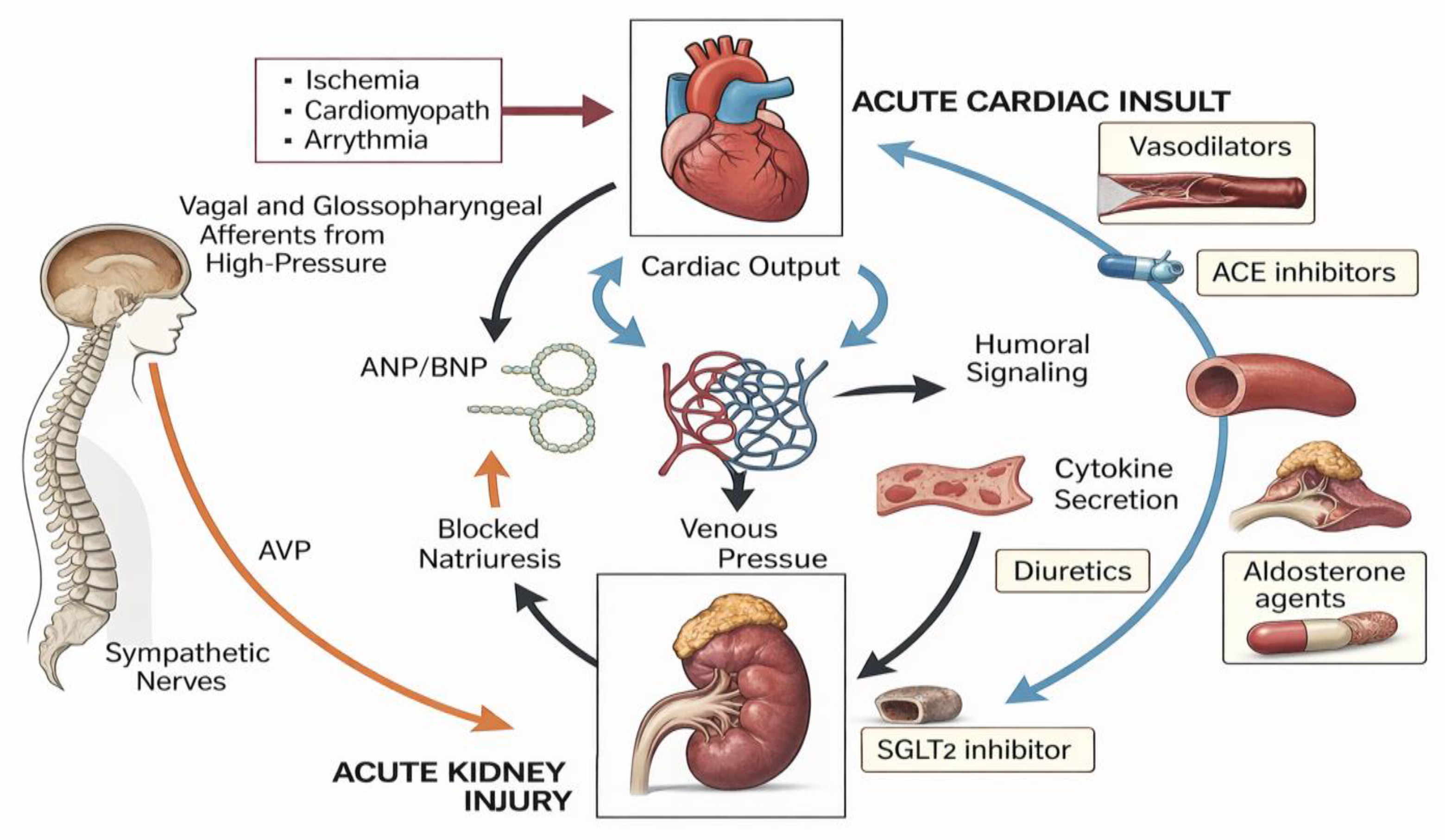
CRS complications (arrhythmias, worsening ventricular function, progression to CKD, diuretic resistance, electrolyte disturbances) arise from intertwined hemodynamic, neurohormonal, inflammatory, and structural mechanisms. Understanding the “why” behind each complication guides preventive and therapeutic strategies. Figure 1 represents mechanisms and treatment for CRS.
6.1. Arrhythmias: Why They Increase in CRS
Arrhythmias (atrial fibrillation, ventricular tachycardias, bradyarrhythmias) are frequent and have multiple converging causes:
- Ionic and electrolyte disturbances: hypokalemia (diuretics), hypomagnesemia, or hyperkalemia (renal failure or RAAS inhibition) alter repolarization and predispose to both tachyarrhythmias and bradyarrhythmias; hyponatremia is also associated with poor prognosis [60].
- Myocardial ischemia and cellular injury: coronary hypoperfusion in hypotension or coexisting coronary disease can generate irritative foci and elevated troponin with arrhythmic risk [61].
- Pharmacologic effects and drug accumulation.
- Systemic inflammation and sympathetic stress [62].
6.2. Worsening Ventricular Function and Progression to Heart Failure
- Reduced output from systolic or diastolic dysfunction leads to renal hypoperfusion; lower renal perfusion worsens sodium/water retention, increasing congestion [63].
- Chronic neurohormonal activation (RAAS, aldosterone, sympathetic tone) causes vasoconstriction, fibrosis, and remodeling that perpetuate HF and renal injury [64].
- Myocardial injury from uremia and uremic toxins: in advanced CKD, accumulation of metabolites promotes myocardial dysfunction (uremic cardiomyopathy) [65].
- Inflammation and oxidative stress accelerate myocyte loss and replacement with fibrotic tissue [66].
6.3. Diuretic Resistance and Failure of Depletive Therapy
- Pharmacokinetic mechanisms: reduced renal perfusion may diminish diuretic delivery to the tubule [67].
- Shift in reabsorption site: RAAS and aldosterone activation increase distal sodium reabsorption, counteracting loop inhibition [68].
- Proximal sodium recovery and tubular adaptation (braking phenomenon) [69].
6.4. Electrolyte Disorders and Therapeutic Limitations
6.5. Thrombotic Risk and Other Systemic Complications
Venous stasis, chronic inflammation–associated hypercoagulability, and endothelial dysfunction increase thrombotic risk (deep vein thrombosis, pulmonary embolism) [71].
7. Practical Implications and How to Translate Diagnosis into Prevention of Complications
Early diagnosis and subphenotyping: implementing protocols that combine early natriuresis, renal biomarkers, and VExUS allows identification of patients with intrarenal congestion and intervention before established AKI develops [45,48,52]. Prioritized correction of venous congestion when present: guided decongestion (natriuresis, VExUS) is more effective than indiscriminate increases in diuretics; consider sequential nephron blockade and, in refractory cases, advanced therapies [2,49]. Management of arrhythmias and correction of precipitants: strict electrolyte control, review of accumulated drugs, optimization of hemodynamics, and ischemia prevention [56,60,61].
Preservation of guideline-directed medical therapies: when possible, use strategies (closer monitoring, adjunctive treatments) that allow maintaining RAAS inhibitors, beta-blockers, and SGLT2 inhibitors while protecting renal function [54,64,72]. Mcamurray
Integrated care models: cardiology, nephrology, clinical pharmacy, and specialized nursing improve adherence to diagnostic and therapeutic strategies and enable proactive follow-up [55].
8. Prevention of Cardio-Renal Syndromes
Before addressing the treatment of CRS it is essential to highlight the importance of prevention. The first step is to identify patients at risk of developing the different types of cardiorenal syndrome and initiate preventive measures early as a strategy to reduce the likelihood of occurrence and improve outcomes should they develop.
8.1. Acute Cardio-Renal Syndrome (CRS1)
- ○
- Non-modifiable risk factors:
- ●
- History of diabetes mellitus
- •
- Previous hospitalizations for heart failure
- •
- History of acute myocardial infarction
- •
- Severe ventricular dysfunction
- •
- Baseline renal impairment
- ○
- Modifiable risk factors:
- •
- Use of high doses of diuretics
- •
- Vasodilator therapy
- •
- Drugs promoting sodium and volume retention, such as NSAIDs, thiazolidinediones, and corticosteroids
- ○
- Radiographic contrast agents.
Several risk-prediction tools exist for estimating the likelihood of acute kidney injury in specific contexts such as heart failure [22] (exposure to iodinated contrast during interventional procedures [75,76,77] or after cardiac surgery [78,79,80] These tools incorporate clinical variables [22,76,78,79], procedural characteristics [75,76,77,78,79,80], and novel approaches including machine learning [81,82], enabling accurate identification of at-risk patients and facilitating preventive and early diagnostic strategies for CRS.
- Comprehensive control of cardiovascular risk factors: smoking cessation, blood pressure optimization, lipid management, intensified glycemic control, and weight management.
- Avoidance of excessive or unnecessary use of radiographic contrast, and application of renal protection protocols when contrast is required. Risk factors for contrast-induced nephropathy include pre-existing CKD, diabetes, chronic heart failure, hypotension, advanced age, and high contrast volumes. Evidence supports the use of isotonic fluid administration as the most effective preventive measure, along with low-osmolar, non-ionic contrast agents such as iopamidol [83].
Additional strategies include statins, which reduce the risk of contrast-induced nephropathy and slow CKD progression in select populations, although heterogeneity in study designs limits generalizability [84], and N-acetylcysteine, which in recent meta-analyses has shown benefit in reducing contrast nephropathy but without impact on dialysis or mortality [85].
- Avoidance of nephrotoxic medications, or use at the minimum necessary dose and duration.
- Avoidance of sodium-retaining drugs.
- Implementation of cardiovascular therapies, pharmacological and invasive, appropriate for the underlying cardiac disease, with the aim of improving cardiac function and preventing heart failure decompensation: optimization of hemodynamic and volume status; modulators of neurohormonal and stress responses; treatment of anemia or malnutrition; cardiac resynchronization therapy or defibrillator implantation when indicated; mechanical circulatory support or transplantation in end-stage HF or end stage renal disease (ESRD); optimal renal replacement therapy; and disease-specific therapies for CVD, CKD, or HF.
- Close monitoring of renal function and electrolytes in patients with cardiac decompensation.
8.2. Chronic Cardio-Renal Syndrome (CRS 2)
All patients with chronic heart disease, particularly those with chronic heart failure, carry a potential risk of CRS2, especially individuals older than 75 years, those with diabetes, or with baseline renal dysfunction [23,73]. In this setting, therapies proven to improve the natural history of heart failure with preserved ejection fraction (SGLT2 inhibitors (SGLT2i)) or reduced ejection fraction (renin-angiotensin system inhibitors (RASi), beta-blockers,SGLT2i, mineralocorticoid receptor antagonists (MRA), cardiac resynchronization therapy or defibrillator therapy in indicated cases, mechanical circulatory support or transplantation in patients with end-stage HF and other specific therapies for CVD serve as preventive strategies for CRS2 [6,7,23,86].
8.3. Acute Reno-Cardiac Syndrome (CRS3)
- ○
- Non-modifiable risk factors:
- •
- Age >75 years
- •
- History of diabetes mellitus
- •
- Chronic heart failure
- •
- Peripheral vascular disease
- •
- Liver disease
- •
- Sepsis
- ○
- Modifiable risk factors:
- •
- Use of nephrotoxic and cardiotoxic medications
- •
- Hypotension
- •
- Electrolyte and fluid imbalances.
Preventive measures for CRS3 focus on avoiding or minimizing the use of potentially nephrotoxic or cardiotoxic drugs, ensuring adequate organ perfusion, and maintaining appropriate hydration while avoiding hypervolemia to prevent cardiac decompensation. Additionally, careful management of uremic changes, hyperkalemia, and pro-inflammatory mediators with adverse cardiac consequences is essential[23,73,87,88].
8.4. Chronic Reno-Cardiac Syndrome (CRS4)
CRS4 is a common manifestation in the natural history of CKD progression. Preventive strategies therefore aim at optimizing control of factors associated with CKD progression as proteinuria, diabetes, hypertension, atherosclerosis, left ventricular hypertrophy, systolic and diastolic dysfunction, and prothrombotic or proinflammatory states. Slowing CKD progression may reduce the incidence of CRS type 4 [6,7,14,23,73].
8.5. Secondary Cardio-Renal Syndrome (CRS5)
9. Treatment of Cardio-Renal Syndrome
Management of CRS is challenging due to its pathophysiological heterogeneity, five subtypes, and frequent comorbidities influencing treatment response. Therefore, therapy should be individualized, focused on the dominant pathology, and often multidisciplinary [1,23]. Specific evidence is limited, as most drugs have not been tested in CRS-specific trials, and patients with reduced glomerular filtration were typically excluded. No dedicated CRS guidelines exist, so management relies on HF and acute or chronic kidney disease recommendations [7,89,90]. Drugs improving HF prognosis and slowing renal progression are central. Optimal treatment of underlying cardiac disease is key in CRS types 1 and 2, while controlling baseline nephropathy is critical in types 3 and 4. In CRS type 5, focus is on the primary systemic disease [23].
9.1. Acute Cardio-Renal Syndrome (CRS1):
In CRS1, associated with acute HF, persistent congestion and renal deterioration worsen prognosis, making effective decongestion while preserving renal perfusion the main goal [1,23].Management includes oxygen therapy, congestion control, hemodynamic support, discontinuation of nephrotoxics, and arrhythmia management [73].
Loop diuretics are first-line; high-dose IV accelerates decongestion, with transient renal impairment generally considered hemodynamic without prognostic impact [49,73]. Dose should be adjusted early according to urine output or urinary sodium [1,89,90]. If response is insufficient, combinations with thiazides, acetazolamide, high-dose MRA, or tolvaptan may be used safely, though without additional cardiorenal prognostic benefit. In contrast, iSGLT2 provide proven cardiorenal benefit [5,91]. Ultrafiltration shows heterogeneous results. While some studies demonstrated effective decongestion [92,93], others reported greater renal deterioration and adverse effects [94].Therefore, current guidelines reserve this technique for volume overload refractory to optimal medical therapy [73,89].Peritoneal dialysis may be considered in selected patients with advanced CKD and refractory HF, with good hemodynamic tolerance [1].
9.2. Chronic Cardio- Renal Syndrome (CRS2):
Management of chronic HF in CRS2 targets underlying etiology and symptom control to prevent progression and decompensation of HF and CKD, requiring a comprehensive patient-centered approach including adherence, lifestyle, and guideline-directed drugs and devices [5,6,23,73].
In LVEF <40%, RASi (ACEi,ARBs, sacubitril/valsartan), beta-blockers, MRA, and iSGLT2 are first-line, reducing morbidity and mortality. In diabetic or obese patients, aGLP-1 add cardiovascular benefit. In patients with LVEF >40%, only iSGLT2 and, in diabetic or obese patients, aGLP-1 have demonstrated significant cardiovascular benefit. Diuretics improve symptoms without affecting mortality. Finerenone reduces HF hospitalizations in CKD with diabetes [5,6,23,73,89,90]. CRS2 patients often exhibit hypervolemia, requiring intensive loop diuretics, often combined. Initiation and titration of RASi and MRA may cause transient renal impairment or hyperkalemia, requiring close monitoring and sometimes new potassium binders. RASi, iSGLT2, finerenone, and aGLP-1 reduce proteinuria and iSGLT2 slow CKD progression. Correction of anemia and iron deficiency improves symptoms and prognosis[1,7,23,73].
9.3. Acute Reno -Cardiac Syndrome (CRS3):
Prevention is key, particularly in CRS3 associated with contrast nephrotoxicity or cardiac surgery. Once established, management focuses on treating AKI, controlling systemic complications, and close hemodynamic monitoring. Proper volume management, avoiding hypo- or hypervolemia, and correcting uremia, acid-base, and electrolyte disturbances are critical, as these can exacerbate cardiac dysfunction, arrhythmias, hemodynamic compromise, and catecholamine resistance. If conventional therapy fails, renal replacement therapy should be considered early [23,73,88].
9.4. Chronic Reno-Cardiac Syndrome (CRS4):
Management focuses on comprehensive CKD care and mitigating cardiovascular impact. Lifestyle measures (sodium, potassium, protein restriction) and glycemic control are essential.Blood preassure should be managed with RASi (ACEi or ARBs) and beta-blockers for cardio- and renoprotection, though use is limited in renal replacement therapy [14,73].
iSGLT2 improve renal and cardiovascular outcomes even in non-diabetic CKD [95]. Finerenone reduces renal progression and CV events in CKD with type 2 diabetes [96], and aGLP-1 improve metabolic control and cardiovascular risk, potentially benefiting renal outcomes in diabetic or obese patients [97].
Metabolic acidosis should be corrected with bicarbonate or citrate, associated with slower CKD progression and improved survival. Anemia management is controversial: iron and erythropoiesis-stimulating agents improve cardiac and renal function, but overcorrection increases CV events; target Hb 11–12 g/dl, avoiding >13 g/dl and excluding iron deficiency. Calcium-phosphate metabolism control is crucial to prevent vascular calcification[14,23,73].
9.5. Secondary Cardio-Renal Syndrome (CRS5):
Management is based on treating the primary disease and supporting renal and cardiovascular function. Early hypotension correction with volume resuscitation and, if needed, inotropes or vasopressors is critical to prevent organ dysfunction. Hemodynamic monitoring guides interventions. Persistent renal deterioration may require continuous renal replacement therapy to maintain function and modulate inflammation via cytokine removal. Avoid nephrotoxics and adjust medications according to perfusion and renal function, using an early multidisciplinary approach to optimize outcomes [23,73].
9.6. Innovative Treatments in CRS:
Emerging therapies aim to modulate inflammation, oxidative stress, gut microbiota, and non-coding RNA expression [86]. Investigational approaches include new vasodilators, cytokine-targeting antibodies, strategies to overcome diuretic resistance, and devices optimizing cardiac and renal preload and afterload [5]. These experimental avenues may offer new therapeutic options in CRS management.
10. Conclusions
CRS management poses multiple clinical challenges. Classification into five types is not always straightforward, and distinguishing true AKI from hemoconcentration-related functional changes can be difficult [1,7,23,73]. Assessing fluid overload, individualizing diuretic therapy, and determining optimal decongestion rates remain challenging, though controlled rapid decongestion does not worsen renal prognosis or increase mortality[1,23,73].
Patient evaluation should integrate renal function with symptoms, vital signs, urine output, and CKD history, while excluding secondary causes such as persistent hypotension, nephrotoxics, or renovascular disease[1,23,73]. Multidisciplinary cardio-renal units and bedside echocardiography are key tools for prevention, early detection, and timely management of CRS.
Supplementary Materials
The following supporting information can be downloaded at: Preprints.org,. Figure S1 Graphical Abstract: Global management of CRS from subtypes to treatment. Table S1: RIFLE, AKIN, KDIGO and WRF classification. Table S2: Evidence Table of Clinical Outcomes with Therapeutic Interventions in Cardiorenal Syndromes.
Author Contributions
Conceptualization, MM and JR; resources MF and LPB ;investigation, MF and LPB.; writing —original draft preparation: MM, MF, LPB and JR.; writing—review and editing MM, MF, LPB and JRC ; visualization, MM, MF, LPB and JR; supervision ,MM and JR . All authors have read and agreed to the published version of the manuscript.
Funding
This research received no external funding.
Institutional Review Board Statement
“Not applicable” for studies not involving humans or animals.
Data Availability Statement
No new data were created or analyzed in this study. Data sharing is not applicable to this article. All the sources are listed in the references.
Conflict of Interests
about the present manuscript authors do not have any conflict of interests.
Abbreviations
The following abbreviations are used in this manuscript
| CRS | Cardio–renal Syndrome |
| HF | Heart Failure |
| CKD | Chronic Kidney Disease |
| ADQI | Acute Dialysis Quality Initiative |
| CV | Cardiovascular |
| AKI | Acute Kidney injury |
| ADQI | Acute Dialysis Quality Initiative |
| AHF | Acute Heart Failure |
| WRF | Worsening Renal Function |
| RIFLE | Risk Injury Failure Loss of Kidney Function and End-stage Kidney Disease |
| AKIN | Acute Kidney Injury Network |
| KDIGO | Kidney Disease: Improving Global Outcomes |
| CHF EF |
Congestive Heart Failure Ejection Fraction |
| RAAS GFR SNS ATII ACE TNF-α ILE-1 ILE-6 CRP EPO eGFR ECG GLS PCPW IVC RVSI AVP ANP BNP VExUS POCUS ESRD iSGLT2 RASi ACEi ARBs MRA aGLP-1 RRRT NSAiDs LVEF LE |
Renin-angiotensin-aldosterone-system Glomerular Filtration Rate Sympathetic Nervous System Angiotensin II Angiontensin-Converting Enzyme Tumor necrosis factor-α Interleukin-1 Interleukin-6 C- Reactive protein Erythropoietin Estimated Glomerural Filtration Rate Electrocardiogram Global longitudinal Strain Pulmonary capillary wedge pressure Inferior vena cava Renal venous stasis index Arginin-vasopresin Atrial natriuretic peptide Brain natriuretic peptide Venous Excess Ultrasound Score Point of Care Ultrasound End Stage Renal Disease SGLT 2 inhibitors Renin-angiotensin system inhibitors Angiotensin-converting Enzime inhibitors Angiotensin Receptor Blockers Mineralocorticoid receptor antagonists Glucagon –like-peptide 1 agonists Renal replacement therapy Non steroidal antiinflamatory drugs Left Ventricle Ejection Fraction |
References
- McCallum W, Sarnak MJ. Cardiorenal Syndrome in the Hospital. Clin J Am Soc Nephrol. julio de 2023;18(7):933-45.
- Ronco C, Haapio M, House AA, Anavekar N, Bellomo R. Cardiorenal Syndrome. J Am Coll Cardiol. 2008;52(19):1527-39.
- Gallo G, Lanza O, Savoia C. New Insight in Cardiorenal Syndrome: From Biomarkers to Therapy. Int J Mol Sci 2023;24(6):5089. [CrossRef]
- Patel KP, Katsurada K, Zheng H. Cardiorenal Syndrome: The Role of Neural Connections Between the Heart and the Kidneys. Circ Res. 2022;130(10):1601-17. [CrossRef]
- McCallum W, Testani JM. Updates in Cardiorenal Syndrome. Med Clin North Am. j 2023;107(4):763-80.
- Schefold JC, Filippatos G, Hasenfuss G, Anker SD, Von Haehling S. Heart failure and kidney dysfunction: Epidemiology, mechanisms and management. Nat Rev Nephrol. 2016;12(10):610-23. [CrossRef]
- Rangaswami J, Bhalla V, Blair JEA, Chang TI, Costa S, Lentine KL, Lerma EV, Mezue K, Molitch M, Mullens W, Ronco C, Tang WHW, McCullough PA; American Heart Association Council on the Kidney in Cardiovascular Disease and Council on Clinical Cardiology. Cardiorenal Syndrome: Classification, Pathophysiology, Diagnosis, and Treatment Strategies: A Scientific Statement From the American Heart Association. Circulation. 2019 Apr 16;139(16):e840-e878. PMID: 30852913. [CrossRef]
- Sheikh O, Nguyen T, Bansal S, Prasad A. Acute kidney injury in cardiogenic shock: A comprehensive review. Catheter Cardiovasc Interv. 2021 Jul 1;98(1):E91-E105. Epub 2020 Jul 29. PMID: 32725874. [CrossRef]
- Bright R. Cases and observations illustrative of renal disease accompanied by the secretion of albuminous urine. Guys Hospital Reports. 1836: 338–400.
- National Heart, Lung, and Blood Institute. NHLBI Working Group: Cardiorenal connections in heart failure and cardiovascular disease, 2004. 2004. https://www.nhlbi.nih.gov/events/2004/cardio-renal-connections-heartfailure- and-cardiovascular-disease. Accessed February 15, 2018.
- Ronco C, House AA, Haapio M. Cardiorenal syndrome: Refining the definition of a complex symbiosis gone wrong. Intensive Care Med 2008;34:957–962. [CrossRef]
- Valika AA, Costanzo MR. The acute cardiorenal syndrome type I: Considerations on physiology, epidemiology, and therapy. Curr Heart Fail Rep. 2014 Dec;11(4):382-92. PMID: 25224320. [CrossRef]
- Núñez J, Miñana G, Santas E, Bertomeu-González V. Cardiorenal Syndrome in Acute Heart Failure: Revisiting Paradigms. Rev Esp Cardiol (Engl Ed). 2015 May;68(5):426-35. Epub 2015 Mar 7. PMID: 25758162. [CrossRef]
- Kidney Disease: Improving Global Outcomes (KDIGO). KDIGO clinical practice guidelines for the prevention, diagnosis, evaluation, and treatment of hepatitis C in chronic kidney disease. Kidney Int Suppl. 2008;109:S1–99.
- Mehta RL, Kellum JA, Shah SV, Molitoris BA, Ronco C, Warnock DG; et al. Acute Kidney Injury Network: Report of an initiative to improve outcomes in acute kidney injury. Crit Care. 2007;11:R31. [CrossRef]
- Bellomo R, Ronco C, Kellum JA, Mehta RL, Palevsky P; Acute Dialysis Quality Initiative workgroup. Acute renal failure—definition, outcome measures, animal models, fluid therapy and information technology needs: The Second International Consensus Conference of the Acute Dialysis Quality Initiative (ADQI) Group. Crit Care. 2004 Aug;8(4):R204-12. [CrossRef]
- Heywood JT, Fonarow GC, Costanzo MR, Mathur VS, Wigneswaran JR, Wynne J. High prevalence of renal dysfunction and its impact on outcome in 118,465 patients hospitalized with acute decompensated heart failure: A report from the ADHERE database. J Card Fail 2007;13:422–430. [CrossRef]
- Jois P, Mebazaa A. Cardio-renal syndrome type 2: Epidemiology, pathophysiology, and treatment. Semin Nephrol. 2012 Jan;32(1):26-30. PMID: 22365159. [CrossRef]
- De Vecchis R, Baldi C. Cardiorenal syndrome type 2: From diagnosis to optimal management. Ther Clin Risk Manag. 2014 Nov 12;10:949-61. PMID: 25419141; PMCID: PMC4235492. [CrossRef]
- Yap SC, Lee HT. Acute kidney injury and extrarenal organ dysfunction: New concepts and experimental evidence. Anesthesiology. 2012 May;116(5):1139-48. PMID: 22415388. [CrossRef]
- Kelly KJ. Distant effects of experimental renal ischemia/reperfusion injury. J Am Soc Nephrol. 2003 Jun;14(6):1549-58. PMID: 12761255. [CrossRef]
- Forman DE, Butler J, Wang Y, Abraham WT, O’Connor CM, Gottlieb SS, Loh E, Massie BM, Rich MW, Stevenson LW, Young JB, Krumholz HM. Incidence, predictors at admission, and impact of worsening renal function among patients hospitalized with heart failure. J Am Coll Cardiol. 2004 Jan 7;43(1):61-7. PMID: 14715185. [CrossRef]
- Ronco C, McCullough P, Anker SD, Anand I, Aspromonte N, Bagshaw SM, Bellomo R, Berl T, Bobek I, Cruz DN, Daliento L, Davenport A, Haapio M, Hillege H, House AA, Katz N, Maisel A, Mankad S, Zanco P, Mebazaa A, Palazzuoli A, Ronco F, Shaw A, Sheinfeld G, Soni S, Vescovo G, Zamperetti N, Ponikowski P. Cardio-renal syndromes: Report from the Consensus Conference of the Acute Dialysis Quality Initiative. Eur Heart J. 2010;31:703– 711. [CrossRef]
- Clementi A, Virzi GM, Brocca A; et al. Cardiorenal syndrome type 4: Management. Blood Purif. 2013;36:200---9. [CrossRef]
- Kumar U, Wettersten N, Garimella PS. Cardiorenal Syndrome: Pathophysiology. Cardiol Clin. 2019 Aug;37(3):251-265. Epub 2019 May 21. PMID: 31279419; PMCID: PMC6658134. [CrossRef]
- Bock JS, Gottlieb SS. Cardiorenal syndrome. New perspectives. Circulation. 2010;121:2592–600.Bock JS, Gottlieb SS. Cardiorenal syndrome. New perspectives. Circulation. 2010;121:2592–600.
- Nohria A, Hasselblad V, Stebbins A, Pauly DF, Fonarow GC, Shah M, Yancy CW, Califf RM, Stevenson LW, Hill JA. Cardiorenal interactions: Insights from the ESCAPE trial. J Am Coll Cardiol. 2008;51:1268 –1274.
- F Gnanaraj J, Haehling von S, Anker SD; et al. The relevance of congestion in the cardiorrenal syndrome. Kidney Int. 2013; 83: 384-391.
- Verbrugge FH, Dupont M, Steels P; et al. Abdominal Contributions to Cardiorrenal Dysfunction in Congestive Heart Failure. J Am Coll Cardiol. 2013. [CrossRef]
- Maeder MT, Holst DP, Kaye DM. Tricuspid regurgitation contributes to renal dysfunction in patients with heart failure. J Card Fail. 2008 Dec;14(10):824-30. Epub 2008 Sep 6. PMID: 19041045. [CrossRef]
- Kawano H, Do YS, Kawano Y; et al. Angiotensin II has multiple profibrotic effects in human cardiac fibroblasts. Circulation. 2000; 101: 1130-1137. [CrossRef]
- Kim S, Iwao H. Molecular and cellular mechanisms of angiotensin II-mediated cardiovascular and renal diseases. Pharmacol Rev. 2000; 52: 11-34. [CrossRef]
- Ronco C, Cicoira M, McCullough PA. Cardiorenal syndrome type 1: Pathophysiological crosstalk leading to combined heart and kidney dysfunction in the setting of acutely decompensated heart failure. J Am Coll Cardiol. 2012 Sep 18;60(12):1031-42. Epub 2012 Jul 25. PMID: 22840531. [CrossRef]
- Barreto DV, Barreto FC, Liabeuf S; et al. Plasma interleukin-6 is independently associated with mortality in both hemodialysis and pre-dialysis patients with chronic kidney disease. Kidney International 2010;77(6):550–556. [PubMed: 20016471]. [CrossRef]
- Kim B-S, Jeon DS, Shin MJ; et al. Persistent Elevation of C-Reactive Protein May Predict Cardiac Hypertrophy and Dysfunction in Patients Maintained on Hemodialysis. AJN 2005;25(3):189–195. [CrossRef]
- Silverberg DS, Wexler D, Blum M, Wollman Y, Schwartz D, Sheps D, Keren G, Iaina A. The interaction between heart failure, renal failure and anemia—the cardio-renal anemia syndrome. Blood Purif. 2004;22(3):277-84. Epub 2004 May 27. PMID: 15166489. [CrossRef]
- Marques Vidas M, Portolés J, Cobo M, Gorriz JL, Nuñez J, Cases A. Anemia Management in the Cardiorenal Patient: A Nephrological Perspective. J Am Heart Assoc. 2025 Mar 4;14(5):e037363. Epub 2025 Mar 3. PMID: 40028884; PMCID: PMC12132626. [CrossRef]
- Damman K, Valente MA, Voors AA, O’Connor CM, van Veldhuisen DJ, Hillege HL. Renal impairment, worsening renal function, and outcome in patients with heart failure: An updated review. Eur Heart J. 2014. [CrossRef]
- Ronco C, Bellasi A, Di Lullo L. Cardiorenal syndrome: An updated classification. Heart Fail Clin. 2020;16(1):1–16.
- McCullough PA, Kellum JA, Ronco C. Cardiorenal syndromes: Pathophysiology and biomarkers. Nephron Clin Pract. 2010;116(2):c65–c70.
- Mullens W, Abrahams Z, Skouri HN, Francis GS, Starling RC, Tang WH. Elevated central venous pressure is associated with impaired renal function and mortality in patients with advanced heart failure. J Am Coll Cardiol. 2009;53(7):582–8.
- Miller WL. Fluid volume overload and congestion in heart failure: Time to reconsider pathophysiology and treatment. Circulation. 2020;142(19):1838–48.
- Ponikowski P, Voors AA, Anker SD, Bueno H, Cleland JGF, Coats AJS; et al. 2021 ESC Guidelines for the diagnosis and treatment of acute and chronic heart failure. Eur Heart J. 2021;42(36):3599–726.
- Logeart D, Thabut G, Jourdain P, Chavelas C, Beyne P, Beauvais F; et al. Predischarge B-type natriuretic peptide assay for identifying patients at high risk of re-admission after decompensated heart failure. J Am Coll Cardiol. 2004;43(4):635–41. [CrossRef]
- Beaubien-Souligny W, Rola P, Haycock K, Bouchard J, Lamarche Y, Spiegel R; et al. Quantifying systemic congestion with point-of-care ultrasound: Development of the venous excess ultrasound grading system. Crit Care. 2020;24:1–10. [CrossRef]
- Kashani K, Al-Khafaji A, Ardiles T, Artigas A, Bagshaw SM, Bell M; et al. Discovery and validation of cell cycle arrest biomarkers for acute kidney injury. Crit Care. 2013;17(1):R25. [CrossRef]
- Nijst P, Verbrugge FH, Dupont M, Steels P, Tang WH, Mullens W. The pathophysiological role of intrarenal venous flow alterations in cardio-renal syndrome type 1. Circ Heart Fail. 2015;8(4):637–48.
- Ter Maaten JM, Valente MAE, Damman K, Hillege HL, Navis G, Voors AA. Diuretic response in acute heart failure—Pathophysiology, evaluation, and therapy. Eur Heart J. 2015;36(20):1241–53. [CrossRef]
- Felker GM, Lee KL, Bull DA, Redfield MM, Stevenson LW, Goldsmith SR; et al. Diuretic strategies in patients with acute decompensated heart failure. N Engl J Med. 2011;364(9):797–805. [CrossRef]
- Gargani L, Pang PS, Frassi F, Miglioranza MH, Dini FL, Landi P; et al. Persistent pulmonary congestion before discharge predicts rehospitalization in heart failure: A lung ultrasound study. Eur J Heart Fail. 2015;17(7):688–95. [CrossRef]
- Melenovsky V, Borlaug BA, Rosen B, Hay I, Ferruci L, Morell CH; et al. Cardiovascular features of heart failure with preserved ejection fraction preserve systemic perfusion at the expense of renal perfusion. Circulation. 2012;126(23):2809–19.
- Rola P, Miralles-Aguiar F, Beaubien-Souligny W, Haycock K, Bouchard J, Spiegel R; et al. Clinical applications of venous excess ultrasound (VExUS): A prospective observational study. J Crit Care. 2021;61:76–82.
- Iida N, Seo Y, Sai S; et al. Clinical implications of intrarenal hemodynamic evaluation by Doppler ultrasonography in patients with heart failure. CircCardiovascImaging. 2014;7(3):416–23. [CrossRef]
- Mullens W, Verbrugge FH, Nijst P, Tang WHW. Insights from invasive hemodynamic monitoring in heart failure. Eur Heart J. 2017;38(19):1501–9.
- House A, Anand I, Bellomo R, Cruz D, Endre Z, Hillege H; et al. Definition and classification of cardiorenal syndromes: Workgroup statements from the 7th ADQI Consensus Conference. KidneyInt. 2010;76(4):456–62. [CrossRef]
- Ling LH, Kistler PM, Ellims AH, Iles LM, Lee G, Hughes GL; et al. Diffuse ventricular fibrosis in atrial fibrillation: Noninvasive evaluation and relationships with atrial remodeling. J Am Coll Cardiol. 2012;60(25):2402–8.
- Park J, Kim TH, Uhm JS, Joung B, Hwang C, Pak HN. High left atrial pressure as a predictor of atrial fibrillation occurrence after ablation. Heart Rhythm. 2014;11(6):953–9.
- de Boer RA, Pinto YM, van Veldhuisen DJ. The imbalance between fibrosis formation and degradation in heart failure. Eur J Heart Fail. 2012;14(4):404–12.
- González A, Schelbert EB, Díez J, Butler J. Myocardial interstitial fibrosis in heart failure: Biological and translational perspectives. Heart. 2018;104(10):775–81.
- Gheorghiade M, Rossi JS, Cotts W, Shin DD, Hellkamp AS, Pina IL; et al. Relation between admission serum sodium concentration and clinical outcomes in hospitalized patients with heart failure. Eur Heart J. 2007;28(8):980–6.
- Metra M, Nodari S, Parrinello G, Bordonali T, Bugatti S, Danesi R; et al. Worsening renal function in patients hospitalized for acute heart failure: Clinical implications and prognostic importance. Circ Heart Fail. 2012;5(1):54–62. [CrossRef]
- Kaye DM, Lefkovits J, Jennings GL, Bergin P, Broughton A, Esler MD. Adverse consequences of high sympathetic nervous activity in the failing human heart. Circulation. 1995;92(9):2464–71. [CrossRef]
- Verbrugge FH, Dupont M, Steels P, Franken P, Tang WHW, Mullens W. Abnormal hemodynamics in heart failure patients with preserved ejection fraction: Importance of combined forward and backward failure. J Am Coll Cardiol. 2013;62(2):200–8.
- Pfeffer MA, Braunwald E. Ventricular remodeling after myocardial infarction: Experimental observations and clinical implications. N Engl J Med. 1990;323(6):362–9. [CrossRef]
- London GM, Pannier B, Guerin AP, Marchais SJ, Stimpel M. Cardiac hypertrophy, aortic compliance, peripheral resistance and wave reflection in end-stage renal disease. J Am SocNephrol. 2001;12(12):2628–36.
- Tang WHW, Francis GS. The oxidative stress hypothesis of heart failure: New insights. J Am Coll Cardiol. 2012;60(22):2105–8.
- Ellison DH. Diuretic resistance: Physiology and therapeutics. N Engl J Med. 2011;364(24):2295–304.
- Luther JM, Brown NJ. The renin–angiotensin–aldosterone system and sodium handling: Physiology and therapeutic implications. Hypertension. 2011;57(3):373–9.
- Wilcox CS. Diuretics and the kidney: Braking phenomenon. Hypertension. 1991;17(5):661–9.
- Rossi J, Bayram M, Udelson JE, Lloyd-Jones D, Adams KF, O’Connor CM; et al. Improvement in hyponatremia during hospitalization for heart failure is associated with improved outcomes. Am Heart J. 2007;154(2):S46.
- Piazza G, Goldhaber SZ. Venous thromboembolism and atherothrombotic disease: Epidemiology and pathophysiology. Circulation. 2010;121(14):1521–31.
- McMurray JJV, Solomon SD, Inzucchi SE, Køber L, Kosiborod MN, Martinez FA; et al. Dapagliflozin in patients with heart failure and reduced ejection fraction. N Engl JMed. 2019;381(21):1995–2008.
- Quiroga B, Santamaría Olomo R, Gorostidi M. Síndrome Cardiorrenal. Nefrol Al Día [Internet]: https://www.nefrologiaaldia.org/555.
- Goldstein SL, Jaber BL, Faubel S, Chawla LS, Acute Kidney Injury Advisory Group of American Society of Nephrology. AKI transition of care: A potential opportunity to detect and prevent CKD. Clin J Am Soc Nephrol CJASN. 2013;8(3):476-83.
- Mehran R, Owen R, Chiarito M, Baber U, Sartori S, Cao D; et al. A contemporary simple risk score for prediction of contrast-associated acute kidney injury after percutaneous coronary intervention: Derivation and validation from an observational registry. Lancet Lond Engl. 2021;398(10315):1974-83. [CrossRef]
- Mehran R, Aymong ED, Nikolsky E, Lasic Z, Iakovou I, Fahy M; et al. A simple risk score for prediction of contrast-induced nephropathy after percutaneous coronary intervention: Development and initial validation. J Am Coll Cardiol. 2004;44(7):1393-9. [CrossRef]
- Uzendu A, Kennedy K, Chertow G, Amin AP, Giri JS, Rymer JA; et al. Contemporary Methods for Predicting Acute Kidney Injury After Coronary Intervention. JACC Cardiovasc Interv. 2023;16(18):2294-305. [CrossRef]
- Thakar CV, Arrigain S, Worley S, Yared JP, Paganini EP. A clinical score to predict acute renal failure after cardiac surgery. J Am Soc Nephrol. 2005 Jan;16(1):162-8. [CrossRef]
- Wijeysundera DN, Karkouti K, Dupuis JY, Rao V, Chan CT, Granton JT; et al. Derivation and Validation of a Simplified Predictive Index for Renal Replacement Therapy After Cardiac Surgery. JAMA. 2007;297(16):1801-9. [CrossRef]
- Lee CPT, Ng KHL, Choi GYS, Wong WT, Wong HMK, Ho KM; et al. Performance of Cleveland, Mehta, and Simplified Renal Index scores for predicting dialysis-requiring acute kidney injury after aortic vs. non-aortic cardiac surgery (2006-2023, 6160 patients). Ren Fail. 2025;47(1):2592437. [CrossRef]
- Chen Y, Gue Y, Lip GYH, Gardner DS, Devonald MAJ. Machine learning prediction of moderate-to-severe acute kidney injury after ICU admission and cardiac surgery with urine trace elements. Eur J Clin Invest. 2025;e70131. [CrossRef]
- Petrosyan Y, Mesana TG, Sun LY. Prediction of acute kidney injury risk after cardiac surgery: Using a hybrid machine learning algorithm. BMC Med Inform Decis Mak. 2022;22(1):137. [CrossRef]
- Lameire N, Kellum JA, KDIGO AKI Guideline Work Group. Contrast-induced acute kidney injury and renal support for acute kidney injury: A KDIGO summary (Part 2). Crit Care Lond Engl. 2013;17(1):205. [CrossRef]
- Chikatimalla R, Trivedi YV, Ruhela N, Singh S, Lal A, Pawa A; et al. Statins and kidney health: Exploring cardiovascular benefits, renal protection, and risks in chronic kidney disease. Postgrad Med. 2025;137(7):588-600. [CrossRef]
- Zhu R, Zheng R, Deng B, Liu P, Wang Y. Association of N-acetylcysteine use with contrast-induced nephropathy: An umbrella review of meta-analyses of randomized clinical trials. Front Med. 2023;10:1235023. [CrossRef]
- Zhao BR, Hu XR, Wang WD, Zhou Y. Cardiorenal syndrome: Clinical diagnosis, molecular mechanisms and therapeutic strategies. Acta Pharmacol Sin. 2025;46(6):1539-55. [CrossRef]
- Kellum JA, Lameire N, KDIGO AKI Guideline Work Group. Diagnosis, evaluation, and management of acute kidney injury: A KDIGO summary (Part 1). Crit Care Lond Engl. 2013;17(1):204. [CrossRef]
- Khwaja A. KDIGO clinical practice guidelines for acute kidney injury. Nephron Clin Pract. 2012;120(4):c179-184. [CrossRef]
- McDonagh TA, Metra M, Adamo M, Gardner RS, Baumbach A, Böhm M; et al. 2021 ESC Guidelines for the diagnosis and treatment of acute and chronic heart failure. Eur Heart J. 2021;42(36):3599-726.
- McDonagh TA, Metra M, Adamo M, Gardner RS, Baumbach A, Böhm M; et al. 2023 Focused Update of the 2021 ESC Guidelines for the diagnosis and treatment of acute and chronic heart failure. Eur Heart J. 2023;44(37):3627-39. [CrossRef]
- Mullens W, Dauw J, Martens P, Verbrugge FH, Nijst P, Meekers E; et al. Acetazolamide in Acute Decompensated Heart Failure with Volume Overload. N Engl J Med. 2022;387(13):1185-95. [CrossRef]
- Bart BA, Boyle A, Bank AJ, Anand I, Olivari MT, Kraemer M; et al. Ultrafiltration versus usual care for hospitalized patients with heart failure: The Relief for Acutely Fluid-Overloaded Patients With Decompensated Congestive Heart Failure (RAPID-CHF) trial. J Am Coll Cardiol. 2005;46(11):2043-6.
- Costanzo MR, Guglin ME, Saltzberg MT, Jessup ML, Bart BA, Teerlink JR; et al. Ultrafiltration versus intravenous diuretics for patients hospitalized for acute decompensated heart failure. J Am Coll Cardiol 2007;49(6):675-83. [CrossRef]
- Bart BA, Goldsmith SR, Lee KL, Givertz MM, O’Connor CM, Bull DA; et al. Ultrafiltration in decompensated heart failure with cardiorenal syndrome. N Engl J Med. 2012;367(24):2296-304.
- Ali MU, Mancini GBJ, Fitzpatrick-Lewis D, Connelly KA, O’Meara E, Zieroth S; et al. The effectiveness of sodium-glucose co-transporter 2 inhibitors on cardiorenal outcomes: An updated systematic review and meta-analysis. Cardiovasc Diabetol. 2024;23(1):72 . [CrossRef]
- Rai B, Dar SF, Alowami M, Sajjad RB, Agarwal T, Khan MH, Mubarak Ali SR, Kamran G, Thomas RP, Mudiyanselage NSBA, AlQudah B, Khan YA, Behary Paray N, Shahzad M, Ahmed M, Ahmed R. Finerenone and Its Cardiorenal Protective Effects: A Meta-Analysis of 21,731 Patients From Randomized Trials. Cardiol Rev. 2025 Oct 13. Epub ahead of print. PMID: 41166566. [CrossRef]
- Sasaki T, Giang SM, Wu J, Yokoo T, Gallagher M, Bellomo R, Wang AY. The effect of GLP-1 receptor agonists on renal outcomes: A systematic review and meta-analysis. Nephrol Dial Transplant. 2025 Sep 22:gfaf193. Epub ahead of print. PMID: 40982219. 98. SOLVD Investigators, Yusuf S, Pitt B, Davis CE, Hood WB, Cohn JN. Effect of enalapril on survival in patients with reduced left ventricular ejection fractions and congestive heart failure. N Engl J Med. 1 de agosto de 1991;325(5):293-302. [CrossRef]
- SOLVD Investigators, Yusuf S, Pitt B, Davis CE, Hood WB, Cohn JN. Effect of enalapril on mortality and the development of heart failure in asymptomatic patients with reduced left ventricular ejection fractions. N Engl J Med. 3 de septiembre de 1992;327(10):685-91.
- Cohn JN, Tognoni G, Valsartan Heart Failure Trial Investigators. A randomized trial of the angiotensin-receptor blocker valsartan in chronic heart failure. N Engl J Med. 6 de diciembre de 2001;345(23):1667-75.
- Pfeffer MA, Swedberg K, Granger CB, Held P, McMurray JJV, Michelson EL; et al. Effects of candesartan on mortality and morbidity in patients with chronic heart failure: The CHARM-Overall programme. Lancet Lond Engl. 6 de septiembre de 2003;362(9386):759-66. [CrossRef]
- Yusuf S, Pfeffer MA, Swedberg K, Granger CB, Held P, McMurray JJV; et al. Effects of candesartan in patients with chronic heart failure and preserved left-ventricular ejection fraction: The CHARM-Preserved Trial. Lancet Lond Engl. 6 de septiembre de 2003;362(9386):777-81. [CrossRef]
- McMurray JJV, Packer M, Desai AS, Gong J, Lefkowitz MP, Rizkala AR; et al. Angiotensin-neprilysin inhibition versus enalapril in heart failure. N Engl J Med. 11 de septiembre de 2014;371(11):993-1004. [CrossRef]
- Solomon SD, McMurray JJV, Anand IS, Ge J, Lam CSP, Maggioni AP; et al. Angiotensin-Neprilysin Inhibition in Heart Failure with Preserved Ejection Fraction. N Engl J Med. 24 de octubre de 2019;381(17):1609-20.
- The Cardiac Insufficiency Bisoprolol Study II (CIBIS-II): A randomised trial. Lancet Lond Engl. 2 de enero de 1999;353(9146):9-13.
- Effect of metoprolol CR/XL in chronic heart failure: Metoprolol CR/XL Randomised Intervention Trial in Congestive Heart Failure (MERIT-HF). Lancet Lond Engl. 12 de junio de 1999;353(9169):2001-7.
- Packer M, Fowler MB, Roecker EB, Coats AJS, Katus HA, Krum H; et al. Effect of carvedilol on the morbidity of patients with severe chronic heart failure: Results of the carvedilol prospective randomized cumulative survival (COPERNICUS) study. Circulation. 22 de octubre de 2002;106(17):2194-9.
- Yamamoto K, Origasa H, Hori M, J-DHF Investigators. Effects of carvedilol on heart failure with preserved ejection fraction: The Japanese Diastolic Heart Failure Study (J-DHF). Eur J Heart Fail. enero de 2013;15(1):110-8. [CrossRef]
- Pitt B, Remme W, Zannad F, Neaton J, Martinez F, Roniker B; et al. Eplerenone, a selective aldosterone blocker, in patients with left ventricular dysfunction after myocardial infarction. N Engl J Med. 3 de abril de 2003;348(14):1309-21. [CrossRef]
- Zannad F, McMurray JJV, Krum H, van Veldhuisen DJ, Swedberg K, Shi H; et al. Eplerenone in patients with systolic heart failure and mild symptoms. N Engl J Med. 6 de enero de 2011;364(1):11-21.
- Pitt B, Zannad F, Remme WJ, Cody R, Castaigne A, Perez A; et al. The effect of spironolactone on morbidity and mortality in patients with severe heart failure. Randomized Aldactone Evaluation Study Investigators. N Engl J Med. 2 de septiembre de 1999;341(10):709-17.
- Pitt B, Pfeffer MA, Assmann SF, Boineau R, Anand IS, Claggett B; et al. Spironolactone for heart failure with preserved ejection fraction. N Engl J Med. 10 de abril de 2014;370(15):1383-92.
- Butler J, Anstrom KJ, Felker GM, Givertz MM, Kalogeropoulos AP, Konstam MA; et al. Efficacy and Safety of Spironolactone in Acute Heart Failure: The ATHENA-HF Randomized Clinical Trial. JAMA Cardiol. 1 de septiembre de 2017;2(9):950-8.
- Agarwal R, Filippatos G, Pitt B, Anker SD, Rossing P, Joseph A; et al. Cardiovascular and kidney outcomes with finerenone in patients with type 2 diabetes and chronic kidney disease: The FIDELITY pooled analysis. Eur Heart J. 10 de febrero de 2022;43(6):474-84. [CrossRef]
- Solomon SD, McMurray JJV, Vaduganathan M, Claggett B, Jhund PS, Desai AS; et al. Finerenone in Heart Failure with Mildly Reduced or Preserved Ejection Fraction. N Engl J Med. 24 de octubre de 2024;391(16):1475-85. [CrossRef]
- Felker GM, Lee KL, Bull DA, Redfield MM, Stevenson LW, Goldsmith SR; et al. Diuretic strategies in patients with acute decompensated heart failure. N Engl J Med. 3 de marzo de 2011;364(9):797-805. [CrossRef]
- Trullàs JC, Morales-Rull JL, Casado J, Carrera-Izquierdo M, Sánchez-Marteles M, Conde-Martel A; et al. Combining loop with thiazide diuretics for decompensated heart failure: The CLOROTIC trial. Eur Heart J. 1 de febrero de 2023;44(5):411-21. [CrossRef]
- Konstam MA, Gheorghiade M, Burnett JC, Grinfeld L, Maggioni AP, Swedberg K; et al. Effects of oral tolvaptan in patients hospitalized for worsening heart failure: The EVEREST Outcome Trial. JAMA. 28 de marzo de 2007;297(12):1319-31. [CrossRef]
- Berg DD, Patel SM, Haller PM, Cange AL, Palazzolo MG, Bellavia A; et al. Dapagliflozin in Patients Hospitalized for Heart Failure: Primary Results of the DAPA ACT HF-TIMI 68 Randomized Clinical Trial and Meta-Analysis of Sodium-Glucose Cotransporter-2 Inhibitors in Patients Hospitalized for Heart Failure. Circulation. 18 de noviembre de 2025;152(20):1411-22. [CrossRef]
- Packer M, Anker SD, Butler J, Filippatos G, Pocock SJ, Carson P; et al. Cardiovascular and Renal Outcomes with Empagliflozin in Heart Failure. N Engl J Med. 8 de octubre de 2020;383(15):1413-24. [CrossRef]
- Anker SD, Butler J, Filippatos G, Ferreira JP, Bocchi E, Böhm M; et al. Empagliflozin in Heart Failure with a Preserved Ejection Fraction. N Engl J Med. 14 de octubre de 2021;385(16):1451-61.
- Biegus J, Voors AA, Collins SP, Kosiborod MN, Teerlink JR, Angermann CE; et al. Impact of empagliflozin on decongestion in acute heart failure: The EMPULSE trial. Eur Heart J. 1 de enero de 2023;44(1):41-50. [CrossRef]
- Kosiborod MN, Abildstrøm SZ, Borlaug BA, Butler J, Rasmussen S, Davies M; et al. Semaglutide in Patients with Heart Failure with Preserved Ejection Fraction and Obesity. N Engl J Med. 21 de septiembre de 2023;389(12):1069-84. [CrossRef]
- Marso SP, Bain SC, Consoli A, Eliaschewitz FG, Jódar E, Leiter LA; et al. Semaglutide and Cardiovascular Outcomes in Patients with Type 2 Diabetes. N Engl J Med. 10 de noviembre de 2016;375(19):1834-44. [CrossRef]
- Perkovic V, Tuttle KR, Rossing P, Mahaffey KW, Mann JFE, Bakris G; et al. Effects of Semaglutide on Chronic Kidney Disease in Patients with Type 2 Diabetes. N Engl J Med. 11 de julio de 2024;391(2):109-21. [CrossRef]
- Costanzo MR, Negoianu D, Jaski BE, Bart BA, Heywood JT, Anand IS; et al. Aquapheresis Versus Intravenous Diuretics and Hospitalizations for Heart Failure. JACC Heart Fail. febrero de 2016;4(2):95-105. [CrossRef]
- Courivaud C, Kazory A, Crépin T, Azar R, Bresson-Vautrin C, Chalopin JM; et al. Peritoneal dialysis reduces the number of hospitalization days in heart failure patients refractory to diuretics. Perit Dial Int J Int Soc Perit Dial. 2014;34(1):100-8. [CrossRef]
- Bertoli SV, Musetti C, Ciurlino D, Basile C, Galli E, Gambaro G; et al. Peritoneal ultrafiltration in refractory heart failure: A cohort study. Perit Dial Int J Int Soc Perit Dial. 2014;34(1):64-70. [CrossRef]
- Moss AJ, Hall WJ, Cannom DS, Klein H, Brown MW, Daubert JP; et al. Cardiac-resynchronization therapy for the prevention of heart-failure events. N Engl J Med. 1 de octubre de 2009;361(14):1329-38. [CrossRef]
- Tang ASL, Wells GA, Talajic M, Arnold MO, Sheldon R, Connolly S; et al. Cardiac-resynchronization therapy for mild-to-moderate heart failure. N Engl J Med. 16 de diciembre de 2010;363(25):2385-95. [CrossRef]
- Bardy GH, Lee KL, Mark DB, Poole JE, Packer DL, Boineau R; et al. Amiodarone or an implantable cardioverter-defibrillator for congestive heart failure. N Engl J Med. 20 de enero de 2005;352(3):225-37. [CrossRef]
- Chen HH, Anstrom KJ, Givertz MM, Stevenson LW, Semigran MJ, Goldsmith SR; et al. Low-dose dopamine or low-dose nesiritide in acute heart failure with renal dysfunction: The ROSE acute heart failure randomized trial. JAMA. 18 de diciembre de 2013;310(23):2533-43.
- Packer M, Colucci W, Fisher L, Massie BM, Teerlink JR, Young J; et al. Effect of levosimendan on the short-term clinical course of patients with acutely decompensated heart failure. JACC Heart Fail. abril de 2013;1(2):103-11. [CrossRef]
- Anker SD, Comin Colet J, Filippatos G, Willenheimer R, Dickstein K, Drexler H; et al. Ferric carboxymaltose in patients with heart failure and iron deficiency. N Engl J Med. 17 de diciembre de 2009;361(25):2436-48. [CrossRef]
- Ponikowski P, Kirwan BA, Anker SD, McDonagh T, Dorobantu M, Drozdz J; et al. Ferric carboxymaltose for iron deficiency at discharge after acute heart failure: A multicentre, double-blind, randomised, controlled trial. Lancet Lond Engl. 12 de diciembre de 2020;396(10266):1895-904. [CrossRef]
- Swedberg K, Young JB, Anand IS, Cheng S, Desai AS, Diaz R; et al. Treatment of anemia with darbepoetin alfa in systolic heart failure. N Engl J Med. 28 de marzo de 2013;368(13):1210-9. [CrossRef]
Figure 1.
Mechanisms and treatment for CRS (detailed explanation in the text). AVP: arginin –vasopresin. ANP/BNP : Atrial natriuretic peptide/Brain natriuretic peptide.
Figure 1.
Mechanisms and treatment for CRS (detailed explanation in the text). AVP: arginin –vasopresin. ANP/BNP : Atrial natriuretic peptide/Brain natriuretic peptide.
Disclaimer/Publisher’s Note: The statements, opinions and data contained in all publications are solely those of the individual author(s) and contributor(s) and not of MDPI and/or the editor(s). MDPI and/or the editor(s) disclaim responsibility for any injury to people or property resulting from any ideas, methods, instructions or products referred to in the content. |
© 2025 by the authors. Licensee MDPI, Basel, Switzerland. This article is an open access article distributed under the terms and conditions of the Creative Commons Attribution (CC BY) license (http://creativecommons.org/licenses/by/4.0/).
Copyright: This open access article is published under a Creative Commons CC BY 4.0 license, which permit the free download, distribution, and reuse, provided that the author and preprint are cited in any reuse.


