Preprint
Article

This version is not peer-reviewed.

An Unexpected Inverse Relationship Between Biofilm Formation and Antibiotic Resistance in Stenotrophomonas maltophilia

A peer-reviewed article of this preprint also exists.

Submitted:

29 December 2025

Posted:

30 December 2025

You are already at the latest version

Abstract

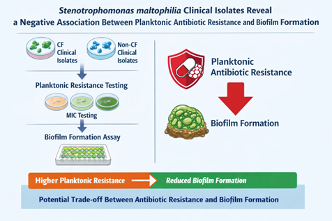
Background/Objectives: Stenotrophomonas maltophilia is an emerging opportunistic pathogen associated with severe infections, particularly in patients with cystic fibrosis (CF). Its intrinsic multidrug resistance and ability to form biofilms significantly complicate treatment. While biofilm growth is widely linked to antimicrobial tolerance, the relationship between biofilm-forming capacity and planktonic antibiotic resistance in S. maltophilia remains unclear. This study aimed to investigate the association between antibiotic resistance profiles and biofilm formation in clinical isolates from CF and non-CF patients. Methods: A total of 86 clinical S. maltophilia isolates (40 from CF airways and 46 from non-CF patients) were analyzed. Antibiotic susceptibility to seven agents was assessed by disk diffusion, with results interpreted according to EUCAST and CLSI criteria. Multidrug resistance phenotypes were defined using standard criteria. Biofilm formation was quantified after 24 h using a crystal violet microtiter plate assay and categorized into five levels of production. Statistical analyses were performed to compare biofilm formation across resistance profiles and clinical origins and to assess correlations between biofilm biomass and multidrug resistance. Results: Overall, high resistance rates were observed, particularly to meropenem (87.2%), ciprofloxacin (80.2%), and rifampicin (72.1%). CF isolates showed significantly higher resistance to piperacillin/tazobactam and a higher prevalence of multidrug resistance. Biofilm production was detected in 94.2% of isolates, with strong and powerful biofilm producers predominating. However, isolates from CF patients formed significantly less biofilm than those from non-CF patients. Notably, resistance to piperacillin/tazobactam and meropenem was associated with significantly reduced biofilm formation, as reflected in both median biomass and the proportion of high biofilm producers. Across the entire collection, the number of antibiotic resistances displayed by an isolate was negatively correlated with biofilm biomass. These trends were maintained after stratification by clinical origin, although some comparisons did not reach statistical significance. Conclusions: These findings demonstrate an unexpected inverse relationship between planktonic antibiotic resistance and biofilm-forming efficiency in S. maltophilia. Enhanced biofilm production may represent an alternative persistence strategy in more antibiotic-susceptible strains, with important implications for infection management and therapeutic failure.

Keywords: 
;  ;  ;  ;  ;  

1. Introduction

Once regarded as a low-virulence microorganism, Stenotrophomonas maltophilia has emerged as a clinically relevant opportunistic pathogen responsible for a broad range of conditions involving multiple organ systems, including the respiratory, gastrointestinal, and urinary tracts. Clinical manifestations include pneumonia, catheter-associated bacteremia and septicemia, osteochondritis, mastoiditis, meningitis, and endocarditis [1]. The bacterium is particularly prevalent in patients with cystic fibrosis (CF), in whom it is frequently isolated from the respiratory tract, with reported prevalence rates ranging from approximately 10% to 30% [2].
The treatment of S. maltophilia infections remains a major clinical challenge due to the bacterium’s extensive intrinsic and acquired antibiotic resistance mechanisms, which confer resistance to a wide range of broad-spectrum antimicrobial agents [3]. In addition, S. maltophilia readily forms biofilms on both abiotic and host tissues, a phenotype that further compromises the efficacy of clinically relevant antibiotics, including aminoglycosides, fluoroquinolones, and tetracyclines [4,5,6].
Biofilm growth is widely recognized as a major contributor to antimicrobial tolerance, as it limits antibiotic penetration, promotes antibiotic inactivation, and fosters physiological heterogeneity within bacterial populations [7,8]. The elevated cell density and oxidative stress characteristics of biofilms can increase mutation rates and facilitate horizontal gene transfer [9]. Compared with their planktonic counterparts, bacteria in biofilms exhibit greater resistance to nutrient starvation, pH fluctuations, and oxidative stress [10]. Biofilms may also increase resistance by altering the expression of pre-existing antibiotic resistance genes [11] and by increasing the proportion of tolerant or persister cells within the population, due to reduced bacterial metabolic activity within the biofilm interior [12].
Despite extensive evidence linking biofilm formation to increased antibiotic tolerance, the relationship between biofilm-forming capacity and antibiotic resistance in S. maltophilia planktonic cells remains poorly defined, leaving unresolved whether strong biofilm formation is consistently associated with increased planktonic resistance or whether trade-offs between these phenotypes may exist. Moreover, potential differences between isolates from CF and non-CF clinical settings have not been systematically explored.
In this study, we address these gaps by performing a comparative analysis of a large and diverse collection of S. maltophilia clinical isolates obtained from the airways of CF patients and from multiple anatomical sites in non-CF patients. By jointly assessing biofilm-forming ability and efficiency, planktonic antibiotic resistance profiles, and clinical origin, our work provides new insight into the interplay among these traits, revealing an unexpected relationship between biofilm formation and antibiotic resistance and setting the stage for the results presented below.

2. Results

2.1. Antibiotic Resistance

Overall, resistance to meropenem, ciprofloxacin, rifampicin, piperacillin/tazobactam, chloramphenicol, levofloxacin, and cotrimoxazole was observed in 87.2%, 80.2%, 72.1%, 50%, 47.7%, 26.7%, and 18.6% of isolates, respectively. Compared with non-CF isolates, those isolated from CF patients exhibited a significantly higher resistance rate to piperacillin/tazobactam (90.0 vs. 52.2%; p=0.0001).
Regarding resistance to multiple antimicrobial agents, the MDR phenotype was observed in a significantly higher proportion of CF isolates than of non-CF isolates (97.5 vs. 67.4%, respectively; p=0.0002). CF isolates also exhibited higher rates of XDR and PDR than the non-CF group, although the differences were not statistically significant (XDR: 60% vs. 54.3%; PDR: 15% vs. 4.3%, respectively, for CF and non-CF isolates).
The antibiotic resistance patterns revealed that most isolates had a high frequency of MDR; specifically, 62 of 86 isolates (72.1%) were resistant to at least 6 of the 7 antibiotics tested. However, no differences in multi-resistance levels were observed between the CF and non-CF groups.

2.2. Biofilm Formation

The cut-off value for biofilm formation – i.e., ODc = mean OD of negative control + (3 × SD of negative control) - was 0.076. This indicated a weak biofilm producer if 0.076 < OD492 ≤ 0.152, a moderate producer if 0.152 < OD492 ≤ 0.304, a strong producer if 0.304 < OD492 ≤ 0.608, and a powerful producer if OD492 > 0.608.
Most S. maltophilia isolates tested (81 out of 86, 94.2%) formed biofilm, with strong and powerful biofilm producer classes being the most prevalent (40.7% and 33.7%, respectively; p values at least 0.0009 vs. other classes). However, trends varied by patient source.
Although CF and non-CF isolates showed comparable biofilm-forming capabilities (90% vs. 97.8%, respectively), CF isolates were less efficient (OD492, median: 0.395 vs. 0.615 for CF and non-CF isolates, respectively; p=0.006). Confirming these findings, a significantly higher proportion of powerful biofilm producers was observed among non-CF isolates than among those isolated from CF patients (50% vs. 15%, respectively; p=0.0007). Conversely, moderate biofilm producers were found more frequently among CF than non-CF isolates (22.5% vs. 2.2%, respectively; p=0.005).

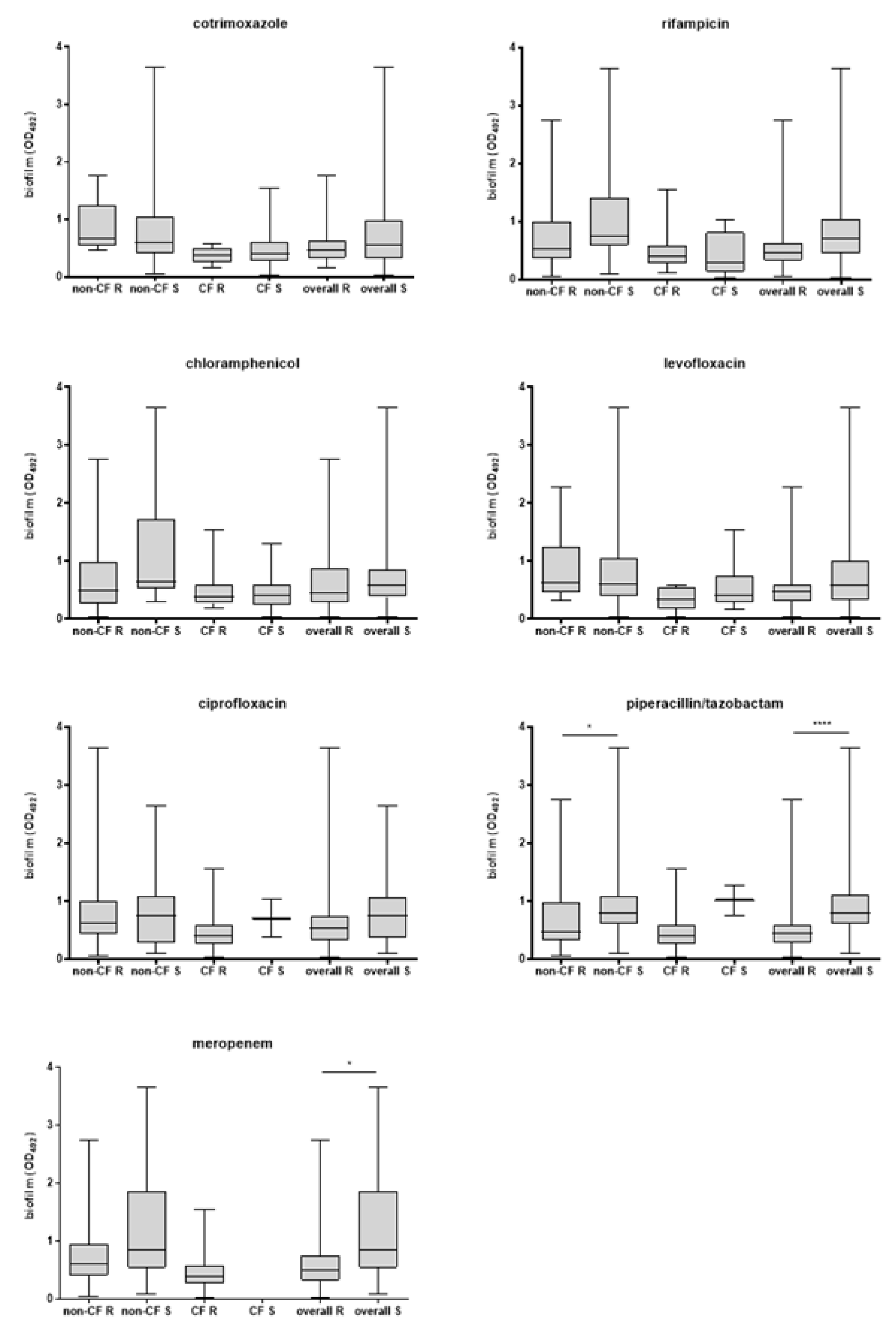
2.3. Correlation Between Antibiotic Resistance and Biofilm Formation

Considering the isolates as a whole, those resistant to piperacillin/tazobactam or meropenem produced significantly less biofilm than susceptible isolates (median OD492; piperacillin/tazobactam: 0.446 vs. 0.793, p<0.0001; meropenem: 0.598 vs. 0.847, p=0.048; respectively for resistant and susceptible isolates) (Figure 1). Confirming these findings, a significantly lower proportion of powerful producers was observed in piperacillin/tazobactam-resistant compared to susceptible isolates (18.3% vs. 69.2%, respectively; p=0.0001). No significant differences were found for other antibiotics.
Stratifying the isolates by patient type and isolation site, airway isolates from non-CF patients that were resistant to piperacillin/tazobactam produced less biofilm than susceptible isolates (OD492, median: 0.470 vs. 0.788, respectively; p=0.012) (Figure 1). No significant differences were found among the biofilm producer groups. Notably, the percentage of isolates categorized as strong biofilm producers was nearly double among susceptible isolates compared to resistant isolates (63.6% vs. 33.3%, respectively); however, this difference did not reach statistical significance due to the small sample size.
Among isolates collected from the airways of CF patients, the proportion unable to form biofilm was significantly lower among resistant than among susceptible isolates for meropenem (0% vs. 100%; p=0.03), ciprofloxacin, and piperacillin/tazobactam (0% vs. 50%; p=0.002).
No statistically significant differences in median biofilm amount were observed among non-MDR, MDR, XDR, and PDR isolates, regardless of the patient group considered (Figure 2). A similar trend was observed in the proportion of biofilm-producing groups among non-MDR, XDR, and PDR isolates. The percentage of non-MDR isolates classified as powerful producers was higher than that observed in MDR, XDR, and PDR isolates, although statistical significance was achieved only in the latter group (50% vs. 0%, p=0.022; for non-MDR and PDR, respectively) (data not shown). The percentage of powerful and strong producers was comparable between MDR and XDR (MDR: 30% vs. 42.9%; XDR: 28.6% vs. 44.9%), but was significantly higher than in other groups (MDR: p at least 0.04 vs. other classes; XDR: p at least 0.004 vs. other classes) (data not shown). PDR isolates were not seen as powerful producers, while the proportion of strong producers was higher than that of moderate and non-producers (75% vs. 12.5% and 12.5%, respectively; p=0.004) (data not shown).
The overall multidrug resistance level – i.e., the number of resistances displayed by an isolate - was negatively associated with the amount of biofilm formed, as indicated by linear regression analysis (p=0.003) (Figure 2). A similar trend was observed after stratification by CF and non-CF isolates; however, it did not reach statistical significance.

3. Discussion

Antibiotic resistance in S. maltophilia is an increasing concern, particularly in the lungs of people with CF, where its prevalence is on the rise. This study reported high levels of resistance to meropenem (87.2%), ciprofloxacin (80.2%), rifampicin (72.1%), piperacillin/tazobactam (50%), and chloramphenicol (47.7%), confirming previous studies [13]. Notably, CF isolates had higher resistance to piperacillin/tazobactam than non-CF ones. This finding likely reflects the frequent use of piperacillin/tazobactam as an antipseudomonal agent in patients with CF experiencing pulmonary exacerbations [14].
The antibiotic resistance of S. maltophilia was also indicated by the overall prevalence of the MDR phenotype, which was 81.4%. Interestingly, MDR isolates were more common in CF than in non-CF isolates. As is well known for Pseudomonas aeruginosa, the development of MDR in S. maltophilia lung isolates from CF patients can be attributed to its ability to adapt to the CF airway microenvironment through various genotypic changes and to develop mutational resistance under high selective pressure [15,16].
Our findings indicated that trimethoprim/sulfamethoxazole and levofloxacin were the most effective drugs tested. However, the susceptibility rates we observed (81.4% and 73.7%, respectively, for trimethoprim/sulfamethoxazole and levofloxacin) were lower than those reported in previous studies from different countries [17,18]. Rhee JY et al. [19] reported even higher resistance rates—over 30%—for both antibiotics. These findings indicate increasing resistance to the last-resort drug for treating multidrug-resistant S. maltophilia infections, underscoring the importance of robust control policies to limit the dissemination of resistant S. maltophilia strains and the need for further research to develop new treatments.
Most bacteria in nature exist in aggregated communities known as biofilms. Cells within a biofilm demonstrate significant physiological changes compared to their planktonic counterparts [20]. Biofilms are associated with numerous infections that can severely impact patients [21]. Indeed, infections involving a biofilm component are often chronic and highly resistant to antibiotic therapy [21]. Several studies have shown that biofilms are crucial in the persistence of S. maltophilia healthcare-associated infections, especially in patients with mechanical ventilation devices and CF patients [22,23]. Our findings confirmed that S. maltophilia has a significant propensity to form biofilms [22]. Over 94% of isolates produced biofilm. Notably, most isolates exhibited high biofilm-forming efficiency and were classified as strong or powerful producers. Decreased efficiency is a distinctive feature of isolates from CF patients, as indicated by a significantly lower proportion of powerful biofilm producers and a higher proportion of moderate biofilm producers compared to isolates from non-CF patients. These findings confirm that S. maltophilia adapts to a stressed environment, such as the CF lung [24].
The correlation between antibiotic resistance and the biofilm-forming ability of planktonic cells has been studied in Gram-positive and Gram-negative pathogens [25,26,27], raising questions about the mechanisms underlying the balance between these biological phenomena. Here, we evaluated, for the first time, the potential relationship between antibiotic resistance and the biofilm-forming capacity of S. maltophilia, leading to several conclusions.
First, isolates resistant to piperacillin/tazobactam or meropenem formed less biofilm than susceptible isolates, as indicated by differences in median biofilm quantity and the prevalence of the high-producing class. A similar trend appears to be specific to non-CF isolates. In contrast to our findings, Liaw et al. [28], evaluating the roles of integrons, efflux pumps, SpgM, melanin, and biofilm in MDR among 40 clinical isolates of S. maltophilia, observed that MDR isolates formed biofilm more readily than non-MDR isolates. Additionally, high biofilm formation was more prevalent among resistant than among susceptible isolates to piperacillin/tazobactam, whereas no difference was observed with meropenem. Differences in growth conditions (i.e., Luria-Bertani rather than TSB) and susceptibility breakpoints (i.e., established by CLSI rather than EUCAST) may explain the discrepancies with our findings.
Second, the percentage of non-CF isolates classified as high biofilm producers was nearly double among susceptible isolates compared to resistant ones, and the proportion of CF isolates unable to form biofilm was significantly lower among resistant isolates than among susceptible ones for meropenem, ciprofloxacin, and piperacillin/tazobactam.
Third, although the prevalence of high- and strong-producer classes was higher among MDR and XDR isolates than among non-MDR isolates, the number of resistances exhibited by an isolate was negatively correlated with the amount of biofilm formed.
Together, these results indicate that in S. maltophilia, there is a negative correlation between antibiotic resistance and biofilm-forming efficacy. Biofilms are known to confer greater antibiotic resistance and host immunity on microorganisms. From this perspective, high biofilm-forming efficiency may be considered an alternative strategy that antibiotic-susceptible strains adopt to escape antimicrobial treatments and persist longer within the host [25]. This adaptive strategy could be responsible for unexplained treatment failures and recurrences in susceptible isolates [29].

4. Materials and Methods

4.1. Bacterial Strains

Eighty-six S. maltophilia isolates were investigated: 40 isolated from respiratory specimens collected from CF patients and 46 from different sites of non-CF patients (i.e., 29 from the respiratory tract, 11 from blood, and 6 from other sources).

4.2. Antibiotic Susceptibility Tests

The agar disk-diffusion technique was used to evaluate the antibiotic susceptibility pattern of S. maltophilia isolates as described by EUCAST [30]. Antibiotic discs used for susceptibility testing were meropenem (10 µg), ciprofloxacin (5 µg), rifampicin (5 µg), piperacillin/tazobactam (30/6 µg), chloramphenicol (10 µg), levofloxacin (5 µg), and trimethoprim/sulfamethoxazole (1.25/23.75 µg). Escherichia coli ATCC 25922 and Pseudomonas aeruginosa ATCC 27853 were used as control strains.
Interpretation of zone diameters was based on the current breakpoint tables at http://www.eucast.org [30]. When no EUCAST breakpoints were available, CLSI breakpoints were considered [31]. Multidrug resistance (MDR) was defined as nonsusceptibility to ≥1 agent in ≥3 antimicrobial categories; extensively drug-resistant (XDR) as susceptibility limited to ≤2 categories; pan drug resistance (PDR), as nonsusceptibility to all agents in all antimicrobial categories [32].

4.3. Biofilm Formation Assay

The ability of each isolate to form biofilm was assessed in a 96-well microtiter plate assay after 24 h of incubation at 37 °C and quantified as optical density at 492 nm (OD492) using a crystal violet colorimetric assay, as previously described [22]. The cut-off value for biofilm formation (ODc) was calculated as the three standard deviations (SD) above the mean OD of the negative control: ODc = average OD of negative control + (3 × SD of negative control); a negative value was recorded as zero, while any positive value indicated biofilm production. Isolates were classified according to Stepanovic et al. [33] with minor modifications: OD ≤ ODc = no biofilm producer; ODc < OD ≤ 2 × ODc = weak biofilm producer; 2 × ODc < OD ≤ 4 × ODc = moderate biofilm producer; 4 × ODc < OD ≤ 8 × ODc = strong biofilm producer; and 8 × ODc < OD = powerful biofilm producer.

4.4. Statistical Analysis

Each experiment was carried out in triplicate and repeated twice (n = 6). The D’Agostino & Pearson normality test indicated that the data were not normally distributed. Therefore, the Mann-Whitney test was chosen to evaluate differences in median biofilm biomass between the CF and non-CF groups and between susceptible and resistant isolates. Fisher’s exact test was used to evaluate differences between proportions. The correlation between biofilm formation efficiency and antibiotic resistance level was also assessed using linear regression. Statistical analysis was performed using Prism software, version 7 (GraphPad Software, Boston, MA, USA), with p-values < 0.05 considered statistically significant.

Author Contributions

A.P. and G.D.B. conceived the study, contributed to conceptualization, designed and conducted the experiments, visualized the results, acquired funds, and wrote the original manuscript, as well as edited the revised version. A.P. contributed to writing the original version of the manuscript. G.D.B. contributed to editing both the original and revised manuscripts. G.D.B. supervised experimental activity. All authors have read and agreed to the published version of the manuscript.

Funding

This study has been partially funded by the Department of Medical, Oral and Biotechnological Sciences, University of Chieti-Pescara (“ex-60%” grant).

Institutional Review Board Statement

Not applicable.

Informed Consent Statement

Not applicable.

Data Availability Statement

Data is unavailable due to privacy and ethical restrictions.

Acknowledgments

During the preparation of this manuscript, the authors used ChatGPT 5.2 to create the graphical abstract. The authors have reviewed and edited the output and take full responsibility for the content of this publication.

Conflicts of Interest

The authors declare that the research was conducted in the absence of any commercial or financial relationships that could be construed as a potential conflict of interest.

Abbreviations

The following abbreviations are used in this manuscript:
CF Cystic fibrosis
OD Optical density
MDR Multidrug resistance
XDR Extensively drug resistance
PDR Pandrug resistance
EUCAST European committee for antimicrobial susceptibility testing
CLSI Clinical laboratory standards institute

References

  1. Brooke, JS. Advances in the Microbiology of Stenotrophomonas maltophilia. Clin Microbiol Rev. 2021, 34(3), e0003019. [Google Scholar] [CrossRef] [PubMed]
  2. Blanchard, AC; Waters, VJ. Opportunistic Pathogens in Cystic Fibrosis: Epidemiology and Pathogenesis of Lung Infection. J Pediatric Infect Dis Soc. 2022, 11 Supplement_2, S3–S12. [Google Scholar] [CrossRef] [PubMed]
  3. Monardo, R; Mojica, MF; Ripa, M; Aitken, SL; Bonomo, RA; van Duin, D. How do I manage a patient with Stenotrophomonas maltophilia infection? Clin Microbiol Infect 2025, 31(8), 1291–1297. [Google Scholar] [CrossRef] [PubMed]
  4. Di Bonaventura, G; Spedicato, I; D’Antonio, D; Robuffo, I; Piccolomini, R. Biofilm formation by Stenotrophomonas maltophilia: modulation by quinolones, trimethoprim-sulfamethoxazole, and ceftazidime. Antimicrob. Agents Chemother. 2004, 48, 151–160. [Google Scholar] [CrossRef]
  5. Pompilio, A; Crocetta, V; Confalone, P; Nicoletti, M; Petrucca, A; Guarnieri, S; Fiscarelli, E; Savini, V; Piccolomini, R; Di Bonaventura, G. Adhesion to and biofilm formation on IB3-1 bronchial cells by Stenotrophomonas maltophilia isolates from cystic fibrosis patients. BMC Microbiol 2010, 10, 102. [Google Scholar] [CrossRef]
  6. Sun, E; Liang, G; Wang, L; Wei, W; Lei, M; Song, S; Han, R; Wang, Y; Qi, W. Antimicrobial susceptibility of hospital acquired Stenotrophomonas maltophilia isolate biofilms. Braz. J. Infect. Dis. 2016, 20, 365–373. [Google Scholar] [CrossRef]
  7. Amanatidou, E; Matthews, AC; Kuhlicke, U; Neu, TR; McEvoy, JP; Raymond, B. Biofilms facilitate cheating and social exploitation of β-lactam resistance in Escherichia coli. NPJ Biofilms Microbiomes 2019, 5(1), 36. [Google Scholar] [CrossRef]
  8. Liu, HY; Prentice, EL; Webber, MA. Mechanisms of antimicrobial resistance in biofilms. NPJ Antimicrob Resist 2024, 2(1), 27. [Google Scholar] [CrossRef]
  9. Michaelis, C; Grohmann, E. Horizontal Gene Transfer of Antibiotic Resistance Genes in Biofilms. Antibiotics (Basel) 2023, 12(2), 328. [Google Scholar] [CrossRef]
  10. Zhao, A; Sun, J; Liu, Y. Understanding bacterial biofilms: From definition to treatment strategies. Front Cell Infect Microbiol 2023, 13, 1137947. [Google Scholar] [CrossRef]
  11. Høiby, N; Bjarnsholt, T; Givskov, M; Molin, S; Ciofu, O. Antibiotic resistance of bacterial biofilms. Int J Antimicrob Agents 2010, 35(4), 322–32. [Google Scholar] [CrossRef] [PubMed]
  12. Wood, TK; Knabel, SJ; Kwan, BW. Bacterial persister cell formation and dormancy. Appl Environ Microbiol 2013, 79(23), 7116–21. [Google Scholar] [CrossRef] [PubMed]
  13. Farrell, DJ; Sader, HS; Jones, RN. Antimicrobial susceptibilities of a worldwide collection of Stenotrophomonas maltophilia isolates tested against tigecycline and agents commonly used for S. maltophilia infections. Antimicrob Ag Chemother. 2010, 54(6), 2735–7. [Google Scholar] [CrossRef] [PubMed]
  14. Rolsma, SL; Sokolow, A; Patel, PC; Sokolow, K; Jimenez-Truque, N; Fissell, WH; Ryan, V; Kirkpatrick, CM; Nation, RL; Gu, K; Teresi, M; Fishbane, N; Kontos, M; An, G; Winokur, P; Landersdorfer, CB; Creech, CB. Population Pharmacokinetic Modeling of Cefepime, Meropenem, and Piperacillin-Tazobactam in Patients With Cystic Fibrosis. J Infect Dis. 2025, 231(2), e364–e374. [Google Scholar] [CrossRef]
  15. Marvig, RL; Sommer, LM; Molin, S; Johansen, HK. Convergent evolution and adaptation of Pseudomonas aeruginosa within patients with cystic fibrosis. Nat Genet. 2015, 47(1), 57–64. [Google Scholar] [CrossRef]
  16. Sanz-Garcia, F; Hernando-Amado, S; Martinez, JL. Mutation-driven evolution of Pseudomonas aeruginosa in the presence of either ceftazidime or ceftazidime/avibactam. Antimicrob Agents Chemother. 2018, 10, 62. [Google Scholar] [CrossRef]
  17. Duan, Z; Qin, J; Liu, Y; Li, C; Ying, C. Molecular epidemiology and risk factors of Stenotrophomonas maltophilia infections in a Chinese teaching hospital. BMC Microbiol 2020, 20(1), 294. [Google Scholar] [CrossRef]
  18. Bostanghadiri, N; Ardebili, A; Ghalavand, Z; Teymouri, S; Mirzarazi, M; Goudarzi, M; Ghasemi, E; Hashemi, A. Antibiotic resistance, biofilm formation, and biofilm-associated genes among Stenotrophomonas maltophilia clinical isolates. BMC Res Notes 2021, 14, 151. [Google Scholar] [CrossRef]
  19. Rhee, JY; Song, JH; Ko, KS. Current Situation of Antimicrobial Resistance and Genetic Differences in Stenotrophomonas maltophilia Complex Isolates by Multilocus Variable Number of Tandem Repeat Analysis. Infect Chemother 2016, 48(4), 285–293. [Google Scholar] [CrossRef]
  20. Squyres, GR; Newman, DK. Biofilms as more than the sum of their parts: lessons from developmental biology. Curr Opin Microbiol 2024, 82, 102537. [Google Scholar] [CrossRef]
  21. Schulze, A; Mitterer, F; Pombo, JP; Schild, S. Biofilms by bacterial human pathogens: Clinical relevance - development, composition and regulation - therapeutical strategies. Microb Cell 2021, 8(2), 28–56. [Google Scholar] [CrossRef] [PubMed]
  22. Pompilio, A; Ranalli, M; Piccirilli, A; Perilli, M; Vukovic, D; Savic, B; Krutova, M; Drevinek, P; Jonas, D; Fiscarelli, EV; Tuccio Guarna Assanti, V; Tavío, MM; Artiles, F; Di Bonaventura, G. Biofilm Formation among Stenotrophomonas maltophilia Isolates Has Clinical Relevance: The ANSELM Prospective Multicenter Study. Microorganisms 2020, 9(1), 49. [Google Scholar] [CrossRef] [PubMed]
  23. Mikhailovich, V; Heydarov, R; Zimenkov, D; Chebotar, I. Stenotrophomonas maltophilia virulence: a current view. Front Microbiol 2024, 15, 1385631. [Google Scholar] [CrossRef] [PubMed]
  24. Esposito, A; Pompilio, A; Bettua, C; Crocetta, V; Giacobazzi, E; Fiscarelli, E; Jousson, O; Di Bonaventura, G. Evolution of Stenotrophomonas maltophilia in Cystic Fibrosis Lung over Chronic Infection: A Genomic and Phenotypic Population Study. Front Microbiol 2017, 8, 1590. [Google Scholar] [CrossRef]
  25. Qi, L; Li, H; Zhang, C; Liang, B; Li, J; Wang, L; Du, X; Liu, X; Qiu, S; Song, H. Relationship between Antibiotic Resistance, Biofilm Formation, and Biofilm-Specific Resistance in Acinetobacter baumannii. Front Microbiol 2016, 7, 483. [Google Scholar] [CrossRef]
  26. Kwon, AS; Park, GC; Ryu, SY; Lim, DH; Lim, DY; Choi, CH; Park, Y; Lim, Y. Higher biofilm formation in multidrug-resistant clinical isolates of Staphylococcus aureus. Int J Antimicrob Agents 2008, 32(1), 68–72. [Google Scholar] [CrossRef]
  27. García-Castillo, M; Morosini, MI; Valverde, A; Almaraz, F; Baquero, F; Cantón, R; del Campo, R. Differences in biofilm development and antibiotic susceptibility among Streptococcus pneumoniae isolates from cystic fibrosis samples and blood cultures. J Antimicrob Chemother 2007, 59(2), 301–4. [Google Scholar] [CrossRef]
  28. Liaw, SJ; Lee, YL; Hsueh, PR. Multidrug resistance in clinical isolates of Stenotrophomonas maltophilia: roles of integrons, efflux pumps, phosphoglucomutase (SpgM), and melanin and biofilm formation. Int J Antimicrob Agents 2010, 35(2), 126–30. [Google Scholar] [CrossRef]
  29. Junco, SJ; Bowman, MC; Turner, RB. Clinical outcomes of Stenotrophomonas maltophilia infection treated with trimethoprim/sulfamethoxazole, minocycline, or fluoroquinolone monotherapy. Int J Antimicrob Agents 2021, 58(2), 106367. [Google Scholar] [CrossRef]
  30. EUCAST Disk Diffusion Method for Antimicrobial Susceptibility Testing. Version 13.0 (January 2025). www.eucast.org.
  31. Clinical Laboratory Standards Institute - CLSI M02, Performance Standards for Antimicrobial Disk Susceptibility Tests. March 2024.
  32. Magiorakos, AP; Srinivasan, A; Carey, RB; Carmeli, Y; Falagas, ME; Giske, CG; Harbarth, S; Hindler, JF; Kahlmeter, G; Olsson-Liljequist, B; Paterson, DL; Rice, LB; Stelling, J; Struelens, MJ; Vatopoulos, A; Weber, JT; Monnet, DL. Multidrug-resistant, extensively drug-resistant and pandrug-resistant bacteria: an international expert proposal for interim standard definitions for acquired resistance. Clin Microbiol Infect. 2012, 18(3), 268–81. [Google Scholar] [CrossRef]
  33. Stepanović, S; Vuković, D; Hola, V; Di Bonaventura, G; Djukić, S; Cirković, I; Ruzicka, F. Quantification of biofilm in microtiter plates: overview of testing conditions and practical recommendations for assessment of biofilm production by staphylococci. APMIS 2007, 115(8), 891–9. [Google Scholar] [CrossRef]
Figure 1. Figure 1. Biofilm formation of S. maltophilia according to susceptibility (S) or resistance (R) to several antibiotics, and stratified by patient type (CF, cystic fibrosis; non-FC, noncystic fibrosis). Results are shown as box-and-whisker plots; each box shows the median, with the bottom and top edges indicating the 25th and 75th percentiles, respectively, and the whiskers extend to the most extreme data points not considered outliers. Statistical significance by Mann-Whitney test: * p<0.05, **** p<0.0001.
Preprints 191983 g001
Figure 2. Biofilm formation and multidrug resistance (MDR, multidrug resistance; XDR, extensively drug resistance; PDR, pandrug resistance) in S. maltophilia isolated from cystic fibrosis (CF) and non-CF patients. Left side: results are shown as box-and-whisker plots (the central mark indicates the median, the bottom and top edges of the box indicate the 25th and 75th percentiles, respectively, and the whiskers extend to the most extreme data points not considered outliers). Right side: correlation between biofilm formation efficiency and antibiotic resistance level, as assessed by linear regression analysis.
Preprints 191983 g002
Disclaimer/Publisher’s Note: The statements, opinions and data contained in all publications are solely those of the individual author(s) and contributor(s) and not of MDPI and/or the editor(s). MDPI and/or the editor(s) disclaim responsibility for any injury to people or property resulting from any ideas, methods, instructions or products referred to in the content.
Copyright: This open access article is published under a Creative Commons CC BY 4.0 license, which permit the free download, distribution, and reuse, provided that the author and preprint are cited in any reuse.
Prerpints.org logo

Preprints.org is a free preprint server supported by MDPI in Basel, Switzerland.

Subscribe

Disclaimer

Terms of Use

Privacy Policy

Privacy Settings

© 2026 MDPI (Basel, Switzerland) unless otherwise stated