Preprint
Review

This version is not peer-reviewed.

Immune Disadvantage, Infection, Vaccination and Inflammation: Key Processes Leading to Immune Paralysis and Sudden Infant Death Syndrome

Submitted:

29 December 2025

Posted:

30 December 2025

You are already at the latest version

Abstract

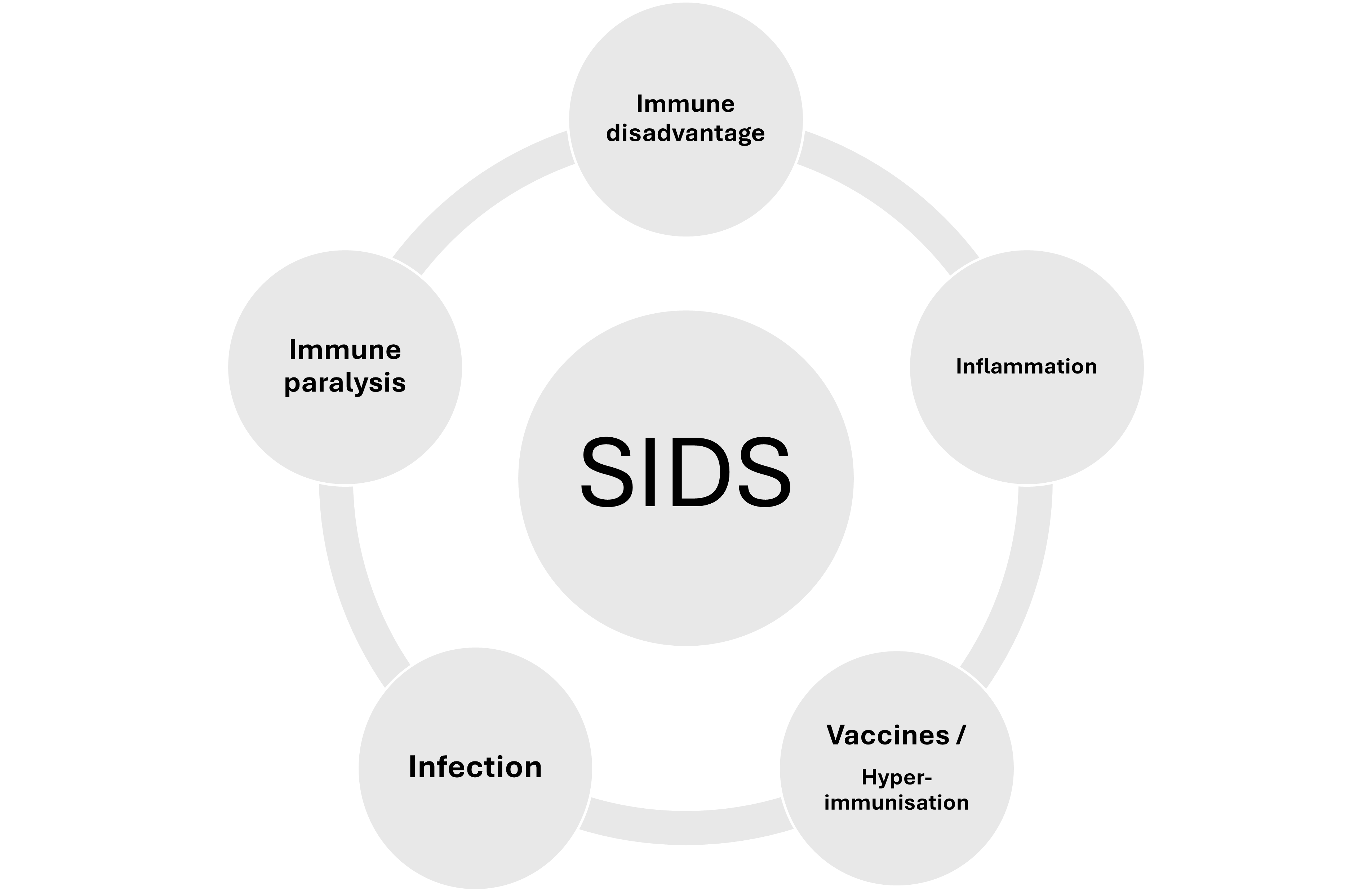
Within Sudden Infant Death Syndrome (SIDS) resides several primary phenomena; these include a state of immune immaturity, susceptibility to infection, and an inflammatory state. Most SIDS risk factors pertain in some way or another to higher risk of infection (prematurity, lack of breastfeeding, low or absent transplacental antibody, ethnicity, genetics, risky gene polymorphisms, poverty, etc.). Most SIDS cases display evidence of an inflammatory state (raised inflammatory markers and inflammatory cytokines). The pattern of inflammation is very similar to that observed following vaccination, which for achieving successful levels of protective immunity requires components that induce high reactogenicity. It is this reactogenicity which, under certain circumstances can cause immune paralysis. Immune paralysis leaves a vulnerable infant open to infection and systemic inflammatory response syndrome leading to shock. Such a mechanism is explored in this rapid review in the context of the aetiopathogenesis of SIDS.

Keywords: 
;  ;  ;  ;  ;  ;  ;  
Immune disadvantage and inflammation are common in SIDS
  • Could Immune Disadvantage, Inflammation and hyperimmunization lead to SIDS?
  • Most infants receive multiple (≥69) vaccine antigens and reactogenic adjuvants in the first year of life; this antigen/adjuvant load could act as hyperimmunization in vulnerable infants.
  • Hyperimmunization is known to cause immune paralysis.
  • Immune paralysis therefore could be a pathway to SIDS.

1. Introduction and Background

Sudden Infant Death Syndrome (SIDS) is defined as the sudden unexpected death of an apparently healthy infant under 1 year of age, which remains unexplained after a thorough case investigation, including a complete autopsy, examination of the death scene, and review of the clinical history [1]. It is important to recognize that SIDS is not only a diagnosis of exclusion but also a distinct entity within the broader category of sudden unexpected infant death (SUID) or sudden unexpected death in infancy (SUDI). SUID/SUDI encompasses all unexpected deaths in infants under 1 year of age, including those due to explained causes such as accidental suffocation, metabolic disorders, or infections, as well as those that remain unexplained after investigation. SIDS specifically refers to those cases where no cause is identified after a comprehensive evaluation as per the above definition [2].
For many decades there has been an unresolved problem in categorizing SIDS: the problem lies in the fact that a majority of SIDS cases bear features of infection ]3-5], and the decision is problematical in naming these cases as SIDS or a death occurring in conjunction with infection that is not considered serious enough to have caused the death. Such deaths are therefore given the name SUID/SUDI. The ‘diagnosis’ depends on the individual perspectives held by the pathologist or coroner responsible for making the ‘diagnosis.’ To gain perspective in this regard, and to emphasize infection-related features commonly observed in SIDS, a summary of these features is given further on.
In considering the pathogenesis of SIDS, infections and infection-related ones remain as primary considerations, especially in the context of the much relied-upon ‘triple risk hypothesis’ (in which a genetically or developmentally vulnerable infant at a critical developmental stage is exposed to exogenous stressors such as smoke exposure, prone sleeping or overheating [6]. Infection and its accompanying inflammatory response are rarely mentioned but are accepted stressors. This generally downplayed position of the role of infection in SIDS pathogenesis could have been one of the biggest mistakes in investigating the SIDS problem.
Numerous studies on the role of infection in SIDS have been published [3,4,5,7], however, this body of work has been ignored by mainstream researchers.
The hypothesis that infection is the most likely factor in the SIDS story, because of its strong association with most SIDS risk factors, and the diminished immunological status of the infant (lack of breastfeeding, prematurity leading to low maternal transplacental antibodies, etc.), representing “immune disadvantage,” and the consistent pathological finding of inflammation [8,9] stands as a well-supported idea. The concentration by mainstream researchers on sleep and homeostatic control and arousal failure has produced little if any data of real substance despite decades of endeavour. The best support for a sleep-related homeostatic failure hypothesis is contained in the findings of CNS inflammation with microglial and astroglial activation, apoptosis, raised CSF levels of inflammatory cytokines (e.g. IL-6). The recently published paper by Opdal et al. [10] entitled ‘The vicious spiral in Sudden Infant Death Syndrome’ provides a good summary of the present state of research but continues to invoke the triple risk hypothesis as the central tenet. The authors tended to favour CNS inflammatory responses as the key feature for the final pathway to death through disturbed homeostatic function [10] rather than seeing inflammation in a broader context. One can apply the triple risk hypothesis to most, if not all infectious diseases in infancy: i.e., a genetically or developmentally vulnerable infant at a critical developmental stage is exposed to an exogenous stressor (i.e. infection).
Further support for the infection model was shown by the rate of sudden infant death syndrome (SIDS increasing during the COVID-19 pandemic). This increase was most evident in 2021, with SIDS rates rising by ~10% compared to the pre-pandemic period, and a marked monthly increase observed from June to December 2021 (northern hemisphere) [11]. Additionally, the overall rate of sudden unexpected infant death (SUID), which includes SIDS, plus cases resulting from an unsafe sleeping environment, and deaths of unknown cause, also increased during the active pandemic period, but the rise was primarily driven by SIDS rather than other SUID categories [11,12]. While associated pandemic-related factors associated with the increased rates of SIDS (such as disruptions in healthcare access, changes in sleep practices—especially co-sleeping), and altered patterns of respiratory infections, particularly off-season resurgences of respiratory syncytial virus (RSV) were main features and add weight to the role of infection in SIDS [11,13,14], and actual CNS viral infection is now recognised as part of the problem.[15]. Perhaps the strongest evidence of infection playing a role in SIDS is the association of Staphylococcus aureus and prone sleep position the combination of which carried a high relative risk of SIDS [16,17].

2. Extending the Infection Hypothesis

The aim of this rapid review is to seek additional putative factors that could explain the SIDS enigma: the notion of pseudo-infection (vaccination) and how this could impact on infants in their first year of life. The review utilised PubMed, Google Scholar and Open Evidence to source relevant literature.
Vaccination/immunization is a pseudo-infection
It is obvious that where infection resides there is always involved an infectious agent (or agents-in-combination). However, until recently there has been little consideration of so-called pseudo-infections (and attenuated real infections) which are represented by immunisations. This issue has been summarised in a recent paper [18]. The paper raised the potential problem of hyperimmunization wherein multiple and/or repeated antigenic exposure causes serious potentially fatal immunopathological responses. Studies cited have shown increased infant mortality with increasing number of vaccines received [19,20]. The 2011 study by Goldman and Miller [21] examined approximately 38,000 infants who were either hospitalized or died after receiving the scheduled vaccines. The researchers grouped the cases which had resulted in serious adverse events by number of vaccine doses received which ranged from one to eight. Among the infants who received two doses of vaccine, the hospitalization rate was 11%; for three doses it was 12.4%. The rise continued for each additional vaccine dose and had reached 23.4% following eight doses. Infants who had five to eight doses had significantly higher mortality rates than those who received one to four doses.
The evidence for an association between vaccination and SIDS resides mainly in the data included in Vaccine Adverse Event Registries (VAERs) which, is collected passively and therefore represents only a tiny fraction of the true number of vaccine-associated deaths and the categorization of causes is questionable. More direct studies provide somewhat stronger evidence and may reflect problems with specific vaccines or adjuvants [22]. By contrast, numerous authors [23,24,25] have concluded that no vaccine-SIDS association exists. As was observed with the VAERS papers, the studies claiming the lack of association are also flawed but in different ways: in their design, especially in terms of lack of recency analysis and low statistical power.
The evidence for an association between vaccines and infant mortality must be regarded with care but should raise a level of concern to warrant serious investigation. Many of the studies tended to use unadjusted analyses and lacked control for confounding variables, which is a significant methodological flaw in epidemiological research on vaccines and neurodevelopmental problems and mortality, but to ignore these findings would be scientifically negligent when considering infant health and wellbeing in the context of the future of childhood immunization.
The problems in interpretation of these and other studies was recently discussed by Cauchi et al. (2022) [26] in the context of vaccines and neurodevelopmental disorders: the authors found it can be difficult to interpret available data and put these into context and to effectively communicate this information. The contemporary methodologies employed to investigate possible associations between vaccination and adverse neurological outcomes were examined and showed that determining causality can be challenging especially the understanding and communication of the data accurately to clinicians and the general population under so-called ‘infodemic’ conditions facilitated by the electronic media. Jacobson & Jacobson (2005) [27] provided a more detailed examination of the problems involved in study design and interpretation of data in the context of neurotoxins and neurodevelopmental disorders. They focussed on multiple comparisons, effect modifiers, and clinical significance. They examined alternatives to the Bonferroni approach regarding multiple comparisons (entailing unacceptably high risk of Type II error), including limiting the number of primary endpoints for hypothesis testing, evaluating the consistency of effects across multiple related endpoints, and replication. They showed that effect modifiers are important for understanding the marked individual differences in vulnerability that characterize most neurodevelopmental exposures. In evaluating effect modification, separate analyses comparing high and low risk groups could provide an alternative to statistical interaction terms, which are demonstrably underpowered in most broad longitudinal field studies. So, the risk of misinterpretation applies to studies both for and against a specific apparent correlation. Thus, it is now incumbent on vaccine manufacturers to conduct trials designed to overcome the above problems and elevate interpretation of results into less controversial status. Conflict of interest remains a problem and should require manufacturers to have independent researchers formally involved in their trials.
As mentioned in the twin SIDS paper [18], the 2024 study by Jablonowski et al. [22] in which 1,542,076 vaccine combinations administered to infants <1 year - old were evaluated for neurodevelopmental and other outcomes, the findings raised concern. While the study did not examine risk of SIDS or SUDI, it showed that for each additional vaccine given, the number of diseases (neurodevelopmental, respiratory, or suspected infection) diagnosed more than doubled [22], despite methodological weakness of the study, we would be negligent if we did not pay serious attention to this potentially adverse “dose effect,” possibly occurring through hyperimmunization. The review now examines this phenomenon.

3. Hyperimmunization

Because the phenomenon of hyperimmunization occurs with multiple immunizations, then it is essential and necessary to determine if this potentially adverse mechanism could plausibly occur in infants who receive multiple immunizations via the infant immunization schedule. If the data supports this, then gaining an understanding of how this could result in fatalities would be required. It is the purpose of this rapid review to discuss these mechanisms in as much detail as current knowledge allows.
Hyperimmunization: vaccine antigens or adjuvants?
Firstly, what possible adverse outcomes are observed following hyperimmunization?
In 1902, Zenoni [28] first described the development of amyloidosis in horses hyperimmunized with diphtheria toxin. Numerous other immunopathological outcomes are now well known to occur under similar excessive antigenic exposures. However, closer examination would indicate that the problem could reside in both the antigen(s) but also in the adjuvant(s). While there is sufficient evidence to raise concern regarding multiple exposures to antigens [19,29,30], concern regarding multiple dosing of adjuvant(s), particularly those that are highly inflammatory is also a concern [31,32,33].
According to Petrovsky [33], symptoms such as fever, headache, malaise, nausea, diarrhoea, arthralgia, myalgia and lethargy relate to systemic reactogenicity manifested through adjuvant-associated innate immune activation and inflammatory responses. Adjuvants are used to exaggerate activation of innate immune receptors. Certain adjuvants function as pathogen-associated molecular patterns (PAMPs) and induce systemic reactogenicity. Toll-like receptor (TLR) ligands, such as monophosphoryl lipid A (MPL), flagellin, lipoarabinomannan, peptidoglycan or acylated lipoprotein [34] are examples. Adjuvants that induce local tissue damage (e.g. oil emulsions and saponins), induce the release of endogenous damage-associated molecular patterns (DAMPs) that activate innate immune receptors and induce inflammation [35] and consequent systemic reactogenicity.
The duration of inflammation-associated adjuvant reactogenicity (markers of inflammation/reactogenicity such as C-reactive protein (CRP) and interleukin-6 (IL-6)), have been shown to typically rise within hours after immunisation in infants and generally return to baseline within 48–72 hours. In premature infants, CRP and IL-6 elevations are evident after administration of whole-cell pertussis-containing vaccines, peaking within the first 24–48 hours and normalizing by 48–72 hours post-immunisation [36,37]. However, transcriptomic changes, including overexpression of interferon-stimulated and inflammation-related genes, are most pronounced at 7 days post-vaccination and generally resolve by one month after immunisation in term infants [38].
It is important to clarify if there is any difference between the administration of a single vaccine or of multiple vaccines to infants on one occasion. Pourcyrous et al. [39] demonstrated abnormal elevation of CRP level occurred in 85% of premature infants (<35 weeks gestation) administered multiple vaccines and up to 70% of those given a single vaccine. Overall, 16% of infants had vaccine-associated cardiorespiratory events within 48 hours postimmunization. In logistic regression analysis, abnormal CRP values were associated with multiple vaccines (OR, 15.77; 95% CI 5.10-48.77) and severe intraventricular haemorrhage (IVH) (OR, 2.28; 95% CI 1.02-5.13). Cardiorespiratory events were associated marginally with receipt of multiple injections (OR, 3.62; 95% CI 0.99-13.25) and significantly with gastroesophageal reflux (GER) (OR, 4.76; 95% CI 1.22-18.52). The vaccines used included DTaP (Infanrix, SK Beecham, Philadelphia, Pa), Hib (ActHIB, Aventis, Swiftwater, Pa), HBV (Engerix-B, SK Beecham, Philadelphia, Pa), IPV (Inactivated-IPOLTM, Aventis, Swiftwater, Pa), and PCV7 (Prevnar, Lederle, Pearl River, NY). Infants were assigned to one of five vaccines, each with different adjuvants, making it challenging to interpret combined results. However, it was clear that DTaP caused the highest average CRP (12.1 mg/dL), the next highest being ActHIB (7.1mg/dL). DTaP incorporates Alum as its adjuvant. The adjuvant in ActHIB is tetanus protein (PRP-T) conjugated to Hib capsular polysaccharide (PRP). When all these vaccines were given in the same day to infants the average CRP was the second highest recorded overall (10.8mg/dL). The study revealed that some vaccines, including DTaP, when administered alone do not avoid cardiorespiratory adverse events or abnormal CRP values in premature infants in the NICU. But it is noteworthy that the incidence of these events was higher following simultaneous administration of multiple vaccines compared with administration of a single vaccine. The acute phase reaction to infection is characterized by IL-1β and IL-6 production, followed by CRP synthesis in the liver and release into the bloodstream. CRP begins increasing within 6 h of onset of infection, peaks around 48 h post-infection [40] but can remain increased for up to 6 days after death [41]. It is interesting that in the context of SIDS compared to infection the inflammatory markers of IL-1 and CRP seemed to be higher in the infection group than in SIDS but there was no difference between the two groups in seventeen other (mostly pro-inflammatory) immune biomarkers.[42]. In febrile infants younger than 3 months, post-immunisation leucocytosis was observed in almost half the children. These cases met the criteria for formal sepsis workup, yet they had no clinical symptoms or signs of infection [43].
Many infants are iron deficient in the highest SIDS risk months. Such hypoferronaemia can have serious impacts on the immune response to both infection and vaccinations [44]. The underlying mechanisms are complex and therefore will be dealt with in a separate paper.
If SIDS is the result of an infection (or preudo-infection) then we should question the idea of comparing SIDS with defined infections. As we have seen, it is difficult to separate the two based on blood levels of a long list of pro-inflammatory markers. Such an approach is comparing like with like. However, the findings in relation to vaccine reactogenicity only strengthens the notion that immunisation (especially involving adjuvants) mimics infection in terms of the pro-inflammatory and reactogenic molecules produced.
So, in summary, post-vaccination inflammatory markers develop in the first few days of the injection and resolve in most recipients within a week but can extend to a month. There is a suggestion that the degree of reactogenicity is related to the adjuvant used but most importantly the timing of the reactogenicity coincides with the incidence of unexpected infant deaths reported following immunisation (summarised in Goldwater 2025 [18] ), with higher mortality in the week following immunisation compared to mortality at 8 weeks post-immunisation, just before the next series of vaccines are scheduled.
Von Kries et al. [45] assessed whether temporal associations of mortalities could be attributed to chance. The study used standardised mortality ratios (SMR) for deaths within 1 to 28 days after administration of either of two hexavalent vaccines in the 1st and 2nd year of life. SMRs were determined using the respective annual rates for sudden unexpected deaths (SUDs) from national vital statistics. The study found for one of the vaccines (vaccine B), all SMRs were well below one. For the other (vaccine A), SMRs exceeded one insignificantly on the 1st day after vaccination in the 1st year of life. In the 2nd year of life, however, the SMRs for SUD cases within 1 day of vaccination with vaccine A were 31.3 (95% CI 3.8-113.1; two cases observed; 0.06 cases expected) and 23.5 (95% CI 4.8-68,6) for within 2 days after vaccination (three cases observed; 0.13 cases expected). Extensive sensitivity analyses were unable to attribute these findings to limitations of the data sources.
The authors concluded that the findings based on spontaneous reporting did not prove a causal relationship between vaccination and sudden unexpected deaths. However, the data constituted a “signal” for one of the two hexavalent vaccines examined which should act as a prompt for intensified surveillance for unexpected infant deaths following vaccination.
The report of six infants and children by Zinka et al. [46] involved three male and three female, aged between 4 and 17 months. Five children had been vaccinated with Hexavac®, one with Infanrix Hexa® during the previous 48 h before death. Shortly after the vaccination, three of the children developed tiredness, loss of appetite, fever up to 39C and insomnia. All children were found dead without explanation 1–2 days after the vaccination. The reported clinical features indicate that inflammation, through the effects of pro-inflammatory cytokines, is likely involved as part of the vaccines' reactogenic effects.

4. The Role of Inflammation

It is generally accepted that for successful immunisation, inflammation (which occurs with natural infection), is also essential for achieving protective immunity, as it facilitates the development of adaptive immunity by activating innate immune pathways and promoting cytokine and chemokine production [47,48,49]. This local and systemic inflammatory response is typically transient and manifests as common, mild adverse effects such as pain, erythema, swelling at the injection site, and fever or malaise [49,50,51]. These symptoms are generally self-limited and correspond with increased levels of proinflammatory cytokines (e.g., IL-1β, IL-6, TNF-α, G-CSF) and seem to be more pronounced with adjuvanted vaccines [49,50,51].
In a minority of cases, inflammation can lead to more significant adverse events, including febrile seizures, arthralgia, arthritis, cutaneous reactions, and rarely, immune-mediated phenomena such as uveitis, idiopathic thrombocytopenic purpura, or acute disseminated encephalomyelitis [52,53,54,55]. Exacerbation or new onset of immune-mediated inflammatory diseases (e.g., psoriasis, hidradenitis suppurativa) has also been reported, particularly following mRNA COVID-19 vaccination, though causality remains under investigation [56]. In a small study by Tukaj et al. [57], COVID-19 mRNA vaccine did not affect cytokine levels associated with major T helper cell subpopulations, including Th1, Th2, Th17, and Th22, which are commonly involved in the pathogenesis of autoimmune diseases. These cytokines are central to the differentiation and effector functions of their respective T helper cell subsets and are implicated in autoimmune pathogenesis [58]. However, anti-Hsp (heat shock) autoantibodies which are associated with autoimmune diseases were shown to be restricted to mRNA vaccine recipients and were not found in unvaccinated and never-infected controls. This finding should raise concern and require serious investigation [57].
In infants (especially preterm) inflammation can induce apnoeic episodes [59] which carries risk of sudden unexpected death. It is now becoming apparent that intermittent hypoxia in preterm babies produces an inflammatory response in much the same way as it does in adults [60]. This phenomenon of infant apnoea and intermittent hypoxia has been an area of intense interest for mainstream SIDS researchers wherein the focus has been on respiratory homeostasis rather than on inflammation. The natural progression of this work I consider should include the inflammatory effects of vaccination as discussed above.

5. Immune Paralysis (Immunoparalysis)

Vaccination can, in addition to causing inflammation, result in other forms of immunotoxicity such as immunoparalysis. Immunoparalysis is an acquired immune suppression marked by decreased innate and adaptive responses, often seen in critical illness or sepsis. Experimental and clinical data indicate that some vaccines, particularly those containing strong adjuvants or administered in high antigenic loads, can induce transient or persistent immunosuppressive effects. For example, randomized trials have shown that the diphtheria-tetanus-pertussis (DTP) vaccine induces immunotolerance, with long-term repression of monocyte-derived cytokine responses and T-cell reactivity to unrelated antigens, a phenomenon partially reversed by concurrent or subsequent BCG vaccination [61,62].
In mice, non-specific immunization with high antigen loads leads to adaptive immunosuppression as occurs in sepsis, lowering antibody responses and T-cell proliferation, partly due to the effects of regulatory T cells (Treg) [63]. Additionally, vaccine adjuvant-induced inflammation recruits inflammatory monocytes that suppress T-cell responses, acting as a counter-regulatory mechanism to limit excessive immune activation [64].
While these immunosuppressive effects are generally transient and rarely result in clinically significant immunoparalysis in healthy individuals, they highlight the complex immunomodulatory potential of vaccines beyond their intended antigen-specific effects. The clinical relevance of vaccine-induced immunoparalysis remains under investigation, but current evidence supports the possibility of immunosuppressive sequelae, particularly in the context of repeated or high-dose immunization, or in individuals with underlying immune dysregulation. Infants, particularly those born prematurely could be subject to vaccine-induced immunoparalysis especially when exposed to high antigen loads provided through polyvalent vaccines given under the conditions imposed by national childhood immunisation schedules wherein (and applying scheduled repeats) up to 69 vaccine targets (or more, especially for Aboriginal infants) in the first year of life [18].

6. Mechanisms Involved in Immunoparalysis

The mechanisms by which immunoparalysis occurs are complex and involve several different processes which fall into three main categories: Immune cell death, anergy and anti-inflammatory state [66]. Recently it has been shown that immune paralysis involves several influencing processes including immune-metabolic dysfunction which adversely affects immune cell energy metabolism, transcriptomics changes affecting gene expression, and epigenetic effects. How sepsis, or pseudo-infection (vaccines and their adjuvants) could affect these processes remains to be determined and is a subject requiring urgent investigation.
It is reasonable to state there is now enough evidence to show that immune paralysis does occur in infancy during or after a severe systemic inflammatory insult such as sepsis (as may occur in critically unwell Intensive Care babies) and is characterized by a marked diminution of both innate and adaptive immune responses accompanied by decreased expression of monocyte human leukocyte antigen-DR (HLA-DR) and demonstrably reduced in vitro production of proinflammatory cytokines including tumor necrosis factor-alpha (TNF-α) and interleukin-6 (IL-6) [66,67]. Affected infants are at risk of nosocomial infections and further sepsis. The pathophysiology involves a compensatory anti-inflammatory response following the initial proinflammatory insult and leads to downregulation of leukocyte function and altered gene expression that dampens immune activation [65,66,67]. Genomic profiling in affected infants has revealed increased expression of genes that dampen immune responses and decreased expression of genes involved in immune activation and regulation, particularly those related to antigen processing and negative regulation of interleukin-6 production [68].
Weiss et al. [69], were the first to describe mitochondrial dysfunction as an important player in immune paralysis, the evidence of which was derived from both animal models and human cases. The processes involved are multifactorial and affect both the innate and adaptive arms of the immune system. Mitochondrial dysfunction in peripheral blood mononuclear cells contributes to immune paralysis, through disruption of cellular energy metabolism and immune competence [69].
Expansion of myeloid-derived suppressor cells (MDSCs), mainly polymorphonuclear subsets generated via the PD-1/PD-L1 pathway, further suppresses T-cell activation and cytokine production, perpetuating immune paralysis [70].
In infants, the immaturity of their immune system plus transplacental maternal antibodies further modulate immune responses, so immune paralysis as described above is a distinct ‘acquired’ phenomenon, most often triggered by severe systemic inflammation or infection [71,72]. It remains unclear if the presence of maternal antibodies contributes in any way to immune paralysis per se.
Immune cell death or apoptosis, as mentioned, is a key process involved in immune paralysis. Sepsis in human subjects results in progressive apoptosis-induced loss of immune cells including CD4 T+-cells, CD8 T-cells, B-cells, natural killer (NK) cells and follicular dendritic cells. Apoptosis occurs through 2 pathways: (1) the death-receptor pathway; and (2) the mitochondrial-mediated pathway. The apoptotic debris dealt with by monocytes, macrophages and dendritic cells may result in increased anti-inflammatory cytokine (e.g. IL-10) release by Treg cells or result in a state of anergy furthering immunosuppressive effects with decreased monocyte CD14/human leucocyte antigen (HLA)-DR co-expression.
Recognizing that hyperimmunization is known to cause immune paralysis, it is not too large a step to reason that hyperimmunization, consequent to infant immunisation and purported hyperimmunization, through multiple antigen exposures at one time, could also be responsible for this.
The consequences of hyperimmunization or infection include systemic inflammatory response syndrome (SIRS) which is usually accompanied by a “cytokine storm.” Often this is followed by CARS (Compensatory Anti-inflammatory Response Syndrome) which is the immune system's counter-inflammatory response to sepsis, a mechanism to restore homeostasis following SIRS. While SIRS is a proinflammatory response to fight infection, CARS is a systemic immunosuppression that can increase susceptibility to secondary infections and lead to poor clinical outcomes [73].
In the case of overwhelming CARS, immune paralysis occurring through the processes discussed above (immune cell apoptosis and functional impairment of lymphocytes and phagocytes), with associated increased anti-inflammatory and decreased pro-inflammatory cytokine production [74,75,76,77]. The role of Treg lymphocytes in immune suppression is becoming better understood and shows that mitochondrial and lysosomal signalling orchestrates Treg cell metabolic and functional fitness [78].
Overshooting of the anti-inflammatory response predisposes the host to bacterial infection, and/or reactivation of latent viruses [79,80]. In contrast to the initial causative insult of sepsis, which if recognized, can be treated, no therapeutic or preventive strategies are presently available to combat the deleterious effects of immune paralysis during the period of CARS [81]. In conclusion, the immune cell phenotype of patients who die from sepsis appears to represent features consistent with immunosuppression [82].
Ferrante et al. [83], reported another potential contributor to immune paralysis that involves downregulation of myeloid differentiation primary response gene 88 (Myd88) in cerebral tissue from SIDS. Myd88 acts as a signal transducer in pathways that regulate activation of several pro-inflammatory genes which are crucial in both innate and adaptive immune responses. The protein product of Myd88 plays a pivotal role in initiating and sustaining an effective immune response, so a deficiency in this protein may impair the ability to mount an optimal immune reaction and potentially contributes to vulnerability in SIDS [10].
Given that immune paralysis is an established entity in infancy, then it is appropriate for this rapid review to examine if there is a putative role of this immunopathology in SUDI and SIDS. And by extension, the review explores why immune paralysis could be important in the context of infant immunisation and SIDS/SUDI.

7. Evidence of a Sepsis-Like Process in SIDS

While sepsis is an obvious sequela of immune paralysis, could this form of immunopathology arise from a pseudo-infection (that is, hyperimmunization)? Previous studies have demonstrated positive evidence for a sepsis-like process occurring in susceptible babies who die of SIDS (thus opening the possibility that the sepsis is a consequence of immune paralysis). The evidence for a sepsis-like process in SIDS includes:
1) the finding of proinflammatory cytokines in SIDS [15,84,85,86].
2) the finding of bacterial toxins in SIDS tissues [87,88].
3) the isolation of highly pathogenic bacteria (Staphylococcus aureus, Escherichia coli) in normally sterile sites [88,89,90].
4) the finding of elevated fibrin degradation products in sera from SIDS cases [91].
5) the shock-like physiological evidence of tachycardia followed by profound bradycardia occurring before cessation of breathing in cases of SIDS captured on memory monitors [92,93,94].
6) biochemical/clinical chemistry findings of Dr. Hazel L. McGaffey. Her work showed elevated brain and CSF lactate in conjunction with low CSF pH in cohorts of SIDS cases [95,96,97,98,99].
7) the organ weight changes (heavy, wet lungs, large brain and liver) compatible with sepsis, with enlarged thymus reflecting immunological perturbation [100,101,102,103].
8) intrathoracic petechial haemorrhages possibly the consequence of sepsis-related immune-mediated vasculopathy causing capillary leakage through basement membrane disruption [104,105] and shock-like diaphragm histopathology [106,107] with contraction band necrosis and muscle fibre changes frequently reported but are not universally observed.
In neonates, viral infections such as adenovirus and influenza trigger a cascade of morphological changes in the thymus. The initial stage shows enlargement of the thymus with oedema resulting in the collapse of reticular epithelial cells. As the infection progresses, the thymus shrinks with loss of volume in both the compartments with increase in connective tissue (reticulosis and sclerosis) [108]. On the basis of these neonatal findings, the overall significance of thymic enlargement in SIDS suggests that most mortal events occur relatively soon after viral infection (before the process of involution) while there are also cases in which the thymus is smaller than average suggesting a longer duration of infection with possible downstream effects from sepsis and compensatory anti-inflammatory response syndrome (CARS) [73].
According to MacFarlane [8], the thymus is involved in modulating development of the adaptive immune system. The study by Qu and colleagues [109] confirmed previous findings of increased thymic weight in SIDS (supporting aetiological involvement in SIDS). They also found increased concentrations of several pro-inflammatory cytokines compared with infants who did not present with an infectious component associated with their death. The level of some cytokines was within the range (but generally less) than control infants who died with severe infection (sepsis or pneumonia). Most of the cytokines promote thymic (and T cell) development suggesting that there may have been an underlying moderate infection in these infants and this supports the idea of subclinical infection promoted by Goldwater et al. [101]. Moreover, these data highlight the key observation that these thymic differences were unique to SIDS infants < 5 months of age, which coincides with the timing of the peak incidence of SIDS (2-6 months). According to MacFarlane [8], the data indicate a subclinical activation of the immune system in the youngest SIDS infants indicating a lack of an age-dependent thymus maturation. While the hypothesis that SIDS may be associated with a repression of normal immune system development, the alternative based on the observation that the cytokine profiles and thymic weights were already elevated in the youngest SIDS cases (< 5 months of age) suggesting a prematurely developed or over-reactive immune system. Thus, the characteristics of the thymus in the younger (< 5 months) SIDS infants started out higher than in control infants, masking the normal postnatal maturational process. Both ideas support and do not negatively impact on the overwhelming evidence of an infectious component of SIDS. The thymic findings should encourage a more in-depth analysis of thymic and immune system abnormalities in SIDS and should promote study of the development of the immune system in the context of critical stages of vulnerability in SIDS and in healthy subjects.
Animal models have shown that the second week of life, corresponding to a ~ 6-month human infant, is a uniquely vulnerable period of development in a rat model of SIDS during which some of the exemplary features of SIDS are evident including a heightened vulnerability to a relatively mild proinflammatory challenge [110,111,112]. The findings of these studies point strongly to an immunopathology resulting in shock; and their findings do not diminish the idea of subclinical infection being part of the underlying mechanism. Thus, the door remains open to the idea that pseudo-infection (i.e. vaccination or hyperimmunisation) acts as the precipitant.
Bajanowski et al. [113] studied 50 cases of sudden infant death. Cervical, paratracheal and lung hilar lymph nodes, thymus and spleen were examined histologically and immunohistochemically (CD 20, 21, 45RO). The cases were divided into 3 groups based on histology: A) – without pathological changes (N = 12), B) – with minimal to intermediate inflammation (N = 23) and C) – with severe inflammation (N = 15). In accordance with previous results the frequency of “pathological” lymph node changes, such as paracortical lymphoid hyperplasia and variegated hyperplasia of the pulp increased from group A) to group C). B-cell antigens reacted according to this pattern. Pronounced lymphodepletation of the thymus (assumed to indicate long lasting stimulation of the T-cell system) was also observed with the same pattern. No case was indicative of a defect of the T- or B-cell system [113]. The findings are compatible with subclinical infection as an underlying mechanism in SIDS pathogenesis.

8. Limitations

Decades of research has shown that infection and inflammation are consistent features of SIDS, and that immunisation has been shown to induce identical inflammatory cytokine changes to those observed during infections, thus suggesting there are two common pathways to the development of inflammation. Blood-Siegfried 2009 [4] reviewed infection and inflammation in SIDS and concluded a high proportion of cases <6 months of age had these features. On the other hand, brainstem pathology, central to the triple risk hypothesis, occurs in about 50% of cases of SIDS based on Naeye’s [114] finding of brainstem astrogliosis. Decades of research has added brainstem neuronal apoptosis, and microglial activation to astrogliosis as markers of neuroinflammation [115]. These changes are purported to also underlie homeostatic failure of respiration or cardiac function or arousal, according to the triple risk hypothesis. But equally they are most likely the result of infection and related proinflammatory cytokines, given that the observed inflammation must precede supposed effects on homeostasis. Simply ‘cause and effect.’
When vaccines are given simultaneously in multiples or in high dose the phenomenon of immune paralysis is a known sequela. The immature immune system of young infants increases vulnerability to infection and sepsis and can lead to immune paralysis following a “cytokine storm,” so, theoretically both vaccines and sepsis can produce this outcome. The key limitation of this paper is the role of immunisation in this process and proof remains at a relatively low level. However, the above detailed discussion exposes a “signal” representing a possible relationship between vaccines and SIDS and this calls for further investigation of the infant immunisation schedule given the schedule provides vaccinations in a context akin to hyperimmunization.
The level of certainty that vaccines do not cause SIDS is also debatable, wherein the studies looking into this are subject to flaws in design including issues of heterogenicity, weak statistical power, proneness to serious confounding (studies may show those who do or do not receive vaccines are likely to differ in many ways, some of which relate to their subsequent risk of early death, independent of vaccination). Also, proneness to other biases, including subject and control selection, and the selective loss of vaccination records for children who die, etc. [116]. Moreover, infants and children who receive vaccines are likely to be different in many ways from those who do not, and this poses an obvious problem for observational studies. Additionally, the literature on vaccine uptake patterns in different populations has identified factors associated with being vaccinated or not [117,118,119,120,121,122,123,124,125]. Socio-economic status amongst other factors, also carry mortality risks, and are therefore potential confounders in assessing associations between vaccination and mortality. Examples include poverty (and its increased risk of infectious disease), low parental education, orphan status and distance from a health centre. These are associated with low or delayed vaccine uptake and have also been associated with high mortality in many populations [125]. On the other hand, the link between high socio-economic status and high vaccination rates is expected to reduce mortality rate ratios when vaccinees and non-vaccinees are compared. Moreover, the collection of data on such factors, and their use in the adjustment of analyses requires ultimate care.

9. Conclusions

Through the decades of research into the cause or causes of SIDS the overwhelming weight of evidence indicates that inflammation is a strong contender through involvement in the underlying processes that can lead to an infant’s sudden unexpected death. There are two main mechanisms that may lead to inflammation: infection and pseudo-infection (vaccination). Inflammation can lead to immune paralysis, and then, to shock and unexpected death. Inflammation is likely to underlie the mechanisms espoused by mainstream researchers (disruption of homeostasis of breathing, cardiac function and arousal). Both points of view have been discussed in this rapid review. In many respects, the evidence for infection and/or inflammation stands well-supported by a broad swathe of evidence. It seems apparent that both the triple risk hypothesis and the infection/pseudo-infection/inflammation hypothesis can coexist, but, on balance of probability, the former requires the latter for SIDS occur. Whether or not vaccinations, as they are currently administered, are a significant player in this process will be clarified in the fullness of time.
While it is encouraging to see energy put into the development of mucosal vaccines, should we be concerned over reactogenicity of these vaccines in the context of hyperimmunization and immune paralysis? Current mucosal vaccines (intranasal, inhaled and oral) are generally less reactogenic than parenterally administered vaccines. Mucosal vaccines tend to cause fewer systemic adverse reactions (e.g., fever, myalgia, malaise). Systematic reviews and meta-analyses of mucosal vaccines for respiratory pathogens (including COVID-19 and influenza) indicate that their safety profile is comparable to or better than intramuscular vaccines, with adverse events typically mild and transient. No significant increase in serious adverse events has been observed with mucosal vaccines in large clinical trials [126]. However, reactogenicity can vary depending on the vaccine platform (live attenuated, inactivated, or vector-based), the mucosal route and the use of adjuvants [127,128]. It will take time to understand if multiple mucosal vaccines can be given together and avoid hyperimmunization effects observed with parenteral vaccines. The apparent lower reactogenicity of mucosal vaccines, and other advantages [129] should encourage their development especially in view of the issues surrounding the high number of vaccines and needles given in an infant’s first year of life.
The epigraph provided at the beginning of this paper is to encourage readers to reflect upon and question widely “accepted” paradigms. The case of the discovery of Helicobacter pylori and its relationship to peptic ulceration provides an excellent example wherein the mainstream paradigm was shown by Marshall and Warren to be heretical! [130].
Declaration of interests: No interests to declare.

Author Contributions

As sole author, all aspects of the paper were those of the author.

Funding

None.

References

  1. Krous, H.F.; Beckwith, J,B.l Byard, R.W.; Rognum, T.O.; Bajanowski, T.; Corey, T.; Cutz, E.; Hanzlick, R.; Keens, T.G. Mitchell EA. Sudden infant death syndrome and unclassified sudden infant deaths: a definitional and diagnostic approach. Pediatrics. 2004, 114(1), 234–8. [CrossRef] [PubMed]
  2. Shapiro-Mendoza, Carrie K.; Palusci, Vincent J.; Hoffman, Benjamin; Batra, Erich; Yester, Marc; Corey, Tracey S.; Sens, Mary Ann. Aap Task Force on Sudden Infant Death Syndrome, Council On Child Abuse And Neglect, Council On Injury, Violence, And Poison Prevention, Section On Child Death Review And Prevention, National Association Of Medical Examiners. "Half century since SIDS: a reappraisal of terminology. Pediatrics 2021, 148(no. 4), e2021053746. [Google Scholar] [PubMed]
  3. Blackwell, C.; Moscovis, S.; Hall, S.; Burns, C.; Scott, R. J. Exploring the risk factors for sudden infant deaths and their role in inflammatory responses to infection. Frontiers in immunology 6, 44. [CrossRef] [PubMed]
  4. Blood-Siegfried, J. The role of infection and inflammation in sudden infant death syndrome. Immunopharmacol Immunotoxicol. 2009, 31(4), 516–523. [Google Scholar] [CrossRef]
  5. Goldwater, P.N. Infection: the neglected paradigm in SIDS research. Arch Dis Child 2017, 102, 767–772. [Google Scholar] [CrossRef]
  6. Filiano, J.J.; Kinney, H.C. A perspective on neuropathologic findings in victims of the sudden infant death syndrome: the triple-risk model. Biol. Neonate 1994, 65, 194–197. [Google Scholar] [CrossRef]
  7. Morris, J.A. The common bacterial toxins hypothesis of sudden infant death syndrome. FEMS Immunol Med Microbiol. 1999, 25(1-2), 11–7. [Google Scholar] [CrossRef]
  8. MacFarlane, P.M. Inflammation in sudden infant death syndrome. Pediatric Research 2024, 95, 885–886. [Google Scholar] [CrossRef]
  9. Raza, M.W.; Blackwell, C.C. Sudden infant death syndrome, virus infections and cytokines. FEMS Immunol Med Microbiol. 1999, 25, 85–96. [Google Scholar] [CrossRef]
  10. Opdal, S.H.; Stray-Pedersen, A.; Eidahl, J.M.; Vege, Å.; Ferrante, L.; Rognum, T.O. The vicious spiral in Sudden Infant Death Syndrome. Front Pediatr 2025, 13. [Google Scholar] [CrossRef]
  11. Guare, E.G.; Zhao, R.; Ssentongo, P.; Batra, E.K.; Chinchilli, V.M.; Paules, C.I. Rates of sudden unexpected infant death before and during the COVID-19 pandemic. JAMA network open 2024, 7(9), e2435722–e2435722. [Google Scholar] [CrossRef]
  12. Shapiro-Mendoza, C.K.; Woodworth, K.R.; Cottengim, C.R.; Erck Lambert, A.B.; Harvey, E.M.; Monsour, M.; Parks, S.E.; Barfield, W.D. Sudden unexpected infant deaths: 2015–2020. Pediatrics 2023, 151(4), e2022058820. [Google Scholar] [CrossRef] [PubMed]
  13. Pease, A.; Turner, N.; Ingram, J.; Fleming, P.; Patrick, K.; Williams, T.; Sleap, V.; Pitts, K.; Luyt, K.; Ali, B.; Blair, P. Changes in background characteristics and risk factors among SIDS infants in England: cohort comparisons from 1993 to 2020. BMJ open 2023, 13(10), e076751. [Google Scholar] [CrossRef] [PubMed]
  14. Ferrante, L.; Opdal, S.H.; Byard, R.W. Further Exploration of the Influence of Immune Proteins in Sudden Infant Death Syndrome (SIDS). Acta Paediatrica 2025. [Google Scholar] [CrossRef] [PubMed]
  15. Ramachandran, P.S.; Okaty, B.W.; Riehs, M.; Wapniarski, A.; Hershey, D.; Harb, H.; Zia, M.; Haas, E.A.; Alexandrescu, S.; Sleeper, L.A.; Vargas, S.O. Multiomic analysis of neuroinflammation and occult infection in sudden infant death syndrome. JAMA neurology 2024, 81(3), 240–247. [Google Scholar] [CrossRef]
  16. Highet, A.R.; Berry, A.M.; Bettelheim, K.A.; Goldwater, P.N. Gut microbiome in sudden infant death syndrome (SIDS) differs from that in healthy comparison babies and offers an explanation for the risk factor of prone position. Int J Med Microbiol 2014, 304(5-6), 735–41. [Google Scholar] [CrossRef]
  17. Goldwater, P.N. SIDS, prone sleep position and infection: An overlooked epidemiological link in current SIDS research? Key evidence for the “Infection Hypothesis”. Medical Hypotheses 2020, 144, 110114. [Google Scholar] [CrossRef]
  18. Goldwater, P.N. Insights from twin Sudden Infant Death Syndrome studies could reveal an aetiopathogenetic pathway to sudden infant death through immunopathology. Med Res Arch. 2025, 13(7). [Google Scholar] [CrossRef]
  19. Goldman, G.S.; Miller, N.Z. Reaffirming a Positive Correlation Between Number of Vaccine Doses and Infant Mortality Rates: A Response to Critics. Cureus 2023, 15(2), e34566. [Google Scholar] [CrossRef]
  20. Miller, N.Z. Vaccines and sudden infant death: An analysis of the VAERS database 1990–2019 and review of the medical literature. Toxicol Reports 2021, 8, 1324–1335. [Google Scholar] [CrossRef]
  21. Miller, NZ; Goldman, S. : Infant mortality rates regressed against number of vaccine doses routinely given: Is there a biochemical or synergistic toxicity? Hum Exp Toxicol 2011, 30, 1420–1428. [Google Scholar] [CrossRef] [PubMed]
  22. Jablonowski, K.; Hooke, r B. Adverse outcomes are increased with exposure to added combinations of infant vaccines. Int J Vaccine Theor Pract Res. 2024, 3, 1103–11. [Google Scholar] [CrossRef]
  23. Vennemann, M.M.T.; Höffgen, M.; Bajanowski, T.; Hense, H.W.; Mitchell, E.A. Do immunisations reduce the risk for SIDS? A meta-analysis. Vaccine 2007, 25(26), 4875–4879. [Google Scholar] [CrossRef] [PubMed]
  24. Müller-Nordhorn, J.; Hettler-Chen, C.M.; Keil, T.; Muckelbauer, R. Association between sudden infant death syndrome and diphtheria-tetanus-pertussis immunisation: an ecological study. BMC Pediatrics 2015, 15(1), 1. [Google Scholar] [CrossRef]
  25. Brotherton, J.M.; Hull, B.P.; Hayen, A.; Gidding, H.F.; Burgess, M.A. Probability of coincident vaccination in the 24 or 48 hours preceding sudden infant death syndrome death in Australia. Pediatrics 2005, 115(6), e643–e646. [Google Scholar] [CrossRef]
  26. Cauchi, M.; Ball, H.; Ben-Shlomo, Y.; Robertson, N. Interpretation of vaccine associated neurological adverse events: a methodological and historical review. J Neurol 2022, 269(1), 493–503. [Google Scholar] [CrossRef]
  27. Jacobson, J.L.; Jacobson, S.W. Methodological issues in research on developmental exposure to neurotoxic agents. Neurotoxicol Teratol. 2005, 27, 395–406. [Google Scholar] [CrossRef]
  28. Zenoni, C. Toxische Amyloidosis bei der anti-diphtherischen Immunization des Pferdes. Zbl. Allg. Path 1902, 13, 334. [Google Scholar]
  29. Miller, N.Z.; Goldman, S. Infant mortality rates regressed against number of vaccine doses routinely given: Is there a biochemical or synergistic toxicity? Hum Exp Toxicol. 2011, 30, 1420–1428. [Google Scholar] [CrossRef]
  30. Hooker, B.S.; Miller, N.Z. Analysis of health outcomes in vaccinated and unvaccinated children: Developmental delays, asthma, ear infections and gastrointestinal disorders. SAGE Open Medicine 2020, 8, 1–11. [Google Scholar] [CrossRef]
  31. Aaby, P.; Jensen, H.; Rodrigues, A.; Garly, M.L.; Benn, C.S.; Lisse, I.M.; Simondon, F. Divergent female–male mortality ratios associated with different routine vaccinations among female–male twin pairs. Int J Epidemiol 2004, 33(2), 367–373. [Google Scholar] [CrossRef]
  32. Aaby, P.; Jensen, H.; Gomes, J.; Fernandes, M.; Lisse, I.M. The introduction of diphtheria-tetanus-pertussis vaccine and child mortality in rural Guinea-Bissau: an observational study. Int J Epidemiol 2004, 33(2), 374–380. [Google Scholar] [CrossRef] [PubMed]
  33. Petrovsky, N. Comparative Safety of Vaccine Adjuvants: A Summary of Current Evidence and Future Needs. Drug Saf. 38, 1059–1074. [CrossRef] [PubMed]
  34. Petrovsky, N.; Aguilar, J.C. Vaccine adjuvants: current state and future trends. Immunol Cell Biol. 2004, 82(5), 488–96. [Google Scholar] [CrossRef] [PubMed]
  35. Powell, B.S.; Andrianov, A.K.; Fusco, P.C. Polyionic vaccine adjuvants: another look at aluminum salts and polyelectrolytes. Clin Exp Vaccine Res. 2015, 4(1), 23–45. [Google Scholar] [CrossRef]
  36. Pourcyrous, M.; Korones, S.B.; Crouse, D.; Bada, HS. Interleukin-6, C-Reactive Protein, and Abnormal Cardiorespiratory Responses to Immunization in Premature Infants. Pediatrics 1998, 101(3), E3. [Google Scholar] [CrossRef]
  37. Jmaa, W.B.; Hernandez, A.I.; Sutherland, M.R.; Cloutier, A.; Germain, N.; Lachance, C.; Martin, B.; Lebel, M.H.; Pladys, P.; Nuyt, A.M. Cardio-respiratory events and inflammatory response after primary immunization in preterm infants<32 weeks gestational age: a randomized controlled study. The Pediatric Infectious Disease Journal 2017, 36(10), 988–994. [Google Scholar] [CrossRef]
  38. Nouri, N.; Cao, R.G.; Bunsow, E.; Nehar-Belaid, D.; Marches, R.; Xu, Z.; Smith, B.; Heinonen, S.; Mertz, S.; Leber, A.; Smits, G. Young infants display heterogeneous serological responses and extensive but reversible transcriptional changes following initial immunizations. Nature Communications 2023, 14(1), 7976. [Google Scholar] [CrossRef]
  39. Pourcyrous, M.; Korones, S.B.; Arheart, K.L.; Bada, H.S. Primary immunization of premature infants with gestational age <35 weeks: cardiorespiratory complications and C-reactive protein responses associated with administration of single and multiple separate vaccines simultaneously. J Pediatr. 2007, 151(2), 167–72. [Google Scholar] [CrossRef]
  40. Fujita, M.Q.; Zhu, B.L.; Ishida, K.; Quan, L.; Oritani, S.; Maeda, H. Serum C-reactive protein levels in postmortem blood—an analysis with special reference to the cause of death and survival time. Forensic science international 2002, 130(2-3), 160–166. [Google Scholar] [CrossRef]
  41. Uhlin-Hansen, L. C-reactive protein (CRP), a comparison of pre- and post-mortem blood. Forensic Sci. Int. 2001, 124, 32–5. [Google Scholar] [CrossRef] [PubMed]
  42. De Beer, C.; Ayele, B.T.; Dempers, J. Immune biomarkers as an adjunct diagnostic modality of infection in cases of sudden and unexpected death in infancy (SUDI) at Tygerberg Medico-legal Mortuary, Cape Town, South Africa. Human Pathology: Case Reports 2021, 23, 200477. [Google Scholar] [CrossRef]
  43. Prentice, S.; Kamushaaga, Z.; Nash, S.B.; Elliott, A.M.; Dockrell, H.M.; Cose, S. Post-immunization leucocytosis and its implications for the management of febrile infants. Vaccine 2018, 36(20), 2870–2875. [Google Scholar] [CrossRef] [PubMed]
  44. Preston, A.E.; Drakesmith, H.; Frost, J.N. Adaptive immunity and vaccination–iron in the spotlight. Immunotherapy Advances 2021, 1(1), ltab007. [Google Scholar] [CrossRef]
  45. Von Kries, R.; Toschke, A.M.; Straßburger, K.; Kundi, M.; Kalies, H.; Nennstiel, U.; Jorch, G.; Rosenbauer, J.; Giani, G. Sudden and unexpected deaths after the administration of hexavalent vaccines (diphtheria, tetanus, pertussis, poliomyelitis, hepatitis B, Haemophilius influenzae type b): is there a signal? European journal of pediatrics 2005, 164(2), 61–69. [Google Scholar] [CrossRef]
  46. Zinka, B.; Rauch, E.; Buettner, A.; Rueff, F.; Penning, R. Unexplained cases of sudden infant death shortly after hexavalent vaccination. Vaccine 2006, 24(31-32), 5779–5780. [Google Scholar] [CrossRef]
  47. Nakayama, T. An Inflammatory Response Is Essential for the Development of Adaptive Immunity-Immunogenicity Immunotoxicity. Vaccine 2016, 34(47), 5815–5818. [Google Scholar] [CrossRef]
  48. Nakayama, T. Causal Relationship Between Immunological Responses and Adverse Reactions Following Vaccination. Vaccine 2019, 37(2), 366–371. [Google Scholar] [CrossRef]
  49. Kashiwagi, Y.; Maeda, M.; Kawashima, H.; Nakayama, T. Inflammatory Responses Following Intramuscular and Subcutaneous Immunization With Aluminum-Adjuvanted or Non-Adjuvanted Vaccines. Vaccine 2014, 32(27), 3393–401. [Google Scholar] [CrossRef]
  50. Christian, L.M.; Porter, K.; Karlsson, E.; Schultz-Cherry, S. Proinflammatory cytokine responses correspond with subjective side effects after influenza virus vaccination. Vaccine 2015, 33(29), 3360–3366. [Google Scholar] [CrossRef]
  51. Zhuang, C.L.; Lin, Z.J.; Bi, Z.F.; Qiu, L.X.; Hu, F.F.; Liu, X.H.; Lin, B.Z.; Su, Y.Y.; Pan, H.R.; Zhang, T.Y.; Huang, S.J. Inflammation-related adverse reactions following vaccination potentially indicate a stronger immune response. Emerging microbes & infections 2021, 10(1), 365–375. [Google Scholar] [CrossRef]
  52. Dudley, M.Z.; Halsey, N.A.; Omer, S.B.; Orenstein, W.A.; T O'Leary, S.; Limaye, R.J.; Salmon, D.A. The state of vaccine safety science: systematic reviews of the evidence. The Lancet Infectious Diseases 2020, 20(5), e80–e89. [Google Scholar] [CrossRef] [PubMed]
  53. Rosenblatt, A.E.; Stein, S.L. Cutaneous Reactions to Vaccinations. Clinics in Dermatology 2015, 33(3), 327–32. [Google Scholar] [CrossRef] [PubMed]
  54. Zou, Y.; Kamoi, K.; Zong, Y.; Zhang, J.; Yang, M.; Ohno-Matsui, K. Ocular inflammation post-vaccination. Vaccines 2023, 11(10), 1626. [Google Scholar] [CrossRef]
  55. Panozzo, C.A.; Pourmalek, F.; Pernus, Y.B.; Pileggi, G.S.; Woerner, A.; Bonhoeffer, J. Brighton Collaboration Aseptic Arthritis Working Group. Arthritis and arthralgia as an adverse event following immunization: a systematic literature review. Vaccine 2019, 37(2), 372–383. [Google Scholar] [CrossRef]
  56. Liakou, A.I.; Tsantes, A.G.; Routsi, E.; Agiasofitou, E.; Kalamata, M.; Bompou, E.K.; Tsante, K.A.; Vladeni, S.; Chatzidimitriou, E.; Kotsafti, O.; Samonis, G. Could Vaccination against COVID-19 Trigger Immune-Mediated Inflammatory Diseases? Journal of Clinical Medicine 2024, 13(16), 4617. [Google Scholar] [CrossRef]
  57. Tukaj, S.; Sitna, M.; Sitko, K. The Impact of the mRNA COVID-19 Vaccine on the Th-Like Cytokine Profile in Individuals With No History of COVID-19: Insights Into Autoimmunity Targeting Heat Shock Proteins. Front Immunol. 2025, 16, 1549739. [Google Scholar] [CrossRef]
  58. Ayass, M.A.; Tripathi, T.; Zhu, K.; Nair, R.R.; Melendez, K.; Zhang, J.; Fatemi, S.; Okyay, T.; Griko, N.; Ghelan, M.B.; Pashkov, V. T helper (Th) cell profiles and cytokines/chemokines in characterization, treatment, and monitoring of autoimmune diseases. Methods 2023, 220, 115–125. [Google Scholar] [CrossRef]
  59. Beyeler, S.A.; Hodges, M.R. Adrianne, G.; Huxtable, A.G. Impact of inflammation on developing respiratory control networks: rhythm generation, chemoreception and plasticity. Resp PhysiolNeurobiol 2020, 274, 103357. [CrossRef]
  60. Jawdeh, E.G.A.; Huang, H.; Westgate, P.M.; Patwardhan, A.; Bada, H.; Bauer, J.A.; Giannone, P. Intermittent hypoxemia in preterm infants: a potential proinflammatory process. American journal of perinatology 2021, 38(12), 1313–1319. [Google Scholar] [CrossRef]
  61. Blok, B.A.; de Bree, L.C.J.; Langereis, J.D.; Aaby, P.; van Crevel, R.; Benn, C.S.; Netea, M.G. Interacting, Nonspecific, Immunological Effects of Bacille Calmette-Guérin and Tetanus-diphtheria-pertussis Inactivated Polio Vaccinations: An Explorative, Randomized Trial. Clin Infect Dis. 2020, 70(3), 455–463. [Google Scholar] [CrossRef] [PubMed]
  62. Messina, N.L.; Zimmermann, P.; Curtis, N. The Impact of Vaccines on Heterologous Adaptive Immunity. Clin Microbiol Infect. 2019, 25(12), 1484–1493. [Google Scholar] [CrossRef] [PubMed]
  63. Schmoeckel, K.; Mrochen, D.M.; Hühn, J.; Pötschke, C.; Bröker, B.M. Polymicrobial Sepsis and Non-Specific Immunization Induce Adaptive Immunosuppression to a Similar Degree. PloS One 2018, 13(2), e0192197. [Google Scholar] [CrossRef] [PubMed]
  64. Mitchell, L.A.; Henderson, A.J.; Dow, S.W. Suppression of Vaccine Immunity by Inflammatory Monocytes. J Immunol. 2012, 189(12), 5612–21. [Google Scholar] [CrossRef]
  65. Tang, B.M.; Herwanto, V.; McLean, A.S. Immune Paralysis in Sepsis: Recent Insights and Future Development. In Annual Update in Intensive Care and Emergency Medicine; Vincent, J.-L., Ed.; Springer Chamhttps, 2018. [Google Scholar] [CrossRef]
  66. Hall, M.W.; Greathouse, K.C.; Thakkar, R.K.; Sribnick, E.A.; Muszynski, J.A. Immunoparalysis in pediatric critical care. Pediatric Clinics of North America 2017, 64(5), 1089. [Google Scholar] [CrossRef]
  67. Neves, F.L.; Amaral, M.N.G.A.; da Silva, S.F.D.; Silva, I.M.M.; Laranjeira, P.M.D.S.; Pinto, C.R.D.J.; Paiva, A.A.; Dias, A.S.D.S.; Coelho, M.L.A.C.V. Immunoparalysis in critically ill children. Immunology 2023, 168(4), 597–609. [Google Scholar] [CrossRef]
  68. Farhat, M.H.; Shadley, J.D.; Halligan, N.L.; Hall, M.W.; Popova, A.P.; Quasney, M.W.; Dahmer, M.K. Differences in the Genomic Profiles of Immunoparalyzed and Nonimmunoparalyzed Children With Sepsis: A Pilot Study. Pediatric Critical Care Medicine 2022, 23(2), 79–88. [Google Scholar] [CrossRef]
  69. Weiss, S.L.; Zhang, D.; Bush, J.; Graham, K.; Starr, J.; Murray, J.; Tuluc, F.; Henrickson, S.; Deutschman, C.S.; Becker, L.; McGowan, F.X., Jr. Mitochondrial dysfunction is associated with an immune paralysis phenotype in pediatric sepsis. Shock 2020, 54(3), 285–293. [Google Scholar] [CrossRef]
  70. Ao, X.; Yang, Y.; Okiji, T.; Azuma, M.; Nagai, S. Polymorphonuclear myeloid-derived cells that contribute to the immune paralysis are generated in the early phase of sepsis via PD-1/PD-L1 pathway. Infection and Immunity 2021, 89(6), e00771-20. [Google Scholar] [CrossRef]
  71. Yu, J.C.; Khodadadi, H.; Malik, A.; Davidson, B.; Salles, É.D.S.L.; Bhatia, J.; Hale, V.L.; Baban, B. Innate immunity of neonates and infants. Frontiers in immunology 2018, 9, 1759. [Google Scholar] [CrossRef]
  72. Edwards, K.M. Maternal Antibodies and Infant Immune Responses to Vaccines. Vaccine 2015, 33(47), 6469–72. [Google Scholar] [CrossRef] [PubMed]
  73. Bone, R.C.; Grodzin, C.J.; Balk, R.A. Sepsis: a new hypothesis for pathogenesis of the disease process. Chest 1997, 112, 235–43. [Google Scholar] [CrossRef] [PubMed]
  74. Bone, R.C. Toward a theory regarding the pathogenesis of the systemic inflammatory response syndrome: what we do and do not know about cytokine regulation. Crit Care Med. 1996, 24, 163–72. [Google Scholar] [CrossRef] [PubMed]
  75. Muenzer, J.T.; Davis, C.G.; Chang, K.; Schmidt, R.E.; Dunne, W.M.; Coopersmith, C.M.; Hotchkiss, R.S. Characterization and modulation of the immunosuppressive phase of sepsis. Infection and immunity 2010, 78(4), 1582–1592. [Google Scholar] [CrossRef]
  76. Hotchkiss, R.S.; Nicholson, D.W. Apoptosis and caspases regulate death and inflammation in sepsis. Nat Rev Immunol. 2006, 6, 813–22. [Google Scholar] [CrossRef]
  77. Monneret, G.; Debard, A.L.; Venet, F.; Bohe, J.; Hequet, O.; Bienvenu, J.; Lepape, A. Marked elevation of human circulating CD4+ CD25+ regulatory T cells in sepsis-induced immunoparalysis. Critical care medicine 2003, 31(7), 2068–2071. [Google Scholar] [CrossRef]
  78. Saravia, J.; Chapman, N.M.; Sun, Y.; Meng, X.; Risch, I.; Li, W.; Guy, C.; Shi, H.; Hu, H.; Dhungana, Y.; Li, J. Mitochondrial and lysosomal signaling orchestrates heterogeneous metabolic states of regulatory T cells. Science Immunology 2025, 10(112), eads9456. [Google Scholar] [CrossRef]
  79. Boomer, J.S.; Green, J.M.; Hotchkiss, R.S. The changing immune system in sepsis: is individualized immuno-modulatory therapy the answer? Virulence 2014, 5, 45–56. [Google Scholar] [CrossRef]
  80. Brenner, T.; Rosenhagen, C.; Hornig, I.; Schmidt, K.; Lichtenstern, C.; Mieth, M.; Bruckner, T.; Martin, E.; Schnitzler, P.; Hofer, S.; Weigand, M.A. Viral infections in septic shock (VISS-trial)–crosslinks between inflammation and immunosuppression. Journal of Surgical Research 2012, 176(2), 571–582. [Google Scholar] [CrossRef]
  81. Meyer, N.J.; Prescott, H.C. Sepsis and Septic Shock. The New England Journal of Medicine 2024, 391(22), 2133–2146. [Google Scholar] [CrossRef]
  82. Boomer, J.S.; To, K.; Chang, K.C.; Takasu, O.; Osborne, D.F.; Walton, A.H.; Bricker, T.L.; Jarman, S.D.; Kreisel, D.; Krupnick, A.S.; Srivastava, A. Immunosuppression in patients who die of sepsis and multiple organ failure. Jama 2011, 306(23), 2594–2605. [Google Scholar] [CrossRef] [PubMed]
  83. Ferrante, L.; Rognum, T.O.; Vege, A.; Nygard, S.; Opdal, S.H. Altered gene expression and possible immunodeficiency in cases of sudden infant death syndrome. Pediatr Res. 2016, 80(1), 77–84. [Google Scholar] [CrossRef] [PubMed]
  84. Moon, R.Y.; Horne, R.S.; Hauck, F.R. Sudden Infant Death Syndrome. Lancet 2007, 370(9598), 1578–87. [Google Scholar] [CrossRef] [PubMed]
  85. Rognum, I.J.; Haynes, R.L.; Vege, Ǻ.; Yang, M.; Rognum, T.O.; Kinney, H.C. Interleukin-6 and the serotonergic system of the medulla oblongata in the sudden infant death syndrome. Acta neuropathologica 2009, 118(4), 519–530. [Google Scholar] [CrossRef]
  86. Ferrante, L.; Opdal, S.H.; Byard, R.W. Understanding the Immune Profile of Sudden Infant Death Syndrome - Proteomic Perspectives. Acta Paediatrica 2024, 113(2), 249–255. [Google Scholar] [CrossRef]
  87. McKendrick, N.; Drucker, D.B.; Morris, J.A.; Telford, D.R.; Barson, A.J.; Oppenheim, B.A.; Crawley, B.A.; Gibbs, A. Bacterial toxins: a possible cause of cot death. Journal of clinical pathology 1992, 45(1), 49–53. [Google Scholar] [CrossRef]
  88. Weber, M.A.; Klein, N.J.; Hartley, J.C.; Lock, P.E.; Malone, M.; Sebire, N.J. Infection and sudden unexpected death in infancy: a systematic retrospective case review. The Lancet 2008, 371(9627), 1848–1853. [Google Scholar] [CrossRef]
  89. Goldwater, P.N. Sterile Site Infection at Autopsy in Sudden Unexpected Deaths in Infancy. Archives of Disease in Childhood 2009, 94(4), 303–7. [Google Scholar] [CrossRef]
  90. Highet, A.R.; Berry, A.M.; Goldwater, P.N. Novel Hypothesis for Unexplained Sudden Unexpected Death in Infancy (SUDI). Archi Dis Child. 2009, 94(11), 841–3. [Google Scholar] [CrossRef]
  91. . [CrossRef]
  92. Poets, C.F.; Meny, R.G.; Chobanian, M.R.; Bonofiglo, R.E. Gasping and other cardiorespiratory patterns during sudden infant deaths. Pediatric research 1999, 45(3), 350–354. [Google Scholar] [CrossRef] [PubMed]
  93. Guilleminault, C.; Ariagno, R.; Souquet, M.; Dement, W.C. Abnormal Polygraphic Findings in Near-Miss Sudden Infant Death. Lancet 1976, 1(7973), 1326–7. [Google Scholar] [CrossRef]
  94. Moon, R.Y.; Carlin, R.F.; Hand, I. Evidence Base for 2022 Updated Recommendations for a Safe Infant Sleeping Environment to Reduce the Risk of Sleep-Related Infant De McGaffey aths. Pediatrics 2022, 150(1), e2022057991. [Google Scholar] [CrossRef] [PubMed]
  95. McGaffey, H.L. Am J Clin Pathol 1968, 50, 615.
  96. McGaffey, H.L. Crib death: metabolic disturbances reflected in laboratory studies. Am J Clin Pathol. 1970, 54, 70. [Google Scholar]
  97. MacReady, N. SIDS may have a previously unsuspected pathogenesis. American Society for Clinical Pathology 2006 Annual Meeting, October 23, 2006. 5. [Google Scholar]
  98. McGaffey, H.L. Am J Clin Pathol Abstract 34 2006, 126, 636.
  99. Goldwater, P.N.; Gebien, D.J. Metabolic acidosis and sudden infant death syndrome: overlooked data provides insight into SIDS pathogenesis. World J Pediatr 2025, 21, 29–40. [Google Scholar] [CrossRef]
  100. Mychaleckyj, J.C.; Normeshie, C.; Keene, K.L.; Hauck, F.R. Organ Weights and Length Anthropometry Measures at Autopsy for Sudden Infant Death Syndrome Cases and Other Infant Deaths in the Chicago Infant Mortality Study. Am J Hum Biol. 2024, 36(10), e24126. [Google Scholar] [CrossRef]
  101. Goldwater, P.N.; Kelmanson, I.A.; Little, B.B. Increased Thymus Weight in Sudden Infant Death Syndrome Compared to Controls: The Role of Sub-Clinical Infections. Am J Hum Biol. 2021, 33(6), e23528. [Google Scholar] [CrossRef]
  102. Little, B.B.; Kemp, P.M.; Bost, R.O.; Snell, L.M.; Peterman, M.A. Abnormal Allometric Size of Vital Body Organs Among Sudden Infant Death Syndrome Victims. Am J Hum Biol. 2000, 12(3), 382–387. [Google Scholar] [CrossRef]
  103. Kelmanson, I.A. Differences in Somatic and Organ Growth Rates in Infants Who Died of Sudden Infant Death Syndrome. J Perinat Med. 1992, 20(3), 183–8. [Google Scholar] [CrossRef]
  104. Goldwater, P.N. Intrathoracic Petechial Hemorrhages in Sudden Infant Death Syndrome and Other Infant Deaths: Time for Re-examination? Pediatr Dev Pathol. 2008, 11(6), 450–5. [Google Scholar] [CrossRef]
  105. Wynn, J.L.; Wong, H. R. Pathophysiology and Treatment of Septic Shock in Neonates. Clin Perinatol. 2010, 37(2), 439. [Google Scholar] [CrossRef]
  106. Kariks, J. Diaphragmatic muscle fiber necrosis in SIDS. Forensic Sci Int. 1989, 43, 281–91. [Google Scholar] [CrossRef] [PubMed]
  107. Silver, M.M.; Denic, N.; Smith, C.R. Development of the Respiratory Diaphragm in Childhood: Diaphragmatic Contraction Band Necrosis in Sudden Death. Hum Pathol. 1996, 27(1), 57–62. [Google Scholar] [CrossRef] [PubMed]
  108. Johnson, J.M.; Mathew, M. Autopsy-related histomorphological findings in neonatal sepsis: a narrative review. Forensic Sci Med Pathol. 2025, 21, 957–970. [Google Scholar] [CrossRef] [PubMed]
  109. Qu, D.; Preuss, V.; Hagemeier, L.; Radomsky, L.; Beushausen, K.; Keil, J.; Nora, S.; Vennemann, B.; Falk, C.S.; Klintschar, M. Age-related cytokine imbalance in the thymus in sudden infant death syndrome (SIDS). Pediatric Research 2024, 95(4), 949–958. [Google Scholar] [CrossRef]
  110. Blood-Siegfried, J.; Nyska, A.; Geisenhoffer, K.; Lieder, H.; Moomaw, C.; Cobb, K.; Shelton, B.; Coombs, W.; Germolec, D. Alteration in regulation of inflammatory response to influenza a virus and endotoxin in suckling rat pups: a potential relationship to sudden infant death syndrome. FEMS Immunol. Med. Microbiol. 2004, 42, 5–93. [Google Scholar] [CrossRef]
  111. Blood-Siegfried, J.; Nyska, A.; Lieder, H.; Joe, M.; Vega, L.; Patterson, R.; Germolec, D. Synergistic effect of influenza a virus on endotoxin-induced mortality in rat pups: a potential model for sudden infant death syndrome. Pediatric research 2002, 52(4), 481–490. [Google Scholar] [CrossRef]
  112. Rourke, K.S.; Mayer, C.A.; MacFarlane, P.M. A critical postnatal period of heightened vulnerability to lipopolysaccharide. Respir. Physiol. Neurobiol. 2016, 232, 26–34. [Google Scholar] [CrossRef]
  113. Bajanowski, T.; Ortmann, C.; Hernandez, M.; Freislederer, A.; Brinkmann, B. Reaction patterns in selected lymphatic tissues associated with sudden infant death (SID). Int J Leg Med. 1997, 110, 63–68. [Google Scholar] [CrossRef] [PubMed]
  114. Naeye, R.L. Brain-stem and adrenal abnormalities in the sudden-infant-death syndrome. Am J Clin Pathol. 1976, 66(3), 526–30. [Google Scholar] [CrossRef] [PubMed]
  115. Bright, F.M.; Vink, R.; Byard, R.W. Brainstem Neuropathology in Sudden Infant Death Syndrome. In SIDS Sudden Infant and Early Childhood Death: The Past, the Present and the Future;May. Chapter 26; Duncan, JR, Byard, RW, Eds.; University of Adelaide Press: Adelaide (AU), 2018; Available online: https://www.ncbi.nlm.nih.gov/books/NBK513396/.
  116. Fine, P.E.; Williams, T.N.; Aaby, P.; Källander, K.; Moulton, L.H.; Flanagan, K.L.; Smith, P.G.; Benn, C.S. Working Group on Non-specific Effects of Vaccines, Epidemiological studies of the ‘non-specific effects’ of vaccines: I–data collection in observational studies. Tropical Medicine & International Health 2009, 14(9), 969–976. [Google Scholar] [CrossRef]
  117. Cutts, F.T.; Rodrigues, L.C.; Colombo, S.; Bennett, S. Evaluation of factors influencing vaccine uptake in Mozambique. Int J Epidemiol. 1989, 18, 427–433. [Google Scholar] [CrossRef] [PubMed]
  118. Cutts, F.T.; Diallo, S.; Zell, E.R.; Rhodes, P. Determinants of vaccination in an urban population in Conakry, Guinea. Int J Epidemiol. 1991, 20, 1099–1106. [Google Scholar] [CrossRef]
  119. Fine, P.E.; Chen, R.T. Confounding in studies of adverse reactions to vaccines. Am J Epidemiol. 1992, 136, 121–135. [Google Scholar] [CrossRef]
  120. Kristensen, I.; Aaby, P.; Jensen, H. Routine vaccinations and child survival: follow up study in Guinea-Bissau, West Africa. BMJ. 2000, 321, 1435–1438. [Google Scholar] [CrossRef]
  121. Breiman, R.F.; Streatfield, P.K.; Phelan, M.; Shifa, N.; Rashid, M.; Yunus, M. Effect of infant immunisation on childhood mortality in rural Bangladesh: analysis of health and demographic surveillance data. The Lancet 2004, 364, 2204–2211. [Google Scholar] [CrossRef]
  122. Vaugelade, J.; Pinchinat, S.; Guiella, G.; Elguero, E.; Simondon, F. Non-specific effects of vaccination on child survival: prospective cohort study in Burkina Faso. BMJ 2004, 329, 1309. [Google Scholar] [CrossRef]
  123. Moulton, L.H.; Rahmathullah, L.; Halsey, N.A.; Thulasiraj, R.D.; Katz, J.; Tielsch, J.M. Evaluation of non-specific effects of infant immunizations on early infant mortality in a southern Indian population. Trop Med Int Health 2005, 10, 947–955. [Google Scholar] [CrossRef]
  124. Chan, G.J.; Moulton, L.H.; Becker, S.; Munoz, A.; Black, R.E. Non-specific effects of diphtheria–tetanus–pertussis vaccination on child mortality in Cebu, The Philippines. International journal of epidemiology 2007, 36(5), 1022–1029. [Google Scholar] [CrossRef]
  125. Jahn, A.; Floyd, S.; Mwinuka, V.; Mwafilaso, J.; Mwagomba, D.; Mkisi, R.E.; Katsulukuta, A.; Khunga, A.; Crampin, A.C.; Branson, K.; McGrath, N. Ascertainment of childhood vaccination histories in northern Malawi. Trop Med Int Health 2008, 13, 129–138. [Google Scholar] [CrossRef]
  126. Chen, J.; Lin, W.; Yang, C.; Lin, W.; Cheng, X.; He, H.; Li, X.; Yu, J. Immunogenicity, Safety, and Protective Efficacy of Mucosal Vaccines Against Respiratory Infectious Diseases: A Systematic Review and Meta-Analysis. Vaccines 2025, 13(8), 825. [Google Scholar] [CrossRef]
  127. Lavelle, E.C.; Ward, R.W. Mucosal Vaccines - Fortifying the Frontiers. Nat Rev Immunol. 2022, 22(4), 236–250. [Google Scholar] [CrossRef]
  128. Dotiwala, F.; Upadhyay, A.K. Next Generation Mucosal Vaccine Strategy for Respiratory Pathogens. Vaccines 2023, 11(10), 1585. [Google Scholar] [CrossRef]
  129. Song, Y.; Mehl, F.; Zeichner, S.L. Vaccine Strategies to Elicit Mucosal Immunity. Vaccines 2024, 12(2), 191. [Google Scholar] [CrossRef]
  130. Marshall, B. Helicobacter pylori--a Nobel pursuit? Can J Gastroenterol 2008, 22(11), 895–6. [Google Scholar] [CrossRef]
Disclaimer/Publisher’s Note: The statements, opinions and data contained in all publications are solely those of the individual author(s) and contributor(s) and not of MDPI and/or the editor(s). MDPI and/or the editor(s) disclaim responsibility for any injury to people or property resulting from any ideas, methods, instructions or products referred to in the content.
Copyright: This open access article is published under a Creative Commons CC BY 4.0 license, which permit the free download, distribution, and reuse, provided that the author and preprint are cited in any reuse.
Prerpints.org logo

Preprints.org is a free preprint server supported by MDPI in Basel, Switzerland.

Subscribe

Disclaimer

Terms of Use

Privacy Policy

Privacy Settings

© 2026 MDPI (Basel, Switzerland) unless otherwise stated