Preprint
Article

This version is not peer-reviewed.

Cutibacterium Culture Isolation Following Total Hip and Total Knee Arthroplasty

Submitted:

22 December 2025

Posted:

24 December 2025

You are already at the latest version

Abstract

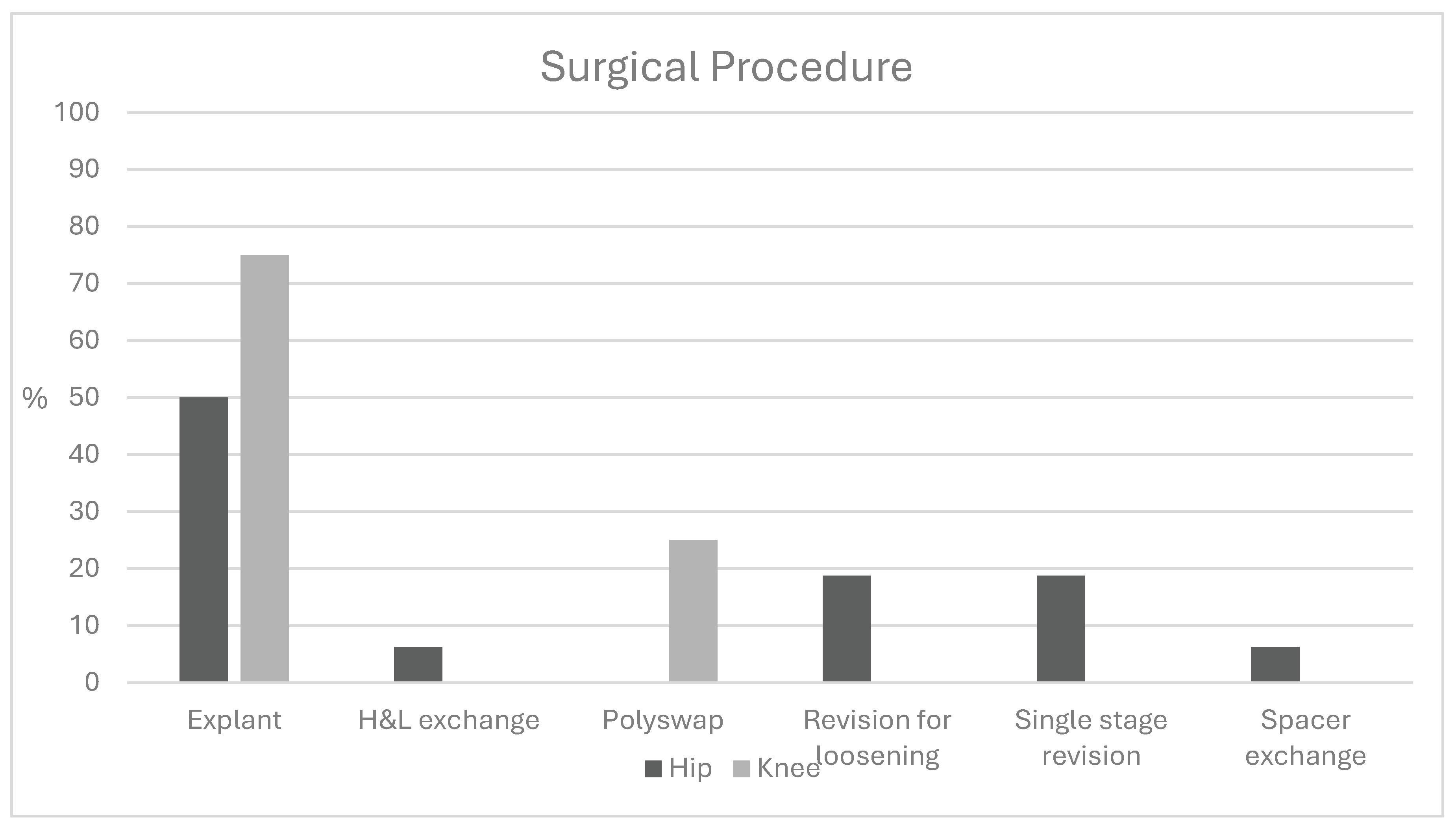
Introduction Cutibacterium species, common commensal gram-positive bacteria, present a diagnostic challenge for arthroplasty surgeons. While Cutibacterium infections have been well characterized in shoulder surgery, their presentation and clinical significance in total hip (THA) and total knee arthroplasty (TKA) remain less understood. Methods A retrospective chart review identified patients with positive Cutibacterium cultures following THA or TKA. Demographics, laboratory values, and microbiologic data were collected. Statistical comparisons were performed using t-tests and chi-squared analysis. One-year outcomes were evaluated using the MSIS ORT criteria among patients undergoing further surgical intervention. Results Twenty-nine patients with Cutibacterium-positive cultures were identified (21 THA, 8 TKA); 15 (52%) were polymicrobial. Ten THA patients (47.6%) and seven TKA patients (87.5%) met MSIS criteria for infection. Mean time to culture positivity was similar between THA (6.8 days) and TKA (7.4 days; p = 0.57). Sonication cultures were positive in 24% of THA and 12.5% of TKA cases. Mean ESR was 36.4 mm/h for THA and 51.5 mm/h for TKA (p = 0.21); mean CRP was 35.2 and 36.8 mg/dL, respectively (p = 0.95). Mean synovial cell counts were 27,055 for THA and 22,194 for TKA, with PMN percentages of 68% and 73.9% (p = 0.72, 0.70). Monomicrobial infections demonstrated a mean cell count of 24,143 with 58.9% PMNs, compared to 25,903 and 78.8% in polymicrobial cases. At one year, 72% of patients undergoing subsequent surgery achieved successful outcomes. Higher ASA classification was the only significant predictor of failure (mean 3.0 vs. 2.75). Conclusion Cutibacterium-associated THA and TKA infections often present with delayed culture growth, mild inflammatory markers, and frequent polymicrobial involvement. Most patients experience favorable outcomes following surgical management, though greater medical comorbidity may predict treatment failure.

Keywords: 
;  ;  

1. Introduction

Infection following total hip arthroplasty (THA) and total knee arthroplasty (TKA) remains an important area of study given the significant costs related to these procedures as well as the morbidity associated with clinical management.[1,2,3] While studied more extensively in orthopaedics as a pathogen following shoulder surgery,[4,5] Cutibacterium following THA and TKA remains poorly understood with respect to unique characteristics of the organism and variable clinical presentations. Cutibacterium species therefore pose an interesting diagnostic challenge for orthopaedic arthroplasty surgeons.
Cutibacterium species, formerly known as Propionibacterium species, are commensal gram-positive skin bacterium which can cause implant-associated infections. They are ubiquitous organisms present in skin flora and are generally found in sebaceous glands. They are notably slow growing, facultatively anaerobic, and non-spore forming. Techniques such as prolonged agar cultures for 14 days as well as tissue sonication have increased the detection of these bacteria.[6] Although once thought to be a cutaneous bacteria of little pathologic significance, there has been increasing evidence for its role in both native as well as implant-associated infections.[7,8] Furthermore, Cutibacterium has been implicated in more systemic diseases such as sarcoidosis, benign prostatic hyperplasia, prostate cancer, and possibly Parkinson’s disease.[9,10,11]
Notable prior work has evaluated Cutibacterium in shoulder arthroplasty [12,13,14,15,16,17] as well as infections following anterior cruciate ligament reconstruction[18], shoulder arthroscopy [19,20,21], and clavicle non-unions[22]. However less has been reported about Cutibacterium in total joint arthroplasty. Elkins et. al assessed anterior and lateral thigh skin colonization of Cutibacterium prior to total hip arthroplasty and found that 14 of the 101 (14%) patients had a positive culture, with most localized to the anterior hip (65%)[23]. Prior work by Nodzo et. al compared infection characteristics for patients undergoing revision TKA for infection due to Cutibacterium acnes versus methicillin sensitive Staphylococcal aureus[24]. Although a small study of 16 patients with P. acnes, they did demonstrate that median ESR was significantly higher in the MSSA group (56.0 mm/h; interquartile range [IQR], 44.3-72.9 vs 23.0 mm/h; IQR, 18.5-52.0; respectively, p = .03). Similar findings were noted for CRP however synovial and serum WBC count did not demonstrate any differences between the two groups. This work also noted a mean time to culture of 8.3 ± 2.0 days in the Cutibacterium group compared to 1.8 ± 0.8 days in the MSSA cohort.
Given the potential for varied and perhaps less overt clinical symptoms in patients with Cutibacterium prosthetic joint infection (PJI), this study sought to evaluate TKA and THA Cutibacterium infections with respect to presenting characteristics, objective clinical workup, treatments, and outcomes of patients undergoing surgical intervention at the 1-year mark.

2. Results

Retrospective review identified a total of 29 patients with positive Cutibacterium cultures during the time period, 21 THAs and 8 TKAs. The average age of the THA patients was 65.8 years and 64.7 years for the TKA patients (p = 0.82). Of the 21 patients with THA infections, 10 preoperatively met MSIS criteria (47.6%), while 7 out of 8 patients with TKA infections preoperatively met MSIS criteria for infection (87.5%) (p= 0.13). Of the eight patients with Cutibacterium TKA infections, seven (87.5%) were female. Comparison of the THA and TKA cohorts did not demonstrate any significant differences with respect to age, sex, ASA, BMI, or prior revision procedure. Other demographic data is shown in Table 1.
Evaluation of the culture data demonstrated that 47.6% of the THA infections and 62.5% of the TKA infections were polymicrobial, with Cutibacterium as one of the identified species (Table 2). Average time to culture positivity was 6.8 days for THA and 7.4 days for TKA (p = 0.572). The rate of positive cultures was 0.53 for THA and 0.27 for TKA (p = 0.059). The number of sonicated tissue cultures that were positive for Cutibacterium was not statistically significantly different for THA and TKA, with 5 out of 21 sonicated samples testing positive in the THA group (23.8%), and 1 out of 8 sonicated samples testing positive in the TKA group (p=0.242).
Laboratory markers including ESR and CRP were also recorded pre-operatively or at the time of revision surgery and demonstrated a mean ESR of 36.4 mm/h for THA (range 9-106) and 51.5 mm/h for TKA (range 10-102) (p = 0.21). Mean CRP was 35.2 mg/dL for THA and 36.8 mg/dL for TKA (p = 0.95). Mean cell count and percentage polymorphonuclear cells from intra-articular aspiration was 27,055 cells (range of 34-128,205) with 68% (range 2-98%) PMNs for THA and 22,194 (range of 95-29,829) with 73.9% (range 8-87%) PMNs for TKA (p = 0.72 and p = 0.70, respectively). Mean cell count and percentage polymorphonuclear cells from intra-articular aspiration was 24,143 (range of 95 to 128,205) with 58.9% (range of 8-98%) PMNs for monomicrobial infections and 25,903 (range of 135 to 89,378) with 78.8% (range of 2-98%) PMNs for polymicrobial infections.
Of the 29 patients with THA and TKA Cutibacterium infections, 24 (82.7%) underwent a surgical intervention (Figure 1): 14 explant of arthroplasty implants, 3 revisions for loosening, 2 TKA polyethylene exchange procedures, 1 spacer exchange, 3 single stage revisions, and 1 head and liner exchange. Of the 8 TKA patients, 6 (75%) underwent an explant, while 8 out of the 21 THA patients underwent an explant of the components (38.1%). The other 5 patients were treated non-operatively with antibiotics.
For patients with positive Cutibacterium THA cultures, approach for the primary THA procedure was also further investigated (Table 3). Of the 21 patients in our study, 10 underwent prior THA via an anterior approach and 8 were performed via a posterior approach (p= 0.92). Five of the ten (50%) anterior approach patients had monomicrobial infections, and similarly four of the eight (50%) posterior approach patients had monomicrobial infections.
For those patients who did undergo subsequent surgical intervention following their positive periprosthetic Cutibacterium culture and had follow up at one year (n =22), outcomes for this cohort were evaluated using the MSIS ORT (Table 4). [26] Overall, 16 of the 22 (72%) had successful outcomes at one year. Only ASA was noted to be statistically significantly different between the two groups (3 for the failure group versus 2.75 for the successful outcome group, p = 0.03).

3. Discussion

Detection and management of Cutibacterium prosthetic joint infection can pose a diagnostic challenge for orthopaedic arthroplasty surgeons given more subtle clinical as well as laboratory changes compared to more virulent microbial species, particularly in monomicrobial communities.[27,28,29] Our study showed that half of the THA and half of the TKA patients with Cutibacterium presented with polymicrobial infections with the most common concomitant bacteria being coagulase negative Staphylococcus. This is consistent with prior work demonstrating Cutibacterium as an overall rare monomicrobial pathogen that is not infrequently found in conjunction with other microorganisms.[32,33] We compared laboratory values for monomicrobial and polymicrobial Cutibacterium infections in THA and TKA patients and did not find statistically significant differences. However, average CRP of the monomicrobial group was lower, with an average of 26.5 vs 44.2 mg/dL for the polymicrobial group, regardless of the joint, supporting the potentially indolent nature of the bacteria. There was no significant difference between the frequency of polymicrobial PJI in THA and TKA patients, with 5 out of 8 TKA patients and 10 out of 21 THA patients having polymicrobial Cutibacterium infections. Further, our work showed a delay in time to culture-positivity of approximately 1-week, necessitating the need for extended cultures to assist in diagnosis (mean of 6.5 days for THA and 7.39 days for TKA).
WBC, ESR, CRP, and percentage PNMs on average for both TKA and THA patients with Cutibacterium infections were found to be elevated. Average CRP and ESR values both met MSIS criteria of greater than 30 mm/h and greater than 10 mg/L, respectively.[31] However, it should be noted that when studied in isolation, only 47% of our THA patients met MSIS criteria pre-operatively when compared to 87% of TKA patients. We believe this point further strengthens the believe that Cutibacterium poses more of a threat to THA than TKA patients as commonly accepted diagnostic algorithms may miss the diagnosis.
Prior studies have shown Cutibacterium infections to be more common in males than females who have had shoulder surgery.[34] This is consistent with the fact that Cutibacterium is present in areas with greater concentration of sebaceous glands, and men have a greater concentration of sebaceous glands than women.[35] Our data does not show a statistically significant difference in men compared women with regards to TKA or THA infections with Cutibacterium. This discrepancy could be related to the relative abundance of Cutibacterium acnes around the axilla and shoulder region compared with the hip and knee. Using this theory, other studies have compared the difference between anterior and posterior approaches for THA and the relative rates of Cutibacterium infections. The theory was that perhaps there would be a higher rate of Cutibacterium culture positivity with the anterior approach given the larger concentration of sebaceous glands in this region. One study found increased rates of Cutibacterium avidum[36] in patients with an anterior compared to posterior THA. Our study did not find a statistically significant difference in the rates of Cutibacterium infections based on the approach of the initial arthroplasty procedure. Further studies have shown that the direct anterior approach to primary THA does not increase the risk of periprosthetic joint infection compared to the direct lateral approach. Additionally, there were not significant differences in infecting organisms between anterior and direct lateral approach groups[37]. Interestingly, of the 29 arthroplasty patients with Cutibacterium infections in this study, 21 were from THA’s, with just 8 from TKA’s. This is consistent with the study by Elkins et al, mentioned in the introduction, which showed that the anterior thigh has a high colonization rate of Cutibacterium. [23]
This study has several limitations as a retrospective analysis of a relatively uncommon organism culture in THA and TKA. As mentioned, Cutibacterium are ubiquitous organisms present in skin flora and some have suggested that isolation of the bacteria may represent a contaminant.[38,39] The present study sought to evaluate all patients with a positive culture, though we do acknowledge that of the 21 patients with THA infections, 10 preoperatively met MSIS criteria for PJI (47.6%) while 7 out of 8 patients with TKA infections preoperatively met current MSIS criteria for infection (87.5%) (p= 0.13). This finding may be explained by the less virulent nature of Cutibacterium to elicit common warning signs for PJI such as significant erythema, effusion, or sinus tract as well as the difficulty with culture of the bacteria. However, it is also possible that the Cutibacterium represents a contaminant or a non-pathologic organism in the polymicrobial infections. The purpose of this work was not to decipher such a difference but rather to report laboratory and other characteristics of those with a positive Cutibacterium prosthetic culture. Another limitation of this study is that it utilized conventional culture, which may have missed some positive identification of Cutibacterium possible with other methods (e.g., next generation sequencing), however we suspect the use of traditional cultures is more generalizable given the feasibility of this method. Furthermore, additional cases of Cutibacterium may be missed by cultures not being held for an extended time (14 days in our institution). As this study found, the average time to culture positivity of approximately one week (mean of 6.5 days with a range of 3-11 for THA and 7.3 days with a range of 5-12 for TKA). Lastly, there may have been other patient or clinical characteristics that influence the laboratory values and outcomes evaluated.

4. Methods

Retrospective chart review was performed to identify patients with culture-positivity Cutibacterium infection of a THA or TKA within the time period at our tertiary academic referral center. Using the electronic medical record, all Propionibacterium or Cutibacterium positive cultures from April 2014 to January 2021 were identified. Those that involved a native joint or an infection of a prosthetic joint that was not a hip or knee arthroplasty were excluded (Table S1) All procedures were reviewed to confirm that each had at least one culture with Cutibacterium or Propionibacterium. Using this criteria, 29 total cases were identified for further analysis. Importantly, this study was conducted as a retrospective review of de-identified data. No patient names, medical record numbers, or other identifiable health information were collected, stored, or reported. As such, this project did not meet the definition of human subjects research under 45 CFR 46.102(f) and therefore did not require Institutional Review Board (IRB) approval.
Patient demographics including gender, age, and BMI were collected. American Society of Anesthesiology (ASA) scores were used to evaluate each patient’s relative overall health. Preoperative lab values were collected, including ESR, CRP, WBC count, percentage of Polymorphonuclear leukocytes (% PMNs), and presence of alpha-defensin. Culture data was collected from either synovial fluid and/or from tissue in cases where the patient underwent subsequent operative intervention. Per institutional standards for periprosthetic joints, cultures were incubated for 14 days, with the exception of one sample which was held for seven days. Previous surgical interventions were recorded. Using the 2018 Musculoskeletal Infection Society (MSIS) criteria for prosthetic joint infection, each patient was dichotomized as infected with a score of ≥6 or not definitively infected with a score of ≤5.[25] One-year outcomes for patients who underwent subsequent surgical intervention and had documented clinical follow-up (n = 22) were categorized according to the Musculoskeletal Infection Society Outcome Reporting Tool (MSIS ORT, Table 4).[26] A single physician reviewer (TMB) retrospectively evaluated patient charts and assigned each patient into “success” (Tier 1 and Tier 2) and “failure due to either PJI or failure due to a secondary cause” (all other Tiers).

Statistical Analysis

Analysis of patient, laboratory, microbiologic and surgical characteristics was performed to evaluate the cohort as a whole and to compare THA to TKA. T-test and chi-squared analysis was utilized to compare patient baseline, demographic, and laboratory characteristics. Additional analysis evaluated outcomes at one year per the MSIS ORT for those who underwent surgical intervention and subsequent analysis compared those with “success” (Tiers 1 and 2) versus “failure” ((Tiers 3A, 3B, 3C, 3D, 3E, 3F, 4A, 4B).

5. Conclusions

This study contributes to the current literature on Cutibacterium culture positivity in TKA and THA patients. With longer culture times becoming more standard, Cutibacterium infections will continue to present itself as a factor in PJI. This research illuminates some of the characteristics of monomicrobial and polymicrobial Cutibacterium infections in the hip and knee and demonstrates that Cutibacterium must be considered as a pathogen when suspecting a PJI in the hip and knee. Further research is needed to identify just how prominent Cutibacterium infections in a THA and TKA are, and to identify the ideal treatments to optimize patient outcomes.

Supplementary Materials

The following supporting information can be downloaded at the website of this paper posted on Preprints.org.

Author Contributions

Methodology, B.L., A.D., T.B. and P.W.; Formal analysis, P.W. and W.M.; Investigation, W.M.; Writing – original draft, B.L. and A.D.; Writing – review & editing, B.L., A.D., T.B., P.W. and W.M.; Supervision, W.M.

Funding

This research received no external funding.

Institutional Review Board Statement

Not applicable and IRB was not required for the current study given the use of patient de-identified information.

Informed Consent Statement

Patient consent was waived due to our study not utilizing or requiring any patient identifying information.

Data Availability Statement

The original contributions presented in this study are included in the article/supplementary material. Further inquiries can be directed to the corresponding author.

Conflicts of Interest

The authors declare no conflicts of interest.

References

  1. Boddapati, V; Fu, MC; Mayman, DJ; Su, EP; Sculco, PK; McLawhorn, AS. Revision Total Knee Arthroplasty for Periprosthetic Joint Infection Is Associated With Increased Postoperative Morbidity and Mortality Relative to Noninfectious Revisions. The Journal of Arthroplasty 2018, 33, 521–6. [Google Scholar] [CrossRef] [PubMed]
  2. Bozic, KJ. The Impact of Infection After Total Hip Arthroplasty on Hospital and Surgeon Resource Utilization. J Bone Joint Surg Am 2005, 87, 1746. [Google Scholar] [CrossRef] [PubMed]
  3. Kurtz, SM; Lau, E; Watson, H; Schmier, JK; Parvizi, J. Economic Burden of Periprosthetic Joint Infection in the United States. The Journal of Arthroplasty 2012, 27, 61–65.e1. [Google Scholar] [CrossRef] [PubMed]
  4. Dodson, CC; Craig, EV; Cordasco, FA; Dines, DM; Dines, JS; DiCarlo, E; et al. Propionibacterium acnes infection after shoulder arthroplasty: A diagnostic challenge. Journal of Shoulder and Elbow Surgery 2010, 19, 303–7. [Google Scholar] [CrossRef]
  5. Sperling, JW; Kozak, TK; Hanssen, AD; Cofield, RH. Infection after shoulder arthroplasty. Clin Orthop Relat Res 2001, 206–16. [Google Scholar] [CrossRef]
  6. Aubin, GG; Portillo, ME; Trampuz, A; Corvec, S. Propionibacterium acnes, an emerging pathogen: From acne to implant-infections, from phylotype to resistance. Médecine et Maladies Infectieuses 2014, 44, 241–50. [Google Scholar] [CrossRef]
  7. Khalil, JG; Gandhi, SD; Park, DK; Fischgrund, JS. Cutibacterium acnes in Spine Pathology: Pathophysiology, Diagnosis, and Management. Journal of the American Academy of Orthopaedic Surgeons 2019, 27, e633–40. [Google Scholar] [CrossRef]
  8. Lutz, M-F; Berthelot, P; Fresard, A; Cazorla, C; Carricajo, A; Vautrin, A-C; et al. Arthroplastic and osteosynthetic infections due to Propionibacterium acnes: a retrospective study of 52 cases, 1995-2002. Eur J Clin Microbiol Infect Dis 2005, 24, 739–44. [Google Scholar] [CrossRef]
  9. Leheste, JR; Ruvolo, KE; Chrostowski, JE; Rivera, K; Husko, C; Miceli, A; et al. P. acnes-Driven Disease Pathology: Current Knowledge and Future Directions. Front Cell Infect Microbiol 2017, 7, 81. [Google Scholar] [CrossRef]
  10. Mourad, M; Passley, TM; Purcell, JM; Leheste, JR. Early-Onset Parkinson’s Disease With Multiple Positive Intraoperative Spinal Tissue Cultures for Cutibacterium acnes. Cureus 2021, 13, e17607. [Google Scholar] [CrossRef]
  11. Negi, M; Takemura, T; Guzman, J; Uchida, K; Furukawa, A; Suzuki, Y; et al. Localization of Propionibacterium acnes in granulomas supports a possible etiologic link between sarcoidosis and the bacterium. Mod Pathol 2012, 25, 1284–97. [Google Scholar] [CrossRef] [PubMed]
  12. Koh, CK; Marsh, JP; Drinković, D; Walker, CG; Poon, PC. Propionibacterium acnes in primary shoulder arthroplasty: rates of colonization, patient risk factors, and efficacy of perioperative prophylaxis. Journal of Shoulder and Elbow Surgery 2016, 25, 846–52. [Google Scholar] [CrossRef] [PubMed]
  13. Chuang, MJ; Jancosko, JJ; Mendoza, V; Nottage, WM. The Incidence of Propionibacterium acnes in Shoulder Arthroscopy; The Journal of Arthroscopic & Related Surgery: Arthroscopy, 2015; Volume 31, pp. 1702–7. [Google Scholar] [CrossRef]
  14. Namdari, S; Nicholson, T; Parvizi, J; Ramsey, M. Preoperative doxycycline does not decolonize Propionibacterium acnes from the skin of the shoulder: a randomized controlled trial. Journal of Shoulder and Elbow Surgery 2017, 26, 1495–9. [Google Scholar] [CrossRef] [PubMed]
  15. Kolakowski, L; Lai, JK; Duvall, GT; Jauregui, JJ; Dubina, AG; Jones, DL; et al. Neer Award 2018: Benzoyl peroxide effectively decreases preoperative Cutibacterium acnes shoulder burden: a prospective randomized controlled trial. Journal of Shoulder and Elbow Surgery 2018, 27, 1539–44. [Google Scholar] [CrossRef]
  16. Qiu, B; Al, K; Pena-Diaz, AM; Athwal, GS; Drosdowech, D; Faber, KJ; et al. Cutibacterium acnes and the shoulder microbiome. Journal of Shoulder and Elbow Surgery 2018, 27, 1734–9. [Google Scholar] [CrossRef]
  17. Duvall, G; Kaveeshwar, S; Sood, A; Klein, A; Williams, K; Kolakowski, L; et al. Benzoyl peroxide use transiently decreases Cutibacterium acnes load on the shoulder. Journal of Shoulder and Elbow Surgery 2020, 29, 794–8. [Google Scholar] [CrossRef]
  18. DiBartola, AC; Swank, KR; Flanigan, DC. Anterior cruciate ligament reconstruction complicated by Propionibacterium acnes infection: case series. The Physician and Sportsmedicine 2018, 46, 273–8. [Google Scholar] [CrossRef]
  19. Patzer, T; Petersdorf, S; Krauspe, R; Verde, PE; Henrich, B; Hufeland, M. Prevalence of Propionibacterium acnes in the glenohumeral compared with the subacromial space in primary shoulder arthroscopies. Journal of Shoulder and Elbow Surgery 2018, 27, 771–6. [Google Scholar] [CrossRef]
  20. Khan, U; Torrance, E; Townsend, R; Davies, S; Mackenzie, T; Funk, L. Low-grade infections in nonarthroplasty shoulder surgery. Journal of Shoulder and Elbow Surgery 2017, 26, 1553–61. [Google Scholar] [CrossRef]
  21. Kajita, Y; Iwahori, Y; Harada, Y; Deie, M. Incidence of Propionibacterium acnes in arthroscopic rotator cuff repair. Journal of Orthopaedic Science 2020, 25, 110–4. [Google Scholar] [CrossRef]
  22. Gausden, EB; Villa, J; Warner, SJ; Redko, M; Pearle, A; Miller, A; et al. Nonunion After Clavicle Osteosynthesis: High Incidence of Propionibacterium acnes. Journal of Orthopaedic Trauma 2017, 31, 229–35. [Google Scholar] [CrossRef]
  23. Elkins, JM; Dennis, DA; Kleeman-Forsthuber, L; Yang, CC; Miner, TM; Jennings, JM. Cutibacterium colonization of the anterior and lateral thigh. The Bone & Joint Journal 2020, 102-B, 52–6. [Google Scholar] [CrossRef]
  24. Nodzo, SR; Westrich, GH; Henry, MW; Miller, AO. Clinical Analysis of Propionibacterium acnes Infection After Total Knee Arthroplasty. The Journal of Arthroplasty 2016, 31, 1986–9. [Google Scholar] [CrossRef]
  25. Parvizi, J; Tan, TL; Goswami, K; Higuera, C; Della Valle, C; Chen, AF; et al. The 2018 Definition of Periprosthetic Hip and Knee Infection: An Evidence-Based and Validated Criteria. The Journal of Arthroplasty 2018, 33, 1309–1314.e2. [Google Scholar] [CrossRef] [PubMed]
  26. Fillingham, YA; Della Valle, CJ; Suleiman, LI; Springer, BD; Gehrke, T; Bini, SA; et al. Definition of Successful Infection Management and Guidelines for Reporting of Outcomes After Surgical Treatment of Periprosthetic Joint Infection: From the Workgroup of the Musculoskeletal Infection Society (MSIS). The Journal of Bone and Joint Surgery 2019, 101, e69. [Google Scholar] [CrossRef] [PubMed]
  27. Elston, MJ; Dupaix, JP; Opanova, MI; Atkinson, RE. Cutibacterium acnes (formerly Proprionibacterium acnes) and Shoulder Surgery. Hawaii J Health Soc Welf 2019, 78, 3–5. [Google Scholar] [PubMed]
  28. Zeller, V; Ghorbani, A; Strady, C; Leonard, P; Mamoudy, P; Desplaces, N. Propionibacterium acnes: an agent of prosthetic joint infection and colonization. J Infect 2007, 55, 119–24. [Google Scholar] [CrossRef]
  29. Renz, N; Mudrovcic, S; Perka, C; Trampuz, A. Orthopedic implant-associated infections caused by Cutibacterium spp. – A remaining diagnostic challenge. PLoS ONE 2018, 13, e0202639. [Google Scholar] [CrossRef]
  30. Alijanipour, P; Bakhshi, H; Parvizi, J. Diagnosis of Periprosthetic Joint Infection: The Threshold for Serological Markers. Clinical Orthopaedics and Related Research® 2013, 471, 3186–95. [Google Scholar] [CrossRef]
  31. New Definition for Periprosthetic Joint Infection. The Journal of Arthroplasty 2011, 26, 1136–8. [CrossRef]
  32. Flurin, L; Greenwood-Quaintance, KE; Patel, R. Microbiology of polymicrobial prosthetic joint infection. Diagnostic Microbiology and Infectious Disease 2019, 94, 255–9. [Google Scholar] [CrossRef]
  33. Boisrenoult, P. Cutibacterium acnes prosthetic joint infection: Diagnosis and treatment. Orthopaedics & Traumatology: Surgery & Research 2018, 104, S19–24. [Google Scholar] [CrossRef]
  34. Batten, TJ; Gallacher, S; Thomas, WJ; Kitson, J; Smith, CD. C. acnes in the joint, is it all just a false positive? Eur J Orthop Surg Traumatol 2022. [Google Scholar] [CrossRef]
  35. Portillo, ME; Corvec, S; Borens, O; Trampuz, A. Propionibacterium acnes: An Underestimated Pathogen in Implant-Associated Infections. BioMed Research International 2013, 2013, e804391. [Google Scholar] [CrossRef]
  36. Achermann, Y; Liu, J; Zbinden, R; Zingg, PO; Anagnostopoulos, A; Barnard, E; et al. Propionibacterium avidum: A Virulent Pathogen Causing Hip Periprosthetic Joint Infection. Clin Infect Dis 2018, 66, 54–63. [Google Scholar] [CrossRef] [PubMed]
  37. Shohat, N; Goswami, K; Clarkson, S; Chisari, E; Breckenridge, L; Gursay, D; et al. Direct Anterior Approach to the Hip Does Not Increase the Risk for Subsequent Periprosthetic Joint Infection. The Journal of Arthroplasty 2021, 36, 2038–43. [Google Scholar] [CrossRef] [PubMed]
  38. Namdari, S; Nicholson, T; Parvizi, J. Cutibacterium acnes is Isolated from Air Swabs: Time to Doubt the Value of Traditional Cultures in Shoulder Surgery? Arch Bone Jt Surg 2020, 8, 506–10. [Google Scholar] [CrossRef]
  39. Liew-Littorin, C; Brüggemann, H; Davidsson, S; Nilsdotter-Augustinsson, Å; Hellmark, B; Söderquist, B. Clonal diversity of Cutibacterium acnes (formerly Propionibacterium acnes) in prosthetic joint infections. Anaerobe 2019, 59, 54–60. [Google Scholar] [CrossRef] [PubMed]
Figure 1. Subsequent surgical procedures performed following positive Cutibacterium culture.
Preprints 190942 g001
Table 1. Patient demographic and baseline characteristics with comparison between THA and TKA.
Demographic THA TKA p-value
N 21 8
Male Sex (%) 10 (47.6) 1 (12.5) 0.19
Age (mean (SD)) 65.8 (9.32) 64.7 (12.0) 0.82
BMI (mean (SD)) 34.6 (6.7) 31.2 (11.3) 0.47
ASA (mean (SD)) 2.79 (0.43) 2.62 (0.52) 0.44
Prior revision (%) 0.23
No 17 (81.0) 5 (62.5)
Unknown 0 (0.0) 1 (12.5)
Yes 4 (19.0) 2 (25.0)
Met MSIS Criteria (%) 10 (47.6) 7 (87.5) 0.13
Total Patients 21 8
BMI = body mass index; ASA = American Society of Anesthesiology; MSIS = Musculoskeletal Infection Society.
Table 2. Laboratory data with comparison between THA and TKA.
Laboratory Data THA TKA p-value
Polymicrobial (%) 10 (47.6) 5 (62.5) 0.68
Time to culture positivity (mean (SD)) 6.75 (2.5) 7.39 (2.6) 0.57
Number of positive cultures (mean (SD)) 2.05 (1.6) 1.75 (1.5) 0.66
WBC count (mean (SD)) 27,055 (3,9922) 22,195 (2,9172) 0.77
PMN % (mean (SD)) 67.92 (36.5) 73.88 (29.3) 0.70
Sonicated tissues positive for Cutibacterium (%) 0.24
No 16 (76.2) 7 (87.5)
Yes 5 (23.8) 1 (12.5)
Alpha Defensin (%) 0.41
Unknown 17 (81.0) 8 (100.0)
Negative 2 (9.5) 0 (0.0)
Positive 2 (9.5) 0 (0.0)
ESR mm/h (mean (SD)) 36.38 (27.3) 51.50 (31.1) 0.21
CRP (mean (SD)) 35.23 (63.0) 36.80 (38.0) 0.95
WBC = white blood cells; PMN = polymorphonuclear cells; ESR = erythrocyte sedimentation rate; CRP = C-reactive protein.
Table 3. Initial THA approach and presence of monomicrobial versus polymicrobial cultures. Of note, three patients in the THA group had prior THA via an unknown approach and are not included in this analysis.
THA Anterior Approach THA Posterior Approach
Monomicrobial 6 4
Polymicrobial 4 4
Total 10 8
Table 4. MSIS Outcomes reporting tool utilized for reporting outcomes at 1 year for those patients undergoing surgical intervention after positive Cutibacterium culture.
Tier 1. Infection control with no continued antibiotic therapy
Tier 2. Infection control with the patient on suppressive antibiotic therapy
Tier 3. Need for reoperation and/or revision and/or spacer retention (assigned to subgroups A, B, C, D, E, and F based on the type of reoperation)
Aseptic revision at >1 year from initiation of PJI treatment
Septic revision (including debridement, antibiotics, and implant retention [DAIR]) at >1 year from initiation of PJI treatment (excluding amputation, resection arthroplasty, and arthrodesis)
Aseptic revision at ≤1 year from initiation of PJI treatment
Septic revision (including DAIR) at ≤1 year from initiation of PJI treatment (excluding amputation, resection arthroplasty, and arthrodesis)
Amputation, resection arthroplasty, or arthrodesis
Retained spacer
Tier 4. Death (assigned to subgroups A or B).
Death ≤1 year from initiation of PJI treatment
Death >1 year from initiation of PJI treatment
Legend: Infection management outcome categories, Successful: Tiers 1, 2, Failure directly or indirectly related to PJI or due to secondary causes: Tiers 3A, 3B, 3C, 3D, 3E, 3F, 4A, 4B.
Disclaimer/Publisher’s Note: The statements, opinions and data contained in all publications are solely those of the individual author(s) and contributor(s) and not of MDPI and/or the editor(s). MDPI and/or the editor(s) disclaim responsibility for any injury to people or property resulting from any ideas, methods, instructions or products referred to in the content.
Copyright: This open access article is published under a Creative Commons CC BY 4.0 license, which permit the free download, distribution, and reuse, provided that the author and preprint are cited in any reuse.
Prerpints.org logo

Preprints.org is a free preprint server supported by MDPI in Basel, Switzerland.

Subscribe

Disclaimer

Terms of Use

Privacy Policy

Privacy Settings

© 2025 MDPI (Basel, Switzerland) unless otherwise stated