Preprint
Review

This version is not peer-reviewed.

Mapping the Brain’s Glymphatic System

Submitted:

19 December 2025

Posted:

22 December 2025

You are already at the latest version

Abstract

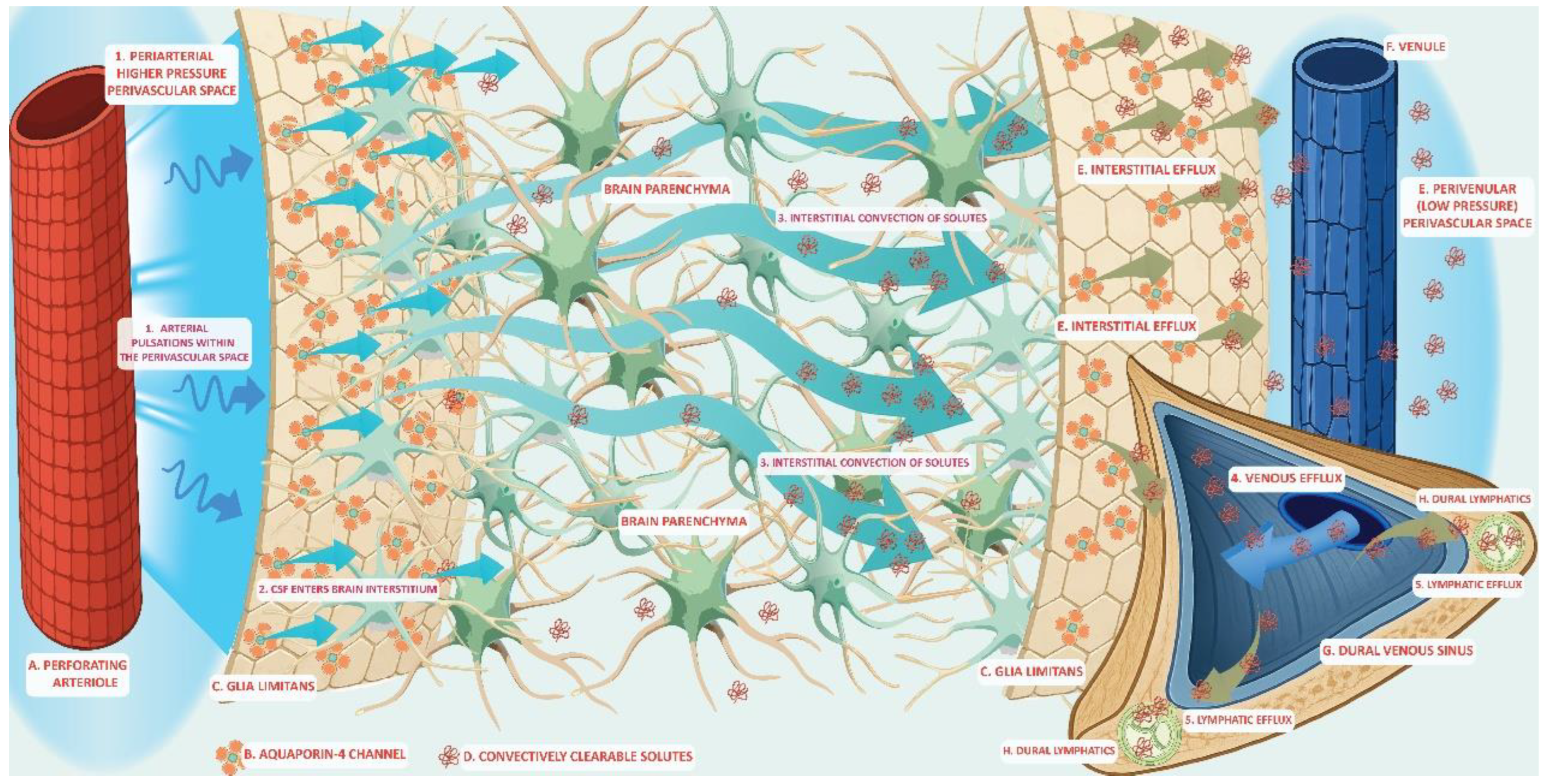
The glymphatic system is a fluid-transport framework in which cerebrospinal fluid (CSF) enters the brain along perivascular routes, exchanges with interstitial fluid (ISF), and exits toward venous, perineural, and meningeal lymphatic pathways enabling waste clearance. Recent studies have clarified the anatomical components that regulate solute movement. The perivascular astrocyte end feet, which is enriched in polarized aquaporin-4 (AQP4) expression, creates a high-permeability water interface that facilitates CSF–ISF exchange. Multiscale physical drivers such as cardiac pulsation, arteriolar vasomotion, and brain-state changes during sleep regulate timing and efficiency of the glymphatic transport. A broad spectrum of solutes is transported through this pathway, from small metabolites to extracellular proteins including amyloid-β and tau, as well as exogenous tracers and some lipid-associated species. Glymphatic redistribution may interface with other clearance systems including the brain-to-blood efflux via blood-brain barrier (BBB) transport, the intramural periarterial drainage (IPAD) that clears along vascular basement membranes and the meningeal lymphatic pathways that drain macromolecules to deep cervical lymph nodes. These different routes may be interconnected and represent a waste clearance network with complementary roles assigned to different mechanisms. Moreover, state dependence (notably sleep) and vascular health modulate glymphatic flux, offering plausible links between glymphatic system dysfunction to aging and neurodegeneration. Methodological advances—from intrathecal contrast magnetic resonance imaging (MRI) to in vivo two-photon imaging and tracer-kinetic modeling—have provided new insights into the anatomical scaffold and kinetics of the glymphatic system. Advances in glymphatic anatomy, together with growing evidence implicating glymphatic dysfunction in neurodegeneration, make a unifying framework urgently needed. Our synthesis spans glymphatic structure, fluid routing, the repertoire of transported solutes, and links to complementary clearance routes, supporting a unified model in which glymphatic clearance represents a core mechanism of cerebral homeostasis. Understanding glymphatic dysfunction may guide the establishment of diagnostic imaging biomarkers that have the potential to assist in therapeutic modulation of neurodegenerative diseases.

Keywords: 
;  ;  ;  ;  ;  ;  ;  ;  ;  

1. Introduction

The central nervous system (CNS) is among the most metabolically active organs consuming nearly 20% of total oxygen at rest and generating large quantities of metabolic by-products, including lactate, reactive oxygen species, and aggregation-prone proteins such as amyloid-β (Aβ) and tau [1,2]. Efficient clearance of these metabolites and solutes from the interstitial extracellular space is essential to prevent toxicity and maintain brain homeostasis [3]. Moreover, clearance mechanisms sustain ionic balance and neurotransmission, both fundamental for neural activity and overall CNS function [4,5]. Additionally, impairment of waste clearance is increasingly recognized as a central element in the pathogenesis of several neurological disorders [3].
In peripheral tissues, waste clearance is efficiently carried out predominantly by the lymphatic system, which drains interstitial fluid (ISF) and solutes toward lymph nodes. However, the brain poses a paradox; the parenchyma lacks conventional lymphatic vessels, apart from the recently characterized meningeal lymphatics that reside in the dura matter [6,7]. For decades, the absence of a classical lymphatic drainage system prompted investigations into alternative pathways capable of clearing waste from the brain.
Early observations established the dynamic circulation of cerebrospinal fluid (CSF) through the ventricles and the subarachnoid spaces as well as an exchange of solutes between the CSF and the cerebral extracellular space [8]. These studies suggested a role for CSF in the waste clearance process via perivascular routes, yet the precise anatomical pathways mediating CSF–ISF exchange remained elusive. Only in the past decade have advances in in vivo imaging and tracer kinetics clarified the pivotal role of CSF in perivascular clearance processes [3].
The present review synthesizes current knowledge on the anatomy and physiology of the glymphatic system, a glia-dependent perivascular network facilitating CSF-ISF exchange and interstitial solute clearance. The following sections delineate the structural components that regulate glymphatic transport, including perivascular spaces (PVS) and aquaporin-4 (AQP4) water channels, describe the variety of solutes conveyed through these routes and discuss the interface between glymphatic flow and complementary clearance mechanisms. Finally, we propose an integrated view of the brain waste removal, emphasizing the interdependence of perivascular, lymphatic and barrier-mediated pathways in maintaining cerebral homeostasis.

2. The Glymphatic System

The discovery of a brain-wide perivascular transport pathway - the glymphatic system - has redefined the understanding of how CSF communicates with ISF to mediate waste clearance. Iliff et al. (2012) first demonstrated that fluorescent tracers infused into the cisterna magna enter the brain along periarterial spaces and exit along perivenous routes [9]. These experiments revealed a directed convective movement of CSF through the parenchyma, suggesting the presence of a glial-dependent transport mechanism coupling CSF flow to the interstitial solute clearance. The dependence by a glial cell type and the functional similarity to the lymphatic transport led tο the designation of this system as the “glymphatic system”.
Using fluorescent tracers and in vivo imaging, Xie et al. (2013) demonstrated that the glymphatic activity is state-dependent with the interstitial space volume expanding by ~60% during natural sleep or anesthesia [10]. The CSF–ISF exchange was enhanced and Aβ clearance was accelerated during sleep, while in the awake state, elevated noradrenergic tone suppressed glymphatic transport. In contrast to passive diffusion, the glymphatic system operates through convective fluxes, driven by multiple physiological forces including arterial pulsatility, slow vasomotion and respiratory cycles [11,12]. These dynamic factors generate oscillating perivascular pressure gradients that propel CSF into the brain parenchyma and facilitate ISF efflux towards perivenous spaces [7].
Whole-brain imaging in rodents using dynamic contrast enhanced magnetic resonance imaging (MRI) confirmed the brain-wide extent of the glymphatic pathway. Two studies from the same research group demonstrated the periarterial CSF influx of intrathecally injected contrast agents and molecular size-dependent CSF-ISF exchange, with subsequent efflux along perivenous spaces [13,14]. Supported by complementary optical microscopy data, these imaging studies provided the first direct evidence for a coordinated, global anatomic framework of the glymphatic system.
Translation of previous findings to humans soon followed through MRI tracer studies. Using intrathecal administration of gadobutrol, a gadolinium-based tracer, researchers visualized CSF influx through periarterial spaces, its parenchymal distribution and subsequent drainage towards cervical lymphatic structures [15,16]. Importantly, tracer clearance was delayed in participants with dementia [15], demonstrating the clinical relevance of this pathway and its potential role in impaired solute elimination.
This conceptual breakthrough consolidated the glymphatic concept across species, providing mechanistic and translational continuity from rodents to humans. The recognition of a dynamic, glia-dependent clearance network has reshaped our understanding of CNS homeostasis and provided new insights into processes potentially underlying aging, neurodegeneration and cerebrovascular disease.

3. The Anatomy and Composition of the Glymphatic System

According to the prevailing model of the glymphatic anatomy, CSF enters the brain along periarterial PVS, also known as or Virchow–Robin spaces. These periarterial compartments are ensheathed by an astrocytic endfeet enriched with AQP-4 channels, which enable water flux across the astrocytic membrane and facilitate CSF-ISF exchange. The resulting CSF–ISF admixture is directed then towards perivenous spaces ultimately draining into meningeal and cervical lymphatic structures (Figure 1) [17,18]. Additionally, recently identified lymphatic vessels in the dura mater provide a structural bridge between perivascular glymphatic clearance and extracranial lymphatic drainage [19]. Therefore, the glymphatic flow is structurally dependent on the volume of the PVS and the polarization of AQP4 channels, features that are further discussed in detail.

3.1. Perivascular Spaces (PVS)

Virchow–Robin spaces are pial-lined, ISF-filled compartments that line penetrating cerebral arterioles, capillaries and venules as those vessels traverse through the brain parenchyma [20]. PVS are located at predictable anatomical locations (e.g. cortex and basal ganglia), and their dimensions vary with age, vascular risk factors as well as disease state [21]. Each space comprises an endothelial vessel wall surrounded by a pial sheath and an outer boundary formed by the astrocytic endfeet densely populated with AQP4 channels underlain by the basal lamina of the glia limitans [22]. Perivascular basement membranes formed by endothelial and glial extracellular matrix components are key structural elements that delineate the trajectory of the glymphatic flow [23].
The role of PVS extends beyond facilitating convective fluid movement and waste clearance. Their topology and CSF-ISF flux patterns provide pathways for the distribution of nutrients, neuromodulators and growth factors across brain regions, thereby supporting metabolic and ionic homeostasis [24,25].
Immune cells that accumulate in PVS during neuroinflammatory states can exit the CNS along glymphatic flow towards the deep cervical lymph nodes, linking central immune surveillance to the meningeal lymphatic system [26]. During physiological conditions, this process promotes immune monitoring, while in pathological contexts, it facilitates the removal of immune cells from the inflamed parenchyma [27]. The presence of immune cells and axonal antigens in the cervical lymph nodes has been documented in several CNS disorders [20]. Concurrently, enlarged PVS have been reported in patients with Alzheimer’s disease (AD) [28], dementia [29] and multiple sclerosis [21] among other conditions. Dysregulation of the PVS-lymphatic system axis and increased immune trafficking to cervical lymph nodes may thus represent converging mechanisms in neurodegenerative pathophysiology.
Tthe PVS compartment also accommodates vascular pulsations that drive glymphatic flow. Arterial pulsations within the central lumen generate perivascular fluid propulsion [30]. Alterations in vessel compliance, such as those induced by hypertension, impair the perivascular pumping and can lead to stagnation of the glymphatic efflux through the PVS [11]. Such dysfunction ultimately compromises waste clearance and parenchymal homeostasis.
Overall, PVS constitute the fundamental anatomic substrate of the glymphatic system, coupling vascular dynamics to interstitial equilibrium. The interplay among mechanical drivers, astrocytic water channels and immune elements supports proteostasis and interstitial stability. Disruption of any of these components may compromise solute clearance and contribute to neurodegenerative processes [20,30].

3.2. Astrocytic Endfeet and AQP4 Water Channels

The astrocytic endfeet refers to the expanded terminal processes of astrocytes that form a continuous sheath around cerebral blood vessels in the brain, positioning these glial cells at the interface between the vascular compartment and the brain parenchyma [7]. Through this close association, the astrocytic endfeet regulate capillary permeability, contributing to the blood-brain-barrier (BBB) integrity while also surrounding PVS and modulating CSF-ISF fluid exchange [31]. This dual positioning renders astrocytes as key regulators of glymphatic transport and subsequently cerebral homeostasis [7].
The endfeet facing the PVS are densely enriched in AQP4, the predominant astrocytic water channel in the CNS. Polarized localization of AQP4 at the vascular endfoot membrane leads to the formation of dense orthogonal arrays that enable bidirectional water flux [23,32]. Proper anchoring of AQP4 depends on the interactions between adaptor proteins, the astrocytic cytoskeleton and the extracellular matrix. Disruption of these interactions results in AQP4 mislocalization or depolarization [23,33].
The subcellular distribution of AQP4 is dynamically regulated. Phosphorylation events and cytoskeletal remodeling can shift its polarization, under physiological or stress conditions [34], allowing adaptive modulation of water transport and coupling between perivascular CSF flux and parenchymal ISF flow [32,33]. Experimental studies in rodents have demonstrated that genetic deletion of AQP4 leads to approximately ~70% reduction in tracer influx from the CSF into the brain parenchyma and severely impairs solute clearance [9,33]. Similar, pharmacological inhibition of AQP4 - for example, with the use of the small molecular inhibitor TGN-020 - diminishes glymphatic flow and promotes accumulation of neurotoxic proteins in the parenchyma [35].
Disruption of AQP4 anchoring to the dystrophin-associated complex or to basal lamina proteins such as agrin, through deletion of Dmd, Snta1 or related genes, result in AQP4 mislocalization [32]. Notably, Snta1 deletion increases Aβ deposition and impairs glymphatic clearance in vivo, highlighting the role of AQP4 polarity in AD [36]. Moreover, AQP4 depolarization of AQP4 induced by LRRK2-mediated phosphorylation reduces clearance of interferon - γ, implicating the glymphatic system in the neuroinflammatory burden of Parkinson’s disease (PD) [37]. Loss of AQP4 perivascular polarization has been also observed with aging [38] and across diverse neuroinflammatory and neurodegenerative disorders [32,39,40,41].
Collectively, the efficiency and polarization of AQP4 in the vascular-glial interface are crucial for maintaining effective glymphatic transport. Loss or mislocalization of AQP4 disrupts the coupling between perivascular CSF flow and parenchymal ISF dynamics, impairing waste clearance, promoting accumulation of neurotoxic metabolites and disturbing ionic and osmotic equilibrium. Defective expression or polarization of AQP4 is increasingly implicated in the pathogenesis of neurodegenerative diseases including AD [42], traumatic brain injury [43] and PD [44].

4. Process of Glymphatic Flow

The principal source of CSF, in mammals, is the choroid plexus epithelium [45], with contribution from extrachoroidal sites, although it is remains debated which anatomical site accounts for the most of the production [46]. The choroid plexus, along with adjacent epithelial cells, filters solutes that enter CSF [45]. The unidirectional ion transport drives isotonic water secretion across the epithelium, generating the bulk CSF that fills the ventricular system and the subarachnoid space [47]. Motile cilia on ependymal cells facilitate CSF movement through the ventricles [48].
Live two-photon imaging with intracisternal fluorescent tracers has shown that subarachnoid CSF enters the brain parenchyma along periarterial routes [9]. After entering these pathways, CSF exchanges with ISF within the parenchyma. Brain ISF arises primarily at the microvascular interface through transendothelial water and solute exchange across the BBB, and the endothelial transport defines both its volume and composition [49]. Convective flow between CSF and ISF enables solute transport deep within the parenchyma [9]. This process is modulated by brain state; during natural sleep or specific anesthetic conditions, the interstitial volume expands by approximately 60%, enhancing CSF-ISF convective exchange and solute clearance [10]. Conversely, human studies confirm suppression of glymphatic function following sleep deprivation [50].
The timing and efficiency of the glymphatic flow reflect the interplay of multiple physiological drivers. Particle-tracking velocimetry in rodents demonstrated that the CSF movement within PVS is pulsatile and tightly linked to the cardiac cycle [11]. In humans, real-time MRI has revealed that the CSF flow also correlates with respiration, highlighting a respiratory contribution to craniospinal CSF dynamics [51]. Two-photon microscopy in awake mice further showed that low-frequency vasomotion, governed by smooth muscle activity, drives perivascular clearance, whereas suppressed vasomotion stalls solute transport [52]. Sleep-related global oscillations have likewise been implicated in glymphatic facilitation [53] and recent work has identified norepinephrine-mediated slow vasomotion as a key mechanism underlying sleep-enhanced clearance [54].
The perivascular astrocytic endfeet are densely packed with orthogonal arrays of AQP4 water channels that form specialized, highly permeable membrane domains at the interface between CSF and the interstitial space, allowing rapid transmembrane water exchange along vessel walls [55]. Polarized localization of AQP directs CSF-ISF flow along vascular trajectories, effectively operating as a unidirectional valve that promotes efficient fluid movement [32]. Distinct AQP4 protein isoforms display selective permeability, suggesting that these channels not only regulate water flux but also influence the access of macromolecules to the parenchyma [56].
Following CSF-ISF exchange and solute loading, efflux proceeds toward dural meningeal lymphatic vessels, which absorb brain-derived macromolecules and drain them to deep cervical lymph nodes [57]. Non-invasive intrathecal MRI studies in humans corroborate this macro-scale drainage route via meningeal lymphatics, showing age-related aberrations [16,58] consistent with declining glymphatic function [59]. Lymphatic vessels in the basal dura and adjacent to the subarachnoid space have also been implicated in CSF macromolecule clearance [60]. Collectively, meningeal lymphatics act as the distal outlet for glymphatic efflux, channeling solutes toward cervical lymphatic basins.

5. Interaction of Waste Clearance Systems

Experimental evidence links the glymphatic system to other cerebral waste clearance mechanisms, suggesting an integrated, multilayered network. Although robust in vivo validation remains limited, the glymphatic pathway appears to interact with the BBB-mediated efflux pathway, the intramural periarterial drainage (IPAD) pathway, the arachnoid granulations and the cellular degradation mechanisms.
The BBB forms a selective interface between circulation and brain parenchyma, maintaining ionic balance within ISF and restricting the entry of circulating factors that may trigger inflammatory cascades [61]. The integrity of BBB depends on coordinated signaling between endothelial cells and astrocytes; any disruption of this interaction can provoke neuroinflammatory and neurodegenerative pathology [62]. Efflux transporters such as endothelial low-density lipoprotein receptor–related protein-1 (LRP1) and P-glycoprotein serve as principal routes for Aβ clearance across the BBB [63,64]. The glymphatic flow, by facilitating bulk ISF movement, redistributes solutes toward vascular surfaces, enabling BBB-mediated removal [59]. Enhanced convective flow and increased interstitial volume fraction during sleep [65] may therefore augment Aβ delivery to endothelial transporters, indirectly boosting BBB-dependent clearance [66]. Thus, BBB efflux and glymphatic redistribution likely operate as complementary components of a coordinated waste-removal system.
The IPAD pathway relies on the vasomotor activity of smooth muscle cells to propel solutes along basement membranes of cerebral capillaries and arteries [67]. In cerebral amyloid angiopathy, fluorescent dextrans have been shown to be cleared preferentially along these intramural routes, rather than via perivenous spaces [68]. Electron microscopy indicates that glymphatic periarterial influx and the IPAD efflux form contiguous, layered channels around the same vessels [69]: the glymphatic component delivers CSF into the parenchyma, whereas the IPAD pathway directs solute drainage along basement membranes within the tunica media. Experimental blockage of IPAD drainage, such as after subarachnoid haemorrhage, impairs solute clearance without affecting the glymphatic influx [70], reinforcing the notion that these two systems are anatomically linked yet functionally distinct.
Arachnoid granulations have traditionally been viewed as CSF drainage portals [71]. Modern microscopy suggests they function as porous, filter-like structures at the interface between CSF and venous circulation. Their location adjacent to meningeal lymphatic channels implies potential co-functionality [72]. Together, arachnoid granulations and lymphatic conduits may form a dual outflow system handling both fluid filtration and solute transport [73].
Cellular degradation pathways provide an additional layer of clearance. Enzymatic mechanisms, such as neprilysin-mediated proteolysis reduce extracellular Aβ burden; down-regulation of neprilysin elevates Aβ levels in vivo [74], whereas neprilysin gene transfer diminishes plaque load in experimental AD models [75]. Similar results have been reported following insulin-degrading enzyme deficiency [76]. Microglia employ digestive exophagy through lysosomal synapses to degrade Aβ depositions [77] while autophagy in pericytes mitigate α-synuclein accumulation [78]. These processes collectively lower the interstitial load of neurotoxic proteins, indirectly complementing glymphatic, BBB, and IPAD-mediated clearance. Together, they form an interdependent defense network against proteostatic stress.
Overall, an interconnectedness between the glymphatic system and other clearance mechanisms may be speculated. These interactions add complexity to the unraveling of the waste clearance network of the brain. Therefore, further research into this topic is warranted.

6. Spectrum of Transported Solutes

The glymphatic system mediates the movement and elimination of a wide range of solutes within the CNS, including metabolic byproducts, neurotoxic proteins, physiological macromolecules, lipids, exogenous tracers and nucleic acid fragments. This diversity underscores its role in maintaining homeostasis and its potential contribution to neurodegenerative disease when impaired. Clearance efficiency varies by solute size and physicochemical properties, while other pathways, particularly BBB efflux, act synergistically [66].
Metabolic byproducts such as lactate and glucose derivatives are efficiently transported via glymphatic flow [79,80]. In rodents, lactate clearance is enhanced during sleep and diminished during wakefulness. Highly diffusible metabolites generated as metabolic waste of the brain, such as urea, may be also transported through the glymphatic system, though passive diffusion across the BBB likely predominates for such small solutes [3].
Neurotoxic proteins, including Aβ and Tau, are among the best-characterized glymphatic cargos. In pivotal experiments, fluorescently labeled Aβ peptides injected into the interstitium were cleared preferentially along perivascular routes [9]. Glymphatic dysfunction due to AQP4 depolarization significantly reduces Aβ eliminations [36,81], while similar impairment promotes Tau accumulation, including pathogenic phosphorylated variants which are relevant to neurodegeneration [35,43,82]. Because the glymphatic efflux is solute size-dependent [83], smaller Aβ fragments (~4 kDa) are cleared more readily than larger species such as tau oligomers and fibrils [84]. Particularly for Aβ, LRP1-mediated BBB efflux may further enhance clearance through combined convective and transporter mechanisms [66].
Beyond pathological aggregates, glymphatic transport distributes physiological proteins including apolipoprotein E (ApoE) and α-synuclein. CSF-derived human ApoE exhibits isoform-specific parenchymal uptake (ApoE2 > ApoE3 > ApoE4) and rapid diffusion through interstitial pathways [56]. In experimental PD models, genetic deletion or pharmacologic inhibition of AQP4 reduces α-synuclein clearance, confirming its dependence on glymphatic transport [44,85].
Glymphatic circulation also contributes to nutrient and signaling molecules delivery. Two-photon imaging confirms perivascular movement of small lipophilic compounds [86], while AQP4 depolarization in PD-like models increases lipid retention, suggesting impaired clearance [87]. Further evidence supports glymphatic involvement in the transportation of neurotransmitters, amino acids [88], growth factors and other neuroactive substances [25]. Ion flux between neighboring neurons via volume transmission may likewise be modulated by glymphatic flow [89].
Although anatomically distinct, the meningeal lymphatic and glymphatic systems are functionally interconnected [90]. Their interaction is evident from the direct CSF-ISF intermix as well as from the immune cell signaling within the meninges during homeostasis [90]. Meningeal lymphatics actively participate in CSF drainage [26], implying that soluble antigens and macromolecules exchange between the parenchyma and peripheral immune circuits. This integration enables CNS immune surveillance by coupling antigen transport to lymphatic communication [91].
Intrathecal administration of exogenous tracers has been instrumental in characterizing glymphatic transport capacity. Small gadolinium-based agents move efficiently along glymphatic routes, visualizing CSF-ISF exchange in vivo [9,14]. Both hydrophobic and hydrophilic solutes can traverse perivascular channels [92], although the molecular size imposes constraints [83]: large molecules (~40 kDa) experience restricted movement due to narrow astrocytic junctions [93], limiting their interstitial diffusion and efflux. Consequently, therapeutic antibodies and other macromolecular drugs may show limited glymphatic permeability, while smaller solutes, such as growth factors, are rapidly transported. Furthermore, the molecular shape and the charge of the solute may influence penetration even more strongly than the size of the solute [92]. Thus, the glymphatic system functions as a size- and property-selective conduit that can inform strategies for intrathecal drug delivery [94].

7. Conclusions and Clinical Implications

Recent evidence has shifted the field from a purely anatomical description of PVS to a process-oriented understanding of fluid and solute dynamics within the brain. The CSF and ISF movement, their intraparenchymal exchange and eventually efflux are governed by multiple physiological parameters and are coordinated across several clearance systems. Within this integrated framework, the glymphatic pathway contributes both to waste elimination and to solute redistribution, functioning in concert with other mechanisms such as the BBB transport, the IPAD pathway and the meningeal lymphatic outflow. The growing recognition of glymphatic interactions with diverse CNS pathways provides a plausible explanation for the disproportionate parenchymal solute accumulation and neurotoxic burden seen following perturbations in any single component, such as reduced vasomotion with vascular aging, loss of AQP4 polarity, sleep distribution or impaired BBB efflux.
Despite substantial progress in defining the anatomy and function of the glymphatic system, critical questions remain regarding the extent to which glymphatic dysregulation contributes to the initiation and progression of neurodegenerative disease. This review sought to highlight mechanisms and interactions that may guide future exploration in this direction. As research in this field continues to expand, glymphatic function assessment may become an important component of diagnostic and prognostic evaluation in disorders characterized by abnormal protein aggregation, such as AD and PD. Recently developed MRI-based techniques offer noninvasive tools for quantifying glymphatic transport and assessing clearance efficiency in vivo [95]. These techniques hold promise for patient stratification, early detection of impaired clearance, and therapeutic monitoring. In parallel, targeted interventions aimed at improving sleep quality and restoring vascular pulsatility or vasomotion may represent novel approaches to enhance glymphatic performance and mitigate neurodegenerative risk.

Author Contributions

Conceptualization, K.I.V.; writing—original draft preparation, K.I.V., S.T.; writing—review and editing, K.I.V., N.S.T., S.T.; supervision, K.I.V. All authors have read and agreed to the published version of the manuscript.

Funding

This research received no external funding.

Data Availability Statement

Not applicable.

Acknowledgments

The authors would like to thank Dr Georgios N. Papadimitropoulos, MD (Consultant Neurologist, Metropolitan Hospital, Piraeus, Greece) for his contribution to the visual concept and artwork of the main figure.

Conflicts of Interest

The authors declare no conflicts of interest.

Abbreviations

The following abbreviations are used in this manuscript:
amyloid-β
AD Alzheimer’s disease
ApoE apolipoprotein E
AQP4 aquaporin-4
BBB blood-brain barrier
CNS central nervous system
CSF cerebrospinal fluid
IPAD intramural periarterial drainage
ISF interstitial fluid
LRP1 lipoprotein receptor–related protein-1
MRI magnetic resonance imaging
PD Parkinson’s disease
PVS perivascular spaces

References

  1. Shokri-Kojori, E; Wang, GJ; Wiers, CE; Demiral, SB; Guo, M; Kim, SW; et al. beta-Amyloid accumulation in the human brain after one night of sleep deprivation. Proc Natl Acad Sci U S A 2018, 115(17), 4483–8. [Google Scholar] [CrossRef] [PubMed]
  2. Raichle, ME; Gusnard, DA. Appraising the brain's energy budget. Proc Natl Acad Sci U S A 2002, 99(16), 10237–9. [Google Scholar] [CrossRef] [PubMed]
  3. Kaur, J; Fahmy, LM; Davoodi-Bojd, E; Zhang, L; Ding, G; Hu, J; et al. Waste Clearance in the Brain. Front Neuroanat. 2021, 15, 665803. [Google Scholar] [CrossRef] [PubMed]
  4. Rasmussen, R; O'Donnell, J; Ding, F; Nedergaard, M. Interstitial ions: A key regulator of state-dependent neural activity? Prog Neurobiol 2020, 193, 101802. [Google Scholar] [CrossRef]
  5. El Kamouh, MR; Lenck, S; Lehericy, S; Benveniste, H; Thomas, JL. Fluid and Waste Clearance in Central Nervous System Health and Diseases. Neurodegener Dis. 2025, 1–22. [Google Scholar] [CrossRef]
  6. Tamura, R; Yoshida, K; Toda, M. Current understanding of lymphatic vessels in the central nervous system. Neurosurg Rev. 2020, 43(4), 1055–64. [Google Scholar] [CrossRef]
  7. Jessen, NA; Munk, AS; Lundgaard, I; Nedergaard, M. The Glymphatic System: A Beginner's Guide. Neurochem Res. 2015, 40(12), 2583–99. [Google Scholar] [CrossRef]
  8. Rennels, ML; Gregory, TF; Blaumanis, OR; Fujimoto, K; Grady, PA. Evidence for a 'paravascular' fluid circulation in the mammalian central nervous system, provided by the rapid distribution of tracer protein throughout the brain from the subarachnoid space. Brain Res. 1985, 326(1), 47–63. [Google Scholar] [CrossRef]
  9. Iliff, JJ; Wang, M; Liao, Y; Plogg, BA; Peng, W; Gundersen, GA; et al. A paravascular pathway facilitates CSF flow through the brain parenchyma and the clearance of interstitial solutes, including amyloid beta. Sci Transl Med. 2012, 4(147), 147ra11. [Google Scholar] [CrossRef]
  10. Xie, L; Kang, H; Xu, Q; Chen, MJ; Liao, Y; Thiyagarajan, M; et al. Sleep drives metabolite clearance from the adult brain. Science 2013, 342(6156), 373–7. [Google Scholar] [CrossRef]
  11. Mestre, H; Tithof, J; Du, T; Song, W; Peng, W; Sweeney, AM; et al. Flow of cerebrospinal fluid is driven by arterial pulsations and is reduced in hypertension. Nat Commun. 2018, 9(1), 4878. [Google Scholar] [CrossRef] [PubMed]
  12. Kedarasetti, RT; Drew, PJ; Costanzo, F. Arterial pulsations drive oscillatory flow of CSF but not directional pumping. Sci Rep. 2020, 10(1), 10102. [Google Scholar] [CrossRef] [PubMed]
  13. Yang, L; Kress, BT; Weber, HJ; Thiyagarajan, M; Wang, B; Deane, R; et al. Evaluating glymphatic pathway function utilizing clinically relevant intrathecal infusion of CSF tracer. J Transl Med. 2013, 11, 107. [Google Scholar] [CrossRef]
  14. Iliff, JJ; Lee, H; Yu, M; Feng, T; Logan, J; Nedergaard, M; et al. Brain-wide pathway for waste clearance captured by contrast-enhanced MRI. J Clin Invest. 2013, 123(3), 1299–309. [Google Scholar] [CrossRef] [PubMed]
  15. Ringstad, G; Valnes, LM; Dale, AM; Pripp, AH; Vatnehol, SS; Emblem, KE; et al. Brain-wide glymphatic enhancement and clearance in humans assessed with MRI. JCI Insight 2018, 3(13). [Google Scholar] [CrossRef]
  16. Eide, PK; Vatnehol, SAS; Emblem, KE; Ringstad, G. Magnetic resonance imaging provides evidence of glymphatic drainage from human brain to cervical lymph nodes. Sci Rep. 2018, 8(1), 7194. [Google Scholar] [CrossRef]
  17. Klostranec, JM; Vucevic, D; Bhatia, KD; Kortman, HGJ; Krings, T; Murphy, KP; et al. Current Concepts in Intracranial Interstitial Fluid Transport and the Glymphatic System: Part I-Anatomy and Physiology. Radiology 2021, 301(3), 502–14. [Google Scholar] [CrossRef]
  18. Ringstad, G; Eide, PK. Cerebrospinal fluid tracer efflux to parasagittal dura in humans. Nat Commun. 2020, 11(1), 354. [Google Scholar] [CrossRef]
  19. Louveau, A; Smirnov, I; Keyes, TJ; Eccles, JD; Rouhani, SJ; Peske, JD; et al. Structural and functional features of central nervous system lymphatic vessels. Nature 2015, 523(7560), 337–41. [Google Scholar] [CrossRef]
  20. Troili, F; Cipollini, V; Moci, M; Morena, E; Palotai, M; Rinaldi, V; et al. Perivascular Unit: This Must Be the Place. The Anatomical Crossroad Between the Immune, Vascular and Nervous System. Front Neuroanat. 2020, 14, 17. [Google Scholar] [CrossRef]
  21. Yang, Y; Wang, M; Luan, M; Song, X; Wang, Y; Xu, L; et al. Enlarged Perivascular Spaces and Age-Related Clinical Diseases. Clin Interv Aging 2023, 18, 855–67. [Google Scholar] [CrossRef] [PubMed]
  22. Barisano, G; Lynch, KM; Sibilia, F; Lan, H; Shih, NC; Sepehrband, F; et al. Imaging perivascular space structure and function using brain MRI. Neuroimage 2022, 257, 119329. [Google Scholar] [CrossRef] [PubMed]
  23. Bojarskaite, L; Nafari, S; Ravnanger, AK; Frey, MM; Skauli, N; Abjorsbraten, KS; et al. Role of aquaporin-4 polarization in extracellular solute clearance. Fluids Barriers CNS 2024, 21(1), 28. [Google Scholar] [CrossRef] [PubMed]
  24. Abdul Hamid, H; Hambali, A; Okon, U; Che Mohd Nassir, CMN; Mehat, MZ; Norazit, A; et al. Is cerebral small vessel disease a central nervous system interstitial fluidopathy? IBRO Neurosci Rep. 2024, 16, 98–105. [Google Scholar] [CrossRef]
  25. Ding, Z; Fan, X; Zhang, Y; Yao, M; Wang, G; Dong, Y; et al. The glymphatic system: a new perspective on brain diseases. Front Aging Neurosci. 2023, 15, 1179988. [Google Scholar] [CrossRef]
  26. Louveau, A; Herz, J; Alme, MN; Salvador, AF; Dong, MQ; Viar, KE; et al. CNS lymphatic drainage and neuroinflammation are regulated by meningeal lymphatic vasculature. Nat Neurosci. 2018, 21(10), 1380–91. [Google Scholar] [CrossRef]
  27. Melloni, A; Liu, L; Kashinath, V; Abdi, R; Shah, K. Meningeal lymphatics and their role in CNS disorder treatment: moving past misconceptions. Front Neurosci. 2023, 17, 1184049. [Google Scholar] [CrossRef]
  28. Menze, I; Bernal, J; Kaya, P; Aki, C; Pfister, M; Geisendorfer, J; et al. Perivascular space enlargement accelerates in ageing and Alzheimer's disease pathology: evidence from a three-year longitudinal multicentre study. Alzheimers Res Ther. 2024, 16(1), 242. [Google Scholar] [CrossRef]
  29. Moses, J; Sinclair, B; Schwartz, DL; Silbert, LC; O'Brien, TJ; Law, M; et al. Perivascular spaces as a marker of disease severity and neurodegeneration in patients with behavioral variant frontotemporal dementia. Front Neurosci. 2022, 16, 1003522. [Google Scholar] [CrossRef]
  30. Wardlaw, JM; Benveniste, H; Nedergaard, M; Zlokovic, BV; Mestre, H; Lee, H; et al. Perivascular spaces in the brain: anatomy, physiology and pathology. Nat Rev Neurol. 2020, 16(3), 137–53. [Google Scholar] [CrossRef]
  31. Verkhratsky, A; Pivoriunas, A. Astroglia support, regulate and reinforce brain barriers. Neurobiol Dis. 2023, 179, 106054. [Google Scholar] [CrossRef] [PubMed]
  32. Salman, MM; Kitchen, P; Halsey, A; Wang, MX; Tornroth-Horsefield, S; Conner, AC; et al. Emerging roles for dynamic aquaporin-4 subcellular relocalization in CNS water homeostasis. Brain 2022, 145(1), 64–75. [Google Scholar] [CrossRef] [PubMed]
  33. Mestre, H; Hablitz, LM; Xavier, AL; Feng, W; Zou, W; Pu, T; et al. Aquaporin-4-dependent glymphatic solute transport in the rodent brain. Elife 2018, 7. [Google Scholar] [CrossRef] [PubMed]
  34. Nagelhus, EA; Ottersen, OP. Physiological roles of aquaporin-4 in brain. Physiol Rev. 2013, 93(4), 1543–62. [Google Scholar] [CrossRef]
  35. Lopes, DM; Wells, JA; Ma, D; Wallis, L; Park, D; Llewellyn, SK; et al. Glymphatic inhibition exacerbates tau propagation in an Alzheimer's disease model. Alzheimers Res Ther. 2024, 16(1), 71. [Google Scholar] [CrossRef]
  36. Pedersen, TJ; Keil, SA; Han, W; Wang, MX; Iliff, JJ. The effect of aquaporin-4 mis-localization on Abeta deposition in mice. Neurobiol Dis. 2023, 181, 106100. [Google Scholar] [CrossRef]
  37. Huang, H; Lin, L; Wu, T; Wu, C; Zhou, L; Li, G; et al. Phosphorylation of AQP4 by LRRK2 R1441G impairs glymphatic clearance of IFNgamma and aggravates dopaminergic neurodegeneration. NPJ Parkinsons Dis. 2024, 10(1), 31. [Google Scholar] [CrossRef]
  38. Xiong, Y; Yu, Q; Zhi, H; Peng, H; Xie, M; Li, R; et al. Advances in the study of the glymphatic system and aging. CNS Neurosci Ther 2024, 30(6), e14803. [Google Scholar] [CrossRef]
  39. Mogensen, FL; Delle, C; Nedergaard, M. The Glymphatic System (En)during Inflammation. Int J Mol Sci. 2021, 22(14). [Google Scholar] [CrossRef]
  40. Lapshina, KV; Ekimova, IV. Aquaporin-4 and Parkinson's Disease. Int J Mol Sci. 2024, 25(3). [Google Scholar] [CrossRef]
  41. Harrison, IF; Ismail, O; Machhada, A; Colgan, N; Ohene, Y; Nahavandi, P; et al. Impaired glymphatic function and clearance of tau in an Alzheimer's disease model. Brain 2020, 143(8), 2576–93. [Google Scholar] [CrossRef] [PubMed]
  42. Xu, Z; Xiao, N; Chen, Y; Huang, H; Marshall, C; Gao, J; et al. Deletion of aquaporin-4 in APP/PS1 mice exacerbates brain Abeta accumulation and memory deficits. Mol Neurodegener 2015, 10, 58. [Google Scholar] [CrossRef] [PubMed]
  43. Iliff, JJ; Chen, MJ; Plog, BA; Zeppenfeld, DM; Soltero, M; Yang, L; et al. Impairment of glymphatic pathway function promotes tau pathology after traumatic brain injury. J Neurosci. 2014, 34(49), 16180–93. [Google Scholar] [CrossRef] [PubMed]
  44. Zhang, Y; Zhang, C; He, XZ; Li, ZH; Meng, JC; Mao, RT; et al. Interaction Between the Glymphatic System and alpha-Synuclein in Parkinson's Disease. Mol Neurobiol. 2023, 60(4), 2209–22. [Google Scholar] [CrossRef]
  45. Lun, MP; Monuki, ES; Lehtinen, MK. Development and functions of the choroid plexus-cerebrospinal fluid system. Nat Rev Neurosci. 2015, 16(8), 445–57. [Google Scholar] [CrossRef]
  46. Wichmann, TO; Damkier, HH; Pedersen, M. A Brief Overview of the Cerebrospinal Fluid System and Its Implications for Brain and Spinal Cord Diseases. Front Hum Neurosci. 2021, 15, 737217. [Google Scholar] [CrossRef]
  47. MacAulay, N; Toft-Bertelsen, TL. Dual function of the choroid plexus: Cerebrospinal fluid production and control of brain ion homeostasis. Cell Calcium 2023, 116, 102797. [Google Scholar] [CrossRef]
  48. Olstad, EW; Ringers, C; Hansen, JN; Wens, A; Brandt, C; Wachten, D; et al. Ciliary Beating Compartmentalizes Cerebrospinal Fluid Flow in the Brain and Regulates Ventricular Development. Curr Biol. 2019, 29(2), 229–41 e6. [Google Scholar] [CrossRef]
  49. Xiang, J; Hua, Y; Xi, G; Keep, RF. Mechanisms of cerebrospinal fluid and brain interstitial fluid production. Neurobiol Dis. 2023, 183, 106159. [Google Scholar] [CrossRef]
  50. Saito, Y; Hayakawa, Y; Kamagata, K; Kikuta, J; Mita, T; Andica, C; et al. Glymphatic system impairment in sleep disruption: diffusion tensor image analysis along the perivascular space (DTI-ALPS). Jpn J Radiol 2023, 41(12), 1335–43. [Google Scholar] [CrossRef]
  51. Dreha-Kulaczewski, S; Joseph, AA; Merboldt, KD; Ludwig, HC; Gartner, J; Frahm, J. Inspiration is the major regulator of human CSF flow. J Neurosci. 2015, 35(6), 2485–91. [Google Scholar] [CrossRef]
  52. van Veluw, SJ; Hou, SS; Calvo-Rodriguez, M; Arbel-Ornath, M; Snyder, AC; Frosch, MP; et al. Vasomotion as a Driving Force for Paravascular Clearance in the Awake Mouse Brain. Neuron 2020, 105(3), 549–61 e5. [Google Scholar] [CrossRef]
  53. Fultz, NE; Bonmassar, G; Setsompop, K; Stickgold, RA; Rosen, BR; Polimeni, JR; et al. Coupled electrophysiological, hemodynamic, and cerebrospinal fluid oscillations in human sleep. Science 2019, 366(6465), 628–31. [Google Scholar] [CrossRef] [PubMed]
  54. Hauglund, NL; Andersen, M; Tokarska, K; Radovanovic, T; Kjaerby, C; Sorensen, FL; et al. Norepinephrine-mediated slow vasomotion drives glymphatic clearance during sleep. Cell. 2025, 188(3), 606–22 e17. [Google Scholar] [CrossRef] [PubMed]
  55. Zhu, DD; Yang, G; Huang, YL; Zhang, T; Sui, AR; Li, N; et al. AQP4-A25Q Point Mutation in Mice Depolymerizes Orthogonal Arrays of Particles and Decreases Polarized Expression of AQP4 Protein in Astrocytic Endfeet at the Blood-Brain Barrier. J Neurosci. 2022, 42(43), 8169–83. [Google Scholar] [CrossRef] [PubMed]
  56. Achariyar, TM; Li, B; Peng, W; Verghese, PB; Shi, Y; McConnell, E; et al. Erratum to: Glymphatic distribution of CSF-derived apoE into brain is isoform specific and suppressed during sleep deprivation. Mol Neurodegener. 2017, 12(1), 3. [Google Scholar] [CrossRef]
  57. Aspelund, A; Antila, S; Proulx, ST; Karlsen, TV; Karaman, S; Detmar, M; et al. A dural lymphatic vascular system that drains brain interstitial fluid and macromolecules. J Exp Med. 2015, 212(7), 991–9. [Google Scholar] [CrossRef]
  58. Albayram, MS; Smith, G; Tufan, F; Tuna, IS; Bostanciklioglu, M; Zile, M; et al. Non-invasive MR imaging of human brain lymphatic networks with connections to cervical lymph nodes. Nat Commun. 2022, 13(1), 203. [Google Scholar] [CrossRef]
  59. Kress, BT; Iliff, JJ; Xia, M; Wang, M; Wei, HS; Zeppenfeld, D; et al. Impairment of paravascular clearance pathways in the aging brain. Ann Neurol. 2014, 76(6), 845–61. [Google Scholar] [CrossRef]
  60. Ahn, JH; Cho, H; Kim, JH; Kim, SH; Ham, JS; Park, I; et al. Meningeal lymphatic vessels at the skull base drain cerebrospinal fluid. Nature 2019, 572(7767), 62–6. [Google Scholar] [CrossRef]
  61. Verheggen, ICM; Van Boxtel, MPJ; Verhey, FRJ; Jansen, JFA; Backes, WH. Interaction between blood-brain barrier and glymphatic system in solute clearance. Neurosci Biobehav Rev. 2018, 90, 26–33. [Google Scholar] [CrossRef] [PubMed]
  62. Manu, DR; Slevin, M; Barcutean, L; Forro, T; Boghitoiu, T; Balasa, R. Astrocyte Involvement in Blood-Brain Barrier Function: A Critical Update Highlighting Novel, Complex, Neurovascular Interactions. Int J Mol Sci. 2023, 24(24). [Google Scholar] [CrossRef] [PubMed]
  63. Storck, SE; Meister, S; Nahrath, J; Meissner, JN; Schubert, N; Di Spiezio, A; et al. Endothelial LRP1 transports amyloid-beta(1-42) across the blood-brain barrier. J Clin Invest. 2016, 126(1), 123–36. [Google Scholar] [CrossRef] [PubMed]
  64. Cirrito, JR; Deane, R; Fagan, AM; Spinner, ML; Parsadanian, M; Finn, MB; et al. P-glycoprotein deficiency at the blood-brain barrier increases amyloid-beta deposition in an Alzheimer disease mouse model. J Clin Invest. 2005, 115(11), 3285–90. [Google Scholar] [CrossRef]
  65. Reddy, OC; van der Werf, YD. The Sleeping Brain: Harnessing the Power of the Glymphatic System through Lifestyle Choices. Brain Sci. 2020, 10(11). [Google Scholar] [CrossRef]
  66. Tarasoff-Conway, JM; Carare, RO; Osorio, RS; Glodzik, L; Butler, T; Fieremans, E; et al. Clearance systems in the brain-implications for Alzheimer disease. Nat Rev Neurol. 2015, 11(8), 457–70. [Google Scholar] [CrossRef]
  67. Aldea, R; Weller, RO; Wilcock, DM; Carare, RO; Richardson, G. Cerebrovascular Smooth Muscle Cells as the Drivers of Intramural Periarterial Drainage of the Brain. Front Aging Neurosci 2019, 11, 1. [Google Scholar] [CrossRef]
  68. Carare, RO; Bernardes-Silva, M; Newman, TA; Page, AM; Nicoll, JA; Perry, VH; et al. Solutes, but not cells, drain from the brain parenchyma along basement membranes of capillaries and arteries: significance for cerebral amyloid angiopathy and neuroimmunology. Neuropathol Appl Neurobiol. 2008, 34(2), 131–44. [Google Scholar] [CrossRef]
  69. Morris, AW; Sharp, MM; Albargothy, NJ; Fernandes, R; Hawkes, CA; Verma, A; et al. Vascular basement membranes as pathways for the passage of fluid into and out of the brain. Acta Neuropathol. 2016, 131(5), 725–36. [Google Scholar] [CrossRef]
  70. Sun, Y; Liu, E; Pei, Y; Yao, Q; Ma, H; Mu, Y; et al. The impairment of intramural periarterial drainage in brain after subarachnoid hemorrhage. Acta Neuropathol Commun. 2022, 10(1), 187. [Google Scholar] [CrossRef]
  71. Ko, AWK; Abdelmonem, A; Taheri, MR. Arachnoid granulations: Dynamic nature and review. Curr Probl Diagn Radiol 2025, 54(2), 265–72. [Google Scholar] [CrossRef] [PubMed]
  72. Shah, T; Leurgans, SE; Mehta, RI; Yang, J; Galloway, CA; de Mesy Bentley, KL; et al. Arachnoid granulations are lymphatic conduits that communicate with bone marrow and dura-arachnoid stroma. J Exp Med. 2023, 220(2). [Google Scholar] [CrossRef] [PubMed]
  73. Proulx, ST. Cerebrospinal fluid outflow: a review of the historical and contemporary evidence for arachnoid villi, perineural routes, and dural lymphatics. Cell Mol Life Sci. 2021, 78(6), 2429–57. [Google Scholar] [CrossRef] [PubMed]
  74. Iwata, N; Tsubuki, S; Takaki, Y; Shirotani, K; Lu, B; Gerard, NP; et al. Metabolic regulation of brain Abeta by neprilysin. Science 2001, 292(5521), 1550–2. [Google Scholar] [CrossRef]
  75. Marr, RA; Rockenstein, E; Mukherjee, A; Kindy, MS; Hersh, LB; Gage, FH; et al. Neprilysin gene transfer reduces human amyloid pathology in transgenic mice. J Neurosci. 2003, 23(6), 1992–6. [Google Scholar] [CrossRef]
  76. Farris, W; Mansourian, S; Chang, Y; Lindsley, L; Eckman, EA; Frosch, MP; et al. Insulin-degrading enzyme regulates the levels of insulin, amyloid beta-protein, and the beta-amyloid precursor protein intracellular domain in vivo. Proc Natl Acad Sci U S A 2003, 100(7), 4162–7. [Google Scholar] [CrossRef]
  77. Jacquet, RG; Gonzalez Ibanez, F; Picard, K; Funes, L; Khakpour, M; Gouras, GK; et al. Microglia degrade Alzheimer's amyloid-beta deposits extracellularly via digestive exophagy. Cell Rep. 2024, 43(12), 115052. [Google Scholar] [CrossRef]
  78. Yokoya, M; Takata, F; Iwao, T; Matsumoto, J; Tanaka, Y; Aridome, H; et al. alpha-Synuclein Degradation in Brain Pericytes Is Mediated via Akt, ERK, and p38 MAPK Signaling Pathways. Int J Mol Sci. 2025, 26(4). [Google Scholar] [CrossRef]
  79. Lundgaard, I; Lu, ML; Yang, E; Peng, W; Mestre, H; Hitomi, E; et al. Glymphatic clearance controls state-dependent changes in brain lactate concentration. J Cereb Blood Flow Metab. 2017, 37(6), 2112–24. [Google Scholar] [CrossRef]
  80. Lundgaard, I; Li, B; Xie, L; Kang, H; Sanggaard, S; Haswell, JD; et al. Direct neuronal glucose uptake heralds activity-dependent increases in cerebral metabolism. Nat Commun. 2015, 6, 6807. [Google Scholar] [CrossRef]
  81. Simon, M; Wang, MX; Ismail, O; Braun, M; Schindler, AG; Reemmer, J; et al. Loss of perivascular aquaporin-4 localization impairs glymphatic exchange and promotes amyloid beta plaque formation in mice. Alzheimers Res Ther. 2022, 14(1), 59. [Google Scholar] [CrossRef] [PubMed]
  82. Ishida, K; Yamada, K; Nishiyama, R; Hashimoto, T; Nishida, I; Abe, Y; et al. Glymphatic system clears extracellular tau and protects from tau aggregation and neurodegeneration. J Exp Med. 2022, 219(3). [Google Scholar] [CrossRef] [PubMed]
  83. Pla, V; Bork, P; Harnpramukkul, A; Olveda, G; Ladron-de-Guevara, A; Giannetto, MJ; et al. A real-time in vivo clearance assay for quantification of glymphatic efflux. Cell Rep. 2022, 40(11), 111320. [Google Scholar] [CrossRef] [PubMed]
  84. Fertan, E; Hung, C; Danial, JSH; Lam, JYL; Preman, P; Albertini, G; et al. Clearance of beta-amyloid and tau aggregates is size dependent and altered by an inflammatory challenge. Brain Commun. 2025, 7(1), fcae454. [Google Scholar] [CrossRef]
  85. Cui, H; Wang, W; Zheng, X; Xia, D; Liu, H; Qin, C; et al. Decreased AQP4 Expression Aggravates a-Synuclein Pathology in Parkinson's Disease Mice, Possibly via Impaired Glymphatic Clearance. J Mol Neurosci. 2021, 71(12), 2500–13. [Google Scholar] [CrossRef]
  86. Rangroo Thrane, V; Thrane, AS; Plog, BA; Thiyagarajan, M; Iliff, JJ; Deane, R; et al. Paravascular microcirculation facilitates rapid lipid transport and astrocyte signaling in the brain. Sci Rep. 2013, 3, 2582. [Google Scholar] [CrossRef]
  87. Lian, X; Liu, Z; Gan, Z; Yan, Q; Tong, L; Qiu, L; et al. Targeting the glymphatic system to promote alpha-synuclein clearance: a novel therapeutic strategy for Parkinson's disease. Neural Regen Res. 2026, 21(1), 233–47. [Google Scholar] [CrossRef]
  88. Lazic, A; Balint, V; Stanisavljevic Ninkovic, D; Peric, M; Stevanovic, M. Reactive and Senescent Astroglial Phenotypes as Hallmarks of Brain Pathologies. Int J Mol Sci. 2022, 23(9). [Google Scholar] [CrossRef]
  89. Li, G; Cao, Y; Tang, X; Huang, J; Cai, L; Zhou, L. The meningeal lymphatic vessels and the glymphatic system: Potential therapeutic targets in neurological disorders. J Cereb Blood Flow Metab. 2022, 42(8), 1364–82. [Google Scholar] [CrossRef]
  90. Louveau, A; Plog, BA; Antila, S; Alitalo, K; Nedergaard, M; Kipnis, J. Understanding the functions and relationships of the glymphatic system and meningeal lymphatics. J Clin Invest. 2017, 127(9), 3210–9. [Google Scholar] [CrossRef]
  91. Bohr, T; Hjorth, PG; Holst, SC; Hrabetova, S; Kiviniemi, V; Lilius, T; et al. The glymphatic system: Current understanding and modeling. iScience 2022, 25(9), 104987. [Google Scholar] [CrossRef]
  92. Munk, AS; Wang, W; Bechet, NB; Eltanahy, AM; Cheng, AX; Sigurdsson, B; et al. PDGF-B Is Required for Development of the Glymphatic System. Cell Rep. 2019, 26(11), 2955–69 e3. [Google Scholar] [CrossRef] [PubMed]
  93. Ayyappan, K; Unger, L; Kitchen, P; Bill, RM; Salman, MM. Measuring glymphatic function: Assessing the toolkit. Neural Regen Res. 2026, 21(2), 534–41. [Google Scholar] [CrossRef] [PubMed]
  94. Paris, J; Angeli, E; Bousquet, G. The Pharmacology of Xenobiotics after Intracerebro Spinal Fluid Administration: Implications for the Treatment of Brain Tumors. Int J Mol Sci. 2021, 22(3). [Google Scholar] [CrossRef]
  95. Boyd, ED; Kaur, J; Ding, G; Chopp, M; Jiang, Q. Clinical magnetic resonance imaging evaluation of glymphatic function. NMR Biomed. 2024, 37(8), e5132. [Google Scholar] [CrossRef] [PubMed]
Figure 1. Principles of classical glymphatic physiology. 1. Perforating arteriole (A) pulsation provides the driving force for the arterial perivascular space cerebrospinal fluid (CSF) to enter (2) the parenchymal interstitium via aquaporin 4 channels (B) of the glia limitans (C). Pressure gradients result in bulk flow convective currents of interstitial fluid (3) that enable the clearance of solutes (D) towards the lower-pressure perivascular space (E) of the venules (F). Solutes are ultimately cleared away through the venous (4) and lymphatic (5) efflux through the dural venous sinuses (G) and their attending dural lymphatic vessels (H).
Preprints 190555 g001
Disclaimer/Publisher’s Note: The statements, opinions and data contained in all publications are solely those of the individual author(s) and contributor(s) and not of MDPI and/or the editor(s). MDPI and/or the editor(s) disclaim responsibility for any injury to people or property resulting from any ideas, methods, instructions or products referred to in the content.
Copyright: This open access article is published under a Creative Commons CC BY 4.0 license, which permit the free download, distribution, and reuse, provided that the author and preprint are cited in any reuse.
Prerpints.org logo

Preprints.org is a free preprint server supported by MDPI in Basel, Switzerland.

Subscribe

Disclaimer

Terms of Use

Privacy Policy

Privacy Settings

© 2025 MDPI (Basel, Switzerland) unless otherwise stated