Preprint
Review

This version is not peer-reviewed.

Photochemical Skin Injury Caused by Highly Oxygenated Compounds of Sosnovsky’s Hogweed

A peer-reviewed article of this preprint also exists.

Submitted:

17 December 2025

Posted:

19 December 2025

You are already at the latest version

Abstract

Sosnovsky’s hogweed (Heracleum sosnowskyi Manden.) is an invasive plant species widely distributed across Eastern Europe and Russia that poses a serious threat to human health due to its pronounced phototoxic properties. Contact with the plant sap, followed by exposure to ultraviolet radiation, frequently results in phytophotodermatitis characterized by erythema, blistering, ulceration, and long-lasting hyperpigmentation. The photochemical injuries are primarily attributed to highly oxygenated secondary metabolites, notably furanocoumarins, which act as potent photosensitizers and induce cellular and DNA damage upon UV activation. This review provides a comprehensive overview of the botanical distribution and invasiveness of H. sosnowskyi, the chemical composition of its biologically active metabolites, and the molecular mechanisms underlying hogweed-induced skin injuries. Particular emphasis is placed on the photochemical transformations of furanocoumarins, including psoralens and their photooxidation products, such as 1,2-dioxetanes, which generate reactive oxygen species and DNA crosslinks. In addition, the review discusses other compounds derived from hogweed biomass, including furan derivatives, aromatic compounds, fatty acids, sterols, and their oxidative products, which may contribute to phototoxic and cytotoxic effects. Clinical manifestations of hogweed burns, their classification, symptomatology, and current therapeutic approaches are critically analyzed, highlighting the lack of standardized treatment guidelines. By integrating chemical, biological, and clinical data, this review aims to elucidate the mechanisms of photochemical skin injury caused by H. sosnowskyi and to support the development of more effective preventive and therapeutic strategies.

Keywords: 
;  ;  ;  ;  

1. Introduction

Heracleum is a genus of herbaceous plants belonging to the family Apiaceae, widely distributed across the temperate regions of the Northern Hemisphere, with some species extending into highland areas as far south as Ethiopia. Members of this genus are commonly known as hogweeds or cow parsnips [1,2,3,4,5]. According to current taxonomic classifications, the genus comprises approximately 147 confirmed species occurring throughout Eurasia, Africa, and North America. The principal centers of species diversity are located in the Caucasus and southwestern China. In Russia, 21 species of Heracleum are found in the North Caucasus, while H. sibiricum and H. dissectum are widespread in European Russia, Western Siberia, Siberia, and the Far East [4,6,7,8,9,10].
Sosnovsky’s hogweed (Heracleum sosnowskyi) is a large perennial herbaceous plant belonging to the family Umbelliferae [11]. The Russian common name “Borshchevik” (hogweed) reflects its historical culinary use. Between the 17th and 19th centuries, young shoots were added to traditional soups such as borscht in certain regions of Russia, similarly to the use of nettle leaves today. In other areas, the plant was referred to as “bear’s paw” due to the characteristic shape of its leaves and its impressive size. The Latin genus name Heracleum is derived from Hercules, emphasizing the plant’s vigorous growth [5].
In the Caucasus and Kamchatka regions, young hogweed leaves were traditionally used to flavor vegetable and meat soups in early spring. Prior to flowering, young shoots were pickled, leaves were salted, stems were candied, and foliage was dried after soaking or boiling to remove essential oils and coumarins. The roots, rich in sugars, were used for sugar extraction, and in some Caucasian regions, vodka was distilled from hogweed-derived substrates during autumn. Historical records indicate that hogweed once represented an important food source for both humans and livestock in these regions [9,10,12,13,14].
Despite its former agricultural and culinary value, H. sosnowskyi is now recognized as a highly dangerous invasive species. The leaves and fruits contain essential oils and aromatic compounds that can induce severe skin irritation and chemical burns upon contact. These effects are primarily associated with furanocoumarins, a class of secondary metabolites that markedly increase skin sensitivity to ultraviolet (UV) radiation [15]. The most severe phototoxic reactions occur following contact with plant sap under sunny conditions or after brief UV exposure. Clinical manifestations include erythema, blistering, and fluid-filled bullae, which may develop several hours or even days after exposure.
Figure 1. Sosnowsky’s hogweed (Heracleum sosnowskyi) distribution and hazard. Sosnowsky’s hogweed is widespread across Central and Eastern Europe, including the Czech Republic, Germany, Poland, Latvia, Estonia, Finland, Sweden, and Russia. Originally introduced and cultivated as a forage crop for cattle, it has since escaped cultivation and become an aggressive invasive species, causing significant ecological damage. The leaves and fruits contain essential oils rich in photosensitizing compounds that increase skin sensitivity to sunlight, leading to phytophotodermatitis and severe skin burns upon exposure.
Preprints 190214 g001
In addition to its phototoxic effects, H. sosnowskyi is a potent allergen capable of inducing both contact and respiratory allergic reactions. The plant emits a strong odor, often compared to kerosene, detectable at distances of up to five meters. Accidental contact of sap with the eyes can result in severe ocular damage, including temporary or permanent blindness [16,17].
The phototoxicity of H. sosnowskyi is attributed to a diverse array of light-sensitive furanocoumarins, including psoralen, isopimpinellin, phellopterin, angelicin, sphondin, bergapten, isobergapten, and pimpinellin. Upon exposure to UV radiation, these compounds undergo photochemical transformations leading to the formation of reactive intermediates, including structures containing a 1,2-dioxetane fragment. These intermediates are highly reactive and capable of damaging cellular components, particularly DNA. Studies by Kulikov and co-workers [18] and Frumin [19] have identified these biologically active metabolites in various tissues of H. sosnowskyi, directly linking them to skin burns and genotoxic effects.
This review summarizes current knowledge on the biologically active metabolites present in different tissues of Heracleum sosnowskyi and focuses on their photochemical properties, mechanisms of skin injury, and DNA-damaging effects following exposure to ultraviolet radiation.

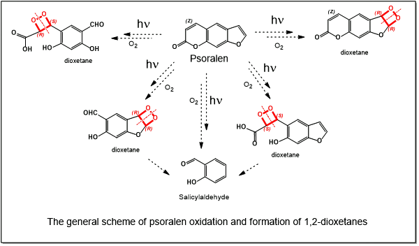
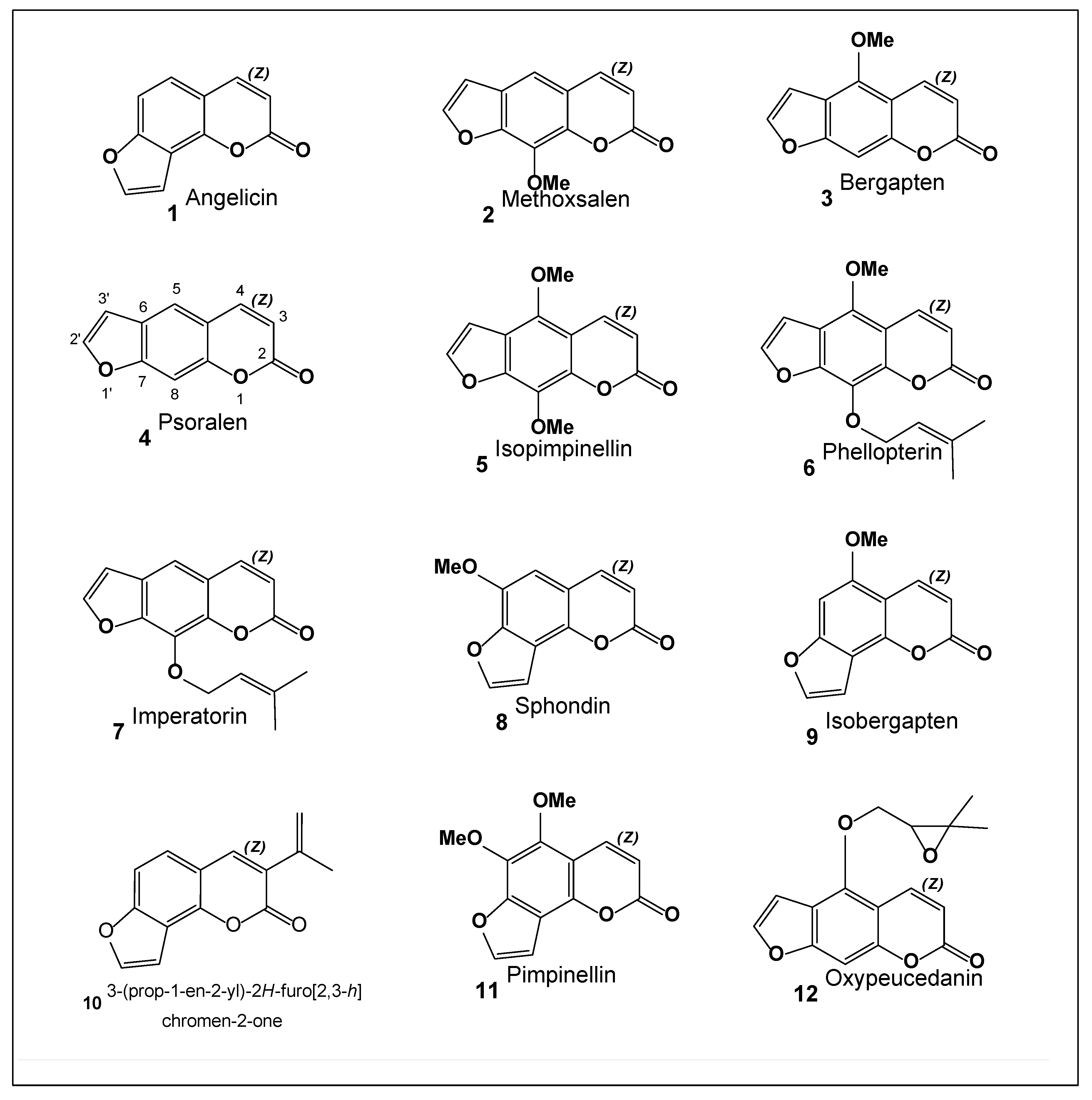
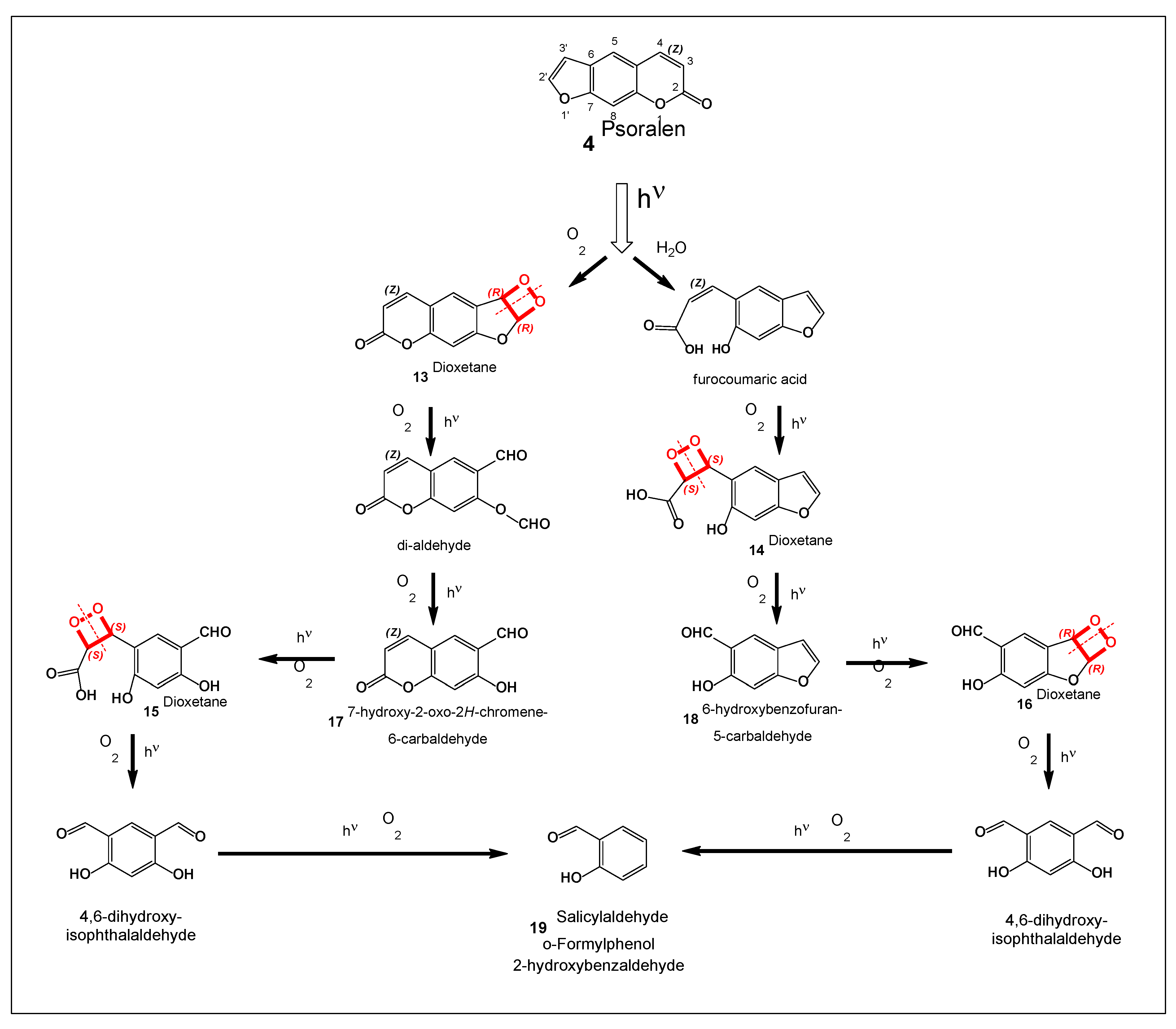
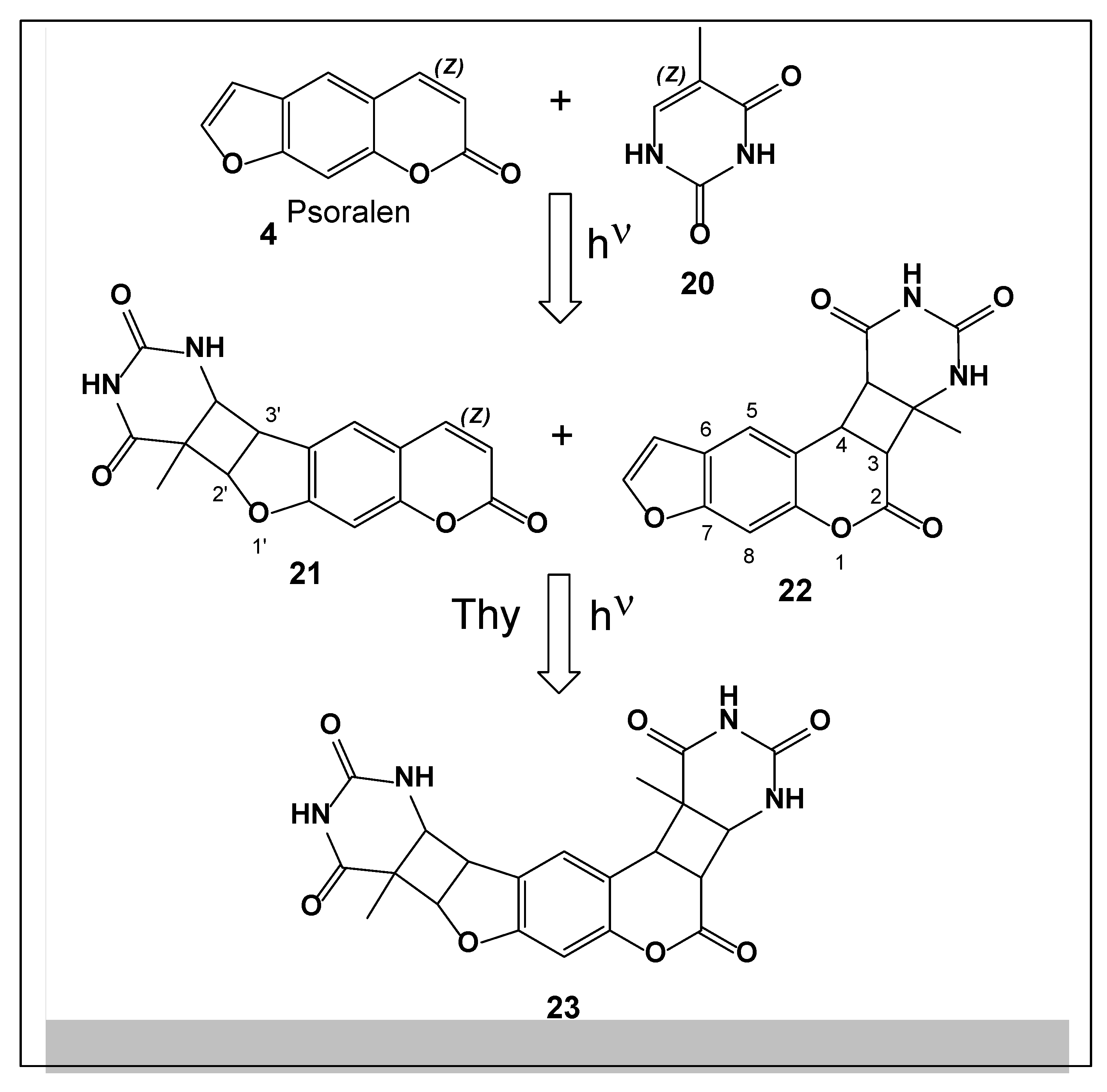
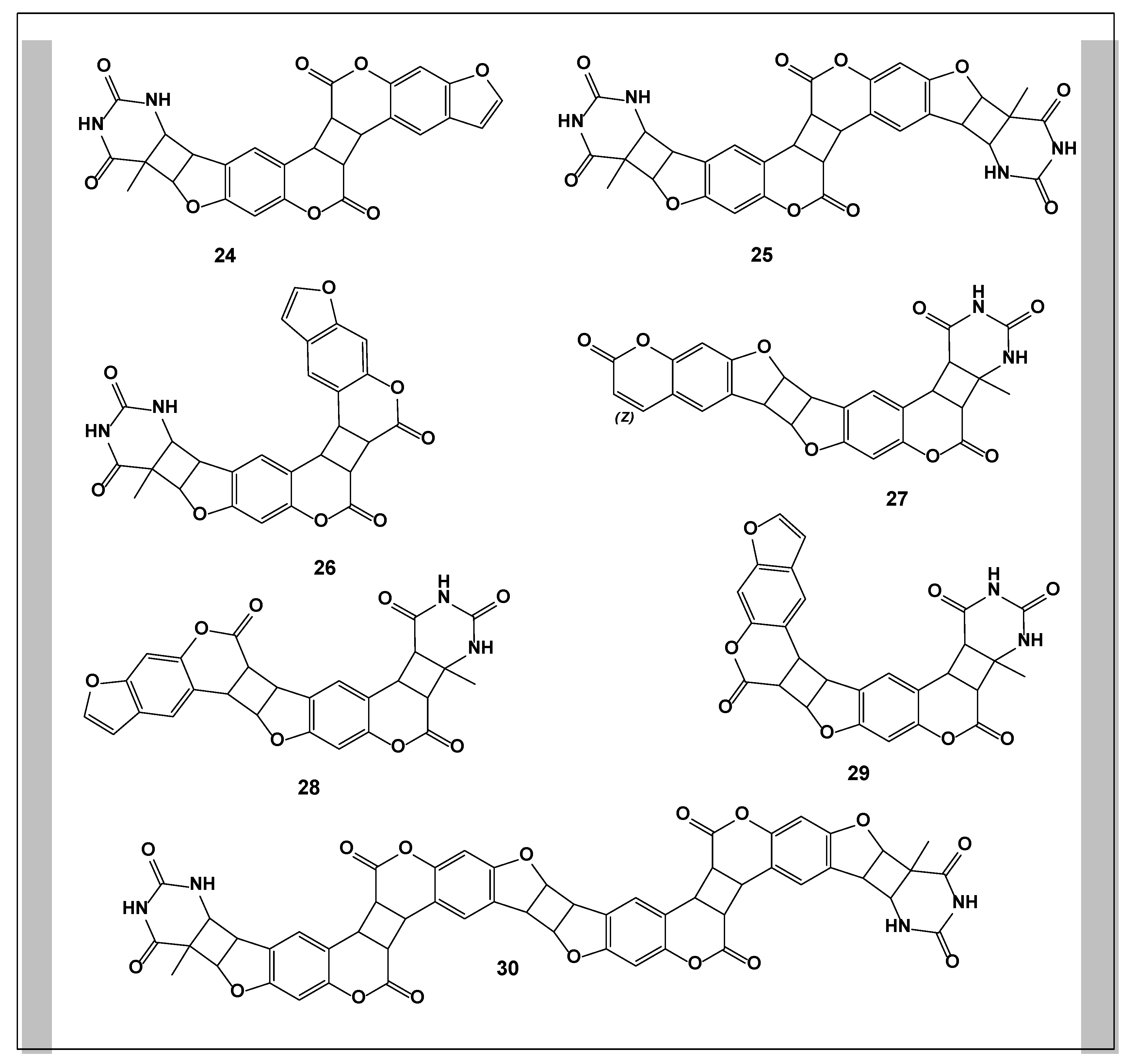
2. Furanocoumarins Produced by Hogweed

Furanocoumarins constitute a class of predominantly plant-derived secondary metabolites, widely recognized for the pronounced photosensitizing properties of several representatives, including angelicin, sphondin, bergapten, psoralen, and methoxsalen. Structurally, furanocoumarins consist of a coumarin nucleus formed by a benzene ring fused to an α-pyrone ring, with an additional furan moiety attached to the core structure (Figure 2). Upon exposure to ultraviolet (UV) radiation, these compounds become photoactivated and can induce severe phototoxic reactions in human skin [4,18,19].
Extensive research on plant-derived furanocoumarins over the past decades has led to the identification and structural elucidation of most naturally occurring members of this compound class. Beyond their well-documented phototoxicity, several furanocoumarins—such as bergapten, psoralen, imperatorin, sphondin, and angelicin—have attracted considerable interest due to their therapeutic potential. These compounds have been investigated for applications in cancer therapy, dermatological treatments (notably PUVA therapy), and as antiseptic, neuroprotective, osteoprotective, and antioxidant agents [4,20,21].
Plants belonging to the family Umbelliferae (Apiaceae) represent the richest known source of furanocoumarins, although these metabolites are also present in species from other plant families. To date, more than fifty naturally occurring furanocoumarins have been described in the scientific literature [22,23]. The present review focuses on furanocoumarins characteristic of umbelliferous plants, using Heracleum sosnowskyi as a representative example. This species is distinguished by its exceptional invasiveness, ecological resilience, and high biomass productivity. Its ability to cause severe photochemical skin burns is directly linked to the high concentration of furanocoumarins accumulated in its tissues.
Despite the significant health risks associated with H. sosnowskyi, several furanocoumarins present in this plant—such as psoralen, bergapten, imperatorin, and pimpinellin—are valuable bioactive compounds extensively studied and utilized in pharmaceutical research. These metabolites have found applications in the treatment of dermatological disorders and are increasingly investigated for potential roles in neurodegenerative disease therapy. Thus, H. sosnowskyi represents a paradoxical biological resource, functioning simultaneously as a hazardous invasive species and a reservoir of pharmaceutically relevant secondary metabolites [23,24]. Figure 1 illustrates furanocoumarins (112) isolated from various tissues of Sosnovsky’s hogweed. Although quantitative content varies depending on geographical origin and environmental conditions, the qualitative composition of these compounds remains relatively stable.
Recent analytical studies have re-examined the furanocoumarin profile of H. sosnowskyi leaves. Plant material collected in Vilnius, Lithuania, was subjected to microwave-assisted extraction, with optimization of key parameters including solvent selection (hexane) and extraction temperature (70 °C). Four major furanocoumarins were identified and quantified using gas chromatography–mass spectrometry (GC–MS) and gas chromatography with flame ionization detection (GC–FID): angelicin (2.3 mg/g), psoralen (0.15 mg/g), methoxsalen (0.76 mg/g), and bergapten (3.14 mg/g) [25].
Complementary findings were reported by Bespalov and Egorov [26], who analyzed the chemical composition of Sosnovsky’s hogweed collected in the St. Petersburg region. In a comparative study of essential oil fractions extracted from the fruits of three Heracleum species (H. mantegazzianum, H. persicum, and H. sosnowskyi), all samples were found to contain a diverse spectrum of furanocoumarins, including angelicin, psoralen, methoxsalen, bergapten, isobergapten, isopimpinellin, pimpinellin, and imperatorin. Notably, isopimpinellin was identified as the dominant compound in H. sosnowskyi, whereas isobergapten predominated in H. mantegazzianum. The fruits analyzed in this study were harvested from wild-growing plants in Wrocław, Poland, in August 2015 [27].

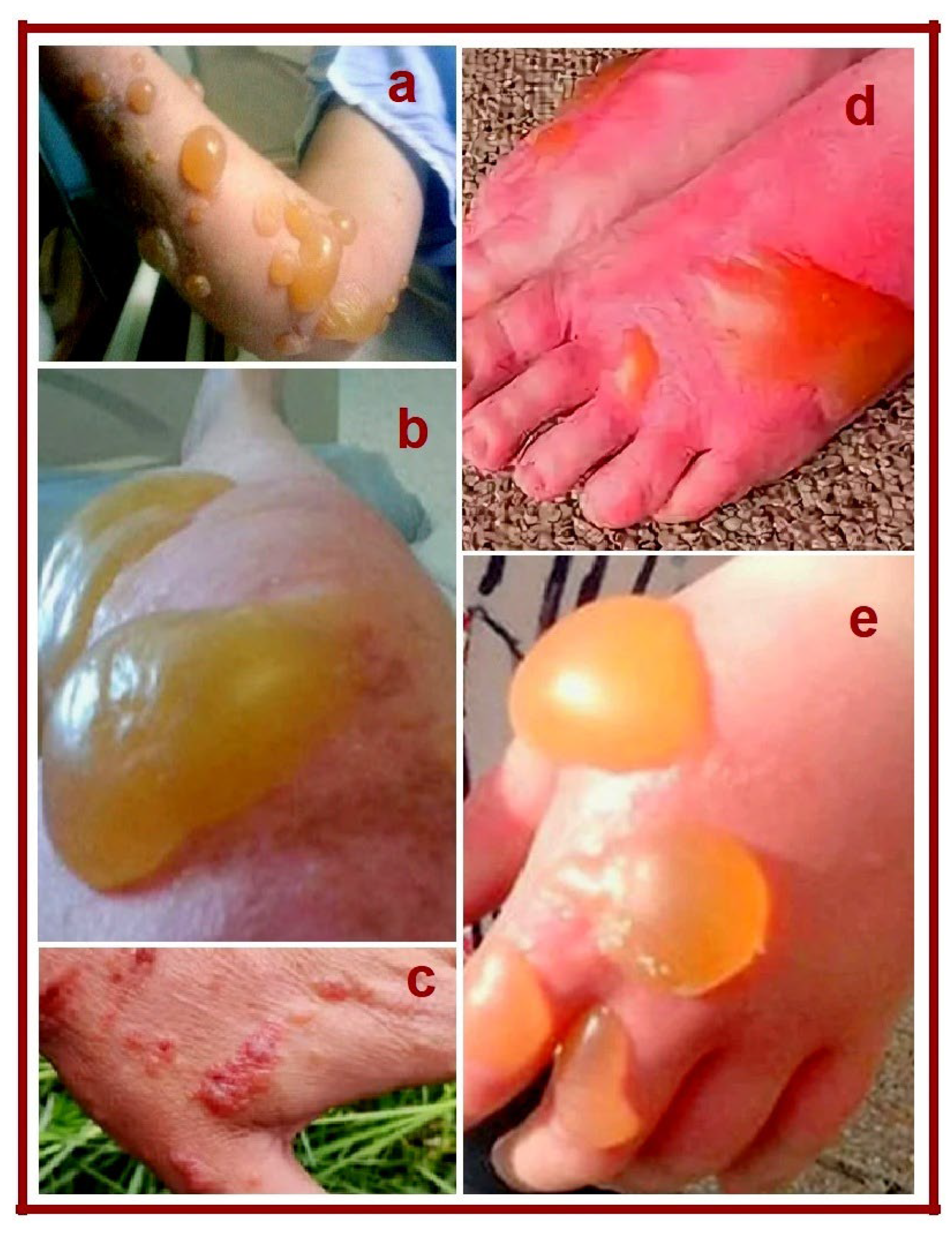
3. Photochemical Burns Caused by Hogweed Juice

The rapid and uncontrolled spread of giant hogweed (Heracleum spp.) represents a serious ecological, economic, and public health problem in many European countries, including Russia. This invasive plant adversely affects biodiversity by displacing native flora, disrupts natural ecosystems, and causes substantial economic losses. In addition, it poses a significant risk to human health due to its pronounced phototoxic properties [11,28,29].
Among representatives of the genus Heracleum, Sosnovsky’s hogweed (Heracleum sosnowskyi) is considered the most aggressive and hazardous species. Introduced in the mid-20th century as a forage crop, it rapidly escaped cultivation due to its high seed productivity and self-seeding ability. As a result, its distribution has expanded dramatically, now encompassing Eastern Europe and almost the entire European part of Russia [30,31].
Isolated reports of toxic effects associated with H. sosnowskyi appeared shortly after its introduction into agriculture in the 1950s. At present, the scale and severity of these effects can be described as alarming [29]. The plant’s juice contains high concentrations of phototoxic secondary metabolites—primarily furanocoumarins such as bergapten, isobergapten, isopimpinellin, xanthotoxin (8-methoxypsoralen, see Figure 2), and psoralen—which are directly responsible for skin damage following exposure to sunlight [4].
Hogweed-induced burns represent a common and severe problem during the summer months. Contact with the plant itself is typically painless, as its leaves lack thorns or mechanical irritants. However, exposure to its sap—often through cut stems or damaged plant tissues—followed by ultraviolet (UV) irradiation leads to delayed but severe photochemical skin injury. Within 4–5 days after contact, or sometimes earlier, sharply demarcated hyperpigmented areas appear on the skin. These lesions subsequently develop into blisters of varying size, which rapidly enlarge and merge, often covering extensive areas of the affected skin (Figure 3). The blisters may rise 0.5–3 cm above the skin surface and are filled with clear serous fluid [32,33].
Rupture of the blisters is strongly discouraged, as removal of the blister roof exposes highly sensitive underlying tissue, resulting in intense pain and increased risk of secondary infection. Even minimal contact with clothing or bedding can cause severe discomfort. Typically, the blisters rupture spontaneously within one week, followed by a stage of profuse exudation. Subsequently, a thin hyperpigmented crust forms, which gradually thickens, dries, and adheres firmly to the wound surface [3,32].
Pain persists for one to two weeks and gradually diminishes as epithelialization progresses. The crust remains for approximately two to three weeks before exfoliating from the edges, revealing newly formed, pink, shiny skin. Intense itching often accompanies the later stages of healing, particularly in children. Although the skin eventually recovers, residual hyperpigmentation may persist for months or even several years after severe burns [3,32,33].
The phototoxic nature of hogweed juice has been attributed primarily to furanocoumarins, especially psoralen and 8-methoxypsoralen. These compounds exhibit strong photosensitizing properties, dramatically increasing skin sensitivity to UV radiation and producing lesions that closely resemble thermal burns. This effect has been well documented in both experimental and clinical studies [18,34,35,36].
The pronounced photosensitizing activity of furanocoumarins is structurally determined by the presence of a furan ring fused to the coumarin core, particularly at positions 6, 7, and 8 (Figure 4). Structural modifications, such as removal or repositioning of the furan ring, result in a significant reduction or complete loss of phototoxic activity, as demonstrated in multiple structure–activity relationship studies [37,38,39].
Importantly, furanocoumarins themselves are relatively inert in the absence of light. Their toxicity arises from photochemical transformations, leading to the formation of highly reactive oxidation products, notably 1,2-dioxetanes [40]. Upon UV irradiation in the presence of molecular oxygen or ozone, the double bond between the 2′ and 3′ positions of the furan ring undergoes oxidation, yielding unstable 1,2-dioxetane intermediates. The formation, stability, and decomposition pathways of these compounds have been extensively investigated [41,42]. In some cases, 1,2-dioxetanes can exist in relatively stable forms when coordinated with specific ligands [43].
1,2-Dioxetanes are potent sources of electronically excited triplet-state carbonyl species generated during their decomposition. In biological systems, these excited intermediates can interact with cellular macromolecules, particularly DNA, leading to strand breaks, base modifications, and cross-link formation. Such DNA damage has been characterized using specialized repair endonucleases and molecular biology techniques [44,45,46].
The mechanisms underlying psoralen photooxidation and the formation of its principal photoproducts have been thoroughly elucidated [47]. Photochemical reactions involving psoralen can be broadly classified into three categories: (i) reactions involving cleavage of the furan ring, (ii) reactions involving cleavage of the pyrone ring, and (iii) deep photolysis leading to extensive molecular fragmentation. Notably, the formation of furan-ring cleavage products proceeds via singlet oxygen, generated during psoralen photooxidation in solution. Singlet oxygen attacks the 2′,3′-double bond of the furan ring, resulting in the formation of a transient dioxetane intermediate, which subsequently decomposes into highly reactive species responsible for cellular and DNA damage [48,49].
All photochemical reactions leading to the formation of psoralen-derived photoproducts can be classified into three major categories:
(i) Reactions involving cleavage of the furan ring,
(ii) Reactions involving cleavage of the pyrone ring, and
(iii) Deep photolysis resulting in extensive molecular fragmentation.
Photoproducts arising from furan-ring cleavage are formed via the participation of singlet oxygen, which is generated during psoralen photooxidation in solution. Singlet oxygen attacks the double bond between the 2′ and 3′ positions of the furan ring, producing a transient 1,2-dioxetane intermediate [48,49]. Subsequent simultaneous cleavage of the O–O and C–C bonds within the dioxetane results in the formation of a dialdehyde intermediate. Hydrolysis of the ester bond of this intermediate ultimately yields the stable photoproduct 6-formyl-7-hydroxycoumarin (17). A similar photooxidative furan-ring opening mechanism has been proposed for 5-methoxypsoralen and 8-methoxypsoralen [50].
Photoproducts resulting from cleavage of the pyrone ring are generated through two related mechanisms. Following photon absorption, an electronically excited psoralen molecule undergoes solvolysis with water, leading to the formation of furocoumaric acid. Subsequent oxidation of the newly formed double bond in the pyrone ring—mediated by dissolved molecular oxygen and potentially involving intermediate species—produces 5-formyl-6-hydroxybenzofuran (18). Thus, both furan- and pyrone-ring cleavage pathways ultimately generate aldehyde-containing photoproducts, including 6-formyl-7-hydroxycoumarin (17) and 5-formyl-6-hydroxybenzofuran (18), via ester bond hydrolysis.
Deep photolysis of psoralen results in a broader spectrum of low-molecular-weight photoproducts. These include compounds with simultaneous cleavage of both the furan and pyrone rings, yielding benzaldehydes and benzodialdehydes, as well as small aldehydes such as formaldehyde and acetaldehyde and their corresponding carboxylic acids. Notably, many of these aldehyde photoproducts absorb UVA radiation and can undergo secondary autoxidation, initiating chain reactions that further amplify photochemical damage [51].
In addition to oxidative phototransformations, psoralen (4) readily reacts with nucleic acids upon UVA irradiation (365 nm). Psoralen forms both monofunctional and bifunctional covalent adducts with pyrimidine bases in DNA (20, Figure 5), along with minor amounts of photodimers and photooxidation products. The formation of monofunctional adducts is kinetically coupled to bifunctional adduct formation, occurring in an approximate ratio of 4:1. Among monofunctional photoproducts, 2′,3′- (21) and 3,4-cycloadducts are formed in higher yields than 4′,5′-cycloadducts. The rate constants for photodimerization and photooxidation are significantly lower than those for cycloadduct formation, indicating that these byproducts represent minor pathways relative to DNA cross-linking reactions [52].
The efficiency of psoralen–DNA photoreactions is strongly influenced by the formation of pre-reactive molecular complexes stabilized by weak noncovalent interactions. Such complexation enables the short-lived excited state of psoralen to react efficiently with DNA bases, minimizing diffusion-controlled losses and solvent quenching of reactive intermediates.
The photoreactivity of psoralens has been elegantly rationalized using the bis-psoralen model [53]. Substantial experimental evidence indicates that psoralens form noncovalent complexes with nucleic acids through intercalation of their planar chromophores (2430, Figure 6) between base pairs of double-stranded DNA or RNA [54]. In contrast, single-stranded DNA and RNA generally exhibit significantly lower affinity for psoralens due to the absence of stable base stacking, making double-stranded and supercoiled DNA the primary biological targets of psoralen-induced photochemical damage.

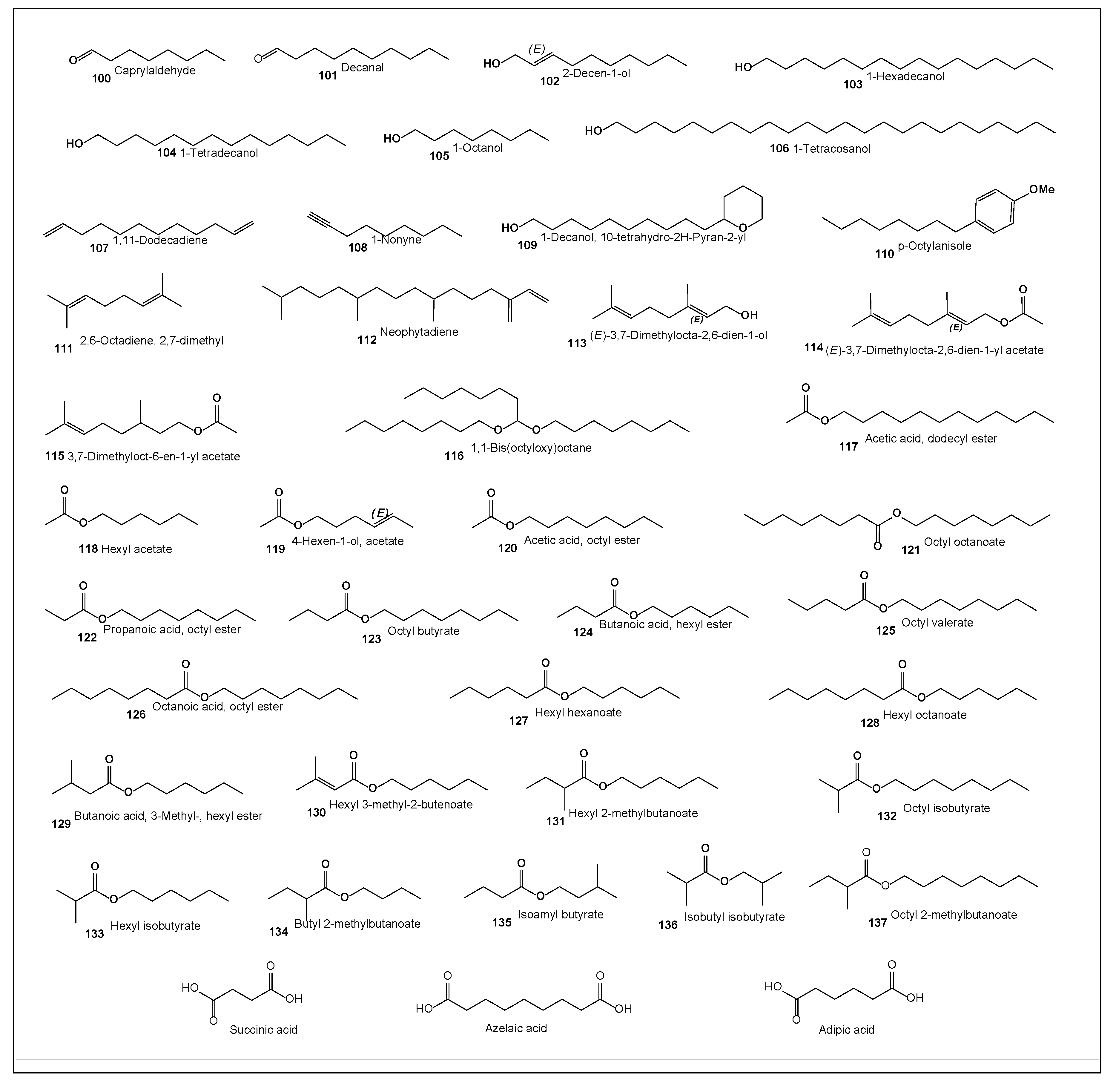
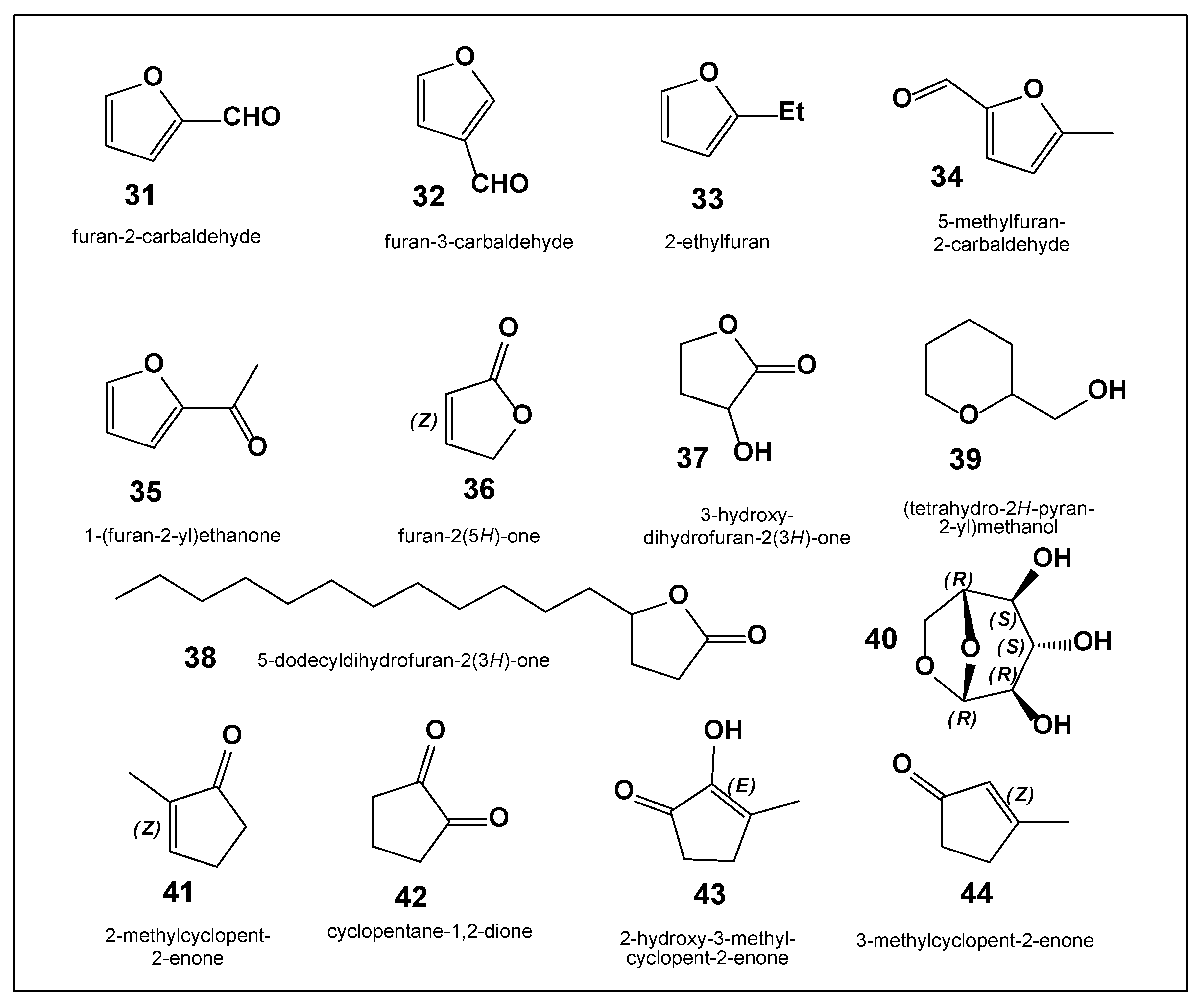
4. Furan, Aromatic, and Other Compounds Derived from H. sosnowskyi Biomass

The chemical composition of products formed during the oxidative thermal decomposition of fuel pellets prepared from Heracleum sosnowskyi biomass has been investigated using gas chromatography–mass spectrometry (GC–MS) [55]. Biomass collected in the village of Vylgort (Komi Republic, Russia) was air-dried to constant weight, pelletized, and subsequently subjected to controlled combustion in a specialized experimental apparatus. The volatile and semi-volatile products generated during oxidative thermal degradation were captured and analyzed using a gas–liquid chromatography system equipped with a mass-selective detector.
Analysis of the chromatographic and mass spectral data allowed the identification of 39 low-molecular-weight compounds among the 47 detected constituents. Both qualitative and quantitative assessments demonstrated that the composition of the degradation products was closely correlated with the biochemical makeup of the biomass. In particular, the relative contents and structural features of cellulose, hemicellulose, and lignin were found to be decisive factors governing the formation of individual compounds.
Cellulose and hemicellulose were shown to undergo thermal degradation more readily, whereas lignin—owing to its condensed aromatic structure—exhibited greater thermal stability. As a result, the pyrolysis products were dominated by carbohydrate-derived compounds, including seven furan derivatives (3138), (tetrahydro-2H-pyran-2-yl)methanol (39), levoglucosan (40), and four cyclopentane derivatives (4144) (Figure 7).
Furfural (2-furaldehyde, 31) is an aromatic aldehyde derived from furan, in which the hydrogen atom at the 2-position is replaced by a formyl group. Furfural plays an important role in the Maillard reaction and participates in various biological and biochemical processes. However, it is also a toxic compound, with exposure risks arising from ingestion, dermal absorption, or inhalation. Furfural has been identified as a natural metabolite in the aerobic bacterium Francisella tularensis and in Angelica gigas (Korean angelica) [56].
2-Ethylfuran (33), characterized by a sweet, burnt, and earthy odor, has been detected in several edible plants. Notably, it occurs at relatively high concentrations in kohlrabi (Brassica oleracea var. gongylodes) [57]. Another furan derivative, 5-methylfurfural (34), has been reported in Camellia sinensis and Swertia japonica [58] and is also produced by the yeast Saccharomyces cerevisiae during fermentation processes [59]. In addition, 5-dodecyldihydrofuran-2(3H)-one (38), a structurally distinct furan derivative, has been isolated from the n-hexane extract of Melilotus officinalis seeds [60].
Levoglucosan (40), also known as 1,6-anhydro-β-D-glucopyranose, is a characteristic product of carbohydrate pyrolysis, particularly from cellulose and starch. Structurally, levoglucosan is a hexose sugar containing a pyranose ring with a 1,6-anhydride bridge linking carbon atoms C-1 and C-6 via an oxygen atom, and three hydroxyl groups located at C-2, C-3, and C-4. This compound has been detected in a variety of plant species, including Lotus burttii and Lotus tenuis [61]. Alongside levoglucosan, several additional sugars were identified in the degradation products, including two isomeric forms of ribose, rhamnose, arabinose, α-mannopyranose, β-mannopyranose, α-glucopyranose, and β-glucopyranose [55,62].
Finally, four cyclic ketones (4144) were identified among the pyrolysis products. These compounds were previously isolated from coffee oil, and their structures—initially proposed based on ultraviolet, infrared, and mass spectrometric data—were later confirmed through targeted chemical synthesis, as reported by Gianturco and co-workers [63].

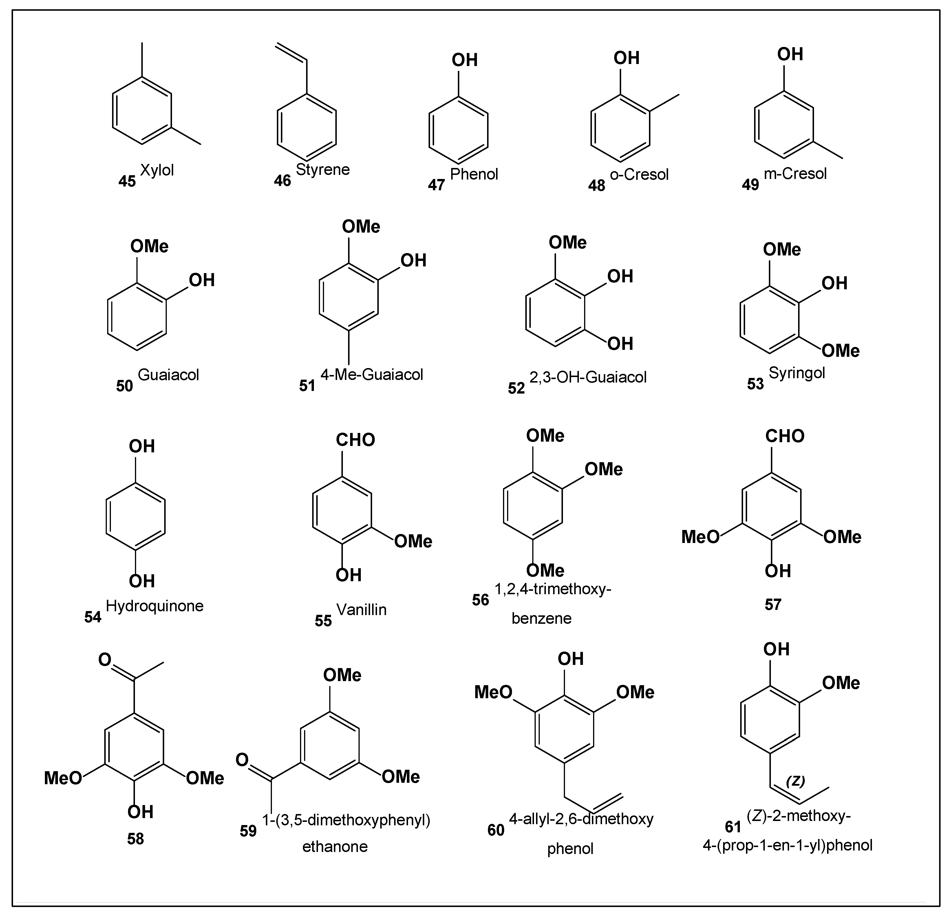
5. Aromatic, Lipid, and Oxidation-Derived Compounds from H. sosnowskyi

Aromatic compounds represent a broad class of organic molecules characterized by a stable cyclic conjugated π-electron system, commonly referred to as an aromatic ring. This electronic configuration imparts distinctive physicochemical properties, including enhanced thermodynamic stability and specific reactivity patterns. The term “aromatic” originally arose from the often pleasant odors associated with early-discovered representatives of this class; however, it is now well established that not all aromatic compounds are odorous [64,65].
Aromatic compounds are widely distributed in nature and are particularly abundant in aromatic plants, which have been utilized since antiquity in traditional medicine, food preservation, and flavoring. Prominent examples include oregano, rosemary, sage, and anise—species originating predominantly from the Mediterranean region [66,67]. These plants are rich in bioactive constituents, especially polyphenols, which exhibit antimicrobial, antioxidant, antiparasitic, antiprotozoal, antifungal, and anti-inflammatory activities [68,69]. Growing demand for natural, environmentally friendly, and generally recognized as safe (GRAS) products has stimulated interest in aromatic plants and their extracts as next-generation agents for human and animal health, nutrition, and sustainable agriculture [70,71].
Oligo- and polyaromatic compounds present in plant biomass may undergo extensive transformation under environmental influences such as microbial degradation or thermochemical decomposition. These processes result in the formation of low-molecular-weight aromatic compounds (4561, Figure 8). Such transformations have been widely investigated, particularly in the context of biomass pyrolysis and oxidative degradation [2,4,55,62,72]. A comprehensive profile of these aromatic degradation products derived from H. sosnowskyi biomass is presented in Figure 7.
The lipid composition of fruits from eight Heracleum species—H. ternatum, H. pyrenaicum subsp. pollinianum, H. verticillatum, H. orphanidis, H. sphondylium, H. sibiricum, H. montanum, and H. pyrenaicum subsp. orsinii—was investigated with respect to fatty acids, sterols, and triterpenes. Plant material collected in southeastern Europe (Slovenia, Serbia, Montenegro, and North Macedonia) was extracted with dichloromethane, and the oil supernatants were subjected to chromatographic analysis. Petroselinic acid (18:1, cis-6-octadecenoic acid) was identified as the dominant fatty acid, accounting for 42.8–56.5% of the total lipid fraction. Linoleic acid (18:2, ω-6, cis,cis-9,12-octadecadienoic acid) was present at levels of 20.3–33.3%, followed by oleic acid (18:1, cis-9-octadecenoic acid), which constituted 12.3–13.7% [73]. Petroselinic acid has also been reported in Agaricus blazei and has been associated with various health-promoting effects, including support of digestive and respiratory functions, regulation of menstrual flow, and immune system modulation. Traditionally, it has been used in herbal preparations such as teas and topical tinctures [74]. This fatty acid has additionally been identified in Angelica ternata, another member of the Apiaceae family [75].
Sterols and triterpenoids identified across multiple hogweed species include Δ⁵,⁷,⁹(¹¹),²²-ergostatetraenol, ergosterol, campesterol, stigmasterol, Δ⁷,²²-ergostadienol, Δ⁷-campesterol, α-amyrin (ursane-type), Δ⁷-stigmastenol, and Δ⁷-avenasterol (3β,24Z-stigmasta-7,24(28)-dien-3-ol). Among these, β-sitosterol was the most abundant sterol, representing 44.9–56.9% of the total sterol fraction, followed by stigmasterol (15.7–25.0%), Δ⁷-stigmastenol (6.6–12.5%), and campesterol (5.2–8.1%) [74].
Borska and co-workers reported detailed lipid profiling of H. sosnowskyi growing in Latvia [76]. Their study revealed a diverse array of positional and geometric fatty acid isomers, with marked variation in concentration among different plant organs. Lipid extracts from H. sosnowskyi exhibited pronounced antimicrobial activity and cytotoxic effects against several cancer cell lines.
In total, 38 distinct fatty acids were identified in extracts obtained from various parts of the hogweed biomass (Table 5). Palmitic acid (16:0) was the most abundant saturated fatty acid. Collectively, mono- and polyunsaturated fatty acids accounted for more than 75% of the total fatty acid content. Roots were particularly rich in linoleic acid (C18:2n-6, cis-isomer) and α-linolenic acid (C18:3n-3), whereas immature seeds contained high levels of 9,12-octadecadienoic acid. Mature seeds were dominated by trans-vaccenic acid (C18:1n-7), while palmitic acid was present in comparable amounts across all plant organs.
Leaves and flowers were identified as the primary reservoirs of fatty acids, largely associated with monogalactosyldiacylglycerols, which typically contain one saturated (palmitic acid) and one unsaturated fatty acid chain, or two unsaturated chains. Numerous positional and geometric isomers of stearic acid derivatives were detected, including trans-isomers (18:1n-9t, 18:1n-11t, 18:1n-13t, 18:1n-14t, and 18:1n-15t) as well as the rare cis-isomer (18:1n-12c), previously reported primarily in maple seed oil [76,77].
Leaves exhibited the highest total fatty acid content (4.398 mg per 100 g dry weight), while stems and roots displayed the greatest diversity (33 fatty acid types). In contrast, mature and immature seeds contained fewer fatty acid species (25 and 23 types, respectively). In leaf tissue, α-linolenic acid (C18:3n-3; Δ9c,12c,15c) plays a central role in lipid metabolism and redox homeostasis, functioning both as an antioxidant and as a precursor in biosynthetic pathways [77]. Approximately 70% of leaf fatty acids belonged to the polyunsaturated fatty acid (PUFA) class, underscoring their significant bioeconomic potential [78].
The unusual fatty acid 7c,10c,13c-hexadecatrienoic acid, detected in leaves and flowers of hogweed, has previously been reported only in potato leaves following alkaline hydrolysis during studies of wound-induced plant responses [79]. Notably, only stem extracts contained detectable amounts (0.21%) of 9-oxooctadeca-10,12-dienoic acid, a potent inhibitor of acetyl-CoA carboxylase (ACC), a key enzyme in fatty acid biosynthesis [80]. This observation suggests a possible regulatory or allelopathic role for this compound, positioning hogweed stems as a potential source of bioherbicidal agents [76].
Fatty acids dominant in hogweed, particularly petroselinic and oleic acids, readily undergo photooxidation in the presence of sunlight and molecular oxygen. This process leads to the formation of unstable peroxide intermediates, including 1,2-dioxetanes and 1,2,3-trioxolanes, respectively (Figure 8). Experimental evidence for these reactions includes the detection of fatty aldehydes (e.g., nonanal, caprylaldehyde) and dicarboxylic acids such as nonanoic, azelaic, succinic, and adipic acids, which arise from peroxide decomposition (Figure 9) [81].
Photooxidation proceeds via photodynamically generated singlet oxygen, which adds to unsaturated fatty acid double bonds. Subsequent hydrogen abstraction from adjacent methylene groups yields hydroperoxylipids that readily decompose. Both dioxetane and trioxolane peroxides are highly reactive and capable of releasing singlet oxygen, thereby amplifying oxidative stress. This reactive environment may synergistically enhance the phototoxic effects of furanocoumarins and contribute to DNA damage.
Finally, essential oils isolated from various parts of H. sosnowskyi contain substantial amounts of alkyl acetates and carboxylic acid esters, collectively encompassing compounds 100137. Several volatile constituents were identified for the first time exclusively in H. sosnowskyi. Compounds 109 and 116 were unique to this species, whereas compounds 115137 had previously been reported only in trace amounts in other plant essential oils. The widespread presence of dicarboxylic acids in hogweed tissues further supports extensive fatty acid oxidation via peroxide intermediates, consistent with earlier observations [82].
Figure 10. Hydrocarbons, fatty aldehydes, fatty alcohols, alkyl acetates, and esters of carboxylic acids derived from leaves, fruits and other parts of the H. sosnovskiyi.
Preprints 190214 g010
Table 1. Saturated fatty (carboxylic, dionic) acid composition of H.sosnovskyi (g/100g DW).
No. Fatty Acids Roots Stems Leaves Flowers
62 8:0 0.7 15.0 0.1 0.7
63 Succinic acid 0.3 3.3 1.6 0.3
64 12:0 0.4 1.7 4.1 1.5
65 14:0 1.6 19.8 33.1 17.2
66 Adipic acid 1.2 14.5 5.8 1.5
67 9-oxo-9:0 1.4. 23.3 3.3 0.7
68 15:0 2.8 18.8 7.9 2.4
69 Azelaic acid 1.6 10.2 4.8 1.2
70 16:0 60.1 884.1 615.8 173.3
71 Iso-17:0 0.1 0.5 0.1 0.1
72 17:0 1.5 15.1 16.9 3.7
73 18:0 5.3 45.4 84.8 18.8
74 22:0 2.8 23.6 24.4 6.6
75 23:0 1.2 17.0 13.4 3.3
76 24:0 5.2 59.4 61.9 13.6
77 25:0 11.2 5.6 39.9 0.9
Table 2. Monoenoic fatty acids.
No. Fatty Acids Roots Stems Leaves Flowers
78 16:1n-11 (Δ5c) 0.6 0.1 1.8 0.5
79 16:1n-9 (Δ7c) 0.8 22.6 2.8 7.1
80 16:1n-7 (Δ9c) 0.5 2.6 7.0 0.6
81 16:1n-5 (Δ11c) 0.4 0.5 0.1 0.3
82 16:1n-3 (Δ12c) 0.3 6.8 86.0 1.1
83 17:1n-7 (Δ10c) 0.3 0.1 0.1 3.9
84 18:1n-9 (Δ9t) 9.5 15.2 77.2 56.1
85 18:1n-7 (Δ11t) 2.4 42.7 9.5 10.4
86 18:1n-6 (Δ12c) 7.6 6.5 15.9 1.6
87 18:1n-3 (Δ14t) 1.2 2.6 2.2 0.5
88 18:1n-2 (Δ15t) 30.0 5.0 35.7 2.6
89 20:1n-6 (Δ14t)
20:1n-9 (Δ11t)
0.5 28.3 3.2 6.8
90 20:1n-11 (Δ9c) 0.1 0.1 0.1 0.1
91 20:1n-9 (Δ11c) 0.7 2.8 11.0 7.1
92 20:1n-7 (Δ13c) 0.1 1.9 1.4 5.6
Table 3. Di- and polyenoic fatty acids.
No. Fatty Acids Roots Stems Leaves Flowers
93 16:2n-6 (Δ7c,10c) 0.2 3.9 5.4 0.9
94 18:2n-6 (Δ9c,12t) 5.5 600.6 10.0 2.7
95 18:2n-6 (Δ9c,12c) 185.0 0.1 856.1 230.5
96 18:2n-6 (Δ10t,12t)(oxo-9) 0.1 4.2 0.1 0.1
97 18:2n-6 (Δ10t,12c) 0.1 0.1 0.1 0.1
98 16:3n-3 (Δ7c,10c,13c) 0.9 55.0 633.2 6.3
99 18:3n-3 (Δ9c,12c,15c) 23.2 71.4 1687.4 135.5

6. Hogweed Burns and Their Possible Treatment

Furanocoumarins present in hogweed sap play a central role in the development of skin burns following contact with Heracleum sosnowskyi. The principal phototoxic constituents include xanthotoxin, bergapten, angelicin, and 5- and 8-methoxypsoralen (Figure 2). These compounds are capable of intercalating into cellular DNA and forming covalent bonds with pyrimidine bases in keratinocytes and melanocytes. However, the manifestation of phototoxic injury requires subsequent exposure to ultraviolet (UV) radiation, which explains why bullous dermatitis typically does not occur immediately after skin contact with hogweed sap unless the affected area is irradiated by sunlight [1,83,84].
Upon UV exposure, furanocoumarins undergo photochemical activation within skin cells, triggering a cascade of reactions that lead to the generation of reactive oxygen species (ROS). This process is accompanied by the release of substantial amounts of thermal and photonic energy. The resulting oxidative stress induces damage to cellular membranes and mitochondria, disrupts metabolic processes, and ultimately leads to apoptosis or necrosis of affected cells [85,86,87].
Clinically, these molecular and cellular events manifest as erythema, edema, and blister formation, characteristic of phytophotodermatitis. In the later stages, enhanced melanocyte activity is observed in the irradiated area, resulting in pronounced hyperpigmentation. This pigmentation response is considered a protective adaptive mechanism that limits further ultraviolet penetration and mitigates additional photochemical damage [22,83,84].

6.1. Classification of Burns Caused by Hogweed

Based on the severity of clinical manifestations, contemporary dermatology distinguishes three principal clinical forms of phytophotodermatitis resulting from exposure to Heracleum sosnowskyi sap [22,88].
The erythematous form is the most common and corresponds to a first-degree thermal burn in terms of tissue involvement. It is characterized by a burning sensation and localized erythema, typically appearing as linear streaks or irregular patches confined to the areas of direct contact with the plant sap. Desquamation of the epidermis usually begins approximately two weeks after exposure and is followed by the formation of persistent hyperpigmented macules.
The erythematous–bullous form is comparable to a second-degree burn with respect to the depth of skin damage. Clinically, it presents with pronounced hyperemia accompanied by the formation of multiple tense vesicles and bullae, which frequently coalesce into large blisters measuring up to 10 cm in diameter. Rupture of the bullae typically occurs within one week, leading to crust formation at the affected sites, followed by residual hyperpigmentation. This form is often associated with signs of moderate systemic intoxication, including malaise and low-grade fever [32,33].
The erosive–ulcerative form represents the most severe manifestation of hogweed-induced photodermatitis. Its initial presentation resembles that of the erythematous–bullous type; however, following rupture of the vesicles, persistent erosions develop and subsequently progress to deep ulcerations. Healing is prolonged and commonly results in the formation of hyperpigmented scars. Systemic intoxication is more pronounced in this form, reflecting extensive tissue damage and inflammatory response.

6.2. Symptoms of Hogweed Burn

The initial local manifestations of skin injury following contact with Heracleum sosnowskyi sap typically develop within several hours to one or two days after exposure. The earliest clinical signs include well-demarcated erythema accompanied by an intense burning sensation. As the inflammatory response progresses, multiple small vesicles form on the affected skin. These vesicles arise against a background of increasing tissue edema and subsequently coalesce into larger bullae.
The resulting blisters are characteristically tense, thick-walled, and occasionally multilocular, containing a clear serous exudate. In the majority of cases, contact with hogweed leads to first- or second-degree burns; however, in rare instances, more severe third-degree burns may occur, particularly following prolonged exposure or extensive skin contact combined with intense ultraviolet irradiation [22,32,33,83].
Within several days, the bullae may rupture spontaneously, exposing a painful wound surface or forming erosive–ulcerative lesions. Re-epithelialization of these lesions is typically slow and is accompanied by crust formation. After detachment of the scab, persistent post-inflammatory hyperpigmentation often develops and may remain visible for several months. The most frequently affected anatomical regions include the shins, forearms, and hands, which are commonly exposed to both direct contact with the plant and sunlight.
In children, atypical patterns of injury are frequently observed due to behavioral factors. Hogweed stems are often used during play as improvised objects such as “telescopes”, blow tubes, swords, or toy horses, resulting in burns localized to unusual areas, including the periorbital and perioral regions, trunk, and inner thighs [22,32,33].
The severity of cutaneous lesions generally correlates with the intensity of systemic intoxication symptoms. These may include fever, chills, headache, general weakness, and decreased appetite. In addition, prolonged inhalation of volatile compounds released from dense hogweed stands may provoke pronounced dizziness and, in severe cases, syncope [89,90,91].

6.3. Treatment for Hogweed Burns

At present, no standardized clinical guidelines have been formally established for the diagnosis and management of phytophotodermatitis caused by exposure to sap from plants of the genus Heracleum [92,93,94]. Consequently, treatment strategies are largely based on clinical experience, case reports, and general principles used in the management of phototoxic and thermal skin injuries.
According to available literature, immediate first aid following contact with hogweed sap is crucial. The affected skin area should be promptly washed thoroughly with copious amounts of running water and soap to remove residual plant sap. After cleansing, an antiseptic solution or topical dexpanthenol may be applied, followed by coverage with a sterile dressing. Importantly, strict avoidance of ultraviolet exposure is recommended for at least 48 h after contact, as UV radiation is the key trigger for the development of phototoxic lesions [95,96,97].
In cases presenting predominantly with erythema, topical corticosteroid preparations may be used to reduce inflammation, while nonsteroidal anti-inflammatory drugs (NSAIDs) can be administered to alleviate pain. For mild erythematous forms of phytophotodermatitis, supportive treatment with over-the-counter analgesics such as acetaminophen or ibuprofen, together with emollients (e.g., petroleum jelly), is often sufficient to relieve symptoms and promote skin recovery [22,32,33,88].
When bullous lesions develop, more intensive medical management is frequently required and may necessitate treatment in a hospital setting [32,88]. Small blisters can be carefully punctured and drained under sterile conditions, whereas larger bullae, extensive epidermal detachment, or pronounced epidermal–dermal exudation should generally be managed conservatively through gentle cleansing and sterile bandaging. In cases of moderate to severe inflammation, systemic corticosteroids may be prescribed to reduce inflammatory responses and limit tissue damage [98,99,100,101].
The erosive–ulcerative form of phytophotodermatitis represents the most severe clinical presentation and may require surgical intervention. Therapeutic measures in such cases can include surgical debridement, bullectomy, or, in rare and complicated situations, fasciotomy, particularly when deep tissue involvement or secondary complications are present [22,32,33,97,102].

7. Conclusions

Sosnovsky’s hogweed (Heracleum sosnowskyi) represents a unique and serious intersection of botanical invasiveness, chemical toxicity, and public health risk. The evidence summarized in this review clearly demonstrates that the severe skin injuries associated with contact with hogweed sap are the result of complex photochemical processes initiated by highly oxygenated secondary metabolites, primarily furanocoumarins. Upon exposure to ultraviolet radiation, these compounds undergo photoactivation and oxidation, leading to the formation of reactive intermediates such as 1,2-dioxetanes, singlet oxygen, and excited carbonyl species. These reactive entities induce damage to cellular membranes, mitochondria, and DNA, resulting in apoptosis, inflammation, blister formation, and long-lasting hyperpigmentation.
Beyond furanocoumarins, H. sosnowskyi biomass contains a wide spectrum of biologically active compounds, including furan derivatives, aromatic compounds, fatty acids, sterols, and triterpenoids. Photooxidation of unsaturated fatty acids further contributes to peroxide formation and oxidative stress, potentially amplifying the phototoxic effects initiated by furanocoumarins. The chemical diversity of hogweed metabolites underscores that its harmful impact is not limited to a single class of compounds but rather arises from synergistic photochemical and oxidative mechanisms.
Clinically, hogweed-induced phytophotodermatitis presents with a broad spectrum of severity, ranging from erythematous reactions to bullous and erosive-ulcerative lesions that resemble second- and third-degree thermal burns. Despite the frequency and severity of these injuries, standardized clinical protocols for diagnosis, treatment, and follow-up remain insufficiently developed. Current management is largely symptomatic and based on general burn and dermatological care principles rather than targeted, mechanism-based therapy.
In summary, H. sosnowskyi is not only an invasive ecological threat but also a significant photochemical hazard to human health. A multidisciplinary approach integrating plant chemistry, photobiology, dermatology, and public health is essential to mitigate the risks associated with hogweed exposure and to develop effective preventive and therapeutic solutions.

Author Contributions

Conceptualization, V.M.D.; methodology, V.M.D.; software, A.O.T.; investigation, V.M.D.; resources, V.M.D.; writing—original draft preparation, A.O.T. and V.M.D.; writing—review and editing, A.O.T. and V.M.D. All authors have read and agreed to the published version of the manuscript.

Funding

This research received no funding.

Data Availability Statement

No new data were created or analyzed in this study. Data sharing is not applicable to this article.

Conflicts of Interest

The authors declare no conflicts of interest.

References

  1. Grzędzicka, E. Invasion of the giant hogweed and the Sosnowsky’s hogweed as a multidisciplinary problem with unknown future—A review. Earth 2022, 3, 287–312. [Google Scholar] [CrossRef]
  2. Matarrese, E.; Renna, M. Prospects of hogweed (Heracleum sphondylium L.) as a new horticultural crop for food and non-food uses: A review. Horticulturae 2023, 9, 246. [Google Scholar] [CrossRef]
  3. Baker, B.G.; Bedford, J.; Kanitkar, S. Keeping pace with the media; Giant Hogweed burns—A case series and comprehensive review. Burns 2017, 43(5), 933–938. [Google Scholar] [CrossRef] [PubMed]
  4. Bahadori, M.B.; Dinparast, L.; Zengin, G. The genus Heracleum: A comprehensive review on its phytochemistry, pharmacology, and ethnobotanical values as a useful herb. Comprehen. Rev. Food Sci. Food Safety 2016, 15(6), 1018–1039. [Google Scholar] [CrossRef]
  5. Annenkov, N.I. Heracleum. Botanical Dictionary; Type. Imp. Academy of Science: St. Petersburg, 1878; p. XXI + 645 p. [Google Scholar]
  6. Liu, M.; Downie, S.R. The phylogenetic significance of fruit anatomical and micromorphological structures in Chinese Heracleum species and related taxa (Apiaceae). Systematic Botany 2017, 42(2), 313–325. [Google Scholar] [CrossRef]
  7. Logacheva, M.; Valiejo-Roman, C.; Pimenov, M. ITS phylogeny of west Asian Heracleum species and related taxa of Umbelliferae–Tordylieae WDJ Koch, with notes on evolution of their psbA-trnH sequences. Plant Syst. Evol. 2008, 270, 139–157. [Google Scholar] [CrossRef]
  8. Yu, Y.; Downie, S.R.; He, X.; Deng, X.; Yan, L. Phylogeny and biogeography of Chinese Heracleum (Apiaceae tribe Tordylieae) with comments on their fruit morphology. Plant Syst. Evol. 2011, 296, 179–203. [Google Scholar] [CrossRef]
  9. Kozhevnikov, A.E.; Probatova, N.S.; Tsvelev, N.N. Heracleum lanatum Michx. Vascular plants of the Soviet Far East; Kharkevich, S.S., Ed.; Nauka, 1987; Volume 2, pp. 269–270. [Google Scholar]
  10. Berkutenko, A.N.; Virek, E.G. Woolly hogweed - Heracleum lanatum. In Medicinal and food plants of Alaska and the Russian Far East; Publishing House of the Far Eastern University: Vladivostok, 1995; pp. P. 105–106. [Google Scholar]
  11. Satsyperova, I.F. Hogweeds of the USSR flora – new forage plants: Prospects for use in the national economy; Nauka Publishin House: Leningrad, 1984; p. 114. [Google Scholar]
  12. Tkachenko, K.G. Sosnowsky’s hogweed (Heracleum sosnowskyi Manden). Recommendations and methods for combating it; Publishing House "First IPH": Saint Petersburg, 2021; p. 68. [Google Scholar]
  13. Taysumov, M.A.; Astamirova, M.M.; Umarov, R.M.; Abdurzakova, A.S.; Magomadova, R.S.; Israilova, S.A.; Khasueva, B.A. Forage plants of Chechnya and classification of natural forage lands. International scientific and practical conference" Agro-SMART-Smart solutions for agriculture"(Agro-SMART 2018), December 2018; Atlantis Press: Russia; pp. 952–957. [Google Scholar]
  14. Paramonova, K.; Chaloupková, V.; Ivanova, T.A. Invasive Heracleum sosnowskyi as a potential feedstock for biorefineries: A review. Industrial Crops and Prod. 2024, 216, 118754. [Google Scholar] [CrossRef]
  15. Sefidkon, F.; Dabiri, M.; Mohammad, N. Analysis of the oil of Heracleum persicum L. (leaves and flowers). J. Essential Oil Res. 2002, 14(4), 295–297. [Google Scholar] [CrossRef]
  16. Osipova, E.S.; Gladkov, E.A. Heracleum Sosnowskyi Manden. as a Source of Valuable Chemicals (Elimination with Utility). Chem. Methodol. 2024, 8(12), 944–956. [Google Scholar]
  17. Voznyakovsky, A.P.; Karmanov, A.P.; Neverovskaya, A.Y.; Voznyakovsky, A.A.; Kocheva, L.S.; Kidalov, S.V. Biomass of Heracleum sosnowskyi as a raw material for obtaining 2D carbon nanostructures. Chem. Plant Raw Material 2020, (4), 83–92. [Google Scholar] [CrossRef]
  18. Kulikov, O.A.; Shlyapkina, V.I.; Brodovskaya, E.P.; Aioub, A.M.A.K.; Ageev, V.P.; Zharkov, M.N.; Sukhorukov, G.B. Phototoxicity in vitro and safety in vivo of the emulsion photosensitizer based on furanocoumarins of Heracleum sosnowskyi. European J. Pharmac. Biopharm. 2024, 198, 114257. [Google Scholar] [CrossRef] [PubMed]
  19. Frumin, G.T. Toxicity of juice of Heracleum sosnowskyi. Russ. J. General Chem. 2023, 93(13), 3483–3487. [Google Scholar] [CrossRef]
  20. Bruni, R.; Barreca, D.; Protti, M.; Brighenti, V.; Righetti, L.; Anceschi, L.; Mercolini, L.; Benvenuti, S.; Gattuso, G.; Pellati, F. Botanical sources, chemistry, analysis, and biological activity of furanocoumarins of pharmaceutical interest. Molecules 2019, 24, 2163. [Google Scholar] [CrossRef]
  21. Sarker, S.D.; Nahar, L. Progress in the chemistry of naturally occurring coumarins. In Progress in the Chemistry of Organic Natural Products; Kinghorn, A.D., Ed.; Springer International Publishing, 2017; Volume 106, p. 241. [Google Scholar]
  22. Klimkina, E.A.; Okolelova, M.S.; Smirnova, E.S. Analysis of data on phytophotodermatitis caused by contact with the sap of plants of the genus Hogweed (Heracleum L.). Bull. Russ. Military Med. Acad. 2024, 43, 183–192. [Google Scholar] [CrossRef]
  23. Shtratnikova, V.Y. Furanocoumarins: History of research, diversity, synthesis, physiological role in the plant, and medical application. Russian J. Plant Physiol. 2023, 70(7), 169. [Google Scholar] [CrossRef]
  24. Schulzová, V.; Hajšlová, J.; Botek, P.; Peroutka, R. Furanocoumarins in vegetables: Influence of farming system and other factors on levels of toxicants. J. Sci. Food Agricul 2007, 87(15), 2763–2767. [Google Scholar] [CrossRef]
  25. Vickackaite, V.; Pilaityte, K.; Poskus, V. Extraction, isolation, and purification of furanocoumarins from invasive Heracleum sosnowskyi. Separations 2025, 12(7), 175. [Google Scholar] [CrossRef]
  26. Bespalov, D.S.; Egorov, D.M. Study of the sum of furanocoumarins of Heracleum sosnowskyi obtained by alkaline extraction method. Scientific creativity of youth to the forest complex of Russia. In Proceedings of the XXI All-Russian Scientific and Technical Conference of Undergraduate and Postgraduate Students, Ekaterinburg, Russia, USFEU, 2025; pp. 891–896. [Google Scholar]
  27. Poliyoeicz, J.; Gebarowska, E.; Prockow, J.; Pietr, S.J.; Szumny, A. Antimicrobial activity of essential oil and furanocoumarin fraction of three Heracleum species. Acta Poloniae Pharm. 2017, 74(2), 723–728. [Google Scholar]
  28. Tkachenko, K.G. Genus Hogweed (Heraclium L.) – economically useful plants. Bulletin of Udmurt University. Biol. Ser. Earth Sciences (Russia) 2014, (4), 27–33. [Google Scholar]
  29. Luneva, N.N. Sosnowsky’s hogweed in Russia: Current status and relevance of its prompt suppression. Bull. Plant Protection (Russia) 2013, (1), 29–43. [Google Scholar]
  30. Tkachenko, K.G.; Krasnov, A.A. Sosnowsky’s hogweed: An ecological problem or an agricultural crop of the future? Bull. Botanical Garden-Institute Far Eastern Branch Russian Acad. Sci. (Russia) 2018, (20), 1–22. [Google Scholar]
  31. Rogozhnikova, D.R.; Abramova, L.M. Some information on the biology of Sosnowsky’s hogweed in Bashkortostan. Izvestia Ufa Scientific Center (Russia) 2018, (3), 94–98. [Google Scholar]
  32. Simonova, A.Y.; Belova, M.V.; Ilyashenko, K.K.; Pidchenko, N.E.; Potskhveria, M.M.; Sachkov, A.V. Photochemical dermatitis due to contact with Sosnowsky’s hogweed juice. Sklifosovsky J. Emergency Medical Care (Russia) 2020, 9(4), 653–658. [Google Scholar] [CrossRef]
  33. Simonova, A.Y.; Belova, M.V.; Ilyashenko, K.K.; Pidchenko, N.E.; Potskhveriya, M.M.; Sachkov, A.V.; Ponomarev, I.N. Photochemical dermatitis due to contact with Sosnovsky hogweed. Sklifosovsky J. Emergency Medical Care (Russia) 2021, 9(4), 653–658. [Google Scholar] [CrossRef]
  34. Klepov, I.D. Vesicular dermatitis of the meadow plant hogweed. Bull. Dermatol. Venereol. (Russia) 1960, (3), 34. [Google Scholar]
  35. Vinokurov, G.I. On dermatitis caused by sweet hogweed plants. Russ. Military Medical J. 1965, (7), 34. [Google Scholar]
  36. Komissarenko, N.F.; Zoz, I.G.; Chernobai, V.T.; Kolesnikov, V.G. Coumarins of hogweed flowers and taxonomy. Biochemistry (Moscow) 1961, 26(6), 980–983. [Google Scholar]
  37. Bruno, R.; Barreca, D.; Protti, M. Botanical sources, chemistry, analysis, and biological activity of furanocoumarins of pharmaceutical interest. Molecules 2019, 24(11), 2163. [Google Scholar] [CrossRef]
  38. Ozek, G.; Yur, S.; Goger, F. Furanocoumarin content, antioxidant activity, and inhibitory potential of Heracleum verticillatum, Heracleum sibiricum, Heracleum angustisectum, and Heracleum ternatum extracts against enzymes involved in alzheimer’s disease and type II diabetes. Chem. Biodiver 2019, 16(4), 1–25. [Google Scholar] [CrossRef]
  39. Patocka, J.; Cupalova, K. Giant Hogweed and photodermatitis. Mil. Med. Sci. Lett. (Voj Zdrav Listy) (Chechia) 2017, 86(3), 135–138. [Google Scholar] [CrossRef]
  40. Adam, W. The chemistry of 1, 2-dioxetanes. Adv. Heterocyclic Chem. 1977, 21, 437–481. [Google Scholar]
  41. Dembitsky, V.M. Highly oxygenated cyclobutane ring in biomolecules: Insights into structure and activity. Oxygen 2024, 4(2), 181–235. [Google Scholar] [CrossRef]
  42. Dembitsky, V.M.; Vil, V.A. Medicinal chemistry of stable and unstable 1,2-dioxetanes: Origin, formation, and biological activities. Science of Synthesis 2019, 3, 333–381. [Google Scholar]
  43. Vil, V.; Terent’ev, A.O.; Al Quntar, A.A.A.; Gloriozova, T.A.; Savidov, N.; Dembitsky, V.M. Oxetane-containing metabolites: Origin, structures, and biological activities. Appl. Microbiol. Biotechnol. 2019, 103, 2449–2467. [Google Scholar] [CrossRef]
  44. Adam, W.; Andler, S.; Ballmaier, D.; Emmert, S.; Epe, B.; Grimm, G.; Stopper, H. Oxidative DNA damage induced by dioxetanes, photosensitizing ketones, and photo-fenton reagents. Risk and Progression Factors in Carcinogenesis 1997, (2), 21–34. [Google Scholar]
  45. Epe, B.; Müller, E.; Adam, W.; Saha-Möller, C.R. Photochemical DNA modifications induced by 1,2-dioxetanes. Chemico-Biological Interact. 1992, 85(2-3), 265–281. [Google Scholar] [CrossRef]
  46. Adam, W.; Beinhauer, A.; Mosandl, T.; Saha-Möller, C.; Vargas, F.; Epe, B.; Wild, D. Photobiological studies with dioxetanes in isolated DNA, bacteria, and mammalian cells. Environ. Health Perspectives 1990, 88, 89–97. [Google Scholar] [CrossRef]
  47. Nevezhin, E.V.; Vlasova, N.V.; Pyatnitskiy, I.A.; Lysenko, E.P.; Malakhov, M.V. Mini-Review: On the mechanism of erythrocyte hemolysis induced by photooxidized psoralen. Biochemistry (Moscow) 2015, 80(6), 763–768. [Google Scholar] [CrossRef]
  48. Logani, M.K.; Austin, W.A.; Shah, B.; Davies, R.E. Photooxidation of 8-MOP with singlet oxygen. Photochem. Photobiol. 1982, 35, 569–573. [Google Scholar] [CrossRef]
  49. Wasserman, H.H.; Berdahl, D.R. The photooxidation of 8-methoxypsoralen. Photochem. Photobiol. 1982, 35, 565–567. [Google Scholar] [CrossRef]
  50. Caffieri, S. Furocoumarin photolysis: Chemical and biological aspects. Photochem. Photobiol. Sci. 2002, 1, 149–157. [Google Scholar] [CrossRef] [PubMed]
  51. Marley, K.A.; Larson, R.A.; Davenport, R. Redox mechanisms of furocoumarin phototoxicity. The Spectrum 1995, 8, 9–14. [Google Scholar]
  52. Dall’Acqua, F.; Magno, S.M.; Zambon, F.; Rodighiero, G. Kinetic analysis of the photoreaction (365 nm) between psoralen and DNA. Photochem. Photobiol. 1979, 29(3), 489–495. [Google Scholar] [CrossRef]
  53. Gervais, J.; Schryver, F.D. Photochemistry of some furo-(3,2-g)-coumarin and 2,3-dihydrofuro-(3,2-g)-coumarin derivatives. Photochem. Photobiol. 1975, 21(2), 71–75. [Google Scholar] [CrossRef]
  54. Song, P.S.; Tapley, K.J. Photochemistry and photobiology of psoralens. Photochem. Photobiol. 1979, 29(6), 1177–1197. [Google Scholar] [CrossRef]
  55. Polina, I.N.; Mironov, M.V.; Belyy, V.A.; Brovarova, O.V. Investigation of the component composition of the oxidative thermal degradation products of fuel pellets fromthe Heracleum sosnowskyi Manden biomass by chromatography-mass-spectrometry. Izv. Vyssh. Uchebn. Zaved. Khim. Khim. Tekhnol. (Russia) 2022, 65, 68–76. [Google Scholar]
  56. Eseyin, A.E.; Steele, P.H. An overview of the applications of furfural and its derivatives. Inter. J. Adv. Chem. 2015, 3(2), 42–47. [Google Scholar] [CrossRef]
  57. Wieczorek, M.N.; Drabińska, N.; Jeleń, H.H. Thermal processing-induced changes in volatilome and metabolome of Brussels sprouts: Focus on glucosinolate metabolism. European Food Res. Technol. 2023, 249(8), 2165–2174. [Google Scholar] [CrossRef]
  58. Chen, W.; Hu, D.; Miao, A.; Qiu, G.; Qiao, X.; Xia, H.; Ma, C. Understanding the aroma diversity of Dancong tea (Camellia sinensis) from the floral and honey odors: Relationship between volatile compounds and sensory characteristics by chemometrics. Food Control 2022, 140, 109103. [Google Scholar] [CrossRef]
  59. Chen, L.; Li, D.; Ren, L.; Song, S.; Ma, X.; Rong, Y. Effects of simultaneous and sequential cofermentation of Wickerhamomyces anomalus and Saccharomyces cerevisiae on physicochemical and flavor properties of rice wine. Food Sci. Nutrition 2021, 9(1), 71–86. [Google Scholar] [CrossRef] [PubMed]
  60. Sisay, M.A.; Mammo, W.; Yaya, E.E. Phytochemical studies of Melilotus officinalis. Bull. Chem Soc. Ethiopia 2021, 35(1), 141–150. [Google Scholar] [CrossRef]
  61. Yu, S.; Wang, L.; Li, Q.; Zhang, Y.; Zhou, H. Sustainable carbon materials from the pyrolysis of lignocellulosic biomass. Materials Today Sustainab 2022, 19, 100209. [Google Scholar] [CrossRef]
  62. Kushakova, A.S.; Tkachenko, K.G.; Zenkevich, I.G. Determination of the component composition of the essential oils of hogweed heraculum using the chromatic-distribution method. Khim. Rastit. Syr’ya (Russia) 2010, (4), 111–114. [Google Scholar]
  63. Gianturco, M.A.; Giammarino, A.S.; Pitcher, R.G. The structures of five cyclic diketones isolated from coffee. Tetrahedron 1963, 19(12), 2051–2059. [Google Scholar] [CrossRef]
  64. Costa, D.C.; Costa, H.S.; Albuquerque, T.G.; Ramos, F.; Castilho, M.C.; Sanches-Silva, A. Advances in phenolic compounds analysis of aromatic plants and their potential applications. Trends Food Sci. Technol. 2015, 45(2), 336–354. [Google Scholar] [CrossRef]
  65. Biniecka, M.; Caroli, S. Analytical methods for the quantification of volatile aromatic compounds. TrAC Trends Anal. Chem. 2011, 30(11), 1756–1770. [Google Scholar] [CrossRef]
  66. Christaki, E.; Bonos, E.; Giannenas, I.; Florou-Paneri, P. Aromatic plants as a source of bioactive compounds. Agriculture 2012, 2(3), 228–243. [Google Scholar] [CrossRef]
  67. Shelepova, O.V.; Baranova, E.N.; Tkacheva, E.V.; Evdokimenkova, Y.B.; Ivanovskii, A.A.; Konovalova, L.N.; Gulevich, A.A. Aromatic Plants Metabolic Engineering: A Review. Agronomy 2022, 12(12), 3131. [Google Scholar] [CrossRef]
  68. Coutts, I.G.C. Aromatic compounds. Annual Reports Section (Organic Chemistry) 1991, 88, 131–148. [Google Scholar] [CrossRef]
  69. Chorlton, A.P. Aromatic compounds. Annual Reports Section" B"(Organic Chemistry) 1994, 91, 165–206. [Google Scholar] [CrossRef]
  70. Abdel-Shafy, H.I.; Mansour, M.S. A review on polycyclic aromatic hydrocarbons: Source, environmental impact, effect on human health and remediation. Egyptian J. Petroleum 2016, 25(1), 107–123. [Google Scholar] [CrossRef]
  71. Juana, F.L.; Angel, P.A.J.; Manuel, V.M. Beneficial health effects of bioactive compounds present in spices and aromatic herbs. Studies in Natural Products Chem. 2012, 37, 115–134. [Google Scholar]
  72. Kozlova, S.V.; Tsvetov, N.S. Optimization of conditions for ultrasonic water-ethanol extraction of polyphenolic components from Heracleum sosnowskyi Manden. Inflorescences. Bull. State Nikita Botan. Gard. (Russia) 2025, 154, 78–86. [Google Scholar]
  73. Ušjak, L.; Sofrenić, I.; Tešević, V.; Drobac, M.; Niketić, M.; Petrović, S. Fatty acids, sterols, and triterpenes of the fruits of 8 Heracleum taxa. Nat. Prod. Commun. 2019, 14(6), 1934578–19856788. [Google Scholar] [CrossRef]
  74. Kaur, R.; Sood, A.; Kanotra, M.; Arora, S.; Subramaniyan, V.; Bhatia, S.; Behl, T. Pertinence of nutriments for a stalwart body. Environ. Sci. Pollution Res. 2021, 28(39), 54531–54550. [Google Scholar] [CrossRef]
  75. Hilditch, T.P.; Jones, E.E. Seed fats of the Umbelliferae: Heracleum sphondylium and Angelica sylvestris. Biochemical J. 1928, 22(2), 326. [Google Scholar] [CrossRef]
  76. Borska, E.; Kviesis, J.; Ramata-Stunda, A.; Nikolajeva, V.; Ansone-Bertina, L.; Boroduskis, M.; Klavins, M. Bioactive lipids and allelopathic potential of the invasive plant Heracleum sosnowskyi: Insights into its fatty acid composition, antimicrobial and cytotoxic effects. Front. Pharmacol. 2025, 16, 1582694. [Google Scholar] [CrossRef]
  77. Zi, X.; Zhou, S.; Wu, B. Alpha-linolenic acid mediates diverse drought responses in maize (Zea mays L.) at seedling and flowering stages. Molecules 2022, 27(3), 771. [Google Scholar] [CrossRef]
  78. Mititelu, M.; Lupuliasa, D.; Neacsu, S.M.; Olteanu, G.; Busnatu, S.S.; Mihai, A. Polyunsaturated fatty acids and human health: A key to modern nutritional balance in association with polyphenolic compounds from food sources. Foods 2025, 14(1), 46. [Google Scholar] [CrossRef]
  79. Weber, H.; Vick, B.A.; Farmer, E.E. Dinor-oxo-phytodienoic acid: A new hexadecanoid signal in the jasmonate family. Proc. Natl. Acad. Sci. U. S. A 1997, 94, 10473–10478. [Google Scholar] [CrossRef]
  80. Watanebe, J.; Kawabata, J.; Kasai, T. 9-Oxooctadeca-10,12-dienoic acids as Acetyl-CoA carboxylase inhibitors from red pepper (Capsicum annuum L.). Biosci. Biotechnol. Biochem. 1999, 63(3), 489–493. [Google Scholar] [CrossRef] [PubMed]
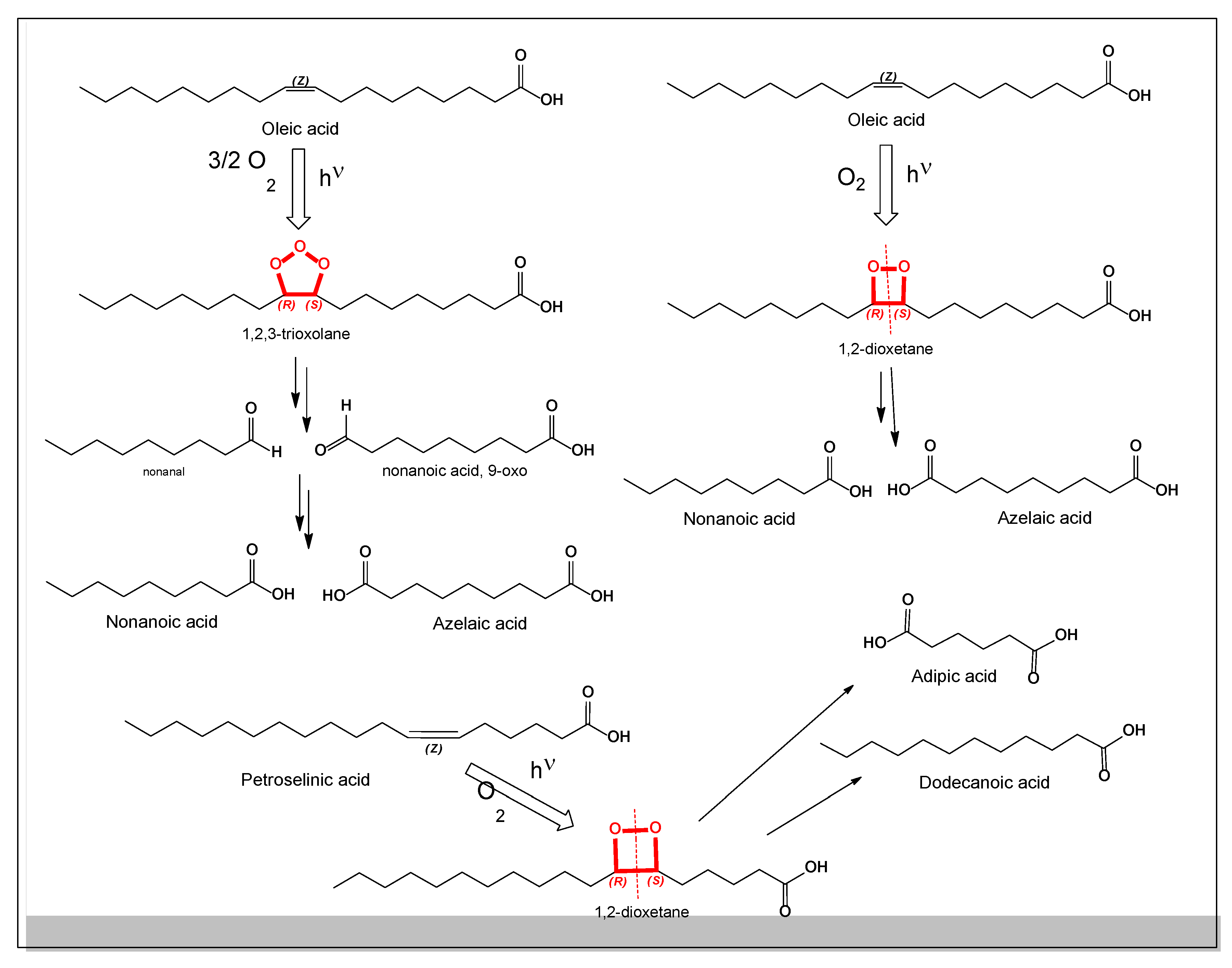
  81. Tejero, I.; González-Lafont, A.; Lluch, J.M.; Eriksson, L.A. Photo-oxidation of lipids by singlet oxygen: A theoretical study. Chem. Physics Lett. 2004, 398(4-6), 336–342. [Google Scholar] [CrossRef]
  82. Dembitsky, V.M.; Goldshlag, P.; Srebnik, M. Occurrence of dicarboxylic (dioic) acids in some Mediterranean nuts. Food Chem. 2002, 76(4), 469–473. [Google Scholar] [CrossRef]
  83. Baker, B.G.; Bedford, J.; Kanitkar, S. Keeping pace with the media; Giant Hogweed burns—A case series and comprehensive review. Burns 2017, 43(5), 933–938. [Google Scholar] [CrossRef]
  84. Pfurtscheller, K.; Trop, M. Phototoxic plant burns: Report of a case and review of topical wound treatment in children. Pediatric Dermatol 2014, 31(6), e156–e159. [Google Scholar] [CrossRef]
  85. Zangerl, A.R.; Berenbaum, M.R. Furanocoumarins in wild parsnip: Effects of photosynthetically active radiation, ultraviolet light, and nutrients. Ecology 1987, 68(3), 516–520. [Google Scholar] [CrossRef]
  86. Kasperkiewicz, K.; Erkiert-Polguj, A.; Budzisz, E. Sunscreening and photosensitizing properties of coumarins and their derivatives. Letters Drug Design & Discovery 2016, 13(5), 465–474. [Google Scholar]
  87. Sumorek-Wiadro, J.; Zając, A.; Maciejczyk, A.; Jakubowicz-Gil, J. Furanocoumarins in anticancer therapy–For and against. Fitoterapia 2020, 142, 104492. [Google Scholar] [CrossRef]
  88. Janusz, S.C.; Schwartz, R.A. Botanical briefs: Phytophotodermatitis is an occupational and recreational dermatosis in the limelight. Cutis 2021, 107(4), 187–189. [Google Scholar] [CrossRef]
  89. Rzymski, P.; Klimaszyk, P.; Poniedziałek, B. Invasive giant hogweeds in Poland: Risk of burns among forestry workers and plant distribution. Burns 2015, 41(8), 1816–1822. [Google Scholar] [CrossRef] [PubMed]
  90. Chan, J.C.; Sullivan, P.J.; O’Sullivan, M.J.; Eadie, P.A. Full thickness burn caused by exposure to giant hogweed: Delayed presentation, histological features and surgical management. J. Plastic, Reconstruct. Aesthetic Surg. 2011, 64(1), 128–130. [Google Scholar] [CrossRef] [PubMed]
  91. Flanagan, K.E.; Blankenship, K.; Houk, L. Botanical Briefs: Phytophotodermatitis caused by giant hogweed (Heracleum mantegazzianum). Cutis 2021, 108, 5. [Google Scholar] [CrossRef] [PubMed]
  92. Kelly, A. Phyto-photo dermatitis. The Ulster Medical J. 1969, 38(1), 51. [Google Scholar]
  93. Morris, A.J.; Rueckeis, C.A. Sap and sun: A case of phytophotodermatitis. Wilderness & Environ. Med. 2023, 34(4), 532–535. [Google Scholar]
  94. Downs, J.W.; Cumpston, K.L.; Feldman, M.J. Giant hogweed phytophotodermatitis. Clinical Toxicology 2019, 57(9), 822–823. [Google Scholar] [CrossRef]
  95. King, A.; Pope, E. Dermatitis versus nonaccidental trauma: A systematic review of initial pediatric misdiagnoses. Pediatric Dermatology 2024, 41(2), 215–220. [Google Scholar] [CrossRef]
  96. Xu, Y.; Luan, X.; He, P.; Zhu, D.; Mu, R.; Wang, Y.; Wei, G. Fabrication and functional regulation of biomimetic interfaces and their antifouling and antibacterial applications: A review. Small 2024, 20(21), 2308091. [Google Scholar] [CrossRef]
  97. Grosu, C.; Jîjie, A.R.; Manea, H.C.; Moacă, E.A.; Iftode, A.; Minda, D.; Vlad, C.S. New insights concerning phytophotodermatitis induced by phototoxic plants. Life 2024, 14(8), 1019. [Google Scholar] [CrossRef]
  98. Panahi, Y.; Dadjo, Y.; Pishgoo, B.; Akbari, A.; Sahebkar, A. Clinical evaluation of the anti-inflammatory effects of Heracleum persicum fruits. Comp. Clinical Pathology 2015, 24(4), 971–974. [Google Scholar] [CrossRef]
  99. Piersiala, K.; Loroch, A.; Kaik, J.; Dadej, D.; Kierepa, A.; Mozer-Lisewska, I. Burn caused by exposure to giant hogweed (Heracleum sosnowskyi, Sosnowsky’s hogweed) and delayed wound healing in a 46 years old HIV and HCV positive patient-a case report. Polish Annals of Medicine 2019, 26((1).). [Google Scholar] [CrossRef]
  100. Dar, N.A.; Raja, W.Y.; Tewari, D.; Bhat, Z.A. Pharmacognostic study of roots and aerial parts of less explored Heracleum candicans Wall. ex DC. from Betaab Valley, Pahalgam, Kashmir, India. Indian J. Nat. Prod. Resour. 2022, 13(3), 362–373. [Google Scholar] [CrossRef]
  101. Patocka, J.; Cupalova, K. Giant Hogweed and photodermatitis. Mil. Med. Sci. Lett. (Voj Zdrav Listy) 2017, 86(3), 135–138. [Google Scholar] [CrossRef]
  102. Agrawal, D.; Singh, M.P.; Sharma, G.K. Pharmacodynamic approaches of phytoconstitutents in wound healing mechanisms. Pharmacological Research-Natural Prod. 2024, 5, 100119. [Google Scholar] [CrossRef]
Figure 2. Furanocoumarins derived from Sosnovsky’s hogweed.
Preprints 190214 g002
Figure 3. Photochemical dermatitis caused by Sosnowsky’s hogweed sap. Contact with the sap of Heracleum sosnowskyi followed by exposure to sunlight induces photochemical dermatitis, resulting in skin lesions of varying severity. The lesions most frequently affect the upper and lower limbs and clinically resemble superficial thermal burns of grades I, II, and IIIA. Panels a, c and e show lesions on the hands, while panels b and d illustrate lesions on the legs. Management of photochemical dermatitis should include systemic measures such as detoxification, administration of antihistamines, analgesics, hormonal therapy, and antibacterial treatment when indicated. Local therapy should follow standard clinical algorithms used for the treatment of thermal burns.
Preprints 190214 g003
Figure 4. The general scheme of psoralen oxidation and formation of 1,2-dioxetanes (13-16) and oxidation to salicylaldehyde (19).
Preprints 190214 g004
Figure 5. Photochemical interaction of psoralens with nucleic acids. The reaction of psoralens with DNA is governed by three key factors: kinetic, spatial, and electronic parameters. Upon exposure to ultraviolet radiation, psoralen molecules interact with thymidine residues in DNA, leading to the formation of cyclobutane pyrimidine dimers. These dimers consist of a four-membered cyclobutane ring formed by the [2+2] cycloaddition of double bonds from two adjacent pyrimidine bases. Psoralens present in Sosnowsky’s hogweed sap therefore induce DNA damage upon irradiation with UV or sunlight and are also capable of damaging RNA under near-UV light. When psoralen molecules intercalate between DNA base pairs, they undergo highly specific photochemical reactions that facilitate efficient cross-linking and nucleic acid modification.
Preprints 190214 g005
Figure 6. Psoralen–thymidine photo-adducts and their structural variability. Photo-adduct structures, predominantly minor cyclobutane-containing complexes formed between psoralen and thymidine, exhibit considerable structural variability. The size and extent of these isomeric complexes can range from short adducts to extended structures involving up to 200 linked units, depending on factors such as ultraviolet radiation intensity, exposure duration, pH, oxygen availability, and the local DNA environment. The length and complexity of these psoralen–DNA photo-adducts directly influence the severity of DNA damage, as larger and more stable cross-linked structures more effectively disrupt DNA replication, transcription, and repair processes.
Preprints 190214 g006
Figure 7. Furan derivatives and other compounds derived from H. sosnowskyi biomass.
Preprints 190214 g007
Figure 8. Aromatic compounds derived from H. sosnowskyi biomass.
Preprints 190214 g008
Figure 9. The general scheme of fatty acid oxidation and formation of both types of peroxides: 1,2-dioxetane and 1,2,3-trioxolane.
Preprints 190214 g009
Disclaimer/Publisher’s Note: The statements, opinions and data contained in all publications are solely those of the individual author(s) and contributor(s) and not of MDPI and/or the editor(s). MDPI and/or the editor(s) disclaim responsibility for any injury to people or property resulting from any ideas, methods, instructions or products referred to in the content.
Copyright: This open access article is published under a Creative Commons CC BY 4.0 license, which permit the free download, distribution, and reuse, provided that the author and preprint are cited in any reuse.
Prerpints.org logo

Preprints.org is a free preprint server supported by MDPI in Basel, Switzerland.

Subscribe

Disclaimer

Terms of Use

Privacy Policy

Privacy Settings

© 2026 MDPI (Basel, Switzerland) unless otherwise stated