
Submitted:
17 December 2025
Posted:
19 December 2025
You are already at the latest version
Abstract
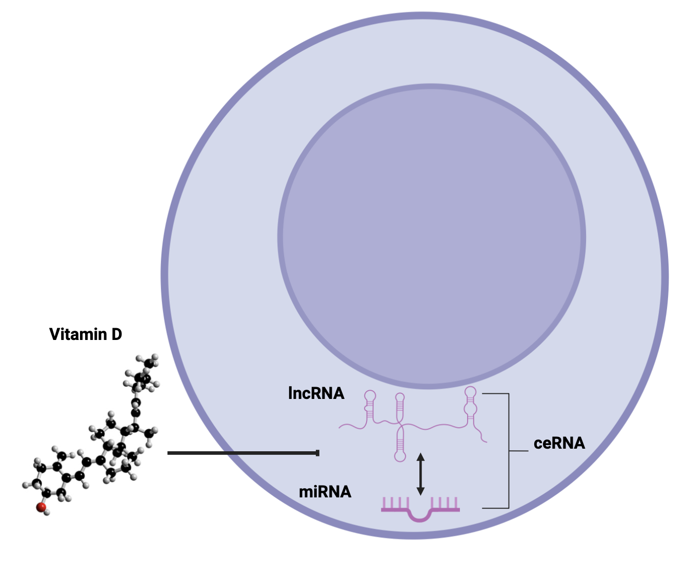
Zika virus (ZIKV), a mosquito-borne flavivirus, is associated with congenital malformations and neuroinflammatory disorders, highlighting the need to identify host factors that shape infection outcomes. Macrophages, key targets and reservoirs of ZIKV, orchestrate both antiviral and inflammatory responses. Vitamin D (VitD) has emerged as a potent immunomodulator that enhances macrophage antimicrobial activity and regulates inflammation. To investigate how VitD shapes macrophage responses to ZIKV, we reanalyzed publicly available RNA-seq and miRNA-seq datasets from monocyte-derived mac-rophages (MDMs) of four donors, differentiated with or without VitD and subsequently infected with ZIKV. Differential expression analysis identified long non-coding RNAs (lncRNAs), microRNAs (miRNAs), and mRNAs, integrated into competing endogenous RNA (ceRNA) networks. In VitD-conditioned and ZIKV-infected MDMs, 65 lncRNAs and 23 miRNAs were significantly modulated. Notably, lncRNAs such as HSD11B1-AS1, Lnc-FOSL2, SPIRE-AS1, and PCAT7 were predicted to regulate immune and metabolic genes, including G0S2, FOSL2, PRELID3A, and FBP1. Among the miRNAs, let-7a and miR-494 were downregulated, while miR-146a, miR-708, and miR-378 were upregulated, all of which have been previously implicated in antiviral immunity. Functional enrichment analysis revealed pathways linked to metabolism, stress responses, and cell migration. ceRNA network analysis suggested that SOX2-OT and SLC9A3-AS1 may act as molecular sponges, modulating regulatory axes relevant to immune control and viral response. Despite limitations in sample size and experimental validation, this study provides an exploratory map of ncRNA–mRNA networks shaped by VitD during ZIKV infection, highlighting candidate molecules and pathways for further studies on host–virus interactions and VitD-mediated immune regulation.
Keywords:
competing endogenous RNA
; lncRNA
; miRNA
; mRNA
; pro-inflammatory response
; antiviral response
; virus
; bioinformatics
1. Introduction
Zika virus (ZIKV) is a mosquito-borne flavivirus that has garnered global attention due to its association with congenital malformations, particularly microcephaly, and its potential to cause neurological complications, such as Guillain-Barré syndrome, in adults[1,2]. In recent years, substantial progress has been made in elucidating the immune mechanisms engaged during ZIKV infection. Both innate and adaptive responses are crucial, but their dysregulation can lead to immunopathology[3,4]. These insights have supported the development of vaccine candidates, some of which have to clinical evaluations. At the same time, ZIKV has been shown to exploit host pathways, including miRNA regulation, to evade immune surveillance[5]. Given the absence of specific antiviral therapies, a deeper understanding of host immune responses remains essential for guiding the development of effective interventions.
Recent research has highlighted the immunomodulatory effects of Vitamin D (VitD), a fat-soluble steroid hormone, in bolstering macrophage-mediated antiviral responses[6,7]. VitD promotes an antiviral state by upregulating the expression of antimicrobial peptides and proinflammatory cytokines in macrophages [8]. For instance, VitD supplementation has been shown to reduce ZIKV replication in vitro [9]. This dual role, enhancing host immune defense while directly impairing viral replication, underscores the potential of VitD as a key modulator in the host response to ZIKV. Yet, despite this recognized potential, the molecular mechanisms through which VitD shapes antiviral immunity during ZIKV infection remain only partially understood.
MicroRNAs (miRNAs) and long non-coding RNAs (lncRNAs) are emerging as critical regulators of gene expression in diverse biological processes, including antiviral immunity [10,11]. Beyond their individual roles, these ncRNAs participate in intricate regulatory circuits, where miRNAs and lncRNAs interact with mRNA targets to modulate the expression of genes involved in the antiviral defense machinery[5,12] A key example is the competing endogenous RNA (ceRNA) mechanism, in which lncRNAs act as a molecular sponge that sequesters miRNA, thereby relieving repression of specific mRNA and modulating antiviral defense pathways [13,14]. Interestingly, several VitD-modulated genes during ZIKV infection are targets of miRNA and lncRNA [15,16], suggesting the involvement of complex post-transcriptional regulatory mechanisms. However, the contribution of ceRNA interaction to VitD-mediated immune modulation in ZIKV infection remains largely unexplored.
Considering our previous findings [15,16], this study aims to investigate how VitD shapes lncRNA-mRNA interaction in ZIKV-infected macrophages. To this end, we reanalyzed RNA-Seq and miRNA-Seq datasets from monocyte-derived macrophage (MDMs) differentiated with or without VitD before ZIKV infection. Differentially expressed lncRNAs and miRNAs were integrated with mRNA targets to construct a ceRNA regulatory network. This genome-wide approach provides novel insights into the molecular pathway and regulatory circuits through which VitD may modulate host response to ZIKV.
2. Materials and Methods
2.1. Ethics Statement
This study was conducted with the approval of the Ethics Committee at the “Sede de Investigación Universitaria-Universidad de Antioquia”, Medellín, Colombia (approval code: 16-08-702; April 28, 2016). These ethical approvals are in accordance with the Declaration of Helsinki. All patients in this study provided written informed consent before blood collection.
2.2. Culturing of Human Monocytes and Differentiation into Monocyte-Derived Macrophages
Peripheral blood was obtained from four healthy donors and collected in tubes containing 4% v/v EDTA. Peripheral blood mononuclear cells (PBMCs) were isolated by gradient centrifugation using Ficoll-Histopaque (Sigma-Aldrich) at 650 x g for 30 minutes. Platelet depletion was achieved through three washes with 1X PBS (Sigma-Aldrich) at 250 x g for 10 minutes. The percentage of CD14+ cells was determined by flow cytometry, with 5x10⁵ cells stained with 1 μl of anti-CD14 (eBiosciences, San Diego, CA) for 30 minutes. Monocytes were isolated from total PBMCs using a plastic adherence procedure. Non-adherent cells were removed by washing twice with 1x PBS, after which monocyte-derived macrophages (MDM) were differentiated either in the absence (MDM) or the presence of 1 nM 1,25-dihydroxyvitamin D3 (D3-MDMs; Sigma-Aldrich). Cells were cultured for 6 days in RPMI 1640 medium supplemented with 10% autologous serum and maintained at 37 °C with 5% CO2, with the medium replaced every 24 hours until the completion of differentiation. The control group (MDMs) was treated with vehicle [0.1% ethanol (EtOH)]. This methodology follows a previously published protocol [17] in which differentiation into macrophages was confirmed by morphology and surface marker expression. All experiments were performed using cells from four independent healthy donors (n=4), yielding four biological replicates per experimental condition. Each donor’s cells were processed independently through all experimental steps (differentiation, treatment, infection, and RNA extraction)
2.3. ZIKV Infection and Treatments
On day 6 of culture, MDMs and D3-MDMs were infected by ZIKV strain COL345Si-Asian (kindly gifted by Professor Blanco P. Universidad de Sucre, Colombia) at a multiplicity of infection (MOI) of 5 (ZIKV-MDM and ZIKV-D3-MDM, respectively) in serum-free RPMI-1640. The cells were incubated for 2 hours at 37 °C with 5% CO2. Subsequently, the supernatant was removed, and the cells were washed with 1× PBS. The medium was then replenished with RPMI containing 10% FBS and cultured at 37 °C with 5% CO2. Harvesting of MDMs was performed at 24 hours post-infection (hpi).
2.4. RNA Isolation, Library Preparation, and miRNA-Seq
Total RNA was extracted using the Direct-zol RNA MiniPrep kit (Zymo Research) following the manufacturer’s instructions. RNA integrity was evaluated by assessing the ratio of 28S and 18S rRNA on a 1% agarose gel. For miRNA sequencing, libraries were prepared from two infected groups (ZIKV-MDM and ZIKV-D3-MDM), and two uninfected controls (MDM and D3-MDM) using a single-adapter ligation and circularization strategy [18]. Libraries were sequenced as 50 bp single-end reads, yielding depths of 7 to 12 million reads per sample.
2.5. Read Alignment and Differential miRNA Expression Analysis
FASTQ files from the miRNA libraries were quality-filtered with a Phred score cutoff of ≥ 30, and Illumina adapter sequences were trimmed using Trimmomatic (V0.32). Single-end reads were aligned to miRBase v22 with Bowtie using the parameters -n 0 -l 8 -a --best --strata --phred33-quals. miRNA counts were generated using FeatureCounts (default settings) and normalized with DESeq2 (version 1.32). Differential expression was defined as an absolute fold change (|fold change |≥ 2 and a false discovery rate (FDR) ≤ 0.05. Curated miRNA annotations were obtained from MirGeneDB.
2.6. Read Alignment and Differential lncRNA Expression Analysis
RNA-seq data from ZIKV-MDMs and ZIKV-D3-MDMs, and their respective controls (MDM and D3-MDM), are publicly available in the NCBI GEO database under accession number GSE209698. Each experimental group consisted of four biological replicates (n=4 independent donors). Data were reanalyzed by mapping to the human reference genome (hg38) using STAR, and differential expression was assessed with DESeq2 under a negative binomial distribution model. Expression profiles were obtained for mRNAs RefSeq databases) and lncRNAs (NONCODE database). Differentially expressed lncRNAs were defined as those with an absolute | fold change | ≥ 2 and FDR ≤ 0.05.
2.7. mRNA Differential Expression
For mRNA expression analysis, we used publicly available data from the Gene Expression Omnibus (GEO; accession number GSE209698). Differential expression was assessed following the methodology outlined in [19]. Briefly, we performed differential gene expression analysis using DESeq2, incorporating donor index as a covariate in the design formula to control for inter-individual genetic background differences. To minimize false-positive discoveries, we applied stringent statistical thresholds, considering genes differentially expressed (DEGs) only when they met both criteria: false discovery rate (FDR) ≤ 0.05 and an absolute log2 fold change > 1. This conservative approach ensured robust identification of biologically meaningful expression changes while accounting for donor-specific variability.
2.8. VDR Motif Analysis
We investigated VDR (Vitamin D Receptor) binding to the promoters of differentially expressed genes in the THP-1 macrophage cell line. THP-1 cells were stimulated with 1,25-dihydroxyvitamin D3 (1,25(OH)2D3) to activate VDR-mediated signaling, and ChIP-Seq data (GEO; accession number GSE2096981) were used to map VDR binding sites. Visualization with the Integrated Genome Viewer (IGV) version 2.3.90 enabled the identification of potential VDR occupancy within the target gene promoters.
2.9. Gene Enrichment Analysis
Gene enrichment analysis was performed using EnrichR, with the Kyoto Encyclopedia of Genes and Genomes (KEGG) pathways and Gene Ontology (GO) Biological Process database. Enrichment results were expressed as the percentage of genes per term, calculated by EnrichR as overlap. KEGG and GO terms were further integrated into the analysis using an over-representation criterion (log2 combined score > 2), where the combined score represents a comprehensive measure of enrichment significance that incorporates both the p-value from Fisher’s exact test and the z-score for deviation from expected rank, thereby providing enhanced statistical power to identify biologically relevant pathways while controlling for both significance and effect size.
2.10. miRNA Target Prediction
Candidate miRNA-mRNA interactions were predicted using TargetScan 5.1, considering both conserved and non-conserved binding sites. Filtering was restricted to differentially expressed mRNA and miRNA identified by RNA-Seq, requiring an inverse correlation between mRNA and miRNA expression levels. Target similarity among differentially expressed genes was assessed using the Jaccard index. The resulting miRNA-mRNA interaction network was visualized in Cytoscape v3.6.1 (https://cytoscape.org/).
2.11. Construction of the ceRNA Network
A potential ceRNA network was constructed in two steps. First, mRNA-miRNA interactions were identified based on DEG. Second, mRNA-lncRNA and miRNA-lncRNA interactions were retrieved by integrating data from three databases: miRTarBase, lncRNA2Target, and lncbasev3. Before merging, filtering was applied to retain only differentially expressed mRNA, miRNA, and lncRNA, requiring an inverse correlation between lncRNA and miRNA expression levels, and including all lncRNA-mRNA interactions. The final lncRNA-miRNA-mRNA ceRNA regulatory network was visualized using Cytoscape v.3.9.1.
2.12. lncRNA-mRNA Correlation
For the statistical analysis of lncRNA-mRNA cis-acting interactions, Pearson correlation analysis was performed. Correlation coefficients (r) were calculated for each lncRNA-mRNA pair across all samples (n=16; 4 donors × 4 conditions) to quantify the strength and direction of their association. Corresponding p-values were computed to assess statistical significance, and the Benjamini-Hochberg procedure was applied to control the false discovery rate (FDR < 0.05) for multiple testing correction. Principal Component Analysis (PCA) was performed to assess overall variance and group separation. K-means clustering was applied to categorize expression profiles of differentially expressed lncRNAs.
3. Results
3.1. Vitamin D–Mediated Regulation of Non-Coding RNA Networks in ZIKV-Infected Macrophages
3.1.1. Vitamin D-Mediated Modulation of lncRNA Expression in ZIKV-Infected MDMs
Previously, we reported the efficacy of vitamin D in reducing ZIKV infection [9]. Here, we extended these findings by investigating the underlying regulatory mechanisms, with a particular focus on the role of lncRNAs as potential modulators of gene expression in ZIKV-infected macrophages differentiated in the presence or absence of VitD (ZIKV-D3-MDMs vs ZIKV-MDM). To this end, we conducted a comprehensive analysis of lncRNA expression profiling using RNA-Seq. A total of 403 lncRNAs were found to be significantly altered across all the experimental groups (S1 Table). These differentially expressed lncRNAs were further classified according to their genomic features, revealing the following distribution: 55.1% (n = 222) long intergenic non-coding RNAs (lincRNAs), 19.1% (n = 77) pseudogene-derived lncRNAs, 17.1% (n = 69) antisense lncRNAs, and 5.7% (n = 23) divergent lncRNAs (Figure 1a).
Next, we explore whether the expression profiles of the 403 lncRNAs could distinguish between the four experimental groups. Principal Component Analysis (PCA) reveals a clear separation between uninfected macrophages (MDMs and D3-MDMs) and their ZIKV-infected counterparts (ZIKV-MDM and ZIKV-D3-MDM) (Figure 1b). Furthermore, PCA shows that they exhibit specific, unique features despite the high similarity among ZIKV-infected macrophages. PCA also highlighted distinct transcriptional features, suggesting that VitD modulated the baseline expression of macrophages and shaped unique regulatory patterns upon viral infection. Consequently, we focused on lncRNAs exhibiting up- or down-regulation in response to VitD treatment during ZIKV infection. Out of the 403 lncRNAs analyzed, 83.9% (n=338) displayed no significant changes in their expression levels when comparing ZIKV-D3-MDMs to ZIKV-MDMs. However, 12.9% (n=52) were upregulated while 3.2% (n=13) displayed downregulation (Figure 1c and Table S2).
We applied the k-means clustering to categorize the expression profiles of 65 VitD-modulated lncRNAs (Figure 1d and Figure 1f). For the upregulated lncRNAs, six distinct expression clusters were identified (Figure 1d). Cluster 1 comprises 11 VitD-dependent lncRNAs whose expression was further enhanced upon ZIKV infection (Figure 1d and Figure 1f). Notably, this cluster contained ITGA-AS1, a lncRNA known to regulate ITGA6, a gene critical for cell adhesion and migration [20]. Cluster 2 encompasses 8 lncRNAs that show decreased expression levels following ZIKV infection (ZIKV-MDM) but increased expression in D3-MDM, with expression levels still notably stronger in ZIKV-D3-MDM (Figure 1d and Figure 1f). Among these, SOX2-OT (SOX2 overlapping transcript) was of particular interest, as it overlaps with the SOX2 gene and plays a role in modulating its expression[21]. Furthermore, SOX2-OT is associated with vital cellular processes, including inflammation and oxidative stress[22].
Cluster 3 comprises four genes with an expression profile similar to Cluster 2, but distinguished by their basal up-regulation under VitD conditioning (Figure 1d and Figure 1f). Within this cluster, DPP4-DT (dipeptidyl peptidase-4 distant transcript) was identified, which has been previously linked to ferroptosis [23]. Cluster 4 displays a particularly distinctive profile, as the lncRNAs are highly expressed in ZIKV-D3-MDM (Figure 1d and Figure 1f). Among them, HSD11B1-AS1 has been associated with the modulation of inflammation [24]. Cluster 5 delineates a group of 9 lncRNAs specifically modulated in VitD-conditioned macrophages (D3-MDM), but whose expression decreases when these macrophages are subsequently infected with ZIKV (ZIKV-D3-MDM) (Figure 1d and Figure 1f). Notably, DBH-AS1 was among these, an lncRNA reported as elevated in the peripheral blood of COVID-19-infected patients [25] and linked to the regulation of autophagy [26,27]. Finally, Cluster 6 consists of lncRNAs that are downregulated upon ZIKV infection in both MDM and D3-MDM. However, in VitD-conditioned (D3-MDM), their expression levels reamis substantially higher compared to unconditioned (MDM) (Figure 1d and Figure 1f). This cluster includes 9 lncRNAs,with SLC9A3-AS1 being particularly notable, as it has been described as a regulator of glycolysis [28].
Conversely, for the downregulated lncRNAs, we identified four distinct expression clusters (Figure 1e and Figure 1f). Cluster 1 comprises lncRNAs expressed exclusively in infected MDMs (ZIKV-MDM), with no modulation observed under VitD conditioning (D3-MDM) or in infected macrophages (ZIKV-D3-MDM). This cluster contained 4 lncRNAs, among them ROR1-AS1, which has been previously described as an immunomodulator during SARS-CoV-2 infection [29]. Cluster 2, similar to Cluster 1, includes lncRNAs whose downregulation is exclusively driven by VitD, regardless of infection status (ZIKV-D3-MDM and D3-MDM, respectively) (Figure 1d and Figure 1f). Notably, LINC02345 belongs to this group and has been associated with the regulation of copper-induced cell death[30]. Lastly, clusters 3 and 4 are characterized by decreased lncRNA expression in both VitD-conditioned (D3-MDM) and ZIKV-infected MDMs (ZIKV-D3-MDM), with three and two lncRNAs, respectively (Figure 1d and Figure 1f). To date, however, no functional evidence has been reported for the lncRNAs in these two clusters.
3.1.2. Vitamin D Modulates Cis-Acting lncRNAs to Regulate Gene Expression
One of the key molecular mechanisms by which lncRNAs exert their function is cis-acting regulation, whereby they physically link to specific genomic loci to control the expression of neighboring genes. A common strategy to infer the potential roles of lncRNAs in biological and pathological processes is co-expression analysis, which evaluates the correlation between lncRNA and mRNA expression profiles. To identify co-expressed lncRNA-coding gene pairs, we calculated Pearson correlation coefficients (PCC) based on the expression level of each differentially expressed lncRNA-mRNA pair, as previously described [31]. This approach provides valuable insights into the mechanistic contribution of cis-acting lncRNAs during viral infection. In the case of ZIKV-D3-MDMs, our analysis focused on lncRNA-coding gene pairs with a PCC> 0.80 and a significance level of p < 0.05.
We identified a total of 129 lncRNA-mRNA interaction pairs, of which 124 exhibited positive correlations and 5 displayed negative regulations (S3 Table). Since these interactions were located on the same chromosome, they are suggestive of a potential cis-acting regulatory relationship between the lncRNAs and their neighboring mRNAs.
To increase the specificity of our findings, we further refined the analysis by incorporating the genomic proximity between lncRNAs and coding genes. We specifically focused on interactions in which the distance between the lncRNA and its neighboring coding gene was less than 300 kilobases (Kb), either upstream or downstream. Applying this stringent criterion, we identified 15 lncRNA-coding gene pairs that fulfilled these conditions (S4 Table). Notably, several interactions stood out, including SLC5A4-AS1-RFPL2 mRNA, PCAT7-FBP1 mRNA, DBH-AS1-SARDH mRNA, and LINC02345-SPRED1 mRNA. These lncRNAs have been previously implicated in the transcriptional regulation of their target [32], the modulation of viral pathogenesis [25], and the induction of endoplasmic reticulum stress. [33]
Next, we sought to determine whether VitD regulated these 15 lncRNA-mRNA pairs via the VDR signaling pathway. Our analysis revealed a statistically significant positive correlation between VitD exposure and the coordinated expression of specific lncRNAs-mRNA pairs, including SPIR1-AS1-PRELID3A, lnc-FOSL2-FOSL2, PCAT7-FBP1, HSD11B1-AS1-LAMB3, and HSD11B1-AS1- G0S2, all of which are located within the same genomic loci (Figure 2a). Importantly, chromatin immunoprecipitation sequencing (ChIP-Seq) analysis demonstrated that VDR binding peaks are positioned within 50 kb of the transcription start site (TSS) of either the lncRNA or its paired coding gene. As shown in Figure 2b, genomic tracks of VDR ChIP-seq data illustrate enrichment profiles in THP-1 macrophages under control conditions (red) and following vitamin D treatment (blue), spanning up to 103 kb. The proximity of VDR binding sites to the promoters of both lncRNAs and their target genes strongly supports the notion that VitD/VDR signaling directly regulates the transcription of these cis-acting pairs. Taken together, these results indicate that VitD modulates cis-acting lncRNAs through VDR binding to regulatory regions of nearby genes, providing a mechanistic explanation for the coordinated upregulation observed in ZIKV-infected macrophages.
3.1.3. Vitamin D Modulates miRNA Expression in ZIKV-Infected Macrophages
Next, we investigate whether the expression levels of miRNAs were altered in MDM, D3-MDM, ZIKV-MDM, and ZIKV-D3-MDM. Out of 2,654 reported human mature miRNAs, 857 were detected across all four groups. PCA performed on these 857 miRNAs revealed a clear separation between the groups, indicating that their expression profiles can effectively discriminate each condition (Figure 3a). These findings suggest distinct sets of miRNAs are associated with the different experimental groups.
Building on our previous investigations into miRNAs modulated by VitD or ZIKV infection in macrophages [15,16,34], the present study specifically aimed to identify the expression pattern in VitD-conditioned and ZIKV-infected MDMs (ZIKV-D3-MDMs) compared to ZIKV-MDMs. We identified 23 differentially expressed (DE) miRNAs in ZIKV-D3-MDM (FDR ≤ 0.05 and |fold change| ≥ 1.5), of which 14 were up- and 9 were down-regulated (Figure 3b). To characterize these differences further, we applied a k-means clustering algorithm to the mean relative expression values of the 23 DE miRNAs. This analysis classified them into eight distinct clusters (clusters 1-8; Figure 3b) based on their dynamic expression profiles across the infected MDMs groups. Clusters 1-4 comprised miRNAs upregulated in response to VitD, whereas clusters 5-8 contained miRNA that were downregulated following VitD treatment (Figure 3b).
We next examined whether the differential expression of miRNAs was mediated through VDR. To address this, we analyzed publicly available VDR ChIP-seq data from THP1 macrophages treated with VitD for 24 hours [35]. Our analysis revealed VDR binding peaks associated with the genomic loci of miR-378a-5p (cluster 2), located within the intronic region of the PPARGC1B gene, and miR-542-3p (Figure 3c). These findings indicated that, among the miRNAs differentially regulated in ZIKV-D3-MDMs, only miR-378a-5p and miR-542-3p appear to be direct transcriptional targets of VDR. This supports the notion that VDR binding events in proximity of the transcription start site of these miRNAs contribute to the modulation of their expression patterns in ZIKV-D3-MDMs.
3.1.4. Vitamin D-Regulated miRNAs Modulate Key Biological Processes in ZIKV-Infected Macrophages
To gain insights into the functional roles of the miRNAs modulated in ZIKV-D3-MDM, we examined the inverse relationship between miRNA expression and the expression of their predicted mRNA targets within the same samples. Predicted mRNA targets were identified for 17 of the 23 DE miRNAs, encompassing up- and down-regulated miRNAs (Figure 4a). This analysis uncovered 78 putative miRNA-mRNA interactions, underscoring the potential for target multiplicity in those 17 miRNAs. Furthermore, the up-regulated miRNA, miR-3145-5p, exhibited the highest number of predicted target sites (24) among mRNAs, while the down-regulated miRNA, miR-494-3p and miR-542-3p displayed the most significant number of predicted targets, with 31 and 27sites, respectively (Figure 4a).
Based on the integrative miRNA-mRNA analysis, we identified signaling pathways enriched among deregulated genes targeted by DE miRNAs (Figure 4b). Gene set enrichment analysis revealed that VitD-modulated miRNAs in ZIKV-infected MDMs predominantly regulate transcripts involved in cellular stress responses and metabolic processes. Specifically, we observed a downregulation of genes associated with the pentose phosphate pathway and, conversely, an upregulation of genes implicated in polyamine biosynthesis. Moreover, several genes are linked to cellular stress pathways were upregulated, including those involved in the IRE1-mediated unfolded protein response (UPR) and the regulation of mitochondrial membrane permeability, suggesting that VitD may influence both metabolic reprogramming and stress adaptation during ZIFV infection.
Up-regulated miRNAs (miR-146a-3p, miR-551b-5p, miR-551b-3p, miR-371a-3p, miR-31-5p, miR-511-3p, miR-378a-5p, miR-3145-5p, and miR-708-5p) were predicted to mediate the downregulation of genes involved in cell migration (PLXNA2, CASS4, SEMA3A, PIEZO1, MB21D2, and PODXL), metabolism (PHACTR1 and SULT1B1), and stress response (FPR1, P2RY6, STON2 and FES) (Figure 4c). On the other hand, down-regulated miRNAs (miR-3142, miR-3200-3p, miR-542-3p, miR-876-5p, miR-766-5p, miR-3611, miR-494-3p, and let-7a-2-3p) were associated with the up-regulation of metabolism genes, including SAT1, ABCD2, SARM1, and ABHD17C (Figure 4c). Furthermore, several target genes of miR-494-3p were related to cell migration (FZD4, EDN1, ADAM28, and CTNNA3) (Figure 4c). In summary, these findings highlight an intricate regulatory network in which VitD-modulated miRNAs exert opposing effects on gene expression programs related to cell migration, metabolism pathways, and cellular stress response, underscoring their multifaceted roles in shaping macrophage responses to ZIKV under VitD conditioning.
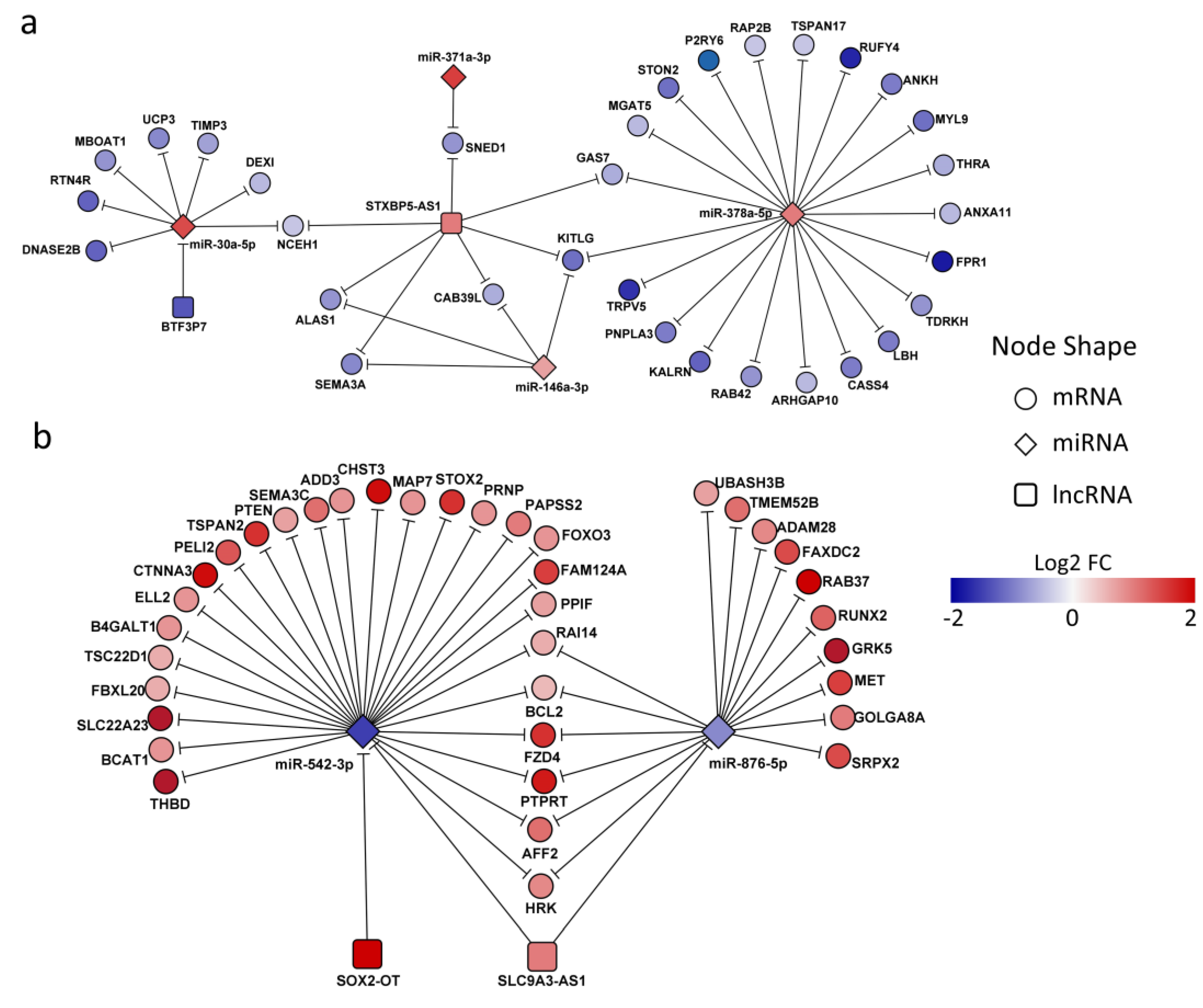
3.1.5. Regulation of Gene Expression by Vitamin D via Competing Endogenous RNA Mechanisms in ZIKV-Infected Macrophages: A Network Approach of lncRNA-miRNA-mRNA Interactions
To comprehensively understand posttranscriptional mRNA regulation, it is essential to recognize than miRNAs do not act in isolation; instead, their crosstalk with other non-coding RNA (ncRNAs), such as lncRNAs, plays a pivotal role. To address this, we constructed a ceRNA network modulated in ZIKV-D3-MDM. Specifically, we examined the relationships between co-differentially expressed RNAs in VitD-conditioned and ZIKV-infected MDMs (ZIKV-D3-MDM vs. ZIKV-MDM). Using curated lncRNA-targets databases, we filtered potential interactions based on expression data to identify cases in which lncRNA-miRNA and lncRNA-mRNA interactions display opposite regulatory patterns. This analysis revealed that several downregulated mRNAs in ZIKV-D3-MDM were influenced by the lncRNA STXBP5-AS1, which cooperatively interacted with multiple miRNAs, including miRNA 378a-5p, miRNA 146-3p, miRNA 371a-3p, and miRNA 30a-5p (Figure 5a). Among these, miRNA 378a-5p emerged as the central node, showing the highest connectivity with 21 mRNA targets and the lncRNA, STXBP5-AS1, highlighting its prominent regulatory role within the ceRNA network. This interaction leads to down-regulation of GAS7, KITLG, CAB39L, SEMA3A, ALAS1, SNED1, and NCEH1. These finding suggests that lncRNA STXBP5-AS1 primarily exerts its effect through direct targeting of mRNAs rather than regulating miRNAs. The affected genes are functionally linked to cell growth (GAS7, KITIG, SEMA3A, and SNED1) and metabolism (ALAS1 and NCEH1). Furthermore, our analysis identified BTF3P7, a pseudogene that negatively regulated miR-30a-5p, supporting a potential sponge effect. In ZIKV-D3-MDM, the downregulation of BTF3P7 may relieve its inhibitory effect on miR-30a-5p, thereby enhancing miR-30a-5p activity and contributing to the downregulation of NCEH1, UCP3, and MBOAT1, all involved in the metabolism process [36,37], as well as TIMP3, a regulator of extracellular matrix remodeling and macrophage proinflammatory response [38].
For the upregulated mRNA in ZIKV-D3-MDM, ceRNA network analysis identified miR-542-3p as the central hub, interacting with 27 mRNAs and two lncRNAs, SOX2-OT and SLC9A3-AS1 (Figure 5b). This network contributes to the upregulation of target genes involved in diverse biological processes. For instance, several miR-542-3p target genes are associated with cell proliferation, differentiation, and apoptosis, highlighting their role in maintaining cellular homeostasis. Notably, PTEN, FOXO3, and BCL2 regulate survival and apoptotic pathways, whereas CTNNA3 and FZD4 are involved in cell-cell adhesion and signaling cascades. Additionally, genes such as THBD and SEMA3C play a role in angiogenesis and vascular development. Beyond miR-542-3p interactions, our analysis also revealed that lncRNA SLC9A3-AS1 acts as a sponge for miR-876-5p, thereby promoting the upregulation of genes, including GRK5, RBA37, and PTPRT (Figure 5b), which are implicated in virus-host interactions and immune modulation. Collectively, these findings suggest that ceRNA-mediated regulation by SOX2-OT and SLC9A3-AS1 in ZIKV-D3-MDM involves miRNA sponging mechanisms that fine-tune the expression of genes governing cellular responses and virus-host dynamics during infection.
4. Discussion
ZIKV infection remains a major global health concern, underscoring the urgent need for an understanding of virus–host interactions to develop effective therapeutic strategies [18,19]. VitD has emerged as a promising immunomodulatory factor in several viral infections, including DENV, although the precise mechanisms highlighting its antiviral effects remain unclear [20,21]. Previous studies have shown that VitD modulates immune responses by influencing the expression of both miRNAs and mRNAs [15]. Moreover, lncRNAs have been identified as direct targets of VitD and may act as mediators of it signaling cascade [22]. Notably, both miRNAs and lncRNAs are key regulators of protein-coding genes [13]. Despite this evidence, the coordinate interplay among lncRNAs, miRNAs, and mRNAs in VitD-conditioned macrophages during ZIKV infection remains broadly understood.
This study investigates the molecular mechanisms underlying VitD-mediated immunomodulation during ZIKV infection, with particular focus on ncRNAs, specifically miRNAs and lncRNAs. Previous reports have shown that VitD can modulate the expression of specific lncRNAs [23]. By integrating RNA-seq and miRNA-seq data, we characterized the transcriptional landscape in VitD-conditioned macrophages after 24 hours of ZIKV infection. Our analyses uncovered significant alterations in 65 lncRNAs and 23 miRNAs, with enrichment of pathways related to metabolism and immune responses. Given the critical role of ncRNAs in fine-tuning gene expression, these findings establish a novel regulatory framework that provides new insights into VitD–ZIKV interactions.
Notably, VitD-conditioned macrophages infected with ZIKV displayed distinct alterations in lncRNA expression, including the upregulation of lincRNAs, pseudogenes, antisense, and divergent transcripts. Among these, SOX2-OT, DPP4-DT, HSD11B1-AS1, DBH-AS1, and PCAT7 were significantly upregulated. Although these lncRNAs have not previously been associated with VitD, they have been linked to key biological processes such as cell adhesion, migration, inflammation, oxidative stress, ferroptosis, and autophagy. For instance, SOX2-OT has been shown to bind ILF3 and promote STAT3 phosphorylation, thereby activating TGF-β signaling [24]. DBH-AS1 regulates cell-cycle genes, enhancing proliferation and resistance to apoptosis [25], and has been linked to COVID-19 severity [26]. Interestingly, it is downregulated in hepatitis virus-associated hepatocellular carcinoma [27] but can be induced by HBx protein to promote proliferation through MAPK signaling [28]. Similarly, lncRNA PCAT7 has been implicated in tumor progression via the ErbB/PI3K/Akt signaling pathway [29].
We also identified key lncRNAs involved in cis-regulatory mechanisms. For example, HSD11B1-AS1 regulates G0S2 in monocytes [30] and has been linked to protective roles in melanoma [31]. Other antisense lncRNAs, such as Lnc-FOSL2-3 and SPIRE-AS1, may regulate nearby genes like FOSL2 and PRELID3A, respectively, both of which harbor VDR binding sites within 50 kb. FOSL2 is a regulator of antiviral cytokine responses [32] and has been reported to be upregulated in severe COVID-19 [33], underscoring its relevance in antiviral immunity. In contrast, PRELID3A plays a central role in mitochondrial lipid metabolism and the synthesis of cardiolipin (CL) [34]. Mitochondrial lipids, including CL, are critical regulators of immune function, and their depletion impairs cytokine production in macrophages[35] Additionally, PRELID3A prevents apoptosis by facilitating phosphatidic acid (PA) transport within mitochondria under oxidative stress [36], maintaining mitochondrial membrane potential, enhancing respiratory chain function, and reducing reactive oxygen species production[37,38]. These functions suggest that PRELID3A could influence to ZIKV pathogenesis in VitD-treated macrophages. Beyond antisense transcripts, other lncRNAs may exert regulatory effects on neighboring genes due to chromosomal proximity [39]. For instance, PCAT7 may regulate the adjacent gene FBP1, a critical enzyme in gluconeogenesis with established roles in macrophage polarization and immune responses [40].
Studies have shown that FBP1 is activated during monocytic differentiation induced by 1,25-(OH)₂D₃, highlighting its responsiveness to VitD [41]. Moreover, FBP1 expression is directly regulated by VDR binding to its promoter [42,43]. During infection, IFN-γ enhances 1α-hydroxylase activity, leading to the conversion of VitD into its active form and in turn, promoting FBP1 expression [44]. This interplay between VitD, IFN-γ, and FBP1 underscores the potential for PCAT7 and VitD to co-regulate immune responses during ZIKV infection. Nevertheless, experimental validation is still required to confirm PCAT7-mediated regulation of FBP1.
miRNA expression was also markedly altered by VitD in ZIKV-infected macrophages. Consistent with previous reports of VitD-mediated modulation of miRNAs in other arboviral infections [20,45,46]. Here, we identified several miRNAs involved in regulating metabolism, stress responses, and macrophage function [47,48,49]. Among the most notable, let-7a was significantly downregulated; this miRNA directly targets Tet2, a key regulator of IL-6 production and TCA cycle metabolites in macrophages [50,51]. Another downregulated miRNA, miR-494-3p, exhibits dynamic expression during macrophage polarization, being suppressed in M1 and induced in M2 phenotypes [52]. It also regulates components of the Wnt signaling pathway [53] and modulates C/EBP-β activation via Nrdp1 binding[54,55]. In contrast, among the upregulated miRNAs, miR-146a-3p emerged as a central regulator of the immune response. This miRNA suppresses pro-inflammatory and antiviral responses in microglial cells during ZIKV infection [56], by targeting TRAF6, a key adaptor protein in antiviral signaling [57]. Additionally, upregulated miRNAs include miR-3145, which has been shown to inhibit influenza virus replication [58], as well as miR-708-5p and miR-378a-5p, both previously linked to immune regulation and viral infections [59,60]
We further identified several ceRNA networks that may underlie the observed changes in mRNA expression. Our findings are consistent with studies in other biological systems, highlighting the broad significance of ceRNA regulation across viral infections and VitD-mediated exposure [61,62,63]. Among these, STXBP5-AS1 emerged as a key ceRNA, supporting multiple miRNAs, and thereby promoting the downregulation of target mRNAs related to cell growth and metabolism. This observation aligns with findings in SARS-CoV-2-infected brain tissue, where STXBP5-AS1 functions as a ceRNA and potentially contributes to post-COVID neurological symptoms [64].
Additionally, lncRNAs such as SOX2-OT and SLC9A3-AS1 acted as sponges for miR-542-3p and miR-876-5p, respectively, leading to the upregulation of genes involved in metabolism and immune defense. This supports previous findings showing that SOX2-OT regulates inflammatory responses via miR-455-3p and miR-215-5p [65], and that SLC9A3-AS1 enhances gene expression by sequestering miRNAs such as miR-760 and miR-486-5p [66,67]. Furthermore, lncRNA STXBP5-AS1 has been reported to be upregulated by Rh2 via promoter hypomethylation and to function as a ceRNA by sequestering the oncogenic miR-4425 [68]. This lncRNA also promotes proliferation and invasion in cervical cancer cells by modulating the miR-96-5p/PTEN axis [69]. Altogether, these findings, along with our results, support a model in which ceRNA-mediated gene upregulation contributes to the molecular mechanisms underlying viral pathogenesis.
Interestingly, our study also identified BTF3P7, a pseudogene acting as a sponge for miR-30a-5p. Although research on pseudogenes as ceRNAs is still emerging, their regulatory potential is increasingly recognized. For instance, IFITM4P has been shown to function as a ceRNA for miR-24-3p in influenza A virus-infected cells, thereby modulating the expression of antiviral proteins IFITM1–3 [91]. Our results contribute to this growing body of evidence, suggesting that pseudogene-mediated ceRNA networks may play a role in shaping antiviral responses.
Together, these findings reveal a complex regulatory network of lncRNA–miRNA–mRNA interactions modulated by VitD in ZIKV-infected macrophages. This landscape uncovers previously unrecognized layers of post-transcriptional gene regulation during infection, suggesting potential molecular targets for therapeutic intervention. Nonetheless, our study is limited by the lack of functional validation. Future work employing gene knockdown, overexpression, luciferase reporter assays, and RNA immunoprecipitation will be essential to confirm these predicted interactions and establish their biological relevance.
In summary, both miRNAs and lncRNAs are dynamically regulated by VitD during ZIKV infection, orchestrating immune and metabolic responses through diverse regulatory mechanisms. By integrating RNA-seq and miRNA-seq analyses, this study reveals previously unrecognized regulatory layers in VitD signaling in macrophages and identifies novel ncRNA candidates with potential as therapeutic targeting in viral infections and immune-related disorders.
Supplementary Materials
The following supporting information can be downloaded at the website of this paper posted on Preprints.org, Supplementary Table S1, Differentially expressed lncRNAs across all pairwise comparisons; Supplementary Table S2, Differentially expressed lncRNAs in ZIKV-D3-MDM group compared with ZIKV-MDM as reference group; Supplementary Table S3, Cis acting lncRNAs pais modulated in ZIKV-D3-MDM; Supplementary Table S4, Pairs of cis-acting lncRNAs modulated in ZIKV-D3-MDM within up to 500 kb.
Author Contributions
Conceptualization: GJF, SUI; Data curation: JMRM, GJF, SUI; Formal analysis: JMRM, GJF; Funding acquisition: GJF, SUI; Investigation: JMRM, GJF, SUI; Methodology: GJF, SUI; Project administration: SUI; Resources: SUI; Software: JMRM, GJF; Supervision: SUI; Validation: JMRM, GJF; Visualization: JMRM, GJF; Writing – original draft: JMRM, GJF; Writing – review & editing: JMRM, GJF, SUI. All the authors have read the paper and have agreed to be co-authors.
Funding
This work was supported by Minciencias/Colciencias [grant No. 111574455028, contrato 641-2017 and contrato No. 455-2019], and Universidad de Antioquia-CODI, acta No. 2015-7803. GJF received Minciencias/Colciencias fellowships (grants # call 811 of 2018 and # 455-2019). The funders played no role in the study design, data collection and analysis, decision to publish, or preparation of the manuscript.
Institutional Review Board Statement
This study was conducted with the approval of the Ethics Committee at the “Sede de Investigación Universitaria-Universidad de Antioquia”, Medellín, Colombia. (approval code: 16-08-702; April 28, 2016). These ethical approvals are in accordance with the Declaration of Helsinki.
Informed Consent Statement
All patients in this study provided written informed consent before blood collection.
Data Availability Statement
All relevant data are within the paper and its Supporting Information files. Raw sequencing data are available in the GEO database under accession number GSE209698.
Acknowledgments
The authors would like to thank the blood bank of the “Escuela de Microbiología, UdeA, Medellín Colombia”, for providing us with leukocyte-enriched blood units from healthy individuals and the personnel at the institutions where the study was performed.:
Conflicts of Interest
The authors declare no conflict of interest.
Abbreviations
The following abbreviations are used in this manuscript:
| VitD | Vitamin D |
| ZIKV | Zika virus |
| MDMs | Monocyte-derived macrophages |
| lncRNAs | Long non-coding RNAs |
| miRNAs | MicroRNAs |
| ceRNA | Competing endogenous RNA |
| Kb | Kilobases |
| ChIP-Seq | Chromatin immunoprecipitation sequencing |
| UPR | Unfolded protein response |
References
- Igbinosa, II; Rabe, IB; Oduyebo, T; Rasmussen, SA. Zika Virus: Common Questions and Answers. Am Fam Physician 2017, 95, 507–513. [Google Scholar]
- Cerbino-Neto, J; Mesquita, EC; Souza, TML; Parreira, V; Wittlin, BB; Durovni, B; et al. Clinical Manifestations of Zika Virus Infection, Rio de Janeiro, Brazil, 2015. Emerg Infect Dis 2016, 22, 1318–1320. [Google Scholar] [CrossRef]
- Lazear, HM; Diamond, MS. Zika Virus: New Clinical Syndromes and Its Emergence in the Western Hemisphere. J Virol 2016, 90, 4864–4875. [Google Scholar] [CrossRef]
- Grant, A; Ponia, SS; Tripathi, S; Balasubramaniam, V; Miorin, L; Sourisseau, M; et al. Zika Virus Targets Human STAT2 to Inhibit Type I Interferon Signaling. Cell Host Microbe 2016, 19, 882–890. [Google Scholar] [CrossRef] [PubMed]
- Trobaugh, DW; Klimstra, WB. MicroRNA Regulation of RNA Virus Replication and Pathogenesis. Trends Mol Med 2017, 23, 80–93. [Google Scholar] [CrossRef]
- Adams, JS; Hewison, M. Unexpected actions of vitamin D: new perspectives on the regulation of innate and adaptive immunity. Nat Clin Pract Endocrinol Metab 2008, 4, 80–90. [Google Scholar] [CrossRef] [PubMed]
- Kow, CS; Hadi, MA; Hasan, SS. Vitamin D Supplementation in Influenza and COVID-19 Infections Comment on: “Evidence that Vitamin D Supplementation Could Reduce Risk of Influenza and COVID-19 Infections and Deaths” Nutrients 2020, 12(4), 988. Nutrients 2020, 12. [Google Scholar] [CrossRef]
- Liu, PT; Stenger, S; Li, H; Wenzel, L; Tan, BH; Krutzik, SR; et al. Toll-like receptor triggering of a vitamin D-mediated human antimicrobial response. Science 2006, 311, 1770–1773. [Google Scholar] [CrossRef]
- Fernandez, GJ; Ramírez-Mejía, JM; Castillo, JA; Urcuqui-Inchima, S. Vitamin D modulates expression of antimicrobial peptides and proinflammatory cytokines to restrict Zika virus infection in macrophages. Int Immunopharmacol 2023, 119, 110232. [Google Scholar] [CrossRef] [PubMed]
- Bartel, DP. MicroRNAs: genomics, biogenesis, mechanism, and function. Cell 2004, 116, 281–297. [Google Scholar] [CrossRef]
- Rinn, JL; Chang, HY. Genome regulation by long noncoding RNAs. Annu Rev Biochem 2012, 81, 145–166. [Google Scholar] [CrossRef]
- Ouyang, J; Hu, J; Chen, J-L. lncRNAs regulate the innate immune response to viral infection. Wiley Interdiscip Rev RNA 2016, 7, 129–143. [Google Scholar] [CrossRef] [PubMed]
- Ala, U. Competing Endogenous RNAs, Non-Coding RNAs and Diseases: An Intertwined Story. Cells 2020, 9. [Google Scholar] [CrossRef]
- Cesana, M; Cacchiarelli, D; Legnini, I; Santini, T; Sthandier, O; Chinappi, M; et al. A long noncoding RNA controls muscle differentiation by functioning as a competing endogenous RNA. Cell 2011, 147, 358–369. [Google Scholar] [CrossRef] [PubMed]
- Fernandez, GJ; Ramírez-Mejía, JM; Urcuqui-Inchima, S. Vitamin D boosts immune response of macrophages through a regulatory network of microRNAs and mRNAs. J Nutr Biochem 2022, 109, 109105. [Google Scholar] [CrossRef]
- Fernandez, GJ; Ramírez-Mejía, JM; Urcuqui-Inchima, S. Transcriptional and post-transcriptional mechanisms that regulate the genetic program in Zika virus-infected macrophages. Int J Biochem Cell Biol 2022, 153, 106312. [Google Scholar] [CrossRef]
- Barberán-Soler, S; Vo, JM; Hogans, RE; Dallas, A; Johnston, BH; Kazakov, SA. Decreasing miRNA sequencing bias using a single adapter and circularization approach. Genome Biol 2018, 19, 105. [Google Scholar] [CrossRef]
- Ferrasi, AC; Fernandez, GJ; Grotto, RMT; Silva, GF; Goncalves, J; Costa, MC; et al. New LncRNAs in Chronic Hepatitis C progression: from fibrosis to hepatocellular carcinoma. Sci Rep 2020, 10, 9886. [Google Scholar] [CrossRef]
- Al-Obaide, M; Ishmakej, A; Brown, C; Mazzella, M; Agosta, P; Perez-Cruet, M; et al. The potential role of integrin alpha 6 in human mesenchymal stem cells. Front Genet 2022, 13, 968228. [Google Scholar] [CrossRef] [PubMed]
- Wang, Z; Tan, M; Chen, G; Li, Z; Lu, X. LncRNA SOX2-OT is a novel prognostic biomarker for osteosarcoma patients and regulates osteosarcoma cells proliferation and motility through modulating SOX2. IUBMB Life 2017, 69, 867–876. [Google Scholar] [CrossRef]
- Gu, Q; Wang, B; Zhao, H; Wang, W; Wang, P; Deng, Y. LncRNA promoted inflammatory response in ischemic heart failure through regulation of miR-455-3p/TRAF6 axis. Inflamm Res 2020, 69, 667–681. [Google Scholar] [CrossRef]
- Qin, Y; Zhang, D; Zhang, H; Hou, L; Wang, Z; Yang, L; et al. Construction of a ferroptosis-related five-lncRNA signature for predicting prognosis and immune response in thyroid carcinoma. Cancer Cell Int 2022, 22, 296. [Google Scholar] [CrossRef]
- Okabe, M; Takarada, S; Miyao, N; Nakaoka, H; Ibuki, K; Ozawa, S; et al. G0S2 regulates innate immunity in Kawasaki disease via lncRNA HSD11B1-AS1. Pediatr Res 2022, 92, 378–387. [Google Scholar] [CrossRef]
- Badr, EA; El Sayed, IE; Gabber, MKR; Ghobashy, EAE; Al-Sehemi, AG; Algarni, H; et al. Are Antisense Long Non-Coding RNA Related to COVID-19? Biomedicines 2022, 10. [Google Scholar] [CrossRef]
- Feng, Q; Wang, J; Cui, N; Liu, X; Wang, H. Autophagy-related long non-coding RNA signature for potential prognostic biomarkers of patients with cervical cancer: a study based on public databases. Ann Transl Med 2021, 9, 1668. [Google Scholar] [CrossRef] [PubMed]
- Qiu, Y; Wang, H-T; Zheng, X-F; Huang, X; Meng, J-Z; Huang, J-P; et al. Autophagy-related long non-coding RNA prognostic model predicts prognosis and survival of melanoma patients. World J Clin Cases 2022, 10, 3334–3351. [Google Scholar] [CrossRef] [PubMed]
- Zhao, L; Zhang, H; Ren, P; Sun, X. LncRNA SLC9A3-AS1 knockdown increases the sensitivity of liver cancer cell to triptolide by regulating miR-449b-5p-mediated glycolysis. Biotechnol Genet Eng Rev 2023, 1–17. [Google Scholar] [CrossRef]
- Chattopadhyay, P; Mishra, P; Mehta, P; Soni, J; Gupta, R; Tarai, B; et al. Transcriptomic study reveals lncRNA-mediated downregulation of innate immune and inflammatory response in the SARS-CoV-2 vaccination breakthrough infections. Front Immunol 2022, 13, 1035111. [Google Scholar] [CrossRef]
- Hou, C; Wu, X; Li, C; Wang, C; Liu, J; Luo, Q. A cuproptosis-associated long non-coding RNA signature for the prognosis and immunotherapy of lung squamous cell carcinoma. Biomolecules & Biomedicine 2023, 23, 624–633. [Google Scholar] [CrossRef]
- Zheng, J; Guo, J; Zhu, L; Zhou, Y; Tong, J. Comprehensive analyses of glycolysis-related lncRNAs for ovarian cancer patients. J Ovarian Res 2021, 14, 124. [Google Scholar] [CrossRef] [PubMed]
- Seroussi, E; Kedra, D; Pan, HQ; Peyrard, M; Schwartz, C; Scambler, P; et al. Duplications on human chromosome 22 reveal a novel Ret Finger Protein-like gene family with sense and endogenous antisense transcripts. Genome Res 1999, 9, 803–814. [Google Scholar] [CrossRef]
- Bai, X; Geng, J; Li, X; Wan, J; Liu, J; Zhou, Z; et al. Long Noncoding RNA LINC01619 Regulates MicroRNA-27a/Forkhead Box Protein O1 and Endoplasmic Reticulum Stress-Mediated Podocyte Injury in Diabetic Nephropathy. Antioxid Redox Signal 2018, 29, 355–376. [Google Scholar] [CrossRef]
- Fernandez, GJ; Castillo, JA; Giraldo, DM; Urcuqui-Inchima, S. Vitamin D Regulates the Expression of Immune and Stress Response Genes in Dengue Virus-infected Macrophages by Inducing Specific MicroRNAs. Microrna 2021, 10, 240–249. [Google Scholar] [CrossRef] [PubMed]
- Neme, A; Seuter, S; Carlberg, C. Selective regulation of biological processes by vitamin D based on the spatio-temporal cistrome of its receptor. Biochim Biophys Acta Gene Regul Mech 2017, 1860, 952–961. [Google Scholar] [CrossRef]
- Rousset, S; Alves-Guerra, M-C; Mozo, J; Miroux, B; Cassard-Doulcier, A-M; Bouillaud, F; et al. The biology of mitochondrial uncoupling proteins. Diabetes 2004, 53 Suppl 1, S130–5. [Google Scholar] [CrossRef] [PubMed]
- Valentine, WJ; Shimizu, T; Shindou, H. Lysophospholipid acyltransferases orchestrate the compositional diversity of phospholipids. Biochimie 2023, 215, 24–33. [Google Scholar] [CrossRef]
- Gill, SE; Gharib, SA; Bench, EM; Sussman, SW; Wang, RT; Rims, C; et al. Tissue inhibitor of metalloproteinases-3 moderates the proinflammatory status of macrophages. Am J Respir Cell Mol Biol 2013, 49, 768–777. [Google Scholar] [CrossRef] [PubMed]
- Faria, NR; Azevedo R do S da, S; Kraemer, MUG; Souza, R; Cunha, MS; Hill, SC; et al. Zika virus in the Americas: Early epidemiological and genetic findings. Science 2016, 352, 345–349. [Google Scholar] [CrossRef]
- Cao-Lormeau, V-M; Blake, A; Mons, S; Lastère, S; Roche, C; Vanhomwegen, J; et al. Guillain-Barré Syndrome outbreak associated with Zika virus infection in French Polynesia: a case-control study. Lancet 2016, 387, 1531–1539. [Google Scholar] [CrossRef]
- Martínez-Moreno, J; Hernandez, JC; Urcuqui-Inchima, S. Effect of high doses of vitamin D supplementation on dengue virus replication, Toll-like receptor expression, and cytokine profiles on dendritic cells. Mol Cell Biochem 2020, 464, 169–180. [Google Scholar] [CrossRef]
- Kanemoto, Y; Nishimura, K; Hayakawa, A; Sawada, T; Amano, R; Mori, J; et al. A long non-coding RNA as a direct vitamin D target transcribed from the antisense strand of the human HSD17B2 locus. Biosci Rep 2022, 42. [Google Scholar] [CrossRef]
- Shahrzad, MK; Gharehgozlou, R; Fadaei, S; Hajian, P; Mirzaei, HR. Vitamin D and Non-coding RNAs: New Insights into the Regulation of Breast Cancer. Curr Mol Med 2021, 21, 194–210. [Google Scholar] [CrossRef]
- Wang, R; Yang, Y; Wang, L; Shi, Q; Ma, H; He, S; et al. SOX2-OT Binds with ILF3 to Promote Head and Neck Cancer Progression by Modulating Crosstalk between STAT3 and TGF-β Signaling. Cancers (Basel) 2023, 15. [Google Scholar] [CrossRef] [PubMed]
- Huang, J; Ren, T; Cao, S; Zheng, S; Hu, X; Hu, Y; et al. HBx-related long non-coding RNA DBH-AS1 promotes cell proliferation and survival by activating MAPK signaling in hepatocellular carcinoma. Oncotarget 2015, 6, 33791–33804. [Google Scholar] [CrossRef] [PubMed]
- Zhang, Q; Matsuura, K; Kleiner, DE; Zamboni, F; Alter, HJ; Farci, P. Analysis of long noncoding RNA expression in hepatocellular carcinoma of different viral etiology. J Transl Med 2016, 14, 328. [Google Scholar] [CrossRef] [PubMed]
- Huang, J; Ren, T; Cao, S; Zheng, S; Hu, X; Hu, Y; et al. HBx-related long non-coding RNA DBH-AS1 promotes cell proliferation and survival by activating MAPK signaling in hepatocellular carcinoma. Oncotarget 2015, 6, 33791–33804. [Google Scholar] [CrossRef]
- Lang, C; Dai, Y; Wu, Z; Yang, Q; He, S; Zhang, X; et al. SMAD3/SP1 complex-mediated constitutive active loop between lncRNA PCAT7 and TGF-β signaling promotes prostate cancer bone metastasis. Mol Oncol 2020, 14, 808–828. [Google Scholar] [CrossRef]
- Zhou, J; Zhang, S; Luo, M. LncRNA PCAT7 promotes the malignant progression of breast cancer by regulating ErbB/PI3K/Akt pathway. Future Oncol 2021, 17, 701–710. [Google Scholar] [CrossRef]
- Liu, K; Zhang, L; Li, X; Zhao, J. High expression of lncRNA HSD11B1-AS1 indicates favorable prognosis and is associated with immune infiltration in cutaneous melanoma. Oncol Lett 2022, 23, 54. [Google Scholar] [CrossRef]
- Meng, Q; Xia, Y. c-Jun, at the crossroad of the signaling network. Protein Cell 2011, 2, 889–898. [Google Scholar] [CrossRef]
- Vadillo, E; Taniguchi-Ponciano, K; Lopez-Macias, C; Carvente-Garcia, R; Mayani, H; Ferat-Osorio, E; et al. A Shift Towards an Immature Myeloid Profile in Peripheral Blood of Critically Ill COVID-19 Patients. Arch Med Res 2021, 52, 311–323. [Google Scholar] [CrossRef]
- Tatsuta, T; Langer, T. Intramitochondrial phospholipid trafficking. Biochim Biophys Acta Mol Cell Biol Lipids 2017, 1862, 81–89. [Google Scholar] [CrossRef]
- Reynolds, MB; Hong, HS; Michmerhuizen, BC; Lawrence, A-LE; Zhang, L; Knight, JS; et al. Cardiolipin coordinates inflammatory metabolic reprogramming through regulation of Complex II disassembly and degradation. Sci Adv 2023, 9, eade8701. [Google Scholar] [CrossRef] [PubMed]
- Potting, C; Tatsuta, T; König, T; Haag, M; Wai, T; Aaltonen, MJ; et al. TRIAP1/PRELI complexes prevent apoptosis by mediating intramitochondrial transport of phosphatidic acid. Cell Metab 2013, 18, 287–295. [Google Scholar] [CrossRef] [PubMed]
- McKeller, MR; Herrera-Rodriguez, S; Ma, W; Ortiz-Quintero, B; Rangel, R; Candé, C; et al. Vital function of PRELI and essential requirement of its LEA motif. Cell Death Dis 2010, 1, e21. [Google Scholar] [CrossRef]
- Kim, BY; Cho, MH; Kim, KJ; Cho, KJ; Kim, SW; Kim, HS; et al. Effects of PRELI in Oxidative-Stressed HepG2 Cells. Ann Clin Lab Sci 2015, 45, 419–425. [Google Scholar]
- Guil, S; Esteller, M. Cis-acting noncoding RNAs: friends and foes. Nat Struct Mol Biol 2012, 19, 1068–1075. [Google Scholar] [CrossRef]
- Reales-Calderón, JA; Aguilera-Montilla, N; Corbí, ÁL; Molero, G; Gil, C. Proteomic characterization of human proinflammatory M1 and anti-inflammatory M2 macrophages and their response to Candida albicans. Proteomics 2014, 14, 1503–1518. [Google Scholar] [CrossRef] [PubMed]
- Solomon, DH; Raynal, MC; Tejwani, GA; Cayre, YE. Activation of the fructose 1,6-bisphosphatase gene by 1,25-dihydroxyvitamin D3 during monocytic differentiation. Proc Natl Acad Sci U S A 1988, 85, 6904–6908. [Google Scholar] [CrossRef]
- Zhu, W; Zhu, Y; Zhang, S; Zhang, W; Si, Z; Bai, Y; et al. 1,25-Dihydroxyvitamin D regulates macrophage activation through FBP1/PKR and ameliorates arthritis in TNF-transgenic mice. J Steroid Biochem Mol Biol 2023, 228, 106251. [Google Scholar] [CrossRef]
- Fujisawa, K; Umesono, K; Kikawa, Y; Shigematsu, Y; Taketo, A; Mayumi, M; et al. Identification of a response element for vitamin D3 and retinoic acid in the promoter region of the human fructose-1,6-bisphosphatase gene. J Biochem 2000, 127, 373–382. [Google Scholar] [CrossRef]
- Besançon, F; Just, J; Bourgeade, MF; Van Weyenbergh, J; Solomon, D; Guillozo, H; et al. HIV-1 p17 and IFN-gamma both induce fructose 1,6-bisphosphatase. J Interferon Cytokine Res 1997, 17, 461–467. [Google Scholar] [CrossRef]
- Diosa-Toro, M; Echavarría-Consuegra, L; Flipse, J; Fernández, GJ; Kluiver, J; van den Berg, A; et al. MicroRNA profiling of human primary macrophages exposed to dengue virus identifies miRNA-3614-5p as antiviral and regulator of ADAR1 expression. PLoS Negl Trop Dis 2017, 11, e0005981. [Google Scholar] [CrossRef]
- Arboleda, JF; Fernandez, GJ; Urcuqui-Inchima, S. Vitamin D-mediated attenuation of miR-155 in human macrophages infected with dengue virus: Implications for the cytokine response. Infection, Genetics and Evolution 2019, 69, 12–21. [Google Scholar] [CrossRef] [PubMed]
- Liu, Y; Wang, W; Zou, Z; Fan, Q; Hu, Z; Feng, Z; et al. Monocyte chemoattractant protein 1 released from macrophages induced by hepatitis C virus promotes monocytes migration. Virus Res 2017, 240, 190–196. [Google Scholar] [CrossRef] [PubMed]
- Sukhorukov, VN; Khotina, VA; Bagheri Ekta, M; Ivanova, EA; Sobenin, IA; Orekhov, AN. Endoplasmic Reticulum Stress in Macrophages: The Vicious Circle of Lipid Accumulation and Pro-Inflammatory Response. Biomedicines 2020, 8. [Google Scholar] [CrossRef]
- Batista-Gonzalez, A; Vidal, R; Criollo, A; Carreño, LJ. New Insights on the Role of Lipid Metabolism in the Metabolic Reprogramming of Macrophages. Front Immunol 2019, 10, 2993. [Google Scholar] [CrossRef]
- Jiang, S; Yan, W; Wang, SE; Baltimore, D. Dual mechanisms of posttranscriptional regulation of Tet2 by Let-7 microRNA in macrophages. Proc Natl Acad Sci U S A 2019, 116, 12416–12421. [Google Scholar] [CrossRef]
- Magryś, A; Bogut, A. MicroRNA hsa-let-7a facilitates staphylococcal small colony variants survival in the THP-1 macrophages by reshaping inflammatory responses. Int J Med Microbiol 2021, 311, 151542. [Google Scholar] [CrossRef] [PubMed]
- Jiang, S; Yan, W; Wang, SE; Baltimore, D. Dual mechanisms of posttranscriptional regulation of Tet2 by Let-7 microRNA in macrophages. Proc Natl Acad Sci U S A 2019, 116, 12416–12421. [Google Scholar] [CrossRef]
- Shao, G; Zhou, C; Ma, K; Zhao, W; Xiong, Q; Yang, L; et al. MiRNA-494 enhances M1 macrophage polarization via Nrdp1 in ICH mice model. J Inflamm (Lond) 2020, 17, 17. [Google Scholar] [CrossRef]
- van Ingen, E; Foks, AC; Woudenberg, T; van der Bent, ML; de Jong, A; Hohensinner, PJ; et al. Inhibition of microRNA-494-3p activates Wnt signaling and reduces proinflammatory macrophage polarization in atherosclerosis. Mol Ther Nucleic Acids 2021, 26, 1228–1239. [Google Scholar] [CrossRef] [PubMed]
- Shao, G; Zhou, C; Ma, K; Zhao, W; Xiong, Q; Yang, L; et al. MiRNA-494 enhances M1 macrophage polarization via Nrdp1 in ICH mice model. J Inflamm (Lond) 2020, 17, 17. [Google Scholar] [CrossRef] [PubMed]
- Zhang, S; He, K; Zhou, W; Cao, J; Jin, Z. miR-494-3p regulates lipopolysaccharide-induced inflammatory responses in RAW264.7 cells by targeting PTEN. Mol Med Rep 2019, 19, 4288–4296. [Google Scholar] [CrossRef]
- Shukla, A; Rastogi, M; Singh, SK. Zika virus NS1 suppresses the innate immune responses via miR-146a in human microglial cells. Int J Biol Macromol 2021, 193, 2290–2296. [Google Scholar] [CrossRef] [PubMed]
- Pu, J; Wu, S; Xie, H; Li, Y; Yang, Z; Wu, X; et al. miR-146a Inhibits dengue-virus-induced autophagy by targeting TRAF6. Arch Virol 2017, 162, 3645–3659. [Google Scholar] [CrossRef]
- Khongnomnan, K; Makkoch, J; Poomipak, W; Poovorawan, Y; Payungporn, S. Human miR-3145 inhibits influenza A viruses replication by targeting and silencing viral PB1 gene. Exp Biol Med (Maywood) 2015, 240, 1630–1639. [Google Scholar] [CrossRef]
- Mohamadkhani, A; Bastani, F; Khorrami, S; Ghanbari, R; Eghtesad, S; Sharafkhah, M; et al. Negative Association of Plasma Levels of Vitamin D and miR-378 With Viral Load in Patients With Chronic Hepatitis B Infection. Hepat Mon 2015, 15, e28315. [Google Scholar] [CrossRef]
- Li, W-T; Zhang, Q. MicroRNA-708-5p regulates mycobacterial vitality and the secretion of inflammatory factors in Mycobacterium tuberculosis-infected macrophages by targeting TLR4. Eur Rev Med Pharmacol Sci 2019, 23, 8028–8038. [Google Scholar] [CrossRef]
- Yu, P; Song, H; Gao, J; Li, B; Liu, Y; Wang, Y. Vitamin D (1,25-(OH)2D3) regulates the gene expression through competing endogenous RNAs networks in high glucose-treated endothelial progenitor cells. J Steroid Biochem Mol Biol 2019, 193, 105425. [Google Scholar] [CrossRef]
- Long, M; Wang, H; Ning, X; Jia, F; Zhang, L; Pan, Y; et al. Functional analysis of differentially expressed long non-coding RNAs in DENV-3 infection and antibody-dependent enhancement of viral infection. Virus Res 2022, 319, 198883. [Google Scholar] [CrossRef]
- Shao, L; Liang, L; Fang, Q; Wang, J. Construction of novel lncRNA-miRNA-mRNA ceRNA networks associated with prognosis of hepatitis C virus related hepatocellular carcinoma. Heliyon 2022, 8, e10832. [Google Scholar] [CrossRef]
- Das, D; Podder, S. Deregulation of ceRNA Networks in Frontal Cortex and Choroid Plexus of Brain during SARS-CoV-2 Infection Aggravates Neurological Manifestations: An Insight from Bulk and Single-Cell Transcriptomic Analyses. Adv Biol 2022, 6, e2101310. [Google Scholar] [CrossRef]
- Yi, C; Gu, T; Li, Y; Zhang, Q. Depression of long non-coding RNA SOX2 overlapping transcript attenuates lipopolysaccharide-induced injury in bronchial epithelial cells via miR-455-3p/phosphatase and tensin homolog axis and phosphatidylinositol 3-kinase/protein kinase B pathway. Bioengineered 2022, 13, 13643–13653. [Google Scholar] [CrossRef]
- Dodangeh, F; Sadeghi, Z; Maleki, P; Raheb, J. Long non-coding RNA SOX2-OT enhances cancer biological traits via sponging to tumor suppressor miR-122-3p and miR-194-5p in non-small cell lung carcinoma. Sci Rep 2023, 13, 12371. [Google Scholar] [CrossRef]
- Zhu, W; Peng, F; Cui, X; Li, J; Sun, C. LncRNA SOX2OT facilitates LPS-induced inflammatory injury by regulating intercellular adhesion molecule 1 (ICAM1) via sponging miR-215-5p. Clin Immunol 2022, 238, 109006. [Google Scholar] [CrossRef]
- Li, J; Li, D; Zhang, X; Li, C; Zhu, F. Long noncoding RNA SLC9A3-AS1 increases E2F6 expression by sponging microRNA-486-5p and thus facilitates the oncogenesis of nasopharyngeal carcinoma. Oncol Rep 2021, 46. [Google Scholar] [CrossRef]
- Huang, X; Huang, M; Chen, M; Chen, X. lncRNA SLC9A3-AS1 Promotes Oncogenesis of NSCLC via Sponging microRNA-760 and May Serve as a Prognosis Predictor of NSCLC Patients. Cancer Manag Res 2022, 14, 1087–1098. [Google Scholar] [CrossRef] [PubMed]
- Park, JE; Kim, HW; Yun, SH; Kim, SJ. Ginsenoside Rh2 upregulates long noncoding RNA STXBP5-AS1 to sponge microRNA-4425 in suppressing breast cancer cell proliferation. J Ginseng Res 2021, 45, 754–762. [Google Scholar] [CrossRef] [PubMed]
- Shao, S; Wang, C; Wang, S; Zhang, H; Zhang, Y. LncRNA STXBP5-AS1 suppressed cervical cancer progression via targeting miR-96-5p/PTEN axis. Biomed Pharmacother 2019, 117, 109082. [Google Scholar] [CrossRef] [PubMed]
- Xiao, M; Chen, Y; Wang, S; Liu, S; Rai, KR; Chen, B; et al. Long Noncoding RNA IFITM4P Regulates Host Antiviral Responses by Acting as a Competing Endogenous RNA. J Virol 2021, 95, e0027721. [Google Scholar] [CrossRef] [PubMed]
Figure 1.
Alteration of lncRNA Profiles by Vitamin D in Zika Virus-Infected MDMs. (a) Categories of differentially expressed non-coding RNAs. (b) Principal component analysis of differentially expressed lncRNAs Each principal component is showed the percentage of the variance. (c) Pie chart illustrating the up-regulated and down-regulated lncRNA responses to Vitamin D in ZIKV-infected MDMs. (d) Up-regulated and (e) Down-regulated genes were categorized into six and four clusters, respectively, using k-means clustering based on their expression patterns across the four experimental groups. “n” indicates the number of lncRNAs in each cluster. (f) A table delineates the up-regulated and down-regulated lncRNAs associated with each expression profile.
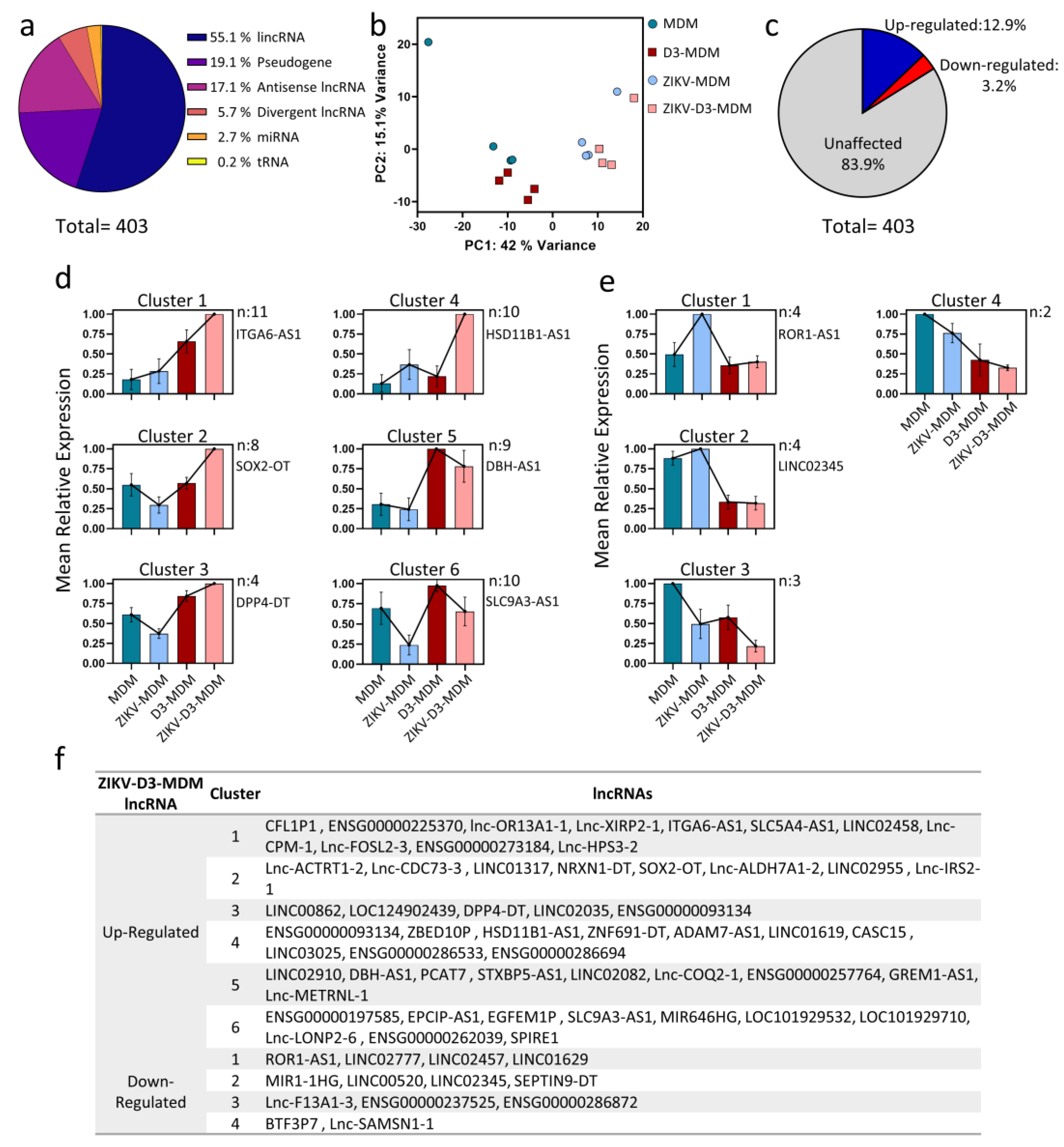
Figure 2.
Vitamin D regulates cis-acting lncRNAs to modulate gene expression. (a) Pearson correlation between specific lncRNAs and mRNA. Each dot is a pair of samples. The dotted line presents the error. Pearson correlation value is shown inset. (b) Genomic tracks show VDR ChIP-seq data, illustrating VDR enrichment profiles in THP1 control macrophages (red) and vitamin D-treated cells (blue). These profiles span up to 103 Kb, highlighting the proximity of VDR binding sites to the promoters of lncRNAs and their target genes.
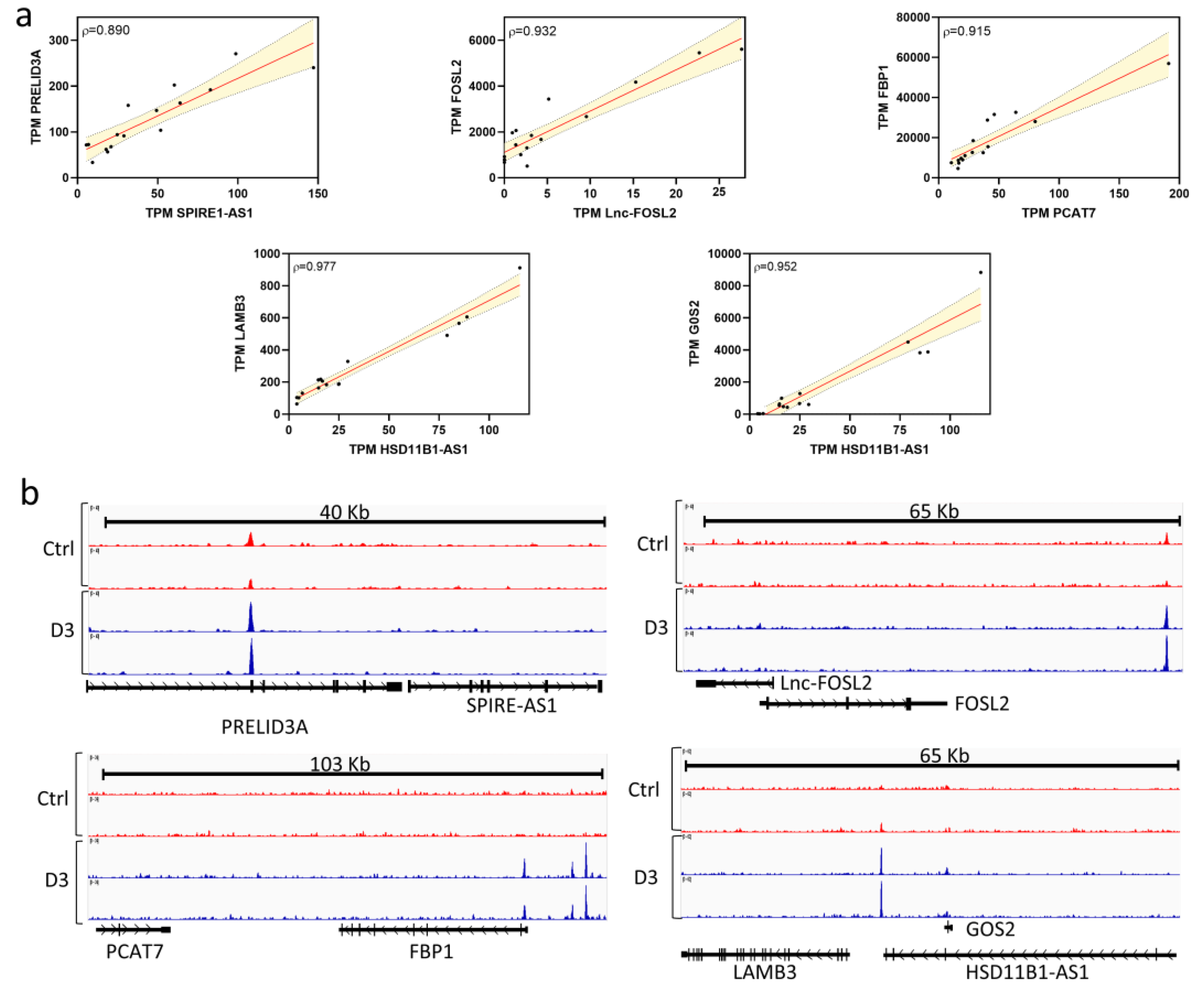
Figure 3.
Vitamin D modulates miRNA expression in macrophages infected with Zika virus. (a) Principal component analysis of miRNA expression profile. Each principal component (PC1 and PC2) is showed the percentage of the variance. (b) Heatmap of differentially expressed miRNAs divided into eight clusters, using k-means, based on their pattern of transcript levels in the four samples. (c) Genomic tracks illustrate VDR ChIP-seq data in THP1 control macrophages (red) and vitamin D-treated macrophages (blue), covering a region up to 60 kb around the TSS of the miRNAs.
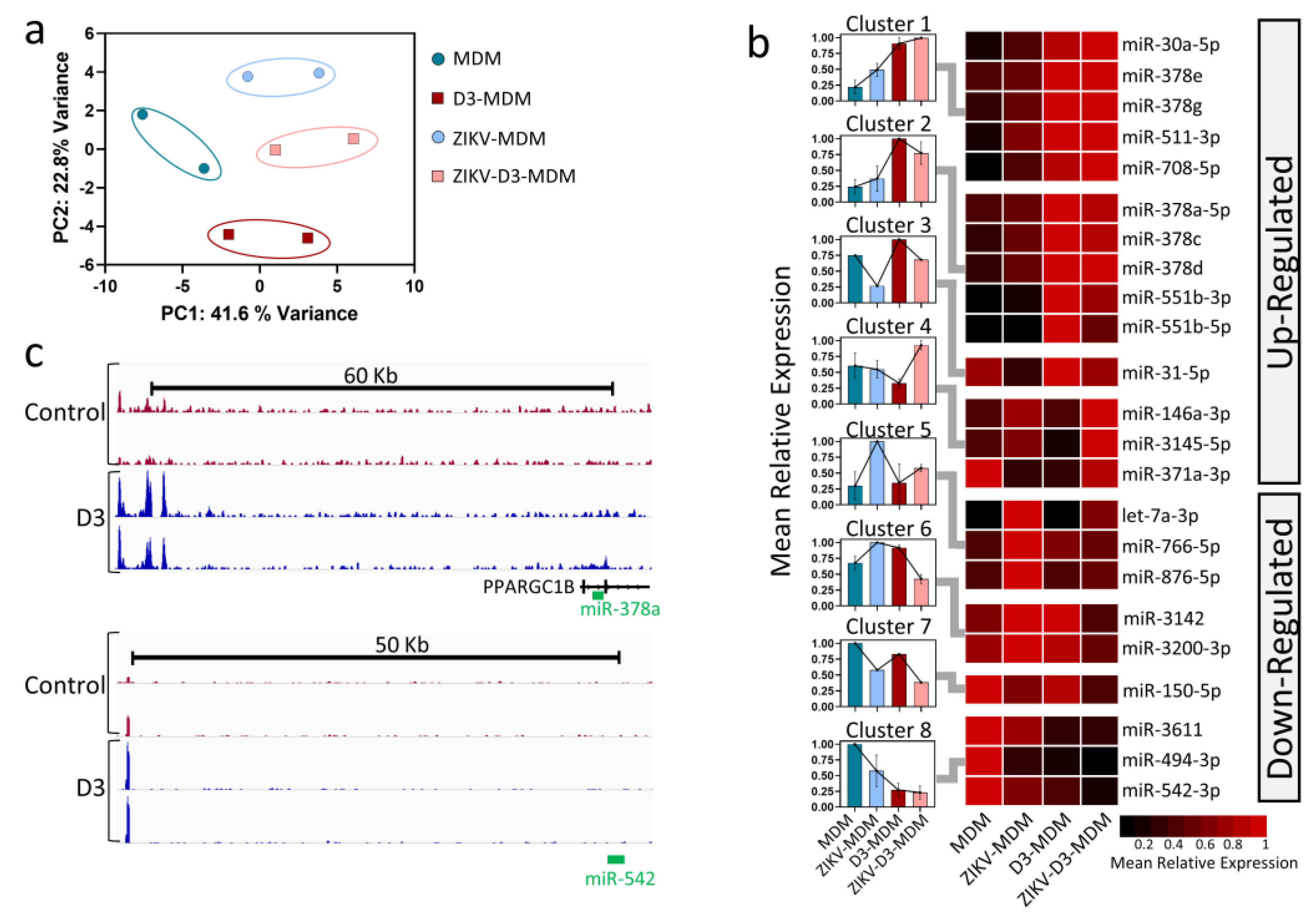
Figure 4.
Vitamin D modulates miRNA-mRNA interactions in macrophages infected with Zika virus. (a) Network demonstrating the potential impact of miRNA regulation on mRNA. Up-regulated miRNAs are depicted by red nodes, while down-regulated miRNAs are shown in blue. The size of each node corresponds to the number of miRNA-target mRNA gene transcripts, while the width of the black edges indicates the degree of overlap between miRNA-target mRNA gene transcripts. (b) Gene-Ontology analysis of the mRNAs predicted as regulated by microRNAs. Each horizontal black bar represents the ontology term enrichment. The vertical-colored bars (y-axis) represent major gene terms modules. PTM; Post translational modification. (c) Interactome between mRNA and the respective potential regulatory microRNAs.
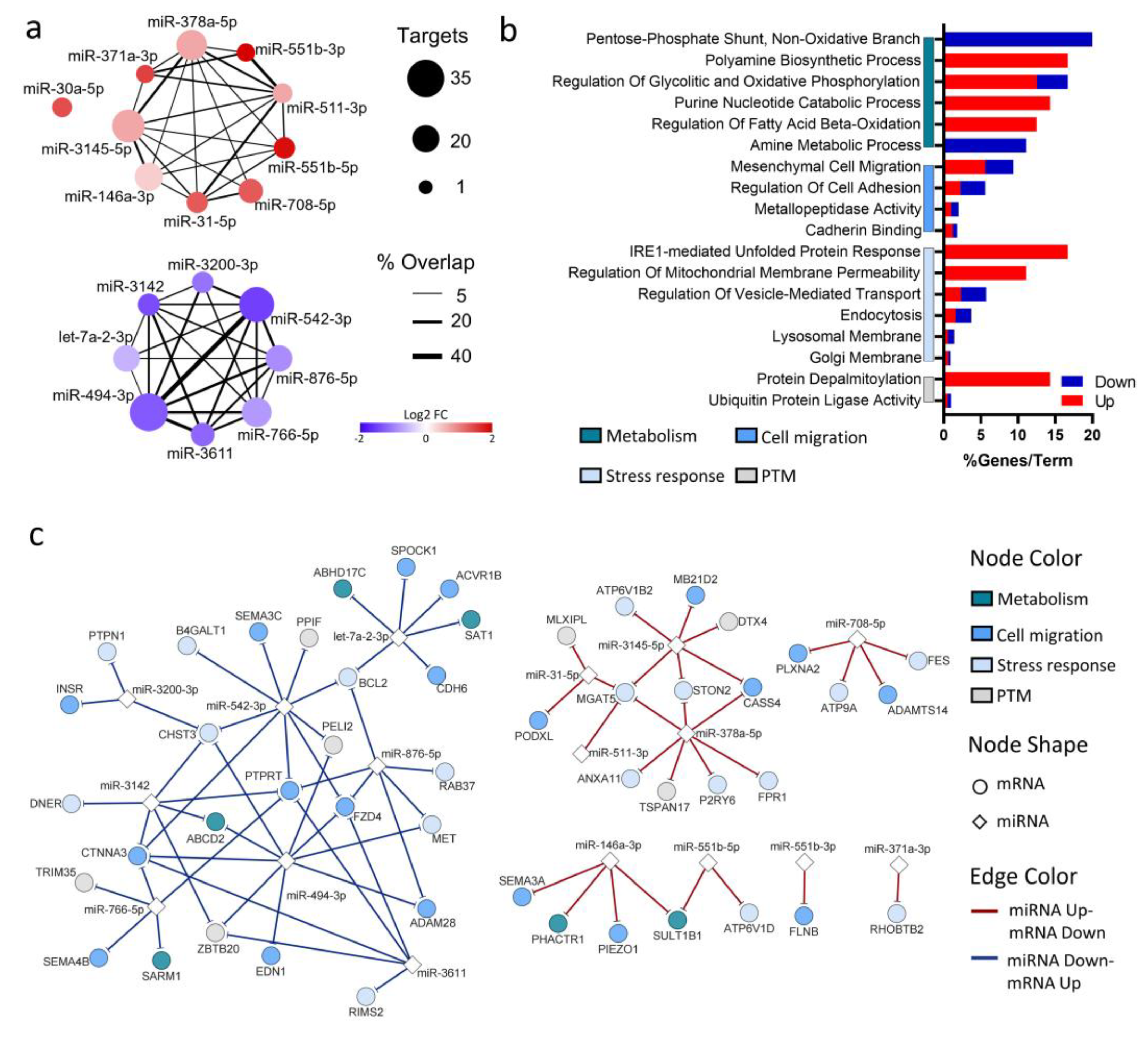
Figure 5.
Figure 5. Vitamin D regulates of gene expression by through Competing Endogenous RNA Mechanisms in Zika Virus-Infected Macrophages. Interactome of potential cross-regulations among mRNAs, miRNAs, and lncRNAs for both (a) down-regulated and (b) up-regulated genes. Circular nodes represent mRNAs, diamond-shaped nodes represent miRNAs, and square nodes represent lncRNAs.
Disclaimer/Publisher’s Note: The statements, opinions and data contained in all publications are solely those of the individual author(s) and contributor(s) and not of MDPI and/or the editor(s). MDPI and/or the editor(s) disclaim responsibility for any injury to people or property resulting from any ideas, methods, instructions or products referred to in the content. |
© 2025 by the authors. Licensee MDPI, Basel, Switzerland. This article is an open access article distributed under the terms and conditions of the Creative Commons Attribution (CC BY) license (http://creativecommons.org/licenses/by/4.0/).
Copyright: This open access article is published under a Creative Commons CC BY 4.0 license, which permit the free download, distribution, and reuse, provided that the author and preprint are cited in any reuse.


