
Submitted:
15 December 2025
Posted:
16 December 2025
You are already at the latest version
Abstract
Berries are an excellent source of bioactive compounds such as polyphenols and widely used in folklore medicine, particularly by Mapuches people in southern of Chile. The objective of this study was to conduct a phytochemical composition analysis of a hydroalcoholic extract of Berberis congestiflora Gay (BE) and to evaluate its potential as an antioxidant, the vasorelaxant effect as relaxation effects in rat aorta, plus the inhibitory enzyme potential in relation to chronic non-communicable diseases, including a possible underlying vasodilatory mechanism in isolated rat aorta. Antioxidant activities were assayed by oxygen radical absorbance capacity (ORAC), by scavenging DPPH (2,2-diphenyl-1-picrylhydrazyl), ABTS (2,2-azinobis-(3-ethylbenzothioazolin-6-sulfonic acid) radicals, and ferric reducing antioxidant power (FRAP). Vascular response of Berberis extract was studied by means of isometric tension recording experiments in ex vivo rat thoracic aortic rings model. Identification of the chemical compounds present in BE was determined by HPLC-DAD-MS for the first time. The BE caused, per se, a dose-dependent contraction at 100 and 1000 µg/mL, and relaxation of the aorta pre-contracted with phenylephrine at 100 and 1000 µg/ml, with maximum contraction of 71% and relaxation of 70% both at 1000 µg/mL. The extract induces calcium-mediated contraction, mainly by calcium release from the sarcoplasmic reticulum and, to a lesser extent, by extracellular Ca2+ influx. The relaxation mechanism was found to depend on the NO/cGMP pathway and the endothelium. The extract also demonstrated cholinesterase, glucosidase and amylase enzyme inhibitor capacities (IC50: 7.33 ± 0.32, 243.23 ± 0.3 and 27.21 ± 0.03 µg/mL, respectively). Docking calculations were additionally performed for a selection of compounds in the berries. These results demonstrate that berries are an excellent source of bioactive compounds with potential antioxidants and endothelium-dependent vasodilator effect, providing evidence for its traditional use and for the inhibition of enzymes, making it a promising candidate for the development of phytotherapeutic products, especially as a supplement against chronic diseases.
Keywords:
endemic Chilean berries
; Berberis
; Berberidaceae
; antioxidant capacity
; docking calculations
; enzyme inhibition
; cardiovascular properties
1. Introduction
The Chilean terroir is home to a variety of unique species, some of which produce edible berries with potentially beneficial compounds. However, these species are endangered by climate change, the growing population, and the timber industry [1,2]. Previous research has identified phenolic compounds including anthocyanins in some endemic berries collected in Chile. For instance, from Azara dentata Ruiz & Pav. diverse phenolic compounds were detected using UHPLC-DAD-TIMS-TOF-MS and demonstrated the inhibition of specific enzymes related to chronic non-communicable diseases (CNCDs) [3]. From several other dark berries several anthocyanin glycosides were detected and quantified for the first time [4], while from Mapuche canelo fruits several phenolic were detected for the first time also with proved anti enzymatic properties related to (CNCDs) [5].
In traditional Mapuche medicine, plants of the genus Berberis have been widely used for their hypoglycemic [6] hepatoprotective [7,8], anti-inflammatory [9] anti-carcinogenic, sometimes related to their content of berberine [10], among others [11]. Berberis congestiflora Gay (with local name including michei or michay) (Figure 1) is a shrub up to 3 m tall. Its leaves with entire or spiny margins and 5-7 pairs of clustered spines, leathery, oval or elliptical shape. The blade is glabrous, with revolute margins. The flowers are grouped in racemes composed of about 25 flowers: 2.5-3 mm long and yellow. The fruit is a globose, dark blue berry, approximately 6 mm in diameter, containing 4-5 black seeds. The plant grows from Santiago metropolitan region to Los Rios region of Chile. Some phenolic acids, alkaloids, and flavonols were identified in the leaves [12]. Recently Reyes-Farias et al., 2015 concluded that extracts of Chilean native fruits Aristotelia chilensis (maqui) and Berberis microphylla (calafate) have important inhibitory characteristics on the inflammatory response [13]. Martínez et al., 1997 isolated in the Berberis chilensis species the alkaloid O-methylisofalicberin (O-MI), very similar to other alkaloids previously described in the literature as antagonists of Ca2+ channels [14]. To date, the edible fruits of Berberis species in Chile have been the subject of only a handful of chemical and bioactivity investigations, leaving a wealth of untapped potential for discovering new medicinal compounds and refining chemotaxonomic relationships. Chile in particular stands out as a hotspot of phytochemical diversity—harboring carotenoids, flavonoids, tannins, anthocyanins, alkaloids and phenolic acids with strong antioxidant capabilities—and thus offers a fertile ground for studying natural interventions against chronic diseases (e.g., diabetes, hypertension, obesity, cardiovascular disorders, inflammation, cancer and age-related ailments) [3,5,15,16,17,18]. This work aims to measure the enzyme inhibitory properties (glucosidase, amylase, BuChE, and AChE), the vasorelaxant effects as relaxation effects in rat aorta and antioxidant properties of the blue berries from a native Berberis species, as well as to analyze the phenolic profiles using UHPLC-DAD, and HPLC-ESI(-)-MS for the first time.
2. Results and Discussion
The mature fruits of Berberis congestiflora were collected during February 2017 in the Region of Araucanía, Chile. Hydroalcoholic extracts were prepared and analyzed regarding their components as well as the several bioactivities commented below.
2.1. Antioxidant Activity and Content of Phenolics and flavonoids
Oxidative stress generates reactive oxygen species (ROS)—for example, hydroxyl radicals—and non-radical oxidants like hydrogen peroxide [19]. These agents readily attack cellular constituents—amino acids, sugars, lipids, proteins and nucleic acids—oxidizing them and thereby initiating disease processes or degrading the quality of foods and cosmetics [20]. A range of analytical assays have been developed to quantify the antioxidant capacity of natural products and gauge their suitability as protective agents. In this study, the total phenolic compounds, total anthocyanins and total flavonoid according to spectrometry were measured (76.35 ± 0.01, and 63.2 ± 0.2/g dry weight), being higher than those of Valdivian A. dentata berries (57 mg GAE/g dry weight)[3], while the total anthocyanins were deemed to be high, as measured using spectroscopy (32.26 ± 1.23 mg c3g/100 g fruit) compared to the Chilean blueberries (21.42 mg/g dry fruits) [21]. The Oxygen Radical Absorbance Capacity (ORAC) assay uses fluorescence to deliver a highly sensitive measure of antioxidant activity [22], whereas the ABTS method offers excellent reproducibility by quantifying an extract’s ability to donate hydrogen atoms and convert the ABTS radical into a stable, non-radical species [23]. In this study, the BE extract achieved an IC50 of 5.32 ± 0.5 μg/mL and 6.78 ± 0.04 μg/mL with the DPPH (2,2-diphenyl-1-picrylhydrazyl) and ABTS (2,2-azinobis-(3-ethylbenzothioazolin-6-sulfonic acid) antiradical methods, respectively. This antioxidant capacity of the extract can be compared with natural antioxidants used commercially (gallic acid DPPH: IC50 = 2.32 ± 0.5; quercetin: IC50 = 12.23 ± 0.8 µg/mL). Furthermore, FRAP (148.7 ± 0.03 μmol Trolox/g dry fruit, respectively) was higher than that of Berberis microphilla and Vaccinium corimbosum berries (124.4 μmol and 96.15 Trolox/g dry fruit, respectively) [21] (Table 2). ORAC (175.9 ± 3.43 μmol Trolox/g dry fruit) is close to that published for Ugni molinae berries (222 μmol Trolox/g dry fruit) [24]. Due to these activities, the extract could be included in supplements or food products as antioxidant additives to protect them from oxidation or for skin care against the effects of free radicals.
2.2. B. congestiflora Extract Causes Contraction in Rat Aorta
To evaluate the effect of B. congestiflora extract alone on vascular response, cumulative doses were added to the bath in intact and endothelial-denuded rat aorta. As shown in Figure 2A, extract concentrations of 100 and 1000 μg/mL caused significant vascular contraction in intact rat aorta. In Figure 2B, the data showed an increase of contraction in intact rat aorta (control): 41 ± 13% at 100 μg/mL and 71 ± 9% at 1000 μg/mL. In the denuded-endothelium rat aorta (denuded), at the same concentrations produced higher contractions: 115 ± 11% at 100 μg/mL and 134 ± 18% at 1000 μg/mL. Vascular contraction of B. congestiflora showed an EC50 of 96 μg/mL in intact rat aorta (control) and an EC50 of 44 μg/mL in denuded-endothelium rat aorta (denuded).
We did not expect B. congestiflora to increase vascular tone per se. After detecting it, subsequent experiments were conducted to understand the mechanism of action of the extract. The protocol consisted of contracting the tissue with a single concentration (1000 µg/mL) of extract, in the presence or absence of a voltage-gated calcium channel blocker (nimodipine), to evaluate whether the extract’s contractile response depends on extracellular calcium, or using caffeine to release calcium from the sarcoplasmic reticulum that might be necessary to induce extract contraction.
As shown in Figure 2C and 2D, preincubation with caffeine reduced the contractile response to B. congestiflora in a dose-dependent manner: 104 ± 8% in the control group, 23 ± 9% (p < 0.01) with 10 mM caffeine, and 4 ± 0% (p < 0.001) with 20 mM caffeine. Interestingly, the reduction in contraction with caffeine was greater than that with nimodipine: 57 ± 13% (p < 0.05).
2.3. B. congestiflora Extract Produces Vasorelaxation Endothelium-Dependent, via NO, in Rat Aorta
When the vasorelaxation effect of the hydroalcoholic extract of B. congestiflora was evaluated at cumulative doses of 1-1000 μg/mL in precontracted aortic rings (with 10-6 M phenylephrine), the control group showed vasorelaxation 25 ± 8% with 100 μg/mL and 70 ± 4% at 1000 μg/mL (Figure 3A). However, removing the vascular endothelium (Figure 3A), or preincubating with 10-4 M L-NAME (Figure 3B) or 10-6 M ODQ (Figure 3C), significantly blunted the vasorelaxation effect of the extract. After endothelium removal, vasorelaxation was 1 ± 1% (p<0.001) with 100 μg/mL and 5 ± 1% with 1000 μg/mL. For L-NAME, the effect was 3 ± 1% (p<0.001) with 100 μg/mL and 6 ± 2% with 1000 μg/mL. For ODQ, the results were 3 ± 1% with 100 μg/mL and 61 ± 2% with 1000 μg/mL.
2.3. Enzyme Inhibitory Properties
Several important enzymes related to CNCD were analyzed using the hydro-ethanolic extract of the endemic fruits of B. congestiflora. The results are shown in Table 2. Enzymes such as glucosidases, amylases, and lipases contribute to metabolic syndrome. In particular, α-glucosidase and α-amylase hydrolyze glycogen and starch to release glucose, linking them to diabetes as well as infections and cancer [25]. Blocking these two enzymes slows the breakdown and absorption of carbohydrates, thereby mitigating post-meal blood sugar spikes [26]. In our work, BE extract inhibits -amylase and α-glucosidase; and was shown to be active against acetylcholinesterase (AChE) and butyrylcholinesterase (BuChE). Cholinesterase enzymes catalyze the breakdown of acetylcholine into choline and acetate, a key factor in Alzheimer’s disease. Inhibiting these enzymes, sometimes with phenolic compounds, helps maintain acetylcholine levels, which strengthens cholinergic transmission and alleviates the symptoms of Alzheimer’s disease [27]. Berberine the main alkaloid from leaves of B. vulgaris was shown to inhibit cholinesterase enzymes [28]. In this study, some notable results were those of acetylcholinesterase (IC50: 7.33 ± 0.22), which was several times less active than the standard galantamine (IC50: 0.40 + 0.02), and butyrylcholinesterase (IC50: 19.45 ± 0.32). on the other hand, the inhibitory effect of Berberis vulgaris crude extract on α-glucosidase was shown to be more potent than that of berberine chloride [28], however, our inhibition of α-amylase was moderate (IC50: 27.21 ± 0.03) and inhibition of α-glucosidase was high (IC50: 243.23 ± 0.3) compared to acarbose, being the extract chemically characterized mainly of anthocyanins. Chilean native Azara serrata anthocyanins enriched extract showed IC50 of 371.6 and 7.23 μg/mL, in glucosidase and amylase inhibition tests, respectively, and IC50 3.92 μg/mL in inhibition of AChE that can be attributed in part to the content of active anthocyanins [17]. Cholinesterase’s inhibition activity could lead to the discovery of a naturally occurring drug that could be useful for further treatment (Table 2). For future studies, it is important to test new concentrations to validate the potential of the B. congestiflora fruit extract (BE) in relation to the inhibition of the cholinesterase, α-glucosidase and α-amylase enzymes; it is also important to isolate its major anthocyanin compounds, which may show better inhibitory activity. Conversely, the extract exhibits notable inhibition of cholinesterase enzymes—particularly acetylcholinesterase—highlighting its potential to modulate cholinergic signaling in disorders such as Alzheimer’s disease. Nonetheless, further work is required to isolate and characterize its active constituents and to fully elucidate their mechanisms of action. Likewise, exploring additional bioactivities of the B. congestiflora extracts such as its anti-cancer effects—will be crucial to confirm its broader therapeutic potential.
2.4. Analysis of the Phenolic and Anthocyanin Profile of BE
2.4.1. Chromatographic Analysis
Anthocyanins are a class of natural pigments that are known for their colors, ranging from blue to red and purple, in relation to fruits, vegetables, and flowers, as well as for the bioactivity that some of them possess. Anthocyanin and phenolic profile were performed using UHPLC-DAD, HPLC-ESI (+)-MSn, and UHPLC-ESI (-)-IT-MS (Figure 5). Identification was performed for anthocyanins using MSn and DAD data. Anthocyanins have characteristic absorption spectra, so the wavelengths of the maxima were determined. Molecular ions were determined using ESI (+)-MSn measurements. Thirty-one peaks (Figure 5 and Figure 6, Table 2) were tentatively identified for the first time in the BE extract using positive and negative UHPLC-ESI- ionization mode. Table 2.
Anthocyanins.
Nine known anthocyanins were identified in Michay fruits, (peaks 4, 5, 6,7, 8, 11, 12, 13, 14 and 15, Figure 6) with molecular ions in positive mode at m/z 609, 479, 609, 465, 505, 625, 639, 449, 493 and 331 and showing characteristic MS2 ions at m/z: 463 (MS3 ions at m/z 301, 286 and 147), 303 (MS3 ion at m/z 257), 301 (MS3 ion at m/z 286), 317(MS3 ion at m/z 302), 331 (MS3 ion at m/z 299, 179) 299 (MS3 ion at m/z 179) respectively, corresponding to petunidin 3-O-glucose, (λ max: 275-343sh-512 nm), peonidin 3-O-rutinose, (λ max: 268, 357sh, 503 nm), delphinidin 3-O-glucose (λ max: 275-341sh-512 nm), peonidin 3-O-glucose, (λ max: 268-347sh-512 nm), peonidin 3-O-6’’-O-acetyl-glucoside, peonidin 3, 5-di-O-glucose, malvidin 3-O-rutinose , malvidin 3-O-glucose, and malvidin, respectively. Other minor compounds were peaks 1-3 with pseudomolecular cations at m/z: 611, 595 and 641, and thus identified as, cyanidin 3,5-O-diglucose, cyanidin 3-O-(6’’-O-p-coumaroyl)-glucose and petunidin 3,5-O-diglucose respectively. Some of the identities were corroborated by co-elections with some standard anthocyanins in our laboratory and literature data [29,30].
Phenolic acids
Peaks 9 and 10 with molecular anion at 515 and 353 showing MS daughter ion at 191 (quinic acid) were related to the phenolics: diccafeoyl quinic acid and caffeoyl quinic acid, respectively [31]. Peak 25 with a pseudomolecular ion at 529 and showing a MS2 ion at 367 was identified as feruloyl-caffeoyl-quinic acid [32], while peak 26 was identified as the lignan hydroxypinoresinol [33]. Peak 27 with a pseudo molecular ion a diagnostic (2M-H-) ion at 742 was identified as hydroxyferulic acid [34] Peak 30 was identified as caffeoyl-2-hydroxyethane-1,1,2-tricarboxylic acid [35].
Flavonoids
Peak 16 with a parent molecular ion at 463, showing two UV maxima at 255 and 355, characteristic of flavonols and a daughter ion at 301, 179 151 was identified as quercetin-3-O-glucoside while peak 17 as quercetin-3-O-2¨¨acetyl-glucoside. Peak 18 was identified as isorhamnetin-3-O-glucose while peak 19 with a molecular anion at 623 producing a MS2 ion at 315 was identified as isorhamnetin 3-O-rutinoside, [36] while peak 24 with a parent ion at 519 and daughter ions at 477 and 315, as isorhamnetin-3-O-6’’-O-acetyl-glucoside [37]. Peak 20 was identified as quercetin-3-O-glucoside, and peak 21 as rutin. Peak 22 was identified as luteolin 3-O-glucoside [38], and peak 23 as phloretin 3,5-C-diglucoside [39]. Finally, peaks 28 and 29 were identified as the isomers isorhamnetin and rhamnetin, and peak 31 as quercetin, respectively.
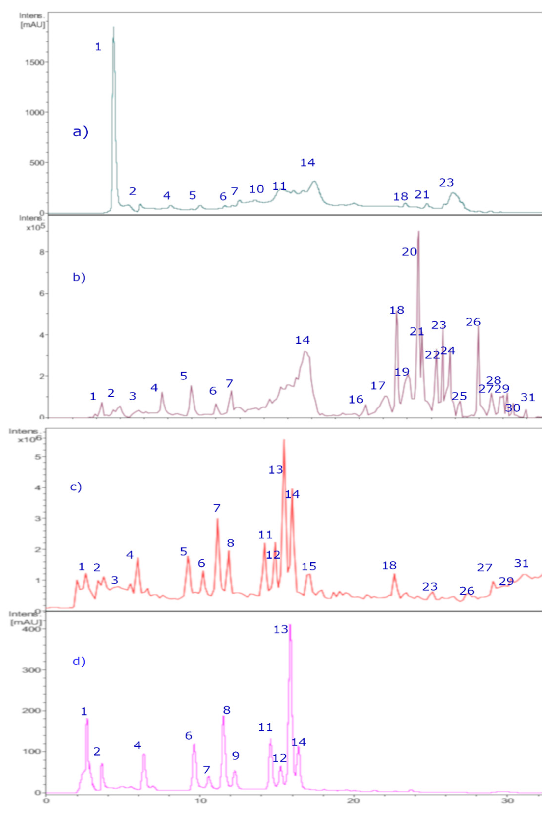
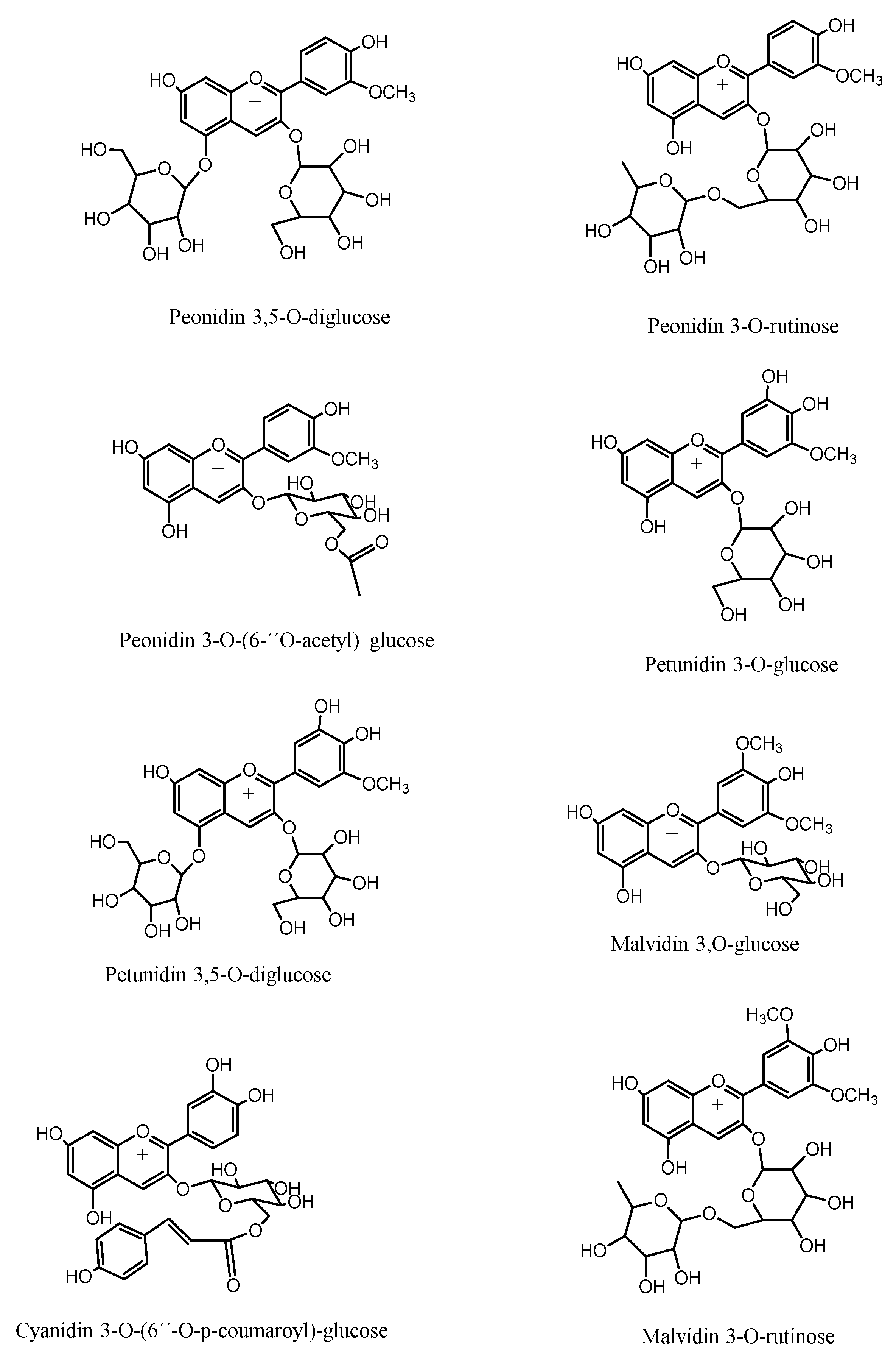
Figure 5.
HPLC-DAD chromatograms of Michay fruit extracts. a) chromatograms at 280 nm. b) TIC (total ion current, negative mode c) chromatograms at 520 nm. d) TIC (total ion current, positive mode.
Figure 5.
HPLC-DAD chromatograms of Michay fruit extracts. a) chromatograms at 280 nm. b) TIC (total ion current, negative mode c) chromatograms at 520 nm. d) TIC (total ion current, positive mode.

Table 1.
Radical scavenging activity of 1,1-diphenyl-2-picrylhydrazyl radical (DPPH), ABTS radical, total phenolic content (TPC), total anthocyanin content (TAC), total flavonoid content (TFC), cholinesterase inhibition capacity, and glucosidase and amylase inhibition capacity of the hydroalcoholic extract of B. congestiflora (BE).
| Sample | DPPHa | ABTSa | ORACb | FRAPb | TPCc | TACd | TFCe | AChEf | BuChEf | α-Glucosidaseg | α-Amylaseg |
| BE extract | 5.32 ± 0.5 | 6.78 ± 0.04 | 175.9 ± 3.43 | 148.7 ± 0.03 | 76.35 ± 0.01 | 32.26 ± 1.23 | 63.2 ± 0.2 | 7.33 ± 0.32 | 19.45 ± 0.32 | 243.23 ± 0.3 | 27.21 ± 0.03 |
| Gallic acid | 2.30 ± 0.5 | 16.5± 0.04 | - | - | - | - | - | - | - | - | - |
| Acarbose | - | - | - | - | - | - | - | - | - | 138.9 ± 0.01 | 10.04 ± 0.02 |
| Galantamine | - | - | - | - | - | - | - | 0.402 + 0.02e | 5.45 ± 0.01 | - | - |
| Quercetin | 12.25 ± 0.6 | 15.65 ± 0.05 | - | - | - | - | - | - | - | - | - |
a DPPH antiradical and ABTS activities are expressed as IC50 in μg/mL. b Expressed as μmol Trolox/g dry fruit. c Total phenolic content (TPC) expressed as mg gallic acid equivalent GAE/g dry weight. c Total antohcyanin content (TAC) expressed as mg c3g equivalent /g dry weight e Total flavonoid content (TFC) expressed as mg quercetin 100 g dry weight. fInhibitory enzymes of cholinesterases, gα-glucosidase, and α-amylase enzymes in IC50 expressed in µg/mL. Values in the same column are significantly different (p < 0.05). Dash (-) means not tested (i.e., not applicable).
Table 2.
Identification of phenolic compounds in Michay fruits by LC-DAD, LC–MS and MS/MS data.
| Peak |
Rt (min) |
HPLC DAD λ max (nm) |
ESI mode |
[M-H] – (m/z) |
MS-MS ions (m/z) |
Tentative identification |
| 1 | 2.2 | 268, 357sh, 503 | + | 611 | 287 | Cyanidin 3,5-O-diglucose |
| 2 | 3.1 | 268, 357sh, 503 | + | 595 | 287 | Cyanidin 3-O-(6’’-O-p-coumaroyl)-glucose |
| 3 | 3.3 | 275, 343sh, 512 | + | 641 | 317, 302 | Petunidin 3,5-O-diglucose |
| 4 | 6.1 | 275, 343sh, 512 | + | 479 | 317, 302 | Petunidin 3-O-glucose |
| 5 | 8.2 | 268, 357sh, 503 | + | 609 | 463, 301, 286 | Peonidin 3-O-rutinose |
| 6 | 9.8 | 275, 341sh, 512 | + | 465 | 303, 257 | Delphinidin 3-O-glucose |
| 7 | 11 | 268, 357sh, 503 | + | 463 | 301, 286 | Peonidin 3-O-glucose |
| 8 | 11.2 | 268, 357sh, 503 | + | 505 | 317, 302 | Peonidin 3-O-6’’-O-acetyl-glucoside |
| 9 | 12.1 | 246, 310 | - | 515 | 353, 191, 179 | Di-caffeoyl-quinic acid |
| 10 | 13.2 | 246, 310 | - | 353 | 191, 179 | Chlorogenic acid |
| 11 | 12.3 | 268, 357sh, 503 | + | 625 | 317, 302 | Peonidin 3, 5-di-O-glucose |
| 12 | 14.6 | 275, 343sh, 512 | + | 639 | 331, 299, 179 | Malvidin 3-O-rutinose |
| 13 | 16 | 278, 503 | + | 449 | 287, 213, 147 | Cyanidin- 3-O-glucose |
| 14 | 16.3 | 275, 343sh, 512 | + | 493 | 331, 299, 179 | Malvidin 3-O-glucose* |
| 15 | 16.7 | 275, 343sh, 512 | + | 331 | 299, 179 | Malvidin |
| 16 | 17.2 | 255, 354 | - | 463 | 301, 179, 151 | Quercetin-3-O-glucoside |
| 17 | 18.7 | 255, 354 | - | 505 | 463, 301, 179, 151 | Quercetin-3-O-2’’acetyl-glucoside |
| 18 | 22.5 | 265, 354 | + - | 477, 479 | 955 (2M-H) 315, 300 | Isorhamnetin-3-O-glucose |
| 19 | 23.4 | 255, 354 | - | 623 | 315 | Isorhamnetin 3-O-rutinoside |
| 20 | 23.8 | 255, 354 | - | 463 | 301, 179, 151 | Quercetin-3-O-glucoside* |
| 21 | 24.2 | 254, 354 | - | 609 | 301, 179, 151 | Rutin * |
| 22 | 24.9 | 255, 354 | - | 447 | 287 | Luteolin 7-O-glucoside |
| 23 | 25.3 | 255, 354 | - | 597 | 287 | Phloretin 3,5,C-diglucoside |
| 24 | 25.7 | 255, 354 | - | 519 | 477, 315, 179, 151 | Isorhamnetin-3-O-6-O-acetyl-glucoside |
| 25 | 26.1 | 255, 354 | - | 529 | 367 | Feruloyl-caffeoyl-quinic acid |
| 26 | 26.5 | 240-290 | - | 373 | 171 | Hydroxypinoresinol |
| 27 | 28.3 | 246, 310 | - | 371 | 209, 742 (2M-H-) | Hydroxyferulic acid |
| 28 | 29.7 | 265, 354 | + - | 315, 317 | 300, 179, 151 | Isorhamnetin* |
| 29 | 30.2 | 265, 354 | + - | 315, 317 | 300, 179, 151 | Rhamnetin |
| 30 | 31.3 | 246, 310 | - | 339 | 295 | Caffeoyl-2-hydroxyethane-1,1,2-tricarboxylic acid |
| 31 | 31.6 | 255, 354 | + - | 301, 303 | 295 | Quercetin* |
*Compounds marked by * were identified by co-spiking experiments using UHPLC-DAD (Figure 2).
Figure 6.
Structures of representative anthocyanin compounds identified in Chilean Michay berries.
2.5.1. Docking Calculations
To more accurately assess and elucidate the inhibition results for Acetylcholinesterase, Butyrylcholinesterase, Glucosidase, and α-Amylase, we considered the data shown in Table 2. Thus, four anthocyanins (Peonidin 3-O-glucose, Peonidin 3-O-rutinose, Malvidin 3-O-glucose, and Malvidin 3-O-rutinose), two phenolic acids (Hydroxyferulic acid and Feruloyl-caffeoyl-quinic acid), and two flavonoids (Isorhamnetin 3-O-rutinoside and Isorhamnetin 3-O-rutinoside), Figure 7, along with the known inhibitors Galantamine and Acarbose, were selected to perform docking analyses. Each docking assay was performed within the catalytic site of each enzyme to evaluate the docking energy descriptor and the molecular interactions between the active-site residues and the selected metabolites. This step seeks to rationalize the inhibitory activities of the extract toward the enzymes. Table 3 shows the binding energies of the selected anthocyanins, phenolic acids, and flavonoids.
Acetylcholinesterase (TcAChE) Docking Results
The ethanolic extract of Berberis congestiflora Gay (BE) exhibited moderate acetylcholinesterase inhibition (IC50: 7.33 ± 0.22 µg/mL), yet overall, quite reasonable, being only one order of magnitude higher than that of galantamine. The energies shown in Table PP indicate that the compounds Peonidin 3-O-glucose, Peonidin 3-O-rutinose, Malvidin 3-O-glucose, Quercetin-3-O-glucoside, and Isorhamnetin 3-O-rutinoside possess favorable binding energies. This outcome could be attributed to the fact that the whole extract contains a combination of compounds that may compete for the catalytic site of the enzyme, thereby interfering with a more specific inhibition. Thus, it is possible that measuring the inhibitory activity of each isolated derivative would reveal strong acetylcholinesterase inhibition. Overall, all the anthocyanins exhibited optimal binding energies superior to that of galantamine, except for Malvidin 3-O-rutinose, which showed a value of −11.632 kcal/mol. The anthocyanins Peonidin 3-O-glucose and Peonidin 3-O-rutinose and Malvidin 3-O-glucose exhibited favorable binding profiles within the catalytic site, suggesting a relevant contribution to the overall inhibition pattern observed in the docking analysis. All the anthocyanins are stabilized within the acetylcholinesterase catalytic site mainly through hydrogen-bonding interactions, but they also exhibit π–π and T-shaped interactions. Peonidin 3-O-glucose establishes seven hydrogen bond interactions through the oxygenated functions of its glucose moiety and the methoxyphenylchromenylium core of its structure with the amino acids Tyr70, Gln74, Tyr121, Glu199, Phe330, and His440. In addition, Peonidin 3-O-glucose forms a π–π interaction with Trp84 and a T-shaped interaction with Tyr121 (Figure 8-A). Peonidin 3-O-rutinose arranges within the catalytic site in a similar manner to Peonidin 3-O-glucose. Therefore, it also forms hydrogen-bond interactions with the amino acids Gln69, Tyr130, Glu199, and Tyr121. Likewise, through its methoxyphenylchromenylium core, it forms a π-π interaction with Trp84 and a T-shaped interaction with Tyr121(Figure 8-B). The anthocyanin Malvidin 3-O-glucose, like the previous two compounds, also forms hydrogen bond interactions with Gln69, Asp72, Gly117, Tyr121, Glu199, and His440, as well as a T-shaped interaction with Tyr121(Figure 8-C). On the other hand, the anthocyanin Malvidin 3-O-rutinose, which showed a binding energy of −11.632 kcal/mol, although exhibiting an energy profile quite like that of Galantamine, forms only four hydrogen bond interactions with Asp72, Trp84, Tyr130, and Gly441, along with a T-shaped interaction with His440 (Figure 8-D).The evaluated phenolic acids, Hydroxyferulic acid and Feruloyl-caffeoyl-quinic acid, did not exhibit favorable binding energies (−8.647 kcal/mol and −10.068 kcal/mol, respectively). The energy values for both derivatives are lower than those of the anthocyanins discussed above, as well as that of Galantamine (Table 3). To assess the relevance of the sugar moieties, Hydroxyferulic acid was selected as a compound lacking a sugar portion, whereas Feruloyl-caffeoyl-quinic acid contains a carbohydroxy framework. In this regard, Feruloyl-caffeoyl-quinic acid showed a better interaction profile within the acetylcholinesterase catalytic. Therefore, Hydroxyferulic acid performs four hydrogen-bond interactions with Tyr121, Glu199, Ser200, and His440, as well as a T-shaped interaction with Trp84(Figure SS-E)., whereas Feruloyl-caffeoyl-quinic acid performs only two hydrogen-bond interactions with Asn85 and Glu199(Figure 8-F). The flavonoids Quercetin-3-O-glucoside and Isorhamnetin 3-O-rutinoside also showed favorable energy profiles (Table 3), although not superior to those of the anthocyanins. Likewise, their main interactions within the catalytic site of the enzyme were hydrogen bond interactions. In this regard, Quercetin-3-O-glucoside establishes four hydrogen bond interactions with Tyr121, Ser122, Glu199, and Phe288; two π–π interactions with Phe330 and Tyr334; and one T-shaped interaction with His440 (Figure SS-G). Isorhamnetin 3-O-rutinoside also forms hydrogenbond interactions with the amino acids Gly80, Ser81, Asn85, Tyr121, Tyr130, and Glu199, as well as two T-shaped interactions with Phe331 and Tyr334(Figure 8-H).
Butyrylcholinesterase (hBuChE) Docking Results
Docking simulations performed on human butyrylcholinesterase (hBuChE) provided insights into the binding behavior of the analyzed metabolites. Overall, the assayed anthocyanin, phenolic acid, and flavonoid derivatives exhibited favorable binding energies (Table 3), except for Hydroxyferulic acid, which could be attributed to the absence of a saccharide moiety in its structure (-6.753 kcal/mol).
Peonidin 3-O-glucose, which exhibits a binding energy of −12.461 kcal/mol, establishes several hydrogen bond interactions with different residues within the butyrylcholinesterase catalytic site, including the amino acids Trp82, Gly117, Thr120, Tyr128, Glu197, Ser198, and Leu286. Also forms a π–cation interaction with His438, whose imidazole ring is protonated at physiological pH of 7.4 (Figure 9-A). The anthocyanin Peonidin 3-O-rutinose exhibited a favorable binding energy of −13.129 kcal/mol, with its main interactions consisting of hydrogen bonds with Trp82, Gly115, Ser198, Leu286, and Tyr440, along with a T-shaped interaction with Phe329. It is noteworthy that the latter anthocyanins differ in the glycosidic framework of their structures, which allow them to fit differently within the catalytic site of the enzyme (Figure 9-B). A similar pattern is observed for the analogues Malvidin 3-O-glucose and Malvidin 3-O-rutinose, in which the former anthocyanin displays a less favorable binding energy compared with the latter (Table 3). In this sense, Malvidin 3-O-glucose forms hydrogen-bond interactions with Asp70, Trp82, Tyr128, Glu197, Glu325, and Ala328, as well as a π–cation interaction with His438 (Figure 9-C). Likewise, Malvidin 3-O-rutinose carry out several hydrogen bond interactions with Asn68, Asn83, Gly115, Glu197, Ser198, Leu286, Tyr332 and His438, as well as a T-shaped interaction with Phe329 (Figure 9-D). Given the evidence presented above, anthocyanin derivatives bearing rutinose moieties exhibit a better binding profile toward butyrylcholinesterase and therefore could act as more effective inhibitory agents. Hydroxyferulic acid, which lacks a glucoside moiety, also performs hydrogen bond interactions through its oxygen functions with Trp82, Gly116, Glu197, and Ser198. Additionally, this derivative exhibited a π–cation interaction with His438 and a T-shaped interaction with Trp82 (Figure 9-E). The phenolic acid feruloyl-caffeoyl-quinic acid, which does contain a glycosidic framework in its structure, exhibited a lower number of hydrogen bond interactions (with residues Thr120 and Tyr128) and a π–π interaction with Trp82. Nevertheless, its binding energy is improved due to the number of hydrophobic interactions that the glycosidic framework itself may be generating (Figure 9-F). In the case of the flavonoids quercetin-3-O-glucoside and isorhamnetin-3-O-rutinoside, the presence of the rutinoside framework again improves the docking descriptors. Accordingly, the first derivative, which exhibits an energy of –12.808 kcal/mol, shows hydrogen bond interactions with Gly116, Gly117, Tyr128, Glu197, Ser198, and Tyr332, as well as a T-shaped interaction with Phe398 and a π–cation interaction with His438 (Figure 9-G). Conversely, the second derivative engages in several hydrogen bond interactions with residues Asp70, Trp82, Gly116, Tyr128, Glu197, Ser198, Ala328, and His438, thereby stabilizing this compound within the catalytic site of butyrylcholinesterase (Figure 9-H).
Glucosidase Docking Results
The results over Glucosidase enzyme reveal distinct differences in how derivatives accommodate within the catalytic pocket, highlighting the contribution of specific substituents to hydrogen bonding, other interactions and overall stabilization of the enzyme–ligand complex. The binding energies shown in Table PP indicate that the anthocyanins are the derivatives that would presumably behave as the most effective inhibitors of this enzyme; however, none of them exhibits a better energetic profile than the reference inhibitor acarbose. Peonidin-3-O-glucoside, Peonidin-3-O-rutinoside, and Malvidin-3-O-glucoside exhibit highly favorable binding energy values (Table 3). These three derivatives, which share the phenylchromenylium framework, are predominantly stabilized by hydrogen bond interactions, but they also engage in π–π interactions and π–cation interactions. Peonidin-3-O-glucoside engages in hydrogen bond interactions with Asp1157, Asp1279, Asp1420, Lys1460, and Asp1526, as well as π–π and π–cation interactions with Trp1355 (Figure 10-A). Peonidin-3-O-rutinoside likewise forms hydrogen bond interactions with Asp1157, Asp1526, Thr1528, and Gln1561, as well as a π–cation interaction with Tyr1251 and three T-shaped interactions with Trp1355, Phe1559, and Phe1569 (Figure 9-B). Malvidin-3-O-glucoside derivative, which exhibits the best energetic profile (Table 3), shows hydrogen bond interactions with Asp1157, Asp1279, Arg1377, Asp1420, Asp1526, and Lys1460, along with a π–π interaction with Trp1355 (Figure 10-C). Similarly, Malvidin-3-O-rutinoside, forms eight hydrogen bond interactions with residues Asp1157, Asp1279, Trp1355, Asp1420, and Arg1510, along with a π–π interaction with Phe1560 and a π–cation interaction with Phe1560 (Figure 10-D). The phenolic acid hydroxyferulic acid, due to the nature of its structure, which lacks a carbohydrate core, engages only in hydrogen bond interactions with Asp1279, Asp1526, and His1584 (Figure 10-E), which likely accounts for its binding energy of –8.017 kcal/mol. In contrast, the structural complexity of feruloyl-caffeoyl-quinic acid allows it to fit within the glucosidase catalytic site in a manner that gives rise to hydrogen bond interactions with Asp1157, Gln1158, Lys1164, and Asp1279, as well as two additional key bondings, a π–cation interaction with Arg1510 and a salt-bridge interaction with Lys1460 (Figure MM-F), which ultimately results in a more favorable binding energy (Table 3).
Finally, the flavonoids Quercetin-3-O-glucoside and Isorhamnetin-3-O-rutinoside present in the Berberis congestiflora Gay (BE) ethanolic extract may be considered good candidates as potential glucosidase inhibitors; however, they do not reach the favorable profiles observed for the aforementioned anthocyanins (Peonidin-3-O-glucoside, Peonidin-3-O-rutinoside, and Malvidin-3-O-glucoside). In this sense, the main interactions of Quercetin-3-O-glucoside are hydrogen bond interactions with Asp1157, Asp1279, Trp1369, Asp1420, Lys1460, Arg1510, and Asp1526, as well as a π–π interaction with Trp1355 (Figure 10-G). For Isorhamnetin-3-O-rutinoside, the interactions observed are predominantly hydrogen bond interactions, with no other types of noncovalent contacts detected. This could account for the weaker binding energy of this derivative compared with Quercetin-3-O-glucoside (Table PP). The amino acids involved in the hydrogen bond interactions of Isorhamnetin-3-O-rutinoside are Asp1157, Asp1279, Asp1420, Lys1460, Asp1526, His1584, and Thr1586 (Figure 10-H).
α-Amylase Docking Results
Docking analysis performed against α-amylase provides insight into the structural determinants governing ligand accommodation within the catalytic pocket. As shown in Table 3, the results reveal distinct differences in binding energies among the derivatives, highlighting the molecular basis for interpreting their potential inhibitory effects on α-amylase activity. Although the anthocyanins exceeding those of the reference inhibitor acarbose in the α-amylase docking assays, their values can still be considered moderately acceptable overall. Notably, Malvidin-3-O-rutinoside was the only derivative that displayed the most favorable energetic profile, with a binding energy of –14.120 kcal/mol. The derivatives bearing a glucose moiety in their structures exhibit poorer binding energies compared with those containing the rutinose core. In this regard, although Peonidin-3-O-glucoside engages in a salt-bridge interaction with Asp300 and a T-shaped interaction with His201, these appear to be insufficient, even when considering the hydrogen bond interactions, it forms with Tyr151, Glu233, Asp300, and Gly306 (Figure 11-A). Peonidin-3-O-rutinoside accommodates itself within the α-amylase catalytic site in a distinct manner compared to Peonidin-3-O-glucoside, which likely accounts for its more favorable binding-energy pattern. This derivative establishes hydrogen bond interactions with Tyr62, Thr163, Glu233, and His305, as well as a π–π interaction between the imidazole ring of His201 and the phenyl ring of the phenylchromenylium moiety of this anthocyanin (Figure 11-B). Malvidin-3-O-glucoside likewise engages in an interaction with His201, specifically a T-shaped involving the imidazole ring of the amino acid. Furthermore, the residues involved in hydrogen-bond interactions with this derivative are Asp197, Glu233, Ile235, His299, and Asp300 (Figure 11-C). Malvidin 3-O-rutinose also forms a T-shaped interaction with His201; as expected, it additionally establishes hydrogen bond interactions with Ile148, Tyr151, Glu233, Ile235, and Gly306, which contribute to stabilizing this compound within the catalytic site (Figure 11-D). The phenolic acids Hydroxyferulic acid and Feruloyl-caffeoyl-quinic acid, similarly to what was observed for the other enzymes, were the derivatives that displayed the most unfavorable binding-energy performance (Table PP). This outcome could be attributed to the number of interactions established by each of the derivatives. Specifically, hydroxyferulic acid engages only two hydrogen bond interactions with Gln63 and Asp300 (Figure 11-E), whereas feruloyl-caffeoyl-quinic acid also forms two hydrogen-bond interactions with Gln63 and Asp197, in addition to a π–π interaction with Tyr62 (Figure 11-F). Regarding the two flavonoids assayed Quercetin-3-O-glucoside forms hydrogen bond interactions with Tyr151, Glu233, Ile235, Asp300, and His305, along with a T-shaped contact involving His201 within the α-amylase catalytic site (Figure 11-G). In contrast, Isorhamnetin-3-O-rutinoside engages in hydrogen bond interactions with Trp59, Gln63, Glu233, His299, and Gly306, while similarly establishing a T-shaped interaction with His201 (Figure ZZ-H). These interaction patterns suggest that both flavonoid derivatives can achieve a stable binding pose, as reflected in the binding energies reported in Table 3.
3.1. Chemicals, Reagents, and Materials
Ultrapure water, ethyl acetate, ethanol, Folin–Ciocalteau reagent, ascorbic acid, AlCl3, FeCl3, gallic acid, magnesium metal, CH3CO2K 1M, quercetin, dimethyl sulfoxide, FeSO4, 2,2-diphenyl-1-picrylhydrazyl (DPPH), 2,4,6, tripyridyl-s-triazine (TPTZ), 2,20-azo-bis (2-amidinopropane dichlorohydrate), and analytical-grade solvents were obtained from Merck® (Germany). Trolox, β-Apo-8`-carotenal, and DMSO with a purity higher than 95% were purchased from Sigma-Aldrich Chem. Co. (St Louis, MO, USA), Phytolab gmbH & Co. KG (Verstenbergsgreuth, Germany), or Extrasynthese (Genay, France). Acetylcholinesterase (TcAChE, EC 3.1.1.7), butyrylcholinesterase (hBuChE, EC 3.1.1.8), 4-nitrophenyldodecanoate, phosphate buffer, dinitrosalicylic acid, trichloroacetic acid (Merck, Darmstadt, Germany), fetal calf serum (FCS, Gibco, Grand Island, NY, USA), L-glutamine (Merck, Darmstadt, Germany), α-amylase, α-glucosidase, standard p-nitrophenyl-D-glucopyranoside, acarbose, sodium persulfate sodium carbonate, ferrous sulfate, sodium acetate, sodium sulfate anhydrous, and absolute ethanol (99.5%) were obtained from Sigma Aldrich Chem. Co. (Sigma, St. Louis, MO, USA). Double-deionized water was obtained from Nanopure® (Werner GmbH, Leverkusen, Germany). Methanol (HPLC grade) was purchased from Fisher Scientific (Loughborough, U.K.). Disodium hydrogen phosphate dihydrate (≥99.0%, p.a.) and citric acid (≥99.5%, p.a.) were obtained from Carl Roth GmbH & Co. KG (Karlsruhe, Germany). The solvents used for the UHPLC-DAD analyses were water (LC-MS grade) and methanol (UHPLC-MS-grade), which were purchased from TH. Geyer GmbH & Co. KG (Renningen, Germany). L-phenylephrine hydrochloride, acetylcholine chloride, indomethacin, sodium nitroprusside dihydrate (SNP), and Nω-nitro-L-arginine methyl ester (L-NAME) from Sigma (St Louis, USA). 1H-[1,2,4]oxadiazolo [4,3-a]quinoxalin-1-one (ODQ) from Cayman Chemical (MI, USA). Phenylephrine and ACh were dissolved in distilled water, and the remaining drugs were dissolved in dimethyl sulfoxide. The final concentration of vehicles in rat procedures was no more than 0.1% (v/v).
3.2. Plant Material
The mature fruits of Berberis congestiflora (Figure 1) plant were collected during February 2017 in the farm called “Las Hortensias” located in the Region of Araucanía, Chile. Subsequently the fruits were lyophilized (Labconco Freeze Dry Systems, Model 7670541 2.5 Liter Palo Alto, CA, USA, -50 oC, vacuum 0.13 barr) and stored at -80 oC in ultrafreezer (Haier, biomedical model Dw86L388A, Qingdao, P.R. China). Plant and fruits were identified by the botanist Alicia Marticorena from Universidad de Concepcion, Chile.
3.3. Berry Extract Preparation
Dried and ground fruits (200 g) were macerated in a mixture EtOH: H2O (1:1 v:v) for 1 h with sonication (3 times). Subsequently, the extract mixture was combined and filtered on Whatman Nº4 paper and subjected to a rotary evaporator at a temperature of 50ºC to evaporate the ethanol. The extract obtained (18.3 g) was lyophilized and stored at 4ºC until its use.
3.4. Chemical Analyses
3.4.2. Total Polyphenol, Anthocyanins and Flavonoids Quantification
Standardized protocols were used to determine the total phenolic content (TPC), total anthocyanins (TAC) and total flavonoid content (TFC). Absorbance was recorded in a multiplate reader (Synergy HTX, Billerica, MA, USA). The calibration curves for TPC were created using gallic acid and for TFC using a quercetin standard [16,17]. The phenolic compound content and flavone content were expressed as μg of gallic acid equivalent (GAE) per mL (μg GAE/mL) and mg of quercetin per g of sample, respectively. The assessment of total anthocyanin content (TAC) was carried out by the pH differential method according to AOAC as described previously using cyanidin 3-O-glucoside equivalents [21].
3.4.3. HPLC Analysis and Mass Spectrometric Conditions
A Merck-Hitachi (LaChrom, Tokyo, Japan) instrument equipped with an L-7100 pump, an L-7455 UV diode array detector, a D-7000 chromato-integrator and a column compartment was used for analyses. The sample was separated on a Purospher star-C18 column (250 mm x 5 mm, 4.6 mm i.d., Merck, Germany). The mobile phase consisted of 10% formic acid in water (A) and acetonitrile (B). A gradient program was used for HPLC-DAD and ESI-MS as follows: 90% A in the first 4 min, linear gradient to 75% A over 25 min, then linear gradient back to initial conditions for other 15 min. The mobile phase flow rate was 1 mL/min. The column temperature was set at 25 oC; the detector was monitored at 520 nm for anthocyanins and 320-280 nm for other compounds while UV spectra from 200 to 600 nm were recorded for peak characterization. An Esquire 4000 Ion Trap mass spectrometer (Bruker Daltoniks, Germany) was connected to an Agilent 1100 HPLC instrument via ESI interface for HPLC-ESI-MS analysis. Full scan mass spectra were measured between m/z 150 and 2000 u in positive ion mode for anthocyanins and negative ion mode for other compounds. High purity nitrogen was used as nebulizer gas at 27.5 psi, 350 ºC and at a flow rate of 8 l/min. The mass spectrometric conditions for negative ion mode were: electrospray needle, 4000 V; end plate offset, -500 V; skimmer 1, -56.0 V; skimmer 2, -6.0 V; capillary exit offset, -84.6 V; and the operating conditions for positive ion mode were: electrospray needle, 4000 V; end plate offset, -500 V; skimmer 1, 56.0 V; skimmer 2, 6.0 V; capillary exit offset, 84.6 V; capillary exit, 140.6 V. Collisionally induced dissociation (CID) spectra were obtained with a fragmentation amplitude of 1.00 V (MS/MS) using ultrahigh pure helium as the collision gas.
3.5. Antioxidant Activity
The free radical scavenging and antioxidant capacity of the different extracts was determined with spectrophotometric methods using a microplate reader (Synergy HTX, Billerica, MA, USA).
3.5.1. Oxygen Radical Absorbance Capacity (ORAC) Assay
The ORAC assay measures radical--scavenging activity by treating samples with 2,2′-azobis(2-amidinopropane) dihydrochloride (AAPH). Fluorescence was recorded at excitation and emission wavelengths of 485 nm and 530 nm, respectively, and quantified against a Trolox standard curve. The results are expressed in μmol Trolox/g of dry fruit [17]. A detailed protocol for this test can be found in the Supplementary Material.
3.5.2. Ferric Reducing Antioxidant Power (FRAP) Assay
This method quantifies antioxidant capacity by reducing the ferric–TPTZ complex (Fe3+–TPTZ) to its ferrous form (Fe2+–TPTZ), which imparts a blue color. Absorbance is read at 593 nm, and results are calculated against a Trolox calibration curve, then reported as μmol Trolox per gram of dry fruit [17]. A full protocol is available in the Supplementary Material.
3.5.3. DPPH Scavenging Activity
Antioxidant activity is determined by the decolorization of the DPPH radical as it accepts protons, yielding a colorless solution. The decrease in absorbance at 515 nm is measured spectrophotometrically and compared to a gallic acid standard curve. Results are expressed as the IC50 (μg/mL)—the concentration required to inhibit 50% of the DPPH radical [40]. Detailed steps appear in the Supplementary Material.
3.5.4. ABTS Scavenging Activity
The assay employed ABTS (2,2′-azinobis-(3-ethylbenzothiazoline-6-sulfonic acid); Sigma Aldrich, St. Louis, MO, USA). Absorbance readings were taken at 1 and 6 minutes after initiating the reaction, and percent decolorization was calculated [23]. All measurements were performed in triplicate, and the IC50—the concentration required to neutralize 50% of the ABTS radicals—was determined. A full protocol is provided in the Supplementary Material.
Vascular reactivity experiments.
All animal procedures were approved by the Institutional Bioethics Committee of the Universidad de Antofagasta and in accordance with the Guide for the Care and Use of Laboratory Animals (115-2018). Male Sprague-Dawley rats (180-200 g) were killed by cervical dislocation and thoracic aorta was removed, placed in a cold physiological Krebs-Ringer Bicarbonate (KRB) solution (in mM: 4,2 KCl, 1,19 KH2PO4, 120,0 NaCl, 25,0 Na2HCO3, 1,2 MgSO4, 1,3 CaCl2, 5,0 D-Glucose, pH 7,4) gassed with 95% O2 and 5% CO2, and maintained at 37ºC. Fat and connective tissue were removed, and thoracic aorta was cut into equal-sized ring segments of 2-3 mm in length. In some cases, the endothelium was removed by gentle rubbing of the luminal surface with a piece of cotton. Each ring was suspended in an organ bath containing 5 mL of KRB solution and then allowed to equilibrate for a period of 60 min under 1g of resting. Isometric tension was recorded using an isometric force transducer (Radnoti, Monrovia, U.S.A.) connected to an acquisition system (PowerLab 8/35, ADInstruments, CA, U.S.A.).
3.6. Aortic Experimental Protocols
After a 60 min of equilibration period, all aortic rings were initially exposed twice to 60 mM KCl. Tissues that failed to produce a 1 g increase in tension were rejected. Endothelial integrity was tested by the action of acetylcholine (10-5 M) in segments previously contracted with phenylephrine (10-6 M). A relaxation equal to or greater than 70% was considered as demonstrative of the functional integrity of the endothelium. After a washout period, increasing concentrations of B. congestiflora extract (0,1-1000 µg/ml) were applied, and concentration-response curves were obtained in aortic rings previously contracted with phenylephrine (10-6 M). In some rings, the effects of the following drugs were evaluated: (1) the nonselective NOS inhibitor Nω-nitro-l-arginine methyl ester (L-NAME, 10-4 M); (2) the soluble guanylyl cyclase inhibitor ODQ (10-6 M). These drugs were added 30 min before the experiments.
3.7. Enzymatic Inhibitory Activity
3.7.1. Acetylcholinesterase and Butyrylcholinesterase Inhibition Assays
The inhibitory activity of the cholinesterase enzymes was evaluated as described previously [16]. Briefly, a solution with 5-dithio-bis (2-nitrobenzoic acid) (DTNB) was prepared in Tris-HCl buffer (pH 8.0) containing 0.02 M MgCl2 and 0.1 M NaCl. Then, the hydroethanolic extract of Michay (50 μL, 2 μg/mL) was mixed in a 96-well microplate with 125 mL of DTNB solution, acetylcholinesterase (TcAChE), or butyrylcholinesterase (hBuChE) (25 mL). It was dissolved in Tris-HCl buffer (pH 8.0) and incubated for 15 min at 25 °C. The reaction was initiated by the addition of acetylthiocholine iodide (ATCI) or butyrylthiocholine chloride (BTCl) (25 µL). After 10 min of reaction, the absorbance at a wavelength of 405 nm was measured and the IC50 (μg/mL) was calculated [17].
3.7.2. α-Glucosidase Inhibition Assay
Solutions were read at λ = 415 nm in a microplate reader over a one-minute interval for a total of 20 min, employing an acarbose standard curve. The stock solution of the α-glucosidase enzyme was prepared in 2 mL at 20 U/mL of buffer for subsequent dilution. The results are expressed as (IC50) in μg/mL [22].
3.7.3. α-Amylase Inhibition Assay
Solutions were read via spectrophotometry at λ = 515 nm using an acarbose standard curve. The α-amylase enzyme at a concentration of 0.5 mg/mL was placed in 5 mL of 20 mM phosphate-buffered solution at pH 6.9. The results are expressed in μg/mL (IC50) [22].
3.8. Docking Calculations
Docking simulations were performed for every compound shown in Figure 7 which belong to the Michay (Berberis congestiflora Gay) ethanolic extract [41]. The partial charges plus the geometries of all compounds were fully set using the DFT method with set B3LYP-6-311G+ (d p) as the standard basis [42,43] in Gaussian 09W software [44]. Crystallographic enzyme structures of Torpedo Californica acetylcholinesterase (TcAChE; PDBID: 1DX6 code [45]), human butyrylcholinesterase (hBChE; PDBID: 4BDS code [46]) human Maltase-Glucoamylase (Glucosidase; PDBID: 3TOP code [47]), and human pancreatic alpha-amylase (Amylase; PDBID: 1B2Y code [48]) were acquired from the Protein Data Bank RCSB PDB [49]. Enzyme optimizations were acquired using the Protein Preparation Wizard from Maestro software, where water molecules and ligands of the crystallographic protein active sites were removed. In the same way, all polar hydrogen atoms at pH 7.4 were added. Appropriate ionization states for acid and basic amino acid residues were pondered. The OPLS3e force field was employed to minimize protein energy. The enclosing box size was set to a cube with sides of 26 Å length. The presumed catalytic site of each enzyme in the centroid of selected residues were chosen, considering their accepted catalytic amino acids: Ser200 for acetylcholinesterase (TcAChE) [50,51], Ser198 for butyrylcholinesterase (hBChE) [52,53], Asp1526 for glucosidase [47], and Asp197 for amylase [48]. The Glide Induced Fit Docking protocol has been used for the final pairings [54]. Compounds were pointed by the Glide scoring function in the extra-precision mode (Glide XP; Schrödinger, LLC) [55] and were picked by the best scores and best RMS values (cutting criterion: less than 1 unit), to get the potential intermolecular interactions between the enzymes and compounds plus the binding mode and docking descriptors. The different complexes were inspected with Visual Molecular Dynamics (VMD) and PyMOL software [56].
3.9. Statistical Analysis
All assays were performed at least three times with three different samples. Each experimental value is expressed as mean ± standard deviation (SD). Results were expressed as mean ± S.E.M. and were considered statistically significant when P <0.05. Multiple comparisons were determined using ANOVA in Prism (version 6.0, (GraphPad Software Inc., San Diego, CA).
4. Conclusions
In this study, the phenolic and anthocyanin composition of B. congestiflora extracts was described, as well as their potential use as antioxidants and against chronic non-communicable diseases. In summary, these findings expand our knowledge of secondary metabolites in these native Berberis species and validate their bioactivity, especially their potential as biphasic vascular response (contraction/vasodilation) and as supplements in Alzheimer’s disease (AChE enzyme inhibition) and diabetes or metabolic syndrome (glucosidase and amylase enzyme inhibition), laying the groundwork for future studies. Future studies should focus on evaluating the biological effects of key, unknown, pure anthocyanin compounds in animal or cellular models, along with pharmacodynamic and pharmacokinetic investigations to elucidate their mechanisms of action. Furthermore, the development of biotechnological or chemical synthesis strategies based on the detected compounds could enhance their future biological applications.
Supplementary Materials
The following supporting information can be downloaded at The following supporting information can be downloaded at the website of this paper posted on Preprints.org. Figure S1:.MS spectra of some identified compounds
Author Contributions
conceptualization: M.J.S., F.C., and J.P.; methodology: A.L., J.R-P., F.C., and M.J.S.; validation: F.C and J.R-P.; formal analysis—experiments performed: A.P and J.R-P.; investigation—experiments performed: F.C., M.J.S, J.P., J.R-P., A.L. and A.P.; investigation—analyzed and interpreted the data: J.R-P. and I.G.; resources: M.J.S., J.P., and F.C.; visualization: M.J.S., I.G., and J.R-P; writing—original draft preparation: M.J.S., F.C., A.P., and J.R-P; writing—review and editing: M.J.S, J.P., I.G., A:L., and J.R-P; supervision: M.J.S. and J.P.; project administration: M.J.S. and F.C.; funding acquisition: J.P. and M.J.S. All authors have read and agreed to the published version of the manuscript.
Funding
This research was funded partially by FONDECYT 1220075.
Data Availability Statement
The original contributions presented in this study are included in the article/supplementary material. Further inquiries can be directed to the corresponding author(s).
Conflicts of Interest
The authors declare no conflicts of interest.
References
- Fuentes-Castillo, T.; Hernández, H.J.; Pliscoff, P. Hotspots and ecoregion vulnerability driven by climate change velocity in Southern South America. Reg Environ Chang 2020, 20, 1–15. [Google Scholar] [CrossRef]
- Tecklin, D.; DellaSala, D.A.; Luebert, F.; Pliscoff, P. Valdivian Temperate Rainforests of Chile and Argentina. In Temperate and Boreal Rainforests of the World: Ecology and Conservation; Island Press/Center for Resource Economics, 2011; pp. 132–153.
- Brito, A.; Areche, C.; Sepúlveda, B.; Kennelly, E.J.; Simirgiotis, M.J. Anthocyanin characterization, total phenolic quantification and antioxidant features of some chilean edible berry extracts. Molecules 2014, 19. [Google Scholar] [CrossRef] [PubMed]
- Ramos, L.C.; Palacios, J.; Barrientos, R.E.; Gómez, J.; Castagnini, J.M.; Barba, F.J.; Tapia, A.; Paredes, A.; Cifuentes, F.; Simirgiotis, M.J. UHPLC-MS Phenolic Fingerprinting, Aorta Endothelium Relaxation Effect, Antioxidant, and Enzyme Inhibition Activities of Azara dentata Ruiz & Pav Berries. Foods 2023, 12. [Google Scholar] [CrossRef] [PubMed]
- Hopfstock, P.; Romero-Parra, J.; Winterhalter, P.; Gök, R.; Simirgiotis, M. In Vitro Inhibition of Enzymes and Antioxidant and Chemical Fingerprinting Characteristics of Azara serrata Ruiz & Pav. Fruits, an Endemic Plant of the Valdivian Forest of Chile. Plants 2024, 13. [Google Scholar] [CrossRef] [PubMed]
- Jara-Seguel, P.; Zúniga, C.; Romero-Mieres, M.; Palma-Rojas, C.; von Brand, E. Luzuriagaceae), Karyotype study in Luzuriaga radicans (Liliales: Luzuriagaceae). Biologia (Bratisl) 2010, 65, 813–816. [Google Scholar] [CrossRef]
- Zhang, Y.-J.; Gan, R.-Y.; Li, S.; Zhou, Y.; Li, A.-N.; Xu, D.-P.; Li, H.-B. Antioxidant Phytochemicals for the Prevention and Treatment of Chronic Diseases. Molecules 2015, 20, 21138–21156. [Google Scholar] [CrossRef]
- Li, P.; Zhang, H.; Chen, J.; Shi, Y.; Cai, J.; Yang, J.; Wu, Y. Association between dietary antioxidant vitamins intake/blood level and risk of gastric cancer. Int. J. Cancer 2014, 135, 1444–1453. [Google Scholar] [CrossRef]
- Gan, R.Y.; Kuang, L.; Xu, X.R.; Zhang, Y.A.; Xia, E.Q.; Song, F.L.; Li, H.B. Screening of natural antioxidants from traditional Chinese medicinal plants associated with treatment of rheumatic disease. Molecules 2010, 15, 5988–5997. [Google Scholar] [CrossRef]
- Rahaman, M.M.; Hossain, R.; Herrera-Bravo, J.; Islam, M.T.; Atolani, O.; Adeyemi, O.S.; Owolodun, O.A.; Kambizi, L.; Daştan, S.D.; Calina, D.; Sharifi-Rad, J. Natural antioxidants from some fruits, seeds, foods, natural products, and associated health benefits: An update. Food Sci Nutr. 2023, 11, 1657–1670. [Google Scholar] [CrossRef]
- Charles, D.; Mgina, C. Proximate composition of Vitex doniana and Saba comorensis fruits. Sci Rep. 2023, 13, 20553. [Google Scholar] [CrossRef]
- Agu, K.C.; Okolie, P.N. Proximate composition, phytochemical analysis, and in vitro antioxidant potentials of extracts of Annona muricata (Soursop). Food Sci Nutr. 2017, 5, 1029–1036. [Google Scholar] [CrossRef] [PubMed]
- Abdi Bellau, M.L.; Chiurato, M.A.; Maietti, A.; Fantin, G.; Tedeschi, P.; Marchetti, N.; Tacchini, M.; Sacchetti, G.; Guerrini, A. Nutrients and Main Secondary Metabolites Characterizing Extracts and Essential Oil from Fruits of Ammodaucus leucotrichus Coss. & Dur. (Western Sahara). Molecules 2022, 27, 5013. [Google Scholar] [CrossRef]
- Kalili, A.; El Ouafi, R.; Aboukhalaf, A.; Naciri, K.; Tbatou, M.; Essaih, S.; Belahyan, A.; Belahsen, R. Chemical composition and antioxidant activity of extracts from Moroccan fresh fava beans pods (Vicia Faba L.). Rocz Panstw Zakl Hig. 2022, 73, 79–86. [Google Scholar] [CrossRef] [PubMed]
- Ilić, T.; Đuričić, I.; Kodranov, I.; Ušjak, L.; Kolašinac, S.; Milenković, M.; Marčetić, M.; Božić, D.D.; B Vidović, B. Nutritional Value, Phytochemical Composition and Biological Activities of Lycium barbarum L. fruits from Serbia. Plant Foods Hum Nutr. 2024, 79, 662–668. [Google Scholar] [CrossRef] [PubMed]
- Katunzi-Kilewela, A.; Rweyemamu, L.M.; Kaale, L.D.; Kibazohi, O.; Fortunatus, R.M. Proximate composition, pasting and functional properties of composite flour blends from cassava and chia seeds flour. Food Sci Technol Int. 2023, 29, 217–227. [Google Scholar] [CrossRef]
- Checa, J.; Aran, J.M. Reactive oxygen species: Drivers of physiological and pathological processes. J Inflamm Res 2020, 13, 1057–1073. [Google Scholar] [CrossRef]
- Lobo, V.; Patil, A.; Phatak, A.; Chandra, N. Free radicals, antioxidants and functional foods: Impact on human health. Pharmacogn Rev 2010, 4, 118–126. [Google Scholar] [CrossRef]
- Vargas--Arana, G.; Merino--Zegarra, C.; Riquelme--Penaherrera, M.; Nonato--Ramirez, L.; Delgado--Wong, H.; Pertino, M.W.; Parra, C.; Simirgiotis, M.J. Antihyperlipidemic and antioxidant capacities, nutritional analysis and uhplc--pda--ms characterization of cocona fruits (Solanum sessiliflorum dunal) from the peruvian amazon. Antioxidants 2021, 10. [Google Scholar] [CrossRef]
- Torres-Benítez, A.; Ortega-Valencia, J.E.; Jara-Pinuer, N.; Ley-Martínez, J.S.; Velarde, S.H.; Pereira, I.; Sánchez, M.; Gómez-Serranillos, M.P.; Sasso, F.C.; Simirgiotis, M.; et al. Antioxidant and Antidiabetic Potential of the Antarctic Lichen Gondwania regalis Ethanolic Extract: Metabolomic Profile and In Vitro and In Silico Evaluation. Antioxidants 2025, 14, 298. [Google Scholar] [CrossRef]
- Conta, A.; Simirgiotis, M.J.; Martínez Chamás, J.; Isla, M.I.; Zampini, I.C. Extraction of Bioactive Compounds from Larrea cuneifolia Cav. Using Natural Deep Eutectic Solvents: A Contribution to the Plant Green Extract Validation of Its Pharmacological Potential. Plants 2025, 14, 1016. [Google Scholar] [CrossRef]
- INTA PORTAL ANTIOXIDANTES Available online: https://portalantioxidantes.com/orac-base-de-datos-actividad-antioxidante-y-contenido-de-polifenoles-totales-en-frutas/.
- Assefa, S.T.; Yang, E.Y.; Chae, S.Y.; Song, M.; Lee, J.; Cho, M.C.; Jang, S. Alpha glucosidase inhibitory activities of plants with focus on common vegetables. Plants 2020, 9. [Google Scholar] [CrossRef]
- Van De Laar, F.A.; Lucassen, P.L.; Akkermans, R.P.; Van De Lisdonk, E.H.; Rutten, G.E.; Van Weel, C. α-Glucosidase inhibitors for patients with type 2 diabetes: Results from a Cochrane systematic review and meta-analysis. Diabetes Care 2005, 28, 154–163. [Google Scholar] [CrossRef] [PubMed]
- Kumar, A.; Singh, A. Ekavali A review on Alzheimer’s disease pathophysiology and its management: An update. Pharmacol Reports 2015, 67, 195–203. [Google Scholar] [CrossRef] [PubMed]
- Frye, R.E.; Rossignol, D.A. Treatments for biomedical abnormalities associated with autism spectrum disorder. Front Pediatr 2014, 2, 66. [Google Scholar] [CrossRef] [PubMed]
- Rolinski, M.; Fox, C.; Maidment, I.; Mcshane, R. Cholinesterase inhibitors for dementia with Lewy bodies, Parkinson’s disease dementia and cognitive impairment in Parkinson’s disease. Cochrane Database Syst Rev 2012, 2012. [Google Scholar] [CrossRef]
- Matsumoto, H.; Ikoma, Y.; Kato, M.; Kuniga, T.; Nakajima, N.; Yoshida, T. Quantification of carotenoids in citrus fruit by LC-MS and comparison of patterns of seasonal changes for carotenoids among citrus varieties. J Agric Food Chem 2007, 55, 2356–2368. [Google Scholar] [CrossRef]
- Petry, F.C.; Mercadante, A.Z. Composition by LC-MS/MS of New Carotenoid Esters in Mango and Citrus. J Agric Food Chem 2016, 64, 8207–8224. [Google Scholar] [CrossRef]
- Lux, P.E.; Carle, R.; Zacarías, L.; Rodrigo, M.J.; Schweiggert, R.M.; Steingass, C.B. Genuine Carotenoid Profiles in Sweet Orange [Citrus sinensis (L.) Osbeck cv. Navel] Peel and Pulp at Different Maturity Stages. J Agric Food Chem 2019, 67, 13164–13175. [Google Scholar] [CrossRef]
- Rodrigues, D.B.; Mercadante, A.Z.; Mariutti, L.R.B. Marigold carotenoids: Much more than lutein esters. Food Res Int 2019, 119, 653–664. [Google Scholar] [CrossRef]
- Wen, X.; Hempel, J.; Schweiggert, R.M.; Ni, Y.; Carle, R. Carotenoids and Carotenoid Esters of Red and Yellow Physalis (Physalis alkekengi L. and P. pubescens L.) Fruits and Calyces. J Agric Food Chem 2017, 65, 6140–6151. [Google Scholar] [CrossRef]
- Kiokias, S.; Gordon, M.H. Antioxidant properties of carotenoids in vitro and in vivo. Food Rev Int 2004, 20, 99–121. [Google Scholar] [CrossRef]
- Miller, N.J.; Sampson, J.; Candeias, L.P.; Bramley, P.M.; Rice-Evans, C.A. Antioxidant activities of carotenes and xanthophylls. FEBS Lett 1996, 384, 240–242. [Google Scholar] [CrossRef] [PubMed]
- Young, A.J.; Lowe, G.M. Antioxidant and prooxidant properties of carotenoids. Arch Biochem Biophys 2001, 385, 20–27. [Google Scholar] [CrossRef] [PubMed]
- Mueller, L.; Boehm, V. Antioxidant activity of β-carotene compounds in different in vitro assays. Molecules 2011, 16, 1055–1069. [Google Scholar] [CrossRef]
- Böhm, V.; Puspitasari-Nienaber, N.L.; Ferruzzi, M.G.; Schwartz, S.J. Trolox equivalent antioxidant capacity of different geometrical isomers of alpha-carotene, beta-carotene, lycopene, and zeaxanthin. J Agric Food Chem. 2002, 50, 221–226. [Google Scholar] [CrossRef]
- Britton, G.; Liaaen-Jensen, S.; Pfander, H. Carotenoids. Volume 5: Nutrition and Health.; Birkhäuser-Verlag.: Berlin, 2009.
- Guerra, F.; Peñaloza, P.; Vidal, A.; Cautín, R.; Castro, M. Seed Maturity and Its In Vitro Initiation of Chilean Endemic Geophyte Alstroemeriapelegrina L. Horticulturae 2022, 8, 464. [Google Scholar] [CrossRef]
- Aros, D.; Barraza, P.; Peña-Neira, Á.; Mitsi, C.; Pertuzé, R. Seed Characterization and Evaluation of Pre-Germinative Barriers in the Genus Alstroemeria (Alstroemeriaceae). Seeds 2023, 2, 474–495. [Google Scholar] [CrossRef]
- Guerra, F.; Cautín, R.; Castro, M. In Vitro Micropropagation of the Vulnerable Chilean Endemic Alstroemeria pelegrina L. Horticulturae 2024, 10, 674. [Google Scholar] [CrossRef]
- Wang, Z.; Li, X.; Chen, M.; Yang, L.; Zhang, Y. Molecular and Metabolic Insights into Anthocyanin Biosynthesis for Spot Formation on Lilium leichtlinii var. maximowiczii Flower Petals. Int. J. Mol. Sci. 2023, 24, 1844. [Google Scholar] [CrossRef]
- Murillo, E.; Nagy, V.; Menchaca, D.; Deli, J.; Agócs, A. Changes in the Carotenoids of Zamia dressleri Leaves during Development. Plants 2024, 13, 1251. [Google Scholar] [CrossRef]
- Aurori, M.; Niculae, M.; Hanganu, D.; Pall, E.; Cenariu, M.; Vodnar, D.C.; Fiţ, N.; Andrei, S. The Antioxidant, Antibacterial and Cell-Protective Properties of Bioactive Compounds Extracted from Rowanberry (Sorbus aucuparia L.) Fruits In Vitro. Plants 2024, 13, 538. [Google Scholar] [CrossRef] [PubMed]
- Bhuker, A.; Malik, A.; Punia, H.; McGill, C.; Sofkova-Bobcheva, S.; Mor, V.S.; Singh, N.; Ahmad, A.; Mansoor, S. Probing the Phytochemical Composition and Antioxidant Activity of Moringa oleifera under Ideal Germination Conditions. Plants 2023, 12, 3010. [Google Scholar] [CrossRef] [PubMed]
- Alsharairi, N.A. Experimental Studies on the Therapeutic Potential of Vaccinium Berries in Breast Cancer—A Review. Plants 2024, 13, 153. [Google Scholar] [CrossRef] [PubMed]
- Kovarik, Z.; Radić, Z.; Berman, H.A.; Simeon-Rudolf, V.; Reiner, E.; Taylor, P. Acetylcholinesterase active centre and gorge conformations analysed by combinatorial mutations and enantiomeric phosphonates. Biochem. J. 2003, 373, 33–40. [Google Scholar] [CrossRef]
- Chatonnet, A.; Lockridge, O. Comparison of butyrylcholinesterase and acetylcholinesterase. Biochem. J. 1989, 260, 625–634. [Google Scholar] [CrossRef]
- Howes, M.-J.R.; Perry, N.S.L.; Houghton, P.J. Plants with traditional uses and activities, relevant to the management of Alz-heimer’s disease and other cognitive disorders. Phyther. Res. 2003, 17, 1–18. [Google Scholar] [CrossRef]
- Release, S. Maestro, version 11.8. 2018, Schrodinger, LLC, New York. - References - Scientific Research Publishing.
- Giuffrida, D.; Cacciola, F.; Mapelli-Brahm, P.; Stinco, C.M.; Dugo, P.; Oteri, M.; Mondello, L.; Meléndez-Martínez, A.J. Free carotenoids and carotenoids esters composition in Spanish orange and mandarin juices from diverse varieties. Food Chem 2019, 300, 125139. [Google Scholar] [CrossRef]
- Official Methods of Analysis, 21st Edition (2019) - AOAC International.
- Vargas-Arana, G.; Merino-Zegarra, C.; del-Castillo, Á.M.R.; Quispe, C.; Viveros-Valdez, E.; Simirgiotis, M.J. Antioxidant, Antiproliferative and Anti-Enzymatic Capacities, Nutritional Analysis and UHPLC-PDA-MS Characterization of Ungurahui Palm Fruits (Oenocarpus bataua Mart) from the Peruvian Amazon. Antioxidants 2022, 11. [Google Scholar] [CrossRef]
- Brand-Williams, W.; Cuvelier, M.E.; Berset, C. Use of a free radical method to evaluate antioxidant activity. LWT - Food Sci Technol 1995, 28, 25–30. [Google Scholar] [CrossRef]
- Adamo, C.; Barone, V. Toward reliable density functional methods without adjustable parameters: The PBE0 model Seeking for parameter-free double-hybrid functionals: The PBE0-DH model Accurate excitation energies from time-dependent density functional theory: Assessing the PBE0. Cit J Chem Phys 1999, 110, 2889. [Google Scholar] [CrossRef]
- Petersson, G.A.; Bennett, A.; Tensfeldt, T.G.; Al-Laham, M.A.; Shirley, W.A.; Mantzaris, J. A complete basis set model chemistry. I. The total energies of closed-shell atoms and hydrides of the first-row elements. J Chem Phys 1988, 89, 2193–2218. [Google Scholar] [CrossRef]
- Frisch, A. Gaussian 09W Reference 2009.
- Greenblatt, H.M.; Kryger, G.; Lewis, T.; Silman, I.; Sussman, J.L. Structure of acetylcholinesterase complexed with (-)-galanthamine at 2.3 Å resolution. FEBS Lett 1999, 463, 321–326. [Google Scholar] [CrossRef] [PubMed]
- Nachon, F.; Carletti, E.; Ronco, C.; Trovaslet, M.; Nicolet, Y.; Jean, L.; Renard, P.Y. Crystal structures of human cholinesterases in complex with huprine W and tacrine: Elements of specificity for anti-Alzheimer’s drugs targeting acetyl- and butyrylcholinesterase. Biochem J 2013, 453, 393–399. [Google Scholar] [CrossRef]
- Berman, H.M.; Westbrook, J.; Feng, Z.; Gilliland, G.; Bhat, T.N.; Weissig, H.; Shindyalov, I.N.; Bourne, P.E. The Protein Data Bank. Nucleic Acids Res 2000, 28, 235–242. [Google Scholar] [CrossRef]
- Sussman, J.L.; Harel, M.; Frolow, F.; Oefner, C.; Goldman, A.; Toker, L.; Silman, I. Atomic structure of acetylcholinesterase from Torpedo californica: A prototypic acetylcholine-binding protein. Science (80-) 1991, 253, 872–879. [Google Scholar] [CrossRef]
- Silman, I.; Harel, M.; Axelsen, P.; Raves, M.; Sussman, J. Three-dimensional structures of acetylcholinesterase and of its complexes with anticholinesterase agents. In Proceedings of the Structure, Mechanism and Inhibition of Neuroactive Enzymes; Portland Press Limited., 1994; Vol. 22, pp. 745–749.
- Nicolet, Y.; Lockridge, O.; Masson, P.; Fontecilla-Camps, J.C.; Nachon, F. Crystal Structure of Human Butyrylcholinesterase and of Its Complexes with Substrate and Products. J Biol Chem 2003, 278, 41141–41147. [Google Scholar] [CrossRef]
- Tallini, L.R.; Bastida, J.; Cortes, N.; Osorio, E.H.; Theoduloz, C.; Schmeda-Hirschmann, G. Cholinesterase inhibition activity, alkaloid profiling and molecular docking of chilean rhodophiala (Amaryllidaceae). Molecules 2018, 23, 1–27. [Google Scholar] [CrossRef]
- Sherman, W.; Day, T.; Jacobson, M.P.; Friesner, R.A.; Farid, R. Novel procedure for modeling ligand/receptor induced fit effects. J Med Chem 2006, 49, 534–553. [Google Scholar] [CrossRef]
- Friesner, R.A.; Murphy, R.B.; Repasky, M.P.; Frye, L.L.; Greenwood, J.R.; Halgren, T.A.; Sanschagrin, P.C.; Mainz, D.T. Extra precision glide: Docking and scoring incorporating a model of hydrophobic enclosure for protein-ligand complexes. J Med Chem 2006, 49, 6177–6196. [Google Scholar] [CrossRef]
- PyMOL. Available online: Pyomol.org (accessed on 24 October 2024).
Figure 1.
Berberis congestiflora Gay inmature, (upper), mature fruits (lower) and place of collection in Chile.
Figure 1.
Berberis congestiflora Gay inmature, (upper), mature fruits (lower) and place of collection in Chile.
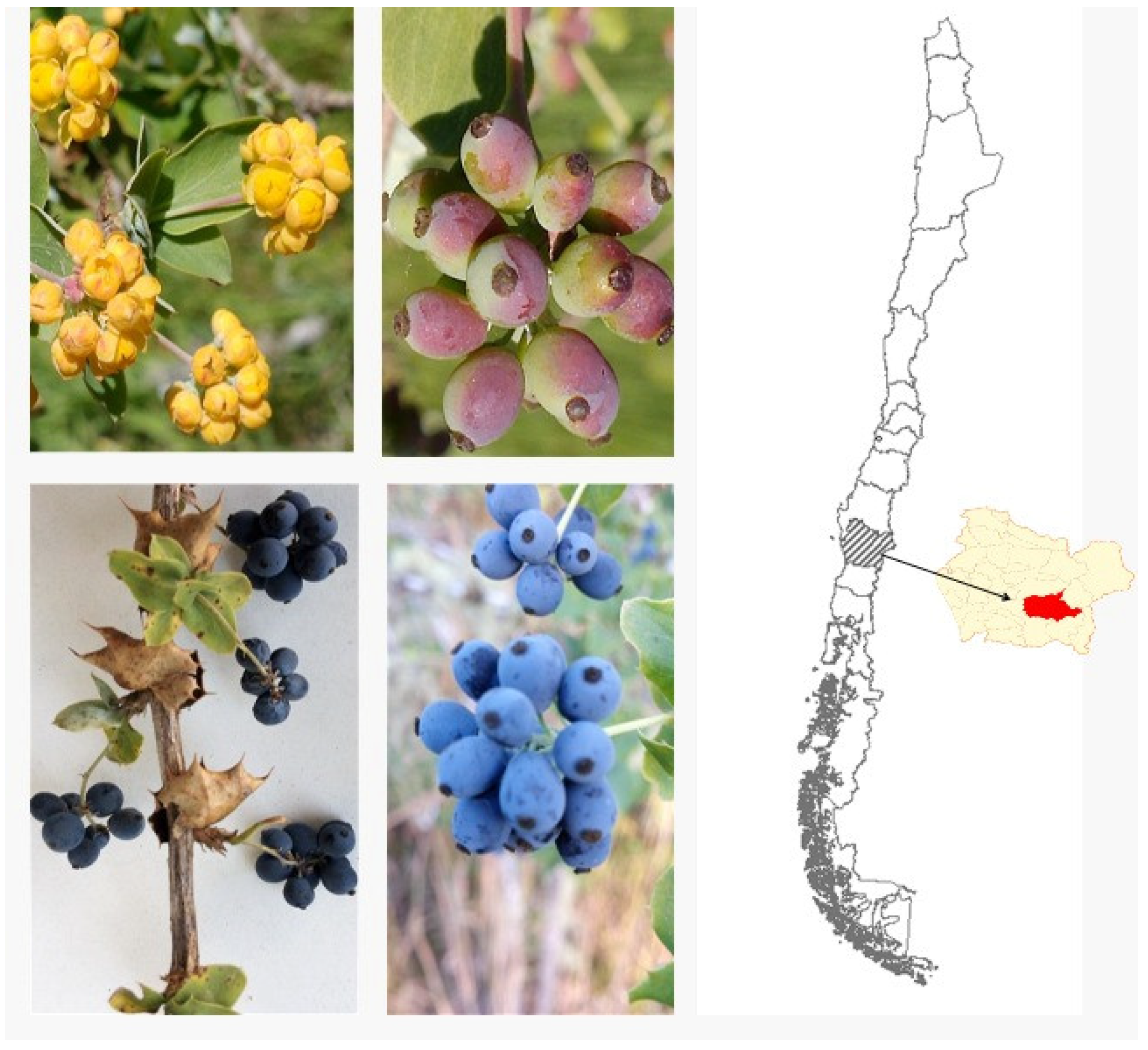
Figure 2.
Contractile effects of B. congestiflora on vascular tone. (A) Representative trace showing the contractile response of intact aortic rings exposed to 0.1-1000 μg/mL B. congestiflora. (B) Concentration-dependent contraction of B. congestiflora in intact (Control) and endothelium-denuded (denuded) aortic rings. Vascular contraction is expressed as a percentage of the submaximum contraction induced by 60 mM KCl. (C) Representative trace showing the contractile response of 1000 μg/mL B. congestiflora on intact aortic rings without or with 20 mM caffeine. (D) Effect of 20-minute preincubation with 10-4 M nimodipine (Nimo), 10 mM caffeine (Caf-10), and 20 mM caffeine (Caf-20). Data are presented as mean ± SEM (n = 5). *p<0.05, **p<0.01, ***p<0.001 vs Control.
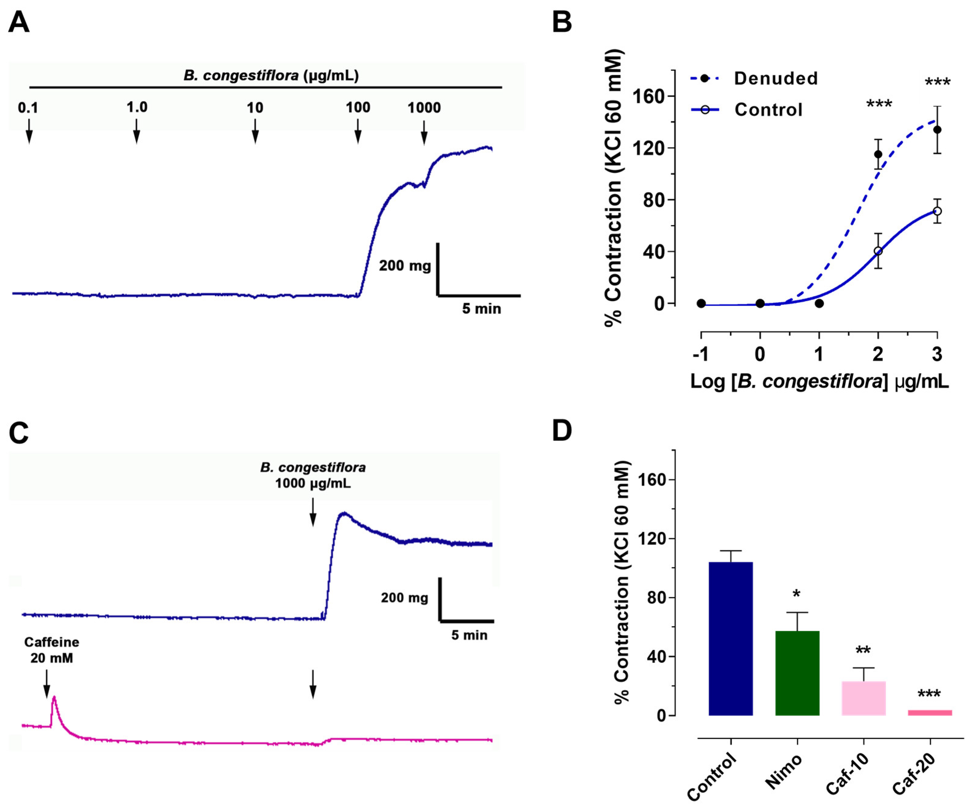
Figure 3.
Endothelium-dependent effects of B. congestiflora on vascular function. A) Representative original trace showing the vascular relaxation of the hydroalcoholic extract of B. congestiflora (0.1 to 1000 μg/mL) in intact aortic rings with endothelium. B) concentration-dependent relaxation to B. congestiflora in intact (Control), endothelium-denuded (Denuded), C) L-NAME-treated, or D) ODQ-treated aortic rings. Data are presented as mean ± SEM (n = 5). ***p<0.001 vs Control.
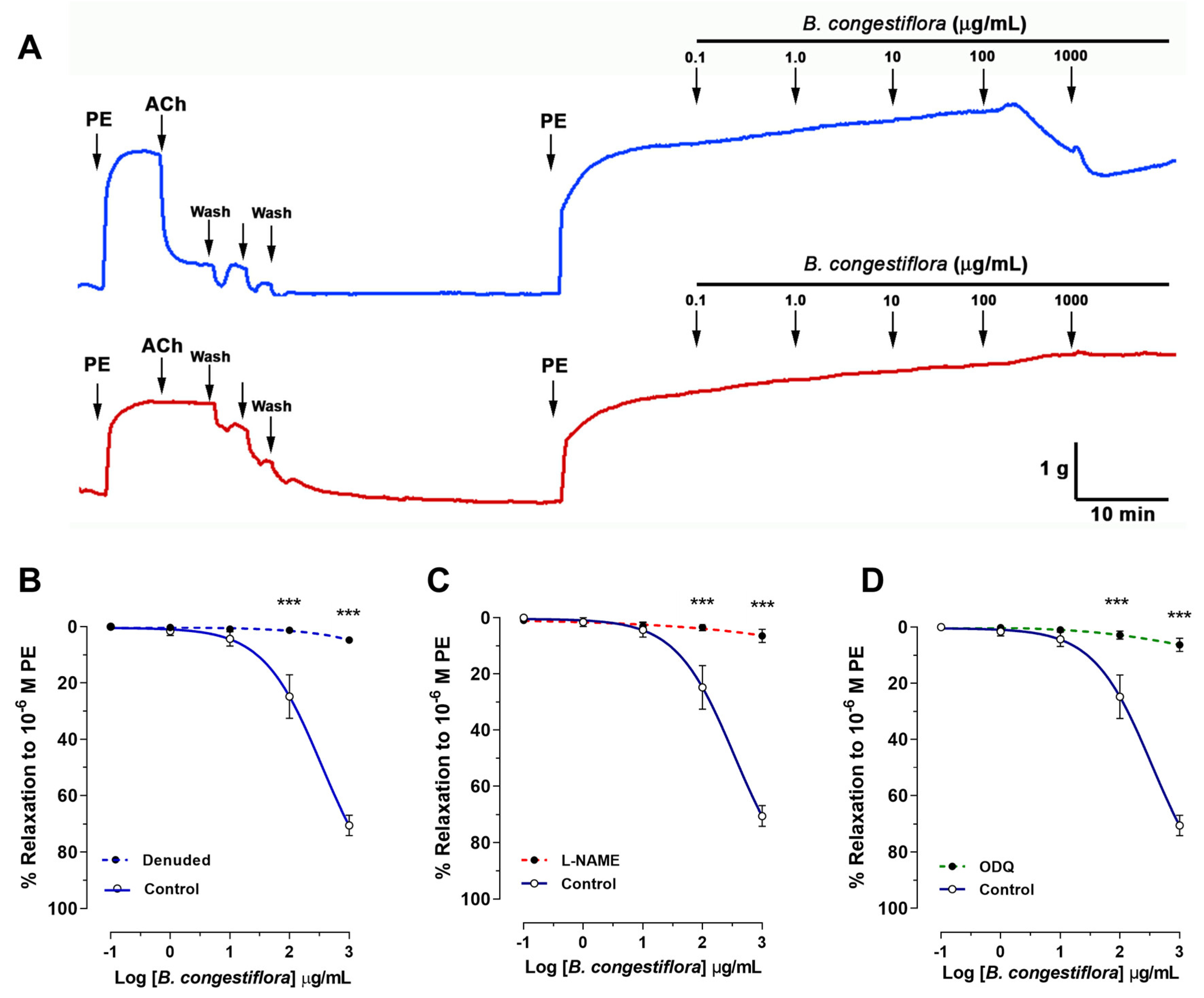
Figure 7.
Compounds subjected to docking assays in the corresponding catalytic sites of acetylcholinesterase, butyrylcholinesterase, Glucosidase and α-amylase.
Figure 7.
Compounds subjected to docking assays in the corresponding catalytic sites of acetylcholinesterase, butyrylcholinesterase, Glucosidase and α-amylase.
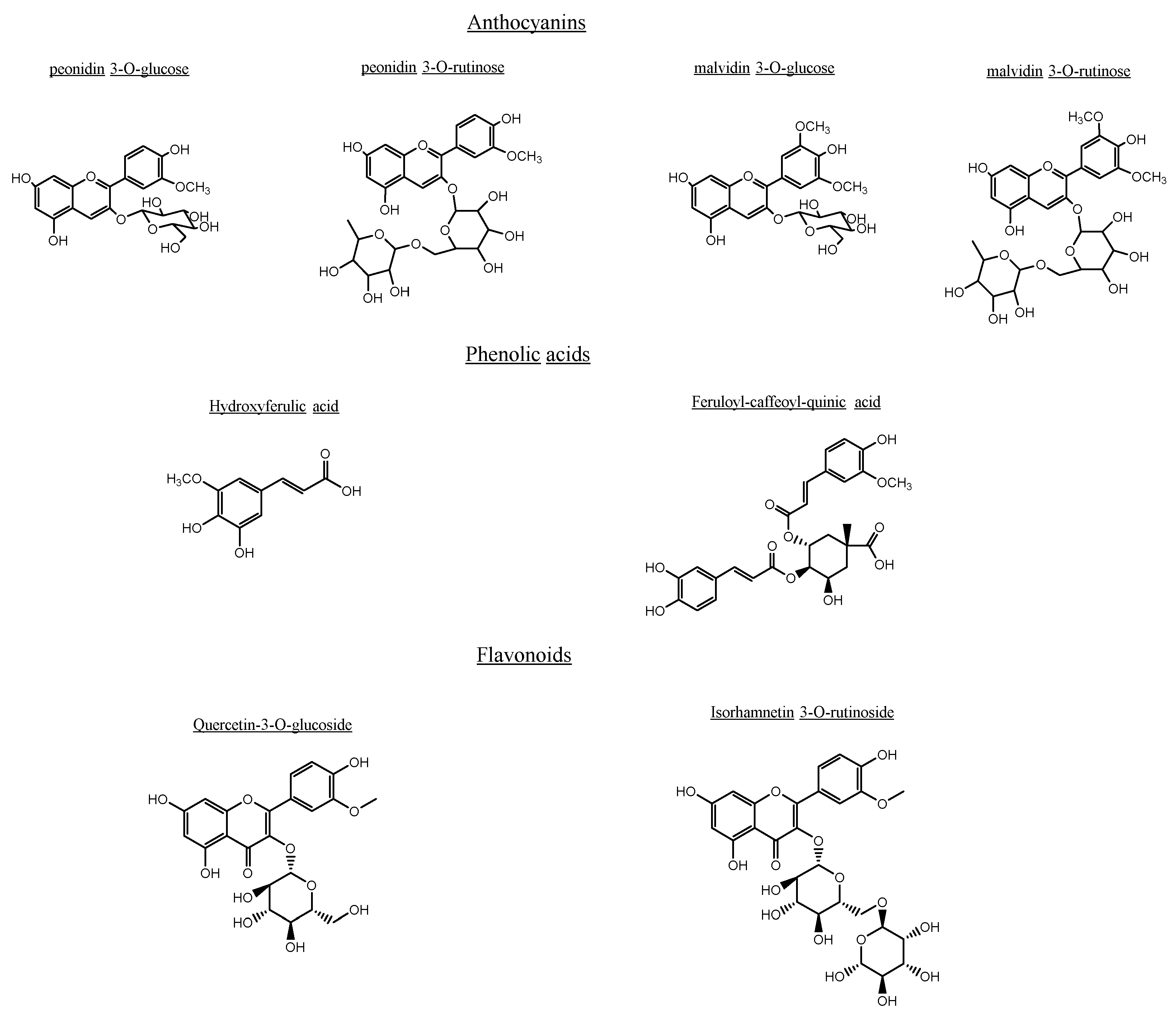
Figure 8.
predicted intermolecular interactions of selected compounds in Berberis congestiflora Gay (BE), ethanolic extract and the residues of Torpedo Californica acetylcholinesterase (TcAChE) catalytic site. Yellow dotted lines indicate hydrogen bond interactions, cyan dotted lines represent π-π interactions, magenta dotted lines represent T-shaped interactions and red dotted lines represents π-cation interactions. A. Peonidin 3-O-glucose in the catalytic site; B. Peonidin 3-O-rutinose in the catalytic site; C. Malvidin 3-O-glucose in the catalytic site; D. Malvidin 3-O-rutinose in the catalytic site; E. Hydroxyferulic acid in the catalytic site; F. Feruloyl-caffeoyl-quinic acid in the catalytic site; G. Quercetin-3-O-glucoside in the catalytic site; H. Isorhamnetin 3-O-rutinoside in the catalytic site.
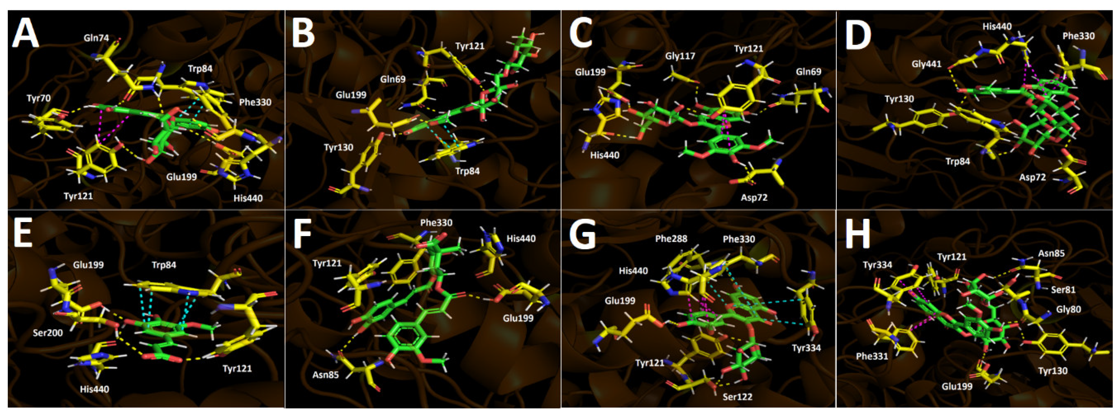
Figure 9.
Predicted intermolecular interactions of selected compounds in Berberis congestiflora Gay (BE) ethanolic extract and the residues of human butyrylcholinesterase (hBuChE) catalytic site. Yellow dotted lines indicate hydrogen bond interactions, cyan dotted lines represent π-π interactions, red dotted lines represent π-cation interactions. A. Peonidin 3-O-glucose in the catalytic site; B. Peonidin 3-O-rutinose in the catalytic site; C. Malvidin 3-O-glucose in the catalytic site; D. Malvidin 3-O-rutinose in the catalytic site; E. Hydroxyferulic acid in the catalytic site; F. Feruloyl-caffeoyl-quinic acid in the catalytic site; G. Quercetin-3-O-glucoside in the catalytic site; H. Isorhamnetin 3-O-rutinoside in the catalytic site.
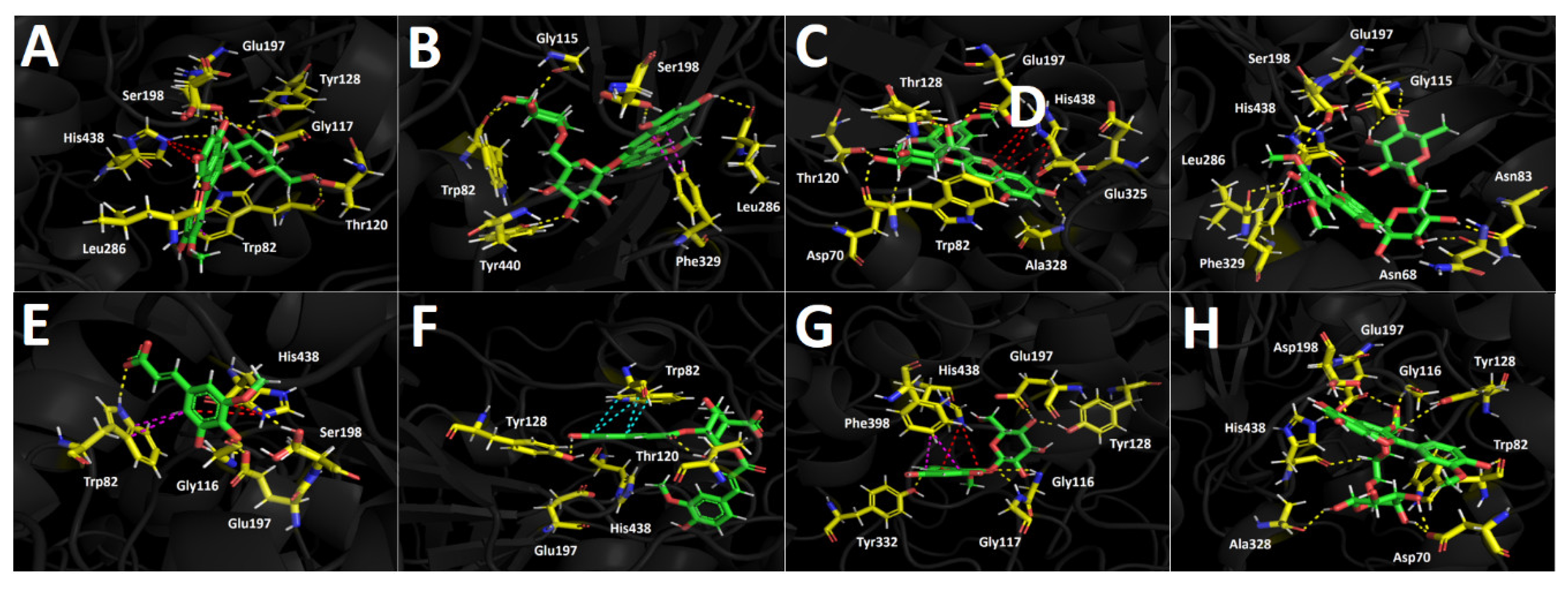
Figure 10.
Predicted intermolecular interactions of selected compounds in Berberis congestiflora Gay (BE) ethanolic extract and the residues of human Glucosidase catalytic site. Yellow dotted lines indicate hydrogen bond interactions, cyan dotted lines represent π-π interactions, magenta dotted lines represent T-shaped interactions and blue dotted lines represent salt bridges. A. Peonidin 3-O-glucose in the catalytic site; B. Peonidin 3-O-rutinose in the catalytic site; C. Malvidin 3-O-glucose in the catalytic site; D. Malvidin 3-O-rutinose in the catalytic site; E. Hydroxyferulic acid in the catalytic site; F. Feruloyl-caffeoyl-quinic acid in the catalytic site; G. Quercetin-3-O-glucoside in the catalytic site; H. Isorhamnetin 3-O-rutinoside in the catalytic site.
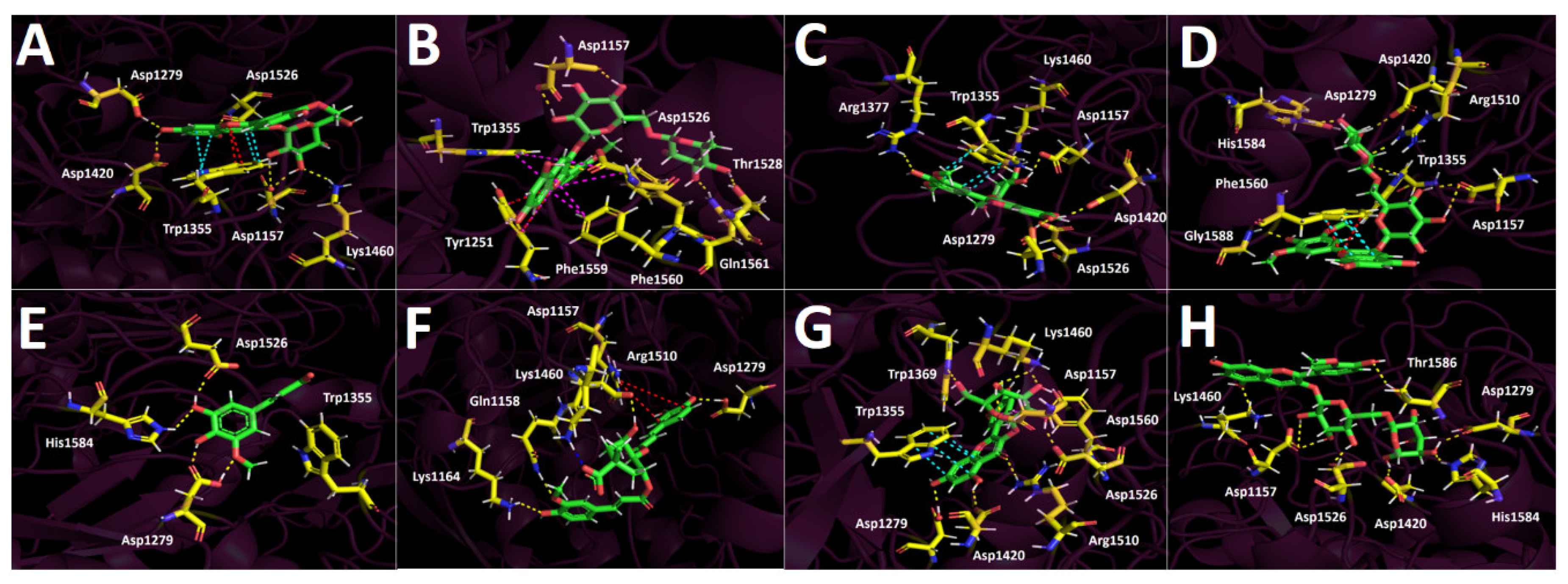
Figure 11.
Predicted intermolecular interactions of selected compounds in Berberis congestiflora Gay (BE) ethanolic extract and the residues of human pancreatic α-amylase catalytic site. Yellow dotted lines indicate hydrogen bond interactions, magenta dotted lines represent T-shaped interactions and blue dotted lines represent salt bridges. A. Peonidin 3-O-glucose in the catalytic site; B. Peonidin 3-O-rutinose in the catalytic site; C. Malvidin 3-O-glucose in the catalytic site; D. Malvidin 3-O-rutinose in the catalytic site; E. Hydroxyferulic acid in the catalytic site; F. Feruloyl-caffeoyl-quinic acid in the catalytic site; G. Quercetin-3-O-glucoside in the catalytic site; H. Isorhamnetin 3-O-rutinoside in the catalytic site.
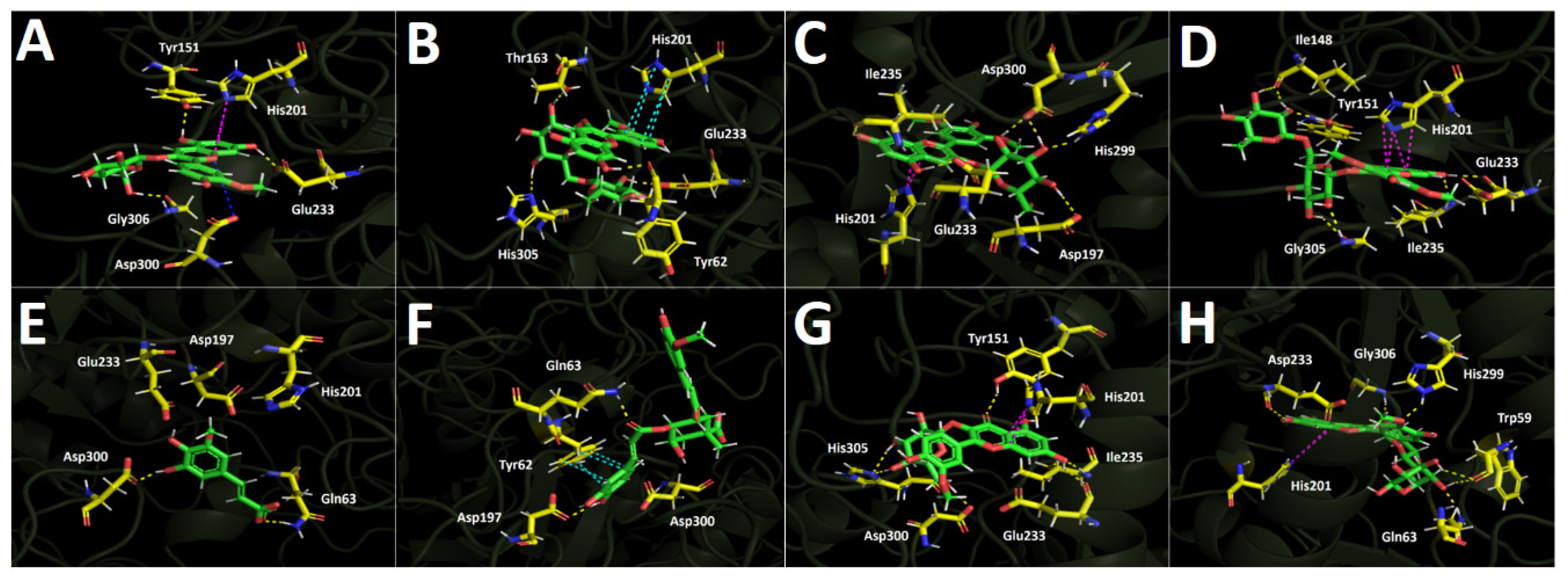
Table 3.
Binding energies obtained from docking experiments of selected compounds from the hydroalcoholic extract of Berberis congestiflora Gay (BE), as well as the known inhibitors galantamine and acarbose over acetylcholinesterase (TcAChE), butyrylcholinesterase (hBChE), Glucosidase and α-Amilase.
| Compound |
Binding energy (kcal/mol) Acetylcholinesterase |
Binding energy (kcal/mol) Butyrylcholinesterase |
Binding energy (kcal/mol) Glucosidase |
Binding energy (kcal/mol) α-Amilase |
| Peonidin 3-O-glucose | -14.837 | -12.461 | -15.462 | -8.597 |
| Peonidin 3-O-rutinose | -15.024 | -13.129 | -15.697 | -11.606 |
| Malvidin 3-O-glucose | -15.793 | -12.944 | -16.176 | -9.793 |
| Malvidin 3-O-rutinose | -11.632 | -13.472 | -12.436 | -14.120 |
| Hydroxyferulic acid | -8.647 | -6.753 | -8.017 | -5.147 |
| Feruloyl-caffeoyl-quinic acid | -10.068 | -10.973 | -10.555 | -7.167 |
| Quercetin-3-O-glucoside | -13.287 | -12.808 | -13.390 | -11.191 |
| Isorhamnetin 3-O-rutinoside | -15.658 | -13.922 | -11.544 | -10.766 |
| Galantamine | -12.989 | -7.125 | --------- | --------- |
| Acarbose | --------- | --------- | -18.591 | -12.626 |
Disclaimer/Publisher’s Note: The statements, opinions and data contained in all publications are solely those of the individual author(s) and contributor(s) and not of MDPI and/or the editor(s). MDPI and/or the editor(s) disclaim responsibility for any injury to people or property resulting from any ideas, methods, instructions or products referred to in the content. |
© 2025 by the authors. Licensee MDPI, Basel, Switzerland. This article is an open access article distributed under the terms and conditions of the Creative Commons Attribution (CC BY) license (http://creativecommons.org/licenses/by/4.0/).
Copyright: This open access article is published under a Creative Commons CC BY 4.0 license, which permit the free download, distribution, and reuse, provided that the author and preprint are cited in any reuse.


