Submitted:
15 December 2025
Posted:
16 December 2025
You are already at the latest version
Abstract
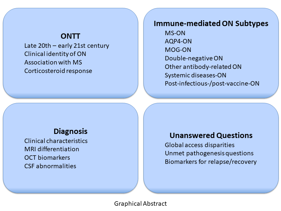
Optic neuritis (ON) has been recognized since antiquity, but its modern clinical identity emerged only in the late 19th century and was definitively shaped by the Optic Neuritis Treatment Trial (ONTT). The ONTT established the natural history, visual prognosis, association with multiple sclerosis (MS), and therapeutic response to corticosteroids, building the foundation for contemporary ON management. Over the past two decades, ON has evolved from a seemingly uniform demyelinating syndrome into a group of biologically distinct disorders. The identification of aquaporin-4-IgG ON (AQP4-ON), myelin oligodendrocyte glycoprotein antibody–associated ON (MOG-ON), and double-negative ON has transformed diagnostic and therapeutic strategies. These subtypes differ in immunopathology, clinical course, MRI features, retinal injury patterns, CSF profiles, and long-term outcomes, making early and accurate differentiation essential. MRI provides key distinctions in lesion length, orbital tissue inflammation, bilateral involvement, and chiasmal or optic tract extension. Optical coherence tomography (OCT) offers complementary structural biomarkers, including severe early ganglion cell loss in AQP4-ON, relative preservation in MOG-ON, and variable patterns in double-negative ON. CSF analysis further refines diagnosis, with oligoclonal bands strongly supporting MS-ON. Together, these modalities enable precise early stratification and timely initiation of targeted immunotherapy, which is critical for preventing irreversible visual disability. Despite major advances, significant unmet needs persist. Access to high-resolution MRI, OCT, cell-based antibody assays, and evidence-based treatments remains limited in many regions, contributing to global disparities in outcomes. The pathogenesis of double-negative ON, reliable biomarkers of relapse and visual recovery, and standardized multimodal diagnostic thresholds remain unresolved. Future research must expand biomarker discovery, refine imaging criteria, and ensure equitable global access to cutting-edge diagnostic platforms and therapeutic innovations. Four decades after the ONTT, ON remains a dynamic field of investigation, with ongoing advances holding the potential to transform care for patients worldwide.
Keywords:
optic neuritis
; optic neuritis treatment trial
; MS-associated optic neuritis
; AQP4-associated optic neuritis
; MOG-associated optic neuritis
; double-negative optic neuritis
; optical coherence tomography
; MRI biomarkers
; CSF biomarkers
1. Introduction
Optic neuritis (ON) encompasses a spectrum of inflammatory disorders affecting the optic nerve, typically presenting with subacute visual loss. The conceptual origins of ON can be traced back to classical antiquity, specifically the 5th to 4th centuries BCE, when early physicians such as Alcmaeon of Croton and Hippocrates first proposed that vision loss could originate from neural structures beyond the eye itself [1]. Since then, the concept has evolved through a series of major milestones, most notably the invention of the ophthalmoscope in the mid-19th century, allowing the in vivo examination of the optic nerve inflammation; the Optic Neuritis Treatment Trial (ONTT) studies with onset in the turn of the 20th century; and the identification of aquaporin 4 (AQP4 IgG) and myelin oligodendrocyte glycoprotein (MOG IgG) antibodies as causes of immune mediated ON [2,3,4].
Recently, an international consortium of experts proposed a classification framework for ON based on its underlying etiology and fundamental pathophysiological mechanisms. This initiative also introduced a set of diagnostic criteria that integrates clinical phenotype, neuroimaging features, and serological biomarkers, aiming to enhance diagnostic accuracy, inform prognosis, and guide therapeutic decision-making [5].
Despite these marked advancements, several unresolved issues persist, particularly regarding biomarker development, pathophysiological characterization, and long-term management of the double negative optic neuritis (DN-ON), defined as AQP4-IgG–negative, MOG-IgG–negative ON not associated with multiple sclerosis (MS). Moreover, there is an urgent need to promote equity across all populations by ensuring access to advanced diagnostic technologies and the most effective therapies.
This review provides a comprehensive synthesis of the evolution of the foundation of the ON concept from Antiquity to Modern Age, and the current status of its knowledge. We examine the state-of-the-art of immune-mediated ON including its clinical characteristics, diagnostic approach, and therapeutic strategies for both typical ON (MS-ON) – as defined by the ONTT and subsequent studies – and atypical ON (non-MS-ON), which predominantly includes AQP4-ON, MOG-ON, and double-seronegative forms of undetermined etiology. Additionally, we explore the major challenges associated with the diagnosis and subclassification of seronegative ON and propose research strategies aimed at defining distinct entities within this group. These include detailed analysis of clinical phenotyping, advanced neuroimaging techniques, and the identification of novel serum autoantibodies that could allow discrimination of other types of ON within this common group. Such efforts are essential for bridging current gaps in the field, ultimately enabling more precise diagnosis, targeted therapy, personalized care and improved clinical outcomes [5,6,7].
2. Historic Foundations of the Concept of Optic Neuritis
The following timeline (Figure 1) highlights the major historical developments that collectively shaped the early foundations of ON.
2.1. Ancient Contribution: The Discovery of the Optic Nerve and Its Function
The discovery of the optic nerves is credited to Alcmaeon of Croton (c. 510–450 BCE), who dissected animals and reportedly traced channels from the eyes to the brain [8]. Prior to his work, Mesopotamians, Egyptians, and early Greek poets and philosophers regarded the eye primarily as a window for light, emotion, or divine influence. Alcmaeon was the first to assert that the brain—not the heart—was the seat of cognition and sensation, including vision, marking a decisive departure from the cardiocentric views of Homeric and early pre-Socratic thought. As summarized in the fragment attributed to him—“Through the eyes, ears, and via the brain, we perceive and understand” (quoted by Theophrastus)—his insight laid the conceptual groundwork for later anatomical and physiological interpretations of sight. Awareness that visual loss could originate beyond the eye appears later in the Hippocratic Corpus (c. 460–370 BCE), although these texts did not yet distinguish clearly between diseases of the eye and those of the optic nerve. The term amaurosis (“darkening”) was sometimes used to describe unexplained blindness [9].
The first known description of the optic chiasm is attributed to Herophilus of Chalcedon (c. 335–280 BCE), a Greek physician and anatomist who lived during the early Hellenistic period. Herophilus was among the earliest practitioners to conduct systematic human dissections, enabling anatomical observations of unprecedented precision in Greek medicine. The term chiasm derives from the Greek letter Χ, denoting the cross-shaped configuration formed by the converging optic nerves. Herophilus characterized this structure as a “crossing” or decussation at the base of the brain [1]. His account was later elaborated by Galen, who proposed that the optic chiasm accommodated a partial decussation of optic fibers—an interpretation that shaped anatomical thought well into the Renaissance.
The occurrence of visual loss in otherwise healthy eyes was first recognized by Rufus of Ephesus in the 1st century CE. He distinguished visual impairment arising from what we would now classify as central or neural causes from that due to primary ocular disease. However, he did not identify inflammation or describe any pathology consistent with modern ON [10]. Rufus also provided one of the earliest detailed anatomical accounts of the optic nerve and its connection to the brain. He described the optic nerves as originating from the brain and extending toward the eyes, explicitly stating that the optic nerve is a solid structure—thereby correcting earlier notions that it functioned as a hollow channel for “visual pneuma,” as proposed by proponents of the extramission theory. He further emphasized the importance of distinguishing visual loss caused by ocular pathology from that resulting from optic nerve or cerebral injury. This conceptual separation remains fundamental to understanding retrobulbar ON.
The notion of optic nerve damage as a cause of visual loss was further advanced by Galen of Pergamon (2nd century CE). Galen argued that vision arises not solely from the eye but from the coordinated interaction among the eye, the optic nerves, and the brain. This perspective helped shift medical thought away from strictly ocular explanations of blindness and toward recognition that lesions affecting the optic pathways or brain could produce visual loss. Despite this conceptual advance, Galenic doctrine maintained that the optic nerves were hollow conduits carrying “pneuma,” or visual spirit, from the anterior ventricles of the brain [1]. Galen’s anatomical writings dominated medical scholarship for more than a millennium, profoundly shaping both Islamic and European medical traditions [11].
2.2. The Middle Age Contribution: Early Clinical Characterization and Treatment of Inflammation of the Optic Nerve
The first clinical characterization of ON is ascribed to Hunayn ibn Ishaq (809–873 CE), one of the most influential physicians of the Islamic Golden Age. His description of ON shows a sophisticated grasp of clinical observation and a remarkable anticipation of modern neurological concepts [12]. In his treatise on the eye, known as The Book of the Ten Treatises on the Eye (Figure 2), Hunayn provides one of the earliest known detailed descriptions of conditions affecting the optic nerve—including what would now be classified as ON. He described cases where patients experienced diminished or lost vision not due to disease of the eye itself, but due to a problem in the “optic spirit” or the pathway leading from the eye to the brain. Hunayn proposed that when this pathway was interrupted vision was impaired. He identified symptoms such as sudden vision loss, a lack of visible damage to the eye itself, pain upon eye movement, and a connection between systemic or brain-related conditions and visual impairment. His explanation—that the optic nerve could be afflicted by “swelling” or “obstruction”—closely aligns with the modern understanding of ON as an inflammatory demyelinating condition. He also noted the possibility of recovery in some patients, acknowledging the variable prognosis of the condition. His synthesis of Greek medical theory with clinical observations advanced the understanding significantly beyond any of his predecessors [13].
The earliest known use of the expression “inflammation of the optic nerve” (waram ʿaṣab al-baṣar) appears in ʿAlī ibn ʿĪsā al-Kahḥāl’s Tadhkirat al-Kahhalīn (Notebook of the Oculists, ca. 1010 CE) (Figure 3). The term referred to a disorder marked by periocular pain, visual impairment, and, in some instances, ocular redness or manifestations of systemic disease. Although clinically perceptive, Ibn ʿĪsā’s anatomical understanding remained rooted in Galenic doctrine, which conceived the optic nerves as hollow conduits carrying pneuma or “visual spirit.”
Important therapeutic and conceptual refinements emerged shortly thereafter in the work of the Persian polymath Ibn Sīnā (Avicenna) (980–1037 CE). Although he did not describe ON specifically, his contributions to ocular physiology and the nosology of visual loss laid essential foundations for later neuro-ophthalmological thought. In his monumental al-Qānūn fī al-Ṭibb (The Canon of Medicine), Avicenna located the faculty of vision in the optic nerve rather than the crystalline lens, directly challenging Galenic tradition and anticipating the later recognition of the optic nerve as a central structure in visual processing [16]. This shift represented a pivotal conceptual advance as, by assigning a primary visual role to the optic nerve, Avicenna provided the intellectual space for understanding visual failure as a neurological disorder rather than a purely ocular one.
Avicenna also provided detailed therapeutic recommendations for acute inflammatory eye diseases. His pharmacopeia included anti-inflammatory and analgesic agents such as willow oil, opium, and vinegar-based poultices, reflecting an early recognition of inflammation as a driver of vision loss. Although he did not explicitly delineate optic nerve inflammation, his therapeutic strategies align with the principles that later guided the management of ON as an inflammatory neuropathy.
2.3. The Renaissance and Early Modern Age: The Definitive Abandonment of Galenic Tradition
The Renaissance and early modern period (14th–18th centuries) constituted a pivotal transitional era in the evolving understanding of optic nerve pathology, marking the definitive departure from Galen’s long-standing theory of vision. For more than a millennium, Galen (129–c.216 CE) had shaped Western and Islamic medical thought with a model in which pneuma psychikon (“psychic spirit”), generated in the brain, was conveyed through the supposedly hollow optic nerves to the crystalline lens—believed to be the primary organ of vision. In Galen’s extramission-based framework, inherited from earlier Greek thinkers such as Euclid and Ptolemy, visual rays were emitted from the eye toward external objects, and visual disturbance resulted from obstruction of the pneuma’s flow due to inflammation, trauma, or imbalance of humors. Within this paradigm, the optic nerves were conceptualized not as neural structures but as tubular channels.
Although ON was not yet recognized as a discrete clinical entity, the anatomical and physiological advances of the Renaissance profoundly reshaped the conceptual foundations necessary for its later identification. The revival of human cadaveric dissection, coupled with a critical reassessment of classical authorities, allowed anatomists to correct Galenic misconceptions directly. Andreas Vesalius (1514–1564), in his seminal De humani corporis fabrica (1543), demonstrated unequivocally that the optic nerves are solid cords of neural tissue rather than hollow tubes. His meticulous descriptions also improved understanding of the optic chiasm and its structural relationships, thereby establishing a more accurate neuroanatomical framework.
In the early 17th century, Thomas Willis (1621–1675) advanced clinico-anatomical correlations by describing how lesions of the optic chiasm produced characteristic visual field defects, a major step toward linking structural damage to functional loss. Johannes Kepler (1571–1630) provided a complementary optical breakthrough by demonstrating that the retina—not the lens—is the true receptive surface for image formation. Kepler’s retinal-image model decisively separated retinal disorders from optic nerve pathology, enabling clinicians for the first time to distinguish vision loss attributable to optic nerve dysfunction [17].
By the 18th century, Enlightenment anatomists further refined ocular anatomy with increasing precision. In Descriptio Anatomica Oculi Humani (1727), Johann Gottfried Zinn (1699–1749) published one of the earliest comprehensive and detailed anatomical treatises of the human eye. Zinn’s systematic descriptions of the optic nerve, its sheath, extraocular muscles, and orbital structures—based entirely on human dissection—represented the culmination of anatomical progress initiated by Vesalius. His work provided the most accurate depiction to date of the optic nerve’s structure and its relationship to surrounding tissues.
Collectively, these advances dismantled the Galenic visual model and replaced it with an anatomically and optically coherent understanding of the visual system. This transformation created the essential conceptual scaffolding upon which the modern recognition and study of optic neuritis would eventually be built.
2.4. The Modern Age: The Invention of the Microscope and the Characterization of Optic Neuritis
The invention of the ophthalmoscope by Hermann von Helmholtz in 1851 revolutionized visual neuroanatomy and enabled direct observation of the optic disc, transforming the recognition, description, and nosology of ON through the possibility of identification of optic disc swelling and pallor in living patients. This diagnostic breakthrough marked the transition from speculative descriptions to objective, reproducible identification of optic nerve inflammation. Shortly after the ophthalmoscope’s spread, Albrecht von Graefe published detailed accounts of cases of ON describing the ophthalmoscopic appearance of swollen optic discs, hyperemia, blurred disc margins, and associated retinal changes [18]. He correlated these with clinical symptoms—subacute visual loss, central scotomas, and ocular pain—and differentiated inflammatory optic disc swelling (papillitis) from other causes of optic nerve head elevation, such as papilledema from raised intracranial pressure [18,19].
Von Graefe recognized that ON could occur as an isolated event or in association with systemic neurological diseases, paving the way for later clinicopathological correlations with MS [18,19,20]. His work provided one of the earliest coherent frameworks for differentiating ON phenotypes, thereby paving the way for subsequent refinement of retrobulbar neuritis versus papillitis.
A major advance in clinical characterization came with Edward Nettleship’s seminal 1884 paper On Retro-Bulbar Neuritis. Analyzing a large series of cases, Nettleship identified key diagnostic features: central scotoma as the characteristic visual field defect, disproportionate color vision loss—particularly for red and green—and pain with eye movement [21]. Crucially, he emphasized that the optic disc often appeared normal in the acute stage, establishing the clinical entity of retrobulbar optic neuritis. This form, defined by acute visual loss with an initially normal fundus, contrasted with papillitis and exhibited variable prognosis.
The late 19th and early 20th centuries also saw recognition of physiological signs that reflected demyelinating conduction failure. Uhthoff’s phenomenon (1890), describing transient visual worsening with heat or exertion in individuals with prior ON, anticipated modern understanding of temperature-dependent conduction block in demyelinated axons, later corroborated by visual evoked potential studies [22,23]. The relative afferent pupillary defect (RAPD)—or Marcus Gunn pupil—described by Stanley Thompson in 1966, emerged as a sensitive indicator of asymmetric pregeniculate pathway dysfunction, often present even when the fundus appears normal.
Another seminal physiological insight was the Pulfrich phenomenon, described by Carl Pulfrich in 1922: a stereo-motion illusion produced by interocular latency differences. Frequently observed after ON, it provided an early functional correlate of slowed neural conduction in the optic nerve [24].
2.5. The Relationship Between Optic Neuritis and Multiple Sclerosis
In 1868, Jean-Martin Charcot (1825-1893) was the first to explicitly recognize ON as a possible manifestation of MS, thereby integrating a visual disorder into the broader clinical spectrum of the disease. In his 1868 Leçons sur les maladies du système nerveux, Charcot defined sclérose en plaques disséminées as a distinct pathological entity characterized by demyelinating plaques in the brain and spinal cord, and he identified ON among its frequent manifestations [25]. His conclusion was grounded meticulous clinicopathological correlation, yet his focus remained on the pathological anatomy and the syndromic constellation—motor, sensory, cerebellar, and visual symptoms—rather than on detailed phenotyping of ON or its prognostic implications [26].
A shift in focus from pathological associations to clinical epidemiology and prognosis was observed in the mid-20th century. A landmark population-based study by Percy, Nobrega, and Kurland in 1972 [27] demonstrated that 13–15% of MS patients presented with ON and that 27–37% developed ON during the course of their illness. They also reported that approximately 17% of idiopathic ON cases progressed to MS, thereby clarifying the strength of the association between the two conditions. Subsequent reviews and cohort studies have consistently confirmed that ON is often the initial symptom of MS and that a significant proportion of adults and children presenting with isolated ON will eventually develop MS [28,29,30,31,32].
These epidemiological insights laid the groundwork for early therapeutic experimentation with adrenocorticotropic hormone (ACTH) in ON during the 1950s. Initial reports—most notably Smith’s 1953 case description—suggested accelerated visual recovery with ACTH treatment [33]. A subsequent double-blind, prospective trial in 1966 demonstrated that ACTH-treated patients regained vision more rapidly than those receiving placebo, although final visual outcomes at one year did not differ significantly [34]. These preliminary therapeutic efforts paved the way for the ONTT, initiated in 1991, which remains the cornerstone of modern ON management [35,36].
3. The Optic Neuritis Treatment Trial and Characterization of Multiple Sclerosis-Related Optic Neuritis
By the 1970s and 1980s, the management of acute ON remained largely empirical. Systemic corticosteroids were widely used, yet the evidence supporting their efficacy was inconsistent and derived mainly from small, uncontrolled studies. Questions persisted regarding the optimal dose, route, and duration of therapy, and uncertainty extended to the natural history of ON itself—particularly its long-term visual prognosis and the magnitude of its association with MS. Although epidemiologic observations suggested that ON frequently served as a presenting symptom of MS, robust prospective data quantifying this risk were not yet available. The advent of magnetic resonance imaging (MRI) in the mid-1980s provided an unprecedented means of detecting clinically silent demyelinating lesions, but its prognostic value in the setting of a first ON episode had not been systematically evaluated. This convergence of therapeutic ambiguity, prognostic uncertainty, and emerging neuroimaging technology created a compelling rationale for a rigorously designed, large-scale clinical trial.
To address these gaps, the Optic Neuritis Study Group (ONSG) was established as a multicenter collaborative consortium supported by the National Eye Institute. Bringing together leading neurologists and neuro-ophthalmologists from 15 clinical centers across the United States, the ONSG undertook the first comprehensive, prospective characterization of demyelinating ON and designed what would become the ONTT. The ONTT was conceived with two principal objectives. The first was to determine whether specific corticosteroid regimens—namely high-dose intravenous methylprednisolone followed by oral prednisone, oral prednisone alone, or placebo—could accelerate visual recovery or improve long-term visual outcomes in acute ON. The second objective was to define the long-term risk of MS following a first episode of ON and to evaluate whether baseline brain MRI could stratify that risk with clinical utility.
Over the next two decades, the ONTT and its extension studies generated an extensive body of work, including seminal reports on treatment effects, visual recovery trajectories, late visual function, quality of life, recurrence rates, and MS conversion risk. These publications collectively transformed the clinical understanding and management of ON [2,37,38,39,40,41,42,43,44,45,46,47,48,49,50,51,52,53,54,55,56,57,58,59,60,61,62,63].
Together, the ONTT and its related investigations established the evidence-based framework that continues to guide the diagnosis, treatment, and prognostication of MS-related ON.
3.1. Demographic and Clinical Characterization of Optic Neuritis in the ONTT
Between 1988 and 1991, the ONTT enrolled 457 patients with a first episode of acute unilateral ON across 15 clinical centers in the United States [35]. The cohort was predominantly Caucasian (85.3%), with smaller proportions of African American (12.7%), Asian (1.5%), and Hispanic (0.4%) participants. Eligibility required age between 18 and 45 years and the presence of a first episode of acute unilateral ON of ≤8 days’ duration, typically characterized by sudden visual loss accompanied by periocular pain. Clinical examination had to be consistent with ON, including an RAPD and either optic disc swelling or a normal disc in retrobulbar cases. Visual acuity (VA) in the affected eye was required to be worse than 20/40 but not no light perception (NLP), while that of the fellow eye had to be better than 20/40. Patients were excluded if they exhibited atypical clinical features—such as marked optic disc swelling with hemorrhages or exudates, progression beyond two weeks, or absence of pain—prior ON in the affected eye, or VA worse than 20/40 in the fellow eye. Additional exclusion criteria included systemic diseases capable of mimicking ON (e.g., sarcoidosis, syphilis, Lyme disease, systemic lupus erythematosus), other ocular or neurological conditions affecting vision, contraindications to corticosteroid therapy (such as uncontrolled hypertension, diabetes, active infection, psychosis, or peptic ulcer disease), pregnancy or lactation, or exposure to systemic corticosteroids within the previous 30 days.
The baseline demographic and clinical characteristics of the ONTT cohort are summarized in Table 1. These data outline the visual function profile and symptomatology of the 457 patients at study entry [35]. The table includes demographic variables, laterality, and the frequency of simultaneous bilateral involvement, as well as key baseline measures such as high-contrast visual acuity (HCVA), color vision, contrast sensitivity (CS), visual field findings, and the presence of an afferent pupillary defect. It also reports the prevalence of eye pain or headache accompanying vision loss, a hallmark feature of acute demyelinating ON in this population.
3.2. Assessment of Corticosteroid Treatment
The selected ONTT participants were randomized to one of three groups: (1) IV methylprednisolone (IVMP) + oral prednisone: 250 mg IV every 6 h for 3 days (1 g/day), then oral prednisone 1 mg/kg/day for 11 days with taper; (2) oral prednisone alone (1 mg/kg/day for 14 days); or (3) oral placebo: 14 days. The study showed that IVMP significantly accelerated recovery of visual function, with earlier improvement in visual fields (P = 0.0001), CS (P = 0.026), and color vision (P = 0.033) compared to placebo, though no long-term VA benefit was observed. Oral prednisone alone was ineffective and increased recurrence risk [2].
At five-year follow-up most patients retained good vision (≥20/20 VA in ~74% of affected eyes). Baseline MRI lesions predicted a ~50% 5-year MS risk vs. ~25% with normal MRI (1997a, 1997b). At 10 years, 74% had ≥20/20 VA, 18% had 20/25–20/40, 5% had <20/40–20/200, and 3% had <20/200. Recurrence occurred in ~35%, more often in MS patients (P < 0.001). At 15-18 years, 72% of affected eyes and two-thirds of patients had ≥20/20 VA bilaterally. Multiple sclerosis remained associated with slightly worse outcomes and quality-of-life measures [57,58,59].
3.3. Multiple Sclerosis Risk and Prognostic Value of MRI
A pivotal contribution of the ONTT was the detailed characterization of the long-term risk of MS following a first episode of acute demyelinating ON meeting the study’s inclusion criteria. These findings established the prognostic utility of baseline brain MRI as the most powerful predictor of MS conversion in isolated ON [37,53,57,61], thereby defining what is now understood as typical demyelinating ON.
In the final long-term ONTT follow-up, conducted 14–18 years after enrollment (mean, 15 years), the cumulative probability of clinically definite MS for the entire cohort was approximately 50% [61]. Baseline brain MRI performed at study entry stratified patients into two distinct prognostic groups. Individuals with one or more characteristic white-matter lesions—defined at the time as ovoid, ≥3 mm in diameter, and located in typical MS regions such as periventricular, juxtacortical, infratentorial areas, or the corpus callosum—had a 15-year MS conversion risk of approximately 72%. In contrast, patients with a normal MRI had a markedly lower conversion probability of approximately 25% [61].
The temporal pattern of MS conversion demonstrated a biphasic distribution. The majority of conversions occurred within the first five years, particularly among patients with MRI abnormalities [53]. Conversions continued at a slower rate thereafter, with very few new cases after ten years among patients with normal baseline MRI, suggesting that long-term risk eventually plateaus [61].
Several clinical factors increased MS risk independently of MRI findings. These included older age at ON onset, female sex, relapsing ON (RON), and the presence of additional neurological symptoms or abnormal neurological examination at presentation [64,65,66]. Conversely, severe visual loss and optic disc swelling—particularly in children—were associated with a lower likelihood of MS conversion, although these features were not strong independent predictors once MRI was taken into account [66].
Optical coherence tomography (OCT) has also emerged as a potential prognostic tool. Thinning of the ganglion cell internal plexiform layer (GCIPL) and the peripapillary retinal nerve fiber layer (pRNFL) in both the affected and the fellow eye may serve as independent predictors of MS conversion after ON [67,68].
Recurrent ON occurred more frequently among patients who subsequently developed MS, affecting approximately one-third of the ONTT cohort [35,58,61]. In many cases, involvement of the fellow eye occurred in close temporal proximity to the MS diagnosis. These observations firmly established MRI as an essential tool for long-term prognostication and directly influenced revisions to the McDonald diagnostic criteria in 2001, 2005, 2010, 2017, and 2024, which increasingly relied on MRI evidence of dissemination in space (DIS) and time (DIT) to confirm MS after a single demyelinating event [69,70,71,72,73].
3.4. The Role of Optic Neuritis in the Diagnosis of Multiple Sclerosis
The evolution of the McDonald criteria over the past two decades has progressively reshaped the diagnostic weight assigned to ON in MS, integrating increasingly sophisticated imaging and laboratory biomarkers into the diagnostic framework [69,70,71,72,73,74,75] (Table 2).
The 2001 and 2005 revisions relied primarily on conventional MRI evidence of DIS and DIT, permitting the diagnosis of MS after a first demyelinating episode—such as ON—when radiological thresholds were fulfilled [69,70]. The 2010 update advanced this framework by allowing DIS and DIT to be demonstrated on a single MRI scan through the simultaneous presence of enhancing and non-enhancing lesions, thereby accelerating MS diagnosis in typical ON presentations [71].
In 2017, cerebrospinal fluid (CSF)-specific oligoclonal bands (OCB) were accepted as an alternative to demonstrating DIT, although the optic nerve itself still did not qualify as a DIS site [72]. This long-standing limitation was fundamentally revised in the 2025 McDonald update, which recognized the optic nerve as the fifth topographic region for DIS, allowing ON-related lesions—identified on orbital MRI, visual evoked potentials (VEP), or OCT—to directly support MS diagnosis [74,76].
This shift is particularly relevant for patients presenting with isolated ON and borderline MRI findings. In such cases, advanced imaging markers now incorporated into the criteria—such as the central vein sign (CVS) and paramagnetic rim lesions (PRLs)—provide additional specificity [77,78,79,80].
The CVS reflects the perivenular origin of demyelinating plaques in MS. It appears as a small central vessel traversing white-matter lesions on susceptibility-weighted or T2* imaging [81], corresponding to classic histopathological descriptions of MS lesions centered on small veins [82]. Quantitative thresholds—such as ≥40% of lesions showing a central vein—have demonstrated high diagnostic accuracy in distinguishing MS from ischemic, migraine-related, and other inflammatory white-matter disorders [83,84].
The PRLs also referred to as “iron rim lesions”, reflect chronic active demyelination in MS. They exhibit a demyelinated, hypocellular core surrounded by iron-laden activated microglia/macrophages, producing a paramagnetic rim on susceptibility-based MRI [85,86,87,88,89]. PRLs are highly specific for MS and rare in ischemic small-vessel disease or neuromyelitis optica spectrum disorder (NMOSD) [90,91]. Their presence correlates with accelerated brain atrophy, greater clinical disability, and worse long-term outcomes [92,93,94,95]. Longitudinal studies have demonstrated that PRLs are relatively specific for MS, distinguishing it from other white matter diseases such as small vessel ischemia or NMOSD, in which PRLs are rare [90,91,96]. The presence of PRLs has been associated with greater clinical disability, faster brain atrophy, and worse long-term outcomes, underscoring their prognostic value [79,90,92,93,94,95,97].
Optical coherence tomography has become an integral component of the 2024 McDonald criteria, reflecting its value as a structural biomarker in patients presenting with optic ON. By quantifying axonal and neuronal integrity in the retina, OCT provides objective evidence of optic nerve involvement that complements MRI and CSF analysis [76,98]. Both, the pRNFL and the GCIPL are particularly informative after ON. They exhibit characteristic thinning following demyelination, with GCIPL loss occurring earlier and correlating more consistently with functional outcomes such as low-contrast visual acuity (LCVA), color vision, and visual field sensitivity [99,100,101,102].
Intereye absolute difference (IEAD) substantially enhances diagnostic sensitivity for prior unilateral ON. Validated thresholds of ≥9 μm for pRNFL and ≥6 μm for GCIPL reliably distinguish affected from unaffected eyes, even when clinical history is uncertain or visual evoked potential (VEP) are inconclusive [103,104].
Importantly, OCT also detects subclinical retinal thinning in MS eyes without a history of ON, reflecting diffuse neuroaxonal injury that parallels global CNS atrophy [105]. This extends OCT’s utility beyond the assessment of ON and supports its use as a marker of neurodegeneration in MS more broadly.
Early OCT changes additionally hold prognostic value as the magnitude of GCIPL and pRNFL thinning in the weeks following ON predicts long-term visual function, and retinal atrophy may progress despite clinical recovery, indicating ongoing neurodegeneration [99,100,101].
Given its reproducibility, quantitative precision, and sensitivity to small structural changes, OCT is now routinely incorporated into multicenter clinical trials evaluating neuroprotective and remyelinating strategies [106]. Its integration into the 2025 diagnostic criteria reflects the growing emphasis on multimodal, objective biomarkers to strengthen the diagnostic framework in clinically isolated ON and early MS.
Kappa free light chains (KFLC) have emerged as a highly sensitive, quantitative marker of intrathecal B-cell activity and are now incorporated into the 2025 McDonald criteria as an accepted alternative to CSF-specific OCB. KFLC are released in excess during immunoglobulin synthesis, and their measurement—typically expressed as the KFLC index—provides a reproducible and automated assessment of intrathecal immunoglobulin production [107,108].
Large multicenter studies have established the strong diagnostic performance of KFLC. In an early, influential investigation, Presslauer et al. reported a diagnostic sensitivity of 95% for intrathecal KFLC synthesis in MS, compared with 93% for OCBs, with both biomarkers demonstrating 95% specificity [107]. These findings were subsequently supported by Leurs et al., who demonstrated a sensitivity of 88% (95% CI 85–90%) for the KFLC index versus 82% (95% CI 79–85%) for OCBs, with specificities of 83% and 92%, respectively [109].
Meta-analytic data further validate the robustness of KFLC. A systematic review encompassing 32 studies reported weighted mean sensitivities of 88% for the KFLC index and 85% for OCBs, with specificities of 89% and 92%, respectively [110]. Complementary findings from Nabizadeh et al. demonstrated pooled KFLC sensitivities of 90–91% and specificities of 86–87%, confirming its diagnostic accuracy across diverse populations [111].
In addition to its diagnostic power, KFLC offers several practical advantages over isoelectric focusing (IEF) for OCB detection, including automation, quantification, reduced inter-observer variability, faster turnaround time, and improved cost-effectiveness [109,112,113,114,115,116]. These operational strengths have led multiple consensus statements to recommend KFLC as a core biomarker for MS diagnosis [108,110].
The integration of KFLC into the 2025 McDonald criteria reflects a broader shift toward multimodal, quantitative, and reproducible biomarkers that enhance diagnostic certainty, reduce delays in identifying MS after a first demyelinating event, and improve differentiation from mimicking inflammatory or infectious conditions. When combined with MRI and OCT findings, KFLC substantially strengthens the diagnostic framework for patients presenting with ON and other clinically isolated syndromes.
4. Beyond the ONTT: The Non-Multiple Sclerosis-Related Optic Neuritis (“Atypical Optic Neuritis”)
The ONTT (1992–2008) established the benchmark clinical phenotype of acute demyelinating ON and clarified the effects of corticosteroid therapy on visual outcomes [2,35,38,39,40,41,42,43,44,45,46,47,48,49,50,51,52,53,55,56,57,58,60,62,63,117,118]. It also defined the risk of conversion to MS over different follow-up periods [37,47,53,57,61]. Clinical features that diverge from the ONTT-defined profile—such as painless presentation, bilateral or rapidly sequential involvement, severe optic disc edema with hemorrhages or exudates, poor visual recovery, and associated systemic signs—were subsequently considered “atypical” and recognized as red flags warranting an expanded diagnostic evaluation [119].
The term “atypical optic neuritis” subsequently emerged as a practical umbrella designation for these phenotypes that deviate from the ONTT-defined profile and encompass a heterogeneous group of inflammatory optic neuropathies, including those associated with NMOSD, MOGAD, chronic relapsing inflammatory optic neuropathy (CRION), infectious etiologies, neurosarcoidosis, and neuroretinitis [119,120].
Table 3 shows the classification of ON with emphasis on the immune-mediated subtypes according to their etiopathogenic mechanisms, with representative references for each category [2,5,6,35,37,38,39,40,41,42,43,44,45,46,47,49,50,51,52,55,56,57,58,59,62,63,117,118,121,122,123,124,125,126,127,128,129,130,131,132,133,134,135,136,137,138,139,140,141,142,143,144,145,146,147,148,149,150,151,152,153,154,155].
The International Consensus Optic Neuritis (ICON) Criteria distinguish Single Isolated Optic Neuritis (SION)—a first episode of ON in individuals who do not fulfil diagnostic criteria for MS, NMOSD, or MOG-antibody disease—from ON occurring in established MS, which reflects disease activity within a recognized MS phenotype [5,156].
This conceptual separation builds on earlier classifications that defined idiopathic isolated ON as a discrete clinical construct within the spectrum of autoimmune optic neuropathies [157].
Despite this categorical distinction, SION and MS-associated ON share nearly identical clinical and paraclinical profiles, including acute unilateral painful visual loss, the presence of a relative afferent pupillary defect, short-segment retrobulbar enhancement on MRI, and characteristic OCT patterns of GCIPL and pRNFL thinning—features consistent with a shared demyelinating pathophysiology [156,158,159].
The 2024 McDonald diagnostic criteria further strengthen this biological continuum. Under the revised framework, MS may be diagnosed at the time of a first demyelinating event—such as SION—when dissemination in space is demonstrated and when supportive MRI or CSF biomarkers, including the central vein sign, paramagnetic rim lesions, or optic nerve involvement, are present. As a result, some cases previously classified as SION now meet diagnostic criteria for MS even in the absence of clinical dissemination in time, underscoring the diagnostic relevance of early radiological markers and the continuum between isolated ON and MS.
This distinction has been operationalized in prospective datasets, including the Acute Optic Neuritis Network (ACON), which stratify patients as SION or MS-ON based on McDonald diagnostic status at presentation [160,161]. Longitudinal studies consistently show that a substantial proportion of patients presenting with SION eventually fulfil criteria for MS, thereby transitioning from an isolated optic neuropathy to MS-associated ON. In the ONTT, approximately 50% of individuals with a first episode of typical ON converted to MS over 15 years, with baseline brain MRI abnormalities representing the strongest predictor of conversion [60]. Similar long-term trajectories have been demonstrated in European and Finnish cohorts evaluating idiopathic ON [162,163].
More recent analyses within the McDonald 2017 and 2024 frameworks confirm that ON frequently represents the first clinical manifestation of MS [164,165].
Collectively, these data indicate that although SION and MS-associated ON are often treated as separate diagnostic categories, they likely represent two temporal expressions of the same underlying demyelinating disease process. Their differentiation primarily reflects the timing of detection of additional MS-typical lesions rather than fundamental distinctions in clinical, radiological, or pathological mechanisms. The 2024 McDonald criteria reinforce this interpretation by enabling MS diagnosis at the first demyelinating event when appropriate supportive biomarkers are present, integrating isolated ON more directly into the MS disease spectrum.
At the time of the ONTT, AQP4-IgG and MOG-IgG antibodies had not yet been discovered. In a recent reanalysis of stored serum from 177 ONTT participants, none tested positive for AQP4-IgG, while 3 patients (1.7%) were positive for MOG-IgG [167]. All MOG-IgG-positive patients presented with optic disc edema and had good recovery of VA, though one had persistent peripheral visual field loss. Two experienced a single episode of recurrent ON, but none developed MS or had demyelinating lesions on MRI during 15 years of follow-up. These results show that MOG-IgG and AQP4-IgG are rare in typical ONTT cases, and that MOG antibody-associated disease (MOGAD) is clinically and prognostically distinct from MS [167].
4.1. Aquaporin 4-Related Optic Neuritis
AQP4-related ON is a severe, relapsing autoimmune disorder with distinct epidemiology, pathophysiology, clinical features, and prognosis. It is a hallmark of NMOSD and is associated with severe visual impairment and a high risk of permanent disability [168].
Epidemiology – The reported prevalence of AQP4-IgG seropositivity in patients with isolated ON varies widely according to age and geographic region, ranging from 4% in non-Asian adults to 27% in Asian adults. In pediatric ON, AQP4-IgG is rare, being detected in only 0.4% of non-Asian children but in up to 15% of Asian children [169].
Population-based data from Olmsted County show the antibody prevalence in 3% of ON cases [170].
Conversely, ON represents a frequent manifestation of NMOSD, accounting for 50–70% of first clinical presentations in seropositive patients [171,172].
The mean age of onset varies across populations, reported at 26.2 ± 11.0 years in a Turkish cohort and 38.6 ± 13.7 years in a Chinese cohort [173,174].
A striking female predominance is observed, with female-to-male ratios ranging from 6.5:1 to 14.7:1 in AQP4-IgG-positive NMOSD cohorts [175]. This sex imbalance is particularly pronounced during reproductive age, when the ratio can reach 23:1 [176].
Epidemiological studies consistently show that Asian, Black, and Latin American patients—both adults and children—are more frequently affected than Caucasians [177,178,179]. These groups also face a higher risk of developing NMOSD and tend to have worse clinical outcomes [180,181,182].
Pathophysiology - In AQP4-ON antibodies of the IgG1 subclass target AQP4 water channel densely expressed at astrocytic endfeet in the optic nerves. Binding of AQP4-IgG triggers complement-dependent cytotoxicity, perivascular deposition of C5b-9, astrocyte injury/necrosis, and secondary oligodendrocyte and axonal loss [127,183,184,185].
Histopathology shows loss of AQP4 and glial fibrillary acidic protein (GFAP) along with perivascular IgG/complement and granulocyte infiltration. These findings indicate a primary astrocytopathy rather than a primary demyelinating process [185]. The optic nerve’s high AQP4 density and relative paucity of complement regulators likely contribute to its particular vulnerability in AQP4-ON [186,187,188].
Experimental models of ON induced by passive transfer of AQP4-IgG have successfully reproduced the histological features observed in NMOSD, including severe visual dysfunction [189,190,191].
Recent transcriptomic analyses suggest that inflammation in AQP4-ON is mediated by damage-associated molecular patterns (DAMPs) and involves selective activation of toll-like receptors (TLR2, TLR5, TLR8, TLR10), with immune cell infiltration correlating with visual impairment [192].
Additionally, gene expression analyses have identified histone modification genes as potential biomarkers, indicating a role for epigenetic regulation in disease pathogenesis [193].
Clinical features - AQP4-ON is consistently associated with profound vision loss at nadir, often reaching 20/200 or worse. In a large Japanese cohort, the median nadir VA in AQP4-ON was 20/2000, and even after treatment, the median final VA improved only to 20/50, which is significantly worse than outcomes in MOG-ON or MS-ON.
In a population-based US study, most AQP4-ON patients had multiple attacks, and two-thirds were left with NLP in at least one eye [170].
Similarly, a large Chinese cohort found that 42.9% of AQP4-ON eyes remained ≤20/200 at final follow-up, while only 42.9% achieved ≥20/40, highlighting the high risk of permanent legal blindness [194].
Chiasmal involvement is a notable feature in AQP4-ON, occurring in approximately 20% of cases and demonstrating microstructural damage that correlates with reduced VA and pRNFL thinning [186], a prevalence similar to that seen in MOG-IgG-ON but with distinct patterns—AQP4-ON more often affects the posterior chiasm, whereas MOG-IgG-ON typically shows LEON lesions extending from the orbit to the chiasm [195].
Treatment and outcome – Acute attacks of AQP4-ON are primarily managed with high-dose IVMP, which should be administered ideally within three days of onset to optimize visual recovery as even a short delay can significantly worsen prognosis [196,197,198]. In cases where response to steroids is inadequate, plasma exchange (PLEX) is recommended and has been shown to improve visual outcomes, especially when initiated early [199].
Long-term relapse prevention relies on immunosuppressive therapies, including rituximab, azathioprine, and mycophenolate mofetil, with recent evidence supporting the preferential use of monoclonal antibodies such as ravalizumab, inebilizumab, and satralizumab [200,201,202].
Visual acuity at nadir is the strongest predictor of long-term outcome across all ON subtypes, including AQP4-ON. In patients with NMOSD, AQP4-ON at disease presentation is a predictor of poorer outcome than when it occurs in the course of the disease [203].
Maintenance immunosuppressive therapy also reduces recurrences and improves final VA, with patients on maintenance therapy achieving median VA of 20/20 at one year, compared to 20/200 in those without. Older age at onset and more recurrences are additional risk factors for poor outcome [194,196,204].
4.2. MOG-Related Optic Neuritis
Myelin oligodendrocyte glycoprotein antibody-associated disease (MOGAD) is recognized as a distinct demyelinating disorder of the CNS [205,206] in which ON is the presenting symptom in 70-77% cases, especially in adults and late-adult onset patients patients [207,208].
Epidemiology - MOGAD prevalence ranges from 1.3 - 2.5 per 100,000 inhabitants, while its annual incidence is approximately 3.4 – 4.8 per million, with 20-40% of patients presenting a history of preceding infection or vaccination [209].
The frequency of MOG-ON among all cases of ON as the first sign of demyelinating diseases of the CNS varies by population and age group. Some pediatric studies have found that 18% - 27% of children with acquired demyelinating syndromes and ON were MOG-IgG positive [210,211]. The proportion of MOG-ON among isolated ON cases ranges from 25% to nearly 50% In pediatric cohorts [169,210,212,213].
However, in adult populations, the proportion is lower. In the US and Europe MOG-IgG-MOG-ON represents about 5% of all adult ON cases, while meta- analyses and systematic reviews show that the frequency in some Asian populations can be as high as 8–20% of all adult ON patients [170,214,215,216,217].
MOG-ON affects a wide age range, with a median or mean age at onset typically in the 20s to 40s, but cases have been reported from early childhood to late adulthood [211,212,218,219].
In MOGAD there is no strong female or male predominance: Most large studies report a female-to-male ratio close to 1:1 or slightly higher for females (1.2:1) [209]. However, an US pediatric cohort found 57% female [[220] and a Quebec cohort found an equal sex ratio [221]. In Olmsted County (USA) and Martinique, 38% of MOGAD cases were female, suggesting some variability by region [222]. This is highly distinct from AQP4+ NMOSD, which has a strong female predominance [175].
Studies with European, Asian and North American cohorts show that there is no clear racial preponderance globally as MOGAD has been observed across all racial groups without a strong bias [209,222]. However, some differences in distinct ancestries have been observed. A study in Singapore found a slightly higher prevalence among Indians (2.48/100,000) compared to Malays (1.47/100,000) and Chinese (1.03/100,000) [223]. Another study in Olmsted County and Martinique found prevalence of 3.70/100,000 in Olmsted County and 2.61/100,000 in Martinique, with children respectively representing 29% and 11% of the MOGAD cohorts [222].
Pathophysiology – MOGAD represents a unique autoimmune demyelination driven by perivenous, antibody-mediated myelin injury. The disease is associated with pathogenic serum MOG-IgG, which targets the outermost surface of myelin sheaths, making it susceptible to autoimmune attack. Activated peripheral CD4⁺ T cells and MOG-specific B cells breach the blood–brain barrier, producing antibodies that induce complement activation and antibody-dependent cytotoxicity. Lesions show perivenous demyelination with macrophages, granulocytes, and CD4⁺ T-cell predominance, but relative astrocyte preservation—distinct from AQP4-NMOSD and MS. Pathological studies reveal that MOGAD lesions display both perivenous and confluent white matter demyelination, with a notable over-representation of intracortical demyelinated lesions compared to MS [224,225,226].
Clinical features – MOG-ON is often associated with severe visual loss at onset, with nadir VA frequently ≤20/200 in 50–80% of cases. Most patients experience substantial recovery, with median post-treatment VA improving to 20/20–20/45 [227,228,229,230]. In a large cohort study, only 6% of the MOG-ON patients had a final VA of 20/200 or worse [219]. In children recovery is even better; 56–85% achieve complete recovery, and 89–98% reach at least 20/40 [231,232,233]. Older age, female sex, and longer optic nerve lesion length predict worse final VA [234,235]. Ocular pain, especially with eye movement, is reported in 82–90% [229,236].
Optic disc swelling is a hallmark of MOG-ON, present in 53–100% of cases, and is more common than in AQP4-IgG or MS-associated ON [227,237]. Bilateral involvement is frequent, occurring in 50–84% of cases [230,238].
Relapsing ON (RON) is common: 47–80% of patients experience relapses, with persistent MOG-IgG seropositivity increasing this risk [198,229]. The annualized relapse rate (ARR) in relapsing cohorts ranges from 0.69 to 1.2 attacks per year [236,239]. The most common interval for the first relapse is within the first year, with a median time to first relapse of 5–6 months and 75–80% of relapsing patients experiencing their first relapse within 12 months [167,227,238]. The median number of relapses in relapsing patients is 2 (IQR 1–4), with over half experiencing two or more relapses [238,240].
Diagnosis of MOGAD requires a compatible clinical syndrome (such as ON, myelitis, or ADEM) detection of MOG-IgG in serum using a cell-based assay; exclusion of alternative diagnoses, especially MS and AQP4-IgG-positive NMOSD; and supportive MRI or neurophysiological evidence of demyelination [238].
Treatment - Acute attacks of MOG-ON are primarily managed with high-dose IVMP, which leads to favorable outcomes in most patients, especially when administered promptly; for example, 91% of patients achieved full visual recovery at three months, whereas delayed treatment (>10 days) significantly reduced the likelihood of optimal recovery [241,242].
If response to IVMP is insufficient after 3–5 days, escalation to intravenous immunoglobulin (IVIG), 1–2 g/kg over 1–5 days) or PLEX is recommended as second-line therapies [243]. IVIG has demonstrated significant improvement in disability and visual outcomes in acute attacks, with retrospective studies showing marked improvement in Expanded Disability Status Scale (EDSS) and VA (p < 0.0001) 2. PLEX is also effective, particularly in severe or steroid-resistant cases, and is commonly used after IVMP failure [242,243].
For maintenance therapy to prevent relapses, immunosuppressive agents such as rituximab, azathioprine, mycophenolate mofetil, and monthly IVIG are commonly used [242,243,244,245,246,247].
Observational studies indicate that IVIG provides the lowest annualized relapse rates (ARR: 0–0.13) and highest relapse-free probability (up to 72%), outperforming rituximab (ARR: 0.51), mycophenolate mofetil (ARR: 0.32), and azathioprine (ARR: 0.2) [244,245,246].
In adults, maintenance IVIG at 1 g/kg every 4 weeks is associated with a significant reduction in relapses, with only 17% relapsing at this dose and frequency [246].
4.3. Distinct Clinical, Biomarker, and Imaging Profiles of MS-ON, AQP4-ON and MOG-ON
Patients with MS-ON, AQP4-ON, and MOG-ON exhibit distinct clinical, biomarker, MRI, and OCT profiles. Clinically, AQP4-ON and MOG-ON are more often bilateral and severe, with AQP4-ON showing the worst visual outcomes. MRI reveals longer, more posterior lesions in AQP4-ON and anterior, optic nerve head swelling in MOG-ON, while MS-ON lesions are typically unilateral and anterior. OCT demonstrates greater GCIPL and p-RNFL thinning in AQP4-ON and MOG-ON than MS-ON, correlating with visual impairment severity. Table 4 summarizes their main distinctive features.
4.4. Relapsing Optic Neuritis
Relapsing ON represents a heterogeneous group of ON with diverse etiopathogenesis and outcomes. A study of 246 patients at Mayo Clinic with at least two consecutive demyelinating ON attacks at presentation with or without a subsequent other demyelinating involvement, showed that about one third of these cases were related to AQP4-IgG or MOG-IgG, about 10% were related to MS, and 6% could be classified as CRION. Double negative isolated RON comprised 41% of the entire cohort [240].
That subtype of ON characterized by relapses restricted to the optic nerves without evidence of MS, AQP4 or MOG mediated disease has been classified as relapsing isolated optic neuritis (RION) [5].
Relapsing isolated optic neuritis affects both adults and children, with a slight female predominance in most cohorts. In a recent US cohort [263] the median age at onset was in the mid-30s, pediatric cases accounting for about 15% of the cohort, and over a quarter of these presented with bilateral involvement. No clear preceding infections were identified, and recurrences typically occurred within two months of the initial episode. Ocular or periocular pain is reported in 85-95% of cases, more frequently precedes vision loss by on to two days and is usually less severe than in NMOSD. Visual acuity at onset is generally less severe than in antibody-positive ON. Only 23% of patients presented with VA <20/200, compared to much higher rates in AQP4-ON and MOG-ON. Pediatric cases had a higher rate of severe vision loss at presentation. Visual recovery is typically favorable, and at one month follow-up, nearly 90% of patients achieved a VA of 20/40 or better; this proportion exceeding 95% at the last follow-up. In pediatric cases, all achieved a final VA of 20/40 or better. These outcomes are notably better than those seen in AQP4-ON and are similar to or slightly better than MOG-ON [263].
Optical coherence tomography provides a sensitive marker of cumulative damage following ON attacks. The average retinal nerve fiber layer (RNFL) loss following the first episode is about 20 μm within 3–6 months, and repeated attacks result in progressive thinning, particularly in the temporal quadrant. Ganglion cell–inner plexiform layer (GCIPL) loss parallels visual outcome. Nevertheless, compared with MOGAD or AQP4-IgG ON, RNFL and GCIPL loss in idiopathic RION tend to be milder [152].
4.5. Chronic Relapsing Inflammatory Optic Neuropathy
Chronic relapsing inflammatory optic neuropathy is a rare form of ON, characterized by recurrent, painful ON responsive to corticosteroids but prone to relapse on steroid withdrawal [149]. Unlike demyelinating MS-ON or AQP4-ON, CRION typically presents with normal brain MRI, absence of AQP4-IgG seropositivity, and dependency on long-term immunosuppression for relapse prevention. The hallmark of CRION is relapse upon steroid taper or discontinuation, typically within weeks to months. The median number of relapses across series ranges from 3 to 6, with an inter-relapse interval of about 4–6 months [150]. Some patients experience prolonged remission under maintenance immunotherapy, whereas others relapse repeatedly over years, accumulating optic atrophy and visual disability.
In the study of 122 cases, the median age at onset was 36 years (range 11–70), with a slight female predominance (~58%). Approximately 70% of cases were bilateral, often sequentially affected, and the majority occurred in adults without prior systemic autoimmune disease. Ethnic distribution indicated higher frequency among Caucasians and Asians, but later cohorts identified cases globally, including in Latin America and the Middle East [150,264].
At nadir, vision is often profoundly reduced—count fingers (CF), hand motion (HM), or NLP in up to 60% of eyes [149]. The weighted mean baseline VA across 122 cases was 20/160. Following IVMP, most eyes recovered to ≥20/40 (0.5 decimal) within weeks, but long-term follow-up revealed residual deficits in up to 40% of eyes. The mean final VA across studies was 20/33, though with marked interindividual variability. Patients with MOG-IgG–positive CRION exhibit better short-term recovery than AQP4-ON, yet relapse frequency leads to cumulative axonal loss and poorer OCT outcomes [151].
Recognition of CRION is critical due to its relapsing nature and potential for irreversible visual loss if untreated. MRI of the orbits shows enhancement of affected optic nerves in most cases, while brain MRI is usually normal, further distinguishing CRION from MS or NMOSD. Cerebrospinal fluid findings are usually unremarkable, or show mild pleocytosis without OCB, differing from MS. Optical coherence tomography studies reveal substantial p-RNFL thinning and GCIPL loss after recurrent attacks, supporting cumulative axonal damage even in steroid-responsive cases. Table 5 shows the revised diagnostic criteria for CRION.
A key discriminator of CRION is its steroid dependence and absence of systemic findings. Serological testing for AQP4- and MOG-IgG is mandatory for the diagnosis of any relapsing or bilateral optic neuritis.
The precise pathogenesis of CRION remains uncertain. Steroid responsiveness and dependency reflect a persistent inflammatory drive modulated by adaptive immunity. With the discovery of MOG-IgG, CRION was reinterpreted as part of the spectrum of MOGAD [151]. In the Seoul National University cohort, 11 of 12 patients meeting CRION criteria were MOG-IgG positive [151]. Similarly, other studies reported MOG-IgG in 50–90% of CRION-like cases, suggesting CRION is often a clinical phenotype within MOGAD rather than a distinct nosological entity [152]. MOG-IgG–associated CRION tends to manifest with younger onset (median 30–40 years), bilateral sequential attacks, prominent optic disc swelling (seen in up to 80%), excellent corticosteroid responsiveness but early relapse if tapered rapidly [151].
Visual outcome depends on early recognition and consistent immunotherapy. In the Petzold review, 36% of patients achieved complete recovery (≥20/25), 45% partial recovery (20/40–20/200), and 19% severe permanent loss (<20/200) [150]. Poor prognostic indicators include delayed treatment, high relapse frequency, and optic disc pallor. MOG-IgG–positive CRION has a relatively favorable prognosis, though with risk of cumulative structural damage positive [151].
4.6. GFAP-Related Optic Neuritis
Glial fibrillary acidic protein (GFAP) is an intermediate filament protein expressed by astrocytes and Müller cells that provides cytoskeletal stability and regulates astrocyte–neuronal signaling. GFAP astrocytopathy (GFAP-A) is a distinctive meningoencephalomyelitis characterized by CSF GFAP-IgG antibodies and a monophasic, steroid-responsive course [265].
GFAP-associated-ON (GFAP-ON) is rare, occurring in 6% of all GFAP-A cases. The mechanism of visual involvement in the disease is thought to be related to venous inflammation and perivascular processes, rather than direct demyelination or perineural inflammation typical of other ON etiologies [132]. Recent systematic reviews and clinical series [266,267,268,269,270] have established the clinical and imaging characteristics of the disease. It affects a wide age spectrum (median age 46 years), shows a slight male predominance, and has a worldwide distribution with no ethnic predilection.
When present, visual symptoms often accompany or follow systemic GFAP-A manifestations—headache, fever, meningismus, encephalopathy, or myelitis—reflecting widespread CNS inflammation. Typically, it presents with subacute bilateral, painless visual blurring and marked optic disc edema, occasionally with vitreous cells, mimicking papilledema. It should be suspected in bilateral disc edema without raised intracranial pressure or when accompanied by meningoencephalitis signs [152]. Visual acuity ranges from mild impairment to profound loss. At nadir it is usually 20/40–20/200, but patients recover to near-normal levels in most cases after corticosteroid therapy; mild optic atrophy persisted in 20%. Relapses occur in about 15–20% of patients, particularly in association with coexisting AQP4-IgG or neoplasia. In a pooled review, > 80% of patients experienced favorable visual and neurological recovery after corticosteroids, confirming excellent reversibility [266].
The hallmark MRI feature of GFAP-A is linear, radial perivascular enhancement radiating from the ventricles into the deep white matter, best seen on post-contrast T1-weighted images. This pattern is highly suggestive of the disease and reflects perivenular inflammation. Patchy confluent hyperintense lesions in the periventricular, centrum semiovale, deep brain structures, brainstem, and cerebellum can be found. Spinal MRI may reveal extensive lesions, sometimes with punctate or patchy enhancement. Orbital MRI may show enhancement of the optic nerves, but this is not a consistent finding. Most visual involvement is due to bilateral optic disc edema without classic optic nerve enhancement; more often, optic disc edema occurs without significant MRI abnormalities of the optic nerve [132,271,272,273]. The CSF profile shows lymphocytic pleocytosis (70–90%), elevated protein (0.8–1.5 g/L), and frequent GFAP-IgG detection by cell-based assay. Rarely, elevated opening pressure is observed [266].
For GFAP astrocytopathy, treatment recommendations remain less well defined and may rely on extrapolation from similar autoimmune CNS disorders. High-dose IV corticosteroids for 5 days induce rapid improvement in > 80% of cases [268]. For relapsing prevention or severe cases, mycophenolate mofetil, azathioprine, or rituximab are used as maintenance therapy.
Plasma exchange or IVIG is considered for steroid-refractory disease. Overall prognosis is favorable, though mild visual field defects or optic pallor may persist. Favorable prognosis correlates with early steroid therapy, absence of co-existing antibodies, and monophasic presentation. Poor outcomes relate to paraneoplastic GFAP-A, delayed treatment, or extensive myelitis [274].
4.7. CRMP5-Related Optic Neuritis
Collapsin response-mediator protein 5 antibodies are a marker of paraneoplastic autoimmunity, most frequently found in patients with small cell lung cancer and thymoma. It is associated with a broad spectrum of neurologic abnormalities, but painful polyradiculoneuropathy, ataxia, myelopathy, optic neuropathy, and cranial neuropathies are the most characteristic [275,276,277]. CRMP5-IgG has been identified in patients with paraneoplastic ON, vitritis, retinitis, or a combination thereof. Its frequency among all ON cases is very low, and is estimated to be lower than 1% of all ON cases [5,152,274]. In a series of 76 CRMP5-IgG-positive patients, 29 (38%) had neuro-ophthalmic manifestations (central nystagmus and diplopia), and only 18% ON [131]. Another study found that ON and/or retinitis occurred in 11% of patients with CRMP5 autoimmunity [275].
The pathology is characterized by microvasculitis affecting small venules and capillaries. CRMP5-ON most commonly affects older adults, with a median age of 67 years (range 33–88), and shows a female predominance (about 69%) (about 69%) [131]. At onset, median VA is moderately reduced (20/50, range 20/20 to CF), and the final median VA is similar or slightly improved (20/40, range 20/20 to hand movements) [131]. In all cases there is optic disc edema which is frequently associated with retinitis, vitritis, and uveitis in the patients [131]. Ocular motility dysfunction, such as central nystagmus and diplopia, occurs in about 41% of cases, and MRI typically does not show optic nerve enhancement [131]. Visual outcomes are variable: about half of patients receiving immunosuppressive therapy experience improvement, but overall, recovery is less favorable than in typical ON, and the prognosis is closely linked to the underlying malignancy [131,152].
Management of CRMP5-ON generally follows treatment protocols for atypical ON. Acute attacks are typically treated with high-dose intravenous corticosteroids as first-line therapy, often followed by an oral taper. In severe or refractory cases, plasma exchange (PLEX) may be considered to control the acute episode. Identification and treatment of underlying malignancy is also crucial for optimal outcomes. Long-term immunosuppressive therapy may be necessary to prevent relapses in some cases, and treatment should be tailored to the individual based on the underlying cause and associated symptoms [152].
4.8. Optic Neuritis in Systemic Autoimmune Diseases
Optic neuritis can occur as a manifestation of numerous systemic immune-mediated diseases [5].
While ON is a rare manifestation in these conditions, its occurrence is clinically significant and often requires urgent immunosuppressive therapy to prevent permanent visual deficit. Table 6 summarizes these disorders according to the approximate frequency with which ON has been reported in association with each condition in the medical literature.
Sarcoidosis-associated optic neuritis - Sarcoidosis is a multisystem disease characterized by noncaseating epithelioid granulomas of unknown etiology, with an estimated prevalence of 1–40 cases per 100,000 population, depending on ethnicity and geography [295]. Involvement of the CNS occurs in 5-26% of the reported cases of systemic sarcoidosis, and in up to about one third of patients with neurosarcoidosis, evidence of systemic disease cannot be found [296]. In neurosarcoidosis, optic neuropathy may be due to direct granulomatous infiltration of the nerve parenchyma, inflammation of the nerve sheaths, cavernous sinus involvement, or as a consequence of increased intracranial pressure.
Optic nerve inflammation accounts for 1–5% of all sarcoid patients [297]. However, in a large Japanese cohort of 531 noninfectious ON cases, none were attributed to sarcoidosis [199]. The mean age at onset of ocular sarcoidosis ranges between 35 and 55 years, with a female predominance (F:M ≈ 1.5:1) and a higher frequency among Black and Northern European patients [295]. Optic nerve involvement is typically unilateral at onset but may become bilateral or sequential in up to 25–30% of cases during disease course [295]. It often presents subacutely, and is often painless. Optic disc swelling is common, and VA at nadir is typically severe, ranging from 20/200 to LP, particularly in cases with extensive perineural or chiasmal infiltration [298]. However, recovery is variable: corticosteroid-responsive cases may recover to ≥20/40, while chronic granulomatous infiltration may lead to optic atrophy and poor outcomes. In a review of 52 cases of optic nerve sarcoidosis, mean VA at presentation was approximately 20/400, with improvement to ≥20/60 in 56% following treatment [298]. Bilateral involvement or chiasmal disease conferred a worse prognosis.
Optic perineuritis (OPN), a variant form with primary inflammation of the optic nerve sheath, manifests with progressive visual loss, orbital pain, and MRI evidence of sheath enhancement, often with relative sparing of the nerve core; it typically responds well to corticosteroids but relapses if tapered rapidly [299]. Up to 60% of patients with optic nerve sarcoidosis have concurrent CNS lesions, such as meningeal or hypothalamic-pituitary disease [298]. Sarcoid OPN frequently coexists with leptomeningeal involvement, reflecting contiguous extension from the meninges to the optic sheath. Systemic symptoms (fatigue, cough, erythema nodosum) may be minimal or absent, complicating diagnosis. Cranial neuropathies—particularly facial (VII), trigeminal (V), and oculomotor (III)—occur in up to 50% of neurosarcoidosis cases [299].
Brain MRI may typically show fusiform enlargement of the optic nerve, enhancement that may extend to the chiasm, and frequently, sheath (“tram-track”) enhancement on coronal fat-suppressed T1 post-contrast images, indicative of OPN. Leptomeningeal enhancement is seen in up to 40% of cases, particularly in the basal meninges, where contiguous spread to the optic nerve occurs. In parenchymal neurosarcoidosis, T2-hyperintense lesions predominate in the hypothalamus, pituitary stalk, and brainstem [298]. CSF abnormalities occur in 50–70% of neurosarcoidosis patients, showing lymphocytic pleocytosis (30–100 cells/µL), elevated protein (>45 mg/dL), and occasionally OCB. Elevated CSF angiotensin-conversion enzyme (ACE) levels or soluble interleukin-2 receptor (sIL-2R) can support diagnosis but lack sensitivity. Serum ACE is elevated in about 60% of cases, while newer biomarkers (chitotriosidase, sCD163, neopterin) are under study for reflecting granulomatous activity [295,299].
Sarcoidosis-ON must be distinguished from demyelinating, infectious, and neoplastic causes. MRI features of leptomeningeal or sheath enhancement favor sarcoidosis other causes of ON. Mimickers include tuberculosis, fungal meningitis, IgG4-related OPN, and lymphoma [299]. The 2018 consensus guidelines stress biopsy of accessible non-neural tissue—such as mediastinal lymph nodes or skin—to confirm systemic sarcoidosis and support a diagnosis of probable neurosarcoidosis [298].
Diagnosis relies on compatible clinical and imaging features, exclusion of mimickerss, and histopathologic confirmation of noncaseating granulomas. The Neurosarcoidosis Consortium [298] and the International Workshop on Ocular Sarcoidosis (IWOS) [300] provide frameworks to stratify diagnostic certainty into definite, probable, and possible categories. Table 7 integrates these criteria with the neuro-ophthalmic imaging features of OPN [301]. Definite cases require neural or ocular tissue granulomas; probable cases rely on non-neural biopsy with compatible neuro-ophthalmic findings; possible cases are presumptive diagnoses in the absence of histology after exclusion of mimickers. ‘Tram-track’ and ‘doughnut’ enhancement patterns correspond to perineural inflammation typical of sarcoid OPN. Supportive findings include elevated serum ACE, lymphocytic CSF, and gallium or FDG-PET uptake in active lesions. Biopsy remains the gold standard for confirming granulomatous inflammation and excluding mimics such as vasculitis or neoplasia. Neural biopsy should be reserved for cases lacking systemic evidence or when differential diagnoses remain uncertain.
High-dose corticosteroids are the mainstay of therapy and typically produce rapid improvement in pain and partial recovery of vision. Relapsing or corticosteroid-dependent cases often require immunosuppressants such as methotrexate, azathioprine, or mycophenolate, and biologic agents (infliximab, adalimumab) for refractory disease. Long-term prognosis varies with disease burden, delay to treatment, and extent of optic nerve involvement. Early recognition of sarcoidosis-related ON/OPN—through structured application of the diagnostic framework above—significantly improves visual and systemic outcomes.
Systemic lupus erythematosus-associated optic neuritis - Optic neuritis occurs in 0.6–1% of all cases of SLE; may be the presenting sign of the disease and affect one or both eyes simultaneously. Mechanisms include vasculitis, thrombosis (especially in antiphospholipid syndrome), and direct inflammatory injury [302]. Presenting VA in SLE-ON is poor with the great majority of the patients seeing worse than 20/200 [279]. Visual outcomes are variable, with some patients experiencing permanent loss despite therapy, while others recover with prompt immunosuppression [303]. Early diagnosis and prompt treatment with high-dose corticosteroids has been associated with better visual outcomes, but over one third of the patients maintains a VA worse than 20/200 [279,302].
Diagnosis requires the coexistence of optic neuropathy with serological or clinical criteria for SLE, such as antinuclear antibody and anti-dsDNA positivity. CSF analysis is usually unremarkable, and the absence of OCB helps differentiate it from MS. AQP4-IgG and MOG-IgG testing are recommended to exclude NMOSD or MOGAD overlap. Magnetic resonance imaging frequently shows LEON lesion with gadolinium enhancement. The optic chiasm may also be involved [304]. OCT may detect preclinical structural and microcirculatory changes in the retina and optic nerve, such as reduced RNFL thickness and impaired macular parameters, especially in patients with longer disease duration or antiphospholipid syndrome, indicating early optic nerve damage even before clinical symptoms arise [305]. There is no single, universally established treatment protocol specifically for SLE-ON, but high-dose corticosteroids are typically the first-line therapy, as early intervention can sometimes lead to rapid improvement in vision [306]. For cases that are refractory to corticosteroids and oral immunosuppressants, intravenous cyclophosphamide pulse therapy has shown effectiveness, with one open trial reporting complete or partial visual recovery in 80% of treated eyes, although some patients may not respond [307]. Additional immunosuppressive therapies, such as PLEX, may be considered in severe or resistant cases, especially when there is high disease activity or associated neuropsychiatric involvement [308]. The literature emphasizes the need for early and aggressive treatment to maximize the chance of visual recovery, but also notes that permanent visual loss can still occur despite therapy. Randomized controlled trials are lacking, so current recommendations are based on case series and expert opinion [306,307,308].
Sjogren syndrome-associated optic neuritis - Optic neuritis is uncommon among all Sjogren syndrome (SS) patients, but SS is overrepresented among ON cases with autoimmune features. In a Chinese cohort of 190 ON patients, 7.9% met criteria for SS, with ON more frequently observed in those with recurrent or bilateral disease [309]. Conversely, ON is a rare manifestation among all SS cases, but when present, it often signals underlying NMOSD, especially in AQP4 antibody-positive patients [310,311,312]. Large-scale data show that the odds of NMOSD (which includes ON) are 5.56 times higher in SS patients than in the general population [313].
Visual acuity at onset is typically poor, with some patients experiencing severe vision loss (e.g., 20/800 or CF) [311,314]. Early corticosteroid treatment can lead to significant improvement, but delayed or recurrent attacks may result in incomplete recovery [309,314]. In a review of SS-ON cases, early intervention was associated with better visual outcomes, while long-standing disease led to permanent damage [314]. Immunosuppressive therapy in addition to steroids further improves outcomes and reduces relapses [309]. SS-related ON often presents with acute or subacute vision loss, sometimes bilateral, and may be accompanied by other neurological symptoms such as myelitis or brainstem syndromes, especially in the context of NMOSD. Sicca symptoms (dry eyes, dry mouth) may be absent at ON onset, and diagnosis of SS may follow neurological presentation [311,314]. Female predominance is marked, and relapsing disease is common [309,310,315].
Diagnosis is confirmed by assessment of sicca symptoms, autoantibody testing (anti-SSA/SSB), labial gland biopsy, while ON is diagnosed clinically and with MRI/OCT. Testing for AQP4 antibodies is essential, as positivity indicates NMOSD overlap and guides management [310,315].
Acute management involves high-dose intravenous corticosteroids, often followed by immunosuppressive agents such as cyclophosphamide, azathioprine, or mycophenolate mofetil to prevent relapses [310,311,315]. Plasma exchange is considered for steroid-refractory cases. Long-term immunosuppression is recommended for relapsing disease, especially in AQP4-positive patients [315].
4.9. Post-Infectious and Post-Vaccination Optic Neuritis
Post-infectious-ON represents an immune-mediated demyelinating reaction that develops after recovery from a systemic infection, rather than direct microbial invasion of the optic nerve. It accounts for a notable proportion of bilateral ON in children and young adults and is typically preceded by a viral or, less often, bacterial illness within a few weeks. It has been reported as a complication of a variety of infectious conditions as varicella-zoster, herpes simplex virus, measles, mumps, influenza, and Epstein–Barr virus (EBV) infections [316,317,318]. More recently, Mycoplasma pneumoniae, arboviral infections such as dengue, and SARS-CoV-2 have been recognized as important antecedents [319,320]. The condition most often affects children and young adults, with a female predominance. The interval between infection and the onset of visual symptoms usually ranges from 1 to 6 weeks, occasionally extending to 8 weeks, consistent with a post-infectious autoimmune mechanism. Clinically, patients present with subacute visual loss, often bilateral (40–60%), accompanied by orbital pain on eye movement and dyschromatopsia. Disc swelling is frequent—especially in children—though retrobulbar forms also occur. Mean presenting VA varies widely, from counting fingers to 20/40, with many patients at or below 20/200 at nadir [321].
Magnetic resonance imaging typically shows optic-nerve T2 hyperintensity and gadolinium enhancement, occasionally extending longitudinally over > 50% of the nerve length. Brain MRI often lacks demyelinating lesions, distinguishing post-infectious ON from MS or MOGAD.
Management is primarily immunosuppressive. High-dose intravenous methylprednisolone remains standard, followed by an oral taper over 2–4 weeks. In severe or bilateral cases, or when MOG-IgG positivity is detected, intravenous immunoglobulin or PLEX may be considered. Prognosis is generally excellent. Most series report complete or near-complete visual recovery within 4–8 weeks. In the varicella cohort, the median final VA was 20/25; in Mycoplasma-related and post-COVID-19 cases, 80–90% of eyes recovered to ≥ 20/40. Relapses are rare unless MOG-IgG is present.
Optic neuritis following vaccination – Optic neuritis has been described in temporal association with several vaccines, most frequently after COVID-19 immunization, though isolated reports of a variety of vaccines (Table 8).
However, the role of vaccines in the development of demyelinating ON remains controversial. A large Vaccine Safety Datalink case-centered analysis including all vaccine types found no association between vaccination and ON within biologically plausible windows of 2–42 days or 5–28 days [326].
On the other hand, data from Vaccine Adverse Event Reporting System (VAERS) showed that although the post-vaccination ON rate is in the range expected in the US general population, there is an unbalanced distribution of these cases in the first 6 weeks after vaccination which suggests that the association between vaccination and ON may not be coincidental [323]. Over one half of the ON cases occurs within 8 weeks of vaccination, and 20% within 2 weeks [323].
A 2023 systematic review of post-COVID-19-vaccination cases found mean VA at presentation of 20/170, with bilateral involvement in 35% of patients—less severe than ON associated with SARS-CoV-2 infection [329].
5. Diagnosis of Optic Neuritis
5.1. Clinical History and Examination
The diagnosis of ON is based on a careful clinical history to characterize the onset of the visual loss, its association with eye pain - usually exacerbated by ocular movements - and headache; the evolution of the visual loss in the course of the next days or weeks; the defects of the visual fields observed by the patient; occurrence of premonitory symptoms; history of infectious diseases or vaccination shortly antedating the visual loss; past history of symptoms or signs that could suggest ON mimickers; presence of comorbidities; and family history of autoimmunity and genetic conditions. As an inflammatory condition, ON is characteristically a subacute event characterized by worsening of the visual deficit in hours and few days following its onset. The observation by the patient of an acute loss with no significant worsening in few days, favors a vascular condition. On the other hand, the continued progression of the visual deficit for periods longer than one week should raise suspicion of an expansive lesion.
Eye Pain and Headache - Pain is a hallmark symptom of ON, most commonly presenting as eye pain and headache. The prevalence, timing, and characteristics of eye pain differ markedly according to the underlying etiology of ON. In MS-ON, eye pain is a near-universal symptom, affecting up to 92% of patients in the ONTT, with most reporting pain exacerbated by eye movement [35,332]. The eye movement-related pain is in part due to frequent optic nerve sheath and anterior segment involvement, which trigger nociceptive activation of trigeminal afferents [332,333]. MRI-based studies confirm that pain with eye movements is strongly associated with orbital segment enhancement (73.6%) and is significantly less frequent when enhancement is absent [333]. Pain usually begins two to three days prior to visual decline and resolves spontaneously or with treatment within one week.
In AQP4-ON, pain is reported less consistently. While some cohorts have described pain in ~53–67% of patients, it is typically less severe, less often movement-related, and less temporally predictive of vision loss [199]. This relative paucity of eye pain in AQP4-ON correlates with its predilection for posterior involvement (chiasm, optic tracts, canalicular segments), where extraocular muscle traction has minimal effect [199].
In contrast, MOG-ON is distinguished by very high pain prevalence and severity. Eye pain is reported in 86–92% of patients, often bilateral, and typically precedes vision loss by several days. Headache is notably more common in MOG-ON (50.5%) compared to MS-ON (14%), supporting a role for sheath inflammation in cephalgia [199,334].
Therefore, the pain profile in ON may be clinically useful for differentiating ON subtypes at presentation, particularly before antibody results become available.
Visual Acuity – Questions about the severity of the visual loss are key-point in the history. One of the most striking features of AQP4-ON is the severity of vision loss. As compared with MS-ON, MOGAD-ON, and double negative relapsing ON, AQP4-ON is associated with poorer high-contrast VA outcomes [199,240].
A large-scale cohort study from Japan analyzed 531 cases of noninfectious ON. In this cohort, pre-treatment VA (logMAR) median was 2.6 (1.2-3.1) for the AQP4-ON group, 1.6 (0.7-2.5) for the MOG-ON group, and 1.2 (0.5-2.0) for the DN-ON. In addition, 53% of the AQP4-ON patients showed a VA of CF or worse, as compared with 25% of the DN-ON patients and 22% of the MOG-ON [199]. A more recent and larger study, also from China, involving 1022 ON attacks, showed that 83.6% of the 550 AQP4-ON and 66.9% of the MOG-ON attacks had VA of ≤20/200 [194].
The breakdown of nadir VA in AQP4-ON episodes showed that 34.9% of patients had NLP, 30.9% had LP–CF vision, and 17.8% had vision between 20/1000 and 20/200 [240].
Visual Field Abnormalities – The patient’s complaint about the pattern of visual field (VF) loss may help to distinguish AQP4-ON from other types of ON. While MS-ON most commonly produces a central scotoma, AQP4-ON is known for causing more diverse and often more extensive visual field defects. This diversity is likely a reflection of the underlying pathology, which can involve long segments of the optic nerve and frequently extends to the optic chiasm.
The Japanese nationwide cohort [199] found that central scotoma was the most frequent single VF defect in double-negative ON (61%), but it was also common in AQP4-ON (46%). AQP4-ON nevertheless showed broader heterogeneity, including complete field loss (26%), altitudinal defects (22%), and temporal hemianopia (7%). These distributions highlight that VF abnormalities in immune-mediated ON are diverse and substantially overlapping across serological subtypes. Accordingly, although junctional/chiasmal or altitudinal patterns may raise clinical suspicion for posterior pathway involvement, most commonly found in AQP4-ON, no single VF defect is sufficiently specific to reliably distinguish MS-ON, AQP4-ON, MOG-ON, or DN-ON in isolation [6,335].
These extensive defects are consistent with MRI findings in NMOSD, which often show LEON lesions, sometimes with chiasm involvement [336].
Contrast Sensitivity and Color Vision Abnormalities - While HCVA, as measured by a standard Snellen chart, is the most commonly reported metric, it does not capture the full spectrum of visual dysfunction [337].
Low-contrast-sensitivity measures and color vision consistently reveal abnormalities that matter for diagnosis, prognosis, and long-term disease monitoring. In a mixed demyelinating cohort (MS and NMOSD) evaluated ≥3 months after ON, Sloan 2.5% low-contrast visual acuity (LCVA) best distinguished prior-ON from non-ON eyes, outperforming HCVA and 1.25% LCVA; combining 2.5% LCVA with HCVA identified abnormalities in ≈85% of prior-ON eyes, underscoring complementary diagnostic yield beyond Snellen-type testing [338].
In the acute setting, a cross-sectional study of 75 eyes showed LCVA had 100% sensitivity for ON—exceeding OCT, VEP, and MRI (each <80% sensitivity)—while orbit MRI provided 100% specificity to confirm the diagnosis. The authors concluded that neuro-ophthalmic evaluation including contrast-sensitivity measurement outperformed complementary tests for initial diagnosis [339].
Contemporary practice guidance likewise recommends LCVA and the Colour Assessment & Diagnosis test as more sensitive visual function tests than HCVA for acute ON pathways, noting that historic trials relied on HCVA alone [340].
Assessment of color vision may provide information on ON outcome. In a prospective cohort assessed within 2 weeks of acute ON, between-eye Ishihara color asymmetry at 1 month was the best predictor of later visual performance (Sloan 2.5% LCVA at 6 and 12 months) and of axonal loss (RNFL thinning and multi-focal visual evoked potential (mfVEP) amplitude reduction at 6–12 months. Simple color testing thus provided an early, clinically actionable signal for both function and structure [341].
A separate prospective study following 88 patients for 6 months found that LCVA and visual quality remained moderately impaired and that baseline macular GCIPL thickness independently predicted 6-month LCVA, reinforcing the tight structure–function coupling of low-contrast vision after ON [342].
Bilateral Involvement - AQP4-ON has a higher tendency for bilateral involvement compared to MS-ON, although it is less frequent than in MOG-ON. Attacks can be simultaneous or rapidly sequential. The systematic review noted that bilateral ON is a characteristic of NMOSD [169].
In the Chinese cohort, 12.6% of first-episode AQP4-ON attacks were bilateral, compared to 45.0% in the MOG-ON group [194].
This feature, when present, should strongly raise suspicion for an atypical optic neuritis like AQP4-ON or MOGAD-ON.
Beyond the visual deficits, other clinical features such as pain, optic disc edema, and relative afferent pupillary defect (RAPD) help to build the characteristic picture of ON.
Optic Disc Edema - In ON, the frequency of optic disc edema depends on its underlying etiology. In MS-ON, optic disc swelling was found in approximately 35% of patients, while the majority presented with retrobulbar neuritis and a normal-appearing optic disc at onset [35]. These findings have been confirmed in subsequent MS-ON series, establishing disc edema as a recognized but non-dominant feature of MS-ON. Optic disc edema is also uncommon in AQP4-ON as demonstrated by a Chinese ON study which included cohort including 67 patients with AQP4-ON patients and 31 with MOG-ON. Disc swelling was present in 32.8% of patients in the AQP4-ON group and in 67.7% in the MOG-ON group [217].
Similarly, in a large Japanese multicenter study of 531 ON cases, disc edema was reported significantly less often in AQP4-ON than in MOG-ON [199].
A systematic review and meta-analysis further confirmed this trend, estimating pooled frequencies of 35% for MS-ON; 20%-30% for AQP4-ON; and 50%-80% for MOG-ON [169].
The low prevalence of disc edema in AQP4-ON reflects its predilection to involve the posterior optic nerve, chiasm, and optic tract.
Relative Afferent Pupillary Deffect - The RAPD reflects asymmetric dysfunction of the afferent visual pathway and remains one of the most informative bedside signs in the evaluation of ON. It strongly supports the diagnosis and assists in distinguishing ON from retinal disorders, in which pupillary reflexes are typically preserved [152].
Relative afferent pupillary defect was a mandatory inclusion criterion in the ONTT [2] and continues to constitute a core element of the minimal clinical triad for diagnosing “possible ON,” together with subacute monocular vision loss and pain with eye movements [5]. In unilateral ON, an RAPD is detected in the vast majority of cases; conversely, its absence in this setting represents a diagnostic “red flag,” particularly when accompanied by atypical features such as severe optic disc edema, retinal hemorrhages, or absence of pain [152]. Accurate elicitation of an RAPD requires optimal technical conditions—namely, a bright, focused light source, a darkened environment, a consistent swinging-flashlight rhythm, and stable patient fixation. Inadequate illumination, excessive ambient light, or inconsistent fixation can attenuate pupillary constriction and lead to equivocal or false-negative findings. These vulnerabilities are amplified in real-world clinical practice, where emergency and general ophthalmic settings frequently lack an appropriate testing environment. Examiner inexperience, poor fixation, photophobia, pain, anxiety, severe visual loss, and media opacities may obscure or mimic an RAPD, while macular pathology can further confound interpretation. Additional challenges arise in patients with anisocoria, pharmacologic dilation, or prior ocular surgery, which complicate the assessment without a detailed ocular history. Importantly, RAPD is intrinsically insensitive in bilateral or near-symmetric ON—common in AQP4-IgG– and MOG-IgG–associated disease—where asymmetry may be minimal or absent [274,334].
Thus, although RAPD remains a rapid and highly valuable bedside indicator of optic nerve dysfunction, its interpretation must be contextualized within environmental constraints, technical limitations, and disease patterns, and should be complemented by multimodal investigations such as OCT, MRI, and serological assays [161,274].
5.2. Paraclinical and Supportive Tests
Magnetic resonance imaging – MRI remains indispensable for evaluating ON, providing diagnostic confirmation and enabling reliable distinction among MS-ON, AQP4-ON, and MOG-ON (Table 9; Figure 4). Conventional fat-suppressed T2-weighted and STIR sequences have limited sensitivity, detecting ON abnormalities in only 20–44% of acute episodes [5], whereas gadolinium-enhanced fat-suppressed T1-weighted sequences achieve far superior detection rates of 85–94% [343,344]. Diffusion tensor imaging offers complementary biomarkers, capturing microstructural axonal injury with predictive value for visual recovery [345].
Lesion length and topography are among the strongest radiological discriminators across immune-mediated ON subtypes. MS-ON typically demonstrates short retrobulbar lesions, whereas longitudinally extensive optic nerve lesions (LEONL) (>17–18 mm or >50% of total nerve length) are common in MOG-ON (23–88%) and AQP4-ON (50–79%) [199,251]. Segmental analysis reinforces these distinctions since ≥2-segment involvement appears in 75% of NMOSD-ON and 33% of MS-ON, whereas ≥3 segments are affected in 72% vs. only 7% of cases, respectively [346]. Such patterns reflect the broader spatial distribution of inflammation in AQP4- and MOG-ON.
Differences in the continuity and trajectory of lesions also aid diagnosis. LEONL with contiguous enhancement from orbit to chiasm is strikingly more frequent in MOG-ON (54%) compared with AQP4-ON (7%) [195] helping differentiate these two long-lesion disorders.
Laterality offers additional discriminatory value. Bilateral involvement appears in 84% of MOG-ON and 82% of AQP4-NMO cases but in only 23% of MS-ON cases [251], a finding supported by independent series [206,347].
Despite this shared bilateral tendency, MOG-ON and AQP4-ON present distinct anatomic predilections: MOG-ON more frequently affects anterior segments and often accompanies optic disc edema, whereas AQP4-ON preferentially involves posterior segments, including intracranial optic nerve, chiasm, and optic tracts [195,199,251,335].
Perineural enhancement is a distinctive marker of MOG-ON, observed in up to ~50% of cases [348] reflecting inflammatory involvement of the optic nerve sheath. MOG-ON is also strongly associated with perioptic orbital fat and posterior orbital tissue involvement (31–52%)—findings uncommon in AQP4-ON and extremely rare in MS-ON [349]. These periorbital features provide important radiological clues in early or atypical presentations.
Chiasmal involvement varies substantially among ON phenotypes. AQP4-ON demonstrates the highest prevalence (~20%), followed by MOG-ON (~16%) [195] while MS-ON shows substantially lower frequencies (5–15%) [195,251]. Nonetheless, the pattern of chiasmal involvement differs: in MOG-ON, chiasmal enhancement often represents an extension of a long anterior-posterior lesion, whereas in AQP4-ON it typically localizes to the posterior chiasm [195]. Modern large-scale cohorts suggest that earlier literature may have overestimated chiasmal involvement in AQP4-ON.
In combination, lesion length, the presence of perineural or orbital tissue involvement, laterality, and chiasmal extension provide robust phenotypic signatures. Consequently, thin-slice (≤3 mm) contrast-enhanced fat-suppressed orbital MRI at ≥1.5 T remains essential for accurate subtype differentiation and individualized therapeutic decision-making.
Optic coherence tomography – OCT provides highly sensitive quantitative measures of neuroaxonal injury across ON phenotypes (Table 10; Figure 5). In MS-ON, chronic pRNFL thinning typically ranges from 60–75 μm, with marked temporal-predominant loss reflecting selective vulnerability of small-diameter papillomacular axons. GCIPL values decline to ~55–70 μm compared with 80–85 μm in healthy controls, and subclinical fellow-eye involvement is frequent, illustrating progressive and attack-independent neurodegeneration [354].
AQP4-ON displays a more destructive and diffuse pattern of retinal neurodegeneration. In the CROCTINO cohort, GCIPL thickness averaged 57.4 ± 12.2 µm versus 81.4 ± 5.7 µm in controls, with the first ON attack accounting for the vast majority of irreversible loss (−22.7 µm after the initial episode and only −3.5 µm with additional relapses)[355]. Meta-analytic data confirm similarly severe injury, with reductions of ~33 µm in pRNFL and ~21 µm in GCIPL compared with healthy controls, and thinning more pronounced than in MS-ON [356]. Although the outer retinal layers are generally preserved, except for selective parafoveal OPL thinning [357], the pattern of inner retinal involvement is highly distinctive. In this context, IED metrics provide particularly strong diagnostic discrimination, with a pRNFL IEAD ≥ 5 µm yielding an are under the curve (AUC) of ~0.95 (sensitivity 86%, specificity 82) and a GCIPL IEAD ≥ 4 µm achieving an AUC of ~0.93 (sensitivity 75%, specificity 98). These quantitative asymmetry measures have been incorporated into recently proposed ON diagnostic criteria and enhance the differentiation of AQP4-ON from both healthy controls and AQP4-NMOSD without ON [358].
MOG-ON exhibits a unique structural trajectory, beginning with marked acute optic disc and pRNFL swelling often exceeding 150–200 μm—more pronounced than in MS-ON or AQP4-ON. After edema resolves, pRNFL and GCIPL thinning are generally mild to moderate after a first episode (GCIPL 60–75 μm), though cumulative relapses lead to progressive loss [359]. Fellow-eye structure is typically preserved, consistent with an attack-dependent rather than chronic degenerative process [360,361]. Diagnostic accuracy of IED thresholds remains high in MOG-ON (pRNFL >5 μm; GCIPL >4 μm) [104,354,362,363,364].
Structure–function correlation displays striking phenotype-specific patterns. In adults with MOG-ON, each 10 μm GCIPL loss corresponds to a loss of ~5.2 high-contrast letters, yet this relationship is weaker than in AQP4-ON and MS-ON [365]. Pediatric patients show even greater dissociation: despite similar degrees of GCIPL thinning, 73% regain complete vision compared with 31% of adults, suggesting greater neuroplasticity [233,366]. A defining hallmark of MOG-ON is its pronounced structure–function dissociation. Even with substantial pRNFL and GCIPL thinning—pRNFL ≤75 μm in 69% of eyes, GCIPL frequently 55–65 μm—VA recovers to ≥20/30 in 70–85% of cases [255,367,368,369,370].
Conversely, AQP4-ON produces far poorer visual outcomes despite sometimes comparable structural metrics [257].
MS-ON typically exhibits more proportional structure–function coupling than MOG-ON but less severe injury than AQP4-ON.
Temporal evolution also differs across subtypes. MS-ON shows early GCIPL thinning within 2–4 weeks. In MOG-ON, pronounced pRNFL swelling masks early GCIPL loss, which becomes apparent only after 6–12 weeks [368]. In AQP4-ON, GCIPL loss is abrupt, severe, and most pronounced during the first attack, reflecting astrocytopathic mechanisms [356,368,371]. Retinal thinning correlates with degenerative changes in the lateral geniculate nucleus and occipital cortex, consistent with trans-synaptic degeneration along the visual pathway [372].
OCTA provides complementary microvascular insights. In AQP4-ON, reductions in radial peripapillary capillary density and macular superficial plexus perfusion occur even in clinically unaffected eyes and correlate closely with VA, pRNFL, and GCIPL thinning. MOG-ON may also show reduced capillary density, though with greater variability and typically milder reductions than AQP4-ON, consistent with its more reversible inflammatory profile.
Visual Evoked Potentials as a Supportive Tool for Optic Neuritis Diagnosis and Subtypes Differentiation – VEP provide an objective physiological measure of visual pathway conduction and have enduring value in both the diagnosis of ON and the differentiation of immune-mediated ON phenotypes. VEP quantify two key dimensions of injury, i. e., conduction delay, reflected in P100 latency prolongation, and axonal loss, reflected in reduction of N75–P100 amplitude. The relative predominance of these abnormalities varies among MS-ON, AQP4-ON, and MOG-ON, and these patterns align with the underlying pathophysiology of each disorder.
The International Consensus on Optic Neuritis (ICON) Criteria 2022 explicitly recognize VEP as a supportive diagnostic biomarker, especially when MRI is nondiagnostic or fundoscopic findings are subtle, reinforcing its role in confirming demyelinating optic neuropathy [5].
In MS-ON, VEP predominantly demonstrate prolonged P100 latency with relatively preserved amplitude, consistent with primary demyelination and partial axonal preservation. In a comparative diagnostic study, combined multifocal VEP (mfVEP) amplitude–latency criteria detected abnormalities in ~89% of previously affected MS-ON eyes, compared with 62% by OCT and 72% by automated perimetry, underscoring VEP’s sensitivity to conduction block [374].
Moreover, mfVEP latency delay has prognostic relevance as in clinically isolated syndrome presenting with ON, latency delay predicted a 36.4% one-year conversion to MS, whereas patients with normal latency showed 0% conversion [375]. Low-contrast VEP further increases early detection sensitivity, identifying abnormalities in 76.9% of first episode ON compared with 43.6% using standard high-contrast stimuli [376].
In AQP4-ON, the VEP pattern diverges markedly. Here, severely reduced or extinguished VEP amplitude with only modest or variable latency prolongation is typical, reflecting pronounced and often irreversible axonal damage. Comparative structure–function studies demonstrate that AQP4-ON produces greater amplitude loss and worse visual outcomes than MS-ON, and unlike MS, the fellow eye is usually structurally and electrophysiologically preserved [377].
MOG-ON shows a third, intermediate profile. During the acute phase, large optic disc swelling, and conduction block commonly produce marked P100 latency prolongation, like MS-ON. However, VEP amplitude is frequently better preserved than in AQP4-ON, aligning with OCT and clinical evidence that MOG-ON involves demyelination with relative axonal sparing. Longitudinal electrophysiology confirms that latency may remain prolonged, but amplitude often recovers in parallel with visual function, producing the well-recognized structure–function dissociation [359].
5.3. Diagnostic Criteria for Optic Neuritis
The ICON 2022 criteria provide a pragmatic, structured framework for diagnosing optic neuritis (ON), distinguishing possible from definite ON and standardizing paraclinical corroboration for both clinical practice and research (Table 11). They represent the first international attempt to formalize diagnostic thresholds across diverse ON etiologies—including MS-ON, AQP4-IgG–positive NMOSD-ON, MOG-ON, and idiopathic ON—while integrating modern imaging and serological biomarkers.
The criteria begin with three clinical entry phenotypes: (A) subacute, typically monocular visual loss with pain on eye movement, dyschromatopsia, reduced CS, and a RAPD; (B) a painless presentation otherwise identical to (A); and (C) binocular visual loss with features of (A) or (B), noting RAPD is unreliable in bilateral disease. These clinical constellations anchor a two-tiered scheme in which paraclinical evidence – neuroimaging, OCT, ot immunological markers - upgrades the diagnosis from “possible” to “definite” ON.
Paraclinical corroboration rests on three modality pillars. First, OCT: acute optic disc swelling consistent with ON, or intereye asymmetry measured within 3 months—the consortium prespecifies thresholds of mGCIPL ≥4% or >4 µm, or pRNFL >5% or >5 µm. Second, MRI of the orbits showing acute optic nerve ± sheath gadolinium enhancement, or intrinsic T2 signal increase within 3 months. Third, biomarkers, either serum antibodies (AQP4-IgG, MOG-IgG, or CRMP5-IgG) or CSF-restricted OCB providing biological plausibility and etiologic cues.
ICON then specifies how clinical and paraclinical data combine: definite ON is diagnosed with (A) + ≥1 paraclinical test; (B) + ≥2 paraclinical tests from different modalities; or (C) + two paraclinical tests, one of which must be MRI. Possible ON applies when a patient is assessed acutely with features of (A)/(B)/(C) but paraclinical tests are unavailable and the fundus and clinical course remain typical on follow-up, or when paraclinical tests are positive in a patient with a history strongly suggestive of ON. These explicit combinations address prior variability and aim to minimize both under- and over-diagnosis.
Crucially, the criteria embed “red flags” to prompt evaluation for mimickers (ischemic, compressive, infectious, toxic-nutritional, hereditary). Examples include progressive visual loss over >2 weeks without improvement, severe disc edema or, conversely, unexplained optic atrophy at onset, bilateral simultaneous ON, absence of pain, retinitis or retinal dysfunction on OCT/electrophysiology, focal neurological deficits, fever/systemic signs, and atypical demographic or geographic contexts. This structured differential reduces misclassification and guides ancillary testing.
Early external applications underscore both the utility and the operational nuances of the 2022 framework. In a first-episode cohort, Terrim et al. [160] classified 62% as definite and 38% as possible ON, with seronegative non-MS causes less often meeting “definite” thresholds—highlighting the importance of timely, multimodal testing. A major prospective evaluation arose from the Acute Optic Neuritis Network (ACON), a global, multicenter cohort assessing first-ever ON, which provided critical insights into the operational performance of the ICON criteria in real-world practice. Strict adherence to the ICON clinical entry features—particularly the requirement for RAPD and documented dyschromatopsia—excluded a substantial proportion of clinically diagnosed ON; in fact, nearly half of patients were classified as “not ON” solely because RAPD or color vision impairment was not documented, despite 63% showing MRI-confirmed optic nerve inflammation and 43% harboring disease-defining biomarkers. These observations underscore several limitations of mandatory reliance on RAPD: it is intrinsically insensitive in bilateral ON, a pattern common in NMOSD and MOGAD; it may be subtle or undetectable in mild, early, or partially treated ON; and the swinging-flashlight test is heavily dependent on examiner skill, lighting conditions, and patient cooperation. Furthermore, emergency departments—where most ON presentations occur—frequently lack standardized color vision testing, contributing to under-documentation of dyschromatopsia. In response to these constraints, the ACON investigators proposed a pragmatic refinement designed to increase diagnostic sensitivity without compromising specificity: in patients with otherwise typical painful monocular ON, the absence of RAPD or dyschromatopsia may be offset by concordant paraclinical evidence, specifically MRI abnormalities of the optic nerve plus a second paraclinical abnormality such as OCT asymmetry or a disease-defining biomarker. Applying this adjustment substantially increased ON capture (from 58% to 79%), offering constructive guidance for implementing ICON criteria across diverse clinical environments and supporting a more flexible, evidence-driven diagnostic approach [161].
The structured ICON 2022 approach facilitates accurate etiologic classification of immune-mediated ON by combining clinical phenotype with OCT, MRI, and antibody testing. OCT inter-eye asymmetry and MRI enhancement support confirmation of inflammatory optic neuropathy, while serologic markers differentiate subtypes: AQP4-IgG identifies NMOSD-ON, MOG-IgG characterizes MOG-ON, and the presence of restricted-CSF OCB in specific antibody negative cases, and typical MRI abnormalities favor MS-ON. The diagnostic flowchart (Figure 6) reinforces this stepwise logic, guiding clinicians from initial recognition of typical ON signs toward diagnosis of immune-mediated ON subtypes, while prompting exclusion of ischemic, compressive, infiltrative, and toxic mimics. This framework promotes early, targeted treatment and reduces misclassification.
6. Unanswered Questions and Unmet Research Needs
In spite of the remarkable progress achieved over the last four decades since the ONTT, ON remains an area marked by persistent diagnostic, prognostic, and therapeutic uncertainties. The following subsections outline the principal areas in which uncertainties persist and identify priority directions for future investigation.
6.1. Damage and Repair in MS-ON
In MS-ON, corticosteroids accelerate visual recovery but do not prevent irreversible axonal degeneration. The imperfect relationship between lesion burden, inflammatory markers, and visual prognosis suggests that the determinants of axonal vulnerability, remyelination capacity, and neuroprotection are not fully characterized [378,379]. Addressing these gaps will require longitudinal imaging–histopathology correlation studies and clinical trials of remyelination-promoting and neuroprotective agents, including clemastine and anti-LINGO-1.
6.2. Double-Negative Optic Neuritis
A major unresolved challenge in ON is the subgroup of patients who test negative for both AQP4-IgG and MOG-IgG. In some cohorts, DN-ON represents the majority of cases, implying the presence of unidentified autoantibodies or non-humoral immune mechanisms [199,240,249].
Whether these forms reflect T cell–mediated optic neuropathy, microglial activation, astrocyte dysfunction, or non-canonical complement signaling is unknown. Progress in this area requires systematic antigen discovery using approaches such as proteomic epitope mapping, single-cell immune profiling, and phage-display antibody screening.
6.3. Biomarker Development and Standardization
Although AQP4-IgG and MOG-IgG testing remain the most robust serological tools for diagnosis of atypical ON [5], their optimal testing platforms, timing of sampling, and interpretation in low-titer or pediatric cases require standardization. Cerebrospinal fluid NF-L levels have shown promise as predictors of poor visual outcomes, yet cutoff values and temporal profiles remain poorly defined. Similarly, other potential CSF biomarkers, such as myelin basic protein, osteopontin, and chitinase-3-like-1—require multicenter validation before incorporation into clinical algorithms [380].
6.4. Imaging Biomarkers
MRI lesion characteristics differ across ON etiologies, yet these distinctions are not reliably reproducible across imaging centers [251]. OCT-derived inter-eye GCIPL and pRNFL asymmetry provides a sensitive marker of previous ON but requires device-independent thresholds and ancestry-adjusted interpretation [381]. The emerging role of OCT-A in detecting perfusion deficits is promising, though its diagnostic specificity remains under investigation, lacking large, prospective validation studies [5]. A coordinated effort toward standardized acquisition protocols and shared normative datasets is urgently needed.
6.5. Therapeutic Algorithms and Clinical Trials
Evidence-based treatment of acute attacks sequencing remains underdeveloped across ON subtypes. Comparative trials of rituximab, inebilizumab, ravalizumab, and satralizumab to prevent relapses and avoid disability in AQP4-ON are limited. Escalation strategies are not standardized, particularly in recurrent MOG-ON [6,382]. Clinical trials would benefit from unified outcome measures, incorporating low-contrast VA, contrast sensitivity, OCT structural loss rates, and VEP latency changes to enable cross-study comparability.
6.6. Global Disparities and the for Collaborative Research Networks
Marked geographic differences exist in access to antibody testing, MRI, OCT, and biologic therapies, leading to delayed diagnosis and undertreatment in many regions, especially in low- and middle-income countries. Consequently, diagnostic delays and under-treatment are common [5,380].
Building multinational ON registries and biorepositories encompassing diverse ancestries is vital to capture population-specific immunological and genetic determinants. Collaborative consortia should prioritize harmonized data collection, equitable technology transfer, and cost-effective diagnostic platforms to ensure that advances in ON research translate into global clinical benefit.
7. Conclusions
The Optic Neuritis Treatment Trial (ONTT) established the benchmark clinical phenotype of acute demyelinating ON, demonstrated that intravenous methylprednisolone accelerates—but does not ultimately change—the visual outcome, and clarified the long-term risk of MS after a first ON episode. At that time, ON was largely conceptualized as a monospecific demyelinating event within the MS spectrum. Subsequent discovery of AQP4-IgG and MOG-IgG, together with refined MRI and OCT techniques, has transformed this view, revealing a group of distinct autoimmune optic neuropathies with different immunopathological mechanisms, clinical trajectories, and treatment responses.
Yet, several critical gaps remain. The immunobiology of DN-ON is still poorly understood; here, targeted antigen-discovery programs, single-cell immunophenotyping, and integrative transcriptomic/epigenomic studies represent key investigative avenues.
In MS-ON, longitudinal cohorts combining high-resolution MRI, OCT, VEP, and fluid biomarkers are needed to dissect the determinants of axonal vulnerability, remyelination capacity, and incomplete visual recovery, and to test candidate neuroprotective or remyelinating agents in well-designed trials.
For biomarkers, research priorities include defining disease- and age-adjusted thresholds for neurofilament light chain, validating multimarker CSF and serum panels, and embedding standardized biobanking within multicenter ON registries.
In the domain of imaging, international efforts should focus on harmonizing MRI and OCT acquisition protocols, developing device-independent inter-eye difference thresholds, and clarifying the diagnostic and prognostic value of OCT-angiography across ON subtypes.
Regarding therapy, comparative and stratified randomized controlled trials are required to optimize the sequencing and duration of B-cell–depleting and complement- or interleukin-targeting agents in AQP4-ON and MOG-ON, as well as to define evidence-based approaches for relapsing seronegative ON.
Finally, addressing global disparities will demand multinational registries, context-adapted diagnostic algorithms, and evaluation of cost-effective testing platforms to ensure that advances in classification and treatment translate into improved outcomes worldwide.
Taken together, these converging research programs—spanning immunology, imaging, biomarkers, therapeutics, and health equity—provide a concrete roadmap toward truly phenotype-driven, personalized care for patients with ON globally.
Author Contributions
Conceptualization, M.L.P.; Literature review and investigation, M.L.P., N.C.T., P.P.C.; Writing—original draft preparation, M.L.P.; Writing—review and editing, M.L.P., N.C.T., P.P.C.; Visualization, M.L.P., N.C.T., P.P.C.; Supervision, M.L.P., N.C.T., P.P.C. All authors have read and agreed to the published version of the manuscript.
Funding
This work received no external funding.
Institutional Review Board Statement
Not applicable.
Informed Consent Statement
Not applicable.
Data Availability Statement
Not applicable. No new data were generated.
Conflicts of Interest
The authors declare no conflicts of interest.
Abbreviations
The following abbreviations are used in this manuscript:
| ACE | Angiotensin-conversion enzyme |
| ACTH | Adrenocorticotropic hormone |
| APD | Afferent pupillary defect |
| AQP4 IgG | Aquaporin-4 |
| AQP4-ON | Aquaporin-4-associated optic neuritis |
| ARR | Annualized relapse rate |
| AUC | Area under the curve |
| CF | Count fingers |
| CRION | Chronic relapsing inflammatory optic neuropathy |
| CRMP5 | Collapsin response-mediator protein 5 |
| CS | Contrast sensitivity |
| CSF | Cerebrospinal fluid |
| CVS | Central vein sign |
| DAMPs | Damage-associated molecular patterns |
| DIS | Dissemination in space |
| DIT | Dissemination in time |
| DN-ON | Double negative optic neuritis |
| EBV | Epstein–Barr virus |
| EDSS | Expanded disability status scale |
| GCIPL | Ganglion cell internal plexiform layer |
| GFAP | Glial fibrillary acidic protein |
| GFAP-A | GFAP astrocytopathy |
| GFAP-ON | GFAP-associated-ON |
| HCVA | High-contrast visual acuity |
| HM | Hand motion |
| HPV | Human papillomavirus |
| ICON | International consensus on optic neuritis |
| IEAD | Intereye absolute difference |
| IED | Intereye difference |
| IEF | Isoelectric focusing |
| IEPD | Intereye percentage difference |
| IVMP | IV methylprednisolone |
| IWOS | International Workshop on Ocular Sarcoidosis |
| KFLC | Kappa free light chains |
| LCVA | Low-contrast visual acuity |
| LEON | Longitudinally extensive optic nerve |
| LP | Light perception |
| mfVEP | Multifocal VEP |
| MOG IgG | Myelin oligodendrocyte glycoprotein |
| MOGAD | Myelin oligodendrocyte glycoprotein antibody-associated disease |
| MOG-ON | Myelin oligodendrocyte glycoprotein–associated optic neuritis |
| MRI | Magnetic resonance imaging |
| MS | Multiple sclerosis |
| MS-ON | Multiple sclerosis-associated optic neuritis |
| NLP | No light perception |
| NMOSD | Neuromyelitis optica spectrum disorder |
| non-MS-ON | Atypical ON |
| OCB | Oligoclonal bands |
| OCT | Optical coherence tomography |
| OCT-A | Optic coherence tomography angiography |
| ON | Optic neuritis |
| ONSG | Optic Neuritis Study Group |
| ONTT | Optic neuritis treatment trial |
| OPN | Optic perineuritis |
| PRLs | Paramagnetic rim lesions |
| pRNFL | Peripapillary retinal nerve fiber layer |
| RAPD | Relative afferent pupillary defect |
| RION | Relapsing isolated optic neuritis |
| RNFL | Retinal nerve fiber layer |
| RON | Relapsing ON |
| SS | Sjogren syndrome |
| Tdap | Tetanus, diphtheria, pertussis |
| VA | Visual acuity |
| VAERS | Vaccine Adverse Event Reporting System |
| VEP | Visual evoked potential |
| VZV | Varicella zoster virus |
References
- von Staden, H. Herophilus: The Art of Medicine in Early Alexandria: Edition, Translation and Essays; Cambridge University Press: 1989.
- Beck, R.W.; Cleary, P.A.; Anderson, M.M., Jr.; Keltner, J.L.; Shults, W.T.; Kaufman, D.I.; Buckley, E.G.; Corbett, J.J.; Kupersmith, M.J.; Miller, N.R.; et al. A randomized, controlled trial of corticosteroids in the treatment of acute optic neuritis. The Optic Neuritis Study Group. The New England journal of medicine 1992, 326, 581-588. [CrossRef]
- Lennon, V.A.; Kryzer, T.J.; Pittock, S.J.; Verkman, A.S.; Hinson, S.R. IgG marker of optic-spinal multiple sclerosis binds to the aquaporin-4 water channel. Journal of Experimental Medicine 2005, 202, 473-477. [CrossRef]
- Kitley, J.; Woodhall, M.; Waters, P.; Leite, M.I.; Devenney, E.; Craig, J.; Palace, J.; Vincent, A. Myelin-oligodendrocyte glycoprotein antibodies in adults with a neuromyelitis optica phenotype. Neurology 2012, 79, 1273-1277. [CrossRef]
- Petzold, A.; Fraser, C.L.; Abegg, M.; Alroughani, R.; Alshowaeir, D.; Alvarenga, R.; Andris, C.; Asgari, N.; Barnett, Y.; Battistella, R.; et al. Diagnosis and classification of optic neuritis. Lancet Neurol 2022, 21, 1120-1134. [CrossRef]
- De Lott, L.B.; Bennett, J.L.; Costello, F. The changing landscape of optic neuritis: a narrative review. Journal of Neurology 2022, 269, 111-124. [CrossRef]
- Benard-Seguin, E.; Costello, F. Optic neuritis: current challenges in diagnosis and management. Curr Opin Neurol 2023, 36, 10-18. [CrossRef]
- Kirk, G.S.; Raven, J.E.; Schofield, M. The Presocratic Philosophers: A Critical History with a Selection of Texts, 2 ed.; Cambridge University Press: Cambridge, 1983.
- Hippocrates. Volume X: Aphorisms and Other Works; Harvard University Press: Cambridge, MA, 2012.
- Pormann, P.E.; Savage-Smith, E. Medieval Islamic Medicine; Edinburgh University Press: 2007.
- Temkin, O. Galenism: Rise and Decline of a Medical Philosophy; Cornell University Press: 1973.
- Gutas, D. Greek Thought, Arabic Culture: The Graeco-Arabic Translation Movement in Baghdad and Early ʻAbbāsid Society (2nd-4th/8th-10th Centuries); Routledge: London, 2001.
- Goldstein, I. The Book of the Ten Treatises on the Eye Ascribed to Hunain Ibn Is-Haq (809-877 A. D.). Archives of Ophthalmology 1929, 2, 114-117. [CrossRef]
- Wikimedia. Hunayn ibn Ishaq 9th century CE description of the eye diagram in a copy of his book, Kitab al-Ashr Maqalat fil-Ayn (Ten Treatises on the Eye), in a 12th century CE edition. Available online: https://commons.wikimedia.org/w/index.php?title=File:Hunayn_ibn_Ishaq_9th_century_CE_description_of_the_eye_diagram_in_a_copy_of_his_book,_Kitab_al-Ashr_Maqalat_fil-Ayn_(Ten_Treatises_on_the_Eye),_in_a_12th_century_CE_edition.jpg&oldid=844124575. (accessed on 26 September).
- Wikimedia. Double Opening of the ‘Memorandum for Oculists’ by Ali Ibn Isa al-Kahhal (CBL Ar 4002, ff.103b-104a).jpg. Available online: https://commons.wikimedia.org/w/index.php?title=File:Double_Opening_of_the_%27Memorandum_for_Oculists%27_by_Ali_Ibn_Isa_al-Kahhal_(CBL_Ar_4002,_ff.103b-104a).jpg&oldid=1032259355 (accessed on 26 September).
- Gruner, O.C. A Treatise on the Canon of Medicine of Avicenna: Incorporating a Translation of the First Book; AMS Press: 1973.
- Lindberg, D.C. Theories of Vision from Al-kindi to Kepler; University of Chicago Press: 1976.
- Leber, T. Beiträge zur Kenntniss der Neuritis des Sehnerven. Archiv für Ophthalmologie 1868, 14, 333-378. [CrossRef]
- Duke-Elder, S. System of Ophthalmology: Neuro-ophthalmology; H. Kimpton: 1971.
- Compston, A.; Lassmann, H.; McDonald, I. Chapter 1 - The story of multiple sclerosis. In McAlpine’s Multiple Sclerosis (Fourth Edition), Compston, A., Confavreux, C., Lassmann, H., McDonald, I., Miller, D., Noseworthy, J., Smith, K., Wekerle, H., Eds.; Churchill Livingstone: Edinburgh, 2006; pp. 3-68.
- Nettleship, E. On cases of retro-ocular neuritis. Trans Ophthal Soc UK 1884, 4, 186-226.
- Uhthoff, W. Untersuchungen über die bei der multiplen Herdsklerose vorkonimenden Augenstörungen. Archiv für Psychiatrie und Nervenkrankheiten 1890, 21, 305-410.
- Opara, J.A.; Brola, W.; Wylegala, A.A.; Wylegala, E. Uhthoff`s phenomenon 125 years later - what do we know today? J Med Life 2016, 9, 101-105.
- Pulfrich, C. Die Stereoskopie im Dienste der isochromen und heterochromen Photometrie. Naturwissenschaften 1922, 10, 553-564. [CrossRef]
- Charcot, J.M. Leçons sur les maladies du système nerveux; Médical, B.d.P., Ed.; Paris: Bureaux du Progrès Médical, 1868.
- Zalc, B. One hundred and fifty years ago Charcot reported multiple sclerosis as a new neurological disease. Brain 2018, 141, 3482-3488. [CrossRef]
- Percy, A.K.; Nobrega, F.T.; Kurland, L.T. Optic neuritis and multiple sclerosis. An epidemiologic study. Arch Ophthalmol 1972, 87, 135-139. [CrossRef]
- Kahana, E.; Alter, M.; Feldman, S. Optic neuritis in relation to multiple sclerosis. Journal of Neurology 1976, 213, 87-95. [CrossRef]
- Arnason, B.G. Editorial: Optic neuritis and multiple sclerosis. The New England journal of medicine 1973, 289, 1140-1141. [CrossRef]
- Kennedy, C.; Carter, S. Relation of optic neuritis to multiple sclerosis in children. Pediatrics 1961, 28, 377-387.
- Kale, N. Optic neuritis as an early sign of multiple sclerosis. Eye Brain 2016, 8, 195-202. [CrossRef]
- Alshyaokh, F.; Alhussein, M.; Almulla, A.; Alhelal, F.; Alqhtani, M.; Abulaban, A. 3. Outcome of Multiple Sclerosis with and without Optic Neuritis. Multiple Sclerosis and Related Disorders 2024, 92. [CrossRef]
- SMITH, E.L. INTRAVENOUS USE OF CORTICOTROPIN IN OPTIC NEURITIS. A.M.A. Archives of Ophthalmology 1953, 49, 303-312. [CrossRef]
- Rawson, M.D.; Liversedge, L.A. TREATMENT OF RETROBULBAR NEURITIS WITH CORTICOTROPHIN. The Lancet 1969, 294, 222. [CrossRef]
- The Optic Neuritis Study Group. The clinical profile of optic neuritis. Experience of the Optic Neuritis Treatment Trial. Optic Neuritis Study Group. Arch Ophthalmol 1991, 109, 1673-1678. [CrossRef]
- Beck, R.W.; Group, T.O.N.S. Corticosteroid treatment of optic neuritis. Neurology 1992, 42, 1133-1133, doi:doi:10.1212/WNL.42.6.1133.
- Beck, R.W.; Cleary, P.A.; Trobe, J.D.; Kaufman, D.I.; Kupersmith, M.J.; Paty, D.W.; Brown, C.H. The effect of corticosteroids for acute optic neuritis on the subsequent development of multiple sclerosis. The Optic Neuritis Study Group. The New England journal of medicine 1993, 329, 1764-1769. [CrossRef]
- Beck, R.W.; Kupersmith, M.J.; Cleary, P.A.; Katz, B. Fellow eye abnormalities in acute unilateral optic neuritis. Experience of the optic neuritis treatment trial. Ophthalmology 1993, 100, 691-697; discussion 697-698. [CrossRef]
- Beck, R.W.; Arrington, J.; Murtagh, F.R.; Cleary, P.A.; Kaufman, D.I. Brain magnetic resonance imaging in acute optic neuritis. Experience of the Optic Neuritis Study Group. Arch Neurol 1993, 50, 841-846. [CrossRef]
- Cleary, P.A.; Beck, R.W.; Anderson, M.M., Jr.; Kenny, D.J.; Backlund, J.Y.; Gilbert, P.R. Design, methods, and conduct of the Optic Neuritis Treatment Trial. Control Clin Trials 1993, 14, 123-142. [CrossRef]
- Keltner, J.L.; Johnson, C.A.; Beck, R.W.; Cleary, P.A.; Spurr, J.O. Quality control functions of the Visual Field Reading Center (VFRC) for the Optic Neuritis Treatment Trial (ONTT). Control Clin Trials 1993, 14, 143-159. [CrossRef]
- Beck, R.W.; Cleary, P.A. Recovery from severe visual loss in optic neuritis. Arch Ophthalmol 1993, 111, 300. [CrossRef]
- Beck, R.W.; Diehl, L.; Cleary, P.A. Optic Neuritis Study Group. Pelli-Robson normative data for young adults. . Clinical Vision Science 1993, 8, 207-210.
- Chrousos, G.A.; Kattah, J.C.; Beck, R.W.; Cleary, P.A. Side effects of glucocorticoid treatment. Experience of the Optic Neuritis Treatment Trial. Jama 1993, 269, 2110-2112.
- Beck, R.W.; Cleary, P.A. Optic neuritis treatment trial. One-year follow-up results. Arch Ophthalmol 1993, 111, 773-775. [CrossRef]
- Beck, R.W.; Cleary, P.A.; Backlund, J.C. The course of visual recovery after optic neuritis. Experience of the Optic Neuritis Treatment Trial. Ophthalmology 1994, 101, 1771-1778. [CrossRef]
- Beck, R.W. The optic neuritis treatment trial: three-year follow-up results. Arch Ophthalmol 1995, 113, 136-137. [CrossRef]
- Beck, R.W.; Trobe, J.D. The Optic Neuritis Treatment Trial. Putting the results in perspective. The Optic Neuritis Study Group. Journal of neuro-ophthalmology : the official journal of the North American Neuro-Ophthalmology Society 1995, 15, 131-135.
- Beck, R.W.; Trobe, J.D. What we have learned from the Optic Neuritis Treatment Trial. Ophthalmology 1995, 102, 1504-1508. [CrossRef]
- Rolak, L.A.; Beck, R.W.; Paty, D.W.; Tourtellotte, W.W.; Whitaker, J.N.; Rudick, R.A. Cerebrospinal fluid in acute optic neuritis: experience of the optic neuritis treatment trial. Neurology 1996, 46, 368-372. [CrossRef]
- Trobe, J.D.; Beck, R.W.; Moke, P.S.; Cleary, P.A. Contrast sensitivity and other vision tests in the optic neuritis treatment trial. Am J Ophthalmol 1996, 121, 547-553. [CrossRef]
- Cleary, P.A.; Beck, R.W.; Bourque, L.B.; Backlund, J.C.; Miskala, P.H. Visual symptoms after optic neuritis. Results from the Optic Neuritis Treatment Trial. Journal of neuro-ophthalmology : the official journal of the North American Neuro-Ophthalmology Society 1997, 17, 18-23; quiz 24-18. [CrossRef]
- The Optic Neuritis Study Group. The 5-year risk of MS after optic neuritis. Experience of the optic neuritis treatment trial. Neurology 1997, 49, 1404-1413. [CrossRef]
- The Optic Neuritis Study Group. Visual function 5 years after optic neuritis: experience of the Optic Neuritis Treatment Trial. The Optic Neuritis Study Group. Arch Ophthalmol 1997, 115, 1545-1552.
- Cole, S.R.; Beck, R.W.; Moke, P.S.; Kaufman, D.I.; Tourtellotte, W.W. The predictive value of CSF oligoclonal banding for MS 5 years after optic neuritis. Optic Neuritis Study Group. Neurology 1998, 51, 885-887. [CrossRef]
- Long, D.T.; Beck, R.W.; Moke, P.S.; Blair, R.C.; Kip, K.E.; Gal, R.L.; Katz, B.J. The SKILL Card test in optic neuritis: experience of the Optic Neuritis Treatment Trial. Smith-Kettlewell Institute Low Luminance. Optic Neuritis Study Group. Journal of neuro-ophthalmology : the official journal of the North American Neuro-Ophthalmology Society 2001, 21, 124-131. [CrossRef]
- Beck, R.W.; Trobe, J.D.; Moke, P.S.; Gal, R.L.; Xing, D.; Bhatti, M.T.; Brodsky, M.C.; Buckley, E.G.; Chrousos, G.A.; Corbett, J.; et al. High- and low-risk profiles for the development of multiple sclerosis within 10 years after optic neuritis: experience of the optic neuritis treatment trial. Arch Ophthalmol 2003, 121, 944-949. [CrossRef]
- Beck, R.W.; Gal, R.L.; Bhatti, M.T.; Brodsky, M.C.; Buckley, E.G.; Chrousos, G.A.; Corbett, J.; Eggenberger, E.; Goodwin, J.A.; Katz, B.; et al. Visual function more than 10 years after optic neuritis: experience of the optic neuritis treatment trial. Am J Ophthalmol 2004, 137, 77-83. [CrossRef]
- Beck, R.W.; Smith, C.H.; Gal, R.L.; Xing, D.; Bhatti, M.T.; Brodsky, M.C.; Buckley, E.G.; Chrousos, G.A.; Corbett, J.; Eggenberger, E.; et al. Neurologic impairment 10 years after optic neuritis. Arch Neurol 2004, 61, 1386-1389. [CrossRef]
- The Optic Neuritis Study Group. Visual function 15 years after optic neuritis: a final follow-up report from the Optic Neuritis Treatment Trial. Ophthalmology 2008, 115, 1079-1082.e1075. [CrossRef]
- The Optic Neuritis Study Group. Multiple sclerosis risk after optic neuritis: final optic neuritis treatment trial follow-up. Arch Neurol 2008, 65, 727-732. [CrossRef]
- Keltner, J.L.; Johnson, C.A.; Cello, K.E.; Dontchev, M.; Gal, R.L.; Beck, R.W. Visual field profile of optic neuritis: a final follow-up report from the optic neuritis treatment trial from baseline through 15 years. Arch Ophthalmol 2010, 128, 330-337. [CrossRef]
- Keltner, J.L.; Johnson, C.A.; Spurr, J.O.; Beck, R.W. Baseline visual field profile of optic neuritis. The experience of the optic neuritis treatment trial. Optic Neuritis Study Group. Arch Ophthalmol 1993, 111, 231-234. [CrossRef]
- Heussinger, N.; Kontopantelis, E.; Gburek-Augustat, J.; Jenke, A.; Vollrath, G.; Korinthenberg, R.; Hofstetter, P.; Meyer, S.; Brecht, I.; Kornek, B.; et al. Oligoclonal bands predict multiple sclerosis in children with optic neuritis. Ann Neurol 2015, 77, 1076-1082. [CrossRef]
- Lin, Y.C.; Yen, M.Y.; Hsu, W.M.; Lee, H.C.; Wang, A.G. Low conversion rate to multiple sclerosis in idiopathic optic neuritis patients in Taiwan. Jpn J Ophthalmol 2006, 50, 170-175. [CrossRef]
- Elpers C; Amler S; Grenzebach U; Allkemper T; Fiedler B; Schwartz O; Meuth SG; Kurlemann G. Prediction of Multiple Sclerosis after Childhood Isolated Optic Neuritis. Neonatal and Pediatric Medicine 2015, 1. [CrossRef]
- Zimmermann, H.G.; Knier, B.; Oberwahrenbrock, T.; Behrens, J.; Pfuhl, C.; Aly, L.; Kaminski, M.; Hoshi, M.M.; Specovius, S.; Giess, R.M.; et al. Association of Retinal Ganglion Cell Layer Thickness With Future Disease Activity in Patients With Clinically Isolated Syndrome. JAMA Neurol 2018, 75, 1071-1079. [CrossRef]
- Pihl-Jensen, G.; Wanscher, B.; Frederiksen, J.L. Predictive value of optical coherence tomography, multifocal visual evoked potentials, and full-field visual evoked potentials of the fellow, non-symptomatic eye for subsequent multiple sclerosis development in patients with acute optic neuritis. Multiple Sclerosis Journal 2021, 27, 391-400. [CrossRef]
- McDonald, W.I.; Compston, A.; Edan, G.; Goodkin, D.; Hartung, H.P.; Lublin, F.D.; McFarland, H.F.; Paty, D.W.; Polman, C.H.; Reingold, S.C.; et al. Recommended diagnostic criteria for multiple sclerosis: guidelines from the International Panel on the diagnosis of multiple sclerosis. Ann Neurol 2001, 50, 121-127. [CrossRef]
- Polman, C.H.; Reingold, S.C.; Edan, G.; Filippi, M.; Hartung, H.P.; Kappos, L.; Lublin, F.D.; Metz, L.M.; McFarland, H.F.; O’Connor, P.W.; et al. Diagnostic criteria for multiple sclerosis: 2005 revisions to the “McDonald Criteria”. Ann Neurol 2005, 58, 840-846. [CrossRef]
- Polman, C.H.; Reingold, S.C.; Banwell, B.; Clanet, M.; Cohen, J.A.; Filippi, M.; Fujihara, K.; Havrdova, E.; Hutchinson, M.; Kappos, L.; et al. Diagnostic criteria for multiple sclerosis: 2010 revisions to the McDonald criteria. Ann Neurol 2011, 69, 292-302. [CrossRef]
- Thompson, A.J.; Banwell, B.L.; Barkhof, F.; Carroll, W.M.; Coetzee, T.; Comi, G.; Correale, J.; Fazekas, F.; Filippi, M.; Freedman, M.S.; et al. Diagnosis of multiple sclerosis: 2017 revisions of the McDonald criteria. Lancet Neurol 2018, 17, 162-173. [CrossRef]
- Montalban, X.; Lebrun-Frénay, C.; Oh, J.; Arrambide, G.; Moccia, M.; Pia Amato, M.; Amezcua, L.; Banwell, B.; Bar-Or, A.; Barkhof, F.; et al. Diagnosis of multiple sclerosis: 2024 revisions of the McDonald criteria. The Lancet Neurology 2025, 24, 850-865. [CrossRef]
- Brownlee, W.J.; Vidal-Jordana, A.; Shatila, M.; Strijbis, E.; Schoof, L.; Killestein, J.; Barkhof, F.; Bollo, L.; Rovira, A.; Sastre-Garriga, J.; et al. Towards a Unified Set of Diagnostic Criteria for Multiple Sclerosis. Ann Neurol 2025, 97, 571-582. [CrossRef]
- Levraut, M.; Landes-Chateau, C.; Mondot, L.; Cohen, M.; Lebrun-Frenay, C. The Kappa Free Light Chains Index and Central Vein Sign: Two New Biomarkers for Multiple Sclerosis Diagnosis. Neurol Ther 2025, 14, 711-731. [CrossRef]
- Vidal-Jordana, A.; Rovira, A.; Calderon, W.; Arrambide, G.; Castilló, J.; Moncho, D.; Rahnama, K.; Collorone, S.; Toosy, A.T.; Ciccarelli, O.; et al. Adding the Optic Nerve in Multiple Sclerosis Diagnostic Criteria: A Longitudinal, Prospective, Multicenter Study. Neurology 2024, 102, e200805. [CrossRef]
- Sati, P.; Oh, J.; Constable, R.T.; Evangelou, N.; Guttmann, C.R.; Henry, R.G.; Klawiter, E.C.; Mainero, C.; Massacesi, L.; McFarland, H.; et al. The central vein sign and its clinical evaluation for the diagnosis of multiple sclerosis: a consensus statement from the North American Imaging in Multiple Sclerosis Cooperative. Nat Rev Neurol 2016, 12, 714-722. [CrossRef]
- Maggi, P.; Absinta, M.; Grammatico, M.; Vuolo, L.; Emmi, G.; Carlucci, G.; Spagni, G.; Barilaro, A.; Repice, A.M.; Emmi, L.; et al. Central vein sign differentiates Multiple Sclerosis from central nervous system inflammatory vasculopathies. Ann Neurol 2018, 83, 283-294. [CrossRef]
- Absinta, M.; Sati, P.; Masuzzo, F.; Nair, G.; Sethi, V.; Kolb, H.; Ohayon, J.; Wu, T.; Cortese, I.C.M.; Reich, D.S. Association of Chronic Active Multiple Sclerosis Lesions With Disability In Vivo. JAMA Neurology 2019, 76, 1474-1483. [CrossRef]
- Rjeily, N.B.; Solomon, A.J. Correction to: Misdiagnosis of Multiple Sclerosis: Past, Present, and Future. Curr Neurol Neurosci Rep 2024, 24, 559. [CrossRef]
- Sinnecker, T.; Clarke, M.A.; Meier, D.; Enzinger, C.; Calabrese, M.; De Stefano, N.; Pitiot, A.; Giorgio, A.; Schoonheim, M.M.; Paul, F.; et al. Evaluation of the Central Vein Sign as a Diagnostic Imaging Biomarker in Multiple Sclerosis. JAMA Neurol 2019, 76, 1446-1456. [CrossRef]
- Adams, C.W. Perivascular iron deposition and other vascular damage in multiple sclerosis. Journal of neurology, neurosurgery, and psychiatry 1988, 51, 260-265. [CrossRef]
- Castellaro, M.; Tamanti, A.; Pisani, A.I.; Pizzini, F.B.; Crescenzo, F.; Calabrese, M. The Use of the Central Vein Sign in the Diagnosis of Multiple Sclerosis: A Systematic Review and Meta-analysis. Diagnostics (Basel) 2020, 10. [CrossRef]
- Belov, S.E.; Boyko, A.N.; Dolgushin, M.B. [The central vein sign in the differential diagnosis of multiple sclerosis]. Zhurnal nevrologii i psikhiatrii imeni S.S. Korsakova / Ministerstvo zdravookhraneniia i meditsinskoi promyshlennosti Rossiiskoi Federatsii, Vserossiiskoe obshchestvo nevrologov [i] Vserossiiskoe obshchestvo psikhiat 2024, 124, 58-65. [CrossRef]
- Preziosa, P.; Pagani, E.; Meani, A.; Margoni, M.; Rubin, M.; Esposito, F.; Palombo, M.; Filippi, M.; Rocca, M.A. Soma and neurite density abnormalities of paramagnetic rim lesions and core-sign lesions in multiple sclerosis. J Neurol 2025, 272, 145. [CrossRef]
- Rocca, M.A.; Preziosa, P.; Filippi, M. Juxtacortical Paramagnetic Rim: A New MRI Marker to Characterize Focal Cortical Pathology in Multiple Sclerosis? Neurology 2024, 102, e208085. [CrossRef]
- Krajnc, N.; Schmidbauer, V.; Leinkauf, J.; Haider, L.; Bsteh, G.; Kasprian, G.; Leutmezer, F.; Kornek, B.; Rommer, P.S.; Berger, T.; et al. Paramagnetic rim lesions lead to pronounced diffuse periplaque white matter damage in multiple sclerosis. Multiple sclerosis (Houndmills, Basingstoke, England) 2023, 29, 1406-1417. [CrossRef]
- Absinta, M.; Sati, P.; Fechner, A.; Schindler, M.K.; Nair, G.; Reich, D.S. Identification of Chronic Active Multiple Sclerosis Lesions on 3T MRI. AJNR. American journal of neuroradiology 2018, 39, 1233-1238. [CrossRef]
- Dal-Bianco, A.; Grabner, G.; Kronnerwetter, C.; Weber, M.; Höftberger, R.; Berger, T.; Auff, E.; Leutmezer, F.; Trattnig, S.; Lassmann, H.; et al. Slow expansion of multiple sclerosis iron rim lesions: pathology and 7 T magnetic resonance imaging. Acta Neuropathol 2017, 133, 25-42. [CrossRef]
- Maggi, P.; Sati, P.; Nair, G.; Cortese, I.C.M.; Jacobson, S.; Smith, B.R.; Nath, A.; Ohayon, J.; van Pesch, V.; Perrotta, G.; et al. Paramagnetic Rim Lesions are Specific to Multiple Sclerosis: An International Multicenter 3T MRI Study. Annals of Neurology 2020, 88, 1034-1042. [CrossRef]
- Meaton, I.; Altokhis, A.; Allen, C.M.; Clarke, M.A.; Sinnecker, T.; Meier, D.; Enzinger, C.; Calabrese, M.; De Stefano, N.; Pitiot, A.; et al. Paramagnetic rims are a promising diagnostic imaging biomarker in multiple sclerosis. Multiple Sclerosis Journal 2022, 28, 2212-2220. [CrossRef]
- Reeves, J.A.; Mohebbi, M.; Wicks, T.; Salman, F.; Bartnik, A.; Jakimovski, D.; Bergsland, N.; Schweser, F.; Weinstock-Guttman, B.; Dwyer, M.G.; et al. Paramagnetic rim lesions predict greater long-term relapse rates and clinical progression over 10 years. Multiple Sclerosis Journal 2024, 30, 535-545. [CrossRef]
- Reeves, J.A.; Bartnik, A.; Jakimovski, D.; Mohebbi, M.; Bergsland, N.; Salman, F.; Schweser, F.; Wilding, G.; Weinstock-Guttman, B.; Dwyer, M.G.; et al. Associations Between Paramagnetic Rim Lesion Evolution and Clinical and Radiologic Disease Progression in Persons With Multiple Sclerosis. Neurology 2024, 103, e210004, doi:doi:10.1212/WNL.0000000000210004.
- Klistorner, S.; Usnich, T.; Clarke, M.A.; Pareto, D.; Rovira, À.; Paul, F.; Barnett, M.; Klistorner, A. The Presence of a Paramagnetic Phase Rim is Linked to Lesion Age in Multiple Sclerosis. medRxiv 2025, 2025.2001.2012.25320426. [CrossRef]
- Elliott, C.; Belachew, S.; Wolinsky, J.S.; Hauser, S.L.; Kappos, L.; Barkhof, F.; Bernasconi, C.; Fecker, J.; Model, F.; Wei, W.; et al. Chronic white matter lesion activity predicts clinical progression in primary progressive multiple sclerosis. Brain 2019, 142, 2787-2799. [CrossRef]
- Hemond, C.C.; Dundamadappa, S.K.; Deshpande, M.; Baek, J.; Brown, R.H., Jr.; Ionete, C.; Reich, D.S. Paramagnetic rim lesions are highly specific for multiple sclerosis in real-world data. Brain Commun 2025, 7, fcaf211. [CrossRef]
- Elliott, C.; Rudko, D.A.; Arnold, D.L.; Fetco, D.; Elkady, A.M.; Araujo, D.; Zhu, B.; Gafson, A.; Tian, Z.; Belachew, S.; et al. Lesion-level correspondence and longitudinal properties of paramagnetic rim and slowly expanding lesions in multiple sclerosis. Multiple Sclerosis Journal 2023, 29, 680-690. [CrossRef]
- Bacchetti, A.; McCormack, B.; Lin, T.-Y.; Doosti, R.; Ahmadi, G.; Ezzedin, O.; Pellegrini, N.; Johnson, E.; Kim, A.; Otero-Duran, G.; et al. Test–retest reliability of Cirrus HD-optical coherence tomography retinal layer thickness measurements in people with multiple sclerosis. Multiple Sclerosis Journal – Experimental, Translational and Clinical 2025, 11, 20552173251340957. [CrossRef]
- Kupersmith, M.J.; Garvin, M.K.; Wang, J.-K.; Durbin, M.; Kardon, R. Retinal ganglion cell layer thinning within one month of presentation for optic neuritis. Multiple Sclerosis Journal 2016, 22, 641-648. [CrossRef]
- Moon, Y.; Gim, Y.; Park, K.-A.; Yang, H.K.; Kim, S.-J.; Kim, S.-M.; Jung, J.H. Recurrence-Independent Progressive Inner-Retinal Thinning After Optic Neuritis: A Longitudinal Study. Journal of Neuro-Ophthalmology 2025, 45, 190-196. [CrossRef]
- Sanchez-Dalmau, B.; Martinez-Lapiscina, E.H.; Torres-Torres, R.; Ortiz-Perez, S.; Zubizarreta, I.; Pulido-Valdeolivas, I.V.; Alba-Arbalat, S.; Guerrero-Zamora, A.; Calbet, D.; Villoslada, P. Early retinal atrophy predicts long-term visual impairment after acute optic neuritis. Multiple Sclerosis Journal 2018, 24, 1196-1204. [CrossRef]
- Lee, T.H.; Ji, Y.S.; Park, S.W.; Heo, H. Retinal ganglion cell and axonal loss in optic neuritis: risk factors and visual functions. Eye 2017, 31, 467-474. [CrossRef]
- Xu, S.C.; Kardon, R.H.; Leavitt, J.A.; Flanagan, E.P.; Pittock, S.J.; Chen, J.J. Optical coherence tomography is highly sensitive in detecting prior optic neuritis. Neurology 2019, 92, e527-e535, doi:doi:10.1212/WNL.0000000000006873.
- Bsteh, G.; Hegen, H.; Altmann, P.; Auer, M.; Berek, K.; Zinganell, A.; Pauli, F.D.; Rommer, P.; Deisenhammer, F.; Leutmezer, F.; et al. Validation of inter-eye difference thresholds in optical coherence tomography for identification of optic neuritis in multiple sclerosis. Mult Scler Relat Disord 2020, 45, 102403. [CrossRef]
- Saidha, S.; Naismith, R.T. Optical coherence tomography for diagnosing optic neuritis. Neurology 2019, 92, 253-254, doi:doi:10.1212/WNL.0000000000006866.
- Andorrà, M.; Alba-Arbalat, S.; Camos-Carreras, A.; Gabilondo, I.; Fraga-Pumar, E.; Torres-Torres, R.; Pulido-Valdeolivas, I.; Tercero-Uribe, A.I.; Guerrero-Zamora, A.M.; Ortiz-Perez, S.; et al. Using Acute Optic Neuritis Trials to Assess Neuroprotective and Remyelinating Therapies in Multiple Sclerosis. JAMA Neurol 2020, 77, 234-244. [CrossRef]
- Presslauer, S.; Milosavljevic, D.; Brücke, T.; Bayer, P.; Hübl, W. Elevated levels of kappa free light chains in CSF support the diagnosis of multiple sclerosis. Journal of Neurology 2008, 255, 1508-1514. [CrossRef]
- Konen, F.F.; Schwenkenbecher, P.; Jendretzky, K.F.; Gingele, S.; Sühs, K.-W.; Tumani, H.; Süße, M.; Skripuletz, T. The Increasing Role of Kappa Free Light Chains in the Diagnosis of Multiple Sclerosis. Cells 2021, 10, 3056.
- Leurs, C.; Twaalfhoven, H.; Lissenberg-Witte, B.; van Pesch, V.; Dujmovic, I.; Drulovic, J.; Castellazzi, M.; Bellini, T.; Pugliatti, M.; Kuhle, J.; et al. Kappa free light chains is a valid tool in the diagnostics of MS: A large multicenter study. Multiple Sclerosis Journal 2020, 26, 912-923. [CrossRef]
- Hegen, H.; Arrambide, G.; Gnanapavan, S.; Kaplan, B.; Khalil, M.; Saadeh, R.; Teunissen, C.; Tumani, H.; Villar, L.M.; Willrich, M.A.V.; et al. Cerebrospinal fluid kappa free light chains for the diagnosis of multiple sclerosis: A consensus statement. Multiple Sclerosis Journal 2023, 29, 182-195. [CrossRef]
- Nabizadeh, F.; Mohammadi, M.; Maleki, T.; Valizadeh, P.; Sodeifian, F. Diagnostic value of kappa free light chain and kappa index in Multiple Sclerosis: a systematic review and meta-analysis. Neurology Letters 2024, 3, 50-63. [CrossRef]
- Abid, M.A.; Ahmed, S.; Muneer, S.; Khan, S.; de Oliveira, M.H.S.; Kausar, R.; Siddiqui, I. Evaluation of CSF kappa free light chains for the diagnosis of multiple sclerosis (MS): a comparison with oligoclonal bands (OCB) detection via isoelectric focusing (IEF) coupled with immunoblotting. Journal of Clinical Pathology 2023, 76, 353-356. [CrossRef]
- Valencia-Vera, E.; Garcia-Ripoll, A.M.-E.; Enguix, A.; Abalos-Garcia, C.; Segovia-Cuevas, M.J. Application of κ free light chains in cerebrospinal fluid as a biomarker in multiple sclerosis diagnosis: development of a diagnosis algorithm. Clinical Chemistry and Laboratory Medicine (CCLM) 2018, 56, 609-613, doi:doi:10.1515/cclm-2017-0285.
- Crespi, I.; Sulas, M.G.; Mora, R.; Naldi, P.; Vecchio, D.; Comi, C.; Cantello, R.; Bellomo, G. Combined use of Kappa Free Light Chain Index and Isoelectrofocusing of Cerebro-Spinal Fluid in Diagnosing Multiple Sclerosis: Performances and Costs. Clin Lab 2017, 63, 551-559. [CrossRef]
- Rosenstein, I.; Rasch, S.; Axelsson, M.; Novakova, L.; Blennow, K.; Zetterberg, H.; Lycke, J. Kappa free light chain index as a diagnostic biomarker in multiple sclerosis: A real-world investigation. Journal of Neurochemistry 2021, 159, 618-628. [CrossRef]
- Maroto-García, J.; Mañez, M.; Martínez-Escribano, A.; Hachmaoui-Ridaoui, A.; Ortiz, C.; Ábalos-García, C.; Gónzález, I.; García de la Torre, Á.; Ruiz-Galdón, M. A sex-dependent algorithm including kappa free light chain for multiple sclerosis diagnosis. Scandinavian journal of immunology 2024, 100, e13421. [CrossRef]
- Keltner, J.L.; Johnson, C.A.; Spurr, J.O.; Beck, R.W. Visual field profile of optic neuritis. One-year follow-up in the Optic Neuritis Treatment Trial. Arch Ophthalmol 1994, 112, 946-953. [CrossRef]
- Visual function 5 years after optic neuritis: experience of the Optic Neuritis Treatment Trial. The Optic Neuritis Study Group. Arch Ophthalmol 1997, 115, 1545-1552.
- Balcer, L.J. Clinical practice. Optic neuritis. The New England journal of medicine 2006, 354, 1273-1280. [CrossRef]
- Hickman, S.J.; Ko, M.; Chaudhry, F.; Jay, W.M.; Plant, G.T. Optic Neuritis: An Update Typical and Atypical Optic Neuritis. Neuro-Ophthalmology 2008, 32, 237-248. [CrossRef]
- Ambika, S.; Lakshmi, P. Infectious optic neuropathy (ION), how to recognise it and manage it. Eye (Lond) 2024, 38, 2302-2311. [CrossRef]
- Kahloun, R.; Abroug, N.; Ksiaa, I.; Mahmoud, A.; Zeghidi, H.; Zaouali, S.; Khairallah, M. Infectious optic neuropathies: a clinical update. Eye Brain 2015, 7, 59-81. [CrossRef]
- Huang, Y.; Liu, Y.; Liu, Y.; Li, Q.; Fu, X.; Zou, L.; Xu, Q. Teaching NeuroImage: Rapid Identification of Infectious Optic Neuritis by Next-Generation Sequencing. Neurology 2022, 98, e872-e874. [CrossRef]
- The 5-year risk of MS after optic neuritis. Experience of the optic neuritis treatment trial. Neurology 1997, 49, 1404-1413. [CrossRef]
- Visual function 15 years after optic neuritis: a final follow-up report from the Optic Neuritis Treatment Trial. Ophthalmology 2008, 115, 1079-1082.e1075. [CrossRef]
- Multiple sclerosis risk after optic neuritis: final optic neuritis treatment trial follow-up. Arch Neurol 2008, 65, 727-732. [CrossRef]
- Lennon, V.A.; Wingerchuk, D.M.; Kryzer, T.J.; Pittock, S.J.; Lucchinetti, C.F.; Fujihara, K.; Nakashima, I.; Weinshenker, B.G. A serum autoantibody marker of neuromyelitis optica: distinction from multiple sclerosis. Lancet 2004, 364, 2106-2112. [CrossRef]
- Cree, B.A.C.; Bennett, J.L.; Kim, H.J.; Weinshenker, B.G.; Pittock, S.J.; Wingerchuk, D.M.; Fujihara, K.; Paul, F.; Cutter, G.R.; Marignier, R.; et al. Inebilizumab for the treatment of neuromyelitis optica spectrum disorder (N-MOmentum): a double-blind, randomised placebo-controlled phase 2/3 trial. The Lancet 2019, 394, 1352-1363. [CrossRef]
- Jurynczyk, M.; Messina, S.; Woodhall, M.R.; Raza, N.; Everett, R.; Roca-Fernandez, A.; Tackley, G.; Hamid, S.; Sheard, A.; Reynolds, G.; et al. Clinical presentation and prognosis in MOG-antibody disease: a UK study. Brain 2017, 140, 3128-3138. [CrossRef]
- Cross, S.A.; Salomao, D.R.; Parisi, J.E.; Kryzer, T.J.; Bradley, E.A.; Mines, J.A.; Lam, B.L.; Lennon, V.A. Paraneoplastic autoimmune optic neuritis with retinitis defined by CRMP-5-IgG. Ann Neurol 2003, 54, 38-50. [CrossRef]
- Cohen, D.A.; Bhatti, M.T.; Pulido, J.S.; Lennon, V.A.; Dubey, D.; Flanagan, E.P.; Pittock, S.J.; Klein, C.J.; Chen, J.J. Collapsin Response-Mediator Protein 5-Associated Retinitis, Vitritis, and Optic Disc Edema. Ophthalmology 2020, 127, 221-229. [CrossRef]
- Greco, G.; Masciocchi, S.; Diamanti, L.; Bini, P.; Vegezzi, E.; Marchioni, E.; Colombo, E.; Rigoni, E.; Businaro, P.; Ferraro, O.E.; et al. Visual System Involvement in Glial Fibrillary Acidic Protein Astrocytopathy: Two Case Reports and a Systematic Literature Review. Neurol Neuroimmunol Neuroinflamm 2023, 10. [CrossRef]
- Jia, N.; Wang, J.; He, Y.; Li, Z.; Lai, C. Isolated optic neuritis with positive glial fibrillary acidic protein antibody. BMC Ophthalmology 2023, 23, 184. [CrossRef]
- White, D.; Mollan, S.P.; Ramalingam, S.; Nagaraju, S.; Hayton, T.; Jacob, S. Enlarged and Enhancing Optic Nerves in Advanced Glial Fibrillary Acidic Protein Meningoencephalomyelitis. Journal of neuro-ophthalmology : the official journal of the North American Neuro-Ophthalmology Society 2019, 39, 411-415. [CrossRef]
- Ke, G.; Jian, S.; Yang, T.; Zhao, X. Clinical characteristics and MRI features of autoimmune glial fibrillary acidic protein astrocytopathy: a case series of 34 patients. Front Neurol 2024, 15, 1375971. [CrossRef]
- Flanagan, E.P.; Hinson, S.R.; Lennon, V.A.; Fang, B.; Aksamit, A.J.; Morris, P.P.; Basal, E.; Honorat, J.A.; Alfugham, N.B.; Linnoila, J.J.; et al. Glial fibrillary acidic protein immunoglobulin G as biomarker of autoimmune astrocytopathy: Analysis of 102 patients. Ann Neurol 2017, 81, 298-309. [CrossRef]
- Martinez-Hernandez, E.; Sepulveda, M.; Rostásy, K.; Höftberger, R.; Graus, F.; Harvey, R.J.; Saiz, A.; Dalmau, J. Antibodies to aquaporin 4, myelin-oligodendrocyte glycoprotein, and the glycine receptor α1 subunit in patients with isolated optic neuritis. JAMA Neurol 2015, 72, 187-193. [CrossRef]
- Piquet, A.L.; Khan, M.; Warner, J.E.A.; Wicklund, M.P.; Bennett, J.L.; Leehey, M.A.; Seeberger, L.; Schreiner, T.L.; Paz Soldan, M.M.; Clardy, S.L. Novel clinical features of glycine receptor antibody syndrome: A series of 17 cases. Neurol Neuroimmunol Neuroinflamm 2019, 6, e592. [CrossRef]
- Hamid, S.; Elsone, L.; Waters, P.; Woodhall, M.W.; Mutch, K.; Rafferty, C.; Tang, L.; Solomon, T.; Vincent, A.; Jacob, A. GLYCINE RECEPTOR ANTIBODY—A MARKER FOR NMO/ NON-MS DEMYELINATION? Journal of Neurology, Neurosurgery & Psychiatry 2015, 86, e4-e4. [CrossRef]
- Sun, C.B.; Ma, Z.; Liu, Z. Case Report: Severe Optic Neuritis after Multiple Episodes of Malaria in a Traveler to Africa. Am J Trop Med Hyg 2023, 108, 868-870. [CrossRef]
- Melzi, S.; Aboura, R.; Ladjouze, A.; Bouhafs, N.; Boulesnane, K.; Bouadjar, R.; Mebrouki, L.; Beddek, Z.; Haddad, N.; Tabti, M.; et al. 66 Optic neuritis in children. Rheumatology 2022, 61. [CrossRef]
- Kerrison, J.B.; Lounsbury, D.; Thirkill, C.E.; Lane, R.G.; Schatz, M.P.; Engler, R.M. Optic neuritis after anthrax vaccination. Ophthalmology 2002, 109, 99-104. [CrossRef]
- Shukla, P.; Sharma, N.; Shaia, J.K.; Cohen, D.A.; Singh, R.P.; Talcott, K.E. The Risk of Optic Neuritis following mRNA Coronavirus Disease 2019 Vaccination Compared to Coronavirus Disease 2019 Infection and Other Vaccinations. Ophthalmology 2024, 131, 1076-1082. [CrossRef]
- Stübgen, J.P. A literature review on optic neuritis following vaccination against virus infections. Autoimmun Rev 2013, 12, 990-997. [CrossRef]
- Karussis, D.; Petrou, P. The spectrum of post-vaccination inflammatory CNS demyelinating syndromes. Autoimmun Rev 2014, 13, 215-224. [CrossRef]
- Matiello, M.; Lennon, V.A.; Jacob, A.; Pittock, S.J.; Lucchinetti, C.F.; Wingerchuk, D.M.; Weinshenker, B.G. NMO-IgG predicts the outcome of recurrent optic neuritis. Neurology 2008, 70, 2197-2200. [CrossRef]
- Benoilid, A.; Tilikete, C.; Collongues, N.; Arndt, C.; Vighetto, A.; Vignal, C.; de Seze, J. Relapsing optic neuritis: a multicentre study of 62 patients. Multiple sclerosis (Houndmills, Basingstoke, England) 2014, 20, 848-853. [CrossRef]
- Chalmoukou, K.; Alexopoulos, H.; Akrivou, S.; Stathopoulos, P.; Reindl, M.; Dalakas, M.C. Anti-MOG antibodies are frequently associated with steroid-sensitive recurrent optic neuritis. Neurol Neuroimmunol Neuroinflamm 2015, 2, e131. [CrossRef]
- Kidd, D.; Burton, B.; Plant, G.T.; Graham, E.M. Chronic relapsing inflammatory optic neuropathy (CRION). Brain 2003, 126, 276-284. [CrossRef]
- Petzold, A.; Plant, G.T. Chronic relapsing inflammatory optic neuropathy: a systematic review of 122 cases reported. J Neurol 2014, 261, 17-26. [CrossRef]
- Lee, H.J.; Kim, B.; Waters, P.; Woodhall, M.; Irani, S.; Ahn, S.; Kim, S.J.; Kim, S.M. Chronic relapsing inflammatory optic neuropathy (CRION): a manifestation of myelin oligodendrocyte glycoprotein antibodies. J Neuroinflammation 2018, 15, 302. [CrossRef]
- Bennett, J.L.; Costello, F.; Chen, J.J.; Petzold, A.; Biousse, V.; Newman, N.J.; Galetta, S.L. Optic neuritis and autoimmune optic neuropathies: advances in diagnosis and treatment. Lancet Neurol 2023, 22, 89-100. [CrossRef]
- Li, H.; Zhou, H.; Sun, J.; Wang, H.; Wang, Y.; Wang, Z.; Li, J. Optic Perineuritis and Its Association With Autoimmune Diseases. Front Neurol 2020, 11, 627077. [CrossRef]
- Ma, K.S.; Lee, C.M.; Chen, P.H.; Yang, Y.; Dong, Y.W.; Wang, Y.H.; Wei, J.C.; Zheng, W.J. Risk of Autoimmune Diseases Following Optic Neuritis: A Nationwide Population-Based Cohort Study. Front Med (Lausanne) 2022, 9, 903608. [CrossRef]
- Kim, S.M.; Woodhall, M.R.; Kim, J.S.; Kim, S.J.; Park, K.S.; Vincent, A.; Lee, K.W.; Waters, P. Antibodies to MOG in adults with inflammatory demyelinating disease of the CNS. Neurol Neuroimmunol Neuroinflamm 2015, 2, e163. [CrossRef]
- Hickman, S.J.; Petzold, A. Update on Optic Neuritis: An International View. Neuroophthalmology 2022, 46, 1-18. [CrossRef]
- Petzold, A.; Plant, G.T. Diagnosis and classification of autoimmune optic neuropathy. Autoimmunity Reviews 2014, 13, 539-545. [CrossRef]
- Hickman, S.J.; Dalton, C.M.; Miller, D.H.; Plant, G.T. Management of acute optic neuritis. Lancet 2002, 360, 1953-1962. [CrossRef]
- Toosy, A.T.; Mason, D.F.; Miller, D.H. Optic neuritis. Lancet Neurology 2014, 13, 83-99.
- Terrim, S.; Silva, G.D.; de Sá e Benevides Falcao, F.C.; dos Reis Pereira, C.; de Souza Andrade Benassi, T.; Fortini, I.; Gonçalves, M.R.R.; Castro, L.H.M.; Comerlatti, L.R.; de Medeiros Rimkus, C.; et al. Real-world application of the 2022 diagnostic criteria for first-ever episode of optic neuritis. Journal of Neuroimmunology 2023, 381. [CrossRef]
- Klyscz, P.; Asseyer, S.; Alonso, R.; Bereuter, C.; Bialer, O.; Bick, A.; Carta, S.; Chen, J.J.; Cohen, L.; Cohen-Tayar, Y.; et al. Application of the international criteria for optic neuritis in the Acute Optic Neuritis Network. Annals of Clinical and Translational Neurology 2024, 11, 2473-2484. [CrossRef]
- Ghezzi, A.; Martinelli, V.; Rodegher, M.; Zaffaroni, M.; Comi, G. The prognosis of idiopathic optic neuritis. Neurological sciences : official journal of the Italian Neurological Society and of the Italian Society of Clinical Neurophysiology 2000, 21, S865-869. [CrossRef]
- Siuko, M.; Kivelä, T.T.; Setälä, K.; Tienari, P.J. The clinical spectrum and prognosis of idiopathic acute optic neuritis: A longitudinal study in Southern Finland. Mult Scler Relat Disord 2019, 35, 215-220. [CrossRef]
- Jendretzky, K.F.; Bajor, A.; Lezius, L.M.; Hümmert, M.W.; Konen, F.F.; Grosse, G.M.; Schwenkenbecher, P.; Sühs, K.W.; Trebst, C.; Framme, C.; et al. Clinical and paraclinical characteristics of optic neuritis in the context of the McDonald criteria 2017. Sci Rep 2024, 14, 7293. [CrossRef]
- Lebranchu, P.; Mazhar, D.; Wiertlewski, S.; Le Meur, G.; Couturier, J.; Ducloyer, J.B. One-year risk of multiple sclerosis after a first episode of optic neuritis according to modern diagnosis criteria. Mult Scler Relat Disord 2025, 93, 106213. [CrossRef]
- Stubgen, J.P. A literature review on optic neuritis following vaccination against virus infections. Autoimmunity Reviews 2013, 12, 990-997. [CrossRef]
- Chen, J.J.; Tobin, W.O.; Majed, M.; Jitprapaikulsan, J.; Fryer, J.P.; Leavitt, J.A.; Flanagan, E.P.; McKeon, A.; Pittock, S.J. Prevalence of Myelin Oligodendrocyte Glycoprotein and Aquaporin-4-IgG in Patients in the Optic Neuritis Treatment Trial. Jama Ophthalmology 2018, 136, 419-422. [CrossRef]
- Wingerchuk, D.M.; Lennon, V.A.; Lucchinetti, C.F.; Pittock, S.J.; Weinshenker, B.G. The spectrum of neuromyelitis optica. Lancet Neurol 2007, 6, 805-815. [CrossRef]
- Filippatou, A.G.; Mukharesh, L.; Saidha, S.; Calabresi, P.A.; Sotirchos, E.S. AQP4-IgG and MOG-IgG Related Optic Neuritis-Prevalence, Optical Coherence Tomography Findings, and Visual Outcomes: A Systematic Review and Meta-Analysis. Front Neurol 2020, 11, 540156. [CrossRef]
- Hassan, M.B.; Stern, C.; Flanagan, E.P.; Pittock, S.J.; Kunchok, A.; Foster, R.C.; Jitprapaikulsan, J.; Hodge, D.O.; Bhatti, M.T.; Chen, J.J. Population-Based Incidence of Optic Neuritis in the Era of Aquaporin-4 and Myelin Oligodendrocyte Glycoprotein Antibodies. Am J Ophthalmol 2020, 220, 110-114. [CrossRef]
- Oertel, F.C.; Zimmermann, H.; Paul, F.; Brandt, A.U. Optical coherence tomography in neuromyelitis optica spectrum disorders: potential advantages for individualized monitoring of progression and therapy. Epma j 2018, 9, 21-33. [CrossRef]
- Castro-Suarez, S.; Guevara-Silva, E.; Osorio-Marcatinco, V.; Alvarez-Toledo, K.; Meza-Vega, M.; Caparó-Zamalloa, C. Clinical and paraclinical profile of neuromyelitis optic spectrum disorder in a peruvian cohort. Mult Scler Relat Disord 2022, 64, 103919. [CrossRef]
- Cam, S.; Gulec, B.; Tutuncu, M.; Saip, S.; Siva, A.; Uygunoglu, U. Disease Characteristics of Seropositive Neuromyelitis Optica Spectrum Disorder in a Turkish Cohort. Neurology 2022, 99, S5-S5, doi:doi:10.1212/01.wnl.0000903076.79930.c1.
- Zhou, H.; Zhao, S.; Yin, D.; Chen, X.; Xu, Q.; Chen, T.; Li, X.; Wang, J.; Li, H.; Peng, C.; et al. Optic neuritis: a 5-year follow-up study of Chinese patients based on aquaporin-4 antibody status and ages. J Neurol 2016, 263, 1382-1389. [CrossRef]
- Arnett, S.; Chew, S.H.; Leitner, U.; Hor, J.Y.; Paul, F.; Yeaman, M.R.; Levy, M.; Weinshenker, B.G.; Banwell, B.L.; Fujihara, K.; et al. Sex ratio and age of onset in AQP4 antibody-associated NMOSD: a review and meta-analysis. J Neurol 2024, 271, 4794-4812. [CrossRef]
- Borisow, N.; Kleiter, I.; Gahlen, A.; Fischer, K.; Wernecke, K.D.; Pache, F.; Ruprecht, K.; Havla, J.; Krumbholz, M.; Kümpfel, T.; et al. Influence of female sex and fertile age on neuromyelitis optica spectrum disorders. Multiple sclerosis (Houndmills, Basingstoke, England) 2017, 23, 1092-1103. [CrossRef]
- Kim, S.H.; Mealy, M.A.; Levy, M.; Schmidt, F.; Ruprecht, K.; Paul, F.; Ringelstein, M.; Aktas, O.; Hartung, H.P.; Asgari, N.; et al. Racial differences in neuromyelitis optica spectrum disorder. Neurology 2018, 91, e2089-e2099. [CrossRef]
- Alvarenga, M.P.; Schimidt, S.; Alvarenga, R.P. Epidemiology of neuromyelitis optica in Latin America. Mult Scler J Exp Transl Clin 2017, 3, 2055217317730098. [CrossRef]
- Lana-Peixoto, M.A.; Talim, N.C.; Pedrosa, D.; Macedo, J.M.; Santiago-Amaral, J. Prevalence of neuromyelitis optica spectrum disorder in Belo Horizonte, Southeast Brazil. Mult Scler Relat Disord 2021, 50, 102807. [CrossRef]
- Hor, J.Y.; Asgari, N.; Nakashima, I.; Broadley, S.A.; Leite, M.I.; Kissani, N.; Jacob, A.; Marignier, R.; Weinshenker, B.G.; Paul, F.; et al. Epidemiology of Neuromyelitis Optica Spectrum Disorder and Its Prevalence and Incidence Worldwide. Front Neurol 2020, 11, 501. [CrossRef]
- Sepúlveda, M.; Armangué, T.; Sola-Valls, N.; Arrambide, G.; Meca-Lallana, J.E.; Oreja-Guevara, C.; Mendibe, M.; Alvarez de Arcaya, A.; Aladro, Y.; Casanova, B.; et al. Neuromyelitis optica spectrum disorders. Neurology Neuroimmunology & Neuroinflammation 2016, 3, e225, doi:doi:10.1212/NXI.0000000000000225.
- Poisson, K.; Moeller, K.; Fisher, K.S. Pediatric Neuromyelitis Optica Spectrum Disorder. Semin Pediatr Neurol 2023, 46, 101051. [CrossRef]
- Hinson, S.R.; Pittock, S.J.; Lucchinetti, C.F.; Roemer, S.F.; Fryer, J.P.; Kryzer, T.J.; Lennon, V.A. Pathogenic potential of IgG binding to water channel extracellular domain in neuromyelitis optica. Neurology 2007, 69, 2221-2231. [CrossRef]
- Papadopoulos, M.C.; Verkman, A.S. Aquaporin 4 and neuromyelitis optica. Lancet Neurology 2012, 11, 535-544.
- Jarius, S.; Paul, F.; Weinshenker, B.G.; Levy, M.; Kim, H.J.; Wildemann, B. Neuromyelitis optica. Nat Rev Dis Primers 2020, 6, 85. [CrossRef]
- Saadoun, S.; Papadopoulos, M.C. Role of membrane complement regulators in neuromyelitis optica. Multiple sclerosis (Houndmills, Basingstoke, England) 2015, 21, 1644-1654. [CrossRef]
- Asavapanumas, N.; Ratelade, J.; Papadopoulos, M.C.; Bennett, J.L.; Levin, M.H.; Verkman, A.S. Experimental mouse model of optic neuritis with inflammatory demyelination produced by passive transfer of neuromyelitis optica-immunoglobulin G. J Neuroinflammation 2014, 11, 16. [CrossRef]
- Yao, X.; Verkman, A.S. Complement regulator CD59 prevents peripheral organ injury in rats made seropositive for neuromyelitis optica immunoglobulin G. Acta neuropathologica communications 2017, 5, 57. [CrossRef]
- Morita, Y.; Itokazu, T.; Nakanishi, T.; Hiraga, S.I.; Yamashita, T. A novel aquaporin-4-associated optic neuritis rat model with severe pathological and functional manifestations. J Neuroinflammation 2022, 19, 263. [CrossRef]
- Zhang, Y.; Bao, Y.; Qiu, W.; Peng, L.; Fang, L.; Xu, Y.; Yang, H. Structural and visual functional deficits in a rat model of neuromyelitis optica spectrum disorders related optic neuritis. Exp Eye Res 2018, 175, 124-132. [CrossRef]
- Soerensen, S.F.; Wirenfeldt, M.; Wlodarczyk, A.; Moerch, M.T.; Khorooshi, R.; Arengoth, D.S.; Lillevang, S.T.; Owens, T.; Asgari, N. An Experimental Model of Neuromyelitis Optica Spectrum Disorder-Optic Neuritis: Insights Into Disease Mechanisms. Front Neurol 2021, 12, 703249. [CrossRef]
- Chen, X.; Cheng, L.; Pan, Y.; Chen, P.; Luo, Y.; Li, S.; Zou, W.; Wang, K. Different immunological mechanisms between AQP4 antibody-positive and MOG antibody-positive optic neuritis based on RNA sequencing analysis of whole blood. Front Immunol 2023, 14, 1095966. [CrossRef]
- Cao, Y.; Yao, W.; Chen, F. Brief research report: WGCNA-driven identification of histone modification genes as potential biomarkers in AQP4-Associated optic neuritis. Front Genet 2024, 15, 1423584. [CrossRef]
- Yang, M.; Wu, Y.; Song, H.; Lai, M.; Li, H.; Sun, M.; Zhao, J.; Fu, J.; Xu, X.; Xie, L.; et al. Vision Prognosis and Associated Factors of Optic Neuritis in Dependence of Glial Autoimmune Antibodies. Am J Ophthalmol 2022, 239, 11-25. [CrossRef]
- Tajfirouz, D.; Padungkiatsagul, T.; Beres, S.; Moss, H.E.; Pittock, S.; Flanagan, E.; Kunchok, A.; Shah, S.; Bhatti, M.T.; Chen, J.J. Optic chiasm involvement in AQP-4 antibody-positive NMO and MOG antibody-associated disorder. Multiple sclerosis (Houndmills, Basingstoke, England) 2022, 28, 149-153. [CrossRef]
- Akaishi, T.; Takeshita, T.; Himori, N.; Takahashi, T.; Misu, T.; Ogawa, R.; Kaneko, K.; Fujimori, J.; Abe, M.; Ishii, T.; et al. Rapid Administration of High-Dose Intravenous Methylprednisolone Improves Visual Outcomes After Optic Neuritis in Patients With AQP4-IgG-Positive NMOSD. Front Neurol 2020, 11, 932. [CrossRef]
- Moon, Y.; Jang, Y.; Lee, H.J.; Kim, S.M.; Kim, S.J.; Jung, J.H. Long-Term Visual Prognosis in Patients With Aquaporin-4-Immunoglobulin G-Positive Neuromyelitis Optica Spectrum Disorder. Journal of neuro-ophthalmology : the official journal of the North American Neuro-Ophthalmology Society 2022, 42, 303-309. [CrossRef]
- Min, Y.G.; Moon, Y.; Kwon, Y.N.; Lee, B.J.; Park, K.A.; Han, J.Y.; Han, J.; Lee, H.J.; Baek, S.H.; Kim, B.J.; et al. Prognostic factors of first-onset optic neuritis based on diagnostic criteria and antibody status: a multicentre analysis of 427 eyes. Journal of neurology, neurosurgery, and psychiatry 2024, 95, 753-760. [CrossRef]
- Ishikawa, H.; Kezuka, T.; Shikishima, K.; Yamagami, A.; Hiraoka, M.; Chuman, H.; Nakamura, M.; Hoshi, K.; Goseki, T.; Mashimo, K.; et al. Epidemiologic and Clinical Characteristics of Optic Neuritis in Japan. Ophthalmology 2019, 126, 1385-1398. [CrossRef]
- Kümpfel, T.; Giglhuber, K.; Aktas, O.; Ayzenberg, I.; Bellmann-Strobl, J.; Häußler, V.; Havla, J.; Hellwig, K.; Hümmert, M.W.; Jarius, S.; et al. Update on the diagnosis and treatment of neuromyelitis optica spectrum disorders (NMOSD) - revised recommendations of the Neuromyelitis Optica Study Group (NEMOS). Part II: Attack therapy and long-term management. J Neurol 2024, 271, 141-176. [CrossRef]
- Niino, M.; Isobe, N.; Araki, M.; Ohashi, T.; Okamoto, T.; Ogino, M.; Okuno, T.; Ochi, H.; Kawachi, I.; Shimizu, Y.; et al. Clinical practice guidelines for multiple sclerosis, neuromyelitis optica spectrum disorder, and myelin oligodendrocyte glycoprotein antibody-associated disease 2023 in Japan. Mult Scler Relat Disord 2024, 90, 105829. [CrossRef]
- Paul, F.; Marignier, R.; Palace, J.; Arrambide, G.; Asgari, N.; Bennett, J.L.; Cree, B.A.C.; De Sèze, J.; Fujihara, K.; Kim, H.J.; et al. International Delphi Consensus on the Management of AQP4-IgG+ NMOSD: Recommendations for Eculizumab, Inebilizumab, and Satralizumab. Neurol Neuroimmunol Neuroinflamm 2023, 10. [CrossRef]
- Amaral, J.M.; Talim, N.; Kleinpaul, R.; Lana-Peixoto, M.A. Optic neuritis at disease onset predicts poor visual outcome in neuromyelitis optica spectrum disorders. Mult Scler Relat Disord 2020, 41, 102045. [CrossRef]
- Ueki, S.; Hatase, T.; Kiyokawa, M.; Kawachi, I.; Saji, E.; Onodera, O.; Fukuchi, T.; Igarashi, H. Visual outcome of aquaporin-4 antibody-positive optic neuritis with maintenance therapy. Jpn J Ophthalmol 2021, 65, 699-703. [CrossRef]
- Mader, S.; Gredler, V.; Schanda, K.; Rostasy, K.; Dujmovic, I.; Pfaller, K.; Lutterotti, A.; Jarius, S.; Di Pauli, F.; Kuenz, B.; et al. Complement activating antibodies to myelin oligodendrocyte glycoprotein in neuromyelitis optica and related disorders. Journal of Neuroinflammation 2011, 8, 14. [CrossRef]
- Sato, D.K.; Callegaro, D.; Lana-Peixoto, M.A.; Waters, P.J.; de Haidar Jorge, F.M.; Takahashi, T.; Nakashima, I.; Apostolos-Pereira, S.L.; Talim, N.; Simm, R.F.; et al. Distinction between MOG antibody-positive and AQP4 antibody-positive NMO spectrum disorders. Neurology 2014, 82, 474-481. [CrossRef]
- Dinoto, A.; Cacciaguerra, L.; Vorasoot, N.; Redenbaugh, V.; Lopez-Chiriboga, S.A.; Valencia-Sanchez, C.; Guo, K.; Thakolwiboon, S.; Horsman, S.E.; Syc-Mazurek, S.B.; et al. Clinical Features and Factors Associated With Outcome in Late Adult-Onset Myelin Oligodendrocyte Glycoprotein Antibody-Associated Disease. Neurology 2025, 104, e213557. [CrossRef]
- Ciotti, J.R.; Eby, N.S.; Wu, G.F.; Naismith, R.T.; Chahin, S.; Cross, A.H. Clinical and laboratory features distinguishing MOG antibody disease from multiple sclerosis and AQP4 antibody-positive neuromyelitis optica. Mult Scler Relat Disord 2020, 45, 102399. [CrossRef]
- Hor, J.Y.; Fujihara, K. Epidemiology of myelin oligodendrocyte glycoprotein antibody-associated disease: a review of prevalence and incidence worldwide. Front Neurol 2023, 14, 1260358. [CrossRef]
- Ferilli, M.A.N.; Valeriani, M.; Papi, C.; Papetti, L.; Ruscitto, C.; Figà Talamanca, L.; Ursitti, F.; Moavero, R.; Vigevano, F.; Iorio, R. Clinical and neuroimaging characteristics of MOG autoimmunity in children with acquired demyelinating syndromes. Mult Scler Relat Disord 2021, 50, 102837. [CrossRef]
- Nair, A.; Sankhyan, N.; Sukhija, J.; Saini, A.G.; Vyas, S.; Suthar, R.; Sahu, J.K.; Rawat, A. Clinical outcomes and Anti-MOG antibodies in pediatric optic neuritis: A prospective observational study. Eur J Paediatr Neurol 2024, 49, 1-5. [CrossRef]
- Wendel, E.M.; Baumann, M.; Barisic, N.; Blaschek, A.; Coelho de Oliveira Koch, E.; Della Marina, A.; Diepold, K.; Hackenberg, A.; Hahn, A.; von Kalle, T.; et al. High association of MOG-IgG antibodies in children with bilateral optic neuritis. Eur J Paediatr Neurol 2020, 27, 86-93. [CrossRef]
- Armangue, T.; Olivé-Cirera, G.; Martínez-Hernandez, E.; Sepulveda, M.; Ruiz-Garcia, R.; Muñoz-Batista, M.; Ariño, H.; González-Álvarez, V.; Felipe-Rucián, A.; Jesús Martínez-González, M.; et al. Associations of paediatric demyelinating and encephalitic syndromes with myelin oligodendrocyte glycoprotein antibodies: a multicentre observational study. Lancet Neurol 2020, 19, 234-246. [CrossRef]
- de Mol, C.L.; Wong, Y.; van Pelt, E.D.; Wokke, B.; Siepman, T.; Neuteboom, R.F.; Hamann, D.; Hintzen, R.Q. The clinical spectrum and incidence of anti-MOG-associated acquired demyelinating syndromes in children and adults. Multiple sclerosis (Houndmills, Basingstoke, England) 2020, 26, 806-814. [CrossRef]
- Zhao, Y.; Tan, S.; Chan, T.C.Y.; Xu, Q.; Zhao, J.; Teng, D.; Fu, H.; Wei, S. Clinical features of demyelinating optic neuritis with seropositive myelin oligodendrocyte glycoprotein antibody in Chinese patients. The British journal of ophthalmology 2018, 102, 1372-1377. [CrossRef]
- O’Connell, K.; Hamilton-Shield, A.; Woodhall, M.; Messina, S.; Mariano, R.; Waters, P.; Ramdas, S.; Leite, M.I.; Palace, J. Prevalence and incidence of neuromyelitis optica spectrum disorder, aquaporin-4 antibody-positive NMOSD and MOG antibody-positive disease in Oxfordshire, UK. Journal of neurology, neurosurgery, and psychiatry 2020, 91, 1126-1128. [CrossRef]
- Liu, H.; Zhou, H.; Wang, J.; Sun, M.; Teng, D.; Song, H.; Xu, Q.; Wei, S. The prevalence and prognostic value of myelin oligodendrocyte glycoprotein antibody in adult optic neuritis. J Neurol Sci 2019, 396, 225-231. [CrossRef]
- Jarius, S.; Ruprecht, K.; Kleiter, I.; Borisow, N.; Asgari, N.; Pitarokoili, K.; Pache, F.; Stich, O.; Beume, L.A.; Hümmert, M.W.; et al. MOG-IgG in NMO and related disorders: a multicenter study of 50 patients. Part 2: Epidemiology, clinical presentation, radiological and laboratory features, treatment responses, and long-term outcome. J Neuroinflammation 2016, 13, 280. [CrossRef]
- Chen, J.J.; Flanagan, E.P.; Jitprapaikulsan, J.; López-Chiriboga, A.S.S.; Fryer, J.P.; Leavitt, J.A.; Weinshenker, B.G.; McKeon, A.; Tillema, J.M.; Lennon, V.A.; et al. Myelin Oligodendrocyte Glycoprotein Antibody-Positive Optic Neuritis: Clinical Characteristics, Radiologic Clues, and Outcome. Am J Ophthalmol 2018, 195, 8-15. [CrossRef]
- Virupakshaiah, A.; Schoeps, V.A.; Race, J.; Waltz, M.; Sharayah, S.; Nasr, Z.; Moseley, C.E.; Zamvil, S.S.; Gaudioso, C.; Schuette, A.; et al. Predictors of a relapsing course in myelin oligodendrocyte glycoprotein antibody-associated disease. Journal of neurology, neurosurgery, and psychiatry 2024, 96, 68-75. [CrossRef]
- Boudjani, H.; Fadda, G.; Dufort, G.; Antel, J.; Giacomini, P.; Levesque-Roy, M.; Oskoui, M.; Duquette, P.; Prat, A.; Girard, M.; et al. Clinical course, imaging, and pathological features of 45 adult and pediatric cases of myelin oligodendrocyte glycoprotein antibody-associated disease. Mult Scler Relat Disord 2023, 76, 104787. [CrossRef]
- Cacciaguerra, L.; Sechi, E.; Komla-Soukha, I.; Chen, J.J.; Smith, C.Y.; Jenkins, S.M.; Guo, K.; Redenbaugh, V.; Fryer, J.P.; Tillema, J.M.; et al. MOG antibody-associated disease epidemiology in Olmsted County, USA, and Martinique. J Neurol 2025, 272, 118. [CrossRef]
- Tan, K.; Yeo, T.; Yong, K.P.; Thomas, T.; Wang, F.; Tye, J.; Quek, A.M.L. Central nervous system inflammatory demyelinating diseases and neuroimmunology in Singapore—Epidemiology and evolution of an emerging subspecialty. Neurology and Clinical Neuroscience 2021, 9, 259-265. [CrossRef]
- Höftberger, R.; Guo, Y.; Flanagan, E.P.; Lopez-Chiriboga, A.S.; Endmayr, V.; Hochmeister, S.; Joldic, D.; Pittock, S.J.; Tillema, J.M.; Gorman, M.; et al. The pathology of central nervous system inflammatory demyelinating disease accompanying myelin oligodendrocyte glycoprotein autoantibody. Acta Neuropathol 2020, 139, 875-892. [CrossRef]
- Lopez, J.A.; Denkova, M.; Ramanathan, S.; Dale, R.C.; Brilot, F. Pathogenesis of autoimmune demyelination: from multiple sclerosis to neuromyelitis optica spectrum disorders and myelin oligodendrocyte glycoprotein antibody-associated disease. Clin Transl Immunology 2021, 10, e1316. [CrossRef]
- Corbali, O.; Chitnis, T. Pathophysiology of myelin oligodendrocyte glycoprotein antibody disease. Front Neurol 2023, 14, 1137998. [CrossRef]
- Banwell, B.; Bennett, J.L.; Marignier, R.; Kim, H.J.; Brilot, F.; Flanagan, E.P.; Ramanathan, S.; Waters, P.; Tenembaum, S.; Graves, J.S.; et al. Diagnosis of myelin oligodendrocyte glycoprotein antibody-associated disease: International MOGAD Panel proposed criteria. Lancet Neurol 2023, 22, 268-282. [CrossRef]
- Nakajima, H.; Motomura, M.; Tanaka, K.; Fujikawa, A.; Nakata, R.; Maeda, Y.; Shima, T.; Mukaino, A.; Yoshimura, S.; Miyazaki, T.; et al. Antibodies to myelin oligodendrocyte glycoprotein in idiopathic optic neuritis. BMJ Open 2015, 5, e007766. [CrossRef]
- Shor, N.; Aboab, J.; Maillart, E.; Lecler, A.; Bensa, C.; Le Guern, G.; Grunbaum, S.; Marignier, R.; Papeix, C.; Heron, E.; et al. Clinical, imaging and follow-up study of optic neuritis associated with myelin oligodendrocyte glycoprotein antibody: a multicentre study of 62 adult patients. European journal of neurology : the official journal of the European Federation of Neurological Societies 2020, 27, 384-391. [CrossRef]
- Jarius, S.; Pellkofer, H.; Siebert, N.; Korporal-Kuhnke, M.; Hümmert, M.W.; Ringelstein, M.; Rommer, P.S.; Ayzenberg, I.; Ruprecht, K.; Klotz, L.; et al. Cerebrospinal fluid findings in patients with myelin oligodendrocyte glycoprotein (MOG) antibodies. Part 1: Results from 163 lumbar punctures in 100 adult patients. J Neuroinflammation 2020, 17, 261. [CrossRef]
- Bruijstens, A.L.; Breu, M.; Wendel, E.M.; Wassmer, E.; Lim, M.; Neuteboom, R.F.; Wickström, R.; Baumann, M.; Bartels, F.; Finke, C.; et al. E.U. paediatric MOG consortium consensus: Part 4 - Outcome of paediatric myelin oligodendrocyte glycoprotein antibody-associated disorders. Eur J Paediatr Neurol 2020, 29, 32-40. [CrossRef]
- Akaishi, T.; Himori, N.; Takeshita, T.; Misu, T.; Takahashi, T.; Takai, Y.; Nishiyama, S.; Fujimori, J.; Ishii, T.; Aoki, M.; et al. Five-year visual outcomes after optic neuritis in anti-MOG antibody-associated disease. Mult Scler Relat Disord 2021, 56, 103222. [CrossRef]
- Havla, J.; Pakeerathan, T.; Schwake, C.; Bennett, J.L.; Kleiter, I.; Felipe-Rucián, A.; Joachim, S.C.; Lotz-Havla, A.S.; Kümpfel, T.; Krumbholz, M.; et al. Age-dependent favorable visual recovery despite significant retinal atrophy in pediatric MOGAD: how much retina do you really need to see well? J Neuroinflammation 2021, 18, 121. [CrossRef]
- Fan, Y.; Wang, Z.; Wu, Y.; Zhou, L.; Wang, L.; Huang, W.; Tan, H.; Chang, X.; ZhangBao, J.; Quan, C. Fewer relapses and worse outcomes of patients with late-onset myelin oligodendrocyte glycoprotein antibody-associated disease. Journal of neurology, neurosurgery, and psychiatry 2025, 96, 655-664. [CrossRef]
- Zhao, J.; Chen, X.; Zhang, J.; Liu, L.; Wang, J.; Zhu, L. Isolated myelin oligodendrocyte glycoprotein antibody-associated optic neuritis in adults: The importance of age of onset and prognosis-related radiological features. Mult Scler Relat Disord 2024, 85, 105518. [CrossRef]
- Dai, Y.; Yin, Q.; Han, B.; Yuan, T.; Su, K.; Dai, W.; Wang, D.; Yang, L. MOG-IgG detection in serum and cerebrospinal fluid: diagnostic implications and clinical correlates in adult-onset Chinese MOGAD patients. J Neurol 2025, 272, 528. [CrossRef]
- Burton, J.M.; Youn, S.; Al-Ani, A.; Costello, F. Patterns and utility of myelin oligodendrocyte glycoprotein (MOG) antibody testing in cerebrospinal fluid. J Neurol 2024, 271, 2662-2671. [CrossRef]
- Takai, Y.; Misu, T.; Kaneko, K.; Chihara, N.; Narikawa, K.; Tsuchida, S.; Nishida, H.; Komori, T.; Seki, M.; Komatsu, T.; et al. Myelin oligodendrocyte glycoprotein antibody-associated disease: an immunopathological study. Brain 2020, 143, 1431-1446. [CrossRef]
- Hyun, J.W.; Woodhall, M.R.; Kim, S.H.; Jeong, I.H.; Kong, B.; Kim, G.; Kim, Y.; Park, M.S.; Irani, S.R.; Waters, P.; et al. Longitudinal analysis of myelin oligodendrocyte glycoprotein antibodies in CNS inflammatory diseases. Journal of neurology, neurosurgery, and psychiatry 2017, 88, 811-817. [CrossRef]
- Jitprapaikulsan, J.; Chen, J.J.; Flanagan, E.P.; Tobin, W.O.; Fryer, J.P.; Weinshenker, B.G.; McKeon, A.; Lennon, V.A.; Leavitt, J.A.; Tillema, J.M.; et al. Aquaporin-4 and Myelin Oligodendrocyte Glycoprotein Autoantibody Status Predict Outcome of Recurrent Optic Neuritis. Ophthalmology 2018, 125, 1628-1637. [CrossRef]
- Rode, J.; Pique, J.; Maarouf, A.; Ayrignac, X.; Bourre, B.; Ciron, J.; Cohen, M.; Collongues, N.; Deschamps, R.; Maillart, E.; et al. Time to steroids impacts visual outcome of optic neuritis in MOGAD. Journal of neurology, neurosurgery, and psychiatry 2023, 94, 309-313. [CrossRef]
- Bruijstens, A.L.; Wendel, E.M.; Lechner, C.; Bartels, F.; Finke, C.; Breu, M.; Flet-Berliac, L.; de Chalus, A.; Adamsbaum, C.; Capobianco, M.; et al. E.U. paediatric MOG consortium consensus: Part 5 - Treatment of paediatric myelin oligodendrocyte glycoprotein antibody-associated disorders. Eur J Paediatr Neurol 2020, 29, 41-53. [CrossRef]
- Whittam, D.H.; Karthikeayan, V.; Gibbons, E.; Kneen, R.; Chandratre, S.; Ciccarelli, O.; Hacohen, Y.; de Seze, J.; Deiva, K.; Hintzen, R.Q.; et al. Treatment of MOG antibody associated disorders: results of an international survey. J Neurol 2020, 267, 3565-3577. [CrossRef]
- Chen, J.J.; Flanagan, E.P.; Bhatti, M.T.; Jitprapaikulsan, J.; Dubey, D.; Lopez Chiriboga, A.S.S.; Fryer, J.P.; Weinshenker, B.G.; McKeon, A.; Tillema, J.M.; et al. Steroid-sparing maintenance immunotherapy for MOG-IgG associated disorder. Neurology 2020, 95, e111-e120. [CrossRef]
- Bilodeau, P.A.; Vishnevetsky, A.; Molazadeh, N.; Lotan, I.; Anderson, M.; Romanow, G.; Salky, R.; Healy, B.C.; Matiello, M.; Chitnis, T.; et al. Effectiveness of immunotherapies in relapsing myelin oligodendrocyte glycoprotein antibody-associated disease. Multiple sclerosis (Houndmills, Basingstoke, England) 2024, 30, 357-368. [CrossRef]
- Chen, J.J.; Huda, S.; Hacohen, Y.; Levy, M.; Lotan, I.; Wilf-Yarkoni, A.; Stiebel-Kalish, H.; Hellmann, M.A.; Sotirchos, E.S.; Henderson, A.D.; et al. Association of Maintenance Intravenous Immunoglobulin With Prevention of Relapse in Adult Myelin Oligodendrocyte Glycoprotein Antibody-Associated Disease. JAMA Neurol 2022, 79, 518-525. [CrossRef]
- Lu, Q.; Luo, J.; Hao, H.; Liu, R.; Jin, H.; Jin, Y.; Gao, F. Efficacy and safety of long-term immunotherapy in adult patients with MOG antibody disease: a systematic analysis. J Neurol 2021, 268, 4537-4548. [CrossRef]
- Greco, G.; Colombo, E.; Gastaldi, M.; Ahmad, L.; Tavazzi, E.; Bergamaschi, R.; Rigoni, E. Beyond Myelin Oligodendrocyte Glycoprotein and Aquaporin-4 Antibodies: Alternative Causes of Optic Neuritis. Int J Mol Sci 2023, 24. [CrossRef]
- Zhao, G.; Chen, Q.; Huang, Y.; Li, Z.; Sun, X.; Lu, P.; Yan, S.; Wang, M.; Tian, G. Clinical characteristics of myelin oligodendrocyte glycoprotein seropositive optic neuritis: a cohort study in Shanghai, China. J Neurol 2018, 265, 33-40. [CrossRef]
- Ramanathan, S.; Prelog, K.; Barnes, E.; Tantsis, E.; Reddel, S.; Henderson, A.; Vucic, O.; Gorman, M.; Benson, L.; Alper, G.; et al. Myelin Oligodendrocyte Glycoprotein Antibodies are Associated with Bilateral and Recurrent Optic Neuritis and have a Distinct Radiological Profile to Multiple Sclerosis or Aquaporin-4 Antibody-associated Optic Neuritis (P3.001). Neurology 2016, 86, P3.001, doi:doi:10.1212/WNL.86.16_supplement.P3.001.
- Ramanathan, S.; Prelog, K.; Barnes, E.H.; Tantsis, E.M.; Reddel, S.W.; Henderson, A.P.; Vucic, S.; Gorman, M.P.; Benson, L.A.; Alper, G.; et al. Radiological differentiation of optic neuritis with myelin oligodendrocyte glycoprotein antibodies, aquaporin-4 antibodies, and multiple sclerosis. Multiple sclerosis (Houndmills, Basingstoke, England) 2016, 22, 470-482. [CrossRef]
- Bianchi, A.; Cortese, R.; Prados, F.; Tur, C.; Kanber, B.; Yiannakas, M.C.; Samson, R.; De Angelis, F.; Magnollay, L.; Jacob, A.; et al. Optic chiasm involvement in multiple sclerosis, aquaporin-4 antibody-positive neuromyelitis optica spectrum disorder and myelin oligodendrocyte glycoprotein-associated disease. Multiple sclerosis (Houndmills, Basingstoke, England) 2024, 30, 674-686. [CrossRef]
- Cortese, R.; Prados Carrasco, F.; Tur, C.; Bianchi, A.; Brownlee, W.; De Angelis, F.; De La Paz, I.; Grussu, F.; Haider, L.; Jacob, A.; et al. Differentiating Multiple Sclerosis From AQP4-Neuromyelitis Optica Spectrum Disorder and MOG-Antibody Disease With Imaging. Neurology 2023, 100, e308-e323. [CrossRef]
- Stiebel-Kalish, H.; Lotan, I.; Brody, J.; Chodick, G.; Bialer, O.; Marignier, R.; Bach, M.; Hellmann, M.A. Retinal Nerve Fiber Layer May Be Better Preserved in MOG-IgG versus AQP4-IgG Optic Neuritis: A Cohort Study. PLoS One 2017, 12, e0170847. [CrossRef]
- Pache, F.; Zimmermann, H.; Mikolajczak, J.; Schumacher, S.; Lacheta, A.; Oertel, F.C.; Bellmann-Strobl, J.; Jarius, S.; Wildemann, B.; Reindl, M.; et al. MOG-IgG in NMO and related disorders: a multicenter study of 50 patients. Part 4: Afferent visual system damage after optic neuritis in MOG-IgG-seropositive versus AQP4-IgG-seropositive patients. J Neuroinflammation 2016, 13, 282. [CrossRef]
- Yu, J.; Huang, Y.; Zhou, L.; ZhangBao, J.; Zong, Y.; Quan, C.; Wang, M. Comparison of the retinal vascular network and structure in patients with optic neuritis associated with myelin oligodendrocyte glycoprotein or aquaporin-4 antibodies: an optical coherence tomography angiography study. J Neurol 2021, 268, 4874-4881. [CrossRef]
- Sotirchos, E.S.; Filippatou, A.; Fitzgerald, K.C.; Salama, S.; Pardo, S.; Wang, J.; Ogbuokiri, E.; Cowley, N.J.; Pellegrini, N.; Murphy, O.C.; et al. Aquaporin-4 IgG seropositivity is associated with worse visual outcomes after optic neuritis than MOG-IgG seropositivity and multiple sclerosis, independent of macular ganglion cell layer thinning. Multiple Sclerosis Journal 2020, 26, 1360-1371. [CrossRef]
- Graner, M.; Pointon, T.; Manton, S.; Green, M.; Dennison, K.; Davis, M.; Braiotta, G.; Craft, J.; Edwards, T.; Polonsky, B.; et al. Oligoclonal IgG antibodies in multiple sclerosis target patient-specific peptides. PLoS One 2020, 15, e0228883. [CrossRef]
- Ramdani, R.; Pique, J.; Deschamps, R.; Ciron, J.; Maillart, E.; Audoin, B.; Cohen, M.; Zephir, H.; Laplaud, D.; Ayrignac, X.; et al. Evaluation of the predictive value of CSF-restricted oligoclonal bands on residual disability and risk of relapse in adult patients with MOGAD: MOGADOC study. Multiple sclerosis (Houndmills, Basingstoke, England) 2025, 31, 290-302. [CrossRef]
- Jarius, S.; Lechner, C.; Wendel, E.M.; Baumann, M.; Breu, M.; Schimmel, M.; Karenfort, M.; Marina, A.D.; Merkenschlager, A.; Thiels, C.; et al. Cerebrospinal fluid findings in patients with myelin oligodendrocyte glycoprotein (MOG) antibodies. Part 2: Results from 108 lumbar punctures in 80 pediatric patients. J Neuroinflammation 2020, 17, 262. [CrossRef]
- Kennedy, P.G.E.; George, W.; Yu, X. The elusive nature of the oligoclonal bands in multiple sclerosis. J Neurol 2024, 271, 116-124. [CrossRef]
- Kaneko, K.; Sato, D.K.; Nakashima, I.; Ogawa, R.; Akaishi, T.; Takai, Y.; Nishiyama, S.; Takahashi, T.; Misu, T.; Kuroda, H.; et al. CSF cytokine profile in MOG-IgG+ neurological disease is similar to AQP4-IgG+ NMOSD but distinct from MS: a cross-sectional study and potential therapeutic implications. Journal of neurology, neurosurgery, and psychiatry 2018, 89, 927-936. [CrossRef]
- Hoshina, Y.; Seay, M.; Vegunta, S.; Stulberg, E.L.; Wright, M.A.; Wong, K.H.; Smith, T.L.; Shimura, D.; Clardy, S.L. Isolated Optic Neuritis: Etiology, Characteristics, and Outcomes in a US Mountain West Cohort. Journal of neuro-ophthalmology : the official journal of the North American Neuro-Ophthalmology Society 2025, 45, 36-43. [CrossRef]
- Hervas-Garcia, J.V.; Pagani-Cassara, F. [Chronic relapsing inflammatory optic neuropathy: a literature review]. Rev Neurol 2019, 68, 524-530. [CrossRef]
- Fang, B.; McKeon, A.; Hinson, S.R.; Kryzer, T.J.; Pittock, S.J.; Aksamit, A.J.; Lennon, V.A. Autoimmune Glial Fibrillary Acidic Protein Astrocytopathy: A Novel Meningoencephalomyelitis. JAMA Neurol 2016, 73, 1297-1307. [CrossRef]
- Hagbohm, C.; Ouellette, R.; Flanagan, E.P.; Jonsson, D.I.; Piehl, F.; Banwell, B.; Wickström, R.; Iacobaeus, E.; Granberg, T.; Ineichen, B.V. Clinical and neuroimaging phenotypes of autoimmune glial fibrillary acidic protein astrocytopathy: A systematic review and meta-analysis. European journal of neurology : the official journal of the European Federation of Neurological Societies 2024, 31, e16284. [CrossRef]
- Long, Y.; Liang, J.; Xu, H.; Huang, Q.; Yang, J.; Gao, C.; Qiu, W.; Lin, S.; Chen, X. Autoimmune glial fibrillary acidic protein astrocytopathy in Chinese patients: a retrospective study. European journal of neurology : the official journal of the European Federation of Neurological Societies 2018, 25, 477-483. [CrossRef]
- Ahmed, S.; K, V.C.C.; Kannoth, S.; Raeesa, F.; Misri, Z.; Mascarenhas, D.G.; Nair, S. Autoimmune GFAP Astrocytopathy-Beyond the Known Horizon, India’s First Multifaceted Institutional Experience. Ann Neurosci 2025, 32, 248-258. [CrossRef]
- Gravier-Dumonceau, A.; Ameli, R.; Rogemond, V.; Ruiz, A.; Joubert, B.; Muñiz-Castrillo, S.; Vogrig, A.; Picard, G.; Ambati, A.; Benaiteau, M.; et al. Glial Fibrillary Acidic Protein Autoimmunity: A French Cohort Study. Neurology 2022, 98, e653-e668. [CrossRef]
- Wu, T.; Zhang, H.; Gao, C.; Yu, Q.; Fan, M.; Zhang, L.J.; Zhang, H.; Cong, H.; Wei, Y.; Böttcher, C.; et al. Glial Fibrillary Acidic Protein Astrocytopathy Based on a Two-Center Chinese Cohort Study. Ann Clin Transl Neurol 2025, 12, 1813-1822. [CrossRef]
- Toledano-Illán, C.; Esparragosa Vázquez, I.; Zelaya Huerta, M.V.; Rosales Castillo, J.J.; Paternain Nuin, A.; Arbizu Lostao, J.; García de Eulate, M.R.; Riverol Fernández, M. Autoimmune glial fibrillary acidic protein astrocytopathy: case report of a treatable cause of rapidly progressive dementia. J Neurol 2021, 268, 2256-2258. [CrossRef]
- Sechi, E.; Morris, P.P.; McKeon, A.; Pittock, S.J.; Hinson, S.R.; Weinshenker, B.G.; Aksamit, A.J.; Krecke, K.N.; Kaufmann, T.J.; Jolliffe, E.A.; et al. Glial fibrillary acidic protein IgG related myelitis: characterisation and comparison with aquaporin-4-IgG myelitis. Journal of neurology, neurosurgery, and psychiatry 2019, 90, 488-490. [CrossRef]
- Kong, Y.; McKeon, A.; Koh, O.S.Q.; Chiew, Y.R.; Purohit, B.; Chin, C.F.; Zekeridou, A.; Ng, A.S.L. Teaching NeuroImages: Linear Radial Periventricular Enhancement in Glial Fibrillary Acidic Protein Astrocytopathy. Neurology 2021, 96, e2454-e2455. [CrossRef]
- Kraker, J.A.; Chen, J.J. An update on optic neuritis. J Neurol 2023, 270, 5113-5126. [CrossRef]
- Dubey, D.; Lennon, V.A.; Gadoth, A.; Pittock, S.J.; Flanagan, E.P.; Schmeling, J.E.; McKeon, A.; Klein, C.J. Autoimmune CRMP5 neuropathy phenotype and outcome defined from 105 cases. Neurology 2018, 90, e103-e110. [CrossRef]
- Uysal, S.; Li, Y.; Thompson, N.; Conway, D.; Levin, K.; Kunchok, A. Prognosticating Outcomes in Patients with CRMP5-IgG Associated Paraneoplastic Neurologic Disorders (P5-5.019). Neurology 2023, 100, 1506, doi:doi:10.1212/WNL.0000000000201956.
- Yu, Z.; Kryzer, T.J.; Griesmann, G.E.; Kim, K.; Benarroch, E.E.; Lennon, V.A. CRMP-5 neuronal autoantibody: marker of lung cancer and thymoma-related autoimmunity. Ann Neurol 2001, 49, 146-154.
- Kidd, D.P.; Burton, B.J.; Graham, E.M.; Plant, G.T. Optic neuropathy associated with systemic sarcoidosis. Neurology Neuroimmunology & Neuroinflammation 2016, 3, e270, doi:doi:10.1212/NXI.0000000000000270.
- Lin, Y.-C.; Wang, A.-G.; Yen, M.-Y. Systemic lupus erythematosus-associated optic neuritis: clinical experience and literature review. Acta Ophthalmologica 2009, 87, 204-210. [CrossRef]
- Wei-Qiang, T.; Shi-Hui, W. Primary Sj?gren’s syndrome related optic neuritis. International Journal of Ophthalmology 6, 888-891.
- Kardum, Ž.; Milas Ahić, J.; Lukinac, A.M.; Ivelj, R.; Prus, V. Optic neuritis as a presenting feature of Behçet’s disease: case-based review. Rheumatology International 2021, 41, 189-195. [CrossRef]
- Junek, M.; Khalidi, N. ANCA-Associated Vasculitis For the Allergist and Immunologist: A Clinical Update. Canadian Allergy & Immunology Today 2023, 3, 13–16. [CrossRef]
- Li, J.; Zhang, Y.; Zhou, H.; Wang, L.; Wang, Z.; Li, H. Magnetic resonance imaging indicator of the causes of optic neuropathy in IgG4-related ophthalmic disease. BMC Med Imaging 2019, 19, 49. [CrossRef]
- Vazquez-Romo, K.A.; Rodriguez-Hernandez, A.; Paczka, J.A.; Nuño-Suarez, M.A.; Rocha-Muñoz, A.D.; Zavala-Cerna, M.G. Optic Neuropathy Secondary to Polyarteritis Nodosa, Case Report, and Diagnostic Challenges. Frontiers in Neurology 2017, Volume 8 - 2017. [CrossRef]
- Szydełko-Paśko, U.; Przeździecka-Dołyk, J.; Nowak, Ł.; Małyszczak, A.; Misiuk-Hojło, M. Ocular Manifestations of Takayasu’s Arteritis—A Case-Based Systematic Review and Meta-Analysis. Journal of Clinical Medicine 2023, 12, 3745.
- Clemmensen, K.; Akrawi, N.; Stawowy, M. Irreversible optic neuritis after infliximab treatment in a patient with ulcerative colitis. Scand J Gastroenterol 2015, 50, 1508-1511. [CrossRef]
- Suvajac, G.; Stojanovich, L.; Milenkovich, S. Ocular manifestations in antiphospholipid syndrome. Autoimmun Rev 2007, 6, 409-414. [CrossRef]
- Taga, A.; Wali, A.; Eberhart, C.G.; Green, K.E. Teaching NeuroImage: Giant Cell Arteritis With Optic Perineuritis. Neurology 2024, 103, e209707, doi:doi:10.1212/WNL.0000000000209707.
- El Mayel, A.; Amor, H.B.; Sellem, I.; Mesfar, R.; Khochtali, S.; Ksiaa, I.; Khairallah, M. Intermediate uveitis associated with Cogan’s syndrome. Acta Ophthalmologica 2022, 100. [CrossRef]
- Chou, Y.-S.; Lu, D.-W.; Chen, J.-T. Ankylosing Spondylitis Presented as Unilateral Optic Neuritis in a Young Woman. Ocular immunology and inflammation 2011, 19, 115-117. [CrossRef]
- Prasad, N.; Dubey, A.; Kumar, K. Bilateral Optic Neuropathy in Association with Diffuse Cutaneous Systemic Sclerosis with Interstitial Lung Disease. Ocular immunology and inflammation 2024, 32, 784-787. [CrossRef]
- Mansız-Kaplan, B.; Nacır, B.; Mülkoğlu, C.; Pervane-Vural, S.; Genç, H. Undifferentiated connective tissue disease presenting with optic neuritis and concomitant axial spondyloarthritis: A rare case report. Turk J Phys Med Rehabil 2021, 67, 111-114. [CrossRef]
- Yousef, N.; Alhmood, A.; Mawri, F.; Kaddurah, A.; Diskin, D.; Abuhammour, W. Bilateral optic neuritis in a patient with Kawasaki disease. J Pediatr Infect Dis 2009, 04, 301-303. [CrossRef]
- Wilf-Yarkoni, A.; Zmira, O.; Tolkovsky, A.; Pflantzer, B.; Gofrit, S.G.; Kleffner, I.; Paul, F.; Dörr, J. Clinical Characterization and Ancillary Tests in Susac Syndrome: A Systematic Review. Neurol Neuroimmunol Neuroinflamm 2024, 11, e200209. [CrossRef]
- Della Zoppa, M.; Bertuccio, F.R.; Campo, I.; Tousa, F.; Crescenzi, M.; Lettieri, S.; Mariani, F.; Corsico, A.G.; Piloni, D.; Stella, G.M. Phenotypes and Serum Biomarkers in Sarcoidosis. Diagnostics (Basel) 2024, 14. [CrossRef]
- Fritz, D.; van de Beek, D.; Brouwer, M.C. Clinical features, treatment and outcome in neurosarcoidosis: systematic review and meta-analysis. BMC Neurol 2016, 16, 220. [CrossRef]
- Ungprasert, P.; Tooley, A.A.; Crowson, C.S.; Matteson, E.L.; Smith, W.M. Clinical Characteristics of Ocular Sarcoidosis: A Population-Based Study 1976-2013. Ocular immunology and inflammation 2019, 27, 389-395. [CrossRef]
- Stern, B.J.; Royal, W., 3rd; Gelfand, J.M.; Clifford, D.B.; Tavee, J.; Pawate, S.; Berger, J.R.; Aksamit, A.J.; Krumholz, A.; Pardo, C.A.; et al. Definition and Consensus Diagnostic Criteria for Neurosarcoidosis: From the Neurosarcoidosis Consortium Consensus Group. JAMA Neurol 2018, 75, 1546-1553. [CrossRef]
- Shen, J.; Lackey, E.; Shah, S. Neurosarcoidosis: Diagnostic Challenges and Mimics A Review. Curr Allergy Asthma Rep 2023, 23, 399-410. [CrossRef]
- Mochizuki, M.; Smith, J.R.; Takase, H.; Kaburaki, T.; Acharya, N.R.; Rao, N.A. Revised criteria of International Workshop on Ocular Sarcoidosis (IWOS) for the diagnosis of ocular sarcoidosis. The British journal of ophthalmology 2019, 103, 1418-1422. [CrossRef]
- Gupta, S.; Sethi, P.; Duvesh, R.; Sethi, H.S.; Naik, M.; Rai, H.K. Optic perineuritis. BMJ Open Ophthalmol 2021, 6, e000745. [CrossRef]
- Palejwala, N.V.; Walia, H.S.; Yeh, S. Ocular manifestations of systemic lupus erythematosus: a review of the literature. Autoimmune diseases 2012, 2012, 290898. [CrossRef]
- Giorgi, D.; Balacco Gabrieli, C. Optic neuropathy in systemic lupus erythematosus and antiphospholipid syndrome (APS): clinical features, pathogenesis, review of the literature and proposed ophthalmological criteria for APS diagnosis. Clin Rheumatol 1999, 18, 124-131. [CrossRef]
- Siatkowski, R.M.; Scott, I.U.; Verm, A.M.; Warn, A.A.; Farris, B.K.; Strominger, M.B.; Sklar, E.M. Optic neuropathy and chiasmopathy in the diagnosis of systemic lupus erythematosus. Journal of neuro-ophthalmology : the official journal of the North American Neuro-Ophthalmology Society 2001, 21, 193-198. [CrossRef]
- Pelegrín, L.; Morató, M.; Araújo, O.; Figueras-Roca, M.; Zarranz-Ventura, J.; Adán, A.; Cervera, R.; Casaroli-Marano, R.P.; Budi, V.; Barrera-López, L.; et al. Preclinical ocular changes in systemic lupus erythematosus patients by optical coherence tomography. Rheumatology (Oxford, England) 2023, 62, 2475-2482. [CrossRef]
- Smith, C.A.; Pinals, R.S. Optic neuritis in systemic lupus erythematosus. The Journal of rheumatology 1982, 9, 963-966.
- Galindo-Rodríguez, G.; Aviña-Zubieta, J.A.; Pizarro, S.; Díaz de León, V.; Saucedo, N.; Fuentes, M.; Lavalle, C. Cyclophosphamide pulse therapy in optic neuritis due to systemic lupus erythematosus: an open trial. Am J Med 1999, 106, 65-69. [CrossRef]
- Kovacs, B.; Lafferty, T.L.; Brent, L.H.; DeHoratius, R.J. Transverse myelopathy in systemic lupus erythematosus: an analysis of 14 cases and review of the literature. Annals of the Rheumatic Diseases 2000, 59, 120-124. [CrossRef]
- Li, H.; Zhang, Y.; Yi, Z.; Huang, D.; Wei, S. Frequency of autoantibodies and connective tissue diseases in Chinese patients with optic neuritis. PLoS One 2014, 9, e99323. [CrossRef]
- Prasad, C.B.; Kopp, C.R.; Naidu, G.; Sharma, V.; Misra, D.P.; Agarwal, V.; Sharma, A. Overlap syndrome of anti-aquaporin 4 positive neuromyelitis optica spectrum disorder and primary Sjögren’s syndrome: a systematic review of individual patient data. Rheumatol Int 2024, 44, 2807-2815. [CrossRef]
- Zheng, W.; Liu, X.; Hou, X.; Zhu, Y.; Zhang, T.; Liao, L. Recurrent optic neuritis in a patient with Sjogren syndrome and neuromyelitis optica spectrum disorder: A case report. Medicine (Baltimore) 2020, 99, e23029. [CrossRef]
- Lalji, A.; Izbudak, I.; Birnbaum, J. Cortical blindness and not optic neuritis as a cause of vision loss in a Sjögren’s syndrome (SS) patient with the neuromyelitis optica spectrum disorder (NMOSD): Challenges of ascribing demyelinating syndromes to SS: a case report. Medicine (Baltimore) 2017, 96, e7454. [CrossRef]
- Sami, F.; Sami, S.A.; Manadan, A.M.; Arora, S. Nationwide analysis of neuromyelitis optica in systemic lupus erythematosus and Sjogren’s syndrome. Clin Rheumatol 2024, 43, 59-65. [CrossRef]
- Sun, J.Y.; Liu, Z.; Zhao, P.; Liu, T. Optic neuritis as an initial presentation of primary Sjögren syndrome: A case report and literature review. Medicine (Baltimore) 2016, 95, e5194. [CrossRef]
- Akaishi, T.; Takahashi, T.; Fujihara, K.; Misu, T.; Fujimori, J.; Takai, Y.; Nishiyama, S.; Abe, M.; Ishii, T.; Aoki, M.; et al. Impact of comorbid Sjögren syndrome in anti-aquaporin-4 antibody-positive neuromyelitis optica spectrum disorders. J Neurol 2021, 268, 1938-1944. [CrossRef]
- Selbst, R.G.; Selhorst, J.B.; Harbison, J.W.; Myer, E.C. Parainfectious Optic Neuritis: Report and Review Following Varicella. Archives of Neurology 1983, 40, 347-350. [CrossRef]
- Vianello, F.A.; Osnaghi, S.; Laicini, E.A.; Milani, G.P.; Tardini, G.; Cappellari, A.M.; Lunghi, G.; Agostoni, C.V.; Fossali, E.F. Optic neuritis associated with influenza B virus meningoencephalitis. J Clin Virol 2014, 61, 463-465. [CrossRef]
- Bazzino Rubio, F.; Gonzalez Betlza, M.; Gonzalez Rabelino, G.; Bello Pedrosa, O. Optic neuritis following Epstein-Barr virus encephalitis in immunocompetent children: A case report. Neurologia 2017, 32, 129-131. [CrossRef]
- Choi, S.-Y.; Choi, Y.-J.; Choi, J.-H.; Choi, K.-D. Isolated optic neuritis associated with Mycoplasma pneumoniae infection: report of two cases and literature review. Neurological Sciences 2017, 38, 1323-1327. [CrossRef]
- Ramos, J.M.; Tello, A.; Alzamora, A.; Ramón, M.L. Optic Neuritis in a Traveler Returning From Dominican Republic to Spain With Dengue Virus Infection. Journal of Travel Medicine 2014, 22, 133-135. [CrossRef]
- Farris, B.K.; Pickard, D.J. Bilateral Postinfectious Optic Neuritis and Intravenous Steroid Therapy in Children. Ophthalmology 1990, 97, 339-345. [CrossRef]
- Zhao, D.; Li, X.; Carey, A.R.; Henderson, A.D. Optic Neuritis and Cranial Neuropathies Diagnosis Rates before Coronavirus Disease 2019, in the Initial Pandemic Phase, and Post-Vaccine Introduction. Ophthalmology 2024, 131, 78-86. [CrossRef]
- Patel, J.; Alchaki, A.R.; Eddin, M.F.; Souayah, N. Development of Optic Neuritis after Vaccination, A CDC/FDA Vaccine Adverse Event Reporting System (VAERS) Study, 1990–2017. (P2.146). Neurology 2018, 90, P2.146, doi:doi:10.1212/WNL.90.15_supplement.P2.146.
- Han, S.B.; Hwang, J.M.; Kim, J.S.; Yang, H.K. Optic neuritis following Varicella zoster vaccination: report of two cases. Vaccine 2014, 32, 4881-4884. [CrossRef]
- Mutete, F.; Mwendaweli, N.; Asukile, M.; mulubwa, s.; Mumbi, W.; Saylor, D. MOG Antibody-Associated Bilateral Optic Neuritis Associated with Johnson and Johnson COVID-19 Vaccination (P12-3.009). Neurology 2023, 100, 2691, doi:doi:10.1212/WNL.0000000000202726.
- Baxter, R.; Lewis, E.; Fireman, B.; DeStefano, F.; Gee, J.; Klein, N.P. Case-centered Analysis of Optic Neuritis After Vaccines. Clin Infect Dis 2016, 63, 79-81. [CrossRef]
- Korematsu, S.; Miyahara, H.; Kakita, A.; Izumi, T. Elevated serum anti-phosphatidylcholine IgG antibodies in patients with influenza vaccination-associated optic neuritis. Vaccine 2014, 32, 6345-6348. [CrossRef]
- Arshi, S.; Sadeghi-Bazargani, H.; Ojaghi, H.; Savadi-Oskouei, D.; Hekmat, S.; Jastan, M.; Majidpour, A.; Shahizareh, F. The first rapid onset optic neuritis after measles-rubella vaccination: case report. Vaccine 2004, 22, 3240-3242. [CrossRef]
- Georganta, I.; Chasapi, D.; Smith, C.J.; Kopsidas, K.; Tatham, A. Systematic review exploring the clinical features of optic neuritis after SARS-CoV infection and vaccination. BMJ Open Ophthalmol 2023, 8. [CrossRef]
- Hurissi, E.A.; Abuallut, II; Dibaji, M.Q.; Jaly, A.; Alhazmi, A.H.; Abuageelah, B.M.; Alameer, K.M.; Alyami, Y.M. Ocular Complications after COVID-19 Vaccination: A Systematic Review. Medicina (Kaunas) 2024, 60. [CrossRef]
- Chang, H.; Lee, H.L.; Yeo, M.; Kim, J.S.; Shin, D.I.; Lee, S.S.; Lee, S.H. Recurrent optic neuritis and neuromyelitis optica-IgG following first and second human papillomavirus vaccinations. Clin Neurol Neurosurg 2016, 144, 126-128. [CrossRef]
- Yang, X.; Li, X.; Lai, M.; Wang, J.; Tan, S.; Chan, H.H. Pain Symptoms in Optic Neuritis. Front Pain Res (Lausanne) 2022, 3, 865032. [CrossRef]
- Fazzone, H.E.; Lefton, D.R.; Kupersmith, M.J. Optic neuritis: correlation of pain and magnetic resonance imaging. Ophthalmology 2003, 110, 1646-1649. [CrossRef]
- Asseyer, S.; Hamblin, J.; Messina, S.; Mariano, R.; Siebert, N.; Everett, R.; Küker, W.; Bellmann-Strobl, J.; Ruprecht, K.; Jarius, S.; et al. Prodromal headache in MOG-antibody positive optic neuritis. Mult Scler Relat Disord 2020, 40, 101965. [CrossRef]
- Tanaka, K.; Kezuka, T.; Ishikawa, H.; Tanaka, M.; Sakimura, K.; Abe, M.; Kawamura, M. Pathogenesis, Clinical Features, and Treatment of Patients with Myelin Oligodendrocyte Glycoprotein (MOG) Autoantibody-Associated Disorders Focusing on Optic Neuritis with Consideration of Autoantibody-Binding Sites: A Review. Int J Mol Sci 2023, 24. [CrossRef]
- Huda, S.; Whittam, D.; Bhojak, M.; Chamberlain, J.; Noonan, C.; Jacob, A. Neuromyelitis optica spectrum disorders. Clin Med (Lond) 2019, 19, 169-176. [CrossRef]
- Rosenkranz, S.C.; Kaulen, B.; Zimmermann, H.G.; Bittner, A.K.; Dorr, M.; Stellmann, J.P. Validation of Computer-Adaptive Contrast Sensitivity as a Tool to Assess Visual Impairment in Multiple Sclerosis Patients. Front Neurosci 2021, 15, 591302. [CrossRef]
- Park, S.-H.; Park, C.Y.; Shin, Y.J.; Jeong, K.S.; Kim, N.-H. Low Contrast Visual Acuity Might Help to Detect Previous Optic Neuritis. Frontiers in Neurology 2020, Volume 11 - 2020. [CrossRef]
- Fernandez, V.C.; Villa, A.M. Acute optic neuritis: What do complementary tests add to diagnosis? Mult Scler Relat Disord 2022, 57, 103348. [CrossRef]
- Laviers, H.; Petzold, A.; Braithwaite, T. How far should I manage acute optic neuritis as an ophthalmologist? A United Kingdom perspective. Eye (Lond) 2024, 38, 2238-2245. [CrossRef]
- Nguyen, M.N.L.; Zhu, C.; Kolbe, S.C.; Butzkueven, H.; White, O.B.; Fielding, J.; Kilpatrick, T.J.; Egan, G.F.; Klistorner, A.; van der Walt, A. Early predictors of visual and axonal outcomes after acute optic neuritis. Front Neurol 2022, 13, 945034. [CrossRef]
- Villoslada, P.; Solana, E.; Alba-Arbalat, S.; Martinez-Heras, E.; Vivo, F.; Lopez-Soley, E.; Calvi, A.; Camos-Carreras, A.; Dotti-Boada, M.; Bailac, R.A.; et al. Retinal Damage and Visual Network Reconfiguration Defines Visual Function Recovery in Optic Neuritis. Neurol Neuroimmunol Neuroinflamm 2024, 11, e200288. [CrossRef]
- Hickman, S.J.; Toosy, A.T.; Miszkiel, K.A.; Jones, S.J.; Altmann, D.R.; MacManus, D.G.; Plant, G.T.; Thompson, A.J.; Miller, D.H. Visual recovery following acute optic neuritis--a clinical, electrophysiological and magnetic resonance imaging study. J Neurol 2004, 251, 996-1005. [CrossRef]
- Kupersmith, M.J.; Alban, T.; Zeiffer, B.; Lefton, D. Contrast-enhanced MRI in acute optic neuritis: relationship to visual performance. Brain 2002, 125, 812-822. [CrossRef]
- Tur, C.; Goodkin, O.; Altmann, D.R.; Jenkins, T.M.; Miszkiel, K.; Mirigliani, A.; Fini, C.; Gandini Wheeler-Kingshott, C.A.; Thompson, A.J.; Ciccarelli, O.; et al. Longitudinal evidence for anterograde trans-synaptic degeneration after optic neuritis. Brain 2016, 139, 816-828. [CrossRef]
- Mealy, M.; Whetstone, A.; Calabresi, P.; Levy, M. Differentiating NMO and MS-Associated Optic Neuritis by MRI (S63.002). Neurology 2014, 82, S63.002, doi:doi:10.1212/WNL.82.10_supplement.S63.002.
- Kitley, J.; Waters, P.; Woodhall, M.; Leite, M.I.; Murchison, A.; George, J.; Kuker, W.; Chandratre, S.; Vincent, A.; Palace, J. Neuromyelitis Optica Spectrum Disorders With Aquaporin-4 and Myelin-Oligodendrocyte Glycoprotein Antibodies A Comparative Study. Jama Neurology 2014, 71, 276-283. [CrossRef]
- Leitner, U.; Chew, S.H.; Blanch, J.; Viswanathan, M.; Patil, S.; Ward, K.; Bhuta, S.; Zhang, P.; Sun, J.; Broadley, S.A. Characteristics of MRI lesions in AQP4 antibody-positive NMOSD, MOGAD, and multiple sclerosis: a systematic review and meta-analysis. J Neurol 2025, 272, 560. [CrossRef]
- Jeyakumar, N.; Lerch, M.; Dale, R.C.; Ramanathan, S. MOG antibody-associated optic neuritis. Eye 2024, 38, 2289-2301. [CrossRef]
- Baumann, M.; Bartels, F.; Finke, C.; Adamsbaum, C.; Hacohen, Y.; Rostásy, K. E.U. paediatric MOG consortium consensus: Part 2 - Neuroimaging features of paediatric myelin oligodendrocyte glycoprotein antibody-associated disorders. Eur J Paediatr Neurol 2020, 29, 14-21. [CrossRef]
- Akaishi, T.; Sato, D.K.; Nakashima, I.; Takeshita, T.; Takahashi, T.; Doi, H.; Kurosawa, K.; Kaneko, K.; Kuroda, H.; Nishiyama, S.; et al. MRI and retinal abnormalities in isolated optic neuritis with myelin oligodendrocyte glycoprotein and aquaporin-4 antibodies: a comparative study. Journal of neurology, neurosurgery, and psychiatry 2016, 87, 446-448. [CrossRef]
- Messina, S.; Mariano, R.; Roca-Fernandez, A.; Cavey, A.; Jurynczyk, M.; Leite, M.I.; Calabrese, M.; Jenkinson, M.; Palace, J. Contrasting the brain imaging features of MOG-antibody disease, with AQP4-antibody NMOSD and multiple sclerosis. Multiple sclerosis (Houndmills, Basingstoke, England) 2022, 28, 217-227. [CrossRef]
- Ciotti, J.R.; Eby, N.S.; Brier, M.R.; Wu, G.F.; Chahin, S.; Cross, A.H.; Naismith, R.T. Central vein sign and other radiographic features distinguishing myelin oligodendrocyte glycoprotein antibody disease from multiple sclerosis and aquaporin-4 antibody-positive neuromyelitis optica. Multiple sclerosis (Houndmills, Basingstoke, England) 2022, 28, 49-60. [CrossRef]
- Petzold, A.; Chua, S.Y.L.; Khawaja, A.P.; Keane, P.A.; Khaw, P.T.; Reisman, C.; Dhillon, B.; Strouthidis, N.G.; Foster, P.J.; Patel, P.J. Retinal asymmetry in multiple sclerosis. Brain 2021, 144, 224-235. [CrossRef]
- Oertel, F.C.; Specovius, S.; Zimmermann, H.G.; Chien, C.; Motamedi, S.; Bereuter, C.; Cook, L.; Lana Peixoto, M.A.; Fontanelle, M.A.; Kim, H.J.; et al. Retinal Optical Coherence Tomography in Neuromyelitis Optica. Neurol Neuroimmunol Neuroinflamm 2021, 8. [CrossRef]
- Huang, L.; Wang, Y.; Zhang, R. Retina thickness in clinically affected and unaffected eyes in patients with aquaporin-4 immunoglobulin G antibody seropositive neuromyelitis optica spectrum disorders: a systematic review and meta-analysis. J Neurol 2023, 270, 759-768. [CrossRef]
- Lu, A.; Zimmermann, H.G.; Specovius, S.; Motamedi, S.; Chien, C.; Bereuter, C.; Lana-Peixoto, M.A.; Fontenelle, M.A.; Ashtari, F.; Kafieh, R.; et al. Astrocytic outer retinal layer thinning is not a feature in AQP4-IgG seropositive neuromyelitis optica spectrum disorders. Journal of neurology, neurosurgery, and psychiatry 2022, 93, 188-195. [CrossRef]
- Oertel, F.C.; Zimmermann, H.G.; Motamedi, S.; Chien, C.; Aktas, O.; Albrecht, P.; Ringelstein, M.; Dcunha, A.; Pandit, L.; Martinez-Lapiscina, E.H.; et al. Diagnostic value of intereye difference metrics for optic neuritis in aquaporin-4 antibody seropositive neuromyelitis optica spectrum disorders. Journal of neurology, neurosurgery, and psychiatry 2023, 94, 560-566. [CrossRef]
- Barnes, S.; You, Y.; Shen, T.; Hardy, T.A.; Fraser, C.; Reddel, S.W.; Brilot, F.; Ramanathan, S.; Klistorner, A.; Yiannikas, C. Structural and functional markers of optic nerve damage in myelin oligodendrocyte glycoprotein antibody-associated optic neuritis. Multiple Sclerosis Journal – Experimental, Translational and Clinical 2021, 7, 20552173211063126. [CrossRef]
- Vicini, R.; Brügger, D.; Abegg, M.; Salmen, A.; Grabe, H.M. Differences in morphology and visual function of myelin oligodendrocyte glycoprotein antibody and multiple sclerosis associated optic neuritis. J Neurol 2021, 268, 276-284. [CrossRef]
- Havla, J.; Kümpfel, T.; Schinner, R.; Spadaro, M.; Schuh, E.; Meinl, E.; Hohlfeld, R.; Outteryck, O. Myelin-oligodendrocyte-glycoprotein (MOG) autoantibodies as potential markers of severe optic neuritis and subclinical retinal axonal degeneration. Journal of Neurology 2017, 264, 139-151. [CrossRef]
- Coric, D.; Balk, L.J.; Uitdehaag, B.M.J.; Petzold, A. Diagnostic accuracy of optical coherence tomography inter-eye percentage difference for optic neuritis in multiple sclerosis. European journal of neurology : the official journal of the European Federation of Neurological Societies 2017, 24, 1479-1484. [CrossRef]
- Nolan, R.C.; Galetta, S.L.; Frohman, T.C.; Frohman, E.M.; Calabresi, P.A.; Castrillo-Viguera, C.; Cadavid, D.; Balcer, L.J. Optimal Intereye Difference Thresholds in Retinal Nerve Fiber Layer Thickness for Predicting a Unilateral Optic Nerve Lesion in Multiple Sclerosis. Journal of neuro-ophthalmology : the official journal of the North American Neuro-Ophthalmology Society 2018, 38, 451-458. [CrossRef]
- Volpe, G.; Jurkute, N.; Girafa, G.; Zimmermann, H.G.; Motamedi, S.; Bereuter, C.; Pandit, L.; D’Cunha, A.; Yeaman, M.R.; Smith, T.J.; et al. Diagnostic Value of Inter-Eye Difference Metrics on OCT for Myelin Oligodendrocyte Glycoprotein Antibody-Associated Optic Neuritis. Neurol Neuroimmunol Neuroinflamm 2024, 11, e200291. [CrossRef]
- Ross, R.; Kenney, R.; Balcer, L.; Galetta, S.; Krupp, L.; O’Neill, K.; Marini, C.; Grossman, S. Structure-function Correlates in Myelin Oligodendrocyte Glycoprotein Antibody Disease Optic Neuritis (P7-10.004). Neurology 2024, 102, 1229, doi:doi:10.1212/WNL.0000000000204420.
- Jarius, S.; Paul, F.; Aktas, O.; Asgari, N.; Dale, R.C.; de Seze, J.; Franciotta, D.; Fujihara, K.; Jacob, A.; Kim, H.J.; et al. MOG encephalomyelitis: international recommendations on diagnosis and antibody testing. J Neuroinflammation 2018, 15, 134. [CrossRef]
- Deschamps, R.; Philibert, M.; Lamirel, C.; Lambert, J.; Vasseur, V.; Gueguen, A.; Bensa, C.; Lecler, A.; Marignier, R.; Vignal, C.; et al. Visual field loss and structure-function relationships in optic neuritis associated with myelin oligodendrocyte glycoprotein antibody. Multiple sclerosis (Houndmills, Basingstoke, England) 2021, 27, 855-863. [CrossRef]
- Chen, J.J.; Sotirchos, E.S.; Henderson, A.D.; Vasileiou, E.S.; Flanagan, E.P.; Bhatti, M.T.; Jamali, S.; Eggenberger, E.R.; Dinome, M.; Frohman, L.P.; et al. OCT retinal nerve fiber layer thickness differentiates acute optic neuritis from MOG antibody-associated disease and Multiple Sclerosis: RNFL thickening in acute optic neuritis from MOGAD vs MS. Multiple Sclerosis and Related Disorders 2022, 58. [CrossRef]
- Ramanathan, S.; Mohammad, S.; Tantsis, E.; Nguyen, T.K.; Merheb, V.; Fung, V.S.C.; White, O.B.; Broadley, S.; Lechner-Scott, J.; Vucic, S.; et al. Clinical course, therapeutic responses and outcomes in relapsing MOG antibody-associated demyelination. Journal of neurology, neurosurgery, and psychiatry 2018, 89, 127-137. [CrossRef]
- Oertel, F.C.; Sotirchos, E.S.; Zimmermann, H.G.; Motamedi, S.; Specovius, S.; Asseyer, E.S.; Chien, C.; Cook, L.; Vasileiou, E.; Filippatou, A.; et al. Longitudinal Retinal Changes in MOGAD. Ann Neurol 2022, 92, 476-485. [CrossRef]
- Specovius, S.; Zimmermann, H.G.; Oertel, F.C.; Chien, C.; Bereuter, C.; Cook, L.J.; Lana Peixoto, M.A.; Fontenelle, M.A.; Kim, H.J.; Hyun, J.W.; et al. Cohort profile: a collaborative multicentre study of retinal optical coherence tomography in 539 patients with neuromyelitis optica spectrum disorders (CROCTINO). BMJ Open 2020, 10, e035397. [CrossRef]
- Bollo, L.; Arrambide, G.; Cobo-Calvo, A.; Alvarez, J.V.; Alberich, M.; Cabello, S.; Castilló, J.; Galan, I.; Midaglia, L.S.; Acevedo, B.R.; et al. Trans-Synaptic Degeneration in the Visual Pathway in Patients With Myelin Oligodendrocyte Glycoprotein Antibody-Associated Disease. Neurology 2024, 102, e209156. [CrossRef]
- El Ayoubi, N.K.; Ismail, A.; Fahd, F.; Younes, L.; Chakra, N.A.; Khoury, S.J. Retinal optical coherence tomography measures in multiple sclerosis: a systematic review and meta-analysis. Ann Clin Transl Neurol 2024, 11, 2236-2253. [CrossRef]
- Laron, M.; Han Cheng, n.; Bin Zhang, n.; Schiffman, J.S.; Tang, R.A.; Frishman, L.J. Comparison of multifocal visual evoked potential, standard automated perimetry and optical coherence tomography in assessing visual pathway in multiple sclerosis patients. Multiple Sclerosis Journal 2010, 16, 412-426. [CrossRef]
- Fraser, C.; Klistorner, A.; Graham, S.; Garrick, R.; Billson, F.; Grigg, J. Multifocal Visual Evoked Potential Latency Analysis: Predicting Progression to Multiple Sclerosis. Archives of Neurology 2006, 63, 847-850. [CrossRef]
- Park, S.-H.; Park, C.-Y.; Shin, Y.J.; Jeong, K.S.; Kim, N.-H. Low Contrast Visual Evoked Potentials for Early Detection of Optic Neuritis. Frontiers in Neurology 2022, Volume 13 - 2022. [CrossRef]
- Shen, T.; You, Y.; Arunachalam, S.; Fontes, A.; Liu, S.; Gupta, V.; Parratt, J.; Wang, C.; Barnett, M.; Barton, J.; et al. Differing Structural and Functional Patterns of Optic Nerve Damage in Multiple Sclerosis and Neuromyelitis Optica Spectrum Disorder. Ophthalmology 2019, 126, 445-453. [CrossRef]
- Britze, J.; Pihl-Jensen, G.; Frederiksen, J.L. Retinal ganglion cell analysis in multiple sclerosis and optic neuritis: a systematic review and meta-analysis. Journal of Neurology 2017, 264, 1837-1853. [CrossRef]
- Petzold, A.; Wattjes, M.P.; Costello, F.; Flores-Rivera, J.; Fraser, C.L.; Fujihara, K.; Leavitt, J.; Marignier, R.; Paul, F.; Schippling, S.; et al. The investigation of acute optic neuritis: a review and proposed protocol. Nat Rev Neurol 2014, 10, 447-458. [CrossRef]
- Tejeda-Velarde, A.; Costa-Frossard, L.; Sainz de la Maza, S.; Carrasco, Á.; Espiño, M.; Picón, C.; Toboso, I.; Walo, P.E.; Lourido, D.; Muriel, A.; et al. Clinical usefulness of prognostic biomarkers in optic neuritis. European Journal of Neurology 2018, 25, 614-618. [CrossRef]
- Krajnc, N.; Föttinger, F.; Ponleitner, M.; Kornek, B.; Leutmezer, F.; Macher, S.; Rommer, P.; Schmied, C.; Zebenholzer, K.; Zulehner, G.; et al. Diagnostic accuracy of inter-eye difference of ganglion cell layer alone in identifying optic neuritis in multiple sclerosis. Multiple sclerosis (Houndmills, Basingstoke, England) 2025, 31, 1051-1060. [CrossRef]
- Huang-Link, Y.; Yang, G.; Gustafsson, G.; Gauffin, H.; Landtblom, A.M.; Mirabelli, P.; Link, H. The Importance of Optical Coherence Tomography in the Diagnosis of Atypical or Subclinical Optic Neuritis: A Case Series Study. J Clin Med 2023, 12. [CrossRef]
Figure 1.
Milestones in the understanding of optic neuritis from antiquity to the onset of modern era.
Figure 1.
Milestones in the understanding of optic neuritis from antiquity to the onset of modern era.
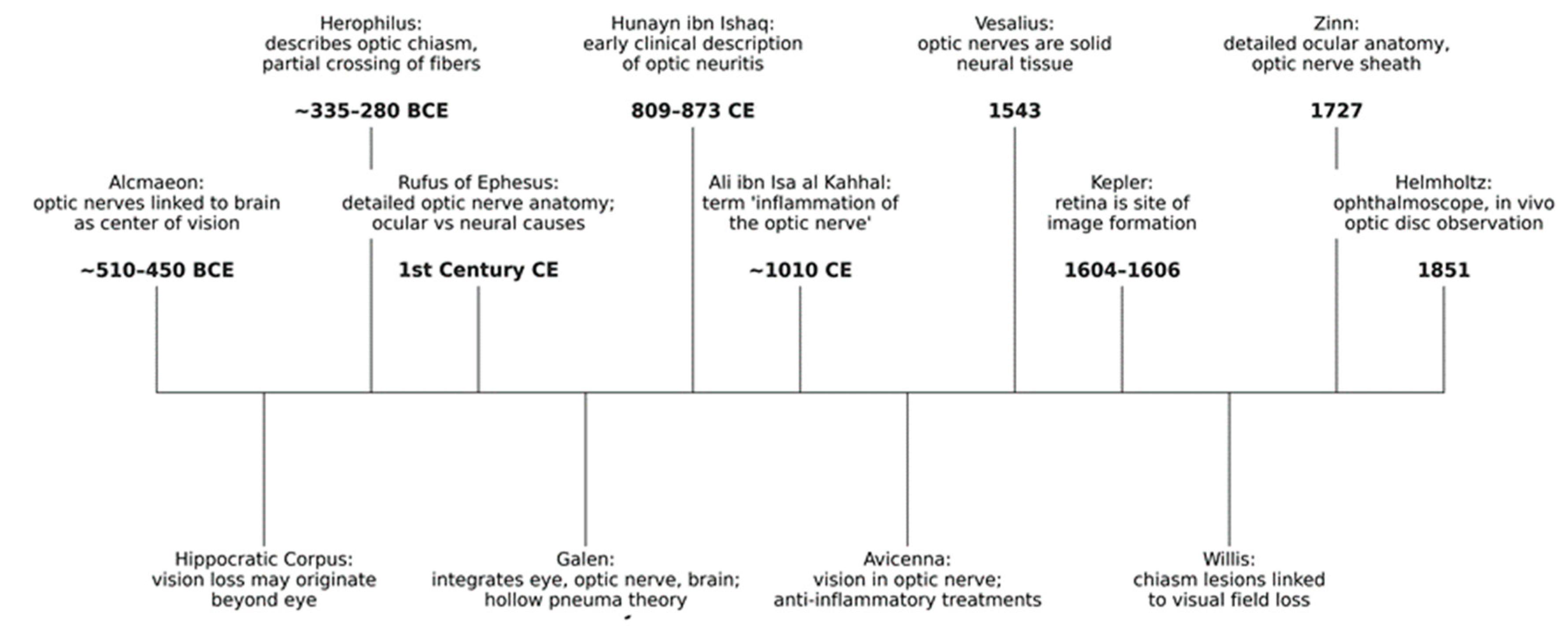
Figure 2.
Hunayn ibn Ishaq 9th century CE description of the eye diagram in a copy of his book, Kitab al-Ashr Maqalat fil-Ayn (“Ten Treatises on the Eye”), in a 12th century CE edition. Source: Wikipedia [14].
Figure 2.
Hunayn ibn Ishaq 9th century CE description of the eye diagram in a copy of his book, Kitab al-Ashr Maqalat fil-Ayn (“Ten Treatises on the Eye”), in a 12th century CE edition. Source: Wikipedia [14].

Figure 3.
The book Tadhkirat al-kahhalin (“Memorandum for Oculists”) (1010) by Ali Ibn Isa al-Kahhal in which the expression “Inflammation of the optic nerve” was first recorded. Source: Wikipedia [15].
Figure 3.
The book Tadhkirat al-kahhalin (“Memorandum for Oculists”) (1010) by Ali Ibn Isa al-Kahhal in which the expression “Inflammation of the optic nerve” was first recorded. Source: Wikipedia [15].

Figure 4.
Magnetic Resonance Imaging Features of Immune-Mediated Optic Neuritis Subtypes. T1-weighted, gadolinium-enhanced orbital MRI illustrating characteristic patterns across immune-mediated optic neuritis. (A,B) MS-ON: axial (A) and coronal (B) views showing a short, focal enhancing lesion of the left optic nerve. (C,D) MOG-IgG–seronegative CRION: axial views demonstrating bilateral, longitudinally extensive enhancement of the optic nerves. (E–G) MOG-ON: axial views showing optic nerve sheath enhancement (E) and enhancement of the posterior orbital perioptic tissues (F), with coronal confirmation of perioptic enhancement (G). (H–L) AQP4-ON: sagittal view (H) showing a longitudinally extensive, thickened and enhancing optic nerve; axial views demonstrating bilateral optic nerve involvement with mild (I) and marked (J) enhancement; and axial (K) and coronal (L) views showing chiasmal involvement.
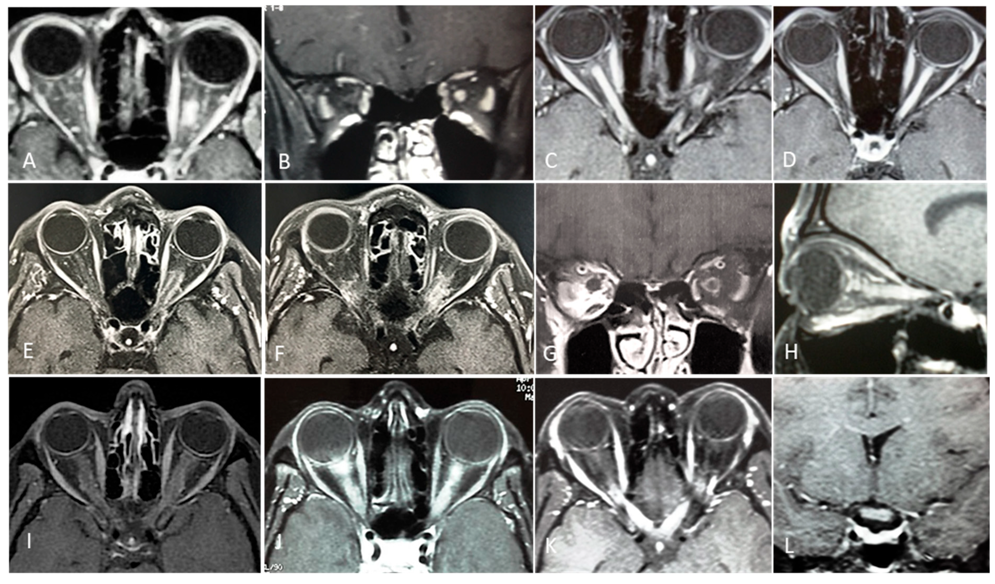
Figure 5.
Optical coherence tomography in immune-mediated optic neuritis subtypes (OS). Upper row: peripapillary retinal nerve fiber layer thickness; lower row: macular ganglion cell (GC) layer thickness. A. MS-ON; B. MOG-ON; C. AQP4-ON. The severity of axonal and ganglion cell loss is lowest in MS-ON, highest in AQP4-ON, and intermediate in MOG-ON. In MS-ON, the temporal quadrant of the pRNFL is preferentially and more severely affected.
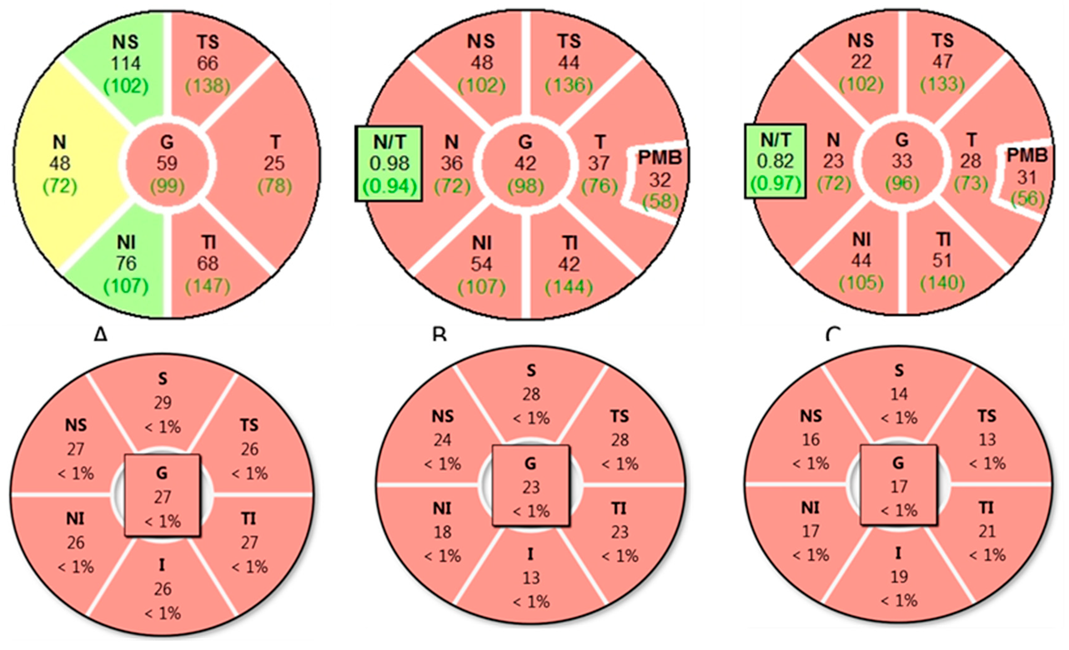
Figure 6.
Diagnostic flowchart for immune-mediated optic neuritis subtypes. RAPD – relative afferent pupillary defect; ICON – International Consensus on Optic Neuritis; CSF cerebrospinal fluid; OCB – oligoclonal bands; OCT – optical coherence tomography; VEP – visual evoked potential; NAION – non-arteritic anterior ischemic optic neuritis; ONSM – optic nerve sheath meningioma; AQP4-ON – aquaporin 4-associated optic neuritis; MOG-ON – myelin oligodencrocyte glycoprotein-associated optic neuritis.
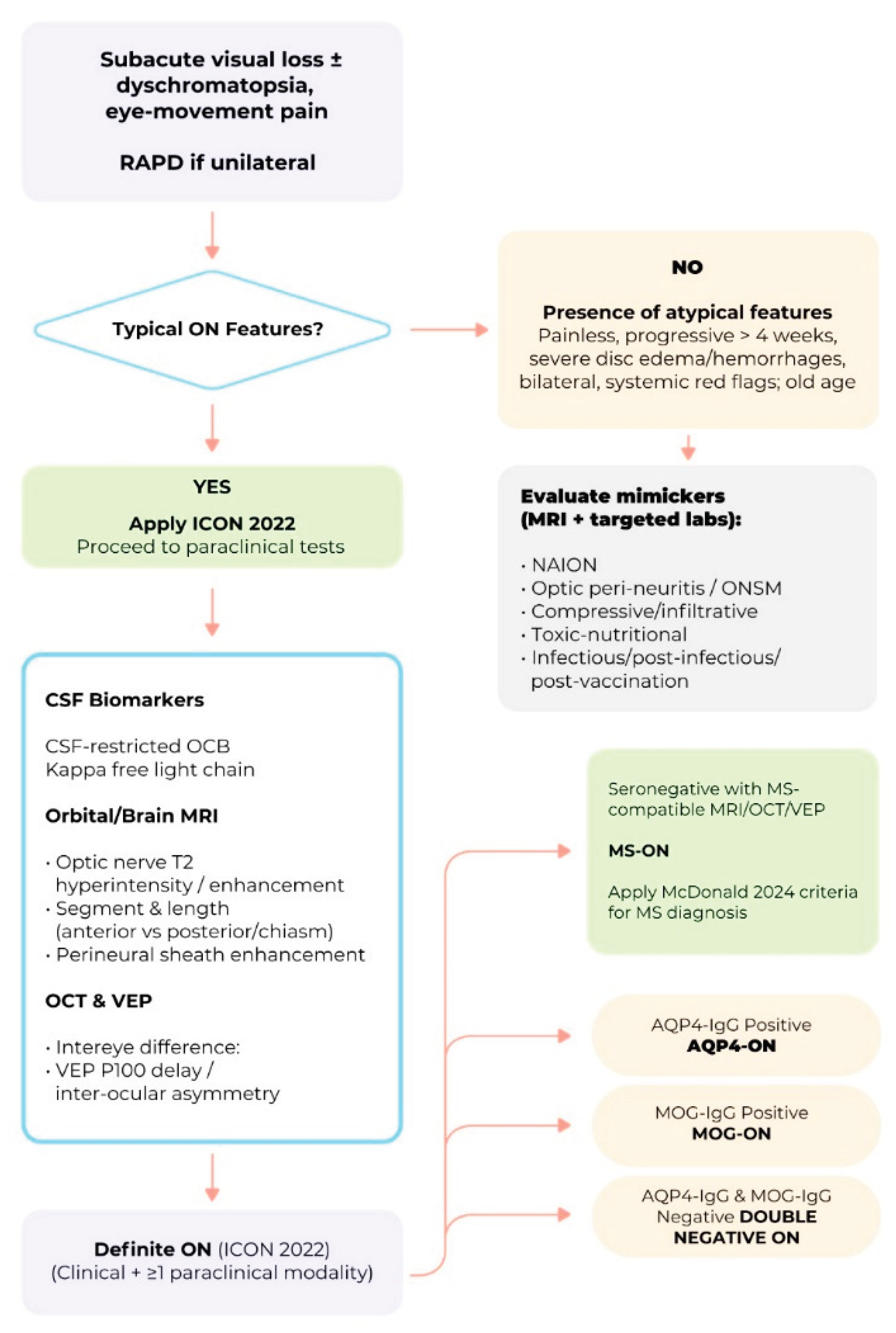

Table 1.
Baseline Demographic and Clinical Characteristics of Patients with Optic Neuritis Enrolled in the ONTT.
Table 1.
Baseline Demographic and Clinical Characteristics of Patients with Optic Neuritis Enrolled in the ONTT.
| Characteristics | Value |
| Number of patients | 457 |
| Age, mean (range), years | 32 (18–45) |
| Sex, female (%) | 352 (77) |
| Race (%) | White: 388 (85) Black: 64 (14) Other: 5 (1%) |
| Affected eye (%) | Right: 229 (50), Left: 228 (50) |
| Simultaneous bilateral involvement (%) | 7 (1.5) |
| Eye pain or headache associated with vision loss (%) |
420 (92) |
| Visual acuity in affected eye % | ≥20/20: 3 20/25–20/40: 6 20/50–20/190: 56 ≤20/200: 35; CF: 14; HM: 10; LP: 3; NLP: <1 |
| Visual acuity in fellow eye (%) | ≥20/20: 96 20/25–20/40: 3 ≤20/50: 1 |
| Color vision defect in affected eye (Hardy–Rand–Rittler plates) |
Median correct plates: 1/14 >90% abnormal, severe defect in 60% |
| Contrast sensitivity abnormality (Pelli–Robson chart) |
Mean log cs: 1.04 ± 0.37 >95% abnormal (<1.65) |
| Visual field defect in affected eye % | Central or cecocentral scotoma: 52 Diffuse depression: 29 Nerve fiber bundle defect: 8 Altitudinal defect: 3 Other: 8 |
| Afferent pupillary defect % | Present: >95 Severe: 47 Moderate: 34 Mild: 14 |
| Abbreviations: ONTT - Optic Neuritis Treatment Trial; APD - afferent pupillary defect; VA - visual acuity; LP - light perception; CS - contrast sensitivity. | |
Table 2.
Evolution of the McDonald’s Criteria and Role of Optic Neuritis in the Diagnosis of Multiple Sclerosis.
Table 2.
Evolution of the McDonald’s Criteria and Role of Optic Neuritis in the Diagnosis of Multiple Sclerosis.
| McDonald’s Criteria |
Key Revisions to McDonald Criteria |
Impact of Optic Neuritis on MS Diagnosis |
References |
| 2001 | First introduction of McDonald criteria; allowed MS diagnosis after a single clinical attack (e.g.,ON) with MRI evidence of DIS and DIT. |
ON recognized as a possible first demyelinating event; MRI could confirm MS diagnosis if DIS and DIT criteria met. | [69] |
| 2005 | Revised criteria allowed DIT to be demonstrated by a new T2 or gadolinium-enhancing lesion on follow-up MRI, replacing the need for clinical evidence of a second attack. |
ON remained a valid first demyelinating event; follow-up MRI showing new lesions could expedite MS diagnosis. | [70] |
| 2010 | DIT could be demonstrated by simultaneous presence of gadolinium-enhancing and non-enhancing lesions on baseline MRI. Allowed earlier diagnosis without waiting for new clinical attacks or follow-up scans. |
ON with baseline MRI fulfilling DIS and DIT criteria could establish MS diagnosis immediately. | [71] |
| 2017 | CSF-specific OCBs accepted as an alternative to demonstrating DIT. DIS still required. |
ON plus MRI showing DIS and positive CSF OCBs could confirm MS diagnosis without requiring DIT. | [72] |
| 2025 | Advanced imaging incorporated: CVS and PRLs recognized as supportive MRI features; inclusion of OCT parameters; CSF KFLC accepted as alternative to OCBs; broader lesion topography definitions. |
ON diagnosis can be supported by advanced MRI markers (CVS, PRLs), OCT evidence of retinal nerve fiber layer loss, and CSF KFLC positivity, enabling earlier and more accurate MS diagnosis when combined with DIS criteria. | [73] |
| Abbreviations: ON - optic neuritis; DIS - dissemination in space; DIT - dissemination in time; CSF - cerebrospinal fluid; OCB - oligoclonal bands; KFLC - kappa free light chains; CVS - central vein sign; PRLs - paramagnetic rim lesions; OCT - optic coherence tomography. | |||
Table 3.
Types of Optic Neuritis According to Etiopathogenic Mechanisms.
| Type | References | |||
| Infectious ON | [5,121,122,123] | |||
| Immune-mediated ON | ||||
| MS-related ON (Typical ON) | [2,35,37,38,39,40,41,42,43,44,45,46,47,48,49,50,51,52,53,55,56,57,58,59,61,62,63,117,118,124,125] | |||
| Non-MS-related ON (Atypical ON) |
||||
| AQP4-Related ON | [127,128] | |||
| MOG-Related-ON | [129,155] | |||
| Paraneoplastic ON (CRMP5-ON) |
[130,131] | |||
| GFAP-Related ON | [132,133,134,135,136] | |||
| Glycine Receptor-Related ON | [137,138,139] | |||
| Post-infection-ON | [140,141] | |||
| Post-vaccination ON | [142,143,145,166] | |||
| Recurring Idiopathic ON RION |
[146,147,148] |
|||
| CRION | [149,150,151] | |||
| ON in systemic autoimmune disorders | [152,153,154] |
|||
| Other immune-mediated ON of undetermined etiopathogenesis | [6,152] |
|||
| ON - optic neuritis; AQP4 - aquaporin-4; MOG - myelin oligodendrocyte glycoprotein; CRMP5 - collapsin response-mediator protein 5; GFAP - glial fibrillary acidic protein; RION - relapsing isolated optic neuritis; CRION - chronic relapsing inflammatory optic neuropathy. | ||||
Table 4.
Distinctive features of Immune-mediated Optic Neuritis Subtypes.
| Feature | MS-ON | AQP4-ON | MOG-ON | Key References |
| Ocular Pain (%) | ~10% | Lower than MOG-ON | 62.5% ; higher than AQP4-ON | [199,228] |
| Severity of VA Loss (Acute) | Mild-moderate | Severe, often profound | Severe, but often recovers | [198,199,240,248] |
| Mean Final VA | 20/25–20/40 | Counting fingers or worse | 20/25 | [199,240,249] |
| Response to Treatment | Good, steroid-responsive | Poorer, limited recovery | Good, steroid-responsive | [198,199,240,248] |
| Optic Disc Edema (Acute, %) | <33% | ~9% | 53–91% | [199,250] |
| Optic Nerve Extensive Lesion (%) | 15 | 64 | 53 | [251] |
| Perineuritis (%) | Rare | Rare | 33 | [198,251,252] |
| Chiasmal Involvement (%) | 15 | 20-64 | 5-16 | [250,251] |
| MRI Lesion Location | Anterior, unilateral | Posterior, bilateral | Anterior, bilateral | [251,253] |
| Optic Nerve Enhancement | Focal, short | Longitudinal, posterior | Longitudinal, anterior | [251,253] |
| Other MRI Features | Brain lesions common | Spinal cord, chiasm | Isolated ON, less brain involvement | [199,253] |
| Mean pRNFL (μm) | ~87.5 | 58–64 | 58–75 | [249,253,254,255,256] |
| Mean GCIPL (μm) | Higher than AQP4/MOG | Lower than MS | Lower than MS | [257] |
| OCT Pattern of Loss | Thinner RNFL, less severe | Severe thinning | Severe thinning, but better preserved | [254,257] |
| OCT-A Differential | Not well defined | Reduced vessel density | Reduced vessel density | [256] |
| CSF OCB (%) | 77-95 | 10-30 | 7-17 | [198,230,258,259,260,261] |
| CSF Pleocytosis (cells/μL) | Rare | 25% | 37.5% | [228,262] |
RNFL - retinal nerve fiber layer; GCIPL - ganglion cell inner plexiform layer; OCT-A - optical coherence tomography angiography; CSF - cerebrospinal fluid; OCB - oligoclonal bands.
Table 5.
Diagnostic criteria for Chronic Relapsing Inflammatory Optic Neuropathy *.
| HISTORY | OPTIC NEURITIS AND AT LEAST ONE RELAPSE |
| CLINICAL | Objective evidence for loss of visual function |
| LABORATORY | AQP4-IgG seronegative status |
| MRI | Contrast enhancement of the acutely inflamed optic nerves |
| TREATMENT | Response to immunosuppressive treatment and relapse on withdrawal or dose reduction |
| *[150,264] | |
Table 6.
Systemic Autoimmune Diseases Associated with Optic Neuritis.
| Disease | Report Frequency * | Typical ON Pattern / Comment | Key References (DOI) |
| Sarcoidosis | Frequent | Granulomatous ON often chiasmal and leptomeningeal involvement. | [278] |
| Systemic lupus erythematosus | Frequent | Severe, ON or ischemic neuropathy; often bilateral and poor visual outcome. | [279] |
| Primary Sjögren’s syndrome | Frequent | ON or optic neuropathy possibly preceding sicca; may overlap with NMOSD. | [280] |
| Behçet’s disease | Rare | Uveitis and retinal vasculitis typical; ON rare but documented. | [281] |
| ANCA-associated vasculitis (GPA/MPA/EGPA) | Rare | Orbital inflammation or vasculitic ON, sometimes bilateral. | [282] |
| IgG4-related disease | Rare | Optic perineuritis or sheath involvement; enhancement along optic nerve; steroid responsiveness. | [283] |
| Polyarteritis nodosa | Rare | ON or Ischemic neuropathy. | [284] |
| Takayasu arteritis | Rare | ON only occasionally reported. Mainly ischemic optic neuropathy secondary to large-vessel stenosis. | [285] |
| Drug-induced ON (TNF-α or checkpoint inhibitors) | Rare | Immune-mediated ON after biologic therapy (e.g., infliximab, nivolumab). | [286] |
| Primary antiphospholipid syndrome | Very rare | Vaso-occlusive or ischemic optic neuropathy; usually secondary to microthrombosis. | [287] |
| Giant cell arteritis | Very rare | True ON extremely rare. Arteritic AION dominant. | [288] |
| Cogan syndrome | Very rare | ON exceptional; typical ocular sign is interstitial keratitis. | [289] |
| Ankylosing spondylitis (HLA-B27) | Very rare | Uveitis typical; ON very rare. | [290] |
| Ulcerative colitis | Very rare | Immune-mediated ON after biologic therapy (e.g., infliximab, nivolumab). | [286] |
| Systemic sclerosis | Very rare | Sporadic optic neuropathy; likely ischemic. | [291] |
| Overlap or rheumatic disease (unspecified CTD) | Very rare | Mixed connective-tissue disease–related ON; anecdotal. | [292] |
| Kawasaki disease | Very rare | Mostly pediatric; isolated ON exceptional.. | [293] |
| Susac syndrome | Very rare | Retinal artery occlusions typical; ON not a core feature. | [294] |
| * Frequent: Supported by cohort studies and systematic reviews; optic nerve involvement appears in 3–10% of systemic cases. Rare: Documented in small but reproducible clusters — roughly a few dozen reported worldwide. Very Rare: Only isolated or single-case literature; prevalence likely <1% of total systemic cases. | |||
Table 7.
Diagnostic framework for sarcoidosis-related optic neuritis and optic perineuritis.
|
Diagnostic domain |
Definite S-ON/ S-OPN |
Probable S-ON/S-OPN |
Possible S-ON/S-OPN |
|
Clinical phenotype |
Acute or subacute optic neuritis or optic perineuritis with visual loss and/or periorbital pain; may coexist with uveitis, cranial neuropathies, or meningitic symptoms |
Same as definite | Same as definite |
| MRI findings | Gadolinium-enhancing optic nerve sheath or optic nerve lesion; often longitudinal, circumferential, or contiguous with meningeal enhancement (‘tram-track’/’doughnut’ signs) |
Same pattern; may be less extensive or limited to leptomeningeal enhancement | Suggestive but non-specific imaging pattern; no tissue confirmation |
|
Systemic and laboratory support |
Typical systemic sarcoidosis evidence (bilateral hilar/mediastinal lymphadenopathy, ↑ACE, ↑sIL-2R, ↑lysozyme); CSF lymphocytic pleocytosis or ↑protein; FDG-PET uptake in sarcoid-like distribution |
Same as definite | Suggestive systemic features or incomplete work-up |
|
Histological confirmation |
Non-caseating granulomas in neural tissue (optic nerve/sheath, meninges, brain, or ocular tissue) |
Non-neural tissue biopsy (e.g., lymph node, skin, salivary gland) showing non-caseating granulomas | No biopsy or non-diagnostic specimen |
|
Exclusion of mimics |
Infectious (TB, syphilis, fungi), neoplastic (meningioma, lymphoma), and immune-mediated (NMOSD, MOGAD, IgG4-RD, GPA) causes ruled out |
Same | Same |
| Level of certainty (Neurosarcoidosis Consortium tier) | Definite neurosarcoidosis | Probable neurosarcoidosis | Possible neurosarcoidosis |
| Clinical implication | Confirms sarcoid origin of optic neuropathy; guides long-term systemic therapy |
Supports diagnosis when neural biopsy unavailable; requires systemic evaluation | Suggests sarcoid etiology when all mimics excluded; biopsy pursuit recommended |
| S-ON - sarcoidosis-associated optic neuritis. S-OPN - sarcoidosis-associated optic perineuritis. ACE - angiotensin converting enzyme. sIL-2R serum interlekin-2 receptor, FDG-PET - fluorodeoxyglucose positron emission tomography. IgG4-RD-IgG4-related disease. GPA granulomatosis with polyangiitis. | |||
Table 8.
Vaccines Associated with Optic Neuritis.
| Vaccine Type | Evidence/Notes | References |
| Influenza (seasonal, H1N1) | Reviews and case series | [144,322] |
| Hepatitis B | Highly reported in VAERS ON | [144,323] |
| Human Papillomavirus (HPV) | Noted in VAERS and case reviews | [144,323] |
| Hepatitis A | Documented in reviews | [144] |
| Rabies | Documented in reviews | [144] |
| Measles, Rubella, Measles-Rubella | Case reports and reviews | [144,324] |
| Varicella Zoster (VZV) | Case reports | [325] |
| Yellow Fever | Documented in reviews | [144] |
| Anthrax | Case reports | [144,326] |
| Meningococcus | Documented in reviews | [144] |
|
Tetanus, Diphtheria, Pertussis (Tdap) |
VAERS, case reports, and reviews | [142,143,144,323] |
| COVID-19 (mRNA, J&J) | Cohort studies and case reports | [143,327,328] |
| Abbreviations: HPV - Human Papillomavirus; VAERS - Vaccine Adverse Event Reporting System; ON optic neuritis; VZV - Varicella Zoster ; Tdap - Tetanus, Diphtheria, Pertussis. | ||
Table 9.
Distinct MRI Features in MS-ON, AQP4-ON, and MOG-ON.
| Feature | MS-ON | AQP4-ON | MOG-ON | Key References |
| Laterality | Usually unilateral | Often bilateral | Often bilateral | [250,251,350] |
| Lesion Length | Short (<1/2 optic nerve) | Longitudinally extensive | Longitudinally extensive | [250,251,350] |
| Optic Nerve Head Swelling | Rare | Rare | Common | [250,251,350] |
| Chiasmal Involvement | Rare (5–15%) | Frequent (20–64%) |
Moderate (15–16%), often as part of long lesion | [250,251,350,351] |
| Optic Tract Involvement | Rare | Common (up to 45%) |
Rare | [250,251,350] |
| Lesion Location (Optic Nerve) | Retrobulbar, posterior | Posterior, intracranial, chiasmal, tract | Anterior, retrobulbar, optic nerve head | [250,251,350] |
| Brain Lesions | Common | Rare | Rare | [253,348,350,352] |
| Central Vein Sign (CVS) | Common | Rare | Rare | [253,353] |
| Enhancement Pattern | Focal, well-demarcated | Patchy, poorly demarcated | Fluffy, perineural, poorly demarcated | [348,350,353] |
| MRI - magnetic resonance imaging ; MS-ON - multiple sclerosis–associated optic neuritis; AQP4-ON – aquaporin 4-associated optic neuritis; MOG-ON - myelin oligodendrocyte glycoprotein antibody–associated optic neuritis; CVS - central vein sign. | ||||
Table 10.
Comparative OCT Characteristics in MS-ON, AQP4-ON, and MOG-ON.
| OCT Parameter | MS-ON | AQP4-ON | MOG-ON | Key-References |
| Typical chronic pRNFL thickness |
60–75 µm | 40–55 µm | 65–85 µm after single ON | [255,257,355,373] |
| Typical chronic GCIPL thickness |
55–70 µm | 57.4 ± 12.2 µm; −22.7 µm after first attack | 60–75 µm after first ON; 55–65 µm recurrent | [255,257,355] |
| Acute pRNFL swelling |
Mild–moderate | Variable | >150–200 µm | [360] |
| Pattern of thinning | Temporal-predominant | Diffuse, pan-retinal | Global, less temporal | [360,361] |
| Outer retinal layers (OPL/ONL) |
Preserved | No consistent thinning | Preserved | [255] |
| Subclinical (non-ON) eye involvement |
Frequent (3–5 µm GCIPL asymmetry) | Less frequent | Rare | [355,373] |
| Inter-Eye Difference thresholds |
GCIPL ≥4 µm; pRNFL ≥5 µm | Validated: pRNFL ≥5 µm (AUC 0.95) | GCIPL ≥7–8 µm or ≥8–10% asym | [358,364] |
| Structure–function relationship | Correlates with VA loss | Strong correlation | Dissociation possible (70–85% recover ≥20/30) |
[256,257] |
| Disease mechanism reflected in OCT |
Mixed demyelination + neurodegeneration (progressive component) |
Astrocytopathy with secondary neuronal loss |
Relapse-driven inflammatory demyelination with relative ganglion cell functional preservation | [255,257] |
| MS-ON - multiple sclerosis–associated optic neuritis; AQP4-ON – aquaporin 4-associated optic neuritis; MOG-ON - myelin oligodendrocyte glycoprotein antibody–associated optic neuritis; pRNFL - peripapillary retinal nerve fiber layer thickness; GCIPL - ganglion cell–inner plexiform layer thickness; IED - intereye difference; VA - visual acuity. | ||||
Table 11.
Optic Neuritis Diagnostic Criteria*.
| Component |
Description |
Key Elements / Thresholds |
| Clinical Entry Phenotypes | Defines clinical presentations consistent with ON. | A. Subacute monocular visual loss, pain with eye movement, dyschromatopsia, reduced contrast sensitivity, RAPD. B. Same as A but without pain. C. Bilateral involvement; note: RAPD unreliable. |
| Core Clinical Features Suggestive of ON | Used to support bedside impression of ON. | Subacute vision loss (hours–days); pain on eye movement (usually); impaired color vision; reduced contrast sensitivity; central/cecocentral scotoma; RAPD (if unilateral/asymmetric). |
| Paraclinical Modalities | Objective confirmation elevates diagnosis from possible to definite. | (1) OCT: - Acute optic disc edema OR - Inter-eye asymmetry within 3 months: • mGCIPL ≥4% or >4 μm • pRNFL ≥5% or >5 μm. (2) MRI (Orbits, Gd/T2): - Gadolinium enhancement or T2 hyperintensity within 3 months. (3) Biomarkers: - AQP4-IgG, MOG-IgG, CRMP5-IgG or - CSF-restricted OCB. |
| Diagnostic Categories | Defines how clinical + paraclinical criteria combine. | Definite ON: • (A) + ≥1 paraclinical test OR • (B) + ≥2 different paraclinical modalities OR • (C) + ≥2 paraclinical tests, one must be MRI. Possible ON: • Typical clinical presentation but insufficient evidence yet OR • Historical ON strongly suggested without contemporaneous testing. |
| Red Flags | Features requiring reconsideration of ON diagnosis to avoid misclassification. | Progressive visual decline >2 weeks; painless ON; absence of RAPD in unilateral cases; severe disc edema or early optic atrophy; simultaneous bilateral ON; retinal findings inconsistent with ON; systemic infectious signs; compressive or ischemic patterns. |
| *Modified from Petzold et al., 2022 [5]. ON - optic neuritis; RAPD - relative afferent pupillary defect; CS – contrast sensitivity; mGCIPL - macular ganglion cell-inner plexiform layer; pRNFL - peripapillary retinal nerve fiber layer | ||
Disclaimer/Publisher’s Note: The statements, opinions and data contained in all publications are solely those of the individual author(s) and contributor(s) and not of MDPI and/or the editor(s). MDPI and/or the editor(s) disclaim responsibility for any injury to people or property resulting from any ideas, methods, instructions or products referred to in the content. |
© 2025 by the authors. Licensee MDPI, Basel, Switzerland. This article is an open access article distributed under the terms and conditions of the Creative Commons Attribution (CC BY) license (http://creativecommons.org/licenses/by/4.0/).
Copyright: This open access article is published under a Creative Commons CC BY 4.0 license, which permit the free download, distribution, and reuse, provided that the author and preprint are cited in any reuse.


