Preprint
Review

This version is not peer-reviewed.

What’s in a Name? A Biochemical Perspective on the Microbial Luciferase-like Monoxygenase Family—The Bac_Luciferases by Another Name

Submitted:

11 December 2025

Posted:

15 December 2025

You are already at the latest version

Abstract

From a biochemical perspective, the recently introduced collective term luciferase-like monooxygenase family (LLM family) is notable for grouping together bacterial enzymes with fundamentally different functional characteristics. Thus not only does the family include non-bioluminescent and well as bioluminescent enzymes, but additionally both anoxybiontic and oxybiontic enzymes. By reviewing both the relatively short history of the LLM family itself, and the more protracted development of our present understanding of a number of the biochemically disparate composite enzyme groups, alternative representational descriptors can be identified that better serve both to succinctly delineate and functionally characterise the discrete groups currently corralled into the LLM family.

Keywords: 
;  ;  ;  ;  ;  

1. Introduction

‘What’s in a name?’. These words, included in the text of William Shakespeare’s 1597 play ‘The tragedy of Romeo and Juliet’, are used in context to express the sentiment that a name is merely a label, and does not define the essence or inherent qualities of a person or thing. Considered from the perspective of current de facto and in silico knowledge of bacterial enzymology, the same sentiment is epitomised by contradictions that arise from corralling functionally disparate enzymes under the same descriptor.
Traditionally, prokaryotic enzymes have been classified de facto, principally on the basis of the recorded biochemistry (chemical reactions) they perform [1]. However, following the development of relevant in silico technologies [2], emphasis has changed to a greater dependence on structure-based perceived evolutionary relationships (molecular evolution analysis). While classifications based on extant outcomes always generate useful empirical relationships, and may additionally promote some limited understanding of the relevant determinants of the activity, they perform poorly in predicting the biochemistry of novel enzymes [3]. Conversely, it has been claimed that a major advantage of the in silico technologies is that the insights provided by tracing the evolutionary history of enzymes can better identify and understand major determinants of their functionality [4].

2. The Luciferase-like Monooxygenase Family (= Bac_Luciferases)—Issues of Perspective

Over the last decade, a small number of bacterial enzymes reported as luciferase-like monooxygenases (LLMs) have been isolated and confirmed to be obligately oxybiontic FMN-dependent enzymes deploying FMNH2 as a cosubstrate, but which do not exhibit any reported evidence of bioluminescence (Figure 1A-C). As LLMs, they belong as members of a structurally-related in silico family, the LLM family [5], given the short name Bac_luciferases. The descriptor Bac_luciferases is used specifically to signify the inclusion of the bacterial luciferases, exclusively prokaryotic FMN-dependent monooxygenases, acknowledged as being the emblematic members of the LLM family. However, in addition to obligate functional dependencies on both molecular oxygen and FMNH2 as joint cosubstrates [9], the canonical feature of the bacterial luciferases, signified by their trivial name, is bioluminescence [10] - the emission of visible light (λmax 490 -500nm [blue-green light], when studied in vitro).
As well as the bioluminescent bacterial luciferases, the LLM family comprises two types of non-bioluminescent prokaryotic enzymes. The minority, like the activities illustrated in Figure 1A-C, are FMN-dependent monooxygenases able to promote a limited range of oxybiontic reactions without the emission of visible light. However, the majority are in silico identified redox enzymes associated with anaerobic biochemistry, a number of which have been purified and functionally characterised as anoxybiontic F420-dependent oxidoreductases for which the deazoflavin serves as an enzyme-bound prosthetic group [11]. In turn, this does raises two relevant and interrelated questions:
Excepting the bacterial luciferases, how ‘luciferase-like` are the other listed LLM family entries? How valid is the LLM family as a concept? To answer these questions requires more extensive knowledge of both the bacterial luciferases and related prokaryotic oxybiontic enzymes, and the LLM family.

3. Bacterial Luciferases—A Historical Overview

The word luciferase (from the Latin word lucifer, meaning ‘lightbearer’) has a long history in traditional biochemistry, the foundations of which are predicated on the successive discoveries of the relevant functional roles of oxygen by Antoine Lavoisier in 1777 [12], and enzymes (diastase – a mixture of amylases) by Anselme Payen in 1833 [13]. The first ever use of the word luciferase in the scientific literature was in an 1887 publication by the French pharmacologist Raphael Dubois, as a direct result of his extensive studies on the characteristic bioluminescence exhibited both by the marine bivalve mollusc Pholas dactylus and the firefly Photinus pyralis [14]. In each case, extensive testing of combinations of both hot- and cold-water extracts enabled Dubois to propose a novel terminology specifying that recorded light generation resulted from the interaction between a relevant enzyme (luciferase), a relevant substrate (luciferin),and molecular oxygen:
luciferase + luciferin + O2 ―――> hv (emitted light = bioluminescence)
A revised version of that simple relationship [15] remains valid to the current day:
luciferin + O2 ――――――> (P)* ――――> P + hv
luciferase excited product product emitted light
In the 140+ years that have elapsed following Dubois’s pioneering studies, extensive research has confirmed that bioluminescence has evolved independently at least 40 times, and is consequently, distributed throughout many different genera [16], for which this oxybiontic luciferase-promoted emission of visible light is deemed to be the canonical feature [17]. This includes seven genera of bioluminescent bacteria, distributed within the Vibrionaceae, Shewanellaceae, and Enterobacteriaceae, these being three families of Gammaproteobacteria [18]. Notably, there is a remarkable consistency to all aspects of the established biochemistry and enzymology of bacterial luciferases. They all function as monooxygenases (EC 1.14.14.3) deploying dioxygen, FMNH2 and the same molecular type of luciferin as cosubstrates to promote a sequence of electron exchanges exhibiting remarkable biochemical unity, thereby serving to generate the same oxygenated product type accompanied by the emission of visible light. Although it has only proved possible to solve the definitive crystal structure of the luciferase from Vibrio (Aliivibrio) harveyi [19,20], there are some other less detailed elements of structural equivalence recognised within the better studied (>20) bacterial luciferases [21], Thus, they are all α/β heterodimers, and while catalytic activity is confined to the α-subunit, the β-subunit does serve an important function by conferring conformational stability to the dimeric complex. Consistently, both subunits fold into (β/α)8 TIM-barrel structures with equivalent internal symmetry. However, unlike bioluminescence, this particular structural feature should not be regarded as a definitive characteristic of the bacterial luciferases, because being of evolutionary ancient origin, it is the most common enzyme fold recorded in the Protein Data Bank database of known protein structures, and has been recorded in many different enzyme families catalysing a wide range of completely unrelated reactions [22,23].

4. De Facto to In Silico Progression—The Advance of the LLM Family

Conversely, the proposal for a LLM family has a much shorter history, having itself developed from the previously used similar term ‘bacterial luciferase family’, introduced in 2004 by Aufhammer et al. [24]. Despite the then current biochemical knowledge confirming that all known bacterial luciferases are heterodimeric oxybiotic FMN-dependent bioluminescent enzymes, the term was chosen to correlate a small number of previously recognised homodimeric anoxybiontic F420-dependent enzymes essential to promote methanogenesis by non-bioluminescent anaerobic prokaryotes such as Methanobacterium thermoautotrophicum [25]. Focussing on exemplar deazaflavin-dependent enzymes that could be sufficiently highly purified, Aufhammer et al.’s. initial 2004 study succeeded in establishing the crystal structure of the homodimeric secondary alcohol dehydrogenase Adf from Methanocellus thermophillicus. They quickly followed this up by establishing the crystal structure of the homotetrameric methylenetetrahydromethanopterin reductase bMer from Methanosarcina barkeri [26]. Further, despite being of limited relevance (vide supra), in both cases Aufhammer et al. made specific reference to significance of the shared similarity of the (β/α)8 TIM-barrel structures of these anoxybiontic enzymes with the established crystal structure of the oxybiontic luciferase from V. harveyi [19] to substantiate his proposal for the bacterial luciferase family
Following Aufhammer et al.’s initial proposal, the related concept of a larger LLM family to collate an expanded group of deazaflavin- and flavin-dependent enzymes subsequently evolved out of a 2010 study by Selengut and Haft [5]. They developed an in silico phylogenetic profiling programme (PPP) based on the known genomic information for the biosynthetic pathway enzymes for F420 present in the facultative anaerobic prokaryote Mycobacterium tuberculosis and other related actinobacteria. The outputs suggested by PPP were then reviewed with the aim of constructing defining protein families based on full-length multiple sequence alignments. This resulted in the identification of a total of 63 top hits belonging to three homology families, each of which included known (deaza)flavin-binding proteins from archaea, actino- and other eubacteria. Because one of the entries in the largest homology family (44 entries) was the luciferase from V. harveyi, all 44 were collectively named the LLM family. It comprised either F420- or FMN-dependent enzymes exhibiting differing degrees of similarity to V. harveyi luciferase based on further analysis using Selengut and Haft’s additional SIMBAL sequence analysis tool. The resultant data indicated that the larger subset of the LLM family comprised anoxybiontic F420-dependent redox enzymes that deploy the reduced deazaflavin as a prosthetic group tightly bound within the TIM-barrel fold. The remainder were all oxybiontic enzymes. Of these, with the exception of the atypical monooxygenase from Acinetobacter baumanii that can function equally effectively with both FMNH2 and FADH2 [27], the others were monooxygenases that bind FMNH2, sourced from a separate flavin reductase, as an active site-bound cosubstrate. Relevant data for the LLM family (short name Bac_luciferase) was subsequently lodged on-line with the intention that it would be further developed into the continuously updated catalogue InterPro IPRO036661 (Pfam PF0296, PROSITE PD016048, SCOP2, and 1nfp).
When considering the LLM family from a conventional biochemical standpoint, the FMN-dependent monooxygenases are the better characterised subset. In addition to the bacterial luciferases (EC 1.14.14.3; vide supra), the easily purified 4-hydroxyphenylacetate 3-monooxygenase from Acinetobacter baumanii (EC 1.14.14.9 [27]), alkanesulfonate monooxygenase (SsuD) from Escherichia coli (EC 1.14.14.5; [28]), and nitrilotriacetate monooxygenase (NTA-MO) from Chelatobacter heinzi (EC 1.14.14.10 [29]) have each been extensively researched (vide infra). While all of these oxybiontic enzymes share much common biochemistry, the bacterial luciferases are unique in being the only bioluminescent members of the entire LLM family. Although historically less well studied, a number of recent reviews have helped considerably to consolidate the known biochemistry and enzymology of several F420-dependent members of the LLM family [11,30], confirming some idiosyncratic mode of action characteristics. The two predominant enzyme types are methylene-H4MPT reductases (MERs) and glucose-6-phosphate dehydrogenases (FGDs). In strict contrast to the FMN-dependent subset which are oxybiontic enzymes that deploy the flavin as a cosubstrate, the F420-dependent subset are anoxybiontic redox enzymes that deploy the deazaflavin as an active site-bound prosthetic group/cofactor. In turn, these significant functional differences between the F420-dependent and FMN-dependent subsets of the LLM family provide directly relevant perspectives that resonate with the previously raised questions concerning the validity of the currently constituted LLM family (vide supra). Interestingly, two recent reviews focussed on relevant in silico issues have both commented on the difficulty of bringing together these two diverse groups of enzymes that were shown by molecular evolution profiling to have diverged from a common ancestor [4,31]. Viewed from a functional perspective, equivalent inferred divergent evolutionary relationships of the LLM family members can be suggested (Figure 2) taking into account the biochemical characteristics of bioluminescence vs non-bioluminescence, FMN- vs F420-dependence, and oxybiontic vs anoxybiontic dependency. The differential biochemical relationship to dioxygen itself reflects the ancient defining impact that resulted from the evolution of oxygenic photosynthesis in the ancestors of modern cyanobacteria, an outcome that served to transform the environmental landscape dramatically and irreversibly [32]. These divergent relationships and associated epistasis (evolutionary entrenchment; [33,34,35,36]), argue strongly that the current concept of a unified luciferase-like family is misleading, and should be replaced by separate terminologies that clearly signal the significant biochemical differences between the two diverse groups of enzymes. While the descriptor ‘F420-dependent oxidoreductases’ accurately serves such a role for the predominant non-bioluminescent anoxybiontic subgroup, reviewing in greater depth the development of our contemporary understanding of the biochemistry of the bioluminescent and non-bioluminescent FMN-dependent monooxygenases will serve to identify an equivalent descriptor that suitably reflects the distinct unifying functional characteristics of the smaller oxybiontic subgroup.

5. The Emergence of a Functional Understanding of the Bacterial Luciferases

It was mankind’s enduring fascination with the phenomenon of bioluminescence, traceable back to earliest recorded observations of ‘cold fire’ by the Ancient Greek philosophers Aristotle and Pliny the Elder [37], that inevitably resulted in the luciferases being the first FMN-dependent monooxygenases to be fully characterised biochemically. However, the current detailed understanding has a long and chequered history that has included a number of significant changes in both perception and nomenclature along the way. While Dubois’s pioneering1887 luciferin-luciferase research was eventually confirmed nearly four decades later by E. Newton Harvey using hot- and cold-water extracts of various species of fireflies [38], serial attempts throughout the first half of the 20th century to conclusively demonstrate bioluminescence with equivalent extracts of various relevant bacteria all reported negative results [37]. However, finally in 1953 bioluminescence was demonstrated but only after specifically adding NADH to a cell-free extract of the luminous bacterium Achromobacter (= Vibrio) fischeri [39]: further, the recorded spectrophotometric changes suggested an undefined involvement of flavin nucleotide biochemistry. A flurry of research activity then quickly confirmed that bioluminescence by the system was stimulated a further 7-fold by the combined addition of FMN [40] plus an extract sourced from powdered kidney cortex termed ‘kidney cortex factor’ [41]. KCF was then subsequently identified as hexadecanal [42], an authentic sample of which was shown to substitute for KCF in promoting bioluminescence in the cell-free extracts of the bacterium. Additional directly related research then further confirmed that hexadecanal could be substituted with any one of the series of C7 to C15 saturated straight-chain aldehydes, albeit with varying degrees of effectiveness: in each case, production of the corresponding carboxylic acid was confirmed [43]. Collectively, this series of outcomes served to generate the consensus seminal proposal that contra to Dubois’s suggestion that ‘luciferase’ was a single functional entity (vide supra), the activity of this novel bacterial system resulted from two separate cooperating enzyme systems - an FMN reductase and a separate biooxygenating enzyme which jointly promoted flavin-dependent bacterial bioluminescence [44].
FMN reductase
NADH + H+ + FMN ―――――――> NAD+ + FMNH2
O2-dependent enzyme
FMNH2 + O2 + ―――――――> FMN + CH3-(CH2)n-COOH +
CH3-(CH2)n-CHO H2O + hv
This important outcome from seven decades ago represents the origin of the recognition that bacterial luciferases function biochemically as two-component oxygen-dependent enzymes (TC-ODEs). In turn this further served to distinguish the V. fischeri luciferase from ‘mushroom phenolase’, a contemporaneously reported but clearly functionally different single-component monooxygenase [45]. While the TC-ODE concept remains equally valid to this day, the suggestion that it could serve as a possible descriptor to collate the bacterial luciferases and the other related two-component FMN-dependent monooxygenases is deficient because it fails to convey other relevant definitive elements of functional information.
The next sequence of important developments that led to a more comprehensive understanding of bacterial luciferases were all greatly influenced by the outcomes of a concerted programme of relevant structural and functional research initiated by the American biochemist Woodland Hastings, a colossus in the field of bacterial bioluminescence. His decade-long studies collectively served to define the molecular mode of action of the bacterial luciferases. From a structural point of view, his initial seminal contribution established that the O2-dependent component of the luciferase activity detected V fischeri (LuxABVf) was a 1:1 α/β heterodimer [46], an outcome that has proved to be a characteristic of all other subsequently studied bacterial luciferases [21]. Functionally, comparisons with existing precedents [47] enabled him to further propose that the dimeric biooxygenating protein should be formally classified as a monooxygenase. Additionally, an extensive spectrophotometric study [48] undertaken in the presence and absence of the thiol group inhibitors sodium arsenite and iodoacetic acid encouraged Hastings to advance a proposal that flavin-dependent peroxidation of the biooxygenating enzyme itself would prove to be a key feature of bacterial bioluminescence. However, the biochemistry of flavins is complex as reflected by the uncertainty that has thwarted efforts to understand relevant mechanisms [9]. Hastings’ initial suggestion was that the sole role of FMNH2 was to reduce a cysteine-cysteine disulfide bridge of the α-subunit of luciferase to generate the equivalent dithiol enzyme that he termed Intermediate I. Subsequent peroxidation of one of the resultant thiol groups by dioxygen would then yield Intermediate II, which further interacted with the aldehyde substrate to generate the transitory unstable Intermediate III. Finally, the rapid decay of Intermediate III resulted in both the emission of visible light and a return of the luciferase to a disulfide-bridged protein. Concommitantly, a significant feature of this initial 1964 proposal was the absence of any suggested direct involvement of the flavin cosubstrate in the relevant aldehyde oxygenation step. However, after further extensive studies with more highly purified enzyme activities isolated from the same bacterium, Hastings then put forward a significantly different proposal (Figure 3; [49]) that redefined the roles of FMNH2, luciferase, and Intermediates I - III.
The novel feature of this proposal was that for the first time a specific defined role was proposed for peroxidation biochemistry of the FMNH2 bound as a cosubstrate in the active site of the luciferase. It was envisaged that the biooxygenating subunit functioned with the reduced flavin nucleotide delivered by the FMN reductase to generate Intermediate I, which then reacted directly with dioxygen to generate a key nucleophilic peroxyflavin corresponding to Intermediate II. Further active site interaction with the aldehyde substrate then generated the transitory unstable Intermediate III. Convinced of the validity of the newly defined roles of each of the Intermediates in this revised outline schematic, Hastings and Eberhard then deployed an extensive stopped-flow spectrophotometric kinetic study to refine and expand these proposals further (Figure 4; [50]). They reiterated the key role played by the formation of Intermediate II, which they chemically characterised more fully as a C(4a)-peroxyflavin anion. In turn, the anion then served to directly react with n-hexadecanal, the archetypal aldehyde substrate of the luciferase, thereby forming a C(4a)-peroxyhemiacetal (Intermediate III). Intermediate III then decomposes to release the corresponding carboxylic acid and an excited state of the C(4a)-hydroxyflavin intermediate, which in turn emits blue-green light on returning to the ground-state species. Significantly, this proposed biochemistry that characterises the emission of visible light by bacterial luciferases remains valid nearly five decades later [51]. Hastings considered that this proposed multi-step mechanism bore some similarity to the schematic previously suggested by Rudolf Criegee in1948 to explain the abiotic oxidation of ketones to their corresponding esters by peracids (Figure 5; [52], a chemical reaction first reported by Adolf von Baeyer and Victor Villiger in 1899 [53]. Criegee’s model envisaged a chemical oxidation that resulted in the cleavage of the bond between a carbonyl carbon and a neighbouring carbon atom, followed by the subsequent insertion of an oxygen atom. By analogy, Hastings’ proposed LuxABVf luciferase model deploys a C(4a)-peroxyflavin intermediate to biooxidise n-hexadecanal to the corresponding carboxylic acid.This suggested mechanism comprised a progressive sequence of electron transfers and rearrangements that involved the cleavage of the bond between the terminal carbon and hydrogen atoms of the aldehyde. While acknowledging that his proposal did differ from Criegee’s model in some respects, Hastings was sufficiently persuaded in his own mind to commence referring to bacterial luciferases as Baeyer-Villiger monooxygenases (BVMOs).

7. What’s in a Name? The Established Value of FD-TCMO as a Moniker, and Its Relevance to Further More Recent Studies

Putting into perspective the last 70+ years of the history of investigating the molecular mode of action of the bacterial luciferases and other directly related enzymes, Ellis’s term FMN-dependent two-component monooxygenases (FC-TCMOs), included in her seminal 2010 review [90], clearly serves as a valuable collective descriptor (Table 2) for the FMN-dependent subgroup currently included within Selengut and Haft’s luciferase-like monooxygenase (LLM) superfamily [5]. Compared to the three previously introduced alternatives (TC-ODEs, Type II BVMOs, and TC-FDMs) the FD-TCMOs is notable for providing the most comprehensive and accurate summary of the key biochemical characteristics that unify both the relevant bioluminescent and non-bioluminescent prokaryotic enzymes.
Further support for the representational value of Ellis’s FD-TCMO moniker can be garnered by reviewing a number of post-2010 studies conducted on functionally characterised two-component prokaryotic enzymes that were reported at the time to be LLM homologues. These are oxybiontic enzymes almost exclusively reported to catalyse reactions of secondary metabolism, being induced exclusively during idiophasic growth of the competent bacterial species. Monooxygenases of various different functional types serve a wide variety of real and proposed roles in microbial secondary metabolism [97], and while some of these activities correspond to extant functionally active proteins, exclusively induced during idiophasic growth, others are encoded in cryptic or silent genes initially detected by in silico screening [98,99]. Considering firstly the relatively few examples of reported idiophasic LLMs that have been extensively studied in vitro, they all function biochemically in a way that conforms explicitly to Ellis’ definition as FD-TCMOs (Figure 1A-C). One such bacterial FD-TCMO-catalysed biotransformation has been confirmed as a key fully characterised step in the biosynthesis of the secondary metabolite neoabssyomicin B, a valuable bioactive antibiotic produced during idiophasic growth by various Streptomyces and Verrucosispora spp. (Figure 1A; [6,100,101]). The relevant genes (abmE2 and abmZ) have been isolated from Streptomyces koyangensis SCSIO 5802, over expressed in E.coli BL21, and the resultant monoxygenase (AbmE2) and flavin reductase (AbmZ) enzymes purified and confirmed to function cooperatively as a two-component NADH + FMN-dependent monooxygenase that efficiently bioxygenates abyssomicin 2 to neoabyssomicin B. In principle, this coordinated activity by AbmE2/AbmZ is equivalent to the cyclic lactonization activity of 2,5-DKCMO from P. putida first recorded by Gunsalus in the early 1960s (vide supra; Figure 5), an activity subsequently designated as a Type II BVMO [62] prior to the introduction of Ellis’s FD-TCMO definition in 2010 [90]. Similarly, a well researched biooxygenation is a key step in the biosynthesis of the secondary metabolite mensacarcin (Figure 1B), a proven powerful anti-tumor drug, by Streptomyces bottropensiss [7,102]. By initially constructing cosmid cos2 which included almost the complete type II polymerise synthase gene cluster, and then using selective deletion and complementation in a heterologous expression system, they were able to show that the msnO3-coded flavin reductase and the msnO8-coded monooxygenase function as an FD-TCMO to catalyse the final step in the biosynthesis of mensacarcin. Interestingly, this oxybiontic step results in the introduction of an epoxy group into the side chain of the relevant mensacarcin precursor. This outcome is notable, because although there are a very few isolated reports of epoxidation reactions undertaken by monomeric FAD-bound Class I BVMOs such as CHMO [103], it represents the first time that epoxidation has been reported as an attribute of an FMN-dependent TCMO. While the relevant mechanistic events in the active site of MsnO8 remain currently unknown, epoxidation and carbonate formation are recognised outcomes of some peracid-catalysed chemical Baeyer-Villiger rearrangements [104,105]. Related studies undertaken using a similar programme of research with S. bottropensis [8] have confirmed that an equivalent biooxygenation catalysed by the concerted activities of Rs101 and Rs102 promotes epoxidation as a key intermediary step in the biosynthesis of rishirilide B, one of a number of tricyclic aromatic Type II polypeptides produced exclusively during idiophasic growth by this bacterium.(Figure 1C). This consolidates the recognition that some reported LLMs function as confirmed FD-TCMOs in bacterial secondary metabolism.
As well as these characterised idiophasic FD-TCMOs for which both participating native enzymes of the functioning partnership have been identified, there are a number of other equivalent candidate activities originally reported as LLMs, but for which there is less definitive evidence for the requisite monooxygenase and/or flavin reductase moieties. An interesting illustrative example is the 2012 study by van der Donk’s research group on the biosynthesis of actagardine (Figure 14) and related lantibiotic secondary metabolites which included a reported LLM activity [106]. Actagardine, isolated from Actinoplanes garbadinensis [107], is a tetracyclic 19-amino acid ribosomally-synthesised peptide that includes four intramolecular thioether linkages (lanthionine bridges). Prior deletion-based genomic studies [108] have confirmed that the garO gene of A. garbadinensis codes for a corresponding GarO monooxygenase that catalyses the sulfoxidation of the 14-S-19 lanthionine bridge of deoxyactagardine to yield actagaradine, the only known lantibiotic containing a sulfoxide group. Based on their previous success in producing other prokaryotic lanthionone bridge-containing peptides by heterologously expressing the corresponding modification enzymes [109,110,111,112], van der Donk’s group firstly generated a source of GarO by cloning garO into a pET28b vector which was then expressed in E.coli [106]. The activity was then purified, and confirmed as monomeric by gel filtration. When the purified GarO monooxygenase was incubated with FMN, NADH and deoxyactagardine, and the stopped reaction mixture then analysed, ‘the mass of the resulting … peptide was increased by 16 Da, consistent with the formation of one sulfoxide group’. It was this outcome that prompted Shi et al.s’ claim [106]that ‘GarO is a luciferase-like monoxygenase that introduces the unique sulfoxide group of actagardine’. While prior studies have reported sulfoxidation as a confirmed activity of both DszC (EC 1.14.14. 21) and DszA (EC 1.14.14. 22), two FD-TCMOs isolated from Rhodococcus erythropolis, these are activities with a shared dependency on the same DszD flavin reductase as the obligate source of NADH (vide supra; Figure 9). However, no native A. garbadinensis flavin reductase was knowingly included by Shi et al. in their assay system, nor was the implied involvement of a flavin reductase activity in facilitating their recorded outcome addressed. So from that point of view, the reassignment of the reported sulfoxidation outcome as a definitive FD-TCMO activity remains problematical. In retrospect, it can be speculated that one or more of the known native flavin reductase enzymes of the E. coli expression system such as FreEc [113] could have cooperated with the GarO monooxygenase, thereby supporting the recorded sulfoxidation activity by a hybrid FD-TCMO. Such a proposed hybrid FD-TCMO would not be unprecedented, as 2,5-DKCMO was confirmed to function efficiently as a hybrid FD-TCMO when the relevant gene was expressed in E. coli and the lactonizing activity of the cloned monooxygenase then tested on a range of alicyclic ketones [114], although again the identity of the relevant flavin reductase(s) was not confirmed. In another recent and more extensively characterised relevant study, the hybrid FD-TCMO concept was confirmed with both purified 2,5-DKCMO and LuxABVf [115]. Highly purified preparations of both monooxygenases were confirmed to catalyse more efficient lactonization of (rac)-bicyclo[3.2.0]hept-2-en-6-one when functioning as hybrid FD-TCMOs coupled with a number of different non-native flavin reductases, including most effectively with FRDAa from Aminobacter aminovorans.
In addition to characterised FD-TCMOs coded for by corresponding genes in normally expressed bacterial genomes, such as SsuD from E. coli (EC 1.14.14.5 [28]), the current InterPro IPR0036661 catalogue contains many in silico-identified entries of proposed LLMs coded for by cryptic genes included in larger clusters corresponding to silent bacterial secondary metabolic pathways. Many of these cryptic genes remain unexpressed, so the functional status of the coded activities is currently not known. One interesting exception is OvmO, coded for by ovmo present in the normally silent ovm biosynthetic gene cluster of Streptomyces olivaceus SCS10 T05 that when activated by metabolic engineering resulted in the production of various lobophorins, anthrancenes and xiamycins [116]. Subsequently, the silent gene cluster was then mutated and reconstructed to generate both S. olivaceus SCS10 T05RXL, a triple-deletion strain mutated at the expense of the production of xiamycins, anthrancenes, and lobophorins, but which included an expressible copy of the ovmo gene, and the corresponding T05RXL/Δovmo strain which was additionaly devoid of the OvmO-coding gene [117]. After culturing the two reconstructed strains for 8 days in separate aliquots of ISP3 medium, the metabolites present in each spent fermentation broth were isolated and characterised. This established olimycin C as the major recovered metabolite produced by SCS10 T05RXL/Δovmo, whereas the directly corresponding ester was the major recovered metabolite produced by SCS10 T05RXL, along with detectible traces of the macrocyclic lactone olimycin A (Figure 15). This outcome prompted
Zhang et al. to suggest that OvmO is ‘a luciferase-like monooxygenase … that catalyses a Baeyer-Villiger oxidation’. While all subsequent attempts to clone and overexpress ovmo in E. coli BL21(DE3) proved unsuccessful, thereby thwarting any attempt to establish the functionality of the monooxygenase directly, in silico analysis indicated a close relationship to both 2,5-DKCMO from P. putida, and Rs101 from S bottropensis, two established FD-TCMOs (vide supra). As in the case of GarO from A. garbadinensis [106], Zhang et al. gave no evident consideration of the requisite involvement of a complementary flavin reductase activity to support their proposed OvmO monooxygenase activity in strain SCS10 T05RXL, thereby making the reassignment of the reported activity as a definitive FD-TCMO problematical. However, a number of constitutively-expressed flavin reductases have been reported subsequently in S. olivaceus [118]. This raises the possibility that the ’Baeyer-Villiger-type oxidation’ reported by Zhang et al. may have resulted from the expressed OvmO functioning as an FD-TCMO in cooperation with one or more of these native flavin reductases, although this suggestions awaits relevant investigation.
In conclusion, research endeavours undertaken across nearly seven decades, from initial trials with acetone-dried powders of whole bacterial cells in the mid-1950s [38] to the current use of selective gene deletion and complementation in heterologous expression systems [8,102], have served to establish the consensus biochemistry that consolidates the bacterial luciferases and functionally equivalent FMN-dependent two-component monooxygenases as a discrete group of prokaryotic oxybiontic enzymes. The value of the corresponding abbreviated descriptor - the FD-TCMOs - is that it succinctly emphasises those canonical biochemical characteristics that serve to unite the group members, and which distinguish them fundamentally from the functionally unrelated anoxybiontic F420-dependent redox enzymes.

Funding

This research received no external funding.

Data availability Statement

Not applicable.

Acknowledgments

A special acknowledgement must go to Raffaella Villa, the research student in my laboratory who was the first to confirm direct functional equivalence between a bacterial luciferase and a corresponding non-bioluminescent bacterial enzyme. The considerable help of Ajza Magamadova with generating the Figures in the manuscript is also gratefully acknowledged.

Conflicts of interest

The author declares no conflicts of interest.

References

  1. Nomenclature Committee of the International Union of Biochemistry and Molecular Biology. Enzyme Nomenclature. Academic Press: New York, NY, USA, 1992.
  2. Orengo, C.A.;Thornton, J.M. Protein families and their evolution - a structural perspective. Ann. Rev. Biochem. 2005, 74, 867-900. [CrossRef]
  3. Mascotti, M.L.; Kumar, H.; Nguyen, Q-T.; Ayub, M.J.; Fraaije,M.W. Reconstituting the evolutionary history of F420-dependent dehydrogenases. Scientific Reports, 2018, 8, 17571. [CrossRef]
  4. Harms, M.J.; Thornton, J.W. Evolutionary biochemistry: revealing the historical and physical causes of protein properties. Nat. Rev, Genet., 2013,14, 559-571. [CrossRef]
  5. Selengut, J.D.; Haft, D.H. Unexpected abundance of coenzyme F420-dependent enzymes in Mycobacterium tuberculosis and other Actinobacteria. J. Bacteriol., 2010, 192, 5788-5798. [CrossRef]
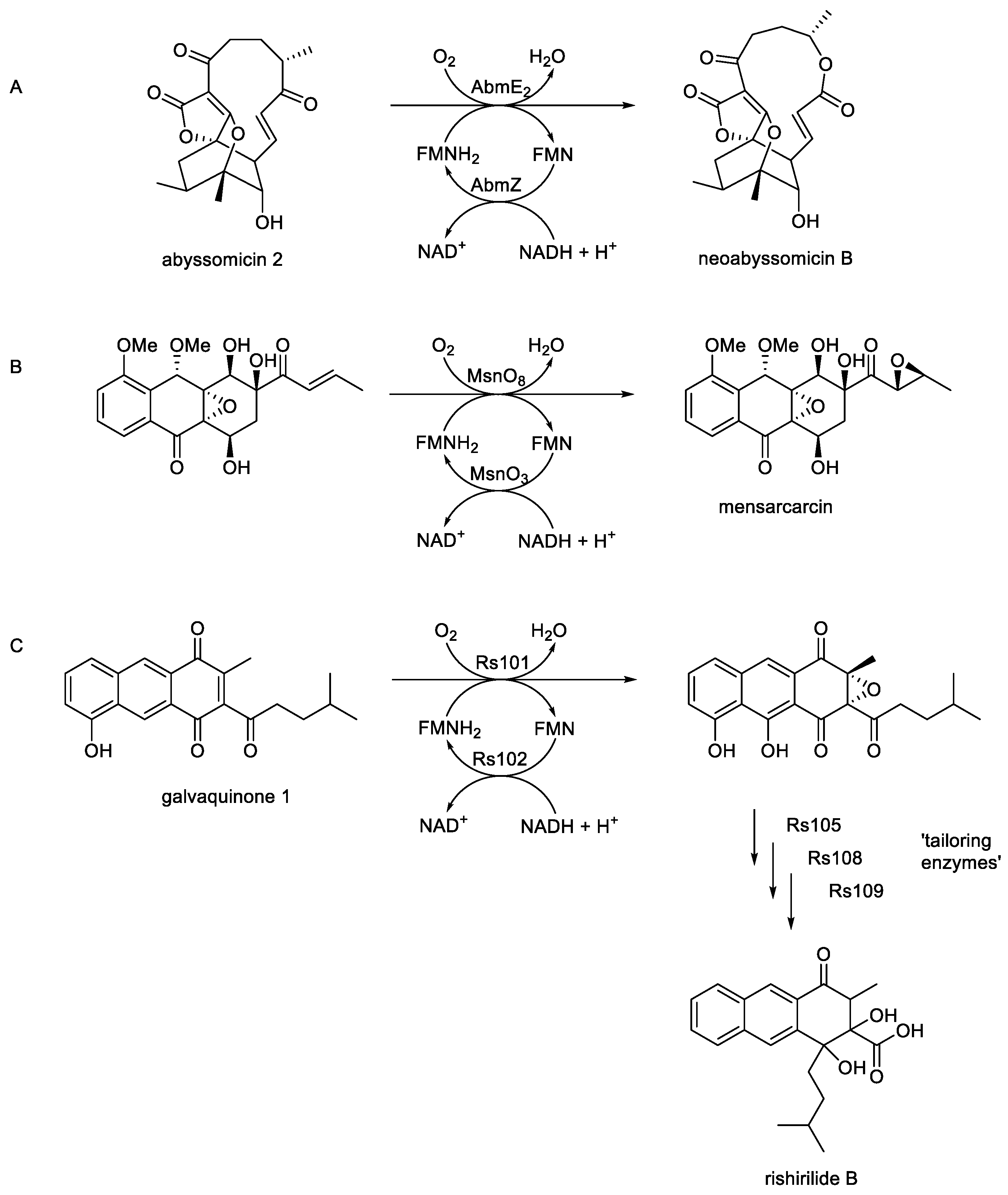
  6. Song, Y.; Li, Q.; Qiu, F.; Sun, C.; Liang, H; Wei, X.; Wong, N.K.; Yi, L.; Zhang, Y.; Shao, M.; Ju, I. Neoabyssomicins A-C, polycyclic macrolactones from deep-sea derived Streptomyces koyangensis SCS 10 5802. Tetrahedron, 2017, 73, 5366-5372.
  7. Maier, S.; Heitzler, T.; Asmus, K.; Brotz, E.; Hardter, U.; Hesselbach, K.; Paululat, T.; Bechthold, A. Functional characterization of different ORFs including luciferase-like monooxygenase genes from the mensacarcin gene cluster. ChemBioChem., 2015, 16, 1175-1182. [CrossRef]
  8. Alali, A.; Zhang, L.; Li, J.; Zuo, C.; Wassouf, D.; Yan, X.; Schwarzer, P.; Gunther, B.; Rinsle, O.; Bechthold, A. Biosynthesis of the tricyclic aromatic Type II polyketide rishirilide: new potential third ring oxygenation after three cyclisation steps. Mol. Biotechnol., 2021, 63, 502-514. [CrossRef]
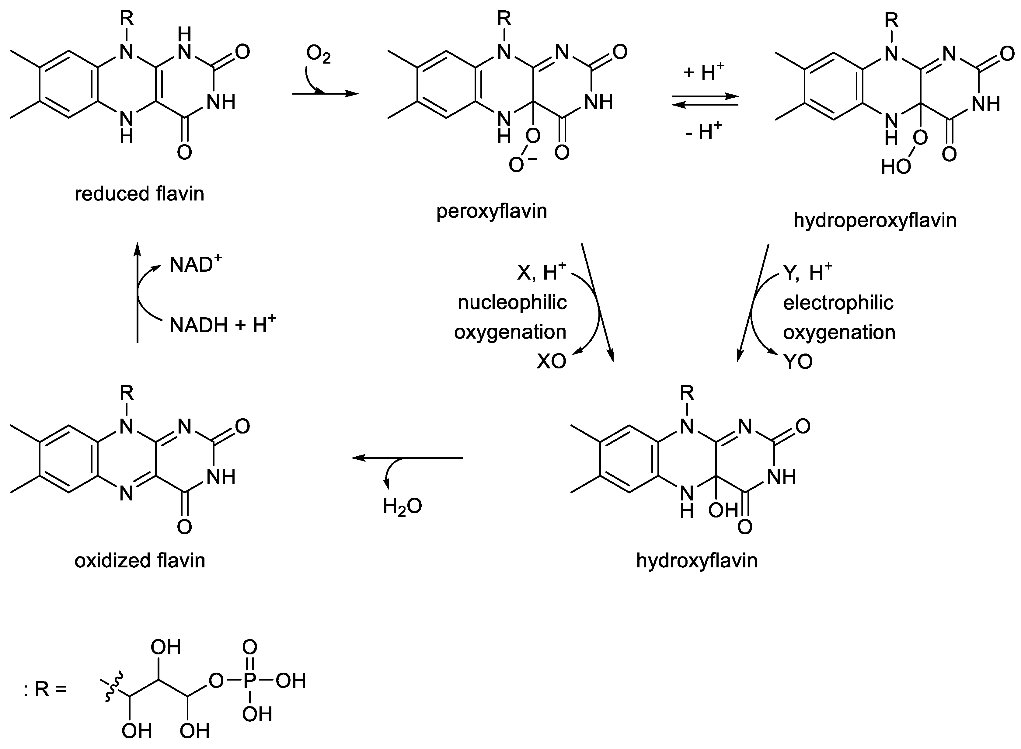
  9. Ghisla, S.; Massey, V. Mechanisms of flavoprotein-catalysed reactions. Eur. J. Biochem., 1989, 181, 1-17.
  10. Hastings, J.W.; Potrikus, C.J.; Gupta, S.C.; Kurfurst, M.; Makemson, J.C. Biochemistry and physiology of bioluminescent bacteria. Adv. Microb. Physiol., 1985, 26, 236-291.
  11. Greening, C.; Ahmed, F.H.; Mohamed, A.E.; Lee, B.M.; Pandey, G.; Warden, A.C.; Scott, C.; Oakeshott, J.G.; Taylor, M.C.; Jackson, C.J. Physiology, biochemistry, and applications of F420-dependent redox reactions. Microbiol. Mol. Biol. 2016, 80, 451-493.
  12. Lavoisier, A. On the combustion of candles in atmospheric air and dephlogistated air. Communicated to the Academie des Sciences, Paris, 3rd May 1777.
  13. Payen, A.; Persoz, J.F. Memoire sur la diastase, les principaux produits de ses reaction, et leurs applications aux arts industrials. Ann. Chim. Phys., 1833, 53, 73-92.
  14. Dubois, R. Sur le mechanisme des fonctions photodermatique et photogenique dans le siphon du Pholas dactylus. Compt. Rend. Soc. Biol., 1887, 3, 564-568.
  15. Hastings, J.W.; Presswood, R.P. Bacterial luciferase: FMNH2-aldehyde oxidase. Meth. Enzymol., 1978, 53, 558-570.
  16. Delroisse, J.; Duchatelet, L.; Flammang, P.; Mallefet, J. Leaving the dark side? Insights into the evolution of luciferases. Front. Mar. Sci., 2021, 8, 673620. [CrossRef]
  17. Haddock, S.H.D.; Moline, M.A.; Case, J.F. Bioluminescence in the sea. Ann. Rev. Mar. Sci., 2010, 2, 443-493. [CrossRef]
  18. Meighen, E.A. Molecular biology of bacterial bioluminescence. Microbiol. Rev. 1991, 55, 123-142. [CrossRef]
  19. Fisher, A.J.; Thompson, T.B.; Thoden, J.B.; Baldwin, T.O.; Rayment, I. The 1.5- Å crystal structure of bacterial luciferase in low salt conditions. J. Biol. Chem., 1986, 271, 21956-21968.
  20. Campbell, Z.T.; Weichsel, A.; Montfort, W.R.; Baldwin, T.O. Crystal . structure of the bacterial luciferase/flavin complex provides insights into the function of the β-subunit Biochem., 2009, 48, 6085-6094.
  21. Deeva, A.A.; Temlyakova, E.A.; Sorokin, A.A.; Nemtseva, E.V.; Kratasyuk, V.A. Structural distinctions of the fast and slow bacterial luciferases revealed by phylogenetic analysis. Bioinform., 2016, 32, 3053-3057. [CrossRef]
  22. Wierenga, R.K. The TIM-barrel fold: a versatile framework for efficient enzymes. FEBS Lett., 2001, 492, 193-198. [CrossRef]
  23. Nagano, N.; Orengo, C.A.; Thornton, J.M. One fold with many functions: the evolutionary relationships between TIM-barrel families based on their sequences, structures, and functions. J. Mol. Biol., 2002, 321, 741-765. [CrossRef]
  24. Aufhammer, S.W.; Warkentin, E.; Berk, H.; Shima, S,; Thauer, R.K.; Ermier, U. Coenzyme binding in F420-dependent secondary alcohol dehydrogenase, a member of the bacterial luciferase family. Structure, 2004, 12, 361-370. [CrossRef]
  25. Nolling, J.; Ishii, M.; Koch, J.; Pihl, T.D.; Reeve, J.N.; Thauer,R.K.; Hedderich, R. Characterization of a 45-kDa flavoprotein and evidence for a rubredoxin, two proteins that could participate in electron transport from H2 to CO2 in methanogenesis in Methanobacterium thermoautotrophicum. Eur. J. Biochem., 1995, 231, 628-638. [CrossRef]
  26. Aufhammer, S.W.; Warkentin, E.; Ermler, U.; Hagemeier, C.H.; Thauer, R.K.; Shima, S. Crystal structure of methylenetetrahydromethanopterin reductase (Mer) in complex with coenzyme F420: archetecture of the F420/FMN binding site of the enzymes within the nonpropyl cis-peptide containing bacterial luciferase family. Protein Sci., 2005, 14, 1840-1849. [CrossRef]
  27. Sucharitakul, J.; Chaiyen, P.; Entsch, B.; Ballou, D.P. The reductase of p-hydroxyphenylacetate 3-hydroxylase requires p-hydroxyphenylacetate for effective catalysis. Biochem., 2005, 44, 10434-10442.
  28. Eichhorn, E.; van der Ploeg, J.R.; Leisinger, T. Characterization of a two-component alkanesulfonate monooxygenase from Escherichia coli. J. Biol. Chem., 1999, 274, 26639-26646. [CrossRef]
  29. Knoebel, H.R.; Egli, T.; van der Meer, J. Cloning and characterization of the genes encoding nitrilotriacetate monooxygenase of Chelobacter heinzii ATCC 29600. J. Bacteriol., 1996, 178, 6123-6132.
  30. Grinter, R.; Greening, C. Cofactor F420: an expended view of its distribution, biosynthesis, and roles in bacteria and archaea. FEBS Microbiol. Rev., 2021, 45, 1-46. [CrossRef]
  31. Mascotti, M.L.; Ayub, M.J.; Fraaije, M.W. On the diversity of .F420-dependent oxidoreductases: a sequence- and structure-based classification. Proteus, 2021, 89, 1497-1507.
  32. Fischer, W.W.; Hemp, J.; Valentine, J.S. How did life survive Earth’s great oxygenation?. Curr. Opin. Chem. Biol., 2016, 166-178. [CrossRef]
  33. Muller, H. Reversibility in evolution considered from the standpoint of genetics. Biol. Rev. Camb. Phil. Soc., 1939, 14, 261-280. [CrossRef]
  34. Riedl, R. A systems-analytical approach to macro-evolutionary . phenomenon. Q. Rev. Biol., 1977, 52, 351-370. [CrossRef]
  35. Shah, P.; McCandlish, D.M.; Plotkin, J.B. Contingency and entrenchment protein evolution under purifying selection. Proc. Natl. Acad. Sci. USA, 2015, E3226-E3225. [CrossRef]
  36. McCandlish, D.M.; Shah, P.; Plotkin, J.B. Epistasis and the dynamics of recession in molecular evolution. Genetics, 2016, 203, 1335-1351. [CrossRef]
  37. Harvey, E.N. A history of bioluminescence: from the earliest times until 1900. American Philosophical Society, Philadelphia, Pennsylvania, USA, 1957.
  38. Harvey, E.N. Oxygen and bioluminescence. Biol. Bull., 1926, 51, 89-97.
  39. Strehler, B.L. Luminescence in cell-free extracts of luminous bacteria. and its activation by DPNH. J. Amer. Chem. Soc., 1953, 75, 1264. [CrossRef]
  40. McElroy, W.D.; Hastings, J.W.; Sonnenfeld, V.; Coulombre, J. The requirement of riboflavin phosphate for bacterial luminescence. . Science, 1953, 118, 385-386. [CrossRef]
  41. Strehler, B.L.; Cormier, M.J. Factors affecting the luminescence of cell-free extracts of the luminous bacterium Achromobacter fischeri. Arch. Biochem. Biophys., 1953, 47, 16-33. [CrossRef]
  42. Cormier, M.J.; Strehler, B.L. The identification of KCF: requirement of long-chain aldehyde for bacterial extract luminescence. J. Amer. Chem. Soc., 1953, 75, 4864-4865. [CrossRef]
  43. Rogers, P.; McElroy, W.D. Enzymatic determination of aldehyde . permeability in luminous bacteria. I. Effect of chain length on light. emission and penetration. Arch. Biochem. Biophys., 1958, 75, 87-105.
  44. Totter, J.R.; Cormier, M.J. The relation of bacterial luciferase to alternative pathways of dihydroflavin mononucleotide oxidation..J. Biol. Chem., 1955, 216, 801-911. [CrossRef]
  45. Mason, H.S.; Fowlks, W.L.; Peterson, E. Oxygen transfer and electron , transport by the phenolase complex. J. Amer. Chem. Soc., 1955, 77, 2914-2915. [CrossRef]
  46. Hastings, J.W.; Riley, W.H.; Massa, J. The purification, properties, and chemiluminescent quantum yield of bacterial luciferase. J. Biol. Chem., 1965, 240, 1473-1481. [CrossRef]
  47. Hayaishi, O. Biological oxidation. Ann. Rev. Biochem., 1963, 31, 25-46.
  48. Hastings, J.W.; Gibson, Q.H.; Greenwood, C. On the molecular mechanism of bioluminescence. I. The role of long-chain aldehyde. Proc. Natl. Acad. Sci. USA, 1964, 52, 1529-1535. [CrossRef]
  49. Meighan, E.A.; Hastings, J.W. Binding site determination from kinetic data: reduced flavin mononucleotide binding to bacterial luciferase. J. Biol. Chem., 1971, 246, 766-767.
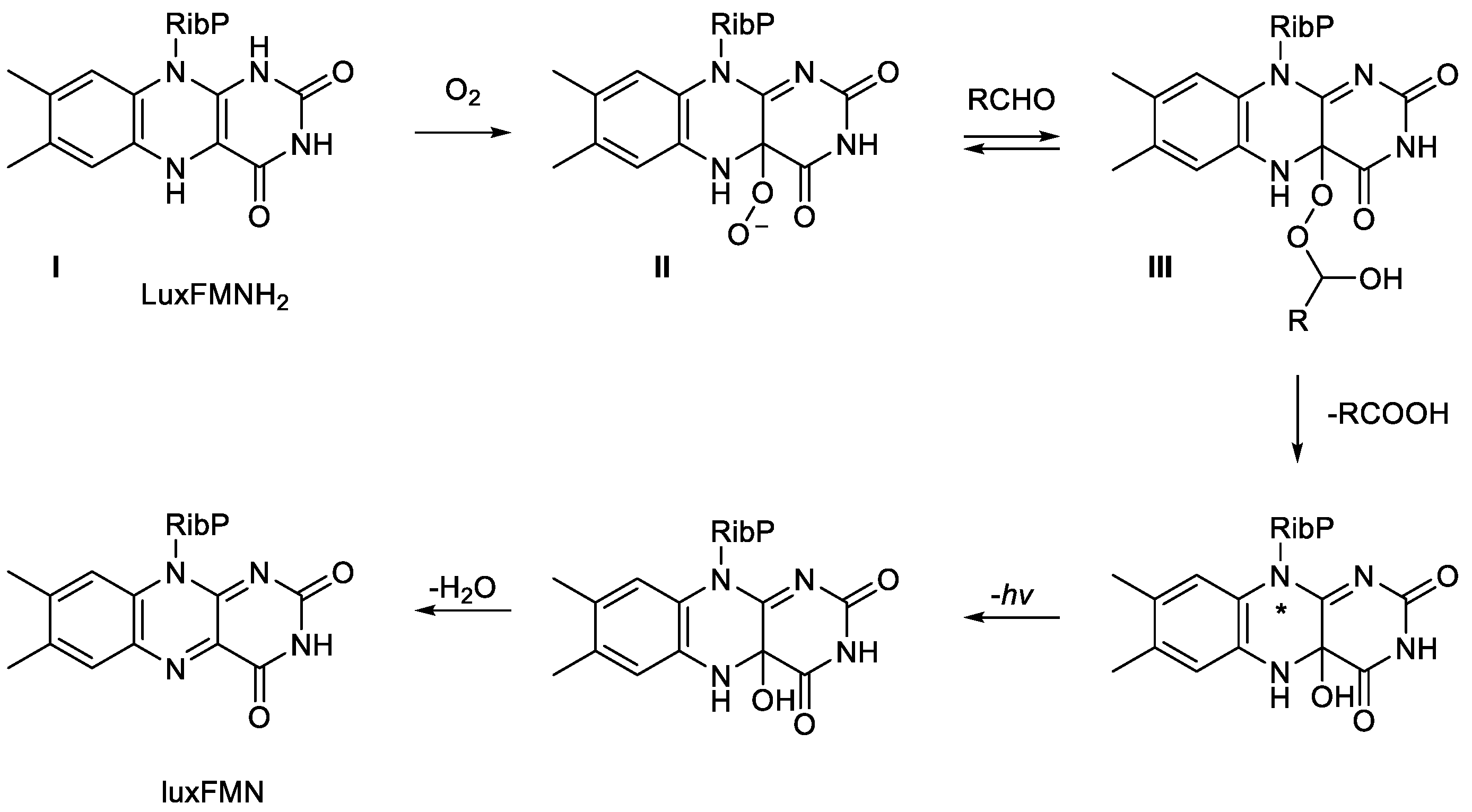
  50. Eberhard, A.; Hastings, J.W. A postulated mechanism for the b . . bioluminescent oxidation of reduced flavin mononucleotide. Biochem. Biophys. Res. Commun., 1972, 47, 348-353. [CrossRef]
  51. Romero, E.; Gomez Castellanos, J.R.; Gadda, G.; Fraaije, M.W.; Mattevi, A. Same substrate, many reactions: oxygen activation in flavoenzymes. Chem. Rev., 2018, 118, 1742-1769. [CrossRef]
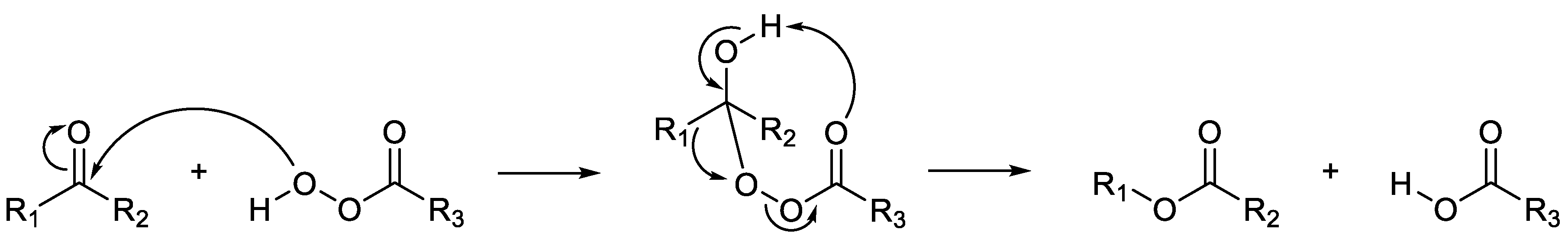
  52. Criegee, R. Die umlangerung de dekalin peroxydester als folge von . . kationischem sauerstoff. Liebigs Ann. Chem., 1948, 560, 127-135.
  53. Baeyer, A.; Villiger, V. Einwirkung des caro’schen reagens auf ketone. Ber. Dtsch. Chem.,1899, 32, 3625-3633. [CrossRef]
  54. Gunsalus, I.C. The biology, chemistry, and physics of oxygen reduction. Fed. Proc., 1976, 35, 1345-1346.
  55. Conrad, H.E.; Lieb, K.; Gunsalus, I.C. An enzyme system for cyclic . . ketolactonization. Biochem. Biophys. Res. Commun., 1961, 6, 293-297. [CrossRef]
  56. Conrad, H.E.; Lieb, K.; Gunsalus, I.C. Mixed function oxidation. III. An electron transport complex in camphor lactonization. J. Biol. Chem., 1965, 240, 4029-4037.
  57. Hastings, J.W.; Nealson, K.H. Bacterial bioluminescence. Ann. Rev. Microbiol., 1977, 31, 549-595.
  58. Eckstein, J.W.; Hastings, J.W.; Ghisla, S. Mechanism of bacterial bioluminescence: 4a,5-dihydroflavin analogues as models for luciferase hydroperoxide intermediates and the effect of substituents at the 8-position of flavin on luciferase kinetics. Biochem., 1993, 32, 404-411. [CrossRef]
  59. Suzuki, K.; Kaidoh, T.; Katagiri, M.; Tsuchiya, T. O2 incorporation into a long-chain fatty acid during bacterial bioluminescence. Biochem. Biophys. Acta, 1983, 722, 297-301. [CrossRef]
  60. Mason, H.S.; Fowlkes, W.L.; Peterson, E. Oxygen transfer and electron transport by a phenolase complex. J. Amer. Chem. Soc., 1955, 77, 2914-2915.
  61. Grogan, G.; Roberts, S.M.; Willetts, A. Camphor-grown Pseudomonas putida, a multifunctional biocatalyst. Biotechnol. Lett., 1993, 15, 913-918. [CrossRef]
  62. Willetts, A. Structural studies and synthetic applications of Baeyer-Villiger monooxygenases. Trends Biotechnol., 1998, 15, 55-62. [CrossRef]
  63. Willetts, A.; Kelly, D.R. Flavin-dependent redox transfers by two-component diketocamphane monooxygenases of camphor-grown Pseudomonas putida NCIMB 10007. Microorganisms, 2016, 4, 38. [CrossRef]
  64. Villa, R.; Willetts, A. Oxidation by microbial NADH plus FMN-dependent luciferases from Photobacterium phosphoreum and Vibrio fischeri. J. Molec. Catal. B: Enzymatic, 1997, 2, 193-197. [CrossRef]
  65. Trudgill, P.W. Bacteria and the challenge of acyclic molecules. In Experiences in Biochemical Perception; Ornston, L.N.; Sligar, S.G., Eds,; Academic Press: New York, NY, USA, 1982; pp59-73.
  66. Xun, L.; Reeder, R.B.; Plymale, A.E.; Girvin, D.C.; Bolton, H. Degradation of metal-trinitriloacetate complexes by nitrilotriacetate monooxygenase. Environ. Sci. Technol., 1996, 30, 1752-1755. [CrossRef]
  67. Xu, Y.; Mortimer, M.W.; Fisher, T.S.; Kahn, M.L.; Brockman, F.J.; Xun, L. Cloning, sequencing, and analysis of a gene cluster from Chelatobacter heintzii ATCC 29600 encoding nitrilotriacetate monooxygenase and NADH:flavin mononucleotide oxidoreductase. J. Bacteriol., 1997, 179, 1112-1116. [CrossRef]
  68. Egli, T. Biodegradation of metal-complexing aminopolycarboxylic acids. J. Biosci. Bioeng., 2001, 92, 89-97.
  69. van der Ploeg, J.R.; Ivanicka-Nowicka, R.; Bykowski, T.; Hryniewicz, M.M.; Leisinger, T. The Escherichia coli ssuEACB gene cluster is required for the utilisation of sulfur from aliphatic sulfonates and is regulated by the transcriptional factor cb1. J. Biol. Chem., 1999, 274, 29358-29365.
  70. Eichhorn, E.; van der Ploeg, J.R.; Leisinger, T. Deletion analysis of the Escherichia coli alkanesulfonate transport system. J. Bacteriol., 2000, 182, 2687-2695.
  71. van der Ploeg, J.R.; Eichhorn, E.; Leisinger, T. Sulfonate-sulfur metabolism and its regulation in Escherchia coli. Arch. Microbiol., 2001, 176, 1-8. [CrossRef]
  72. Payne, J.W.; Bolton, H.J.; Campbell, J.A.; Xun, L. Purification and characterization of EDTA monooxygenase from the EDTA-degrading bacterium BNS1. J. Bacteriol., 1998, 180, 3823-3827.
  73. Zhan, X.; Carpenter, R.A.; Ellis, H.R. Catalytic importance of the substrate binding order for the FMNH2-dependent alkanesulfonate monooxygenase enzyme. Biochem., 2008, 47, 2221-2230. [CrossRef]
  74. Kertesz, M.A.; Schmidt-Larbig, K.; Wuest, T. A novel reduced flavin mononucleotide-dependent methanesulfonate sulfonatase encoded by the sulfur-regulated msu operon of Pseudomonas aeruginosa. J. Bacteriol., 1999, 181, 1464-1473. [CrossRef]
  75. Liew, J.J.M.; El Saudi, I.M.; Nguyen, S.V.; Wicht, D.K.; Douling, D.P. Studies of the alkanesulfonate monooxygenase MsuD provide insights into C-S bond cleavage, substrate scope, and unexpected role for the tetramer. J. Biol. Chem., 2021, 297, 100823. [CrossRef]
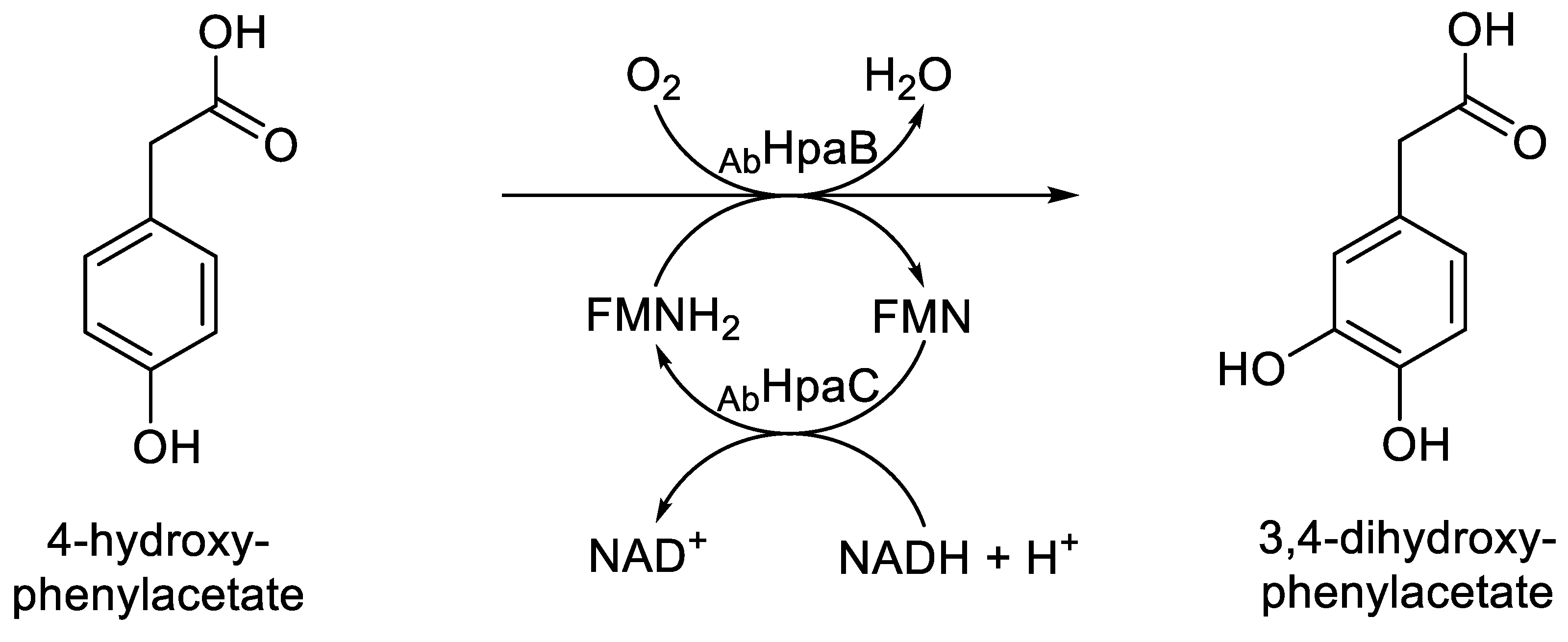
  76. Prieto, M.A.; Garcia, J.L. Molecular characterization of 4-hydroxy- phenylacetate 3-hydroxylase of Escherichia coli: a two-protein component system. J. Biol. Chem., 1994, 269, 22823-22829.
  77. Prieto, M.A.; Diaz, E.; Garcia, J.L. Molecular characterization of the 4-hydroxyphenylacetate catabolic pathway of Escherichia coli W: engineering a mobile aromatic degradative cluster. J. Bacteriol., 1994, 176, 111-120. [CrossRef]
  78. Galan, B.; Diaz, E.; Prieto, M.A.; Garcia, J.L. Functional analysis of the small component of the 4-hydroxyphenylacetate 3-monooxygenase of Escherichia coli W: a prototype of a new flavin:NAD(P)H reductase family. J. Bacteriol., 2000, 182, 627-636. [CrossRef]
  79. Oldfield, C.; Pogrebinsky, O.; Simmonds, J.; Olsen, E.S.; Kulpa, C.F. Elucidation of the metabolic pathway for dibenzothiophene desulfurization by Rhodococcus sp. strain IGTS8 (ATCC 53968). Microbiology, 1997, 143, 2961-2973.
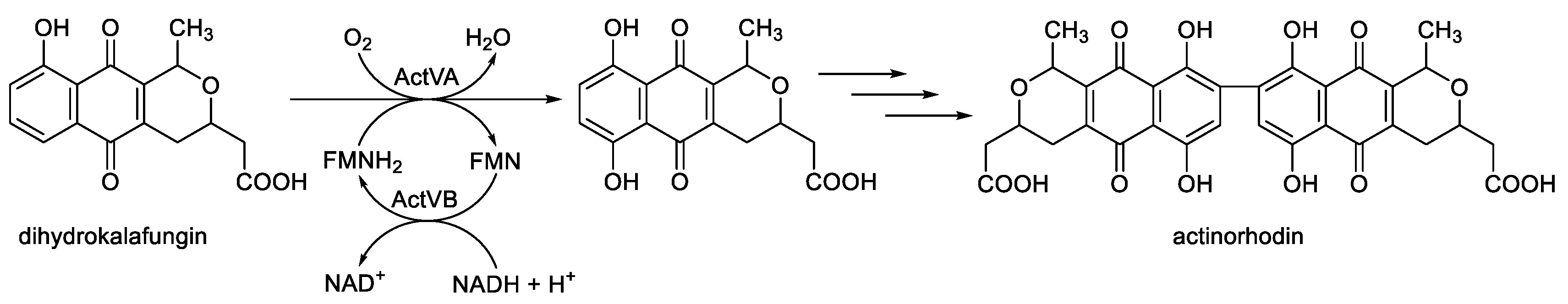
  80. Kendrew, S.G.; Hopwood, D.A.; Marsh, E.N.G. Identification of a monooxygenase from Streptomyces coelicolor A3(2) involved in the biosynthesis of actinorhodin: purification and characterization of the recombinant enzyme. J. Bacteriol., 1997, 179, 4305-4310. [CrossRef]
  81. Sciara, G.; Kendrew, S.G.; Miele, A.E.; Marsh, E.N.G.; Federici, L.; Malatesta, F.; Schimperna, G.; Savino, C.; Valione, B. The structure of ActVA-Orf6, a novel type of monooxygenase involved in actinorhodin synthesis. EMBO J., 2003, 22, 205-215.
  82. Valton, J.; Filisetti, L.; Fontecave, M.; Niviere, V. A two-component flavin-dependent monooxygenase involved in actinorhodin biosynthesis in Streptomyces coelicolor. J. Biol. Chem., 2004, 279, 44362-44369. [CrossRef]
  83. Filisetti, L.; Valton, J,; Fontecave, M.; Niviere, V. The flavin reductase ActVB from Streptomyces coelicolor: characterization of the electron transfer activity of the flavoprotein form. FEBS Lett., 2005, 579, 2817-2820. [CrossRef]
  84. Valton, J.; Fontecave, M.; Douki, T. Kendrew, S.C.; Niviere, V. An aromatic hydroxylation reaction catalysed by a two-component FMN-dependent monooxygenase. The ActVA-ActVB system from Streptomyces coelicolor. J. Biol. Chem., 2006, 281, 27-35. [CrossRef]
  85. Valton, J.; Mathevon, C.; Fontecave, M.; Niviere, V.; Ballou, D.P. Mechanism and regulation of the two-component FMN-dependent monooxygenase ActVA-ActVB from Streptomyces coelicolor. J. Biol. Chem., 2008, 283, 10287-10296. [CrossRef]
  86. Tang, M-C.; Zou, Y.; Watanabe, K.; Walsh, C.T.; Tang, Y. Oxidative cyclization in natural product biosynthesis. Chem. Rev., 2017, 117, 5226-5333. [CrossRef]
  87. Tolmie, C.; Smit, M.S.; Opperman, J. Native roles of Baeyer-Villiger monooxygenases in microbial metabolism of natural products. Nat. Prod. Rev., 2017, 36, 326-353.
  88. Ichinose, K.; Bedford, D.J.; Tornus, D.; Bachthold, A.; Bibb, M.J.; Revill, W.P.; Floss, H.G.; Hopwood, D.A. The granaticin biosynthetic gene cluster of Streptomyces violaceoruber Tu22: sequence analysis and expression in a heterologous host. Chem. Bio., 1998, 6, 647-659. [CrossRef]
  89. Van Berkel, W.J.H.; Kamerbeck, N.M.; Fraaije, M.W. Flavoprotein monooxygenases, a diverse class of oxidative biocatalysts. J. Biotechnol., 2006, 4, 670-689. [CrossRef]
  90. Ellis, H.R. The FMN-dependent two-component monooxygenase systems. Arch. Biochem. Biophys., 2010, 497, 1-12. [CrossRef]
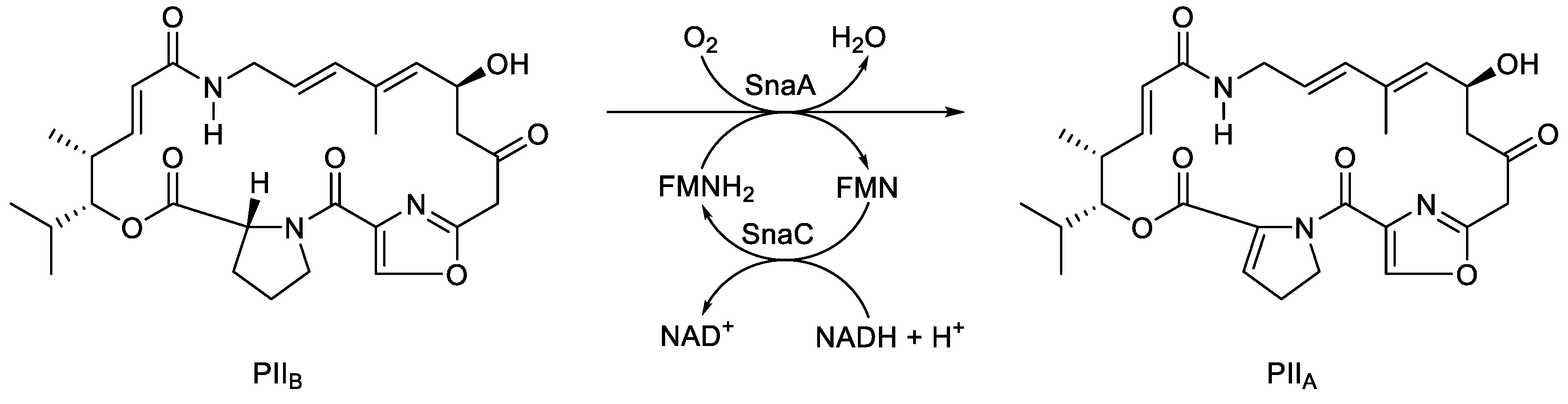
  91. Thibaut, D.; Ratet, N.; Bisch, D.; Faucher, D.; Debussche, L.; Blanche, F. Purification of the two-enzyme system catalysing the oxidation of the D-proline residue of pristinamycin IIB during the last step of pristinamycin IIA biosynthesis. J. Bacteriol., 1995, 177, 5199-5205. [CrossRef]
  92. Blanc, V.; Lagneaux, D.; Didier, P.; Gill, P.; Lacroix, P.; Crouzet, J. Cloning and analysis of structural genes from Streptomyces pristinaespirilis encoding enzymes involved in the conversion of pritinamycinIIB to pristinamycin IIA(PIIA): PIIA synthase and NADH: riboflavin 5’-phosphate oxidoreductase. J. Bacteriol., 1995, 177, 5206-5214. [CrossRef]
  93. Chaiyen, P.; Suadee, C.; Wilairat, P. A novel two-component flavoprotein hydroxylase p-hydroxyphenylacetate hydroxylase from Acinetobacter baumanii. Eur. J. Biochem., 2001, 268, 5550-5561. [CrossRef]
  94. Feng, L.; Wang, W.; Cheng, I.; Wang, L. Genome and proteome of long-chain alkane degrading Geobacillus thermodenitrificans N980-2 isolated from a deep-subsurface oil reservoir. Proc. Natl. Acad. Sci. USA, 2007, 104, 5602-5607. [CrossRef]
  95. Li, L.; Liu, X.; Yang, W.; Xu, F.; Wang, W.; Feng, L.; Bartlam, L.; Wang, L.; Rao, Z. Crystal structure of long-chain alkane monooxygenase (LadA) in complex with coenzyme FMN: unveiling the long-chain alkane hydroxylase. J. Mol. Biol., 2008, 376, 453-465. [CrossRef]
  96. Abbasian, F.; Lockington, R.; Mallavarapu, M.; Naidu, R. A comprehensive review of aliphatic hydrocarbon biodegradability by bacteria. Appl. Biochem. Biotechnol., 2015, 176, 670-699. [CrossRef]
  97. Toplak, M.; Teufel, R. Three rings to rule them all: how versatile flavoproteins orchestrate the structural diversification of natural products. Biochem., 2022, 61, 47-56. [CrossRef]
  98. Doraghazi, J.R.; Albright, J.C.; Goering, A.W.; Ju, K-S.; Haines, R.R.; Tchalukov, K.A.; Labeda, D.P.; Kelleher, N.L.; Metcalf, W.W. A road map for natural product discovery based on large-scale genomics and metabolomics. Nat. Chem. Biol., 2014, 10, 963-968.
  99. Hoskisson, P.A.; Seipke, R.A. Cryptic or silent? The known unknowns, unknown knowns, and unknown unknowns of secondary metabolism. Microbial Interactions, 2020, 11, 5. [CrossRef]
  100. Zapf, C.W.; Harrison, B.D.; Drahl, C.; Sorensen, E.J. A Diels-Alder macrocyclization enables an efficient asymmetric synthesis of the antibacterial natural product abyssomicin C. Angew. Chem. Int, Ed., 2005, 44, 6533-6537. [CrossRef]
  101. Keller, S.; Nicholson, G.; Drahl, C.; Sorensen, E.J.; Fiedler, H.P.; Sussmuth, R.D. Abyssomicins G and H and atrop-abyssomicin C from marine Verrucosispora strain Ass-18-032. J. Antibiot., 2007, 60, 391-394. [CrossRef]
  102. Maier, S.; Heitzler, T.; Asmus, K.; Brotz, K.; Hardter, U.; Hesselbach, K.; Paululat, T.; Bechthold, A. Functional characterization of different ORFs including luciferase-like monoxygenase genes from the mensacarcin gene cluster. ChemBioChem., 2015, 16, 1175-1182. [CrossRef]
  103. Colonna, S.; Gaggero, N.; Carrea, G.; Ottolina, G.; Pasta, P.; Zambianchi, F. First asymmetric epoxidation catalysed by cyclohexanone monooxygenase. Tet. Lett., 2002, 43, 1797-1799. [CrossRef]
  104. Mendelovici, M.; Glotter, E. Epoxidation and Baeyer-Villiger oxidation of γ-hydroxy-α,β-unsaturated ketones on exposure to m-chloroperbenzoic acid. J. Chem. Soc, Perkin Trans I, 1992, 1735-1749.
  105. Osumak, A.; Magolan, J.; Waynant, K.V. One-pot carbonyl reduction and carbonate formation using sodium borohydride in dialkyl carbonate solvents. Tet Lett., 2019, 60, 151203. [CrossRef]
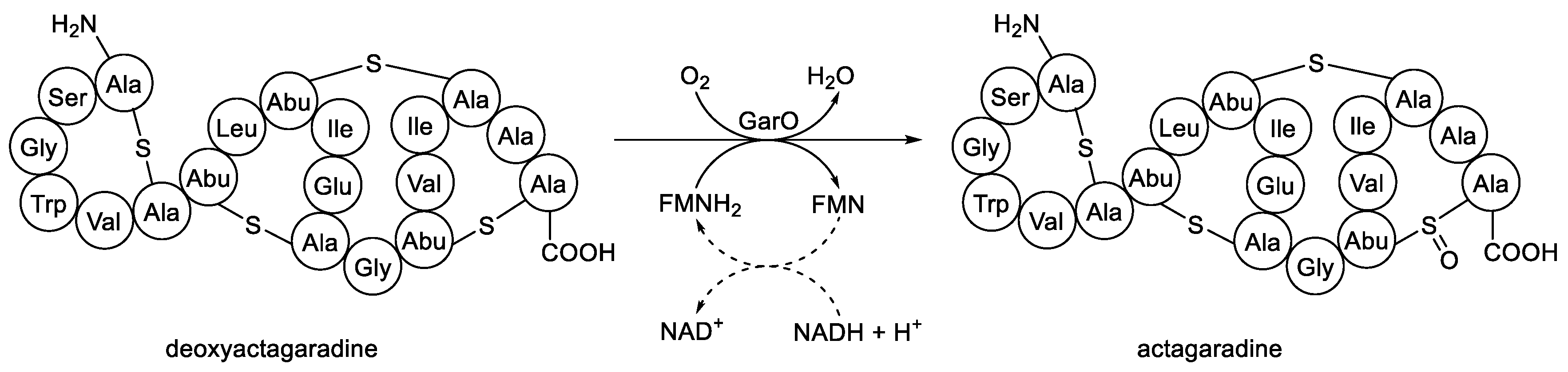
  106. Shi, Y.; Bueno, A.; van der Donk, W.A. Heterologous production of the lantibiotic Ala(0)actagardine in Escherichia coli. Chem. Commun., 2012, 48, 10966-10968. [CrossRef]
  107. Zimmermann, N.; Metzger, J.W.; Jung, G. The tetracyclic lantibiotic actagaradine 1H-NMR and 13C-NMR assignments and revised primary structure. Eur. J. Biochem., 1995, 228, 786-797.
  108. Dawson, M.W.; Appleyard, N.; Cortes-Bargallo, J.; Wadman, S.N. Actagaradine derivatives, and pharmaceutical uses thereof. 2011. WO/2011/095769.
  109. Shi, Y.; Yang, X.; Garg, N.; van der Donk, W.A. Production of lantipeptides in Escherichia coli. J. Amer. Chem. Soc., 2011, 133, 2338-2341.
  110. Okesli, A.; Cooper, L.E.; Fogle, E.J.; van der Donk, W.A. Nine post-translational modifications during biosynthesis of cinnamycin. J. Amer. Chem. Soc., 2011, 133, 13753-13760. [CrossRef]
  111. Garg, N.; Tang, W.; Goto, Y.; van der Donk, W.A. Lantibiotics from Geobacillus thermodenitrificans. Proc. Natl. Acad. Sci. USA, 2012, 109, 5241-5246. [CrossRef]
  112. Tang, W.; van der Donk, W.A. Structural characterization of four prochlorosins: a novel class of lantipeptides produced by planktonic marine cyanobacteria. Biochem., 2012, 51, 4271-4279.
  113. Campbell, Z.; Baldwin, T.O. Fre is the major flavin reductase supporting bioluminescence from Vibrio harveyi luciferase in Escherichia coli. J. Biol. Chem., 2009, 284, 8322-8328. [CrossRef]
  114. Kadow, M.; Sass, S.; Schmidt, M.; Bornscheuer, U.T. Recombinant expression and purification of the 2,5-diketocamphane 1,2-monooxygenase from the camphor-metabolising Pseudomonas strain NCIMB 10007. AMB Express, 2011, 1, 13. [CrossRef]
  115. Willetts, A. Inter-species redox coupling by flavin reductases and FMN-dependent two-componenent monooxygenases undertaking nucleophilic Baeyer-Villiger bioxygenations. Microorganisms, 2023, 11, 71. [CrossRef]
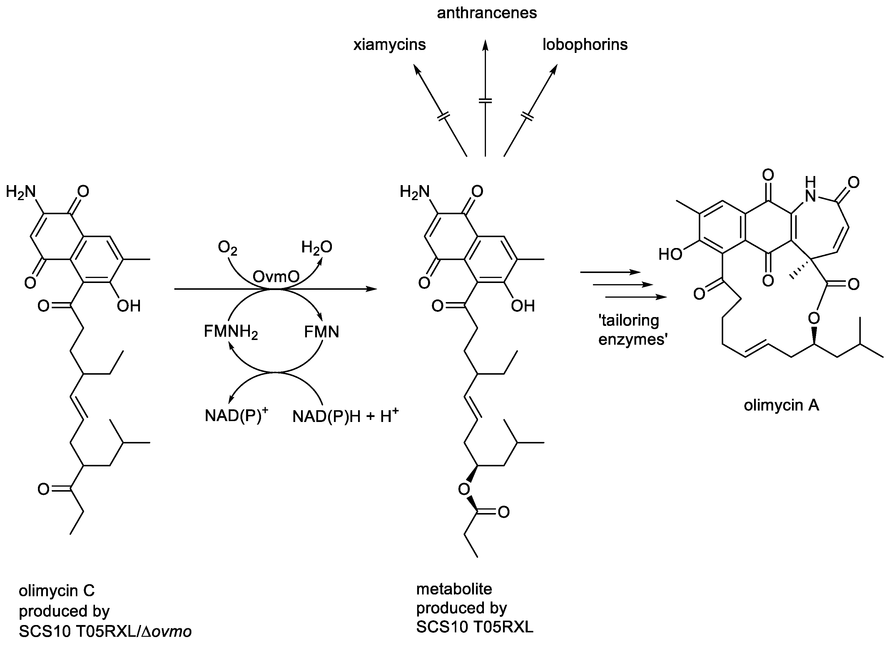
  116. Zhang, C.; Sun, C.; Huang, H.; Gui, C.; Wang, L.; Li, Q.; Ju, J. Biosynthetic Baeyer-Villiger chemistry enables access to two anthracene scaffolds from a single gene cluster in deep-sea-derived Streptomyces olivaceus SCS10 T05. J. Nat. Prod., 2018, 81, 1570-15 . [CrossRef]
  117. Zhang, C.; Zhang, H.; Ju, J. On-PKS Baeyer-Villiger-type O-atom insertion catalysed by luciferase-like monooxygenase Ovmo during olimycin biosynthesis. Org. Lett., 2022, 22, 1780-1784. [CrossRef]
  118. Dong, Y.; Enjing, J.; Wang, R.; Bao, Y.; Li, H. New olimycins from cold-seep-derived Streptomyces olivaceus. Chem. Biodiversity, 2023, 20. [CrossRef]
Figure 1. Key monooxygenase-dependent steps in the biosynthesis of (A) neoabyssomycin B, Song et al. [6]; (B) mensacarcin, Maier et al. [7]; (c) rishirilide B, Alali et al. [8].
Preprints 189269 g001
Figure 2. Inferred divergent evolutionary relationships of LLM family members taking into account their functional and biochemical characteristics.
Preprints 189269 g002
Figure 3. Hastings’ more developed proposal for the role of peroxidation biochemistry of luciferase-bound FMNH2 in bacterial bioluminescence.
Preprints 189269 g003
Figure 4. Hastings’ proposal for a defined role for a luciferase-bound proxide as a key intermediate in bacterial bioluminescence. The symbols I, II, and III are retained to enable comparison with Figure 3. The symbol * indicates a not fully characterised excited state.
Preprints 189269 g004
Figure 5. Criegee’s proposed mechanism for the abiotic oxidation of a ketone to its corresponding ester by peracid catalysis.
Preprints 189269 g005
Figure 6. The established biochemistry of 2,5-diketocamphane 1,2-monooxygenase, an FMN-dependent two-component monooxygenase induced in camphor-grown Pseudomonas putida ATCC 17453.
Preprints 189269 g006
Figure 7. Comparing the fully characterised outcomes of the biooxidation of (rac)-bicyclo[3.2.0]hept-2-en-6-one by 2,5-diketocamphane 1,2-monooxygenase from Pseudomonas putida ATCC 17453 and the luciferase from Vibrio fischeri ATCC 7744.
Preprints 189269 g007
Figure 8. The oxybiontic cleavage of the C-S bond of short-chain alkanesulfonate.
Preprints 189269 g008
Figure 9. The three-step sequential biooxidation of dibenzothiophene to 2-hydroxy-biphenyl-2-sulfinite by Rhodococcus erythropolis deploying successive actions of the FMN-dependent two-component monooxygenases DszC and DszA, both functioning in cooperation with flavin reductase DszD.
Preprints 189269 g009
Figure 10. Generalised summary of the relevant reactions undergone by the reduced flavin cosubstrate and dioxygen in the nucleophilic and electrophilic oxygenations catalysed by FMN-dependent two-component monooxygenases. X =ketone; XO = lactone/aldehyde; Y = organosulfide/sulfoxide; YO = organosulfoxide/sulfone.
Preprints 189269 g010
Figure 11. The oxybiontic hydroxylation of dihydrokalafungin by ActVA, an FMN-dependent two-component monooxygenase from Streptomyces coelicolor.
Preprints 189269 g011
Figure 12. The oxybiontic oxidation of PIIB to PIIA by PIIA synthase, an FMN-dependent two-component monooxygenase isolated from various Streptomyces spp.
Preprints 189269 g012
Figure 13. The oxybiontic hydroxylation of 4-hydroxyphenylacetate by C2 (AbHpaB) monooxygenase, an FMN-dependent two-component monooxygenase from Acinetobacter baumanii.
Preprints 189269 g013
Figure 14. The oxybiontic oxidation of deoxyactagaradine to actagaradine by GarO, an FMN-dependent two-component monooxygenase isolated from Actinoplanes garbadinensis.
Preprints 189269 g014
Figure 15. The oxybiontic oxidation of olimycin C by OvmO, an FMN-dependent two-component monooxygenase from Streptomyces olivaceus SCS10 T05.
Preprints 189269 g015
Table 1. Confirmed examples of FMN-dependent two-component monooxygenases and their corresponding flavin reductase(s) cited by Garcia in 2000 and subsequently by Ellis in 2010. a = monomeric; b = homodimeric; c = heterodimeric; d = homotetrameric.
Preprints 189269 i001
Table 2. Shared functional characteristics of the bacterial luciferases and other directly related prokaryotic enzymes as reflected by the different proposed collective descriptors TC-ODEs, Type II BVMOs, TC-FDMs, and FD-TCMOs.
Preprints 189269 i002
Disclaimer/Publisher’s Note: The statements, opinions and data contained in all publications are solely those of the individual author(s) and contributor(s) and not of MDPI and/or the editor(s). MDPI and/or the editor(s) disclaim responsibility for any injury to people or property resulting from any ideas, methods, instructions or products referred to in the content.
Copyright: This open access article is published under a Creative Commons CC BY 4.0 license, which permit the free download, distribution, and reuse, provided that the author and preprint are cited in any reuse.
Prerpints.org logo

Preprints.org is a free preprint server supported by MDPI in Basel, Switzerland.

Subscribe

Disclaimer

Terms of Use

Privacy Policy

Privacy Settings

© 2026 MDPI (Basel, Switzerland) unless otherwise stated