Preprint
Article

This version is not peer-reviewed.

Establishment of a Novel Anti-Human Cadherin 13 Monoclonal Antibody (Ca13Mab-4) Utilizing CBIS Method

  † contributed equally to this work.

Submitted:

07 December 2025

Posted:

10 December 2025

You are already at the latest version

Abstract

Cadherin 13 (CDH13), also known as T-cadherin or H-cadherin, is a member of the cadherin superfamily. CDH13 is anchored to the plasma membrane via glycosylphosphatidylinositol. CDH13 plays an essential role in the development of the heart and nervous systems, including the brain. Many reports have identified CDH13 as a risk factor for neurodevelopmental disorders. Furthermore, CDH13 has been shown to be expressed in numerous cancers, but its role as a cancer-promoting or -suppressing factor remains unclear. Therefore, the development of highly sensitive and specific anti-CDH13 monoclonal antibodies (mAbs) is necessary to elucidate the biological and pathological functions of CDH13. In this study, we established a novel anti-human CDH13 mAb (clone Ca13Mab-4) using the Cell-Based Immunization and Screening (CBIS) method. Ca13Mab-4 can be used for flow cytometric analysis. Ca13Mab-4 binds specifically to CDH13 and not to other cadherin family members. The dissociation constant values of Ca13Mab-4 for CDH13-overexpressed CHO-K1 and U87MG glioma cells were determined as 2.5 (± 0.6) x 10-8 M and 8.9 (± 2.1) x 10-9 M, respectively. Furthermore, Ca13Mab-4 clearly detected CDH13 in the western blot and immunohistochemistry of cell sections. Therefore, the Ca13Mab-4, established by CBIS method, could be a valuable tool for basic research and is expected to contribute to elucidating the relationship between CDH13 and diseases, including neurodevelopmental disorders and cancer.

Keywords: 
;  ;  ;  ;  

1. Introduction

Cadherins (CDHs) are a superfamily of transmembrane adhesion molecules that regulate tissue morphogenesis, cell-cell adhesion, polarity, and differentiation through Ca2+-dependent homophilic cell-cell interactions [1]. These cadherins are described as “classical” and are single-pass glycoproteins consisting of five extracellular Ca2+-binding sites (EC1-EC5), a transmembrane domain, and a cytoplasmic domain with highly conserved binding sites for p120-catenin and β-catenin, which bind to α-catenin to polymerize actin microfilaments and maintain cytoskeletal stability [2,3]. The partial structures of two cadherins, CDH1 (also known as E-cadherin) and murine CDH2 (also known as N-cadherin), were first reported in 1995 [4,5]. Initially, CDHs were recognized as adhesive molecules, but in recent years they have been shown to act as mediators that regulate signals essential to maintaining cellular homeostasis [1].
CDH13, also known as T-cadherin or H-cadherin, was first described in the nervous system and has an atypical structure distinct from classical CDHs [6]. CDH13 lacks transmembrane and cytoplasmic domains, and is tethered to the plasma membrane through a glycosyl-phosphatidylinositol (GPI) anchor attached to the apical aspect of the plasma membrane. CDH13 also lacks the conserved His-Ala-Val motif required for classical cadherin homophilic binding, although it possesses a hallmark extracellular part of five Ca2+-binding domains (EC1-EC5) like classical CDHs [2]. The expression of CDH13 has been prominently confirmed in nervous and cardiovascular tissues [7]. During early development, CDH13 is highly expressed in the nervous systems and vascular plexus. In adults, CDH13 is widely distributed throughout the central nervous system, including the cerebral cortex, thalamus, midbrain, and medulla, and is enriched in the heart [8].
Adiponectin is an atypical factor secreted from adipocytes as trimers, hexamers, and high-molecular-weight multimers. It is abundantly present in the peripheral circulation [9]. Circulating adiponectin has diverse effects on many different target tissues that express adiponectin receptors (adipoRs). Adiponectin plays a critical metabolic role via adipoRs. It is also involved in calreticulin-dependent dead cell opsonization [9,10]. Plasma adiponectin inversely correlated with several clinical pathophysiological disease states, such as coronary artery disease [11], myocardial infarction [12], and type 2 diabetes [13]. Adiponectin has been observed to bind to CDH13 in addition to adipoRs. CDH13 specifically recognizes hexamers and high-molecular-weight multimers of adiponectin with high affinity via EC1 and EC2 [14,15]. CDH13-deficient mice show increased circulating adiponectin and reduced adiponectin binding to vascular endothelial cells [16]. Interestingly, serum-derived native adiponectin tends to bind to CDH13-expressing cells, not to adipoRs-expressing cells [17]. Thus, CDH13 appears to be a major binding partner of adiponectin. The adiponectin-CDH13 axis might be a pivotal regulator of tissue homeostasis.
CDH13 is highly expressed during brain development, and has been reported to be associated with the risk of various neurodevelopmental disorders, including attention-deficit/hyperactivity disorder (ADHD) [18], autism spectrum disorders (ASD) [19], bipolar disorder [20], schizophrenia [21], and depression [22]. CDH13 is expressed in neurons, including glutamatergic [23], GABAergic [18], and serotonergic neurons [24]. CDH13 exerts its functions through low-adhesive homophilic or heterophilic interactions to regulate neurite outgrowth and axon guidance [25,26]. Analysis of patient samples has suggested that mutations or common genetic variation in CDH13 are one of the risk factors associated with the disease [27]. These findings appear to highlight the critical roles of CDH13 in neuronal networks.
CDH13 is expressed in several tumor types. CDH13 is known as a tumor suppressor gene, and its loss of function due to hypermethylation of the promoter region contributes to the malignant progression of cancers, including colorectal [28], lung [29], bladder [30], ovarian cancers [31], glioblastoma [32], and oral squamous cell carcinoma (SCC) [33]. The expression of CDH13 and programmed cell death-1 ligand 1 (PD-L1) inversely correlated with human papillomavirus-negative head and neck SCC patients [34]. But, some reports have described CDH13 as a tumor promoter in some tumors, such as breast [35] and adrenocortical carcinoma [36]. CDH13-mediated tumor angiogenesis may create a tumor microenvironment and affect cancer progression [37]. Further research is necessary to clarify the role of CDH13 in promoting or suppressing cancer functions.
To clarify the role of CDH13, specific and sensitive antibodies are desired. Previously, we have generated numerous monoclonal antibodies (mAbs) against transmembrane and membrane-anchored proteins using the Cell-Based Immunization and Screening (CBIS) method [38,39,40,41]. This method allows rapid and efficient generation of antibody clones with diverse epitopes that recognize linear or structural epitopes of membrane proteins and modifications of the extracellular domain.
In this study, we have established a novel anti-human CDH13 mAb (clone Ca13Mab-4) that can be used for multiple applications using the CBIS method.

2. Materials and Methods

2.1. Cell Lines

Chinese hamster ovary (CHO)-K1, mouse myeloma P3X63Ag8U.1 (P3U1), human glioblastoma LN229, human glioma U87MG, and human mesothelioma NCI-H2052 cell lines were obtained from the American Type Culture Collection (ATCC, Manassas, VA, USA). Immortalized-normal fibroblast KMST-6 was obtained from the Cell Resource Center for Biomedical Research Institute of Development, Aging, and Cancer at Tohoku University (Miyagi, Japan).

2.2. Establishment of Stable Transfectants

The gene encoding human CDH13 (Catalog No.: IRAK046C21, Accession No.: NM_001257) was obtained from RIKEN BRC (Ibaraki, Japan). The expression plasmid of human CDH13 was subcloned into the pCAG-ble vector (FUJIFILM Wako Pure Chemical Corporation, Osaka, Japan), which deleted amino acids 1st to 138th with N-terminal MAP16 tag (PGTGDGMVPPGIEDKI) [42] or PA16 tag (GLEGGVAMPGAEDDVV) [43]. The pCAG-CDH13 vectors were transfected into cell lines using the Neon transfection system (Thermo Fisher Scientific, Inc., Waltham, MA, USA). Subsequently, LN229 and CHO-K1, which stably overexpressed CDH13 with N-terminus MAP16 or PA16 tags [hereafter described as LN229/MAP16-CDH13 (LN229/CDH13) and CHO/PA16-CDH13 (CHO/CDH13), respectively] were stained with an anti-MAP16 tag mAb (clone PMab-1) and an anti-PA16 tag mAb (clone NZ-1) followed by sorting by the SH800 cell sorter (Sony corp., Tokyo, Japan). The sorted cells were cultured in a medium containing 0.5 mg/mL of Zeocin (InvivoGen, San Diego, CA, USA).
Other stably human CDHs-overexpressed CHO-K1 cells were established as previously reported [44]. In this study, the expression of each CDH was confirmed by using the following mAbs: anti-CDH6 mAb (clone 427909, MAB2715, R&D Systems, Inc., Minneapolis, MN, USA) and NZ-33, another anti-PA16 tag mAb [45].

2.3. Antibodies

An anti-human/mouse Cadherin-13 Antibody (clone 392411, MAB3264, rat IgG2a) was purchased from R&D Systems, Inc. (Minneapolis, MN, USA). An anti-isocitrate dehydrogenase 1 (IDH1) mAb (clone RcMab-1) was developed previously in our lab [46]. A secondary Alexa Fluor 488-conjugated anti-mouse IgG and anti-rat IgG was purchased from Cell Signaling Technology, Inc. (Danvers, MA, USA). Secondary horseradish peroxidase-conjugated anti-mouse IgG and anti-rat IgG were obtained from Agilent Technologies Inc. (Santa Clara, CA, USA) and Merck KGaA (Darmstadt, Germany), respectively. The sources of commercially available anti-CDH antibodies are listed above.

2.4. Development of Hybridomas

For developing anti-CDH13 mAbs, two 6-week-old female BALB/cAJcl mice, purchased from CLEA Japan (Tokyo, Japan), were immunized intraperitoneally with 1 × 10⁸ cells/mouse of LN229/CDH13. The LN229/CDH13 cells as immunogen were harvested after brief exposure to 1 mM ethylenediaminetetraacetic acid (EDTA; Nacalai Tesque, Inc.). Alhydrogel adjuvant 2% (InvivoGen, San Diego, CA, USA) was added as an adjuvant in the first immunization. Three additional injections of 1 × 10⁸ LN229/CDH13 cells/mouse were administered intraperitoneally, without adjuvant, every week. A final booster injection of 1 × 10⁸ LN229/CDH13 cells/mouse was administered intraperitoneally 2 days before harvesting splenocytes from mice. We conducted cell fusion of harvested splenocytes from LN229/CDH13-immunized mice with P3U1 cells using polyethylene glycol 1500 (PEG1500, Roche Diagnostics, Indianapolis, IN, USA) under heated conditions.
Hybridomas were cultured in the RPMI-1640 medium supplemented as shown above, with additional supplements included hypoxanthine, aminopterin, and thymidine (HAT; Thermo Fisher Scientific, Inc.), 5% BriClone (NICB, Dublin, Ireland), and 5 μg/mL of Plasmocin (InvivoGen, San Diego, CA, USA) into the medium. The supernatants from hybridomas were screened by flow cytometry using CHO/CDH13 and parental CHO-K1 cells. The hybridoma supernatant, containing Ca13Mab-4 in serum-free medium, was filtered and purified using Ab-Catcher Extra (ProteNova, Kagawa, Japan).

2.5. Flow Cytometry

Cells were harvested using 1 mM EDTA. Subsequently, cells were washed with 0.1% bovine serum albumin in phosphate-buffered saline (PBS) and treated with primary mAbs for 30 min at 4 °C. Afterward, cells were treated with Alexa Fluor 488-conjugated anti-mouse IgG (1:1000), and fluorescence data were collected using the SA3800 Cell Analyzer (Sony Corp.). Expression of CDHs in each CDHs-overexpressed CHO-K1 cells in Figure 4 was confirmed with specific antibodies, 1 µg/mL of an anti-CDH1 mAb (clone Ca1Mab-5) for CHO/CDH1, 1 µg/mL of an anti-CDH3 mAb (clone MM0508-9V11, abcam) for CHO/CDH6, 0.5 µg/mL of an anti-CDH6 mAb (clone 427909, R&D Systems, Inc.) for CHO/CDH6, 1 µg/mL of an anti-CDH17 mAb (clone CDH17/2618, Novus Biologicals.) for CHO/CDH17, and 0.1 µg/mL of an anti-PA16 tag mAb (clone NZ-33) for CHO/CDH2, CHO/CDH4, CHO/CDH5, CHO/CDH7, CHO/CDH8, CHO/CDH9, CHO/CDH10, CHO/CDH11, CHO/CDH12, CHO/CDH13, CHO/CDH15, CHO/CDH16, CHO/CDH18, CHO/CDH19, CHO/CDH20, CHO/CDH22, CHO/CDH24, and CHO/CDH26, respectively.

2.6. Determination of Dissociation Constant Values Using Flow Cytometry

Cells were treated with serial dilutions of Ca13Mab-4 (50 to 0.003 μg/mL for CHO/CDH13, 100 to 0.006 μg/mL for U87MG). The cells were stained with anti-mouse IgG (H+L)-Alexa Fluor 488 conjugate (1:200 dilution). The dissociation constant (KD) values of Ca13Mab-4 were determined using GraphPad Prism 6 software (GraphPad Software, Inc., La Jolla, CA, USA).

2.7. Western Blotting

Cell lysates were boiled in sodium dodecyl sulfate (SDS) sample buffer (Nacalai Tesque, Inc.). Proteins (10 µg/lane) were electrophoresed on 5%–20% polyacrylamide gels (Wako Pure Chemical Corporation) and transferred onto polyvinylidene difluoride (PVDF) membranes (Merck KGaA). After blocking with 4% non-fat milk (Nacalai Tesque, Inc.), PVDF membranes were incubated with 1 μg/mL of Ca13Mab-4, 0.1 μg/mL of an anti-PA16 tag mAb (clone NZ-33), and 1 μg/mL of an anti-IDH1 mAb (clone RcMab-1), followed by incubation with horseradish peroxidase-conjugated anti-mouse IgG (1:2000, Agilent Technologies Inc.) or anti-rat IgG (1:10000, Merck KGaA). Chemiluminescence signals were developed using Pierce™ ECL Plus (Thermo Fisher Scientific, Inc.) and ImmunoStar LD (Wako Pure Chemical Corporation). The signals were imaged with a Sayaca-Imager (DRC Co. Ltd., Tokyo, Japan).

2.8. Immunohistochemistry (IHC) Using Cell Blocks

The CHO/CDH13 and CHO-K1 cell blocks were prepared using iPGell (Genostaff Co., Ltd., Tokyo, Japan). The sections were stained with 5 μg/mL of Ca13Mab-4 and 0.1 μg/mL of NZ-33 using BenchMark ULTRA PLUS with the ultraView Universal DAB Detection Kit (Roche Diagnostics, Indianapolis, IN, USA).

3. Results

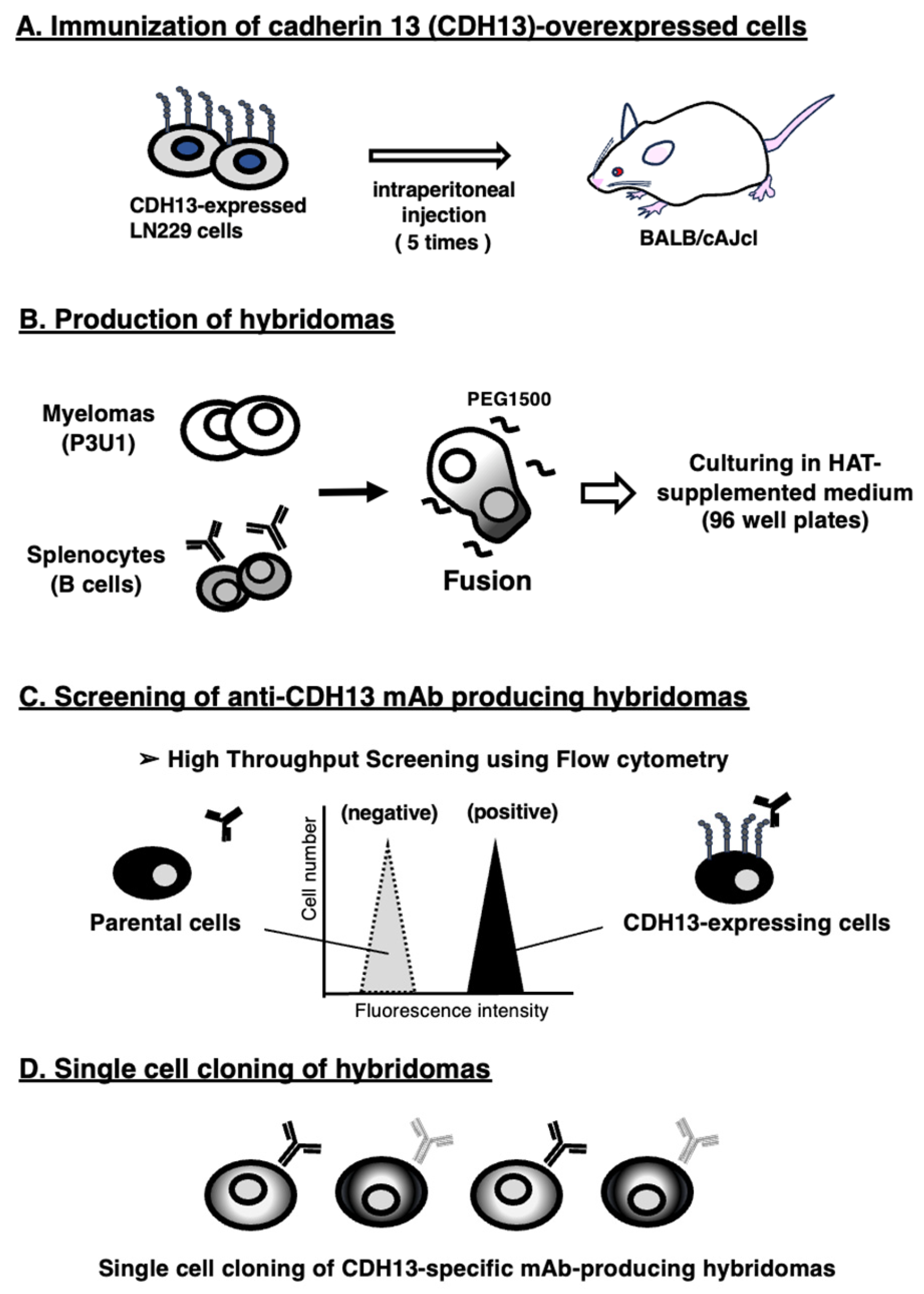
3.1. Development of Anti-CDH13 mAbs Using the CBIS Method

To develop anti-CDH13 mAbs, we employed the CBIS method using CDH13-overexpressed cells. Anti-CDH13 mAbs-producing hybridomas were screened by using flow cytometry (Figure 1). Two female BALB/cAJcl mice were intraperitoneally immunized with LN229/CDH13 (1 × 10⁸ cells/time/mouse) every week, a total of 5 times. Subsequently, mouse splenocytes and P3U1 mouse myeloma cells were fused by PEG1500 under warming conditions. The fused cells were seeded into 96-well plates in HAT medium. After confirming hybridoma formation, flow cytometric screening was conducted to select CHO/CDH13-reactive and parental CHO-K1-nonreactive supernatants of hybridomas. We obtained some highly CHO/CDH13-reactive supernatants of hybridomas. We finally established the highly sensitive clone Ca13Mab-4 (mouse IgG1, kappa) by limiting dilution and additional analysis.

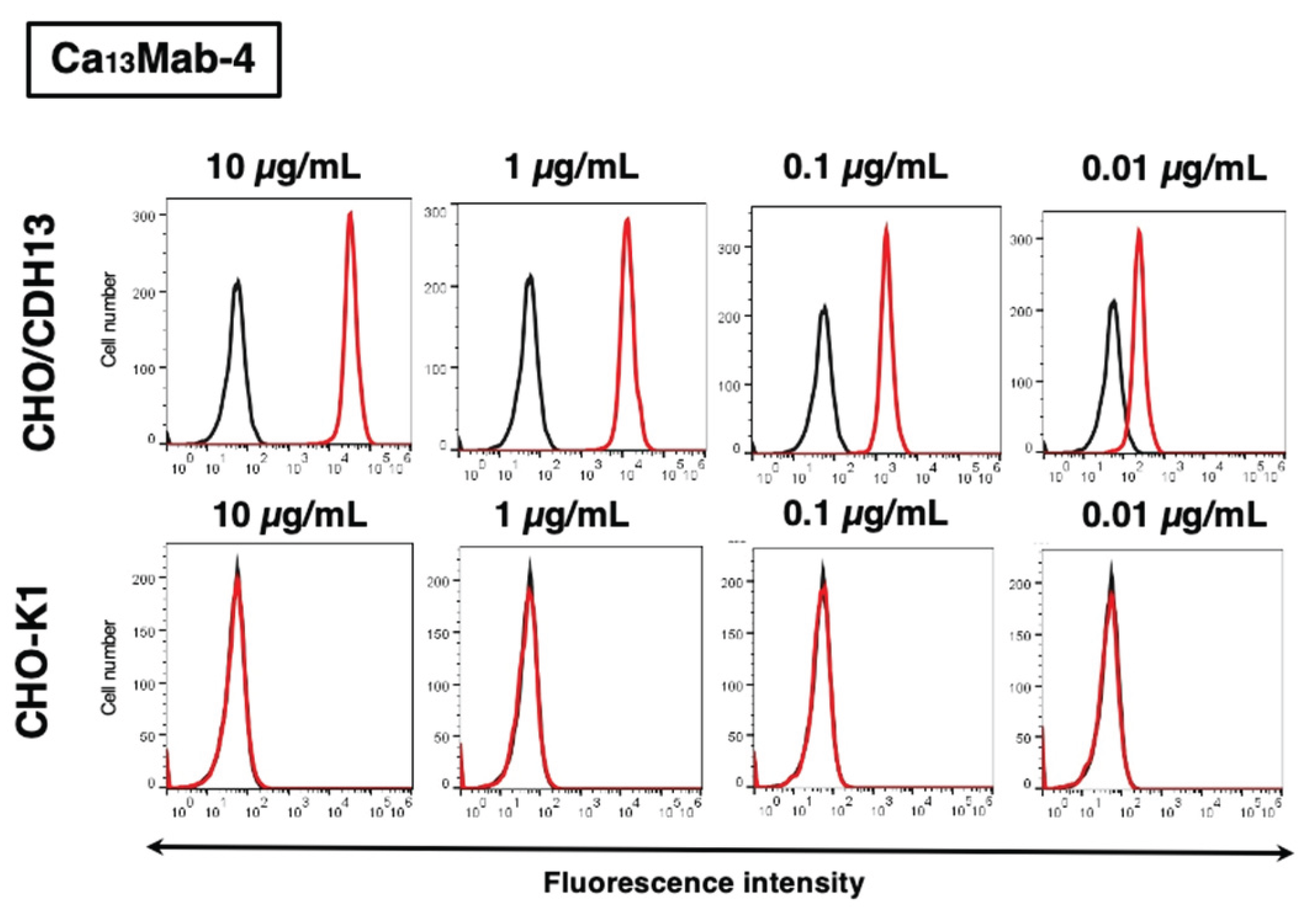
3.2. Evaluation of Ca13Mab-4 Reactivity Using Flow Cytometry

First, flow cytometric analysis was conducted using Ca13Mab-4 against CHO/CDH13 and parental CHO-K1 cells. Results indicated that Ca13Mab-4 recognized CHO/CDH13 dose-dependently (Figure 2 upper). Ca13Mab-4 did not react with parental CHO-K1 cells even at 10 µg/mL (Figure 2, lower). Thus, Ca13Mab-4 can detect CDH13 specifically in flow cytometry.

3.3. Evaluation of Anti-CDH13 mAbs Reactivity Against Endogenous CDH13 Using Flow Cytometry

We conducted flow cytometric analysis using Ca13Mab-4 and a commercially available anti-CDH13 mAb (392411) against U87MG, NCI-H2052, and KMST-6 cells. Results showed that both Ca13Mab-4 and 392411 recognized endogenous CDH13 in a dose-dependent manner (Figure 3). At concentrations of 10 μg/mL and 1 μg/mL of mAbs, Ca13Mab-4 showed almost the same reactivity as 392411. At concentrations of 0.1 μg/mL and 0.01 μg/mL of mAbs, 392411 was more reactive than Ca13Mab-4. Ca13Mab-4 can recognize endogenously expressing CDH13 in flow cytometry.

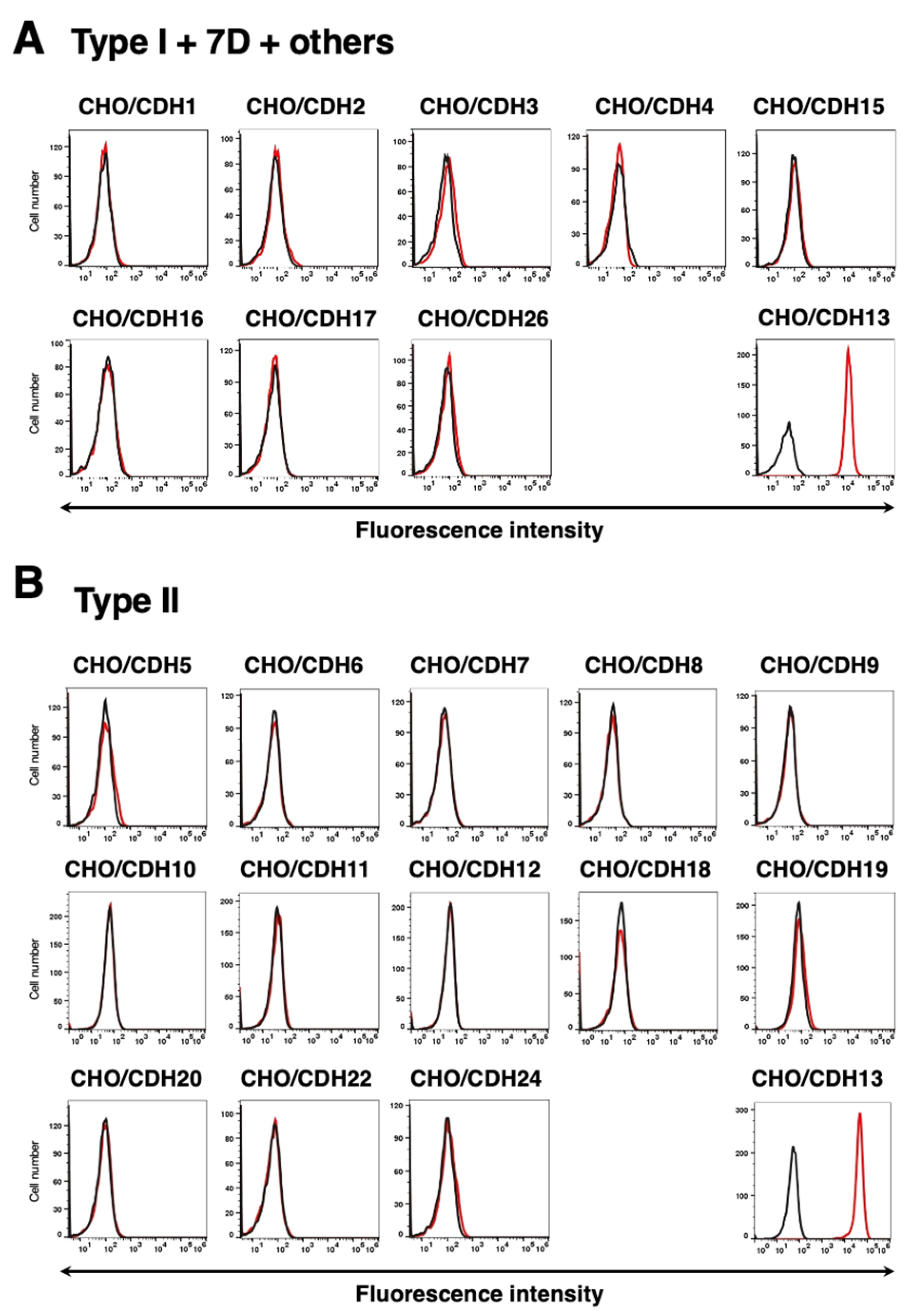
3.4. Specificity of Ca13Mab-4 to CDHs-Overexpressed CHO-K1 Cells

We have established other 22 cell lines of CDHs-overexpressed CHO-K1 cells classified into each clusters [47], including type I CDHs (CHO/CDH1, CHO/CDH2, CHO/CDH3, CHO/CDH4, and CHO/CDH15), type II CDHs (CHO/CDH5, CHO/CDH6, CHO/CDH7, CHO/CDH8, CHO/CDH9, CHO/CDH10, CHO/CDH11, CHO/CDH12, CHO/CDH18, CHO/CDH19, CHO/CDH20, CHO/CDH22, and CHO/CDH24), 7D cluster (CHO/CDH16 and CHO/CDH17) and other (CHO/CDH26). CDH13 belongs to the “other” category. Using the 22 cell lines, the specificity of Ca13Mab-4 was analyzed. As shown in Figure 4, 5 µg/mL of Ca13Mab-4 potently recognized CHO/CDH13, but not other CDHs categorized as type I (7D, other [CHO/CDH26]) (Figure 4A) or type II (Figure 4B). Expression of CDHs in all cell lines was confirmed with corresponding antibodies (Supplemental Figure S1). As a result, it was confirmed that Ca13Mab-4 is a CDH13-specific antibody.

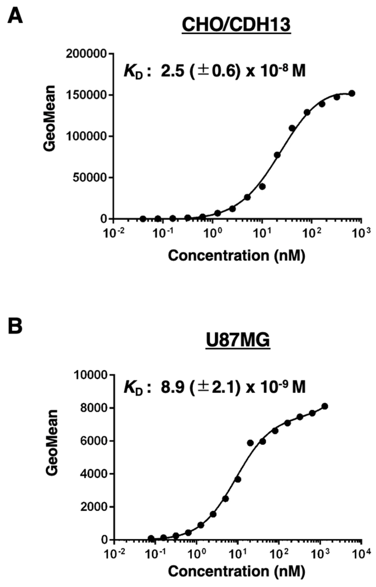
3.5. Calculation of the Apparent Binding Affinity of Ca13Mab-4 Using Flow Cytometry

The binding affinity of Ca13Mab-4 was assessed with CHO/CDH13 and U87MG using flow cytometry. The results indicated that the KD value of Ca13Mab-4 for CHO/CDH13 was 2.5 (± 0.6) × 10−8 M (Figure 5A). The KD value of Ca13Mab-4 for U87MG glioma cell was 8.9 (± 2.1) × 10−9 M (Figure 5B). These results demonstrate that Ca13Mab-4 possesses moderate to high affinity for cell-surface CDH13.

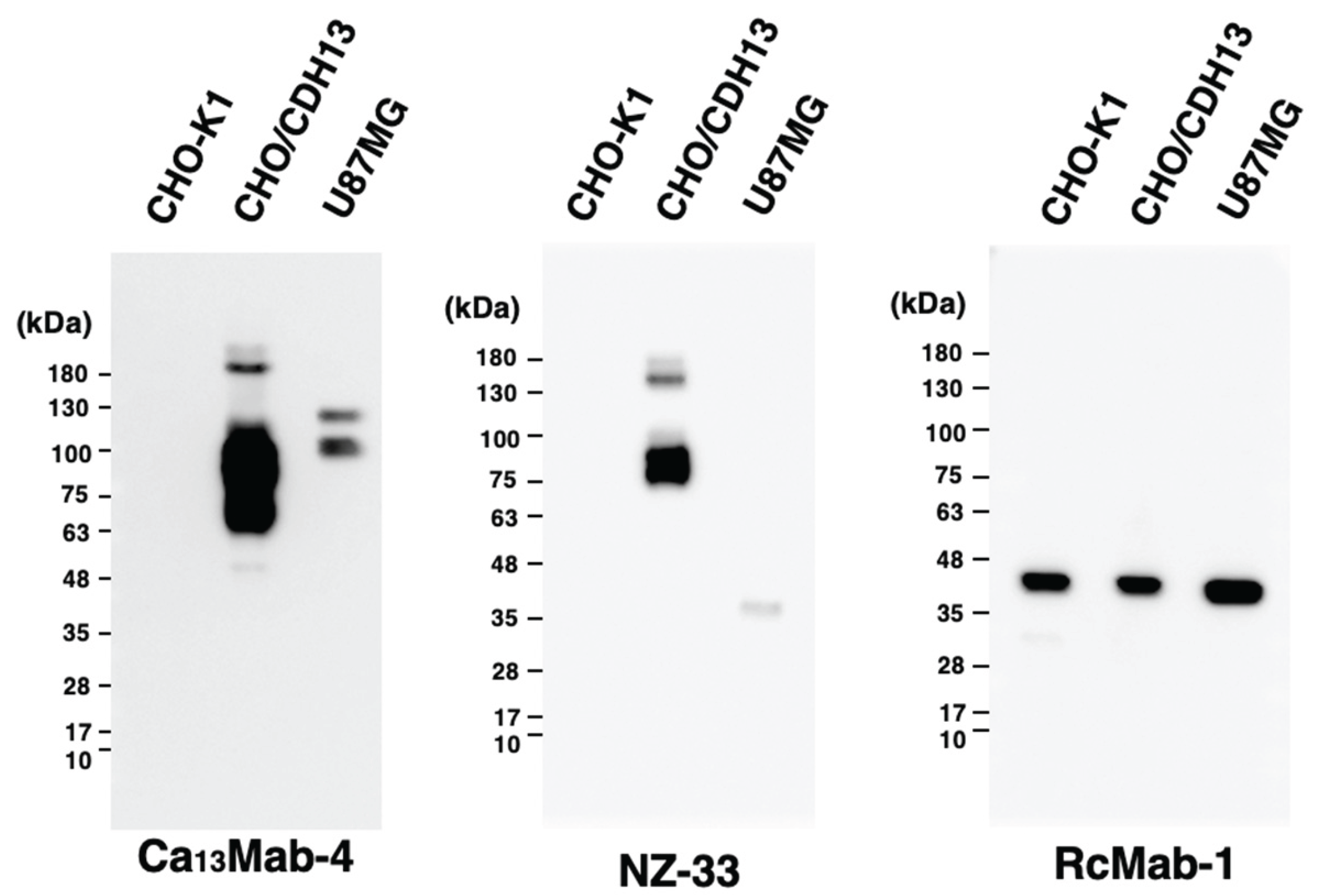
3.6. Western Blot Analyses Using Ca13Mab-4

We investigated whether Ca13Mab-4 can be used for western blot analysis by analyzing cell lysates. As shown in Figure 6, Ca13Mab-4 could detect CDH13 as the major band around 100 kDa in CHO/CDH13 cell lysates, while no band was detected in parental CHO-K1 cells. An anti-PA16 tag mAb (clone NZ-33) could detect a major band in CHO/CDH13 cell lysates at a position very similar to that of Ca13Mab-4. Furthermore, Ca13Mab-4 could detect endogenous CDH13 in U87MG glioma cell lysate (Figure 6). An anti-IDH1 mAb (clone RcMab-1) was used for internal control (Figure 6). These results indicate that Ca13Mab-4 can detect CDH13 in CDH13-expressing cells in western blot analyses.

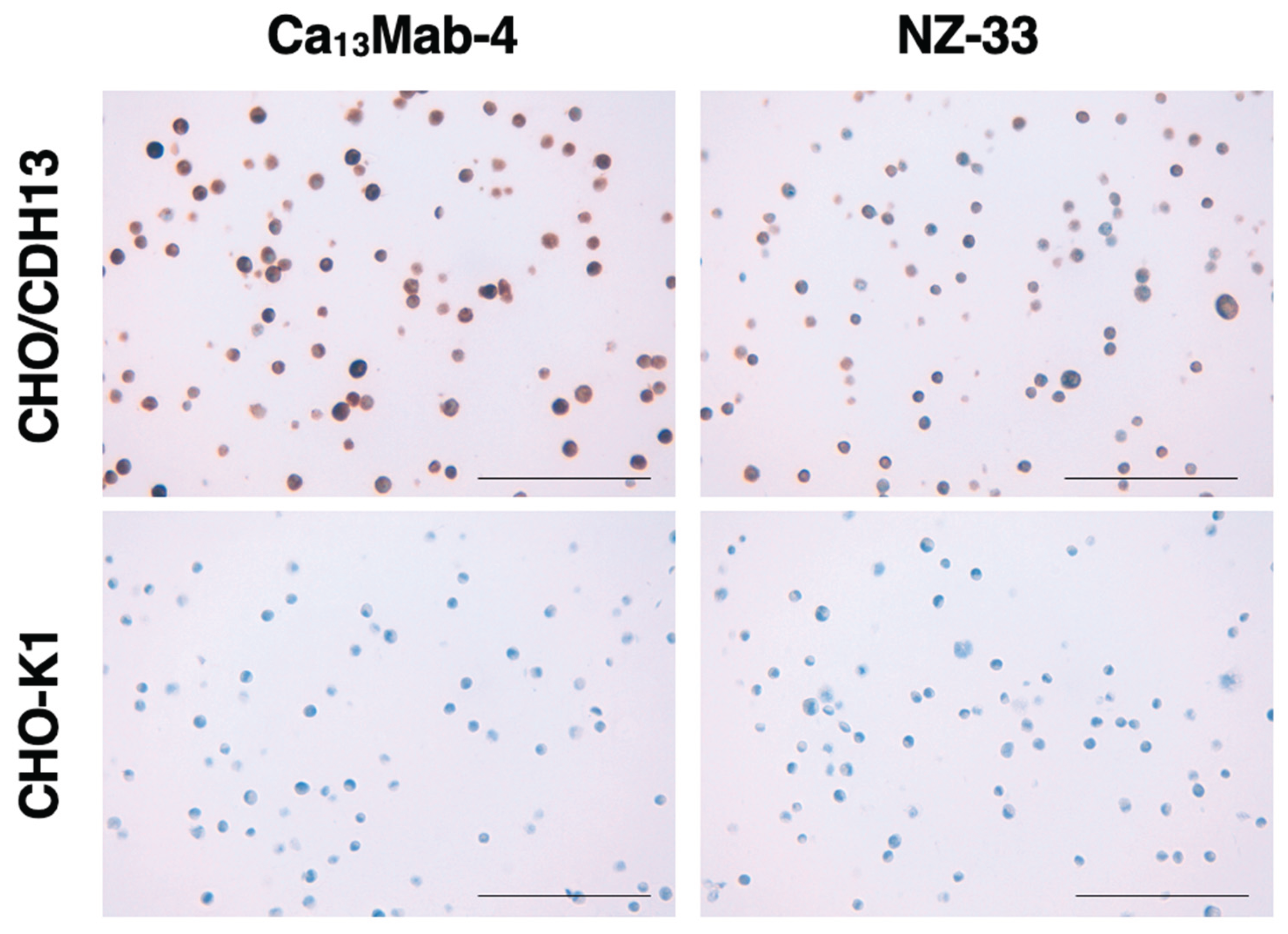
3.7. Immunohistochemistry Using Ca13Mab-4

To investigate whether Ca13Mab-4 can be used for immunohistochemistry (IHC), paraffin-embedded sections of CHO-K1 and CHO/CDH13 were stained with Ca13Mab-4. Apparent staining by Ca13Mab-4 was observed in CHO/CDH13 (Figure 7, upper left). Ca13Mab-4 did not react with the CHO-K1 section (Figure 7, lower left). NZ-33 was used to detect PA16-tagged CDH13 in CHO/CDH13 as a positive control. NZ-33 stained CHO/CDH13 (Figure 7 upper right), but not parental CHO-K1 cell sections (Figure 7 lower right). These results indicate that Ca13Mab-4 is suitable for IHC detection of CDH13-positive cells in paraffin-embedded tissue samples.

4. Discussion

CDH13 is abundantly expressed in the heart and large arteries and localizes to the forming vascular plexus, coincident with active vascular sprouting during cardiovascular development [48]. Systolic blood pressure of CDH13 knockout mice was significantly elevated after physical training [49]. A common single-nucleotide polymorphism located upstream of the Cdh13 gene has been correlated with hypertension in a genome-wide association study [50]. Many reports have demonstrated the close relationship between CDH13 and the cardiovascular system. Similar to its role in normal vessels, CDH13 also contributes to tumor angiogenesis [51,52]. Tumor angiogenesis is vital to supply essential nutrients and oxygen to cancer cells, resulting in an aberrant tumor microenvironment. The overexpression of CDH13 in HMEC-1, human microvascular endothelial cells, leads to the formation of a vascular network within melanoma in a tumor spheroid model [53]. However, in CDH13 the CDH13-deficient MMTV-PyV-mT transgenic breast cancer model, CDH13 deficiency suppresses mammary tumor vascularization and reduces tumor growth, but pathologically advanced and metastasize to the lung [35]. The loss of CDH13 reduces angiogenesis and weakens the function of adhesion factors, which is thought to increase lung metastasis. CDH13 is not considered a monofunctional tumor suppressor, and further analysis is required. The developed Ca13Mab-4 can be applied to numerous experiments, such as flow cytometry (Figure 2, Figure 3, Figure 4 and Figure 5), western blot (Figure 6), and IHC (Figure 7), and will contribute to various analyses of cells and tissues enriched for CDH13 expression.
CDH13 has been identified as a binding partner for adiponectin, hexamers and high-molecular-weight multimers, [15] and low-density lipoproteins (LDL) [54]. LDL-CDH13 clusters induce intercellular calcium signaling from the endoplasmic reticulum [55]. Adiponectin exists in plasma at a high circulating concentration and plays essential roles in tissue homeostasis, including regulation of glucose and lipid metabolism [56]. Disruption of AdipoRs results in increased tissue triglyceride content, inflammation, and oxidative stress, leading to insulin resistance and pronounced glucose intolerance [57]. In contrast, small molecule AdipoR agonist ameliorated diabetes of genetically obese rodent model db/db mice, and extended the shortened lifespan of high-fat diet-fed db/db mice [58]. Interestingly, the soluble form of CDH13 has been reported and plays a pivotal role in pancreatic β-cell proliferation, leading to glucose metabolism via Notch signaling. CDH13-deficient mice exhibited impaired glucose disposal due to attenuated pancreatic β-cell proliferation under high-fat diet conditions [59]. Since CDH13 is a preferred binding partner of adiponectin, it may dramatically regulate glucose metabolism. Polymorphisms in CDH13 are associated with type 2 diabetes and circulating adiponectin levels [60].
Moreover, three forms of soluble CDH13, a 130 kDa form with prodomain, a 100 kDa mature form, and a 30 kDa prodomain form, have been observed in human serum [61]. The 130 kDa form of CDH13 with prodomain positively correlated with plasma adiponectin. Surprisingly, the 30 kDa prodomain form was most strongly associated with several clinical characteristics in patients with type 2 diabetes [61]. According to this report, the effects of different CDH13 forms in cancer may also need to be analyzed, as we detected two clear bands around 100 kDa to 130 kDa in western blot using U87MG glioma cells with Ca13Mab-4 (Figure 6B). Although a correlation between adiponectin and tumor grade of colorectal cancer patients has been reported [62], no evaluation has been conducted that focuses on the relationship between soluble CDH13 or different CDH13 forms and cancer to date. Ca13Mab-4 should be tested to determine whether it can capture these soluble CDH13 forms in colorectal cancer. Along with this evaluation, it is also essential to identify the epitope of Ca13Mab-4. We will determine the epitope of Ca13Mab-4 using cell-based tag-sequence insertion methods, such as PA scanning and the REMAP method [63,64].
Non-cancer-derived KMST-6 cells express CDH13 as shown in Figure 3C. CDH13 is widely known as a tumor suppressor, but it is also associated with some type of cancer malignancy [65]. We have previously established novel mAbs that recognize cancer-specific structure and modification of human epidermal growth factor receptor 2 and podoplanin [66,67]. In future studies, we intend to determine whether cancer-specific epitopes in CDH13 exist by further developing anti-CDH13 mAbs using the same strategy to help distinguish between the two aspects of CDH13 in cancer. It is expected that the development of CDH13 antibodies, including Ca13Mab-4, will contribute to elucidating the pathogenesis of various diseases, including cancer and neurological disorders.

Supplementary Materials

The following supporting information can be downloaded at: Preprints.org.

Credit authorship contribution statement

Yu Kaneko: Investigation. Tomohiro Tanaka: Writing—original draft. Hiroyuki Suzuki: Writing—review and editing. Mika K. Kaneko: Conceptualization. Yukinari Kato: Conceptualization, Funding acquisition, Project administration, Writing—review and editing. All authors have read and agreed to the published version of the manuscript.

Funding Information

This research was supported in part by Japan Agency for Medical Research and Development (AMED) under Grant Numbers: JP25am0521010 (to Y.K.), JP25ama121008 (to Y.K.), JP25ama221153 (to Y.K.), JP25ama221339 (to Y.K.), and JP25bm1123027 (to Y.K.), and by the Japan Society for the Promotion of Science (JSPS) Grants-in-Aid for Scientific Research (KAKENHI) grant nos. 24K18268 (to T.T.) and 25K10553 (to Y.K.).

Institutional Review Board Statement

The animal study protocol was approved by the Animal Care and Use Committee of Tohoku University (Permit number: 2022MdA-001) for studies involving animals.

Informed Consent Statement

Not applicable.

Data Availability Statement

All related data and methods are presented in this paper. Additional inquiries should be addressed to the corresponding authors.

Conflicts of Interest

The authors declare no conflict of interest involving this article.

References

  1. Yulis, M.; Kusters, D.H.M.; Nusrat, A. Cadherins: Cellular adhesive molecules serving as signalling mediators. J. Physiol. 2018, 596, 3883–3898. [Google Scholar] [CrossRef]
  2. Hulpiau, P.; van Roy, F. Molecular evolution of the cadherin superfamily. Int. J. Biochem. Cell Biol. 2009, 41, 349–369. [Google Scholar] [CrossRef]
  3. van Roy, F. Beyond E-cadherin: Roles of other cadherin superfamily members in cancer. Nat. Rev. Cancer 2014, 14, 121–134. [Google Scholar] [CrossRef]
  4. Overduin, M.; Harvey, T.S.; Bagby, S.; Tong, K.I.; Yau, P.; Takeichi, M.; Ikura, M. Solution structure of the epithelial cadherin domain responsible for selective cell adhesion. Science 1995, 267, 386–389. [Google Scholar] [CrossRef]
  5. Shapiro, L.; Fannon, A.M.; Kwong, P.D.; Thompson, A.; Lehmann, M.S.; Grübel, G.; Legrand, J.-F.; Als-Nielsen, J.; Colman, D.R.; Hendrickson, W.A. Structural basis of cell-cell adhesion by cadherins. Nature 1995, 374, 327–337. [Google Scholar] [CrossRef] [PubMed]
  6. Ranscht, B.; Dours-Zimmermann, M.T. T-cadherin, a novel cadherin cell adhesion molecule in the nervous system lacks the conserved cytoplasmic region. Neuron 1991, 7, 391–402. [Google Scholar] [CrossRef] [PubMed]
  7. Wang, H.; Tao, L.; Ambrosio, A.; Yan, W.; Summer, R.; Lau, W.B.; Wang, Y.; Ma, X. T-cadherin deficiency increases vascular vulnerability in T2DM through impaired NO bioactivity. Cardiovasc. Diabetol. 2017, 16, 12. [Google Scholar] [CrossRef]
  8. Takeuchi, T.; Misaki, A.; Liang, S.-B.; Tachibana, A.; Hayashi, N.; Sonobe, H.; Ohtsuki, Y. Expression of T-Cadherin (CDH13, H-Cadherin) in Human Brain and Its Characteristics as a Negative Growth Regulator of Epidermal Growth Factor in Neuroblastoma Cells. J. Neurochem. 2000, 74, 1489–1497. [Google Scholar] [CrossRef]
  9. Straub, L.G.; Scherer, P.E. Metabolic Messengers: Adiponectin. Nat. Metab. 2019, 1, 334–339. [Google Scholar] [CrossRef] [PubMed]
  10. Takemura, Y.; Ouchi, N.; Shibata, R.; Aprahamian, T.; Kirber, M.T.; Summer, R.S.; Kihara, S.; Walsh, K. Adiponectin modulates inflammatory reactions via calreticulin receptor-dependent clearance of early apoptotic bodies. J. Clin. Investig. 2007, 117, 375–386. [Google Scholar] [CrossRef]
  11. Ouchi, N.; Kihara, S.; Arita, Y.; Maeda, K.; Kuriyama, H.; Okamoto, Y.; Hotta, K.; Nishida, M.; Takahashi, M.; Nakamura, T.; et al. Novel modulator for endothelial adhesion molecules: Adipocyte-derived plasma protein adiponectin. Circulation 1999, 100, 2473–2476. [Google Scholar] [CrossRef]
  12. Pischon, T.; Girman, C.J.; Hotamisligil, G.S.; Rifai, N.; Hu, F.B.; Rimm, E.B. Plasma adiponectin levels and risk of myocardial infarction in men. JAMA 2004, 291, 1730–1737. [Google Scholar] [CrossRef]
  13. Hotta, K.; Funahashi, T.; Arita, Y.; Takahashi, M.; Matsuda, M.; Okamoto, Y.; Iwahashi, H.; Kuriyama, H.; Ouchi, N.; Maeda, K.; et al. Plasma concentrations of a novel, adipose-specific protein, adiponectin, in type 2 diabetic patients. Arter. Thromb. Vasc. Biol. 2000, 20, 1595–1599. [Google Scholar] [CrossRef]
  14. Fukuda, S.; Kita, S.; Obata, Y.; Fujishima, Y.; Nagao, H.; Masuda, S.; Tanaka, Y.; Nishizawa, H.; Funahashi, T.; Takagi, J.; et al. The unique prodomain of T-cadherin plays a key role in adiponectin binding with the essential extracellular cadherin repeats 1 and 2. J. Biol. Chem. 2017, 292, 7840–7849. [Google Scholar] [CrossRef] [PubMed]
  15. Hug, C.; Wang, J.; Ahmad, N.S.; Bogan, J.S.; Tsao, T.-S.; Lodish, H.F. T-cadherin is a receptor for hexameric and high-molecular-weight forms of Acrp30/adiponectin. Proc. Natl. Acad. Sci. USA 2004, 101, 10308–10313. [Google Scholar] [CrossRef]
  16. Hebbard, L.W.; Garlatti, M.l.; Young, L.J.T.; Cardiff, R.D.; Oshima, R.G.; Ranscht, B. T-cadherin Supports Angiogenesis and Adiponectin Association with the Vasculature in a Mouse Mammary Tumor Model. Cancer Res. 2008, 68, 1407–1416. [Google Scholar] [CrossRef]
  17. Kita, S.; Fukuda, S.; Maeda, N.; Shimomura, I. Native adiponectin in serum binds to mammalian cells expressing T-cadherin, but not AdipoRs or calreticulin. eLife 2019, 8, e48675. [Google Scholar] [CrossRef] [PubMed]
  18. Rivero, O.; Selten, M.M.; Sich, S.; Popp, S.; Bacmeister, L.; Amendola, E.; Negwer, M.; Schubert, D.; Proft, F.; Kiser, D.; et al. Cadherin-13, a risk gene for ADHD and comorbid disorders, impacts GABAergic function in hippocampus and cognition. Transl. Psychiatry 2015, 5, e655. [Google Scholar] [CrossRef] [PubMed]
  19. Sanders, S.J.; Ercan-Sencicek, A.G.; Hus, V.; Luo, R.; Murtha, M.T.; Moreno-De-Luca, D.; Chu, S.H.; Moreau, M.P.; Gupta, A.R.; Thomson, S.A.; et al. Multiple recurrent de novo CNVs, including duplications of the 7q11.23 Williams syndrome region, are strongly associated with autism. Neuron 2011, 70, 863–885. [Google Scholar] [CrossRef]
  20. Xu, W.; Cohen-Woods, S.; Chen, Q.; Noor, A.; Knight, J.; Hosang, G.; Parikh, S.V.; De Luca, V.; Tozzi, F.; Muglia, P.; et al. Genome-wide association study of bipolar disorder in Canadian and UK populations corroborates disease loci including SYNE1 and CSMD1. BMC Med. Genet. 2014, 15, 2. [Google Scholar] [CrossRef]
  21. Otsuka, I.; Watanabe, Y.; Hishimoto, A.; Boku, S.; Mouri, K.; Shiroiwa, K.; Okazaki, S.; Nunokawa, A.; Shirakawa, O.; Someya, T.; et al. Association analysis of the Cadherin13 gene with schizophrenia in the Japanese population. Neuropsychiatry Dis. Treat. 2015, 11, 1381–1393. [Google Scholar] [CrossRef]
  22. Edwards, A.C.; Aliev, F.; Bierut, L.J.; Bucholz, K.K.; Edenberg, H.; Hesselbrock, V.; Kramer, J.; Kuperman, S.; Nurnberger, J.I.J.; Schuckit, M.A.; et al. Genome-wide association study of comorbid depressive syndrome and alcohol dependence. Psychiatry Genet. 2012, 22, 31–41. [Google Scholar] [CrossRef]
  23. Paradis, S.; Harrar, D.B.; Lin, Y.; Koon, A.C.; Hauser, J.L.; Griffith, E.C.; Zhu, L.; Brass, L.F.; Chen, C.; Greenberg, M.E. An RNAi-based approach identifies molecules required for glutamatergic and GABAergic synapse development. Neuron 2007, 53, 217–232. [Google Scholar] [CrossRef]
  24. Forero, A.; Rivero, O.; Wäldchen, S.; Ku, H.-P.; Kiser, D.P.; Gärtner, Y.; Pennington, L.S.; Waider, J.; Gaspar, P.; Jansch, C.; et al. Cadherin-13 Deficiency Increases Dorsal Raphe 5-HT Neuron Density and Prefrontal Cortex Innervation in the Mouse Brain. Front. Cell Neurosci. 2017, 11, 307. [Google Scholar] [CrossRef]
  25. Ciatto, C.; Bahna, F.; Zampieri, N.; VanSteenhouse, H.C.; Katsamba, P.S.; Ahlsen, G.; Harrison, O.J.; Brasch, J.; Jin, X.; Posy, S.; et al. T-cadherin structures reveal a novel adhesive binding mechanism. Nat. Struct. Mol. Biol. 2010, 17, 339–347. [Google Scholar] [CrossRef]
  26. Hayano, Y.; Zhao, H.; Kobayashi, H.; Takeuchi, K.; Norioka, S.; Yamamoto, N. The role of T-cadherin in axonal pathway formation in neocortical circuits. Development 2014, 141, 4784–4793. [Google Scholar] [CrossRef] [PubMed]
  27. Woodbury-Smith, M.; Lamoureux, S.; Begum, G.; Nassir, N.; Akter, H.; O’rielly, D.D.; Rahman, P.; Wintle, R.F.; Scherer, S.W.; Uddin, M. Mutational Landscape of Autism Spectrum Disorder Brain Tissue. Genes 2022, 13, 207. [Google Scholar] [CrossRef]
  28. Xiang, P.; Li, P.; Yuan, X.; Zhao, X.; Xiao, Z.; Chen, B.; Liu, K.; Bischof, E.; Han, J. Exon 1 methylation status of CDH13 is associated with decreased overall survival and distant metastasis in patients with postoperative colorectal cancer. Discov. Oncol. 2024, 15, 725. [Google Scholar] [CrossRef]
  29. Wang, Y.; Zhang, L.; Yang, J.; Li, B.; Wang, J. CDH13 promoter methylation regulates cisplatin resistance of non-small cell lung cancer cells. Oncol. Lett. 2018, 16, 5715–5722. [Google Scholar] [CrossRef] [PubMed]
  30. Chen, F.; Huang, T.; Ren, Y.; Wei, J.; Lou, Z.; Wang, X.; Fan, X.; Chen, Y.; Weng, G.; Yao, X. Clinical significance of CDH13 promoter methylation as a biomarker for bladder cancer: A meta-analysis. BMC Urol. 2016, 16, 52. [Google Scholar] [CrossRef] [PubMed]
  31. Chmelarova, M.; Baranova, I.; Ruszova, E.; Laco, J.; Hrochova, K.; Dvorakova, E.; Palicka, V. Importance of Cadherins Methylation in Ovarian Cancer: A Next Generation Sequencing Approach. Pathol. Oncol. Res. 2019, 25, 1457–1465. [Google Scholar] [CrossRef] [PubMed]
  32. Martinez, R.; Setien, F.; Voelter, C.; Casado, S.; Quesada, M.P.; Schackert, G.; Esteller, M. CpG island promoter hypermethylation of the pro-apoptotic gene caspase-8 is a common hallmark of relapsed glioblastoma multiforme. Carcinogenesis 2007, 28, 1264–1268. [Google Scholar] [CrossRef]
  33. Wang, Q.; Chen, Y.; Chen, Y.; Jiang, J.; Song, X.; Zhang, L.; He, Q.; Ye, B.; Wu, L.; Wu, R.; et al. Aberrant promoter methylation of T-cadherin in sera is associated with a poor prognosis in oral squamous cell carcinoma. Neoplasma 2021, 68, 528–534. [Google Scholar] [CrossRef]
  34. Wang, Q.; Zhao, Y.; Chen, Y.; Chen, Y.; Song, X.; Zhang, L.; He, Q.; Ye, B.; Wu, L.; Huang, X.; et al. High PD-L1 expression associates with low T-cadherin expression and poor prognosis in human papillomavirus-negative head and neck squamous cell carcinoma. Head. Neck 2023, 45, 1162–1171. [Google Scholar] [CrossRef]
  35. Hebbard, L.W.; Garlatti, M.; Young, L.J.; Cardiff, R.D.; Oshima, R.G.; Ranscht, B. T-cadherin supports angiogenesis and adiponectin association with the vasculature in a mouse mammary tumor model. Cancer Res. 2008, 68, 1407–1416. [Google Scholar] [CrossRef]
  36. Situ, Y.; Deng, L.; Huang, Z.; Jiang, X.; Zhao, L.; Zhang, J.; Lu, L.; Liang, Q.; Xu, Q.; Shao, Z.; et al. CDH2 and CDH13 as potential prognostic and therapeutic targets for adrenocortical carcinoma. Cancer Biol. Ther. 2024, 25, 2428469. [Google Scholar] [CrossRef]
  37. Pfaff, D.; Philippova, M.; Kyriakakis, E.; Maslova, K.; Rupp, K.; A Buechner, S.; Iezzi, G.; Spagnoli, G.C.; Erne, P.; Resink, T.J. Paradoxical effects of T-cadherin on squamous cell carcinoma: Up- and down-regulation increase xenograft growth by distinct mechanisms. J. Pathol. 2011, 225, 512–524. [Google Scholar] [CrossRef] [PubMed]
  38. Kaneko, Y.; Tanaka, T.; Fujisawa, S.; Li, G.; Satofuka, H.; Kaneko, M.K.; Suzuki, H.; Kato, Y. Development of a novel anti-human glypican 5 monoclonal antibody (G5Mab-1) for multiple applications. Biochem. Biophys. Rep. 2025, 43, 102140. [Google Scholar] [CrossRef] [PubMed]
  39. Tanaka, T.; Sakata, T.; Fujisawa, S.; Yamamoto, H.; Kaneko, Y.; Kaneko, M.K.; Suzuki, H.; Kato, Y. Establishment of a novel anti-mouse CD73 monoclonal antibody C73Mab-9 by the Cell-Based Immunization and Screening method. MI 2025. [Google Scholar] [CrossRef]
  40. Tanaka, T.; Suzuki, H.; Taruta, H.; Saga, A.; Li, G.; Fujisawa, S.; Kaneko, M.K.; Kato, Y. Development of a Novel Anti-human EphA1 Monoclonal Antibody, Ea(1)Mab-30, for Multiple Applications. Monoclon. Antib. Immunodiagn. Immunother. 2025, 44, 41–52. [Google Scholar] [CrossRef]
  41. Tanaka, T.; Nanamiya, R.; Takei, J.; Nakamura, T.; Yanaka, M.; Hosono, H.; Sano, M.; Asano, T.; Kaneko, M.K.; Kato, Y. Development of Anti-Mouse CC Chemokine Receptor 8 Monoclonal Antibodies for Flow Cytometry. Monoclon. Antib. Immunodiagn. Immunother. 2021, 40, 65–70. [Google Scholar] [CrossRef]
  42. Fujii, Y.; Kaneko, M.K.; Kato, Y. MAP Tag: A Novel Tagging System for Protein Purification and Detection. Monoclon. Antib. Immunodiagn. Immunother. 2016, 35, 293–299. [Google Scholar] [CrossRef]
  43. Furusawa, Y.; Yamada, S.; Itai, S.; Nakamura, T.; Takei, J.; Sano, M.; Harada, H.; Fukui, M.; Kaneko, M.K.; Kato, Y. Establishment of a monoclonal antibody PMab-233 for immunohistochemical analysis against Tasmanian devil podoplanin. Biochem. Biophys. Rep. 2019, 18, 100631. [Google Scholar] [CrossRef]
  44. Satofuka, H.; Suzuki, H.; Kaneko, M.K.; Kato, Y. Development of Anti-Human Cadherin-26 Monoclonal Antibody, Ca26Mab-6, for Flow Cytometry. Preprints 2025. [Google Scholar] [CrossRef]
  45. Fujisawa, S.; Yamamoto, H.; Tanaka, T.; Kaneko, M.K.; Suzuki, H.; Kato, Y. Development and characterization of Ea7Mab-10: A novel monoclonal antibody targeting ephrin type-A receptor 7. MI 2025. [Google Scholar] [CrossRef]
  46. Ikota, H.; Nobusawa, S.; Arai, H.; Kato, Y.; Ishizawa, K.; Hirose, T.; Yokoo, H. Evaluation of IDH1 status in diffusely infiltrating gliomas by immunohistochemistry using anti-mutant and wild type IDH1 antibodies. Brain Tumor Pathol. 2015, 32, 237–244. [Google Scholar] [CrossRef]
  47. Hulpiau, P.; Gul, I.S.; van Roy, F. New insights into the evolution of metazoan cadherins and catenins. Prog. Mol. Biol. Transl. Sci. 2013, 116, 71–94. [Google Scholar]
  48. Rubina, К.А.; Smutova, V.А.; Semenova, М.L.; Poliakov, А.А.; Gerety, S.; Wilkinson, D.; Surkova, Е.I.; Semina, Е.V.; Sysoeva, V.Y.; Tkachuk, V.А. Detection of T-Cadherin Expression in Mouse Embryos. ActaNaturae 2015, 7, 87–94. [Google Scholar] [CrossRef]
  49. Popov, V.S.; Brodsky, I.B.; Balatskaya, M.N.; Balatskiy, A.V.; Ozhimalov, I.D.; Kulebyakina, M.A.; Semina, E.V.; Arbatskiy, M.S.; Isakova, V.S.; Klimovich, P.S.; et al. T-Cadherin Deficiency Is Associated with Increased Blood Pressure after Physical Activity. Int. J. Mol. Sci. 2023, 24, 14204. [Google Scholar] [CrossRef] [PubMed]
  50. Org, E.; Eyheramendy, S.; Juhanson, P.; Gieger, C.; Lichtner, P.; Klopp, N.; Veldre, G.; Döring, A.; Viigimaa, M.; Sõber, S.; et al. Genome-wide scan identifies CDH13 as a novel susceptibility locus contributing to blood pressure determination in two European populations. Human. Molecular Genet. 2009, 18, 2288–2296. [Google Scholar] [CrossRef]
  51. Philippova, M.; Banfi, A.; Ivanov, D.; Gianni-Barrera, R.; Allenspach, R.; Erne, P.; Resink, T. Atypical GPI-Anchored T-Cadherin Stimulates Angiogenesis In Vitro and In Vivo. Arterioscler. Thromb. Vasc. Biol. 2006, 26, 2222–2230. [Google Scholar] [CrossRef]
  52. Wen, Y.; Ma, L.; Liu, Y.; Xiong, H.; Shi, D. Decoding the enigmatic role of T-cadherin in tumor angiogenesis. Front. Immunol. 2025, 16, 1564130. [Google Scholar] [CrossRef]
  53. Ghosh, S.; Joshi, M.B.; Ivanov, D.; Feder-Mengus, C.; Spagnoli, G.C.; Martin, I.; Erne, P.; Resink, T.J. Use of multicellular tumor spheroids to dissect endothelial cell–tumor cell interactions: A role for T-cadherin in tumor angiogenesis. FEBS Lett. 2007, 581, 4523–4528. [Google Scholar] [CrossRef]
  54. Tkachuk, V.A.; Bochkov, V.N.; Philippova, M.P.; Stambolsky, D.V.; Kuzmenko, E.S.; Sidorova, M.V.; Molokoedov, A.S.; Spirov, V.G.; Resink, T.J. Identification of an atypical lipoprotein-binding protein from human aortic smooth muscle as T-cadherin. FEBS Lett. 1998, 421, 208–212. [Google Scholar] [CrossRef]
  55. Balatskaya, M.N.; Sharonov, G.V.; Baglay, A.I.; Rubtsov, Y.P.; Tkachuk, V.A. Different spatiotemporal organization of GPI-anchored T-cadherin in response to low-density lipoprotein and adiponectin. Biochim. Biophys. Acta (BBA)—Gen. Subj. 2019, 1863, 129414. [Google Scholar] [CrossRef]
  56. Nakano, Y.; Tobe, T.; Choi-Miura, N.H.; Mazda, T.; Tomita, M. Isolation and characterization of GBP28, a novel gelatin-binding protein purified from human plasma. J. Biochem. 1996, 120, 803–812. [Google Scholar] [CrossRef]
  57. Yamauchi, T.; Nio, Y.; Maki, T.; Kobayashi, M.; Takazawa, T.; Iwabu, M.; Okada-Iwabu, M.; Kawamoto, S.; Kubota, N.; Kubota, T.; et al. Targeted disruption of AdipoR1 and AdipoR2 causes abrogation of adiponectin binding and metabolic actions. Nat. Med. 2007, 13, 332–339. [Google Scholar] [CrossRef] [PubMed]
  58. Okada-Iwabu, M.; Yamauchi, T.; Iwabu, M.; Honma, T.; Hamagami, K.-I.; Matsuda, K.; Yamaguchi, M.; Tanabe, H.; Kimura-Someya, T.; Shirouzu, M.; et al. A small-molecule AdipoR agonist for type 2 diabetes and short life in obesity. Nature 2013, 503, 493–499. [Google Scholar] [CrossRef] [PubMed]
  59. Okita, T.; Kita, S.; Fukuda, S.; Fukuoka, K.; Kawada-Horitani, E.; Iioka, M.; Nakamura, Y.; Fujishima, Y.; Nishizawa, H.; Kawamori, D.; et al. Soluble T-cadherin promotes pancreatic β-cell proliferation by upregulating Notch signaling. iScience 2022, 25, 105404. [Google Scholar] [CrossRef] [PubMed]
  60. Nicolas, A.; Aubert, R.; Bellili-Muñoz, N.; Balkau, B.; Bonnet, F.; Tichet, J.; Velho, G.; Marre, M.; Roussel, R.; Fumeron, F. T-cadherin gene variants are associated with type 2 diabetes and the Fatty Liver Index in the French population. Diabetes Metab. 2017, 43, 33–39. [Google Scholar] [CrossRef]
  61. Fukuda, S.; Kita, S.; Miyashita, K.; Iioka, M.; Murai, J.; Nakamura, T.; Nishizawa, H.; Fujishima, Y.; Morinaga, J.; Oike, Y.; et al. Identification and Clinical Associations of 3 Forms of Circulating T-cadherin in Human Serum. J. Clin. Endocrinol. Metab. 2021, 106, 1333–1344. [Google Scholar] [CrossRef]
  62. Polito, R.; Nigro, E.; Fei, L.; De Magistris, L.; Monaco, M.L.; D’Amico, R.; Naviglio, S.; Signoriello, G.; Daniele, A. Adiponectin Is Inversely Associated With Tumour Grade in Colorectal Cancer Patients. Anticancer. Res. 2020, 40, 3751–3757. [Google Scholar] [CrossRef]
  63. Asano, T.; Kaneko, M.K.; Kato, Y. Development of a Novel Epitope Mapping System: RIEDL Insertion for Epitope Mapping Method. Monoclon. Antib. Immunodiagn. Immunother. 2021, 40, 162–167. [Google Scholar] [CrossRef]
  64. Okada, Y.; Suzuki, H.; Tanaka, T.; Kaneko, M.K.; Kato, Y. Epitope Mapping of an Anti-Mouse CD39 Monoclonal Antibody Using PA Scanning and RIEDL Scanning. Monoclon. Antib. Immunodiagn. Immunother. 2024, 43, 44–52. [Google Scholar] [CrossRef]
  65. Philippova, M.; Joshi, M.B.; Kyriakakis, E.; Pfaff, D.; Erne, P.; Resink, T.J. A guide and guard: The many faces of T-cadherin. Cell. Signal. 2009, 21, 1035–1044. [Google Scholar] [CrossRef] [PubMed]
  66. Kato, Y.; Kaneko, M.K. A cancer-specific monoclonal antibody recognizes the aberrantly glycosylated podoplanin. Sci. Rep. 2014, 4, 5924. [Google Scholar] [CrossRef] [PubMed]
  67. Arimori, T.; Mihara, E.; Suzuki, H.; Ohishi, T.; Tanaka, T.; Kaneko, M.K.; Takagi, J.; Kato, Y. Locally misfolded HER2 expressed on cancer cells is a promising target for development of cancer-specific antibodies. Structure 2024, 32, 536–549.e535. [Google Scholar] [CrossRef] [PubMed]
Figure 1. A schematic depiction of the CBIS method for the development of anti-CDH13 mAbs. Simplified steps for mAb development using the CBIS method. (A) LN229/CDH13 cells (1 × 10⁸ cells per mouse per session) were intraperitoneally immunized into two female BALB/cAJcl mice. (B) Spleen cells from LN229/CDH13-immunized mice were fused with P3U1 myeloma cells using PEG1500. (C) The culture supernatants of hybridomas were screened by flow cytometry with CHO-K1 and CHO/CDH13 to select CDH13-specific mAb-producing hybridomas. (D) Single-cell cloning of hybridomas was performed by limiting dilution, followed by additional screening experiments. Ultimately, Ca13Mab-4 (mouse IgG1, kappa) was successfully established.
Preprints 188602 g001
Figure 2. Flow cytometric analysis of Ca13Mab-4. CHO/CDH13 and CHO-K1 cells were treated with 0.01–10 µg/mL of Ca13Mab-4 (red line), followed by treatment with Alexa Fluor 488-conjugated anti-mouse IgG. Fluorescence data were collected using the SA3800 Cell Analyzer. Black line, control (no primary antibody treatment).
Preprints 188602 g002
Figure 3. Flow cytometric analysis of anti-CDH13 mAbs against endogenous CDH13-expressing cells. U87MG (A), NCI-H2052 (B), and KMST-6 (C) cells were treated with 0.01–10 µg/mL of Ca13Mab-4 and 392411 (red line), followed by incubation with Alexa Fluor 488-conjugated anti-mouse or anti-rat IgG. Fluorescence data were collected using the SA3800 Cell Analyzer. Black line, control (no primary antibody treatment).
Preprints 188602 g003
Figure 4. Flow cytometry of Ca13Mab-4 in CDHs-expressed CHO-K1 cells. Cells overexpressing each of the nine CDHs (type I, 7D cluster, and other unaffiliated) (A) and thirteen CDHs (type II) (B) were treated with Ca13Mab-4 (5 µg/mL) (red line). The black line shows the cells treated with control-blocking buffer instead primary antibody. After incubation with primary antibody or control blocking buffer, anti-mouse IgG conjugated with Alexa Fluor 488 was applied. Fluorescence data were collected using the SA3800 Cell Analyzer. CHO/CDH13, which belongs to the ‘other’ categories, was used as a positive control in experiments in A and B.
Preprints 188602 g004
Figure 5. Determination of the binding affinity of Ca13Mab-4. (A) CHO/CDH13 cells were suspended in 100 µL of serially diluted Ca13Mab-4, ranging from 50 µg/mL to 0.003 µg/mL. (B) U87MG cells were suspended in 100 µL of serially diluted Ca13Mab-4, from 100 µg/mL to 0.006 µg/mL. Then, cells were reacted with Alexa Fluor 488-conjugated anti-mouse IgG (dilution ratio 1:200). Subsequently, the geometric mean fluorescence values were obtained using the SA3800 Cell Analyzer, and the KD was calculated with GraphPad PRISM 6 software.
Preprints 188602 g005
Figure 6. Western blot analysis using Ca13Mab-4. Cell lysates of CHO-K1, CHO/CDH13 (10 μg/lane) and U87MG (10 μg/lane) were electrophoresed and transferred onto PVDF membranes. The membranes were incubated with 1 μg/mL of Ca13Mab-4, 0.1 μg/mL of NZ-33, and 1 μg/mL of RcMab-1 and subsequently with horseradish peroxidase-conjugated anti-mouse or anti-rat immunoglobulins.
Preprints 188602 g006
Figure 7. Immunohistochemical staining of paraffin-embedded section of CHO/CDH13 and parental CHO-K1. The sections of CHO/CDH13 and CHO-K1 cells were treated with 5 μg/mL of Ca13Mab-4 or 0.1 μg/mL of NZ-33. The staining was performed using BenchMark ULTRA PLUS with the ultraView DAB IHC Detection Kit. Scale bar = 100 μm.
Preprints 188602 g007
Disclaimer/Publisher’s Note: The statements, opinions and data contained in all publications are solely those of the individual author(s) and contributor(s) and not of MDPI and/or the editor(s). MDPI and/or the editor(s) disclaim responsibility for any injury to people or property resulting from any ideas, methods, instructions or products referred to in the content.
Copyright: This open access article is published under a Creative Commons CC BY 4.0 license, which permit the free download, distribution, and reuse, provided that the author and preprint are cited in any reuse.
Prerpints.org logo

Preprints.org is a free preprint server supported by MDPI in Basel, Switzerland.

Subscribe

Disclaimer

Terms of Use

Privacy Policy

Privacy Settings

© 2026 MDPI (Basel, Switzerland) unless otherwise stated