

Submitted:
04 December 2025
Posted:
05 December 2025
You are already at the latest version
Abstract
Background: Catheter-associated urinary tract infections (CAUTIs) are still the most common healthcare-associated infections globally. Although catheter designs have improved, based on the duration of use, such as indwelling catheters, substantial numbers of infections are still caused by contamination from the collection bag (intraluminal) and from the external catheter's surface (extraluminal). This study assessed the safety and practicality of a novel silver-based device designed for external attachment between a catheter outlet and a urine collection bag to stop bacteria from traveling up from the urine bag into the bladder through the inside of the tube. Methods: The accessory features a silver ion-releasing polymer matrix with a synergistic formulation and an anti-reflux valve to prevent urine backflow. Laboratory tests included microbial adherence, 10-day biofilm formation, and MIC assays against common uropathogens. Biocompatibility was assessed by ISO 10993-5. A pilot clinical trial randomized 157 catheterized patients (80 received the antiseptic accessory, 77 were controls) to evaluate safety and initial efficacy. Results: Laboratory studies showed that the matrix material reduced all tested microbes by at least 5 log and had minimum inhibitory concentrations (MICs) of 0.15–0.3 ppm for silver ions. Cytotoxicity testing found it to be non-cytotoxic (Grade 0–2). In clinical assessments, the accessory was evaluated for CAUTI-related organisms by comparing urine samples from the bag and catheter port. The accessory reduced intraluminal infection incidence in half (5.6% vs. 9.6%) and no device-related adverse events were reported. Conclusions: The accessory device was biocompatible, well tolerated, and showed strong antimicrobial activity against common CAUTI pathogens. It may help prevent intraluminal infections, but not overall CAUTI due to extraluminal infection via the catheter surface. More research is needed to confirm the benefits of this concept.
Keywords:
urinary catheter
; CAUTI
; urine bag
; accessory
; intraluminal
; extraluminal
1. Introduction
Urinary tract infections (UTIs) are a common hospital-acquired infection, with indwelling catheters responsible for about 80% of cases [1,2]. Catheter-associated UTIs (CAUTIs) can increase morbidity, prolong hospital stays, and raise costs globally [3]. Indwelling urinary catheters are vital in perioperative care, urinary retention, critical monitoring, and long-term management [3] but cause around 75% of hospital-acquired UTIs. The duration of catheterization is a key risk factor for CAUTI and bacteriuria [4].
Catheter-associated bacteriuria develops in up to 25% of patients within five days of catheterization, with an estimated daily acquisition risk ranging from 1% to 5% [ 5,6,7] The impact of CAUTI extends beyond infection to include prolonged hospitalization, increased morbidity and mortality, and significant economic costs—estimated at $400–500 million annually. [8,9,10,11,12]
Most infections arise via extraluminal contamination routes, with approximately two-thirds attributable to extraluminal migration and the 1/3 via the intraluminal route [5,6]. Predominant CAUTI pathogens include Gram-negative bacilli (Escherichia coli, Klebsiella pneumoniae, Pseudomonas aeruginosa), Gram-positive cocci (Enterococcus species), and fungi (Candida species), many of which exhibit multidrug resistance (MDR).[7,16]
Although infection-control measures such as aseptic technique and closed drainage systems have been shown to mitigate risk, intraluminal contamination persists, frequently resulting from contaminated collection bags or reflux of colonized urine. [6,13] and protection against intraluminal contamination remains inadequate. [19,20]. Low- and middle-income countries (LMICs) often face deficiencies in surveillance systems and catheter stewardship programs, with limited availability of metrics such as CAUTI incidence and catheter-days [5]. Device innovation is further constrained by economic and logistical challenges.
To address these issues, we designed a silver formula impregnated device to prevent microbes from entering the catheter from the urine collection bag (Patent: US 7,147,625 B2). This study describes its design, microbiological effectiveness, biocompatibility, and initial clinical results.
2. Materials and Methods
2.1. Device Description
The accessory device comprises:
- An antimicrobial Matrix: A Hydrophilic medical-grade resin compounded with silver-salt containing antimicrobial powder formulation (ICET Inc.) via twin-screw extrusion at 135 °C, followed by pelletization and drying.
- Assembly: The compounded polymer is injection-molded (185 °C) using a Sumitomo SE18s machine to form the matrix, integrated with a silicone cone adapter, a duck-bill medical grade, one-way anti-reflux valve and medical grade RTV sealant.
- Sterilization: Ethylene oxide (EO) sterilization per ANSI/AAMI/ISO 10993-7 and AAMI 11135 standards, with residuals <0.1 mg/24h for EO and ethylene chlorohydrin.
The accessory device is shown in Figure 1 and the configuration for its sterile assembly with the catheter.
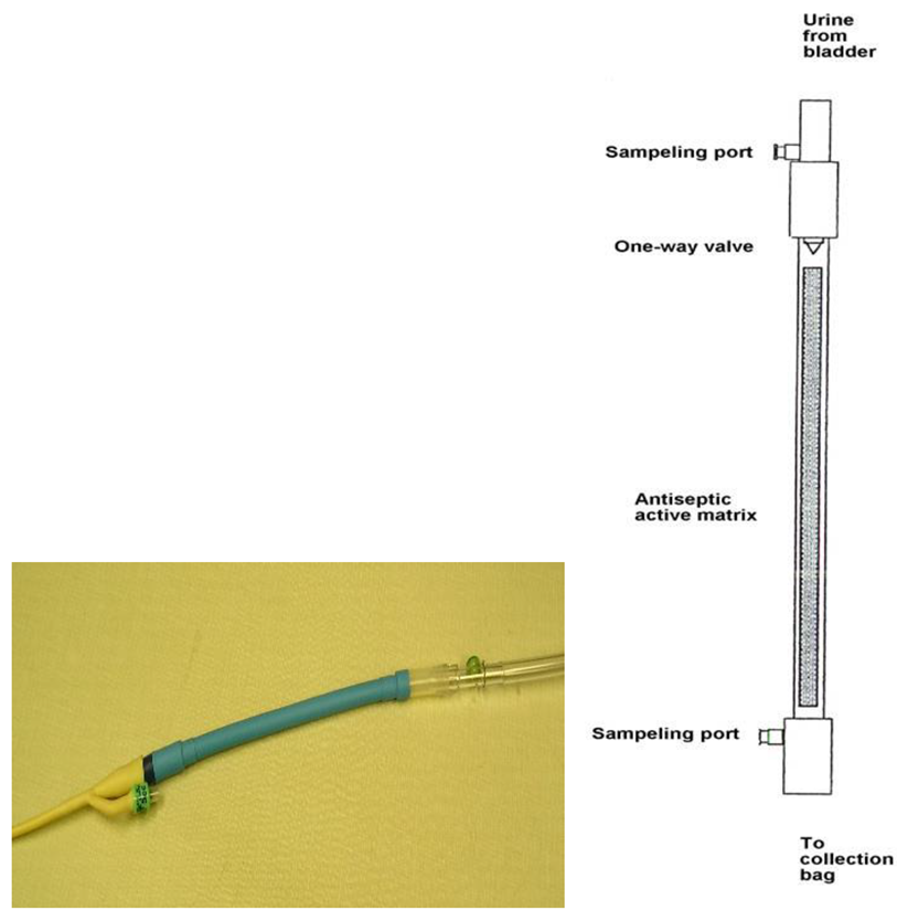
Figure 1 shows the attachment of the accessory (blue) with Bardex silver hydrogel catheter and the tubing for collection bag.
2.2. Microbiology
Organisms Used
Ten clinically relevant uropathogens were sourced from ATCC, USA and clinical isolates from hospital resources, were tested, including E. coli 53498 and a clinical isolate (EC), K. pneumoniae, (Kp) P. aeruginosa, (PA) S. aureus (MRSA), E. faecalis (VRE), C. albicans, CA, Proteus mirabilis, PM Serratia marcescens (SM) and S. aureus ATCC 25923, SA.
Justification for Organisms Used
Organisms used in this study were based upon the recommendations by consulting clinicians. The organisms used are ones often responsible for Catheter Associated Urinary Tract Infections (CAUTI).
Microbial Growth Conditions
Organisms were streaked onto tryptic soy agar from frozen stock. The organisms were incubated overnight at 37 °C. Single colonies were cultured overnight at 37 °C in 1 mL tryptic soy broth. For plating of the contacting solution, biofilm, and roll plate analysis TSA is supplemented with 1% neutralizer (mercaptoacetic acid).
Evaluation of accessory material to reduce microbial growth over time (Catheter 10 day trial, 2x challenge)
Sample Preparation
Sterile packaged samples (EO sterilized) were cut using sterile technique to remove the material from various lots from the plastic tubing. These samples were then placed into 5mL culture tubes. The average weight of the sample matrix is 0.56g, 2.1 cm long and 1.1 cm across. 1950 µl of simulated urine (SU) [14] containing 1.25% Tryptic Soy Broth (TSB) by weight was added to each test tube. A control without antimicrobial was treated similarly. Each sample was prepared duplicate for each organism. The sample and control pieces were incubated at 37℃ for two hours in SU with 1.25% TSB prior to microbial challenge.
Method for a 10 day trial, 2x challenge
Sample Preparation
Sterile packaged samples (EO sterilized) were cut using sterile technique to remove the accessory material (Lot # 080507) from the plastic tubing. Control samples were without antimicrobial additive. These samples were then placed into 5mL culture tubes. 1950 µl of SU containing 1.25% Tryptic Soy Broth (TSB) by weight was added to each test tube. Each sample was prepared duplicate for each organism. The sample and control pieces were incubated at 37oC for two hours in SU with 1.25% TSB prior to microbial challenge.
Inoculum Preparation
On Day 0 microbial cells were inoculated at 106 CFUs/mL into the samples. Samples were then incubated at 37oC. Because K. pneumonia and P. mirabilis create a toxic alkaline environment in synthetic urine under static conditions the methodology was altered. On Day 0, K. pneumonia was inoculated at 105CFUs/mL and P. mirabilis was inoculated at 104 CFUs/mL.
Sample Conditions
Because synthetic urine media under static conditions eventually reduces the viability of growing and dense microbial cultures the test samples were transferred to fresh tubes containing synthetic urine with 1.25% Tryptic soy broth, on Day 4 and Day 8 of the experiment. On day 4, in addition to media refreshment, the test samples are also re-challenged with 106 CFUs/mL of microbe.
P. mirabilis and to a lesser extent K. pneumonia both quickly lose culture viability due to a pH increase over time. To address that effect alterations were made to the methodology for these two organisms. Each morning the control samples from P. mirabilis were washed in 2mL PBS and transferred to fresh SU media with 1.25% TSB. The control and coated samples were transferred to fresh SU media with 1.25% TSB on Day 4. Each day P. mirabilis samples were inoculated with 104 CFUs/mL. The K. pneumonia samples were transferred to fresh SU media with 1.25% TSB on day 4 and day 8, then re-challenged with 105 CFUs/mL on day 4 and day 8. On Day 8 the control K. pneumonia samples were washed with 2mL PBS and transferred to fresh SU media with 1.25% TSB and re-challenged with 104 CFUs/mL.
Contact Solution, Part A
On days 7, and 10 all samples were assayed for growth of the contacting solution. On day 10 the samples were also assayed for biofilm formation on the piece surface. 100µl aliquots from each sample solution is removed on the days assayed. One-tenth serial dilutions in PBS are performed in a final volume of 100µl. For each dilution 10µl is plated into TSA containing 1% neutralizer (Mercaptoacetic acid). After 24hr incubation at 37oC these plates are counted for colony forming units
For P. mirabilis, which received daily re-challenge, the samples were inoculated each morning and incubated for 5 hours before plating. On days when samples were refreshed or re-challenged this step was performed after plating of the contacting solution except for P. mirabilis which was re-challenged each day five hours prior to plating of the contacting solution.
Biofilm Formation, Part B
On day 10 after plating contact solution the piece is removed from the SU medium and placed into 10 mL sterile PBS. The piece is washed by vortexing for 10sec and then transferred to fresh 10 mL sterile PBS. This process is repeated for a total of 3 washes. After the 3rd wash the pieces are rolled across the surface of a TSA plate with neutralizer and incubated for 24 hours at 37oC. The roll plates are examined for growth. Total lawn describes a surface and surrounding area of the catheter/agar interface to be colonized by a lawn of microbe.
2.4. Biocompatibility Testing
In vitro Cytotoxicity tests (ISO 10993-5) were conducted at Toxikon , MA using L929 mammalian cells.
2.5. Clinical Evaluation
A randomized, double-blind pilot enrolled 157 catheterized patients under an IRB approval at the University of Wisconsin Hospital under Dennis Maki, MD.: 80 received the antiseptic accessory connected to the standard of care (silver-hydrogel Bard) catheter, 77 standard-of- care (silver-hydrogel Bardex) catheters. Catheters remained for ≥10 days. Outcomes included CAUTI organisms incidence and infection mechanism (intraluminal vs. extraluminal).
Patients entering with baseline low-level bacteriuria (≤103 cfu/ml) were enrolled since newly-acquired nosocomial bacteriuria, or a subsequent catheter-associated superinfection with a different organism, was the primary outcome measure. The endpoint variables for efficacy determination were: Newly acquired organisms, defined as ≥103 cfu/ml of a genus different from any previously identified. In this study, CAUTI was defined as new appearance of bacteriuria or candiduria > 103 CFU/mL in urine aspirated from the collection port of the drainage system.
On entry into the study and daily thereafter until the catheter is removed, aliquots of urine will be obtained separately from the urine catheter sampling port and from the outlet tube of the collection bag, and cultured using a technique that can detect as few as 1 cfu/ml. The date and time of sample collections were recorded. Organisms were enumerated and identified using standard techniques and criteria9.
3. Results
3.1. In Vitro Efficacy
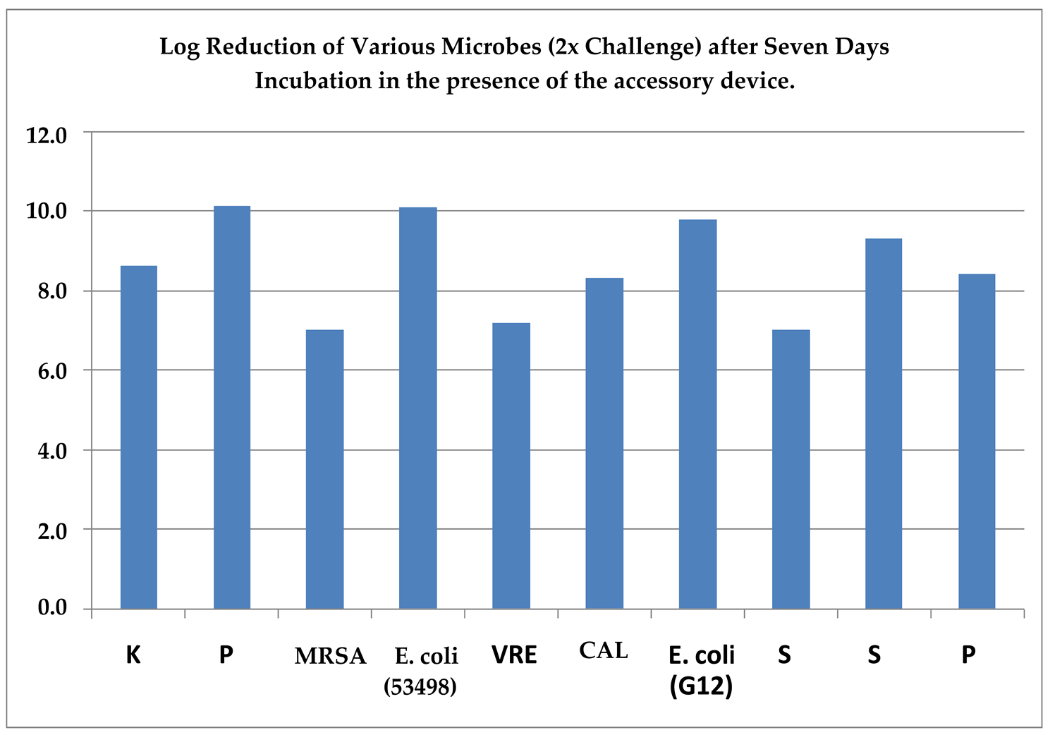
All organisms demonstrated ≥5log reduction in viable counts in contact solution assays compared with controls after 7–10 days.
Assay Criteria: at least 4 logs reduction (99.99%) of growth (CFUs/mL) in the accessory sample to the control. Organisms from left to right: Klebsiella pneumoniae, Pseudomonas aeruginosa, Staphylococcus aureus (MRSA), Escherchia coli, Enterococcus faecalis (VRE), Candida albicans, E. coli, S. aureus, Serratia marcesscens, Proteus mirabilis.
Results from Figure 2 reveal at least 6 logs reduction in growth for all organisms tested (in duplicate) as compared to the control (Limit of Detection is 1000 CFUs/mL) for day 7. The log reduction in growth of the contacting solution is obtained by subtracting the averaged log10 value (CFUs/mL) of the accessory samples from the averaged log10 value of control (CFUs/mL).
Direct application of the sample piece after 10-day incubation onto the surface of agar (Roll Plate) (Table 1) shows the resistance of the accessory material to biofilm formation as compared to the control. Total lawn describes the surface and surrounding area of the catheter/agar interface colonized by a lawn of microbe. NG denotes no detectable growth. Plates were also visually inspected after a 24 hr incubation at 37 °C and again three days later.
3.2. Biocompatibility
Samples of the test device MEM extracts exhibited a reactivity score of “0” following 48 hours of incubation, indicating an absence of cytotoxic response. The test device was classified as non-cytotoxic with respect to direct contact cytotoxicity involving the Accessory Device filter matrix. Mild biological reactivity (Grade 2) was noted in L929 mammalian cells. Overall, the test device is considered non-cytotoxic.
Reactivity was evaluated on a scale of 0–4 against positive and negative controls, with scores of ≤2 interpreted as non-cytotoxic.
3.3. Clinical Outcomes
Human Pilot Study Results
A 10-day human pilot study was conducted in a hospital population of catheterized patients to evaluate the effectiveness of the intraluminal accessory device in assessing organisms causing catheter-associated urinary tract infections (CAUTIs). In this study, the overall incidence of new CAUTI organisms was similar between the groups: 37.5% in the group using the antiseptic accessory device and 38.2% in the control group. However, a difference was observed in the rates of intraluminal infections, which were nearly halved in the accessory device group (5.6%) compared to the control group (9.6%; p = 0.59). This finding indicates a trend toward reduced intraluminal infections with the use of the accessory device, although the difference did not reach statistical significance.
No adverse device-related events were reported during the study period. The infecting organisms identified were primarily bacteria and yeasts. In total, 26 distinct microorganisms were identified among the patient population during this clinical study.
Incidence of Infections in Study Groups
The following table compares the percentage of infections observed in the control group and the group that utilized the intraluminal accessory device. The data were originally presented at the 2004 Interscience Conference on Antimicrobial Agents and Chemotherapy (ICAAC) in Washington, D.C. [#970 Prospective, Randomized Double-Blinded Trial of a Novel Antiseptic Urine-Collection Device for Prevention of Intraluminally-Acquired Catheter-Associated Urinary Tract Infection (CAUTI) D.G. Maki, A.M. Jones, L.L. Narans, University of Wisconsin and subsequently reported in Urology Times in 2005].
Mechanisms of CAUTI in Two Treatment Groups (Duration: average 10 Days or More) (Taken from Dr. Dennis Maki’s presentation at the ICAAC Meeting, November 2004, Washington, DC)
| Control[n=77] Catheters |
Catheters with accessory Device[n=80] |
p-value | |
| Extraluminal (%) Intraluminal (%) Indeterminate (%) |
15 (17.2) 10 (9.6) 18 (14.4) |
16 (14.6) 5 (5.6) 13 (18.0) |
0.82 0.59 0.10 |
Expert Commentary and Interpretation
Dr. Dennis Maki, who presented the findings at the ICAAC meeting in November 2004, commented that the novel device was well-tolerated, but did not significantly reduce the incidence of CAUTI, in part because of the large proportion of extraluminally-acquired infections. We believe that the concept is sound and that there could be benefit for prevention of intraluminally-acquired CAUTIs, possibly with a highly effective microbially-impervious antireflux valve or with a device similar to that tested in this trial. It is very plausible that an intraluminal antiseptic-matrix with far greater surface area and higher level of surface anti-infective activity might prove effective for preventing intraluminally-acquired CAUTIs. The use of an antiseptic, as contrasted with an antibacterial or specific antifungal is highly unlikely to select for resistance to the agent incorporated in the antiseptic matrix.. There was a trend suggesting that it may be of some benefit for preventing intraluminally-acquired CAUTI. The concept is sound, but there may not be sufficient antibacterial activity diffusing off the matrix to reliably kill organisms in urine passing over it.
3.4. Discussion
Ionic silver is well-known for its antimicrobial properties in laboratory settings; however, its effectiveness is often reduced when applied to medical devices. This reduction is attributed to several factors, including the development of biofilms, interactions with bodily fluids, restricted release of silver ions, and various tissue interactions [22,23]. To overcome these challenges, proprietary formulations for the intraluminal accessory along with device design concepts were developed. These formulations were designed to provide controlled silver ion release over a ten-day period and incorporate additional elements to improve their combined antimicrobial efficacy.
The hypothesis behind these formulations was that complexation of silver ions, such as those bound to EDTA type chelators or citrate, possess increased solubility and can help reduce precipitation of silver ions that commonly occurs due to chloride ligands present in urine. The EDTA and silver could act synergistically because EDTA weakens the bacterial outer membrane by removing Mg²⁺ and Ca²⁺, increasing permeability. Even though EDTA and chloride bind some Ag⁺, a portion remains free, and this free Ag⁺ remains at a certain equilibrium concentration. [24,25] The dominance of specific silver complexes in the urinary system is influenced by the salts, protein-based ligands present and competitive ligand reactions, which determine the prevailing silver species in this environment[ 26,27].
Enhancing the urine-contact area of the accessory during routine catheter use may further improve its antimicrobial efficacy and overall performance. Further research is warranted to refine intraluminal accessory designs and formulations, with particular emphasis on increasing antimicrobial efficacy and performance during standard catheter use. Ongoing development of these devices with potential incorporation of probiotics, their derivatives, or similar agents, may contribute to safe and more effective catheter management in clinical practice, ultimately reducing infection rates and improving patient outcomes.
Effective reduction of infection rates associated with indwelling urinary catheters requires optimization of both major pathways (extraluminal and intraluminal) by which microbes can enter the urinary tract. Although the accessory device did not demonstrate a statistically significant reduction in overall CAUTI organism incidence, there was a notable trend toward decreased intraluminal infections.
3.5. Clinical Relevance and Global Implications
Foley catheters are among the most widely used medical devices globally, with nearly 100 million units placed each year. While most of these catheters are intended for temporary use, long-term catheterization remains necessary in many cases. A 2023 systematic review of 67 studies across multiple countries found that among nursing-home residents, the median prevalence of indwelling urinary catheters is 7.3% (interquartile range 4.3 – 10.1 %.[28] Extended use increases the risk of complications, including infection. These challenges are particularly pronounced in low-resource settings, where supply limitations and inadequate training contribute to higher infection rates and persistent public health disparities.
4. Acknowledgements
This work was supported by the National Institutes of Health (NIH) grant 2R44DK55891. Special thanks are extended to Primrose Medical Inc for the fabrication of the accessory and sterilization under a contract with ICET, Inc. The author would like to express gratitude to Dr. Dennis G. Maki for leading the human trial and clinical coordinators Ms. Anne Jones, and L.L Narans at University of Wisconsin Hospitals, under the auspices of this grant.
5. Data Availability
Data is available from the author upon reasonable request. For ongoing developments contact shanthasarangapani@gmail.com
6. Conflicts of Interest
The author declares no conflicts of interest related to this work.
References
- Saint, S.; Trautner, B. W.; Fowler, K. E.; Colozzi, J.; Ratz, D.; Lescinskas, E.; Krein, S. L. A Multicenter Study of the Prevalence of Indwelling Urinary Catheter Use in the United States. Infect. Control Hosp. Epidemiol. 2019, 40(4), 420–423. [Google Scholar]
- Hooton, T. M.; Bradley, S. F.; Cardenas, D. D.; Colgan, R.; Geerlings, S. E.; Rice, J. C.; Saint, S.; Schaeffer, A. J.; Tambyah, P. A.; Nicolle, L. E. Diagnosis, Prevention, and Treatment of Catheter-Associated Urinary Tract Infection in Adults: 2009 International Clinical Practice Guidelines. Clin. Infect. Dis. 2010, 50(5), 625–663. [Google Scholar] [CrossRef]
- Meddings, J.; Rogers, M. A. M.; Krein, S. L.; Fakih, M. G.; Olmsted, R. N.; Saint, S. Reducing unnecessary urinary catheter use and other strategies to prevent catheter-associated urinary tract infection: an integrative review. BMJ Qual. Saf. 2014, 23(4), 277–289. [Google Scholar] [CrossRef]
- Magill, S. S.; Edwards, J. R.; Bamberg, W.; Beldavs, Z. G.; Dumyati, G.; Kainer, M. A.; Lynfield, R.; Maloney, M.; McAllister-Hollod, L.; Nadle, J.; et al. Multistate Point-Prevalence Survey of Health Care–Associated Infections. N. Engl. J. Med. 2014, 370, 1198–1208. [Google Scholar] [CrossRef]
- Tambyah, P. A.; Maki, D. G. Catheter-Associated Urinary Tract Infection Is Rarely Symptomatic: A Prospective Study of 1,497 Catheterized Patients. Arch. Intern. Med. 2000, 160, 678–682. [Google Scholar]
- Maki, D. G.; Tambyah, P. A. Engineering Out the Risk of Infection with Urinary Catheters. Emerg. Infect. Dis. 2001, 7(2), 342–347. [Google Scholar] [CrossRef]
- Flores-Mireles, A. L.; Walker, J. N.; Caparon, M.; Hultgren, S. J. Urinary tract infections: epidemiology, mechanisms of infection and treatment options. Nat. Rev. Microbiol. 2015, 13, 269–284. [Google Scholar] [CrossRef]
- Saint, S.; Greene, M. T.; Krein, S. L.; Rogers, M. A. M.; Ratz, D.; Fowler, K. E.; Edson, B. S.; Watson, S. R.; Meyer-Lucas, B.; Fakih, M. G. A Program to Prevent Catheter-Associated Urinary Tract Infection in Acute Care. N. Engl. J. Med. 2016, 374, 2111–2119. [Google Scholar] [CrossRef]
- Nicolle, L. E. Catheter associated urinary tract infections. Antimicrob. Resist. Infect. Control 2014, 3, 23. [Google Scholar] [CrossRef] [PubMed]
- Trautner, B. W.; Grigoryan, L. Approach to a positive urine culture in a patient without urinary symptoms. Infect. Dis. Clin. North Am. 2014, 28, 15–31. [Google Scholar] [CrossRef] [PubMed]
- Umscheid, C. A.; Mitchell, M. D.; Doshi, J. A.; Agarwal, R.; Williams, K.; Brennan, P. J. Estimating the Proportion of Healthcare-Associated Infections That Are Reasonably Preventable and the Related Mortality and Costs. Infect. Control Hosp. Epidemiol. 2011, 32, 101–114. [Google Scholar] [CrossRef]
- Saint, S.; Kaufman, S. R.; Thompson, M.; Rogers, M. A. M.; Chenoweth, C. E. A Reminder Reduces Urinary Catheterization in Hospitalized Patients. Jt. Comm. J. Qual. Patient Saf. 2005, 31, 455–462. [Google Scholar] [CrossRef] [PubMed]
- Stickler, D. J. Bacterial biofilms and the encrustation of urethral catheters. Biofouling 1996, 9, 293–305. [Google Scholar] [CrossRef]
- Brooks, T and Keevil, CW. A simple artificial urine for the growth of urinary pathogens. Letters in Applied Microbiology 1997, 24, 203–206. [CrossRef] [PubMed]
- Jacobsen, S. M.; Stickler, D. J.; Mobley, H. L. T.; Shirtliff, M. E. Complicated Catheter-Associated Urinary Tract Infections Due to Escherichia coli and Proteus mirabilis. Clin. Microbiol. Rev. 2008, 21, 26–59. [Google Scholar] [CrossRef]
- Warren, J. W. Catheter-associated urinary tract infections. Int. J. Antimicrob. Agents 2001, 17, 299–303. [Google Scholar] [CrossRef] [PubMed]
- Johnson, J. R.; Kuskowski, M. A.; Wilt, T. J. Systematic Review: Antimicrobial Urinary Catheters to Prevent Catheter-Associated Urinary Tract Infection in Hospitalized Patients. Ann. Intern. Med. 2006, 144, 116–126. [Google Scholar] [CrossRef]
- Lo, E.; Nicolle, L. E.; Coffin, S. E.; Gould, C.; Maragakis, L. L.; Meddings, J.; Pegues, D. A.; Pettis, A. M.; Saint, S.; Yokoe, D. S. Strategies to Prevent Catheter-Associated Urinary Tract Infections in Acute Care Hospitals: 2014 Update. Infect. Control Hosp. Epidemiol. 2014, 35, 464–479. [Google Scholar] [CrossRef]
- Fuchs, M. A.; Sexton, D. J.; Thornlow, D. K.; Champagne, M. T. Evaluation of an evidence-based, nurse-driven checklist to prevent hospital-acquired catheter-associated urinary tract infections in intensive care units. J. Nurs. Care Qual. 2011, 26, 101–109. [Google Scholar] [CrossRef]
- Schumm, K.; Lam, T. B. L. Types of urethral catheters for management of short-term voiding problems in hospitalized adults. Cochrane Database Syst. Rev. 2008, CD004013. [Google Scholar]
- McKibben, L.; Horan, T. C.; Tokars, J. I.; Fowler, G.; Cardo, D. M.; Pearson, M. L.; Brennan, P. J. Guidance on Public Reporting of Healthcare-Associated Infections: Recommendations of the Healthcare Infection Control Practices Advisory Committee. Am. J. Infect. Control 2005, 33, 217–226. [Google Scholar] [CrossRef]
- Sun Y, Ren P, Long X (2020) Role of noble metal-coated catheters for short-term urinary catheterization of adults: a meta-analysis. PLoS ONE 15(6): e0233215. [CrossRef]
- Kostenko V, Lyczak J, Turner K, Martinuzzi RJ. Impact of silver-containing wound dressings on bacterial biofilm viability and susceptibility to antibiotics during prolonged treatment. Antimicrob Agents Chemother. 2010 Dec;54(12):5120-31. doi: 10.1128/AAC.00825-10. Epub 2010 Sep 20. [CrossRef] [PubMed] [PubMed Central]
- Atkins, P.; Jones, L. Chemical Principles: The Quest for Insight, 6th ed.; W. H. Freeman: New York, 2012. [Google Scholar]
- Stojicic, S.; Shen, Y.; Qian, W.; et al. Antimicrobial efficacy of silver ions in combination with EDTA. Journal of Endodontics 2013, 39(9), 1245–1250. [Google Scholar]
- Ashley, K.; Janghorbani, M. Speciation of silver in biological fluids. Analytical Chemistry 1980, 52(3), 346–350. [Google Scholar]
- Vicky W.-M. Lee, Hongbo Li, Tai-Chu Lau, Roger Guevremont, and K. W. Michael Siu; Journal of the American Society for Mass Spectrometry, 1998, Volume 9, Issue 8, Pages 760–766.
- Czwikla J, Wandscher K, Helbach J, Fassmer AM, Schmiemann G, Hoffmann F. Prevalence of indwelling urinary catheters in nursing home residents: Systematic review. Int J Nurs Stud. 2023 Sep;145:104555. doi: 10.1016/j.ijnurstu.2023.104555. Epub 2023 Jun 21. [CrossRef] [PubMed]

Table 1.
Roll plate results for biofilm on samples.
| Organism | Control | Accessory |
| Enterococcus faecalis Pseudomonad Aeruginosa |
Total Lawn Total Lawn |
No Growth Detected No Growth Detected |
| Candida albicans | Total Lawn | No Growth Detected |
| Escherichia coli | Total Lawn | No Growth Detected |
| Staphylococcus aureus | Total Lawn | No Growth Detected |
| Serratia marcescens | Total Lawn | No Growth Detected |
Disclaimer/Publisher’s Note: The statements, opinions and data contained in all publications are solely those of the individual author(s) and contributor(s) and not of MDPI and/or the editor(s). MDPI and/or the editor(s) disclaim responsibility for any injury to people or property resulting from any ideas, methods, instructions or products referred to in the content. |
© 2025 by the authors. Licensee MDPI, Basel, Switzerland. This article is an open access article distributed under the terms and conditions of the Creative Commons Attribution (CC BY) license (http://creativecommons.org/licenses/by/4.0/).
Copyright: This open access article is published under a Creative Commons CC BY 4.0 license, which permit the free download, distribution, and reuse, provided that the author and preprint are cited in any reuse.