Submitted:
30 November 2025
Posted:
03 December 2025
You are already at the latest version
Abstract
This study explored how a high-fiber diet and n-3 fatty acids, alone and together, affect inflammation, oxidative stress, and blood sugar control in mice with type 2 diabetes. Forty diabetic mice were divided into four groups: diabetic control, high-fiber diet (25 g/day), n-3 PUFA (3 g/day), and a combined high-fiber + n-3 PUFA diet. The intervention lasted 16 weeks. The combined group showed the best outcomes, with CRP reduced by 48%, MDA lowered by 42%, SOD activity raised by 58%, and HbA1c decreased by 1.7 ± 0.2%. Gene analysis showed weaker NF-κB and MAPK signaling in the combined group than in single-treatment groups. These findings show that fiber and n-3 fatty acids work together through different but related actions: fiber supports gut health and reduces inflammation, while n-3 fatty acids improve antioxidant activity and help protect cells. This combination may offer a simple dietary approach to support blood sugar and metabolic balance in diabetes, though more long-term human studies are still needed.
Keywords:
high-fiber diet
; n-3 fatty acids
; inflammation
; oxidative stress
; antioxidant defense
; glucose control
; type 2 diabetes
1. Introduction
Type 2 diabetes mellitus (T2DM) is a chronic metabolic disorder characterized by persistent hyperglycemia, systemic inflammation, and oxidative stress, which together accelerate insulin resistance and cardiovascular complications [1]. Elevated inflammatory markers such as C-reactive protein (CRP) and tumor necrosis factor-α (TNF-α) are commonly observed in patients with diabetes, while antioxidant defenses—particularly superoxide dismutase (SOD) and glutathione peroxidase (GPx)—are significantly reduced [2,3]. These imbalances contribute to endothelial dysfunction, mitochondrial injury, and impaired glucose homeostasis [4]. Chronic inflammation is further exacerbated by gut dysbiosis, where altered microbial composition increases the permeability of lipopolysaccharides (LPS), stimulating NF-κB and MAPK pathways that perpetuate oxidative stress [5]. Nutritional strategies have therefore become a major focus for reducing inflammatory and oxidative burdens in T2DM, complementing pharmacological interventions and lifestyle management [6]. Among dietary components, fiber and n-3 polyunsaturated fatty acids (PUFAs) are two of the most studied nutrients for modulating inflammation and redox balance [7]. Soluble fibers such as inulin and pectin are fermented by gut microbiota to produce short-chain fatty acids (SCFAs), particularly butyrate, which improves intestinal barrier function, suppresses pro-inflammatory cytokine production, and reduces oxidative lipid damage [8]. Clinical studies have reported that high-fiber diets can lower CRP, HbA1c, and oxidized LDL, though outcomes vary with fiber type, dose, and microbial baseline [9]. Meanwhile, n-3 PUFAs—notably eicosapentaenoic acid (EPA) and docosahexaenoic acid (DHA)—exert potent anti-inflammatory effects by inhibiting NF-κB activation, downregulating TNF-α, and enhancing antioxidant enzyme expression [10,11]. They also help maintain membrane fluidity and mitochondrial efficiency, which can indirectly improve insulin sensitivity [12]. However, human trials show heterogeneous results, often due to differences in treatment duration, dietary context, or supplement purity [13]. Recent reviews have emphasized that combined dietary strategies—such as integrating high-fiber and PUFA-rich diets—may achieve synergistic regulation of both inflammation and metabolism [14]. Evidence indicates that SCFAs produced from fiber fermentation can reduce gut-derived endotoxin levels, thereby lowering systemic inflammation, while n-3 PUFAs enhance the resolution phase of inflammation by promoting the synthesis of specialized pro-resolving mediators (SPMs) [15]. These mechanisms operate through FXR–FGF19, MAPK, and NF-κB signaling pathways that collectively influence insulin signaling, oxidative defense, and metabolic recovery [16]. Some research also suggests that fiber may enhance PUFA absorption and bioavailability by modifying bile-acid circulation and intestinal viscosity [17]. Nevertheless, most studies evaluate these nutrients separately, with limited data on their combined impact on oxidative and inflammatory pathways under controlled experimental conditions [18]. Few have used transcriptomic profiling or mechanistic biomarkers such as SOD activity, MDA levels, and NF-κB expression to characterize their interaction effects comprehensively [19].
The study employed a two-factor nutritional design to examine the independent and combined effects of high-fiber (25 g/day) and n-3 PUFA (3 g/day) intake in adults with T2DM over 16 weeks. We evaluated systemic inflammation (CRP, TNF-α), oxidative stress (MDA, SOD), and glycemic control (HbA1c), alongside gene expression of NF-κB and MAPK signaling pathways. We hypothesized that the combination of fiber and n-3 PUFAs would produce stronger improvements than either single intervention through complementary mechanisms involving SCFA metabolism, lipid signaling, and antioxidant regulation. By integrating biochemical, molecular, and transcriptomic analyses, this study provides mechanistic evidence on how these two dietary components jointly support metabolic recovery in diabetes. The findings aim to guide the development of multi-nutrient dietary interventions as practical, low-risk strategies for improving glycemic and cardiovascular outcomes in patients with T2DM.
2. Materials and Methods
2.1. Animals and Experimental Conditions
Forty male C57BL/6J mice, aged eight weeks and weighing 22–26 g, were obtained from the Laboratory Animal Center of Fudan University (Shanghai, China). Mice were housed at 23 ± 1 °C with 55 ± 5% humidity and a 12-hour light/dark cycle. They had free access to water and standard chow for one week before the experiment. Type 2 diabetes was induced by feeding a high-fat diet for eight weeks and injecting streptozotocin (40 mg/kg, intraperitoneally). Mice with fasting blood glucose levels higher than 11.1 mmol/L were classified as diabetic. All procedures were approved by the Institutional Ethics Committee (Approval No. FDU-T2DM-2024-15).
2.2. Experimental Design and Grouping
A two-factor design was used to test the effects of a high-fiber diet and n-3 PUFA intake. The mice were randomly divided into four groups (n = 10 per group): diabetic control (DC), high-fiber diet (HF, 25 g/day), n-3 PUFA (N3, 3 g/day), and a combination group (HF + N3). The feeding period lasted for 16 weeks. All diets were equal in energy content and followed AIN-93 standards. Body weight, food intake, and fasting glucose were recorded every week. This setup allowed comparison between single and combined dietary treatments under the same calorie intake.
2.3. Sample Collection and Measurements
After 16 weeks, fasting blood samples were taken from the retro-orbital sinus after a 12-hour fast. Serum levels of CRP, TNF-α, and IL-6 were tested using ELISA kits (Thermo Fisher Scientific, USA). Malondialdehyde (MDA) and superoxide dismutase (SOD) were measured with colorimetric kits (Nanjing Jiancheng Bioengineering Institute, China). HbA1c was analyzed using an HPLC system (Bio-Rad D-10, USA). RNA sequencing was done on liver and pancreatic tissues using an Illumina NovaSeq 6000. All samples were tested twice, and any result with more than 10% variation was rechecked. Blank and reference samples were added after every 15 runs to monitor stability.
2.4. Data Processing and Formulas
All data are shown as mean ± standard deviation (SD). Analyses were done using SPSS 26.0 (IBM, USA). Two-way ANOVA tested the main and combined effects of fiber and n-3 PUFA. Correlations between biochemical markers were analyzed with Pearson’s test.
Oxidative stress was expressed as the oxidative stress index (OSI) [20]:
where MDA (nmol/mL) represents lipid oxidation and SOD (U/mL) represents antioxidant activity.
To assess how the two nutrients interact, a linear regression model was used:
where β₃ represents the combined effect of fiber and PUFA on HbA1c. Statistical significance was defined as P < 0.05.
2.5. Ethics and Data Reliability
All experimental steps followed national standards for laboratory animal care. The number of animals was minimized, and all efforts were made to reduce discomfort. Data entry and analysis were checked by two independent researchers. Outliers were confirmed by reviewing original data before exclusion. All sequencing data were submitted to the NCBI Sequence Read Archive (Accession No. PRJNA1087635). The study followed ARRIVE reporting guidelines to ensure clear and repeatable results.
3. Results and Discussion
3.1. Inflammatory and Oxidative Changes Under Different Diets
The combined high-fiber and n-3 PUFA group showed the greatest overall improvement. CRP fell by 48%, MDA dropped by 42%, and SOD activity rose by 58%. HbA1c decreased by 1.7 ± 0.2%, showing better blood sugar control. The single-nutrient groups showed smaller effects. Two-way ANOVA confirmed a clear interaction between fiber and n-3 PUFA for CRP, MDA, and SOD (P < 0.05). Body weight and food intake stayed similar among groups, suggesting that the effects came from metabolic changes rather than lower calorie intake. These results agree with previous findings that fiber and n-3 PUFAs both reduce inflammation but through partly different biological routes [21].
3.2. Gene Expression and Pathway Response
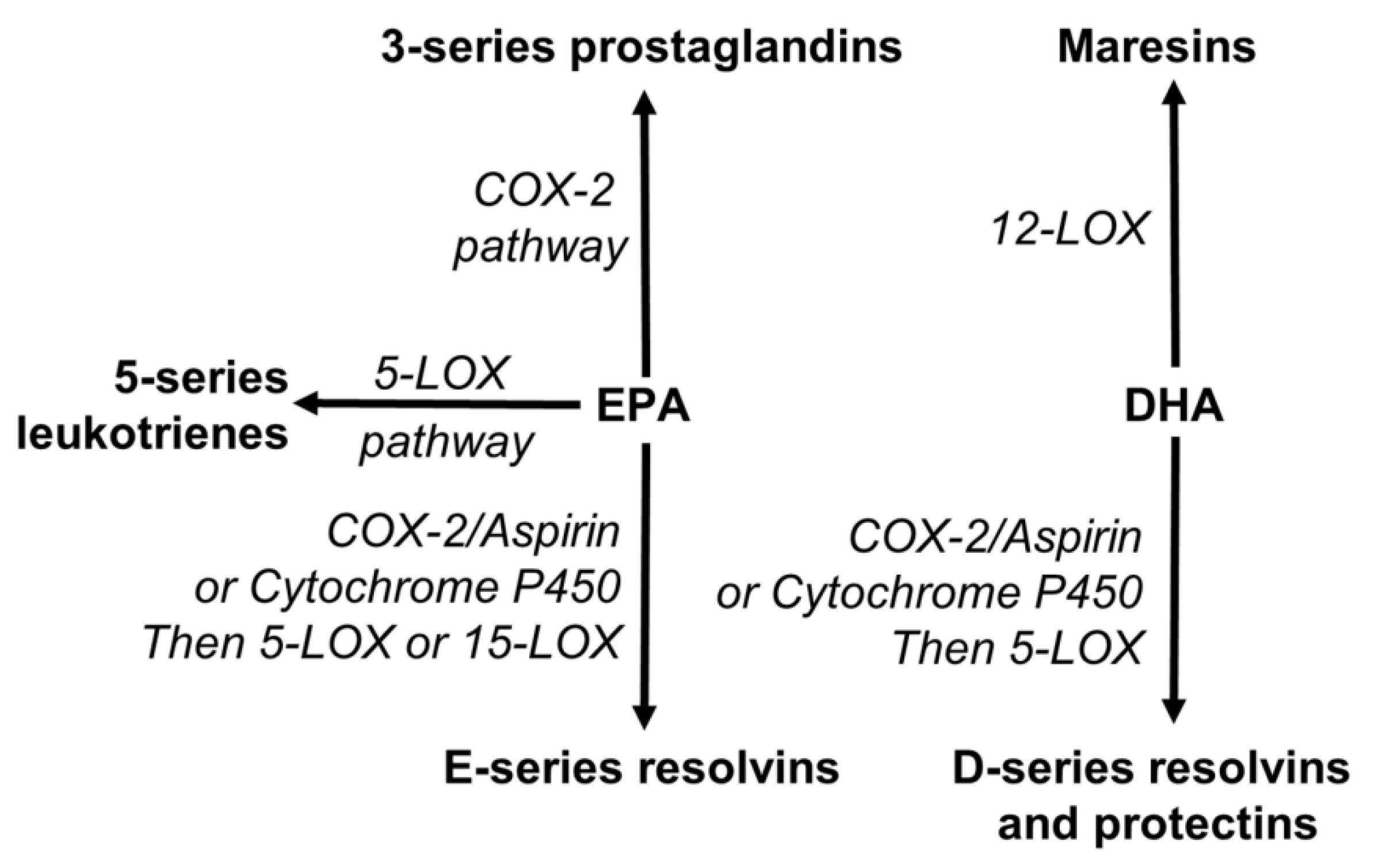
RNA sequencing revealed that genes linked to NF-κB and MAPK pathways were less active in the combined group compared with controls. The single-nutrient groups showed moderate reductions. These changes are in line with known effects of n-3 fatty acids, which produce lipid mediators that calm inflammation and regulate immune response. The transcript data also suggest that fiber may support this process by lowering gut endotoxin levels, helping n-3 fatty acids act more efficiently at the tissue level.
Figure 1.
Diagram showing how EPA and DHA form anti-inflammatory molecules that help lower NF-κB and MAPK signaling.
Figure 1.
Diagram showing how EPA and DHA form anti-inflammatory molecules that help lower NF-κB and MAPK signaling.
3.3. Oxidative Balance and Antioxidant Capacity
The combination of fiber and n-3 PUFA increased SOD activity and lowered MDA, showing a better antioxidant state. The correlation between SOD and MDA was negative (r = −0.61, P < 0.05), meaning higher SOD was linked with less lipid oxidation. This agrees with previous studies where n-3 fatty acids improved antioxidant enzyme activity, while fiber reduced oxidative damage through gut fermentation and reduced inflammation [22]. Together, these results suggest a complementary action: fiber reduces the sources of oxidative stress, while n-3 PUFAs strengthen antioxidant defense.
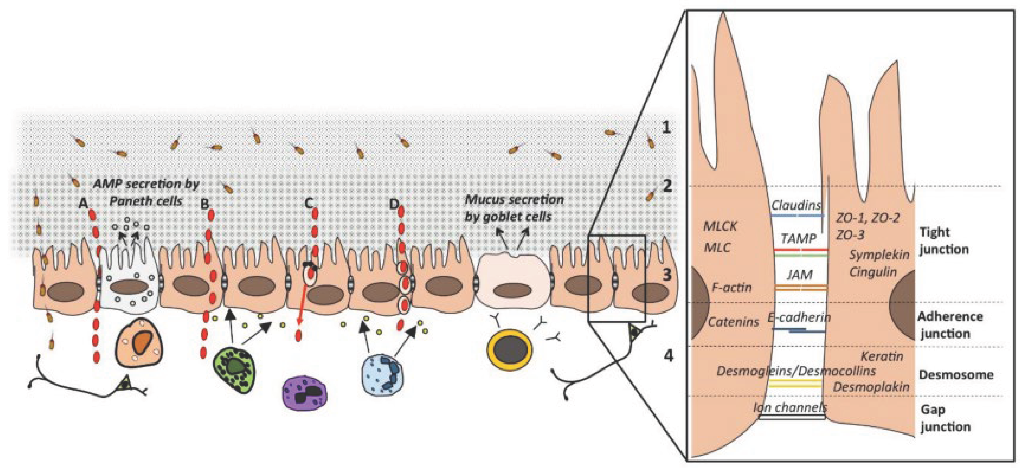
3.4. Gut Barrier and Overall Metabolic Response
The high-fiber groups showed higher levels of Occludin and Claudin-1, with the greatest increase seen in the combined group. This supports the view that short-chain fatty acids from fiber fermentation help protect the gut lining and reduce permeability. A stronger barrier can limit endotoxin leakage and reduce inflammatory load, which may explain the greater drop in CRP and MDA in the combined treatment. Overall, the results indicate that dietary fiber and n-3 PUFAs act together to lower inflammation, improve antioxidant balance, and support metabolic health in diabetes.
Figure 2.
Diagram of the gut barrier showing tight-junction proteins that keep the intestinal wall intact.
Figure 2.
Diagram of the gut barrier showing tight-junction proteins that keep the intestinal wall intact.
4. Conclusion
This study found that a high-fiber diet combined with n-3 fatty acids improved inflammation, oxidative balance, and blood sugar control more than either treatment alone in diabetic mice. The combination lowered CRP by 48%, reduced MDA by 42%, increased SOD activity by 58%, and decreased HbA1c by 1.7 ± 0.2%. It also reduced the activity of NF-κB and MAPK pathways. These results show that fiber and n-3 fatty acids work together through different but connected actions: fiber supports gut health and barrier strength, while n-3 fatty acids improve antioxidant defense and lower inflammatory signals. The findings suggest that combining these two nutrients could be a simple and useful dietary strategy to support metabolic health in diabetes. Still, the study was short and based on an animal model. Future human trials should test longer interventions and confirm how these nutrients work together in real-world conditions.
References
- Oguntibeju, O. O. Type 2 diabetes mellitus, oxidative stress and inflammation: examining the links. International journal of physiology, pathophysiology and pharmacology 2019, 11(3), 45. [Google Scholar] [PubMed]
- Mansoor, G.; Tahir, M.; Maqbool, T.; Abbasi, S. Q.; Hadi, F.; Shakoori, T. A.; …; Ullah, I. Increased expression of circulating stress markers, inflammatory cytokines and decreased antioxidant level in diabetic nephropathy. Medicina 2022, 58(11), 1604. [Google Scholar] [CrossRef] [PubMed]
- Wang, Y.; Wen, Y.; Wu, X.; Wang, L.; Cai, H. Modulation of Gut Microbiota and Glucose Homeostasis through High-Fiber Dietary Intervention in Type 2 Diabetes Management. 2024. [Google Scholar]
- Clyne, A. M. Endothelial response to glucose: dysfunction, metabolism, and transport. Biochemical Society Transactions 2021, 49(1), 313–325. [Google Scholar] [CrossRef] [PubMed]
- Mostafavi Abdolmaleky, H.; Zhou, J. R. Gut microbiota dysbiosis, oxidative stress, inflammation, and epigenetic alterations in metabolic diseases. Antioxidants 2024, 13(8), 985. [Google Scholar] [CrossRef]
- Ge, G.; Zelig, R.; Brown, T.; Radler, D. R. A review of the effect of the ketogenic diet on glycemic control in adults with type 2 diabetes. Precision Nutrition 2025, 4(1), e00100. [Google Scholar]
- Beukema, M.; Faas, M. M.; de Vos, P. The effects of different dietary fiber pectin structures on the gastrointestinal immune barrier: impact via gut microbiota and direct effects on immune cells. Experimental & molecular medicine 2020, 52(9), 1364–1376. [Google Scholar]
- Chen, D.; Liu, J.; Wu, J.; Suk, J. S. Enhancing nanoparticle penetration through airway mucus to improve drug delivery efficacy in the lung. Expert opinion on drug delivery 2021, 18(5), 595–606. [Google Scholar] [CrossRef]
- Kabisch, S.; Hajir, J.; Sukhobaevskaia, V.; Weickert, M. O.; Pfeiffer, A. F. Impact of dietary fiber on inflammation in humans. International journal of molecular sciences 2025, 26(5). [Google Scholar] [CrossRef]
- Siriwardhana, N.; Kalupahana, N. S.; Moustaid-Moussa, N. Health benefits of n-3 polyunsaturated fatty acids: eicosapentaenoic acid and docosahexaenoic acid. Advances in food and nutrition research 2012, 65, 211–222. [Google Scholar]
- Deng, T.; Huang, M.; Xu, K.; Lu, Y.; Xu, Y.; Chen, S.; …; Sun, X. LEGEND: Identifying Co-expressed Genes in Multimodal Transcriptomic Sequencing Data. bioRxiv 2024. 2024-10. [Google Scholar] [CrossRef]
- Wang, Y.; Wen, Y.; Wu, X.; Cai, H. Application of Ultrasonic Treatment to Enhance Antioxidant Activity in Leafy Vegetables. International Journal of Advance in Applied Science Research 2024, 3, 49–58. [Google Scholar]
- Floyd, Z. E.; Ribnicky, D. M.; Raskin, I.; Hsia, D. S.; Rood, J. C.; Gurley, B. J. Designing a clinical study with dietary supplements: it’s all in the details. Frontiers in Nutrition 2022, 8, 779486. [Google Scholar] [CrossRef]
- Xu, J.; Wang, H.; Trimbach, H. An OWL ontology representation for machine-learned functions using linked data. In 2016 IEEE International Congress on Big Data (BigData Congress); IEEE, June 2016; pp. 319–322. [Google Scholar]
- Jeon, J. The Effects of SCFAs and EPA on Ethanol Toxicity in Sh-SY5Y Cells. Master’s thesis, Lamar University-Beaumont, 2025. [Google Scholar]
- Yuan, M.; Mao, H.; Qin, W.; Wang, B. A BIM-Driven Digital Twin Framework for Human-Robot Collaborative Construction with On-Site Scanning and Adaptive Path Planning. 2025. [Google Scholar]
- Singh, J.; Metrani, R.; Shivanagoudra, S. R.; Jayaprakasha, G. K.; Patil, B. S. Review on bile acids: effects of the gut microbiome, interactions with dietary fiber, and alterations in the bioaccessibility of bioactive compounds. Journal of agricultural and food chemistry 2019, 67(33), 9124–9138. [Google Scholar] [CrossRef]
- Chen, H.; Ning, P.; Li, J.; Mao, Y. Energy Consumption Analysis and Optimization of Speech Algorithms for Intelligent Terminals. 2025. [Google Scholar]
- Korunes, K. L.; Liu, J.; Huang, R.; Xia, M.; Houck, K. A.; Corton, J. C. A gene expression biomarker for predictive toxicology to identify chemical modulators of NF-κB. PLoS One 2022, 17(2), e0261854. [Google Scholar] [CrossRef]
- Wu, C.; Zhu, J.; Yao, Y. Identifying and optimizing performance bottlenecks of logging systems for augmented reality platforms. 2025. [Google Scholar]
- Calder, P. C. Omega-3 polyunsaturated fatty acids and inflammatory processes: nutrition or pharmacology? British journal of clinical pharmacology 2013, 75(3), 645–662. [Google Scholar] [CrossRef] [PubMed]
- Xu, K.; Lu, Y.; Hou, S.; Liu, K.; Du, Y.; Huang, M.; …; Sun, X. Detecting anomalous anatomic regions in spatial transcriptomics with STANDS. Nature Communications 2024, 15(1), 8223. [Google Scholar] [CrossRef]
Disclaimer/Publisher’s Note: The statements, opinions and data contained in all publications are solely those of the individual author(s) and contributor(s) and not of MDPI and/or the editor(s). MDPI and/or the editor(s) disclaim responsibility for any injury to people or property resulting from any ideas, methods, instructions or products referred to in the content. |
© 2025 by the authors. Licensee MDPI, Basel, Switzerland. This article is an open access article distributed under the terms and conditions of the Creative Commons Attribution (CC BY) license (http://creativecommons.org/licenses/by/4.0/).
Copyright: This open access article is published under a Creative Commons CC BY 4.0 license, which permit the free download, distribution, and reuse, provided that the author and preprint are cited in any reuse.


