Submitted:
28 November 2025
Posted:
02 December 2025
You are already at the latest version
Abstract
The aim of this study was to characterise postural defects in children aged 10-12 years according to the author’s proposed typology, which was created based on measurements of thoracic kyphosis and lumbar lordosis, taking compensatory mechanisms and postural and movement patterns into account. This will allow for more precise diagnosis of postural defects and the selection of individual corrective exercises. Methods: The research included 303 children aged 10-12 years. Measurements were taken using the Diers Formetric III 4D system, determining angles of thoracic kyphosis (42°-55°) and lumbar lordosis (33°-47°). Based on this, nine postural types were identified, encompassing various combinations of shallow, normal and deep kyphosis as well as lordosis. Results: The analysis revealed that only 29% of children had normal body posture, while 71% demonstrated abnormal spinal alignment. The most common finding was flattened thoracic kyphosis, often combined with varying degrees of lumbar lordosis. Each posture type is characterised by specific muscle patterns—lengthened, shortened, hypoactive and hyperactive muscles—which is crucial for individualising therapy and selecting corrective exercises. Conclusions: In the study, it was confirmed that posture in children aged 10-12 is a dynamic phenomenon which is strongly related to the maturation of the neuromuscular system. The nine-type classification revealed a wide variation in postural pattern—only a minority of children demonstrated normal posture, while the vast majority demonstrated the presence of characteristic compensatory mechanisms. The results clearly suggest that postural disturbances are not the result of deviations in individual spinal curvatures, but are a consequence of multi-segment, interconnected changes in the entire postural chain. Changes in kyphosis and lordosis coexist with disturbances in pelvic positioning, hip function, knee extension or flexion strategies, as well as compensations related to the feet, which are often the final result of deficits in core stabilisation. These relationships confirm that posture is an integrated system in which each segment influences consecutive ones. In this context, the nine-type classification has a distinct advantage over traditional assessments which are primarily focused on curvature angles. The new typology allows for the identification of not only structural deformities but, above all, compensatory patterns, the hierarchy of stabilisation mechanisms and disturbances in proximal-distal control. This makes it a more precise clinical tool and better reflects the child's actual postural organisation. Further longitudinal studies are necessary to clarify the evolution of these patterns during the maturation of the antigravity system and determine their significance in treatment planning.
Keywords:
characteristics of body posture types according author’s typology
; thoracic kyphosis angle
; lumbar lordosis angle
; anti-gravity system
; compensation
; Diers Formetric III 4D
1. Introduction
The body posture of a child results from dynamic neuromuscular maturation shaped by biological, environmental and behavioural factors. Postural development is a non-linear process, strongly dependent on sensory integration, reflex control and the gradual acquisition of cortical motor regulation [1,2,3]. This complexity can be interpreted through chaos theory, in which subtle, often imperceptible changes may trigger long-term dysfunctions, reflecting the “butterfly effect.” Many physiological, including postural, systems show partially chaotic behaviour. In late childhood (10–12 years), intensive changes occur in balance, central stabilisation and muscle tone organisation, crucial for forming mature postural patterns [4,5,6]. Epidemiological data indicate that postural abnormalities exceed 60% in this age group [7,8,9]. These defects arise from interactions between compensatory strategies, sensory system maturation, body loading and environmental influences [10,11,12]. Classical diagnostics focus on sagittal spinal curvature deviations, whereas modern neurophysiology emphasises proximal control, trunk segmentation and stabilisation–mobility synergy [13,14,15,16].
In recent years, there has been a growing need to create more complex and functional classifications of posture that reflect its dynamic nature [17,18,19]. The new typology of posture is an example of a tool that refers to neurodevelopmental mechanisms and allows for a more precise description of postural control strategies present in school-age children [8,20]. Unlike traditional classifications, this approach takes not only anatomical differences into account, but also tension properties, the compensation system, and degree of sensory-motor integration as well as cortical control [21,22]. Understanding these mechanisms is of great clinical significance. Postural disorders can lead to musculoskeletal overload, respiratory dysfunction, difficulties in balance control and coordination problems, thus affecting the child’s physical activity and functioning [23,24,25]. Simultaneously, proper identification of dominant compensatory strategies enables the implementation of targeted therapies appropriate for a specific neurodevelopmental profile [26,27,28]. Therefore, it is necessary to expand knowledge on the diversity of postural patterns and their neurophysiological determinants, especially during periods of intensive antigravity system reorganisation. The aim of this article is to analyse postural profiles of children aged 10-12 years in relation to the proposed nine-type classification and to interpret individual body posture types in light of contemporary knowledge in the field of neurophysiology and postural control.
2. Materials and Methods
The current study involved 303 children aged 10-12 years, including 143 girls (47.18%) and 160 boys (52.82%). The largest group consisted of 10-year-olds, totalling 133 participants (43.85%). The number of included 11-year-olds (35.55%) was 107, while the smallest group comprised 12-year-olds, equalling 62 subjects (20.60%). Before initiation of the study, the children and their parents were informed of its purpose, procedure and duration. All parents provided written, informed consent for their child to participate in the study. The trial was conducted in 2016 at the Laboratory of Posturology, UJK in Kielce. The participants were randomly selected, following the prior establishment of criteria for each group. All research procedures were carried out in accordance with the applicable 1964 Declaration of Helsinki and with the consent of the University Bioethics Committee for Scientific Research of Jan Kochanowski University in Kielce (Resolution No. 5/2015).
- Assessment of body posture type using the Diers Formetric III 4D system
Body posture was assessed using the Diers Formetric III 4D optoelectronic system, based on raster stereography technology, which enables rapid, non-contact and radiation-free measurement of the spine and trunk. The system projects parallel measurement lines onto the surface of the back, and a digital video recorders captures the resulting mesh deformations, allowing for the reconstruction of a three-dimensional model of the trunk an spatial analysis of its parameters. The study was conducted in a darkened room to eliminate the influence of external light. The participants, wearing only shorts, stood with their back two metres away from the projector and video recorder. The measurement was performed using the “Average measurement” mode, which involved a series of 12 averaged images to reduce natural postural fluctuations and increase the repeatability of the results. During the analysis, key inflection points along the spine axis were identified, including the cervicothoracic transition point (ICT), thoracic kyphosis apex (KA), thoracolumbar transition point (ITL), lumbar lordosis apex (LA) and lumbosacral transition point (ILS). Based on their relative positions, angular values were calculated, reflecting the size and distribution of curvatures in the sagittal plane, enabling comprehensive assessment regarding the morphology of the thoracic and lumbar spine. The reliability and clinical utility of the Formetric III 4D system were confirmed in comparative studies utilising radiological analysis, highlighting its high diagnostic value. The results were interpreted in relation to norms, assuming typical ranges between 42°-55° for thoracic kyphosis and 33°-47° for lumbar lordosis [29].
- Methods of statistical analysis
The examined variables were tested for normality of distribution using the Kolmogorov-Smirnov and Shapiro-Wilk tests. Depending on the conformity of the scale variables to normal distribution and the values of skewness and kurtosis, parametric or non-parametric tests were used. For qualitative and discrete variables, frequency distributions and percentages were calculated. The percentage shares (%) of individual posture types in the groups of girls and boys were compared via structure indicator testing.
3. Results
3.1. Characteristics of Postural Defects According to Author’s Typology
Taking the normative ranges of thoracic kyphosis (42°-55°) and lumbar lordosis (33°-47°) into account it is possible to distinguish nine clinically significant types of body posture, resulting from the combination of the values of both spinal curvatures. These configurations include postures characterised by reduced thoracic kyphosis coexisting with reduced, normal or increased lordosis (K < 42°; L < 33°; 33° ≤ L ≤ 47°; L > 47°). Similarly, for kyphosis within the normative range (42° ≤ K ≤ 55°), postures with reduced, normal or increased lumbar lordosis are distinguished. The last set includes configurations in which kyphosis is increased (K > 55°), while lordosis can demonstrate values below, within or above the norm (L < 33°; 33° ≤ L ≤ 47°; L > 47°). This classification model allows for unambiguous categorisation of posture based on precise morphological criteria, constituting an important point of reference in clinical assessment, diagnostic planning and biomechanical analysis of postural disorders (Table 1).
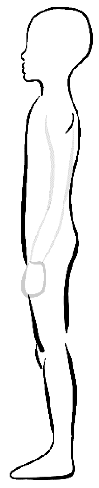
- Reduced thoracic kyphosis, reduced lumbar lordosis (K < 42°; L < 33°)
In recent years, the incidence of this type of posture has intensified, and in the subject-literature, it is increasingly emphasized that modern environmental conditions—especially limited physical activity and greater exposure to static positions—contribute to the flattening of physiological spine curvatures [29,30,31]. In the school-aged population, this configuration occurs in approximately 16% of children, making it one of the most common types of incorrect posture [8].
Such a pattern reflects the predominance of global muscles over segmental ones, which leads to increased stiffness and reduced effectiveness of postural control [28,29]. This is characterised by flattening of thoracic kyphosis and lumbar lordosis, causing reduced load absorption and stiff trunk function. Hyperactivity of the thoracic erector spinae and dominance of the scapular retractors (middle trapezius, rhomboids) coexist with hypoactivity of the serratus anterior and weakened function of the pectoral muscles, which contributes to impaired scapulocostal rhythm [30,31]. In the cervical segment, a typical “head-forward” pattern is observed—head protrusion, decreased cervical lordosis, compensatory upper cervical extension and overloading of the sternocleidomastoid and suboccipital muscles, with simultaneous weakness of the longus colli/capitis [32,33]. In the lumbar-pelvic region, decreased lordosis coexists with posterior pelvic tilt, hyperactivity of the rectus abdominis and hamstrings, as well as hypoactivity of the local stabilisers (transversus abdominis, multifidus). Weakness of the iliopsoas and lumbar erector spinae reduces the ability to maintain stabilisation and proper movement segmentation [34,35]. The gluteus maximus often exhibits increased tonus, while the gluteus medius and minimus are weakened, which impairs hip joint control and affects compensation during weight-bearing of the lower limbs [36,37]. The hips tend to flex due to posterior pelvic tilt and shortened hamstrings. Slight deflection or increased stiffness is observed in the knee joint, which is a reaction to a backwards shift in centre of gravity and a compensatory increase in hamstring tension [38]. Shifting the load axis leads to foot supination, reduced gastrocnemius/soleus activity, and limited foot adaptation during the stance phase [39]. This pattern is associated with trunk stiffness, neck and lower back pain, restricted thoracic rotation, shallow upper rib breathing and reduced postural efficacy. These disturbances impede postural adaptation in dynamic conditions and promote the development of global compensations [40]. Shortened muscles: SCM, suboccipitals, thoracic erector spinae, middle trapezius, rhomboids, rectus abdominis, hamstrings, gluteus maximus. Elongated muscles: longus colli/capitis, serratus anterior, pectoralis major/minor, upper trapezius, iliopsoas, rectus femoris, lumbar erector spinae, TA, multifidus, gastrocnemius/soleus, gluteus medius/minimus [28]. This pattern includes trunk stiffness, neck and lumbar pain, limited thoracic rotation and shallow upper rib breathing. This may include impaired pelvic stabilisation, reduced postural efficacy, rapid fatigability in static positions, as well as a tendency to supinate the feet and overload the hindfoot. The pattern induces global compensations and reduced movement efficiency [41].
Figure 1.
Reduced thoracic kyphosis, reduced lumbar lordosis (K < 42°; L < 33°).
- Reduced thoracic kyphosis, normal lumbar lordosis (K < 42°; 33° ≤ L ≤ 47°)
This type of posture is characterised by a flattening of physiological thoracic kyphosis, with preserved normal lumbar lordosis (Figure 2). The defect occurs in approximately 22% of children [8]. The pattern indicates a predominance of the global muscles of the shoulder girdle and trunk with weakened segmental control, which affects the quality of thoracic spine alignment. It is characterised by flattening of the thoracic spine with preserved lumbar lordosis [30,31,32]. Reduced kyphosis results from dominance of the thoracic erector spinae and predominance of the middle trapezius and rhomboids, which maintain the scapula in retraction and slight depression. The serratus anterior and pectoralis major/minor remain elongated and hypoactive, which disrupts normal scapulocostal rhythm. In the cervical spine, head protrusion and decreased lordosis are often noted, with excessive activity of the sternocleidomastoid and suboccipital muscles as well as weakness of the longus colli/capitis. Lumbar lordosis is preserved, but segmental stabilisers (transversus abdominis, multifidus) may be weakened, which favours compensatory activity of the global muscles. The hip joint is usually closer to neutral position, with a slight tendency to flex [33]. The knee joint typically exhibits a stiff, slightly flexed or neutral position, resulting from a posterior shift of the centre of gravity as well as insufficient pelvic and hip control. The lower limbs usually do not exhibit as strong compensation as in the case of posterior tilt; however, flattening of the thoracic segment can disrupt trunk balance and foot loading. The gluteus maximus is usually normal or shows a slight increase in stabilising tonus, while the gluteus medius may be hypoactive because of limited pelvic control in the frontal plane. Shortened muscles: SCM, suboccipitals, thoracic erector spinae, middle trapezius, rhomboids. Elongated muscles: longus colli/capitis, serratus anterior, pectoralis major/minor, upper trapezius, gluteus medius, and partially segmental stabilizers (TA, multifidus) [28]. Thoracic spine stiffness, limited trunk rotation, neck pain resulting from suboccipital muscle overload and shallow upper rib breathing are observed. Effective scapular function and shoulder girdle stabilisation are hindered, which can lead to neck and shoulder overloading as well as reduced economy of upper limb movement. Lumbar lordosis remains stable, but global trunk mobility and functional flexibility become restricted [34].
- Reduced thoracic kyphosis, increased lumbar lordosis (K < 42°; L > 47°)
This pattern reflects the predominance of global extensor mechanisms in the lumbar and global retraction patterns of the thoracic spine, with reduced activity of segmental stabilizers. It combines flattening of the thoracic spine with increased lumbar lordosis (Figure 3). This pattern fuses reduced thoracic kyphosis with increased lumbar lordosis and occurs in approximately 5% of children [8]. The configuration reflects the predominance of global extensor mechanisms and reduced integration regarding the axis of the body, leading to impaired adjustment and balance responses. Reduced kyphosis results from overactivity of the thoracic erector spinae and scapular retractors (middle trapezius, rhomboids), with concomitant hypoactivity of the serratus anterior and secondary lengthening of the pectoralis major/minor. A consequence of this is limited thoracic mobility and impaired scapular-costal coordination. In the cervical spine segment, protraction of the head, shallowing of central lordosis and compensatory extension of the upper cervical segment (C0-C2) are observed [34]. This arrangement favours dominance of the sternocleidomastoid and suboccipital muscles and inhibition of the longus colli/capitis, which disrupts craniocervical integration and the precision of balance reactions.
Increased lumbar lordosis and anterior pelvic tilt reflect the predominance of the iliopsoas, rectus femoris and lumbar erector spinae over the local stabilisers (transversus abdominis, multifidus). Segmental lumbar stabilisation is reduced and postural control relies on global stiffness. Weakness of the gluteus maximus and medius limits pelvic and hip stabilisation. Extension strategies dominate in the hip joints, which increases loading on the anterior compartment and intensifies kinematic compensations. An anterior shift of the centre of gravity increases forefoot loading and promotes pronation of the feet, requiring compensatory activation of the gastrocnemius, soleus and tibialis posterior. Hyperextension is common in the knee joint, representing a passive stabilisation strategy when proximal control is ineffective. Shortened muscles: thoracic erector spinae, middle trapezius, rhomboids, iliopsoas, rectus femoris, lumbar erector spinae, gastrocnemius, soleus, tibialis posterior, sternocleidomastoid, suboccipitals. Elongated muscles: serratus anterior, pectoralis major/minor, upper trapezius, longus colli/capitis, rectus abdominis, transversus abdominis, multifidus, gluteus maximus, gluteus medius [28,35,36]. Clinically, hyperextended posture, limited trunk rotation, overloading of the cervical and lumbar spine as well as postural control disorders result from dominance of global extension patterns.
- Normal thoracic kyphosis, reduced lumbar lordosis (42° ≤ K ≤ 55°; L < 33°)
This postural type is characterised by a normal degree of thoracic kyphosis with reduced lumbar lordosis (Figure 4). It occurs in approximately 6% of children [8]. The given pattern indicates the predominance of global lumbopelvic flexion strategies with weakened activity of segmental central stabilizers. It is characterised by normal thoracic kyphosis with a shallowing of lumbar lordosis, reflecting the predominance of lumbopelvic flexion patterns and weakened central stabilisation mechanisms. Posterior pelvic tilt results from overactivity of the rectus abdominis and hamstrings, while local stabilizers (transversus abdominis, multifidus) remain hypoactive, limiting the ability to generate pressor control of the trunk. Although thoracic kyphosis remains physiological, compensatory stiffness of the lower segments reduces trunk segmentation and decreases the effectiveness of adjustment responses in a dynamic environment. In the cervical spine, head protraction and decreased cervical lordosis are frequently observed, leading to the dominance of the sternocleidomastoid and suboccipital muscles. Weakness of the longus colli/capitis disrupts craniocervical integration, impairs head-to-trunk adjustment responses, and limits sagittal balance precision. In the lumbar spine, decreased lordosis, elongated iliopsoas and lumbar erector spinae, as well as weakened segmental mechanisms reduce the ability of forward and backward control, which affects the quality of balance responses concerning the entire axis of the body. Hypoactivity of the gluteus maximus and gluteus medius impairs hip and pelvic stabilisation—key reference points in antigravity posture. Flexion dominates in the hips, increasing reliance on trunk compensation in balance strategies. A posterior shift of the centre of gravity promotes foot supination and reduced gastrocnemius/soleus activity. A slight flexion strategy is common in the knees, representing a passive stabilisation response to proximal postural control failure. This pattern results in reduced efficiency of the lower segment balance responses and limited adaptation to load-bearing tasks. Shortened muscles: rectus abdominis, hamstrings, sternocleidomastoid, suboccipitals and sometimes the gluteus maximus (tonic tension). Elongated muscles: iliopsoas, rectus femoris, lumbar erector spinae, transversus abdominis, multifidus, gluteus medius/minimus, longus colli/capitis, gastrocnemius and soleus [28]. A person with this pattern exhibits shallow lumbar lordosis and posterior pelvic tilt, which contribute to low back pain, weakened central stabilisation and ineffective hip extension. Head protrusion leads to neck strain and compensatory work of the global muscles reduces trunk flexibility. Loading the hindfoot and a tendency to supinate can cause ankle strain and balance disorders [42].
- Normal thoracic kyphosis, normal lumbar lordosis (42° ≤ K ≤ 55°; 33° ≤ L ≤ 47°)
This is the only normal type of body posture. It is characterised by correct degrees of thoracic kyphosis and lumbar lordosis (Figure 5). It occurs in approximately 29% of subjects [8]. This suggests that 71% of subjects have defective body posture types [8]. The pattern is characterised by the harmonious cooperation of global and segmental muscles, allowing for optimal maintenance of spinal curvatures and effective postural control. It reflects the correct configuration of spinal curvatures, in which thoracic kyphosis and lumbar lordosis are within the physiological range. This trunk positioning promotes effective load transfer, optimal shock absorption as well as full cooperation of global and local muscles. Balance between the activity of the thoracic erector spinae, scapular stabilisers and abdominal muscles ensures proper postural control without excessive compensation. In the cervical spine, a neutral head position is most often observed with preserved cervical lordosis and balance between the deep (longus colli/capitis) and global muscles (sternocleidomastoid, upper trapezius) [33,34,35,36,37,38]. In the pelvic and lumbar regions, segmental stabilisers—the transversus abdominis and multifidus—work together with the gluteus maximus and medius, ensuring proper stabilisation of the body’s core. The lower limbs demonstrate harmonious functioning with proper foot loading and a stable longitudinal arch. There are no significant advantages related to length or tension. Local and global muscle function is balanced, and any minor symmetries are physiological in nature. There is a lack of patterns typical for dysfunction: neither shortening that induces compensation nor lengthening which leads to decreased stability. The person demonstrates balanced posture, proper mobility and trunk stability, and no characteristic overloads resulting from changes in spinal curvature. The muscular system functions effectively, and the risk of compensation or overloading of the neck, thoracic spine or lumbar spine is minimal. Movements are fluid, economical and do not produce typical pain symptoms [43].
- Normal kyphosis, increased lordosis (42° ≤ K ≤ 55°; L > 47°)
This type of posture is characterised by a normal degree of thoracic kyphosis accompanied by increased lumbar lordosis (Figure 6). It occurs in approximately 12% of subjects [8]. This pattern reflects the dominance of global extension mechanisms in the lumbopelvic segment and the insufficiency of central stabilisation. Anterior pelvic tilt increases the activity of the iliopsoas, rectus femoris and lumbar erector spinae, perpetuating hyperextension and reducing segmentation ability. The local stabilisers (transversus abdominis, multifidus) remain elongated and hypoactive, which impairs tonus control and the precision of adjustment responses. In the cervical spine, normal or slightly protruded head positioning can be observed. The dominance of the sternocleidomastoid and suboccipital muscles compensates for the anterior shift of the trunk, disrupting craniocervical mechanisms and balance responses. The gluteus maximus and gluteus medius muscles are elongated and weakened, which compromises hip stabilisation [43,44,45]. A tendency to extend the hip joint occurs, increasing anterior compartment overload and limiting functional flexion during gait. Hyperextension is common in the knee joint, representing a passive stabilisation strategy with insufficient proximal control. Anterior shift of the centre of gravity increases forefoot loading and promotes foot pronation. This results in compensatory activation of the gastrocnemius, soleus and tibialis posterior. The foot loses the ability to precisely modulate stiffness during the stance phase, reducing the efficacy of balance responses. Shortened muscles: iliopsoas, rectus femoris, lumbar erector spinae, gastrocnemius, soleus, tibialis posterior. Elongated muscles: transversus abdominis, multifidus, rectus abdominis, gluteus maximus, gluteus medius, partially longus col-li/capitis. Clinically, hyperextended posture, lumbar overload, weakened pelvic stabilisation and ineffective balance reactions are predominant.
- Increased kyphosis, decreased lordosis (K > 55°, L < 33°)
This type of posture is characterised by increased thoracic kyphosis accompanied by decreased lumbar lordosis (Figure 7). It occurs in approximately 1% of subjects [8]. The pattern demonstrates a predominance of global flexion strategies in the shoulder girdle and trunk and weakening of segmental central stabilisation, leading to loss in segmented movement. This pattern combines excessive thoracic kyphosis with decreased lumbar lordosis, indicating a predominance of flexion patterns and reduced integration of antigravity mechanisms. Increased kyphosis results from hyperactivity of the pectoralis major/minor and weakness of the back muscles (thoracic erector spinae, middle/lower trapezius, rhomboids), which limits trunk rotation and worsens postural responses. In the cervical spine, head protraction, decreased medial lordosis and compensatory upper cervical extension occur [31,32,33,34,35,36,37]. Dominance of the sternocleidomastoid and suboccipital muscles, along with weakened longus colli/capitis, disrupts head-trunk integration and sagittal balance responses. In the lumbopelvic segment, decreased lordosis and posterior pelvic tilt result from tension in the rectus abdominis and hamstrings, as well as hypoactivity of the local stabilisers (transversus abdominis, multifidus). Weakened iliopsoas and lumbar erector spinae limit the generation of central stabilisation. The gluteus maximus and medius are hypoactive, reducing hip stability. Flexion dominates in the hips, weakening control during weight-bearing tasks. The knees typically exhibit slight flexion, being a passive stabilising strategy in the case of poor proximal control. A posterior shift in centre of gravity promotes foot supination and reduced gastrocnemius/soleus activity. The foot has limited stiffness modulation capacity, which reduces the effectiveness of balance responses. Shortened muscles: pectoralis major/minor, rectus abdominis, hamstrings, sternocleidomastoid, suboccipitals. Elongated muscles: thoracic erector spinae, trapezius (middle/lower), rhomboids, iliopsoas, lumbar erector spinae, transversus abdominis, multifidus, longus colli/capitis, gluteus maximus/medius, gastrocnemius/soleus [28]. Clinically, the following symptoms predominate: thoracic stiffness, cervical and lumbar overloads, limited central stabilisation and ineffective balance reactions [46].
- Increased kyphosis, normal lordosis (K > 55°; 33° ≤ L ≤ 47°)
This posture type is characterised by increased thoracic kyphosis accompanied by normal lumbar lordosis (Figure 8). It occurs in approximately 4% of subjects [8]. This pattern demonstrates the dominance of global flexion patterns in the upper trunk, with weakened segmental thoracic control and central stabilisation. It is characterised by excessive thoracic kyphosis with preserved lumbar lordosis. Excessive thoracic flexion results from predominance of the pectoralis major/minor and weakness of the posterior thoracic muscles, including the thoracic erector spinae, middle/lower trapezius and rhomboids. Increased kyphosis limits the rotational mobility of the thorax and impairs trunk adjustment responses in the sagittal and transverse planes. In the cervical spine, head protraction and compensatory upper neck extension are observed in response to anterior trunk shift. The dominance of the sternocleidomastoid and suboccipital muscles with weakened longus colli/capitis disrupts head-trunk integration and reduces the precision of balance responses. In the lumbar-pelvic segment, lordosis remains normal, but local stabilizers (transversus abdominis, multifidus) exhibit reduced activity. This leads to a decrease in the quality of segmental control and limits the ability to stabilise the lumbar spine during weight-bearing tasks. The gluteus maximus and medius muscles are weakened, reducing hip stability in the frontal plane and affecting adjustment responses. A tendency towards neutral or slight flexion is noted in the hips, resulting from trunk compensation for excessive kyphosis. In the knees, slight flexion strategies predominate, constituting passive stabilisation with limited proximal control. A posterior shift in centre of gravity promotes foot supination and reduced activity of the gastrocnemius/soleus. The foot exhibits limited stiffness modulation, which hinders stance phase adaptation and compromises balance responses. Shortened muscles: pectoralis major/minor, sternocleidomastoid, suboccipitals. Elongated muscles: thoracic erector spinae, trapezius (middle/lower), rhomboids, longus colli/capitis, gluteus maximus/medius, transversus abdominis, multifidus, gastrocnemius/soleus. Clinically, flexed upper trunk posture, cervical overload, limited thoracic mobility and reduced balance response efficiency are predominant.
- Increased kyphosis, increased lordosis (K > 55 °; L > 47°)
This pattern clearly demonstrates the dominance of global muscles over those segmental, leading to dissociation of trunk function and limited ability for precise postural control. This occurs in approximately 7% of subjects [8]. It is characterised by simultaneous deepening of thoracic kyphosis and lumbar lordosis, reflecting a clear dissociation of trunk function: dominance of global flexion patterns in the thoracic spine and global extension patterns in the lumbar spine, with reduced integration of segmental stabilisers (Figure 9). Excessive kyphosis results from predominance of the pectoralis major/minor and a weakening of the posterior thoracic band (thoracic erector spinae, middle/lower trapezius, rhomboids), which limits trunk rotation and worsens adjustment reactions. In the cervical spine, head protraction and compensatory upper neck extension are observed. The global muscles controlling head position remain dominant—sternocleidomastoid and suboccipitals—while the segmental stabilisers (longus colli/capitis) are hypoactive. This results in impaired head-trunk integration and causes reduced balance accuracy. In the lumbar-pelvic region, increased lordosis and anterior pelvic tilt reflect the predominance of the global hip extensors and flexors (iliopsoas, rectus femoris, lumbar erector spinae) over the segmental muscles of central stabilisation (transversus abdominis, multifidus). This pattern increases extension stiffness and reduces the ability to exercise local segmental control. Weakness of the gluteus maximus and medius further reduces hip stability and impairs balance responses in the lower limb. The hips are dominated by extension, which overloads the anterior compartment of the joint and limits the range of functional flexion. Hyperextension is common in the knees, a passive stabilisation strategy used when proximal control is ineffective. Anterior shifting of centre of gravity causes increased forefoot loading and foot pronation, which activates the global calf muscles (gastrocnemius, soleus) and posterior tibialis in a compensatory function. The foot loses its ability to precisely modulate stiffness, reducing the effectiveness of balance responses and load adaptation. Shortened muscles: pectoralis major/minor, sternocleidomastoid, suboccipitals, iliopsoas, rectus femoris, lumbar erector spinae, gastrocnemius, soleus, tibialis posterior. Elongated muscles: thoracic erector spinae, trapezius (middle/lower), rhomboids, longus col-li/capitis, transversus abdominis, multifidus, gluteus maximus/medius [28]. Clinically, a pattern based on global tension, cervical and lumbar overloads, limited thoracic mobility and ineffective balance reactions of the loaded lower limbs are observed. This pattern reflects the dominance of global muscles over local stabilisers, which limits the effectiveness of postural control and predisposes to overloading in the cervical, thoracic and lumbar spine [42].
4. Discussion
The results of the study emphasize that body posture between the ages of 10 and 12 years remains a dynamic phenomenon and strongly embedded in the context of neuromuscular system maturation [1,2,3,4,5]. The implemented nine-type classification allowed to reveal a wide spectrum of postural profiles that cannot be explained solely by structural parameters of the spine [6,7,8,9]. Each of them reflects a different compensatory mechanism resulting from the interaction between postural tone, sensory integration, reflex control and the ability to segment movement [10,11,12,13,14,15]. Only 29% of children presented normal posture, which means that in the majority of children the development of antigravity mechanisms is not fully optimal at this stage of ontogeny [16,17,18]. In particular, types with shallow thoracic kyphosis (types 1-3) are an example of incomplete integration of the extension reaction and insufficient trunk control at the middle and upper levels [19,20,21]. From a neurodevelopmental perspective, flattening of thoracic kyphosis can be explained by the dominance of extension strategies in the body axis, characteristic of children whose deep stabilisers and trunk-rib system have not yet reached full maturity [22,23,24]. Disturbed scapular-costal rhythm, neck muscle overload and compensatory upper-cervical extension indicate excessive involvement of tonic extension mechanisms with insufficient activation of the postural system [25,26]. The first type, in which both kyphosis and lordosis are shallowed, is an expression of global failure regarding central stabilisation mechanisms [27,28,29]. Such posture is typical in children with reduced postural tone—a phenomenon described in the developmental literature as a manifestation of immature muscle coordination, poor proprioceptive conduction and insufficient vestibulospinal integration [30,31,32]. The observed supinated foot position and “hanging” onto passive structures indicate a transfer of stabilisation from the central to the distal level, which is a characteristic sign of neurodevelopmental compensation for proximal deficits [33]. In types with increased lordosis (3 and 6), antigravity strategies based on lumbar extensors, hip flexors and “escape-to-extension” mechanisms dominate [34,35,36]. From a neurodevelopmental perspective, this is a pattern characteristic of children whose centre of gravity is poorly modulated, and trunk control is based on global tension instead of segmental stabilisation [37]. Anterior pelvic tilt, knee hyperextension and forefoot overload indicate maintenance of archaic stabilisation strategies, well-described in the neurophysiological literature as a result of immature adjustment reactions, lack of inhibition of adjustive reflexes, or insufficient vestibular-cortical integration [38,39]. In turn, flexion-dominant types—especially types 7 and 9—indicate the predominance of strategically oriented flexion patterns, typical of children with inefficient mechanisms of separation of the upper and lower trunk [40,41]. Thoracic hyperkyphosis combined with reduced lordosis is characteristic of children in whom the mechanisms of trunk rotation, central stabilisation and diaphragm function have not achieved full synchronisation [42,43]. In these types, compensatory upper neck extension—one of the earliest reflexive extension patterns—is often observed, suggesting that the child stabilises the head through archaic mechanisms rather than through deep neck flexion [44]. One of the most important neurodevelopmental conclusions from the study is that each postural type represents a different stage of balance between reflexive, habitual mechanisms and cortical control [45].
The diversity of types indicates that postural development is not linear but is modulated by numerous factors—sensory, motor, environmental and emotional [46]. The postural system is chaotic, and slight deviations in muscle tonus can lead to a cascade of compensations in various body segments [8,47]. In a practical context, the nine-type postural typology provides a tool for precisely defining the dominant neurodevelopmental profile and, consequently, for targeted therapeutic intervention. Among children with a predominance of extension strategies, strengthening flexion mechanisms, lengthening the posterior line and training central stabilisation are necessary. In types with a predominance of flexion, interventions are needed to restore thoracic mobility, improve diaphragm function and respiratory function, and enable segmental separation of the trunk. In mixed types (e.g., hyperkyphosis + hyperlordosis), simultaneous action on several levels of postural control is necessary.
Clinical implications include the need to treat postural disturbances as a symptom secondary to dysfunctions in the areas of postural tone, muscle tone and sensory integration. Types with hyperlordosis demonstrate an increased risk of lumbar, hip and foot overload, while those with hyperkyphosis experience respiratory disorders, shoulder girdle asymmetry and trunk rotation limitations. These patterns may affect gross motor skills, coordination and balance control, as confirmed by numerous neurophysiological reports [44,45,46,47]. The presented studies, with their cross-sectional nature and narrow age range, limit the ability to assess the dynamics of postural mechanism maturation. The lack of references to psychosensory factors, motor activity or gender differences indicates the need to expand future research to include a neurobehavioural and longitudinal perspective. Overall, the results of the study confirm that postural development is a neurodevelopmental process of high complexity, dependent on the maturation of central structures, sensory integration and modulation of postural and muscular tonus.
Therefore, the nine-type classification is not only a precise diagnostic tool, but it also reflects the neurophysiological maturity of the child’s postural system, helping to understand the complexity of compensatory mechanisms and planning effective therapeutic interventions.
5. Conclusions
In the conducted study, it was confirmed that body posture in children aged 10-12 is a dynamic phenomenon strongly related to the maturation of the neuromuscular system. The nine-type classification revealed a wide variation in postural patterns—only a minority of children demonstrated normal posture, while the vast majority presented with characteristic compensatory mechanisms. The results clearly indicate that postural disturbances are not the result of deviations in individual spinal curvatures, but are a consequence of multi-segment, interconnected changes throughout the entire postural chain. Changes in kyphosis and lordosis coexist with disturbances in pelvic alignment, hip function, knee extension or flexion strategies and foot compensations, which are often the final result of deficits in core stabilisation. These relationships confirm that posture is an integrated system in which each segment influences the following one. In this context, the nine-type classification has a distinct advantage over traditional assessments, which focused primarily on individual curvature angles. The new typology allows for the identification of not only structural deformities but, above all, compensatory patterns, the hierarchy of stabilisation mechanisms and disturbances in proximal-distal control. This makes it a more precise clinical tool and better reflects the child’s actual postural organisation. Further longitudinal studies are necessary to clarify the evolution of these patterns during the maturation of the antigravity system and determine their significance for treatment planning.
Author Contributions
J.W.: Conceptualisation, data collection and analysis, formal analysis, methodology.
Institutional Review Board Statement
All research procedures were performed according to the applicable 1964 Declaration of Helsinki and after obtaining approval from the Bioethics Committee at the Regional Medical Chamber No. 5/2015.
Informed Consent Statement
Written, informed consent was obtained from all subjects involved in the study prior to its initiation.
Data Availability Statement
The data and materials supporting the conclusions of this article can be found within the article.
Conflicts of Interest
The authors have no conflict of interest to declare regarding the publication of this paper.
References
- Saavedra, S.; Woollacott, M.; van Donkelaar, P. Multisensory Contributions to Postural Control in Children. Front. Hum. Neurosci. 2020, 14, 569486.
- Wilson, P.H.; Smits-Engelsman, B.; Caeyenberghs, K.; et al. Neural Mechanisms Underlying Motor Control Difficulties in Children. Nat. Rev. Neurol. 2020, 16, 367–380.
- van Emmerik, R.E.; Rosenstein, M.T.; McDermott, J.M.; Haddad, J.M. Movement Variability from a Health and Pathology Perspective. Hum. Mov. Sci. 2020, 78, 102796.
- Lininger, M.R.; Smith, B.; Jacobs, J.; et al. Sagittal Plane Posture and Associated Motor Behaviors in Youth. Gait Posture 2020, 80, 330–343.
- Houwen, S.; Hartman, E.; Visscher, C. Motor Skill Development Trajectories in Childhood. Dev. Rev. 2021, 60, 100962.
- dos Santos, A.N.; Bunn, P.S.; Armand, S.; et al. Longitudinal Maturation of Postural Sway in Children. Gait Posture 2023, 103, 23–29.
- Czaprowski, D.; Stoliński, Ł.; Tyrakowski, M.; et al. Spinal Curvature Development in Children and Adolescents. Children 2021, 8, 472.
- Wilczyński, J. Own Typology of Body Posture Based on DIERS Formetric III 4D. J. Clin. Med. 2025, 14, 501. [CrossRef]
- Macedo, R.B.; Coelho-e-Silva, M.J.; Valente-dos-Santos, J.; et al. Postural Characteristics of School-Age Children. Front. Pediatr. 2022, 10, 841677.
- Kocjan, J.; Adamek, M.; Gzik-Zroska, B.; et al. Respiratory–Postural Synergies. Respir. Physiol. Neurobiol. 2021, 285, 103609.
- Bernard-Demanze, L.; Dumitrescu, M.; Delarque, A.; et al. Vestibular Contributions to Pediatric Postural Stability. Front. Neurol. 2021, 12, 673256.
- Ghai, S.; Ghai, I. Virtual Reality-Based Postural Training in the Pediatric Population. Phys. Ther. Rev. 2020, 25, 53–64.
- Pavão, S.L.; Rocha, N.A.C.F. Postural Control Assessment in Children. Children 2023, 10, 487.
- Bisi, M.C.; Stagni, R. Complexity and Regularity of Postural Sway in Children. Sensors 2021, 21, 3218.
- Wang, X.; Chen, X.; Lee, K.; et al. Development of Trunk Muscle Activation Patterns in Children. J. Biomech.2022, 132, 110992.
- Kocjan, J.; Gzik-Zroska, B.; et al. Respiratory–Postural Synergies. Respir. Physiol. Neurobiol. 2021, 285, 103609.
- Chow, J.; Choong, A.M.; Bedi, M.; et al. Advances in Pediatric Gait and Posture Biomechanics. Bioengineering2023, 10, 251.
- Lacquaniti, F.; Bosco, G.; Gravano, S.; et al. Multisensory Integration and Body Schema. Curr. Biol. 2022, 32, R175–R188.
- Taverna, L.; Colombo, S.; Fumagalli, G.; et al. Neurodevelopmental Determinants of Posture. Dev. Rev. 2024, 64, 101065.
- Richardson, C. The muscle designation debate: the experts respond. J. Bodyw. Mov. Ther. 2000, 4, 235–236. [CrossRef]
- Liu, J.; Yu, K.; Zhang, L.; et al. Neural Control of Motion-Compensation Strategies. IEEE Trans. Neural Syst. Rehabil. Eng. 2024, 32, 889–897.
- Caronni, A.; Recalcati, M.; Montesano, A.; et al. Machine-Learning–Based Classification of Postural Phenotypes. Sci. Rep. 2025, 15, 1182.
- Gill, S.V.; Hogan, N.; Palisano, R.; et al. Functional Consequences of Impaired Postural Control. Pediatr. Res.2023, 93, 1824–1834.
- Lopes, P.N.; Ribeiro, D.C.; Pinheiro, A.; et al. Trunk Stability and Balance. Gait Posture 2024, 108, 67–73.
- Silva, P.L.; Barros, R.V.; Monteiro, L.; et al. Effects of Targeted Interventions on Spinal Alignment. Children 2024, 11, 203.
- Lininger, M.R.; Smith, B.; Jacobs, J.; et al. Sagittal Plane Posture and Motor Behaviors. Gait Posture 2020, 80, 330–343.
- Suda, E.Y.; de Paula, L.R.; Sacco, I.C.N. Impact of Core Stability Training. Pediatr. Phys. Ther. 2021, 33, 168–175.
- Kendall, F.P.; McCreary, E.K.; Provance, P.G. Muscles: Testing and Function with Posture and Pain; Lippincott Williams & Wilkins: Baltimore, MD, USA, 2005.
- Harzmann H. Methode und Klinische Einsatzmöglichkeiten der dreidimensionalen Rückenoberflächenvermessung mit der Videorasterstereographie (VRS) [Methods and Clinical Applications of Three-Dimensional Back Surface Measurements Using Video Raster Stereography (VRS)]. Individuelle Gesundheitsleistungen (IGEL). Orthopädie 2001: 81-104 (in German).
- Chatelain, L.S.; Dayer, R.; Dubois-Ferrière, V.; Armand, S. Pediatric Spinal Alignment and Spinal Development: A Narrative Review. Spine J. 2024, (online ahead of print).
- Qi, H.; Yang, X.; Hu, Z.; Liu, S.; Zhang, W. Spinopelvic Sagittal Alignment and Normative Values in a Large Pediatric Cohort. Sci. Rep. 2025, 15, 91481.
- Atalay, E.S.; Türker, D.; Soylu, Ç.; Arslan, S.; Erden, Z. Association Between Lumbar Lordosis, Thoracic Kyphosis and Muscle Activations During Lower Back Exercises. Medicina 2025, 61, 986. [CrossRef]
- Grabara, M.; Witkowska, A. Sagittal Spinal Curvatures of Young Adults and Their Relationship with Physical Activity and Somatic Parameters. Sci. Rep. 2024, 14, 12221.
- Macedo, R.B.; Coelho-e-Silva, M.J.; Valente-dos-Santos, J.; et al. Postural Characteristics of School-Age Children: A Systematic Review. Front. Pediatr. 2022, 10, 841677.
- dos Santos, A.N.; Bunn, P.S.; Armand, S.; et al. Longitudinal Maturation of Postural Sway in Children. Gait Posture2023, 103, 23–29.
- Lininger, M.R.; Smith, B.; Jacobs, J.; et al. Sagittal Plane Posture and Motor Behaviors in Youth: A Scoping Review. Gait Posture 2020, 80, 330–343.
- Bisi, M.C.; Stagni, R. Complexity and Regularity of Postural Sway in Children: A Nonlinear Approach. Sensors 2021, 21, 3218.
- Lacquaniti, F.; Bosco, G.; Gravano, S.; et al. Multisensory Integration and Body Schema Development in Humans. Curr. Biol. 2022, 32, R175–R188.
- Kocjan, J.; Adamek, M.; Gzik-Zroska, B.; et al. Respiratory–Postural Synergies: An Updated Review of Mechanisms. Respir. Physiol. Neurobiol. 2021, 285, 103609.
- Gill, S.V.; Hogan, N.; Palisano, R.; et al. Functional Consequences of Impaired Postural Control in Children. Pediatr. Res.2023, 93, 1824–1834.
- Chow, J.; Choong, A.M.; Bedi, M.; et al. Advances in Pediatric Gait and Posture Biomechanics. Bioengineering 2023, 10, 251.
- Liu, J.; Yu, K.; Zhang, L.; Chen, F. Neural Control of Motion-Compensation Strategies in Developing Children. IEEE Trans. Neural Syst. Rehabil. Eng. 2024, 32, 889–897.
- Gomes, C.A.; de Faria, A.; Diniz, A.; et al. Breathing Patterns and Postural Control in Children. Front. Pediatr. 2021, 9, 699048.
- Silva, P.L.; Barros, R.V.; Monteiro, L.; et al. Effects of Targeted Interventions on Spinal Alignment in Youths. Children2024, 11, 203.
- Pavão, S.L.; Rocha, N.A.C.F. Postural Control Assessment in Children: An Updated Perspective. Children 2023, 10, 487.
- Caronni, A.; Recalcati, M.; Montesano, A.; et al. Machine-Learning–Based Classification of Children’s Postural Phenotypes. Sci. Rep. 2025, 15, 1182.
- Houwen, S.; Hartman, E.; Visscher, C. Motor Skill Development Trajectories in Childhood. Dev. Rev. 2021, 60, 100962.
Figure 2.
Reduced thoracic kyphosis, normal lumbar lordosis (K < 42°; 33° ≤ L ≤ 47°).
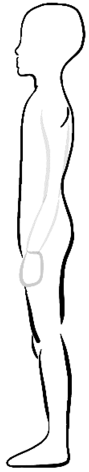
Figure 3.
Reduced thoracic kyphosis, increased lumbar lordosis (K < 42°; L > 47°).
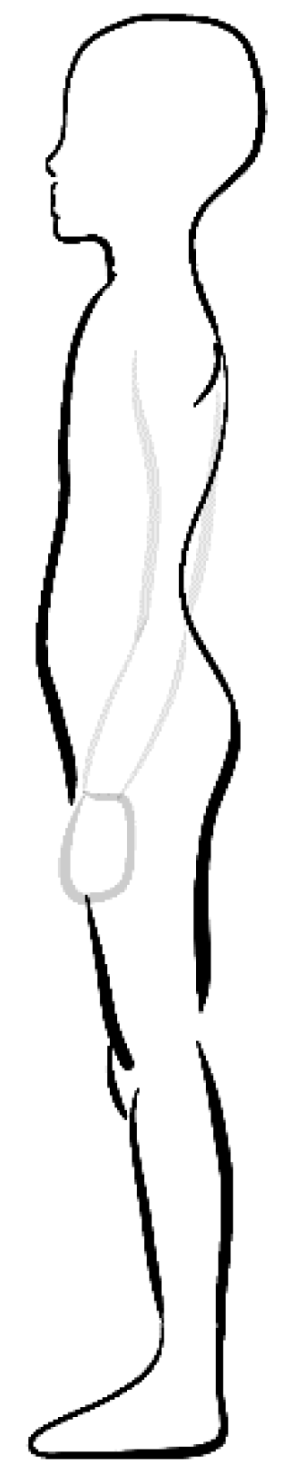
Figure 4.
Normal thoracic kyphosis, reduced lumbar lordosis (42° ≤ K ≤ 55°; L < 33°).
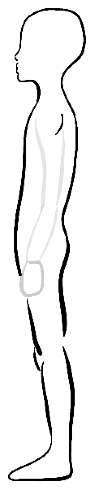
Figure 5.
Correct thoracic kyphosis, correct lumbar lordosis (42° ≤ K ≤ 55°; 33° ≤ L ≤ 47°).
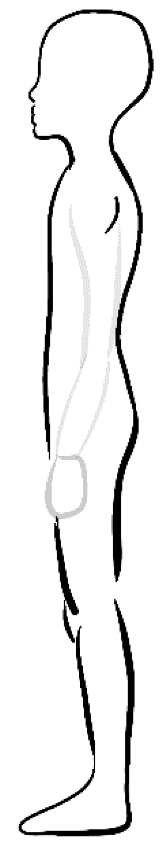
Figure 6.
Normal thoracic kyphosis, increased lumbar lordosis (42° ≤ K ≤ 55°; L > 47°).

Figure 7.
Increased thoracic kyphosis, decreased lumbar lordosis (K > 55°, L < 33°).
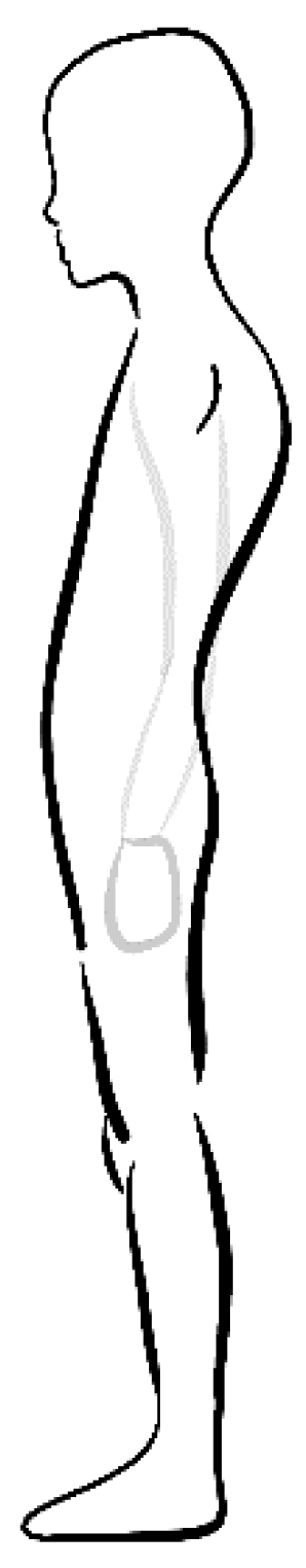
Figure 8.
Increased thoracic kyphosis, normal lumbar lordosis (K > 55°; 33° ≤ L ≤ 47°).
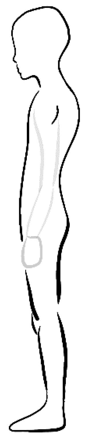
Figure 9.
Increased thoracic kyphosis, increased lumbar lordosis (K > 55°; 33° ≤ L ≤ 47°).
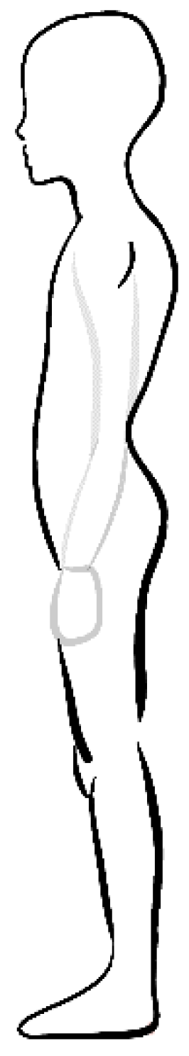

Table 1.
Body posture types in examined group.
| Variable | Girls | Boys | Total | |||
| N | % | N | % | N | % | |
| Reduced kyphosis, reduced lordosis | 22 | 15.49 | 26 | 16.35 | 48 | 15.95 |
| Reduced kyphosis, normal lordosis | 41 | 28.87 | 26 | 16.35 | 67 | 22.26 |
| Reduced kyphosis, increased lordosis | 12 | 8.45 | 2 | 1.26 | 14 | 4.65 |
| Normal kyphosis, reduced lordosis | 1 | 0.7 | 16 | 10.06 | 17 | 5.65 |
| Posture with normal physiological curvatures of spine | 36 | 25.35 | 51 | 32.08 | 87 | 28.9 |
| Normal kyphosis, increased lordosis | 18 | 12.68 | 17 | 10.69 | 35 | 11.63 |
| Increased kyphosis, reduced lordosis | 1 | 0.7 | 1 | 1.60 | 2 | 0.66 |
| Increased kyphosis, normal lordosis | 2 | 1.41 | 10 | 6.29 | 12 | 3.99 |
| Increased kyphosis, increased lordosis | 10 | 7.04 | 11 | 6.92 | 21 | 6.98 |
| Total | 143 | 47.18 | 160 | 52.82 | 303 | 100 |
Disclaimer/Publisher’s Note: The statements, opinions and data contained in all publications are solely those of the individual author(s) and contributor(s) and not of MDPI and/or the editor(s). MDPI and/or the editor(s) disclaim responsibility for any injury to people or property resulting from any ideas, methods, instructions or products referred to in the content. |
© 2025 by the authors. Licensee MDPI, Basel, Switzerland. This article is an open access article distributed under the terms and conditions of the Creative Commons Attribution (CC BY) license (http://creativecommons.org/licenses/by/4.0/).
Copyright: This open access article is published under a Creative Commons CC BY 4.0 license, which permit the free download, distribution, and reuse, provided that the author and preprint are cited in any reuse.


