Preprint
Article

This version is not peer-reviewed.

Epidemiology of Channelopathies in a Mediterranean Pediatric Population

Submitted:

27 November 2025

Posted:

28 November 2025

You are already at the latest version

Abstract

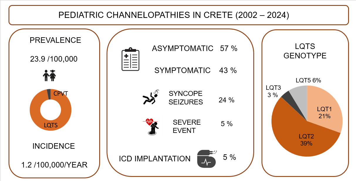
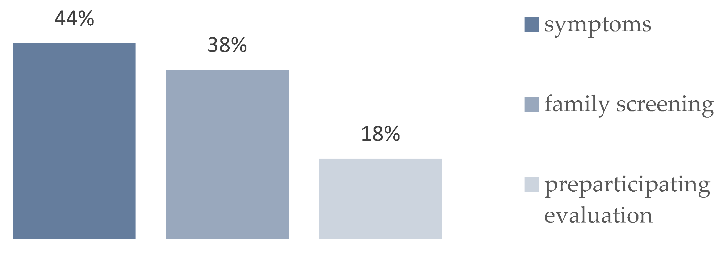
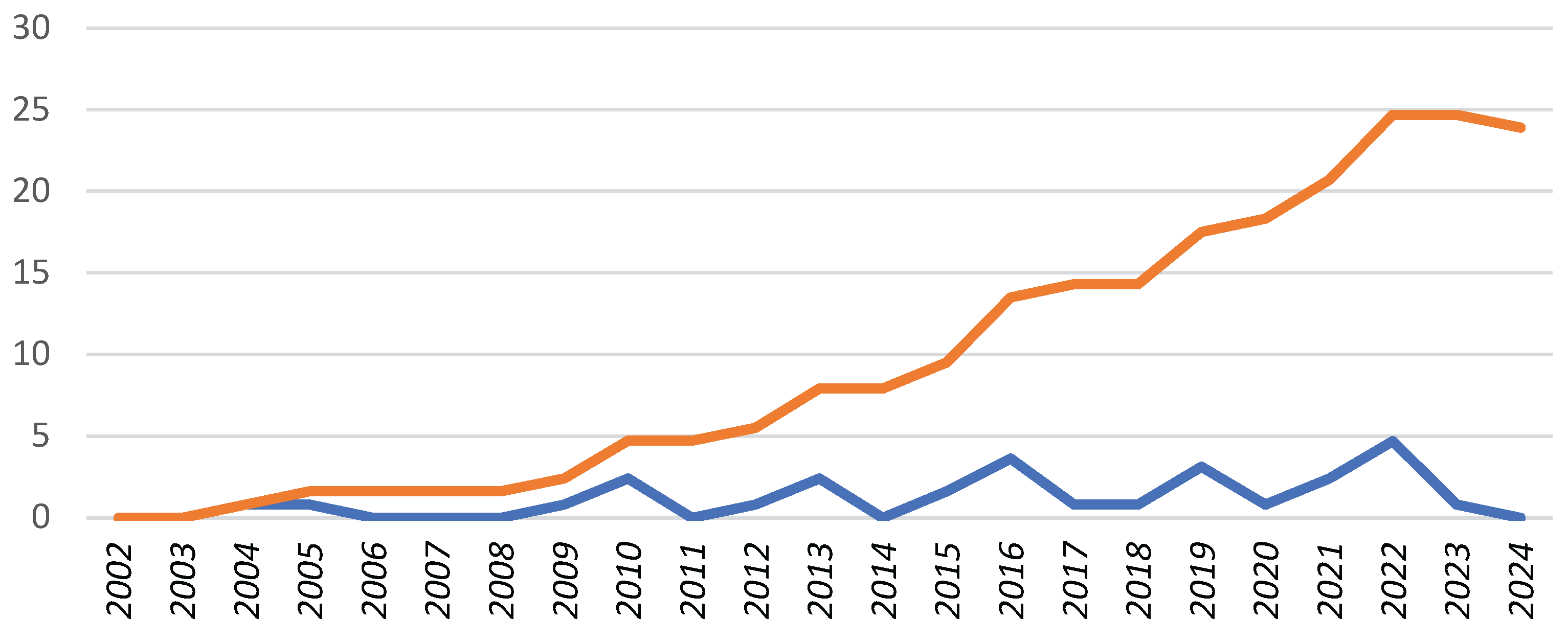
Background: Channelopathies represent a heterogeneous group of rare inherited cardiac diseases associated with life-threatening arrhythmias. Our knowledge of their epidemiology in childhood is limited. The aim of this study is to evaluate the epidemiology of pediatric channelopathies on a Mediterranean island (Crete, Greece). Methods: Retrospective study of children < 18 years followed in the Regional Tertiary Pediatric Cardiology Unit during a 23-year period (2002-2024) and meeting the disease-specific diagnostic criteria. Results: A total of 34 children (27 families) were enrolled, corresponding to an average annual incidence of 1.2 (95% C.I.: 0.8 – 1.6) and a cumulative prevalence of 23.9 (95% C.I.: 16.1 – 34.1) cases per 100, 000 children, with significant though regional incidence differences. Long QT syndrome (n=33) was predominant, with a single exception of catecholaminergic polymorphic tachycardia. Diagnosis was based on symptomatic presentation (n=15, 44 %), preparticipation screening (n=6, 18%) or affected family cascade screening (n=13, 38%). They represented the first diagnosis within affected families (index cases) in 20/34 (58%) of cases. Genetic testing was performed in 27/34 (79%) channelopathy cases and it was positive in a single case of CPVT and in 23 out of 27 (89%) LQT cases in which it was performed, with a genotype of LQT2 in 13 (39%), LQT1 in 7 (21%), LQT3 in 1 (3%) and LQT5 in 2 (6%) cases. Conclusion: The incidence of pediatric channelopathies on the Mediterranean island of Crete was comparable to that reported in the literature, with regional though clusters of significant increased incidence. Further study of the epidemiology of pediatric channelopathies is needed, to document any regional or ethnic differences and for the best design of large-scale screening programs.

Keywords: 
;  ;  ;  ;  ;  ;  

1. Introduction

Channelopathies are a heterogenous group of inherited, primary cardiac electrical diseases critically affecting the function of cardiomyocyte membrane ion channels, responsible for the action potential generation (depolarization and repolarization) [1]. Although rare, with a reported prevalence ranging from 1/2,000 to 1/5,000 in the general population [2,3] with regional and ethnic variability [4] they are extremely important due to their associated significant morbidity and mortality, representing a main cause of sudden cardiac death (SCD) in young people [5,6]. Main clinical diagnoses include Long QT Syndrome (LQTS), Brugada syndrome (BrS), Catecholaminergic Polymorphic Ventricular Tachycardia (CPVT) and Short QT Syndrome (SQTS). Their early detection in childhood is of critical clinical importance as affected children might represent the first cases (index cases) in affected families [7], presenting with syncopal episodes resembling epilepsy, or even with sudden cardiac death [5,8,9]. Our knowledge of their epidemiology in childhood is rather limited, based mainly on a limited number of cross-sectional studies in different ethnic groups [2,10,11].
The Mediterranean island of Crete, inhabited since prehistory with a flourishing civilization interacting with several Mediterranean basin populations, shows a genetically distinct pattern of inherited diseases compared to the Greek mainland [12]. The distinct epidemiology of pediatric cardiomyopathies on Crete has been recently reported [13]. The aim of the present study was to document, for first time, the epidemiology of pediatric channelopathies in a Mediterranean island population, in comparison to that reported in the literature.

2. Materials and Methods

This was a retrospective study of children younger than 18 years of age living in the region of Crete Island, evaluated in the Regional Academic Pediatric Cardiology Center during a 23-year period (January 2002 to December 2024), and meeting established disease-specific ECG/clinical diagnostic criteria for channelopathies [14,15]. The study was performed in line with the principles of the Declaration of Helsinki. The approval for collecting and processing data from the available medical records of each study patient was obtained from the University Hospital Herakleion, Ethics Committee, approval number 1007, 33/2.12.2020. Written informed consent for participation in research was obtained from legal parents/guardians of evaluated children < 18 years of age old. All personal data were anonymized. None of Gen AI tools were used in any stage of data processing.

Study Population

The study population included children, who met disease specific ECG/clinical diagnostic criteria, according to 2022 ESC Guidelines [14,15]. Disease specific diagnostic criteria are summarized in Table 1.

Data Collection

Data collection included demographic characteristics, brief family history, related clinical symptoms - dizziness, palpitations, seizures, deafness and especially the presence of cardiac events (syncope, aborted cardiac arrest or sudden cardiac death), resting 12-lead ECG, 24 hours ECG monitoring (Holter) including ECG monitoring during exercise testing on treadmill in cooperative patients, standard blood analysis, chest radiography and echocardiography. Genetic testing results were documented when performed [16,17,18]. Detailed family history and family pedigrees were documented. Clinical evaluation and cascade screening of 1st degree relatives was recommended.

Epidemiologic Data

The cumulative prevalence at the last year of study enrollment and the average annual incidence rate of channelopathies in the pediatric population of Crete were estimated, including documented cases meeting the disease-specific diagnostic criteria. Regional differences were evaluated as well. All demographic data were obtained from the Hellenic Statistical Authority Office [19].

Statistical Analysis

The incidence (new diagnoses per 100,000 children < 18 years of age per year, for each year of the duration of the study) and the prevalence (total cases per 100,000 children < 18 years) were estimated by the retrospective data analysis. The Hellenic Statistical Service data, regarding total and regional (total of 4 Prefectures) pediatric population of the island of Crete, were used as denominators to calculate the total and regional annual incidence and prevalence of channelopathies [19]. Data were summarized as frequencies and percentages for categorical data and as medians and means for age of presentation. Confidence intervals (95% C.I.) were estimated using free online software [20]. Regional differences regarding demographic and clinical variables were evaluated with appropriate methods for qualitative data (Chi-Square Test, Fisher exact test, Odds Ratio, Relative Risk and 95% C.I.) and numerical data (t-Test, Mann-Whitney U test paired comparisons, ANOVA or Kruskal-Wallis test group comparisons, for variables with normal or non-normal distribution, respectively). SPSS program (IBM SPSS Statistics V26) was used for data analysis. Significant differences (p < 0.05) were documented. None of Gen AI tools were used in any stage of data statistical analysis.

3. Results

Over a 23-year period (2002-2024) 34 new cases of pediatric channelopathy were documented. The average island population during this period was 615,000, including 125,300 children under 18 years of age. The average annual incidence of pediatric channelopathies in Crete was estimated at 1.2 (95% C.I.: 0.8 – 1.6) patients per 100,000 children, although with significant geographical differences. The cumulative prevalence in the last year of enrollment was estimated at 23.9 (95% C.I.: 16.1 – 34.1) patients per 100,000 children < 18 years old. The predominant channelopathy (corresponding to all but one case) was Long QT Syndrome, with a prevalence of 23.9 (95% C.I.: 16.1-34.1) cases per 100,000 children. Table 2 presents the average annual incidence and prevalence at last year of enrollment of channelopathies, while Figure 1 presents the annual incidence and cumulative prevalence, as documented for each year of the study period.
Diagnoses: From the total of 34 diagnoses, 33 corresponded to long QT syndrome (LQTS) with a single child diagnosed with catecholaminergic polymorphic ventricular tachycardia (CPVT). Sex: There were 14 (41%) boys and 20 (59%) girls. Τhe difference in sex distribution was not statistically significant. Age: Mean (median) age of diagnosis was 7.5 (8.5) years (range 0.1 – 15). Mean (median) age of diagnosis in boys was 5.9 (5) years, in girls 11.3 (9) years. This difference was not statistically significant (p=0.08). Geographical distribution: A statistically increased annual incidence of channelopathies (LQT syndrome), reaching 2.2 cases per 100,000 children per year was documented in the Middle-Western Region, compared to other island’s regions, as presented in Table 3.
Table 3. Incidence of pediatric channelopathies in Crete.
Abbreviations: LQTS: Long QT Syndrome; CPVT: Catecholaminergic Polymorphic Ventricular Tachycardia * statistically significant differences.
Diagnostic approach: The diagnosis was based on symptomatic presentation (n=15, 44%), preparticipation screening (n=6, 18%) or family cascade screening (n=13, 38%) as shown in Figure 2. Patients were diagnosed as index cases in 58% (n=20) cases. Symptoms at diagnosis included dizziness n=5 (14%), syncopal episodes n= 8 (24%) and seizures n=2 (6%), while 19 cases (56%) were asymptomatic.
Family history: A positive diagnosis in at least one first-degree relative was recorded in 16/34 (47%) cases. The probability of an affected parent was reported in 10/34 (30%) cases (mother in n=8 cases, father in n=2 cases), of an affected sibling in 6/34 (17%). However the total number of affected siblings was much higher (n=19). Positive history of sudden cardiac death (SCD) was recorded in 9 (26%) cases, being first-degree relative in 1 (3%) case, second degree relative in 8 (23%) cases.
Figure 2. Diagnosis type of pediatric channelopathies on Crete.
Preprints 187127 g002
Genetic evaluation: Genetic testing was performed in 27/34 (79%) channelopathy cases and it was positive in 24/34 (70%) of them, specifically in a single case of CPVT and in 23 out of 27 LQT cases (85% of those tested), in which it was performed.
Type of Channelopathy:
A) Long QT Syndrome. LQTS was the most frequent pediatric channelopathy, documented in 33 cases. The mean age at diagnosis was 7.1 years (range 0.1 – 15). Diagnosis in the first year of life in n=2 (6%) was associated with the most severe phenotype (recorded episode of ventricular tachycardia, resp. recurrent syncopal episodes). Genetic testing was performed in 26/33 (79%) of LQTS patients. Positive genotype was confirmed in n=23/33 (70%) LQT cases. Pathogenic variants in following LQT related genes KCNQ1, KCNH2, SCN5A and KCNE1 were found and consequently 4 LQT genotypes were determined: LQT1 in n=7 (21%), LQT2 in n=13 (39%), LQT3 in n=1 (3%) and LQT5 in n=2 (6%) cases.
B) CPVT. A single case was documented. This was a 5 years old boy who was diagnosed after a symptomatic episode of bidirectional ventricular tachycardia in preschool age and was a career of disease causing RYR2 gene. The prevalence of CPVT in the pediatric population in Crete was estimated at 0.8 cases per 100,000.
C) No cases of Brugada Syndrome or SQT syndrome were documented
Therapy: Standard recommendations regarding avoidance of QT prolongation agents and sport participation according to current guidelines were recommended [21,22]. Medical treatment of LQT patients was genotype specific (when genotype available), including beta blockers, and sodium channel blocker (Mexiletine) in LQT3 patients [23]. Surgical therapeutic methods (left or both sided sympathetic denervation) followed by ICD implantation were indicated in 6% (n=2) patients.
Comparison to the literature: The comparison of the disease prevalence in the present study with that reported in the literature (screening programs) is presented in Table 4.

4. Discussion

The Mediterranean region is characterized by an increased prevalence of many hereditary diseases [12]. Our study group has documented an increased prevalence of pediatric cardiomyopathies on the Mediterranean island of Crete [13]. To our knowledge this is the first study reporting the epidemiology of pediatric channelopathies in the Southeastern Mediterranean region.
The annual average incidence of channelopathies in the pediatric population in Crete is estimated at 1.2 cases per 100,000/year. The prevalence of channelopathies is estimated at 23.9 cases per 100,000 children < 18 years. Long QT Syndrome is the most frequent channelopathy [3] and the only one with a well reported prevalence due to available large scale epidemiological studies in neonates and infants from Italy [2,27] and Japan [28], and in children in Japan [10,30]. The prevalence of LQTS in the present study does not differ from the prevalence of neonatal LQTS in Japan estimated at 23.3/100,000 [28]. Compared to the epidemiological studies of LQTS prevalence in the neonatal period in Italy, estimated at 38/100,000 [2] resp. 42/100,000 [27] and in Germany at 88/100,000 [29], the prevalence of LQTS in the pediatric population of Crete is statistically significantly lower as shown in the Table 4. The prevalence of LQT in the neonatal period was estimated in both above mentioned countries Italy [2,27] and Japan [28] at 1 case per 2,000–2,500 children. In the present study, the estimated prevalence was 1 case per 4,184 children. The prevalence of LQTS in the 6-year and the 12-year age group is reported by Yoshinaga et al. [10] to be 30 cases respective 93 cases per 100,000 children, while in the study by Hayashi et al. [30], the prevalence of LQTS in the 6 to 12-year age group is 37.6 cases per 100,000 children. The comparison of these studies has revealed significant differences, probably due to different study methodologies, as the data of the present study are not based on the results of a mass pediatric population screening program, but on recording of symptomatic cases only.
A slight predominance of girls (60%) in this study correlates with the results of the Japan [10] and Dutch study [31]. The distribution of channelopathies by age correlates with distribution of reported in the literature for pediatric LQTS cases. In this study 6% of cases were diagnosed during the first year of life and were associated with severe clinical symptoms, in accordance with published data [32,33]. The diagnosis of LQTS in infancy and preschool age was approximately equally distributed. The age of elementary school students is considered by some authors to be the most suitable age for implementing screening programs, based on 12-lead ECG recording [10,30], in contrast to the early detection of LQTS in infancy, preferred by other authors [2,34,35].
In previous studies regional geographical differences in pediatric cardiomyopathy epidemiology were documented [36,37], including regional differences observed in our study population [13]. Similarly, in the present study, regarding pediatric channelopathies with LQT predominance, regional clusters were documented. An increased incidence of LQTS cases, detected in the Middle-West area of the island of Crete could be probably explained by the presence of genetically distinct populations, residents of mountain areas and members of large, related families, relatively isolated from the wider population of the island. The results of DNA analysis highlight the presence of mutations unique to each family, strengthening the hypothesis of the presence of “private” mutations that characterize families with common ancestral ties and relatively little interaction with other population, as has been described in studies of rare neuromuscular [38] and metabolic diseases [39]. Reports of “private” gene mutations in channelopathies are well described in the literature [40,41].
Targeted family screening and sports pre-participation screening contributed to the diagnosis of channelopathies in our population similar to previous studies [42,43].
Although not all of our cases underwent genetic testing evaluation, DNA analysis has become an integral part of every patient diagnostic evaluation. In our study, genetic testing was performed in 79% of children and positive results were confirmed in 70% of them. Genetic testing in channelopathies is reported to be positive in 75-80% of LQTS cases [44,45], in 66% of CPVT cases [46], and only in 25-30% cases of Brugada Syndrome [47,48]. The diagnostic yield of genetic testing in LQTS in this study was close to reported data from other studies [7]. In our study LQT2 represented the most frequent genotype (39%). In Rochester LQTS registry, LQT1 predominated (46% vs. 21% in the present study), LQT2 followed (42% vs. 39%) with LQT3 being the least common form (12% vs. 3%) [49]. In the Italian LQTS registry LQT1 also predominated (56%) followed by LQT2 (32%) and LQT3 (12%) [25]. The predominance of LQT2 in the pediatric population of Crete might represent a geographic variation in pediatric LQT epidemiology [4].
In our study a single case of CPVT was found. CPVT is a rare, highly malignant channelopathy. The rarity of CPVT is confirmed by a limited number of recorded cases in international databases [46,50]. Patients with CPVT often present with exercise or emotion induced ventricular tachycardia leading to syncope or cardiac arrest. The heart is structurally normal and the ECG in rest has no pathognomonic feature.
Brugada Syndrome is a rare disease in young patients compared with adult populations and the prevalence in children and the young remains not well defined [51]. Symptoms associated with Brugada syndrome include heart palpitations, fainting or seizures that often happen during febrile conditions, rest or sleep [51]. In our study we did not document any pediatric BrS case.
Reported pediatric cases of Short QT Syndrome are extremely rare [52], similar to our study where no patient was detected.
Limitations of the present study include retrospective analysis methodology, based on available medical records information, in contrast to the majority of available studies in the field reporting large scale ECG screening results. The possibility of individual cases failing to be registered cannot be ruled out while further undetected cases among the reference population cannot be excluded. Due to limited availability of genetic testing in the early years of the documentation period, the identification of patients’ genotype is not complete.
Conclusion
The incidence of pediatric channelopathies on the Mediterranean island of Crete was comparable to that reported in the literature, with regional though clusters of significant increased incidence. Further study of the epidemiology of pediatric channelopathies is needed, to document any regional or ethnic differences and for the best design of large-scale screening programs.

Author Contributions

Conceptualization, A.B. and I.G.; methodology, all authors; software, I.G.; validation, I.P., A.A. and I.G.; formal analysis, A.B.; investigation, A.B.; resources, A.B.; data curation, all authors; writing—original draft preparation, A.B., I.G.; writing—review and editing, all authors; visualization, I.G.; supervision, I.P., A.A., E.G., I.G.; project administration, all authors. All authors have read and agreed to the published version of the manuscript.

Funding

This research received no external funding.

Institutional Review Board Statement

The study was conducted in accordance with the Declaration of Helsinki and approved by Ethics Committee of University Hospital Heraklion, 1007, 33/2.12.2020.

Informed Consent Statement

Informed consent was obtained from all subjects involved in the study.

Data Availability Statement

The data presented in this study are available from the corresponding author upon request. The data are not publicly available due to privacy and ethical restrictions.

Acknowledgments

None.

Conflicts of Interest

The authors declare no conflicts of interest.

References

  1. Schwartz PJ, Crotti L. Ion channel diseases in children: manifestations and management. Curr Opin Cardiol 2008 May;23(3):184-91. doi: 10.1097/HCO.0b013e3282fcc2e3.
  2. Schwartz PJ, Stramba-Badiale M, Crotti L, Pedrazzini M, Besana A, Bosi A et al. Prevalence of the congenital long QT syndrome. Circulation 2009;120: 1761–1767.
  3. Schwartz PJ, Ackerman MJ, Antzelevitch C, Bezzina CR, Borggrefe M, Cuneo BF, Wilde AAM. Inherited cardiac arrhythmias. Nat Rev Dis Primers 2020;6(1):58.
  4. Offerhaus JA, Bezzina CR, Wilde AAM. Epidemiology of inherited arrhythmias Nat Rev Cardiol 2020;17(4):205-215.
  5. Skinner JR, Winbo A, Abrams D, Vohra J, Wilde AA. Channelopathies that lead to Sudden Cardiac Death: Clinical and Genetic Aspects. Heart, Lung and Circulation 2018; 1–9. https://doi.org/10.1016/j.hlc.2018.09.007.
  6. Bagnall RD, Weintraub RG, Ingles J, Duflou J, Yeates L, Lam L et al. A prospective study of sudden cardiac death among children and young adults N Engl J Med. 2016 23;374(25):2441-52. doi: 10.1056/NEJMoa1510687.
  7. Schwartz PJ, Crotti L, Insolia R. Long QT Syndrome: From genetics to management. Circ Arrhythm Electrophysiol. 2012 August 1; 5(4): 868–877. doi:10.1161/CIRCEP.111.962019.
  8. Hazle MA, Shellhaas RA, Bradley DJ, Macdonald Dick 2nd, Lapage MJ. Arrhythmogenic channelopathy syndromes presenting as refractory epilepsy. Pediatr Neurol 2013 Aug;49(2):134-7. doi: 10.1016/j.pediatrneurol.2013.03.017.
  9. Schwartz PJ, Stramba-Badiale M, Giudicessi JR, Tester DJ, Crotti L, Ackermann MJ. Cardiac Channelopathies and Sudden Infant Death Syndrome I. Gussak, C. Antzelevitch (eds.), Electrical Diseases of the Heart, 381DOI 10.1007/978-1-4471-4978-1_24, © Springer-Verlag London 2013.
  10. Yoshinaga M, Kucho Y, Nishibatake M, Ogata H, Nomura Y. Probability of diagnosing long QT syndrome in children and adolescents according to the criteria of the HRS/EHRA/APHRS expert consensus statement. Eur Heart J. 2016; 37:2490–2497.
  11. Oe H, Takagi M, Tanaka A, Namba M, Nishiboro Y, Nishida Y et al. Prevalence and clinical course of the juveniles with Brugada-type ECG in Japanese population. Pacing Clin Electrophysiol 2005;28:549–54.
  12. Drineas P, Tsetsos F, Plantinga A, Lazaridis I, Yannaki E, Razou A et al. Genetic history of the population of Crete. Ann Hum Gen 2019; 83:373-388.
  13. Bagkaki A, Parthenakis F, Chlouverakis G, Anastasakis A, Papagiannis I, Galanakis E, Germanakis, I. Epidemiology of Pediatric Cardiomyopathy in a Mediterranean Population. Children2024;11: 732.
  14. Zeppenfeld K, Tfelt-Hansen J, de Riva M, Gregers Winkel B, Behr ER, Blom NA et al. 2022 ESC Guidelines for the management of patients with ventricular arrhythmias and the prevention of sudden cardiac death Developed by the task force for the management of patients with ventricular arrhythmias and the prevention of sudden cardiac death of the European Society of Cardiology (ESC) Endorsed by the Association for European Paediatric and Congenital Cardiology (AEPC). European Heart Journal2022; 43, 3997–4126. https://doi.org/10.1093/eurheartj/ehac262.
  15. Priori SG, Blomström-Lundqvist C, Mazzanti A, Blom N, Borggrefe M, Camm J et al. 2015 ESC Guidelines for the management of patients with ventricular arrhythmias and the prevention of sudden cardiac death: The Task Force for the Management of Patients with Ventricular Arrhythmias and the Prevention of Sudden Cardiac Death of the European Society of Cardiology (ESC) Endorsed by: Association for European Paediatric and Congenital Cardiology (AEPC). Europace. 2015 Nov;17(11):1601-87.
  16. Schwartz PJ, Ackerman MJ, George AL Jr, Wilde AAM. Impact of genetics on the clinical management of channelopathies. J Am Coll Cardiol. 2013;62:169–180. doi: 10.1016/j.jacc.2013.04.044.
  17. Adler A, Novelli V, Amin AS, Abiusi E, Care M, Nannenberg EA et al. An International, Multicentered, Evidence-Based Reappraisal of Genes Reported to Cause Congenital Long QT Syndrome. Circulation. 2020;141:418–428.
  18. 18. [CrossRef]
  19. Wilde AAM, Semsarian C, Marquez MF, Sepehri Shamloo A, Ackerman MJ, Ashley EA et al. EHRA/HRS/APHRS/LAHRS Expert Consensus Statement on the State of Genetic Testing for Cardiac Diseases. Heart Rhythm 2022;19(7):e4-e49.
  20. Hellenic Statistical Authority Greece population data. Available online: https://www.statistics.gr/el/statistics/-/publication/SPO18 (Accessed 23/9/2025).
  21. Med Calc. Easy to use statistical software. Available at https://www.medcalc.org/calc/rate_comparison.php (Accessed 2/9 2025).
  22. QT drugs lists. Available online: https://crediblemeds.org/ (Accessed 23/9/2025).
  23. Pelliccia A, Sharma S, Gati S, Back M, Borjensson M, Caselli S et al.2020 ESC Guidelines on sports cardiology and exercise in patients with cardiovascular disease: The Task Force on sports cardiology and exercise in patients with cardiovascular disease of the European Society of Cardiology (ESC). European Heart Journal, 2021; 42(1):17–96,.
  24. Bagkaki A, Tsoutsinos A, Hatzidaki E, Tzatzarakis M, Parthenakis F, Germanakis I. Mexiletine Treatment for Neonatal LQT3 Syndrome: Case Report and Literature Review. Front. Pediatr.2021;9:674041. doi: 10.3389/fped.2021.674041.
  25. Gonzalez Corcia MC, de Asmundis C, Chierchia GB, Brugada P. Brugada syndrome in the paediatric population: a comprehensive approach to clinical manifestations, diagnosis, and management. Cardiology in the Young 2016; 1- 12.
  26. Mazzanti A, Maragna R, Vacanti G, Monteforte N, Bloise R, Marino M et al. et al. Interplay Between Genetic Substrate, QTc Duration, and Arrhythmia Risk in Patients with Long QT Syndrome, J Am Coll Cardiol 2018;71:1663–71.
  27. Sieira J, Brugada P. Brugada syndrome: defining the risk in asymptomatic patients. Arrhythm Electrophysiol Rev 2016;5:164–169.
  28. Nosetti L, Zaffanello M, Lombardi C, Gerosa A, Piacentini G, Abramo M et al. Early Screening for Long QT Syndrome and Cardiac Anomalies in Infants: A Comprehensive Study. Clin. Pract. 2024;14:1038–1053.
  29. Yoshinaga M, Ushinohama H, Sato S, Tauchi N, Horigome H, Takahashi H et al. Electrocardiographic screening of 1-month-old infants for identifying prolonged QT intervals. Circ Arrhythm Electrophysiol 2013;6:932–938.
  30. Simma A, Potapow A, Brandstetter S, Michel H, Melter M, Seelbach-Göbelb B et al. Electrocardiographic Screening in the First Days of Life for Diagnosing Long QT Syndrome: Findings from a Birth Cohort Study in Germany. Neonatology2020;117:756–763.],.
  31. Hayashi K, Fujino N, Uchiyama K, Ino H, Sakata K, Konno Teta l. Long QT syndrome and associated gene mutation carriers in Japanese children: results from ECG screening examinations Clinical Science (2009) 117, 415–424.
  32. Vink AS, Clur SB, Wilde AAM, Blom NA. Effect of age and gender on the QTc-interval in healthy individuals and patients with long-QT syndrome. Trends Cardiovasc Med. 2018; 28:64–75.
  33. Wilde AA, Moss AJ, Kaufman ES, Shimizu W, Peterson DR, Benhorin J et al. Clinical aspects of type 3 long QT syndrome. An international multicenter study.Circulation.2016;134:872-882.
  34. Horigome H, Nagashima M, Sumitomo N, Yoshinaga M, Ushinohama H, Iwamoto M et al. Clinical Characteristics and Genetic Background of Congenital Long-QT Syndrome Diagnosed in Fetal, Neonatal, and Infantile Life. A Nationwide Questionnaire Survey in Japan. Circ Arrhythm Electrophysiol. 2010;3:10-17.
  35. Schwartz PJ, Garson AJr, Paul T, Stramba-Badiale M, Vetter VL, Villain E et al. Guidelines for the interpretation of the neonatal electrocardiogram. A Task Force for the European Society of Cardiology. Eur Heart J.2002;23:1329-1344.
  36. Sarquella-Brugada G, Cesar S, Zambrano MD, Fernandez-Falgueras A, Fiol V, Iglesias A et al. Electrocardiographic Assessment and Genetic Analysis in Neonates: A Current Topic of Discussion. Current Cardiology Reviews.2019;15: 30-37.].
  37. Lipshultz SE, Sleeper LA, Towbin JA, Lowe AM, Orav EJ, Cox GF et al. The incidence of pediatric cardiomyopathy in two regions of the United States. N. Engl. J. Med. 2003;348:1647–1655.].
  38. Nugent AW, Daubeney PE, Chondros P, Carlin JB, Cheung M, Wilkinson LC et al. The epidemiology of childhood cardiomyopathy in Australia. N Engl J Med 2003; 348: 1639–1646].
  39. Baban A, Lodato V, Parlapiano G, di Mambro C, Adorisio R, Bertini ES et al. Myocardial and Arrhythmic Spectrum of Neuromuscular Disorders in Children. Biomolecules 2021;11: 1578.].
  40. Lodato V, Parlapiano G, Cal F, Silvetti MS, Adorisio R, Armando M et al. Cardiomyopathies in children and systemic disorders when is it useful to look beyond the heart? J. Cardiovasc. Dev. Dis. 2022;9: 47.].
  41. Bidzimou MTK, Landstrom AP. From diagnostic testing to precision medicine: the evolving role of genomics in cardiac channelopathies and cardiomyopathies in children. Current Opinion in Genetics & Development 2022;76:101978.],.
  42. Rivolta I, Abriel H, Tateyama M, Liu H, Memmi M, Vardas P et al. Brugada and Long QT-3 Syndrome Mutations of a Single Residue of the Cardiac Sodium Channel Confer Distinct Channel and Clinical Phenotypes. Journal of biological chemistry 2001; 276 (33):30623–30630.].
  43. Theilade J, Kanters J, Henriksen FL, Gilsa-Hansen M, Svendsen JH, Eschen O, et al. Cascade Screening in Families with Inherited Cardiac Diseases Driven by Cardiologists: Feasibility and Nationwide Outcome in Long QT Syndrome. Cardiology2013;126:131–13].
  44. Graziano F, Schiavon M, Cipriani A, Savalla F, De Gaspari M, Bauce B et al. Causes of sudden cardiac arrest and death and the diagnostic yield of sport preparticipation screening in children. BrJSportsMed2024;58:255–260.
  45. Schwartz PJ, Ackerman MJ, George Jr AL, Wilde AAM. Impact of Genetics on the Clinical Management of Channelopathies. J Am Coll Cardiol. 2013; 62(3): 169–180.],.
  46. Schwartz PJ, Ackerman MJ. The long QT syndrome: a transatlantic clinical approach to diagnosis and therapy. Eur Heart J. 2013; 34, 3109-3116.],.
  47. Roston TM, Yuchi Z, Kannankeril PJ, Hathaway J, Vinocur JM, Etheridge SP, et al. The clinical and genetic spectrum of catecholaminergic ventricular tachycardia: findings from an international multicenter registry. Europace 2018;20:541-547].
  48. Monasky MM, Micaglio E, Ciconte G, Pappone C., Brugada Syndrome: Oligogenic or Mendelian Disease? Int. J. Mol. Sci. 2020; 21: 1687.
  49. Campuzano O, Sarquella-Brugada G, Fernandez-Falgueras A, Cessar S, Coll M, Mates J et al. Genetic interpretation and clinical translation of minor genes related to Brugada syndrome. Human Mutation 2019; 40:749-764.].
  50. Kutyifa V, Daimee UA, McNitt S, Polonsky B, Lowenstein C, Cutter K, et al., Clinical aspects of the three major genetic forms of long QT syndrome (LQT1, LQT2, LQT3), Ann Noninvasive Electrocardiol. 2018;23:e12537. https://doi.org/10.1111/anec.12537).
  51. Pflaumer A, Davis AM. An update on the diagnosis and management of catecholamine polymorphic ventricular tachycardia. Heart Lung Circ 2019;28:266-9],.
  52. Brugada J, Campuzano O, Arbelo E, Sarquella-Brugada G, Brugada R. Present status of Brugada Syndrome. JACC State-of-the-Art Review. JACC2018; 72(9): 1046-59.
  53. Pereira R, Campuzano O, Sarquella-Brugada G, Cesar S, Iglesias A, Brugada J et al. Short QT syndrome in pediatrics. Clin Res Cardiol 2017;106:393–400.
Figure 1. Epidemiology of channelopathies in pediatric population in Crete, Greece.
Preprints 187127 g001
Table 1. Channelopathies. ECG/clinical diagnostic criteria [14,15].
Diagnostic criteria
Long QT Syndrome a) QTc ≥ 480 ms by Bazett’s formula in repeated 12-lead ECG with or without symptoms in the absence of a secondary cause of LQTS
b) QTc > 460 in repeated 12-lead ECGs in a patient with unexplained syncope or aborted cardiac arrest in the absence of a secondary cause for QT prolongation.
c) LQTS risk score (“Schwartz score”) >3
Brugada Syndrome ECG: Spontaneous or induced (fever, provocative testing) type 1 (“coved pattern”) is considered as the only diagnostic pattern for BrS regardless the clinical symptoms
Catecholaminergic
Polymorphic Ventricular
Tachycardia (CPVT)
The presence of normal resting ECG in a structurally normal heart and exercise- or emotion-induced bidirectional VT or PVT (polymorphic ventricular tachycardia)
Short QT Syndrome a)QTc ≤ 360 ms in the presence of a pathogenic mutation and/or a positive family history of SQTS in the absence of secondary causes of SQTS
b) SQTS should be considered in the presence of a QTc ≤ 320 m.
Table 2. Epidemiology of pediatric channelopathies in Crete, Greece.
Cases Incidence/
100,000<18Y/Year
(95%C.I.)
Prevalence/
100,000<18Y
(95%C.I.)
Total 34 1.2 (0.8-1.6) 23.9 (16.1-34.1)
LQTS 33 1.1 (0.8-1.6) 23.9 (16.1-34.1)
CPVT 1 0.03 (0.01-0.2) 0.8 (0.02-4.4)
Abbreviations. LQTS: Long QT Syndrome; CPVT: Catecholaminergic Polymorphic Ventricular Tachycardia.
Table 4. Epidemiological studies of pediatric channelopathies.
Schwartz Circulation 2009 [2] Nosetti
Clinics and Practice 2024 [27]
Yoshinaga Circ Arrh Electr 2013 [28] Simma
Neonatology
2020 [29]
Hayashi
Clinical Science 2009 [30]
Yoshinaga
European Heart Journal 2016 [10]
Crete, Greece
2025
Channelopathy LQTS LQTS LQTS LQTS LQTS LQTS LQTS
Retrospective/Prospective
Pro Retro Pro Pro Pro Pro Retro
Years (duration)
5 (2001 –06) 18 (2001-17) 2 (2010-11) 4 (2015-18) 2 (2004-5) 6 (2008-13) 23 (2002-24)
Country Italy
Italy
Japan Germany Regensburg Japan
Kanazawa
Japan
Kagoshima
Greece
Crete
Age of children
15-25 days 20-40 days 4 weeks 27 days 6-12 years 6 years 12 years 0-18 years
Screened population 44,596 2,245
4,285 2,251 7,961
33,051
3,751
125,300
Cases
17 27 1 2 3 10 (6 Years)
32 (12 Years)
33
Prevalence
(100,000<18Y)

38*

42*

23.3

88*

37.6
30 (6 Y)
93 (12 Years)
23.9
95% C.I: 16.1 – 34.1
Sex distribution (%) (boys/girls) 51/49 39.9/60.1 60/40 50.9/49.1 50.8/49.2 50/50 6 Years 46.8/53.2 12 Years 41/59
Abbreviations: LQTS: Long QT Syndrome. * statistically significant difference in prevalence (compared to the average incidence in Crete 2002-24).
Disclaimer/Publisher’s Note: The statements, opinions and data contained in all publications are solely those of the individual author(s) and contributor(s) and not of MDPI and/or the editor(s). MDPI and/or the editor(s) disclaim responsibility for any injury to people or property resulting from any ideas, methods, instructions or products referred to in the content.
Copyright: This open access article is published under a Creative Commons CC BY 4.0 license, which permit the free download, distribution, and reuse, provided that the author and preprint are cited in any reuse.
Prerpints.org logo

Preprints.org is a free preprint server supported by MDPI in Basel, Switzerland.

Subscribe

Disclaimer

Terms of Use

Privacy Policy

Privacy Settings

© 2025 MDPI (Basel, Switzerland) unless otherwise stated