Submitted:
23 November 2025
Posted:
25 November 2025
You are already at the latest version
Abstract
This study used a PDMS microfluidic chip to study how nanoparticle size and surface wettability affect their movement in airway mucus. Artificial mucus with 3% mucin was used to test nanoparticles ranging from 50 nm to 300 nm in size and with contact angles between 40° and 90°. When the particle size was smaller than 150 nm and the contact angle was below 60°, the diffusion coefficient increased about 2.7 times, and the retention time in mucus dropped by nearly 45%. A simple regression model combining particle size and contact angle showed strong agreement with experimental data ( ), allowing clear prediction of transport efficiency. Unlike earlier static or single-factor tests, this microfluidic system provided a stable and repeatable environment close to airway conditions. The results offer practical guidance for designing nanoparticles that can cross mucus barriers and improve inhaled drug delivery. Future work should test real human mucus and include ciliary movement to verify the model under physiological conditions.
Keywords:
microfluidic chip
; airway mucus
; nanoparticle transport
; surface wettability
; diffusion coefficient
; mucus penetration
; drug delivery
1. Introduction
Airway mucus is a highly hydrated yet structurally complex gel that normally protects the respiratory tract by trapping pathogens and particulates. However, its dense mucin network, heterogeneous pore structure, and dynamic clearance also hinder the diffusion of inhaled drugs and nanocarriers, preventing them from efficiently reaching the underlying epithelium [1]. The steric obstruction and electrostatic interactions within the mucus matrix lead to restricted or sub-diffusive nanoparticle motion, often resulting in poor therapeutic delivery [2]. Efforts to improve nanoparticle (NP) transport have focused on engineering their physicochemical properties, including size, surface charge, and coating chemistry [3]. Systematic studies indicate that smaller and more hydrophilic particles exhibit higher mobility, whereas larger or hydrophobic particles are more readily immobilized by mucin fibers [4]. Surface modification strategies, such as PEGylation, have been shown to reduce mucoadhesion and improve penetration by preventing strong nonspecific interactions [5]. Importantly, investigations have demonstrated that improving nanoparticle penetration into mucus can enhance drug delivery efficacy in the lungs, underscoring the value of rational nanoparticle design [6]. Despite progress, many existing studies evaluate only a single property at a time and often rely on synthetic or reconstituted mucus that lacks the mechanical and biochemical complexity of native mucus [7]. Particle transport behavior also depends on local microstructure and hydration state, making standardized testing difficult [8]. Furthermore, prior work typically examines particle motion under static or short-term conditions, whereas the airway environment involves continuous ciliary flow and active turnover [9,10]. These variations lead to inconsistent datasets and make it challenging to establish quantitative design rules. Microfluidic technologies have recently emerged as powerful tools to mimic airway physical conditions in small, well-controlled systems. PDMS-based airway-on-chip platforms can reproduce air–liquid interfaces, mucus secretion, and directional flow, thereby enabling real-time tracking of nanoparticle behavior. These systems allow systematic evaluation of how physical and chemical factors influence particle diffusivity and retention under more realistic dynamic constraints [11,12]. However, their integration into predictive frameworks remains limited: most studies focus on narrow ranges of particle sizes, do not standardize surface wettability, or lack sufficient sample sizes for robust multivariate analysis [13]. Surface wettability—commonly assessed by contact angle—is a key determinant of mucoadhesion, yet few studies quantify its effect under concurrent flow and mucus-like viscoelasticity [14,15]. As a result, current guidelines for designing mucus-penetrating nanoparticles remain qualitative, typically summarized as “smaller and more hydrophilic particles diffuse better,” without providing mechanistically informed thresholds or predictive models.
The present work introduces a PDMS-based microfluidic chip that simulates key features of the airway mucus microenvironment. The platform employs a mucin gel with rheological properties comparable to native mucus and allows controlled assessment of nanoparticles spanning a range of sizes (50–300 nm) and surface wettability (contact angle 40°–90°) under defined flow conditions. Using this system, we measured diffusion and retention profiles and applied multivariate regression to quantitatively link particle size and wettability with transport efficiency. By integrating microfluidic testing with statistical modeling, this study establishes a predictive framework for mucus penetration and provides data-driven guidelines to support the rational design of nanocarriers capable of effectively navigating airway mucus barriers for therapeutic delivery.
2. Materials and Methods
2.1. Sample and Study Description
Forty-eight mucus-like samples were prepared to simulate human airway conditions. Each sample contained 3% (w/v) porcine gastric mucin (Type III, Sigma-Aldrich) dissolved in phosphate-buffered saline (PBS, pH 7.4). The mixture was kept at 37 °C for 24 h to allow gel formation. Nanoparticles (NPs) were produced in five sizes—50, 100, 150, 200, and 300 nm—using a standard emulsion–solvent evaporation method. Surface hydrophobicity was adjusted by changing the ratio of poly(ethylene glycol) (PEG) and polylactic acid (PLA) coatings, resulting in contact angles between 40° and 90°. All tests were conducted at 37 °C and 95% relative humidity to reproduce airway conditions. Each experimental setting was repeated three times, generating 240 data sets in total.
2.2. Experimental Design and Control Setup
A PDMS microfluidic chip was produced by soft lithography. The device had a 200 μm-high channel and a 50 μm mucus layer. The experimental group included NPs of different sizes and surface wettability, while the control group used 100 nm PEG-coated particles with a 50° contact angle, which served as a benchmark for mucus penetration. These parameters were selected based on earlier reports showing that diffusion in mucus depends on both particle size and surface adhesion. In each test, 10 μL of particle suspension was injected into the inlet at a steady flow of 5 μL min⁻¹, and fluorescence images were taken every 10 s for 30 min. All chips came from the same PDMS batch and received identical curing and plasma treatment to maintain consistency.
2.3. Measurement Methods and Quality Control
Particle movement in mucus was recorded using a confocal laser scanning microscope (Leica TCS SP8) with a 63× oil-immersion objective. Fluorescent polystyrene nanoparticles (excitation 488 nm, emission 520 nm) were imaged at 1 frame s⁻¹. The diffusion coefficient was calculated from the mean square displacement (MSD) [16]:
where is the average MSD and is the time lag. Temperature was held constant within ±0.2 °C to prevent thermal drift. Laser power and detector gain were kept the same for all runs. Frames with photobleaching greater than 10% or unstable focus were discarded. Glycerol–water calibration samples were used to check the accuracy of the optical system before each session.
2.4. Data Processing and Model Formulation
All data were processed using MATLAB R2024a and OriginPro 2023. Particle tracks were analyzed with the TrackMate plugin in ImageJ to obtain displacement data. The relation between diffusion coefficient (), particle diameter (), and contact angle () was described by multiple linear regression [17]:
where are regression coefficients and is the error term. Model fit was evaluated using the coefficient of determination () and root-mean-square error (RMSE). A normalized permeability index () was also calculated:
where is the diffusion coefficient of particles in water. Outliers beyond ±2 standard deviations were excluded before fitting.
2.5. Statistical Analysis
All data are presented as mean ± standard deviation (SD). Differences among groups were analyzed using one-way ANOVA followed by Tukey’s post-hoc test with a significance level of 0.05. Data normality and variance equality were checked using Shapiro–Wilk and Levene tests. Regression and correlation analyses used 95% confidence intervals. Each experiment was repeated independently three times to confirm reproducibility. Measurement uncertainties were estimated following ISO GUM procedures.
3. Results and Discussion
3.1. Effect of Particle Size on Transport
The diffusion coefficient () decreased as particle size increased from 50 nm to 300 nm. A sharp reduction occurred above 150 nm, where particles began to show restricted movement. When the diameter was smaller than 150 nm, increased by about 2.7 times, and the average retention time dropped by nearly 45%. This pattern agrees with earlier reports describing size-dependent diffusion of nanoparticles in airway mucus [18]. These results confirm that steric resistance dominates at larger diameters, where particles exceed the effective pore size of the mucin network.
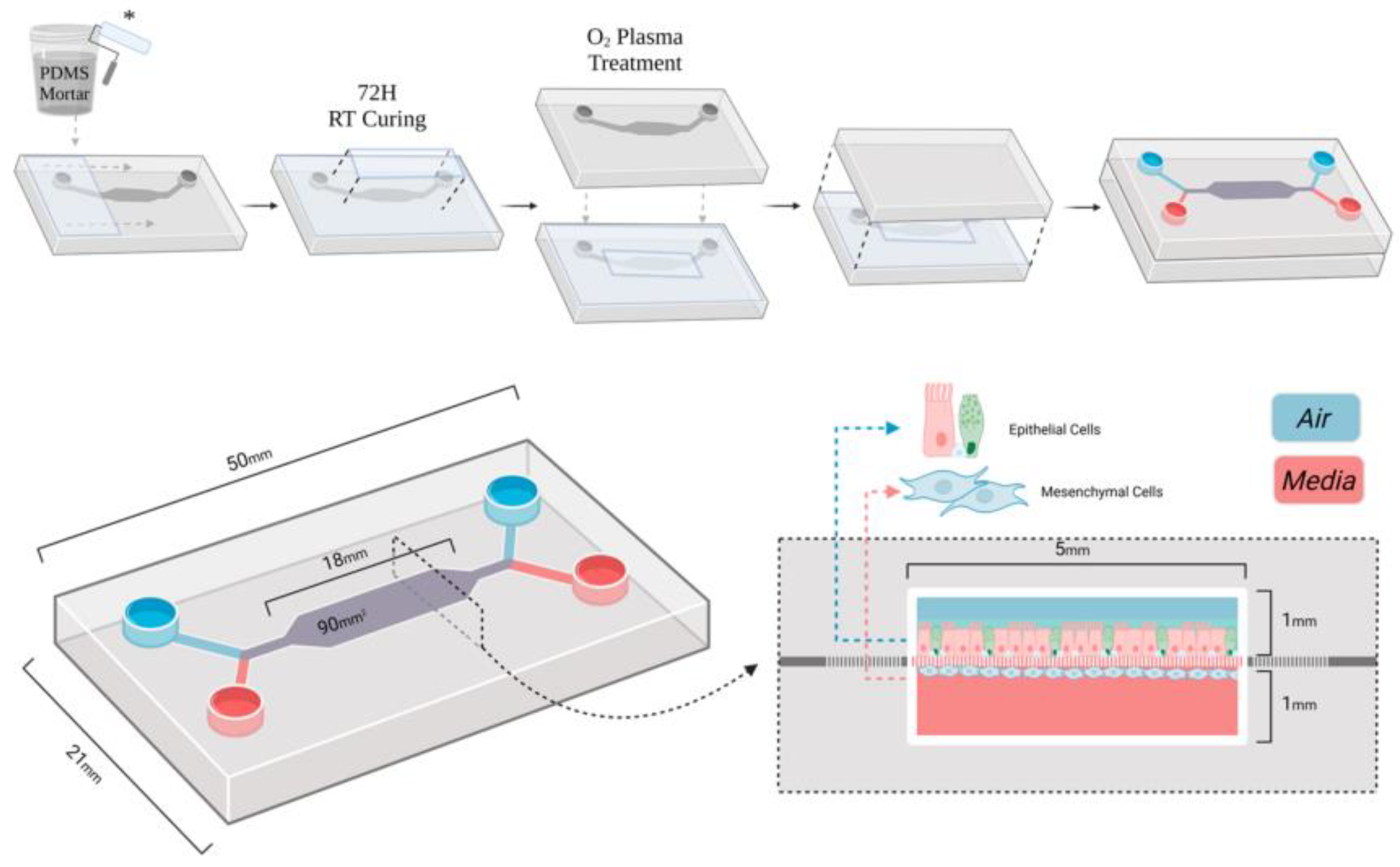
Figure 1.
Layout of the PDMS airway-on-chip showing the airflow channel, mucus layer, and imaging zones for tracking particle movement.
Figure 1.
Layout of the PDMS airway-on-chip showing the airflow channel, mucus layer, and imaging zones for tracking particle movement.
3.2. Role of Surface Wettability in Mucus Penetration
At constant particle size, reducing the water contact angle from 90° to 60° significantly improved diffusion and reduced trapping. Hydrophilic coatings such as PEG reduced adhesion to mucin fibers and promoted more uniform motion. Similar effects were reported for zwitterionic and PEG-modified particles in mucosal media [19]. These findings indicate that weaker hydrophobic interactions lead to less aggregation and greater transport efficiency.
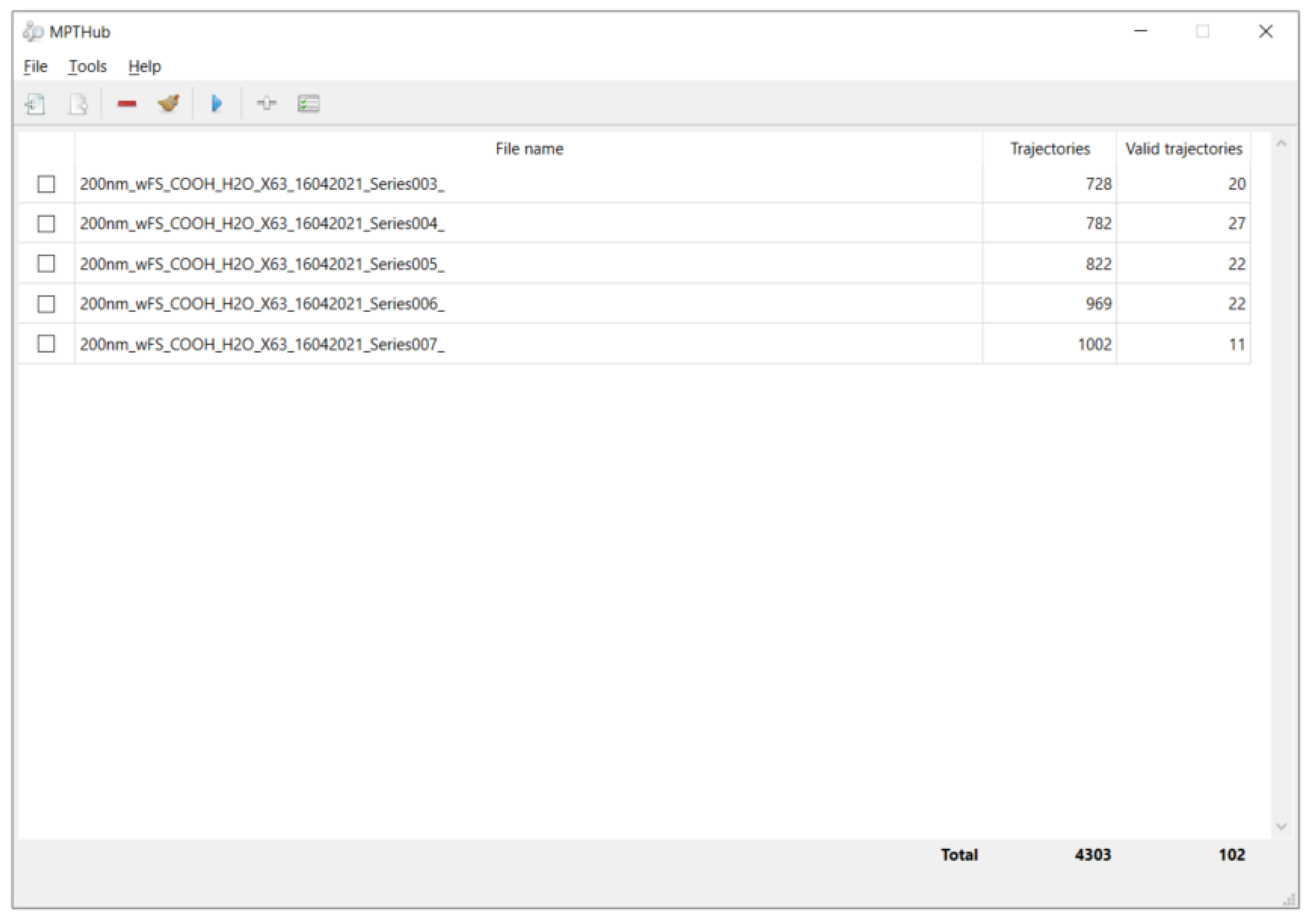
Figure 2.
Step-by-step workflow for fluorescence image processing and mean-square-displacement calculation used in single-particle diffusion analysis.
Figure 2.
Step-by-step workflow for fluorescence image processing and mean-square-displacement calculation used in single-particle diffusion analysis.
3.3. Combined Influence of Size and Wettability
A multiple linear regression model was built to analyze the joint effect of particle size () and surface contact angle () on diffusion:
The model achieved , showing strong predictive power. The regression surface indicated that small, hydrophilic particles () exhibited the best penetration. This outcome aligns with earlier studies that separately analyzed particle geometry or charge but rarely combined both factors under controlled flow [20]. The clear interaction term also highlights how wettability moderates steric hindrance: even moderately larger particles can diffuse efficiently if their surfaces are sufficiently hydrophilic.
3.4. Comparison with Previous Studies and Implications
Compared with conventional PEG-coated mucus-penetrating nanoparticles [21], this study provides specific design thresholds for size and wettability. The identified range—50 to 120 nm in diameter and 40° to 55° contact angle—achieves a balance between mobility and stability in the mucus environment. These quantitative data agree with other microfluidic studies showing that realistic shear conditions can enhance transport compared with static gels. The model framework developed here can serve as a screening tool for inhaled nanomedicine formulations. Future studies should expand this analysis to include surface charge and polymer density and validate results using primary human mucus samples.
4. Conclusion
This study built a PDMS microfluidic system to study how nanoparticle size and surface wettability affect their movement in airway mucus. The results showed that particles smaller than 150 nm with a contact angle below 60° moved faster and stayed for a shorter time in the mucus. Under these conditions, the diffusion coefficient increased about 2.7 times, and the retention time decreased by nearly 45%. A simple regression model combining particle size and contact angle accurately described the diffusion behavior and provided clear design limits for effective mucus penetration. Compared with earlier work that tested only one factor or used static gels, this method gave a more controlled and repeatable way to mimic the airway environment. These findings can guide the design of inhaled drug carriers and diagnostic nanoparticles that need to pass through mucus layers. The main limitation is that the current system used artificial mucus and did not include living cells or ciliary motion. Future work should test real human mucus and include dynamic flow to improve the accuracy and practical value of the model.
References
- Fröhlich, E. Non-Cellular Layers of the Respiratory Tract: Protection against Pathogens and Target for Drug Delivery. Pharmaceutics 2022, 14, 992. [CrossRef]
- Zha, D.; Mahmood, N.; Kellar, R.S.; Gluck, J.M.; King, M.W. Fabrication of PCL Blended Highly Aligned Nanofiber Yarn from Dual-Nozzle Electrospinning System and Evaluation of the Influence on Introducing Collagen and Tropoelastin. ACS Biomater. Sci. Eng. 2025, 11, 6657–6670. [CrossRef]
- Shin, S.W.; Song, I.H.; Um, S.H. Role of Physicochemical Properties in Nanoparticle Toxicity. Nanomaterials 2015, 5, 1351–1365. [CrossRef]
- Lutz, T.M.; Kimna, C.; Lieleg, O. A pH-stable, mucin based nanoparticle system for the co-delivery of hydrophobic and hydrophilic drugs. Int. J. Biol. Macromol. 2022, 215, 102–112. [CrossRef]
- Abdelkawi, A.; Slim, A.; Zinoune, Z.; Pathak, Y. Surface Modification of Metallic Nanoparticles for Targeting Drugs. Coatings 2023, 13, 1660. [CrossRef]
- Chen, D.; Liu, S.; Chen, D.; Liu, J.; Wu, J.; Wang, H.; Su, Y.; Kwak, G.; Zuo, X.; Rao, D.; et al. A Two-Pronged Pulmonary Gene Delivery Strategy: A Surface-Modified Fullerene Nanoparticle and a Hypotonic Vehicle. Angew. Chem. Int. Ed. Engl. 2021, 60, 15225–15229. [CrossRef]
- Wagner, C.; Wheeler, K.; Ribbeck, K. Mucins and Their Role in Shaping the Functions of Mucus Barriers. Annu. Rev. Cell Dev. Biol. 2018, 34, 189–215. [CrossRef]
- Dash, P.K.; Chen, C.; Kaminski, R.; Su, H.; Mancuso, P.; Sillman, B.; Zhang, C.; Liao, S.; Sravanam, S.; Liu, H.; et al. CRISPR editing of CCR5 and HIV-1 facilitates viral elimination in antiretroviral drug-suppressed virus-infected humanized mice. Proc. Natl. Acad. Sci. 2023, 120. [CrossRef]
- Poland, C.A.; Hiéronimus, L.; Okhrimenko, D.V.; Hoffman, J.W. Biopersistence of man-made vitreous fibres (MMVF) / synthetic vitreous fibres (SVF): advancing from animal models to acellular testing. Part. Fibre Toxicol. 2025, 22, 1–27. [CrossRef]
- Xu, K.; Lu, Y.; Hou, S.; Liu, K.; Du, Y.; Huang, M.; Feng, H.; Wu, H.; Sun, X. Detecting anomalous anatomic regions in spatial transcriptomics with STANDS. Nat. Commun. 2024, 15, 1–23. [CrossRef]
- Sharma, V.K.; Srinivasan, H.; Gupta, J.; Mitra, S. Lipid lateral diffusion: mechanisms and modulators. Soft Matter 2024, 20, 7763–7796. [CrossRef]
- Zha, D.; Gamez, J.; Ebrahimi, S.M.; Wang, Y.; Verma, N.; Poe, A.J.; White, S.; Shah, R.; Kramerov, A.A.; Sawant, O.B.; et al. Oxidative stress-regulatory role of miR-10b-5p in the diabetic human cornea revealed through integrated multi-omics analysis. Diabetologia 2025, 1–16. [CrossRef]
- Ogawa, T.; Hirota, M.; Shibata, R.; Matsuura, T.; Komatsu, K.; Saruta, J.; Att, W. The 3D theory of osseointegration: material, topography, and time as interdependent determinants of bone–implant integration. Int. J. Implant. Dent. 2025, 11, 1–37. [CrossRef]
- Xu, J. (2025). Building a Structured Reasoning AI Model for Legal Judgment in Telehealth Systems.
- Wang, Y.; Wen, Y.; Wu, X.; Wang, L.; Cai, H. Assessing the Role of Adaptive Digital Platforms in Personalized Nutrition and Chronic Disease Management. World J. Innov. Mod. Technol. 2025, 8, 24–31. [CrossRef]
- Wen, Y.; Wu, X.; Wang, L.; Cai, H.; Wang, Y. Application of Nanocarrier-Based Targeted Drug Delivery in the Treatment of Liver Fibrosis and Vascular Diseases. J. Med. Life Sci. 2025, 1, 63–69. [CrossRef]
- Tjakra, M.; Lidayová, K.; Avenel, C.; Bergström, C.A.; Hossain, S. Machine learning framework for investigating nano- and micro-scale particle diffusion in colonic mucus. J. Nanobiotechnology 2025, 23, 1–18. [CrossRef]
- Meziu, E. (2025). Nanoparticle interactions with mucosal barriers.
- Jiang, A.Y.; Lathwal, S.; Meng, S.; Witten, J.; Beyer, E.; McMullen, P.; Hu, Y.; Manan, R.S.; Raji, I.; Langer, R.; et al. Zwitterionic Polymer-Functionalized Lipid Nanoparticles for the Nebulized Delivery of mRNA. J. Am. Chem. Soc. 2024, 146, 32567–32574. [CrossRef]
- Zare, M.; Khatibi, M.; Ashrafizadeh, S.N. Synergistic effects of dielectrophoretic and magnetophoretic forces on continuous cell separation via pinched flow fractionation. Phys. Fluids 2025, 37. [CrossRef]
- Chime, S.A.; Momoh, M.A. (2025). PEGylated Nanocarriers for Drug Delivery Applications. In PEGylated Nanocarriers in Medicine and Pharmacy (pp. 107-136). Singapore: Springer Nature Singapore.
Disclaimer/Publisher’s Note: The statements, opinions and data contained in all publications are solely those of the individual author(s) and contributor(s) and not of MDPI and/or the editor(s). MDPI and/or the editor(s) disclaim responsibility for any injury to people or property resulting from any ideas, methods, instructions or products referred to in the content. |
© 2025 by the authors. Licensee MDPI, Basel, Switzerland. This article is an open access article distributed under the terms and conditions of the Creative Commons Attribution (CC BY) license (http://creativecommons.org/licenses/by/4.0/).
Copyright: This open access article is published under a Creative Commons CC BY 4.0 license, which permit the free download, distribution, and reuse, provided that the author and preprint are cited in any reuse.


