
Submitted:
20 November 2025
Posted:
24 November 2025
You are already at the latest version
Abstract
Background: Endometriosis is a chronic inflammatory disorder affecting about 190 million women of reproductive age worldwide. It represents a major health challenge due to its broad impact on physical, reproductive, and psychological well-being and is clinically characterized by pelvic pain, menstrual irregularities, and infertility. Methods: This narrative review synthesized current evidence on the relationship between adiposity, metabolic and inflammatory markers, and endometriosis from a biopsychosocial and intersectional perspective. A comprehensive search was conducted in PubMed, Scopus, and Web of Science for peer-reviewed studies published in English over the past decade. Results: Endometriosis significantly affects inflammatory activity within adipose tissue, in especial in visceral adipose tissue. Studies also reported reduced adipocyte size and altered adipose tissue function. The endometriosis cytokine profile exhibited a pattern of systemic and tissue-specific inflammatory activation (i.e., elevated levels of interleukin-6 and monocyte chemoattractant protein-1). Sociodemographic factors (i.e., age, race/ethnicity, socioeconomic status, and educational level) also play a significant role in differences in symptomatology, disease course, and healthcare access. Conclusions: Endometriosis need to be considered as a multisystem condition related to metabolic, inflammatory, and psychosocial factors. It is necessary to adopt a biopsychosocial and intersectional perspective to improve diagnosis and support more equitable and personalized therapeutic approaches.
Keywords:
endometriosis
; inflammation
; adiposity
; metabolism
; biopsychosocial
; intersectional perspective
; women
1. Introduction
Endometriosis is a chronic inflammatory disorder characterized by severe pelvic pain (i.e., dysmenorrhea, dyspareunia, and chronic pelvic discomfort), abnormal uterine bleeding (i.e., heavy menstrual bleeding and intermenstrual spotting), and infertility, which constitutes one of the most impactful consequences of the condition. Additional symptoms frequently include fatigue, gastrointestinal disturbances consistent with irritable bowel syndrome and urinary symptoms. Endometriosis has a high global prevalence and is estimated to affect approximately 190 million women and girls of reproductive age worldwide, representing a major public health concern [1,2,3,4,5]. The World Health Organization (WHO) acknowledges the significance of endometriosis and its far-reaching impact on sexual and reproductive health, related rights, quality of life, and the overall well-being of affected individuals [1].
There is considerable heterogeneity in the manifestation of symptoms among affected women, which complicates both diagnosis and treatment. In general, symptoms tend to worsen during menstruation, sexual intercourse, and episodes of dysmenorrhea. Furthermore, there is not always a direct association between symptom intensity and the subjective experience of pain. Many women in the early stages of the disease report high levels of pain that can be debilitating, whereas others with advanced-stage endometriosis may remain largely asymptomatic. All of these factors must be carefully considered to ensure an accurate diagnostic process and an effective treatment approach [2,3,4,5].
In addition to its gynecological manifestations, endometriosis is associated with a broad spectrum of psychosocial symptoms that require careful consideration. The condition exerts a pronounced negative impact on mental health, often arising from the interplay between chronic pain, emotional stress, and infertility. These factors contribute to an increased prevalence of mood disorders—particularly anxiety and depression—among affected women. Furthermore, the disease imposes a substantial socioeconomic burden on patients, their families, and healthcare systems, primarily due to the ongoing need for pharmacological treatment and the frequency of surgical interventions. Moreover, in many cases, endometriosis causes debilitating pain that prevents regular participation in work or school. This can result in loss of income—both for affected individuals and their families—and contribute to broader societal costs. Treatments often require direct out-of-pocket expenses, which can be considerable. Collectively, these challenges lead to marked impairments in quality of life, affecting interpersonal relationships, social functioning, and overall well-being [1,2,3,4,5]. These consequences are further compounded by stigma and by prevailing beliefs that tend to minimize or dismiss menstrual pain [1].
In many countries, the general population, family members, and even most healthcare and social care professionals are not fully aware that the chronic pelvic pain associated with endometriosis is not normal. The normalization and stigmatization of pain and other symptoms adversely affect the mental health and general well-being of affected women [1]. There is an urgent need to strengthen research efforts and move away from a patriarchal model of health that has historically centered the male body and experience as the norm. This model has contributed to the underdiagnosis, late diagnosis, and trivialization of women’s symptoms, as well as to the systematic neglect of sex- and gender-specific determinants of health. Promoting gender-sensitive and feminist approaches in health research and care is essential to generate evidence that reflects women’s realities, reduce structural inequities, and improve the quality, accessibility, and relevance of health services for all.
In this regard, several sociodemographic variables—such as age, race or ethnicity, educational attainment, and socioeconomic status—must be considered to achieve a more comprehensive understanding of endometriosis. Evidence shows that these factors influence both the course of the disease, the differential access to timely diagnosis and management, and the lived experiences of affected women. Nevertheless, despite the growing scientific support for this view, diagnostic and treatment models often fail to incorporate biopsychosocial and intersectional perspectives, resulting in a fragmented understanding of the condition and of those who experience it. These models typically emphasize biomedical or psychosocial aspects in isolation. An urgent shift toward integrating these dimensions is needed to ensure a truly comprehensive and effective approach to endometriosis care [2,3,4,5].
Indeed, the etiology of endometriosis remains the subject of ongoing investigation. Current evidence suggests that it is a multifactorial condition arising from the interplay of hormonal, genetic, immunological, and environmental factors. Importantly, these biological mechanisms also interact with broader social determinants, shaping both vulnerability to the disease and its clinical course [2,3,4,5].
This estrogen-dependent gynecological disorder is characterized by the proliferation of functional endometrial glands and stroma outside the uterine cavity. Ectopic endometrial tissue persists through estrogen-dependent inflammatory pathways that promote lesion survival, pathological angiogenesis, and fibrosis. The onset and progression of endometriosis are closely associated with systemic metabolic and inflammatory processes, establishing a biological link between metabolic dysregulation and reproductive health. These interrelated metabolic and hormonal disturbances converge within a chronic inflammatory milieu that further connects obesity to the pathogenesis of endometriosis [6,7].
Obesity appears to influence the risk and progression of endometriosis through multiple, interrelated physiological mechanisms involving inflammation, metabolism, and hormonal regulation. Chronic low-grade inflammation associated with excess adiposity can disrupt estrogen homeostasis and impair endometrial receptivity, thereby compromising reproductive outcomes such as embryo implantation. In parallel, obesity-induced insulin resistance may alter the function of endometrial stromal cells, contributing to the establishment and persistence of ectopic endometrial lesions. Increased oxidative stress—resulting from an imbalance between reactive oxygen species and antioxidant defenses—further exacerbates cellular damage and inflammatory activation within both endometrial and adipose tissues.
Additionally, hormonal disturbances, particularly elevated circulating leptin and estrogen levels, may impair ovarian function and reinforce the estrogen-dependent nature of the disease. Beyond these systemic alterations, the metabolic and inflammatory activity of visceral adipose tissue plays a pivotal role in linking obesity to the pathogenesis of endometriosis. Composite indices such as the Lipid Accumulation Product (LAP) and the Visceral Adiposity Index (VAI) more accurately reflect obesity-related metabolic alterations than conventional anthropometric measures (e.g., body mass index or waist circumference) and offer greater sensitivity for detecting metabolic dysfunction associated with endometriosis. In sum, these mechanisms underscore how obesity amplifies systemic pathophysiological processes that heighten the risk of endometrial dysfunction and the development of endometriosis [6,7].
Furthermore, these mechanisms—particularly obesity-induced chronic inflammation—constitute a systemic condition that bridges metabolic and reproductive dysfunction. Adipose tissue, especially in its visceral form, functions as an active endocrine and immunologic organ that secretes a wide range of adipokines and cytokines, including interleukin-6 (IL-6), tumor necrosis factor-alpha (TNF-α), and monocyte chemoattractant protein-1 (MCP-1). These mediators sustain a pro-inflammatory microenvironment that extends beyond local fat depots to affect distant tissues, including the endometrium [6,7].
The activation of macrophages within hypertrophic or dysfunctional adipose tissue further contributes to immune dysregulation and the continuous release of cytokines, which may, in turn, promote the proliferation and survival of ectopic endometrial cells. Moreover, metabolic disturbances associated with obesity—such as insulin resistance and altered lipid metabolism—intensify oxidative stress and inflammatory signaling. These processes establish a chronic systemic inflammatory milieu that not only exacerbates the pathogenesis of endometriosis but also links the disease to broader metabolic and endocrine abnormalities [6,7].
In consequence, the treatment of endometriosis aims to alleviate symptoms, improve quality of life, and address infertility. Therapeutic strategies are highly individualized, depending on disease severity, reproductive goals, and general health status. Pharmacological approaches remain the first-line option and include nonsteroidal anti-inflammatory drugs (NSAIDs) to reduce pain and inflammation, as well as hormonal therapies designed to suppress ovarian hormone production and the menstrual cycle. Combined oral contraceptives, progestins, gonadotropin-releasing hormone (GnRH) agonists or antagonists, and progestin-releasing intrauterine devices are commonly used to limit the estrogen-dependent growth of endometriotic lesions [3,4,5].
When medical therapy proves insufficient, surgical interventions such as laparoscopy can provide both diagnostic confirmation and removal of endometriotic lesions, adhesions, or ovarian cysts. Hysterectomy, with or without oophorectomy, may be considered in refractory cases in which fertility preservation is not desired. For women experiencing infertility, assisted reproductive technologies—particularly in vitro fertilization (IVF)—represent an effective alternative [3,4,5].
Complementary lifestyle interventions, including anti-inflammatory dietary modifications, regular physical activity, and stress-reduction practices such as mindfulness, may further support symptom management and psychological well-being. Moreover, several emerging pharmacological therapies are currently under investigation and may offer promising therapeutic advances in the future [3,4,5].
Despite the wide range of therapeutic options available, current treatments for endometriosis primarily focus on symptom management rather than addressing the underlying pathophysiological mechanisms of the disease. The high rates of recurrence following both medical and surgical interventions highlight the need for a deeper understanding of the biological processes that drive lesion persistence and systemic manifestations. Growing evidence indicates that endometriosis extends beyond a localized gynecological condition and involves complex interactions among hormonal, immune, and metabolic pathways interconnected with the environment and psychosocial issues [1,4,5].
Chronic inflammation and altered adipose tissue metabolism—particularly within visceral fat depots—appear to play a crucial role in modulating estrogen activity and cytokine secretion, thereby perpetuating disease progression [6,7]. Investigating these metabolic and inflammatory interactions may offer valuable insights into the systemic nature of endometriosis and help identify novel therapeutic targets aimed at restoring tissue homeostasis and reducing recurrence.
Based on the elements discussed above, the present review seeks to analyze the relationship between inflammation, adiposity, and metabolic markers in women with endometriosis from a biopsychosocial and intersectional perspective. This approach aims to address the gaps identified in the current literature and to support the development of a more comprehensive model of sociosanitary healthcare for affected women.
2. Materials and Methods
This study follows a comprehensive narrative review methodology to explore the current evidence on the relationship between adiposity, adipose tissue function, and metabolic markers in women with endometriosis from a biopsychosocial and intersectional perspective. The search was conducted across PubMed, Scopus, and Web of Science databases, focusing on peer-reviewed articles published in English over the past ten years.
2.1. Inclusion and Exclusion Criteria
Inclusion criteria included empirical studies focusing on I) adiposity or body composition indicators (e.g., BMI, fat distribution, etc.); and II) metabolic or inflammatory biomarkers (e.g., cytokines, TNF-α, IL-6, etc.). In addition, given the main objective of this research, sociodemographic factors such as age, race or ethnicity, socioeconomic status, and educational attainment were also included when such variables were available.
Exclusion criteria comprise I) Case reports, conference abstracts, non-human studies; II) Publications lacking primary data; and III) Duplicate records.
2.2. Search Strategy
The search strategy was based on Medical Subject Headings (MeSH) terms to enhance precision and consistency in identifying relevant literature. The followed key terms were included: Endometriosis, Inflammation, Adiposity, and Metabolic. Moreover, Boolean operators were used to combine terms and refine the search results.
2.3. Study Selection Process
The initial screening of studies was developed analysing the titles and abstracts, followed by a full-text review of the final selected articles. Three independent reviewers conducted the selection process to reduce bias and guarantee the quality of the included studies. In cases of disagreement, a fourth reviewer was included to solve it and to reach a consensus.
2.4. Quality Assessment
Due to it is a narrative review, a formal risk-of-bias assessment was not performed. Instead, the selected studies were critically evaluated to analyse their methodological quality and main limitations. In particular, the following issues were appraised: the clarity of study design, adequacy of sample size, validity of measurement instruments, and consistency of reported outcomes. In addition, the methodological limitations and potential sources of bias acknowledged by the original authors were also analysed during the process.
2.5. Data Analysis
As part of the data analysis, the main findings extracted from the final selection of the studies were organized into thematic categories. Descriptive and comparative methods were used to identify the principal patterns and trends in the reviewed literature.
3. Results
This section summarizes current evidence on the biological mechanisms linking adiposity, metabolic dysfunction, and inflammation to the pathophysiology of endometriosis; and determining the influence of sociodemographic factors (i.e., age, race or ethnicity, socioeconomic status, and educational level). Particular attention is given to the differential inflammatory activity of visceral and subcutaneous adipose tissue, alterations in adipocyte morphology and function, the role of adipokines and cytokines in mediating local and systemic immune responses, the metabolic markers of VAT, and the influence of severity and types of endometriosis in the relationship between adiposity and endometriosis. Evidence is also reviewed regarding how these adipose- and inflammation-related mechanisms contribute to disease heterogeneity, severity, and clinical outcomes. To conclude this section, the influence of sociodemographic factors is discussed to highlight their potential implications for understanding and managing endometriosis as a systemic and multifactorial condition.
3.1. Differential Inflammatory Responses in Visceral and Subcutaneous Adipose Tissue of Women with Endometriosis
Abobeleira et al. [8] reported smaller adipocytes in visceral adipose tissue (VAT) and subcutaneous adipose tissue (SAT) of endometriosis patients, along with increased serum IL-6 and reduced norepinephrine levels. VAT from endometriosis patients exhibited upregulated browning-related proteins (UCP-1, PGC-1α) and pro-inflammatory markers (IL-6, MCP-1), alongside reduced CD206 expression, indicating M1 macrophage predominance. SAT showed no significant changes in protein expression. Gene expression analysis revealed trends of decreased browning-related genes in VAT and SAT of endometriosis patients, except for DIO2, which was significantly downregulated on SAT.
Endometriosis significantly affects inflammatory activity within adipose tissue, particularly in VAT. VAT from women with endometriosis exhibited markedly elevated levels of pro-inflammatory markers, including a 3.5-fold increase in interleukin-6 (IL-6) and a 4.5-fold increase in monocyte chemoattractant protein-1 (MCP-1), both associated with macrophage activation and local inflammation. Galectin-3 expression also showed a tendency to increase in VAT, although this change did not reach statistical significance. In contrast, CD206, a marker of anti-inflammatory M2 macrophages, was significantly reduced in the VAT of endometriosis patients, suggesting a shift toward a pro-inflammatory M1 macrophage phenotype. Consistent with these tissue findings, serum IL-6 concentrations were significantly higher in women with endometriosis than in controls, indicating the presence of systemic inflammation. No significant differences were detected in SAT for IL-6, MCP-1, Galectin-3, or CD206, although there was a mild trend toward higher IL-6 and Galectin-3 and lower CD206 expression in the endometriosis group. These findings point out that endometriosis is associated with localized inflammation in VAT, driven by upregulation of pro-inflammatory cytokines and macrophage activity, whereas SAT remains relatively unaffected. This VAT-specific inflammatory response may contribute to adipose tissue dysfunction and play a role in the pathophysiological progression of endometriosis [8].
Moreover, inflammation exerts a profound influence on adipose tissue physiology in endometriosis, particularly in regions adjacent to endometriotic lesions. Histological and molecular analyses reveal that these perilesional adipose areas display marked fibrosis and angiogenesis compared with tissues distant from the lesions or from women without the disease. A pronounced infiltration of macrophages -predominantly of the pro-inflammatory M1 subtype- was observed in these regions, contrasting with the predominance of anti-inflammatory M2 macrophages typically found in the peritoneal fluid of endometriosis patients. Moreover, adipose tissue adjacent to endometriotic lesions exhibited elevated expression of fatty acid-binding protein 4 (FABP4), vascular endothelial growth factor (VEGF), and pro-inflammatory cytokines such as IL-6, TNF-α, and IL-1β, indicating a state of immune and metabolic dysregulation. These findings support the notion that perilesional adipose tissue is an active participant in the local inflammatory microenvironment of endometriosis [9] (See Table 1 for further details).
3.1.1. Reduced Adipocyte Size and Altered Adipose Tissue Function in Women with Endometriosis
Adipocytes in both visceral adipose tissue (VAT) and subcutaneous adipose tissue (SAT) of women with endometriosis were significantly smaller than those observed in control women, with VAT showing the smallest adipocyte size overall. Higher BMI was associated with an increase in SAT adipocyte size in both control and endometriosis groups; however, even under conditions of elevated BMI, women with endometriosis continued to exhibit smaller adipocytes compared to controls. In control women, VAT adipocyte size did not vary significantly with BMI, whereas in patients with endometriosis, VAT adipocyte size increased with higher BMI but remained consistently smaller than in the control group [8] (See Table 1 for further details).
3.2. Adipokine Dysregulation in Women with Endometriosis
Adipokines, bioactive molecules secreted by adipose tissue, have emerged as key modulators of the inflammatory and endocrine pathways implicated in endometriosis. Among them, leptin has been the most extensively studied, showing consistently elevated concentrations in both the serum and peritoneal fluid of affected women. Leptin enhances the proliferation, migration, and invasion of endometrial stromal cells through activation of the JAK2/STAT3 signaling cascade, which upregulates matrix metalloproteinase-2 (MMP-2) and promotes extracellular matrix degradation. Moreover, leptin stimulates angiogenesis by increasing vascular endothelial growth factor A (VEGF-A) expression, thereby facilitating lesion vascularization and progression. Other adipokines, such as chemerin and follistatin, also contribute to the pro-inflammatory and proliferative milieu. Chemerin and its receptor (CMKLR1) are overexpressed in ovarian endometriomas, with elevated levels detected in peritoneal fluid, suggesting a role in local immune activation and inflammation. Follistatin, elevated in both serum and ovarian tissue, inhibits activin A -a cytokine that promotes cell invasion and inflammatory signaling-thereby influencing tissue remodeling. In addition, the apelin receptor (APLNR) has been identified as a potential regulatory gene in endometriosis, although its precise functional role remains under research. These findings related to adipokines reinforce the link between visceral adipose activity, angiogenesis, and chronic inflammation, supporting their contribution to endometriosis pathophysiology [10].
By contrast, adiponectin appears to exert a protective influence against endometriosis progression. As an anti-inflammatory and anti-proliferative adipokine, adiponectin helps maintain immune balance and suppresses the growth of endometrial stromal cells. Experimental studies have shown that recombinant adiponectin inhibits the proliferation and viability of both endometriotic and normal endometrial stromal cells, suggesting its capacity to restrict lesion growth. Clinically, women with endometriosis exhibit significantly lower serum and peritoneal fluid adiponectin levels, particularly in advanced stages of the disease, suggesting that reduced adiponectin bioavailability may diminish this protective effect. The decrease in adiponectin might further amplify inflammatory and metabolic disturbances, exacerbating disease severity and persistence. The divergent profiles of leptin and adiponectin highlight the dual endocrine function of adipose tissue in endometriosis, where an imbalance favoring pro-inflammatory adipokines over anti-inflammatory ones might contribute to sustaining and exacerbating the disorder [10].
3.3. Key Cytokines Linking Systemic and Adipose Tissue Inflammation in Endometriosis
Inflammation in adipose tissue near endometriotic lesions contributes to fibrosis, angiogenesis, and the maintenance of a pro-inflammatory environment that may promote disease progression. In addition, elevated levels of several cytokines have been identified, including interleukin-6 (IL-6), tumor necrosis factor-alpha (TNF-α), interleukin-1 beta (IL-1β), and interleukin-8 (IL-8) in women with endometriosis. These cytokines are increased both in the peritoneal cavity and in adipose tissues adjacent to endometriotic lesions. Their presence reflects an ongoing inflammatory response that supports lesion survival and chronicity. These findings suggest that cytokine overexpression is a key feature of the immune and metabolic dysregulation associated with endometriosis [9].
The cytokine profile associated with endometriosis reveals a pattern of both systemic and tissue-specific inflammatory activation. Serum interleukin-6 (IL-6) levels were significantly higher in women with endometriosis compared with controls, consistent with a state of chronic systemic inflammation. Within VAT, IL-6 expression was also upregulated, contributing to localized inflammatory signaling and potentially promoting adipose tissue browning. Monocyte chemoattractant protein-1 (MCP-1) levels were likewise elevated in VAT, reflecting increased macrophage recruitment and activation within this depot. In contrast, serum transforming growth factor-beta (TGF-β) concentrations did not differ significantly between groups; however, TGF-β has been implicated in the survival and proliferation of ectopic endometrial cells and exhibits mechanistic overlap with cancer-related metabolic dysregulation. These cytokines appear to play complementary roles in mediating both systemic and localized inflammation, contributing to adipose tissue dysfunction and to the pathophysiological progression of the condition [8].
3.4. Metabolic Markers of Visceral Adiposity: Lipid Accumulation Product (LAP) and Visceral Adiposity Index (VAI)
In women with endometriosis, the evaluation of adiposity and metabolic risk has been limited by the constraints of traditional anthropometric measures, such as the Body Mass Index (BMI). BMI does not differentiate between lean mass and fat mass, nor does it provide information about the distribution of adipose tissue. As a result, it frequently underestimates excess body fat and fails to assess the accumulation of VAT, which plays a more critical role in metabolic dysfunction and inflammation. Considering the complex nature of endometriosis and its characterization as a condition with systemic inflammatory and metabolic components, relying solely on BMI is insufficient to meaningfully elucidate its underlying pathophysiological mechanisms. Indeed, current scientific evidence highlights the limitations of BMI in terms of its diagnostic sensitivity and specificity for obesity-related metabolic disturbances, emphasizing the requirement for more reliable and biologically relevant metrics of visceral adiposity [11].
In this context, the Visceral Adiposity Index (VAI) offers a more comprehensive and clinically meaningful assessment of adipose tissue function. The VAI integrates waist circumference, BMI, triglyceride levels, and high-density lipoprotein cholesterol (HDL-C) into a composite index that comprises visceral fat activity and metabolic impairment. Unlike BMI, VAI accounts for sex-specific differences in fat distribution, making it particularly suitable for studying female reproductive disorders such as endometriosis. Elevated VAI values have been associated with insulin resistance, dyslipidemia, and systemic inflammation processes that reflect the metabolic and immunological alterations observed in endometriosis. Furthermore, the VAI’s simplicity, cost-effectiveness, and accessibility enhance its clinical utility for identifying women at increased risk of metabolic dysfunction and endometriosis-related comorbidities. These findings position the VAI as a valuable tool for both research and clinical screening, supporting individualized prevention and management strategies in women with endometriosis [11].
Zhang et al. [11] found a positive linear association between VAI and increased risk endometriosis. This relationship remained significant even after adjusting for several demographic, lifestyle, and reproductive factors. Additionally, subgroup analyses showed that this association was not significantly influenced by factors such as age, education level, marital status, or oral contraceptive use. Nevertheless, the study concluded that additional large-scale prospective research is required to confirm these findings [11].
To further examine the metabolic implications of adipose tissue dysfunction in endometriosis, recent evidence has explored the relationship between two composite metabolic indices -the lipid accumulation product (LAP) and VAI- and the presence of endometriosis. Using data from the National Health and Nutrition Examination Survey (NHANES, 1999–2006), both markers were found to be significantly associated with endometriosis. Women in the highest quartiles of LAP and VAI exhibited 56% and 54% higher odds of having endometriosis, respectively, even after controlling for a comprehensive set of relevant covariates. Subgroup analyses indicated that these associations were more pronounced among women aged 35 years or older, those with a history of pregnancy, users of exogenous female hormones, and individuals without chronic kidney disease. These findings suggest that LAP and VAI, which are noninvasive and cost-effective measures integrating anthropometric and biochemical parameters, might serve as potential tools for early risk stratification and metabolic assessment in women at higher risk for endometriosis. However, the cross-sectional nature of the study and dependence on self-reported diagnostic information limit causal inference, and the generalizability of the findings beyond the U.S. populations. Nevertheless, the observed associations highlight the relevance of considering metabolic dysfunction and visceral adiposity as integral components of endometriosis pathophysiology, warranting further prospective and mechanistic research to confirm their clinical relevance [6].
3.5. Adiposity and Endometriosis Severity and Typology
The study of Byun et al. [12] revealed an inverse relationship between adiposity and endometriosis, showing that women with the condition generally exhibited lower adiposity measures than those without it. This trend was consistent across multiple indicators of body fat and visceral adiposity. When stratified by disease stage, women with stage I or IV endometriosis presented lower adiposity measures than those with stage II or III, suggesting a non-linear association between fat accumulation and disease severity [12].
Endometriosis severity was classified according to the revised American Society for Reproductive Medicine (rASRM) staging system, which categorizes the disease into four stages: stage I (minimal, scores 1–5), stage II (mild, scores 6–15), stage III (moderate, scores 16–40), and stage IV (severe, scores >40)- based on lesion size, location, and adhesions. Staging was determined intraoperatively through laparoscopy or laparotomy using a standardized scoring algorithm [12].
The study also analyzed endometriosis typology following the rASRM classification, distinguishing three main forms: superficial endometriosis (SE), limited to the ovary or peritoneum; deep infiltrating endometriosis (DIE), involving peritoneal invasion deeper than 5 mm or posterior cul-de-sac obliteration; and ovarian endometrioma (OE), consisting of deep ovarian lesions. Cases presenting both OE and DIE were also identified [12].
Different adiposity patterns emerged across these types. Women with ovarian endometrioma (OE) and/or deep infiltrating endometriosis (DIE) had the lowest adiposity indicators (i.e., BMI, skinfold thickness, and body circumferences) while those with superficial endometriosis (SE) showed comparatively higher measures. These findings highlight a possible link between adipose tissue burden and distribution and the severity and clinical presentation of endometriosis [12].
In sum, the association between adiposity and endometriosis appears complex and multifactorial, likely involving biological and metabolic pathways. Although consistent patterns were observed, confidence intervals were wide and overlapping, warranting cautious interpretation. It remains unclear whether reduced adiposity acts as a predisposing factor for endometriosis or results from the disease’s metabolic and inflammatory alterations. Future studies should assess adiposity measures prior to disease onset and investigate how variations in the anatomical distribution of fat contribute to the development and progression of endometriosis [12].
3.6. Integrating Biopsychosocial and Intersectional Perspectives into Adiposity–Endometriosis Research
The studies reviewed indicate that, although there is evidence of relevant sociodemographic differences in the prevalence, clinical expression, and severity of endometriosis, these factors are often not examined in depth. Several works focus almost exclusively on biological, metabolic, and molecular aspects, without adequately integrating variables such as age, race or ethnicity, socioeconomic status, or educational attainment [8,9,10,13] (See Figure 1 for more details).
Regarding age, some studies report meaningful differences between clinical groups. Abobeleira et al. [8] report that women with endometriosis were younger (mean 37.63 years) than those in the control group (mean 45.07 years), a difference interpreted as a methodological limitation due to the potential influence of age on adipose tissue metabolism. Epidemiological evidence aligns with the fact that endometriosis primarily affects women of reproductive age (22–49 years), which may partly explain these group differences. Complementing this, Hong et al. [14] describe that the relationship between obesity and disease severity varies by age: women aged 27–36 and 37–45 years with advanced disease show a lower BMI than those with mild disease, whereas this pattern does not appear in younger women aged 18–27 years. These findings are consistent with Ma et al. [6], who report that endometriosis prevalence is substantially higher in women aged ≥35 years (9.89%) compared with those under 35 (3.77%), and that associations between visceral adiposity indices (LAP and VAI) and endometriosis are stronger in this older age group.
Racial and ethnic differences are also reflected across several studies. According to Ma et al. [6], non-Hispanic White women show the highest prevalence of endometriosis (10.13%), followed by non-Hispanic Black women (6.07%), women from other racial groups (4.33%), and—with the lowest prevalence—Mexican American women (2.41%). A similar pattern appears when examining the distribution of cases: 84.17% of diagnosed women were non-Hispanic White, compared with 7.72% non-Hispanic Black, 5.70% other racial groups, and only 2.41% Mexican American women. Comparable results have been reported in Byun et al. [12] and Zhang et al. [13], where non-Hispanic White women also show greater representation in more severe stages, including stage IV disease.
Socioeconomic factors present a complex relationship with endometriosis. Ma et al. [6] report higher prevalence among women with middle- or high-income levels (PIR > 1; 7.61%) compared with those with lower income (PIR ≤ 1; 4.25%), a difference attributed to factors such as access to healthcare or greater likelihood of receiving a formal diagnosis. However, findings are not uniform across studies. Zhang et al. [13] did not observe significant differences in socioeconomic status—measured through the poverty-to-income ratio—between women with and without endometriosis (p = 0.593), nor relevant interactions between socioeconomic status and the association between visceral adiposity and disease risk. In contrast, Byun et al. [12] point out that women with more severe forms of the disease—such as stage IV endometriosis or deep infiltrating endometriosis—are more likely to be above 180% of the poverty line, suggesting distinct sociodemographic patterns depending on disease subtype and severity.
Educational attainment shows similar patterns. Ma et al. [6] report that endometriosis prevalence is markedly higher among women with education beyond high school (8.02%) compared with those with a high school education (4.39%) or below (1.60%). Zhang et al. [13] also observe significant differences in educational distribution between women with and without the disease, with secondary/GED education more frequent among those with endometriosis (31.74% vs. 21.73%). In terms of severity, Byun et al. [12] show that women with advanced stages (III–IV) and severe subtypes (deep infiltrating endometriosis or ovarian endometrioma) exhibit proportionally higher rates of university education—for example, 46.4% in stage IV and more than 52% in DIE and ovarian endometrioma cases. Although these studies do not establish causality, they suggest that educational attainment may influence the likelihood of seeking healthcare and therefore receiving a diagnosis.
Other sociodemographic variables also appear associated with endometriosis in some studies. Zhang et al. [13] report a higher proportion of married or cohabiting women among those diagnosed with endometriosis (74.88% vs. 67.40% in the non-endometriosis group), whereas Byun et al. [12] find that more severe disease forms are associated with a greater likelihood of never having been pregnant, as well as differences related to place of residence. Additionally, Ma et al. [6] identify variations in the use of female hormones, pregnancy history, use of steroids, and the presence of metabolic and cardiovascular comorbidities between women with and without endometriosis.
Although these findings suggest that sociodemographic factors play a meaningful role in the prevalence, clinical expression, subtype, and severity of endometriosis, the studies seldom explore the potential causes of these differences or the mechanisms underlying them. These limitations highlight the need to integrate biopsychosocial and intersectional approaches to better understand how age, race or ethnicity, education, socioeconomic status, and other determinants combine to shape the experience and manifestation of the disease.
4. Discussion
The present discussion is structured in a first section, which analyzes the main findings of this narrative review, and a second section devoted to the clinical relevance and practical applications of this research, with the aim of providing information that addresses the previously identified research gaps and contributes to more comprehensive social and health care for women with endometriosis.
4.1. Adipose Tissue Dysregulation in Endometriosis: Inflammatory and Metabolic Insights within a Biopsychosocial–Intersectional Framework
Differential inflammatory responses between visceral and subcutaneous adipose tissues in women with endometriosis has been reported. In line with the patterns described in the results section, clear depot-specific differences were observed. On purpose, IL-6 protein levels were markedly elevated in visceral adipose tissue (VAT), whereas subcutaneous adipose tissue (SAT) showed only a mild, non-significant increase and a significant reduction in IL-6 mRNA expression. MCP-1 was significantly upregulated in VAT, reflecting enhanced macrophage recruitment, while SAT remained unchanged. CD206, a marker of anti-inflammatory M2 macrophages, was markedly decreased in VAT, indicating a shift toward a pro-inflammatory phenotype, with only a non-significant downward trend in SAT. Galectin-3 expression tended to increase in VAT but showed no variation in SAT. These findings highlight tissue-specific inflammatory regulation, with VAT exhibiting a stronger pro-inflammatory signature than SAT in the context of endometriosis, supporting the notion that VAT browning and inflammation contribute to adipose tissue dysfunction and altered lipid metabolism [8,15,16].
These depot-specific inflammatory alterations further indicate that inflammation in endometriosis is predominantly localized within VAT rather than being uniformly distributed across adipose depots. The distinct upregulation of pro-inflammatory cytokines and reduction of anti-inflammatory markers in VAT suggest selective activation of immune and macrophage-related pathways that may contribute to local tissue dysfunction. Given that VAT is metabolically and immunologically more active than SAT, these findings reinforce its central role in shaping the systemic inflammatory milieu associated with endometriosis. This pattern also aligns with the idea that VAT inflammation might act as a potential driver of systemic inflammatory signaling and interact with reproductive and metabolic pathways, thereby contributing to disease progression [8]. Its anatomical proximity to endometriotic lesions may further amplify these inflammatory processes. Understanding these depot-specific alterations is clinically relevant, as it highlights VAT as a potential therapeutic target within metabolic and anti-inflammatory treatment strategies [8].
From a broader pathophysiological perspective, adipose tissue inflammation may contribute to the progression of endometriosis through multiple interconnected mechanisms. The secretion of IL-6, TNF-α, IL-1β, and IL-8 sustain chronic inflammation and promote tissue remodeling, while increased VEGF supports angiogenesis and lesion survival. Accumulation of M1 macrophages amplifies inflammatory signaling, creating a persistent feedback loop that maintains lesion activity. FABP4 overexpression appears to link lipid metabolism to inflammatory and angiogenic pathways, reinforcing the metabolic–immune connection. Additionally, reductions in adipocyte size reflect underlying adipose tissue dysfunction and a shift toward enhanced browning and thermogenic activity [8]. These processes establish a self-sustaining inflammatory network between endometriotic lesions and adjacent adipose depots, contributing to fibrosis, angiogenesis, and chronic progression [9,16]. These observations reinforce the bidirectional interaction between endometriotic lesions and adipose tissue, where metabolic and inflammatory signaling mutually amplify one another [16].
Consistent with these mechanisms, inflammation within adipose tissue adjacent to endometriotic lesions contributes to fibrosis, angiogenesis, and the establishment of a persistent pro-inflammatory microenvironment. Elevated IL-6, TNF-α, IL-1β, and IL-8—detected both in the peritoneal cavity and in perilesional adipose tissue—serve as key mediators of chronic inflammation. IL-6 and TNF-α support immune cell recruitment and angiogenic signaling, while IL-1β and IL-8 further amplify inflammatory cascades and vascular remodeling. This sustained cytokine activity reinforces local-to-systemic immunometabolic interactions, promoting the maintenance and progression of endometriotic lesions [9,16].
In parallel, metabolic markers of visceral adiposity also appear relevant. The lipid accumulation product (LAP) emerges as a significant indicator in endometriosis, with women in the highest quartile showing approximately 56% higher odds of developing the condition compared with those in the lowest quartile. As an index integrating waist circumference and triglyceride levels, LAP better reflects visceral fat accumulation than BMI. Because visceral adiposity is closely tied to inflammation, insulin resistance, and oxidative stress, LAP may function as a proxy for metabolic dysfunction linked to endometriosis. Its association is particularly pronounced among women aged ≥35 years, those with pregnancy history, and individuals undergoing hormonal therapy. Clinically, LAP represents a simple, non-invasive tool that may facilitate early identification of women at increased risk [6,17]. Given its associations with VAT-related inflammation, LAP—and to a lesser extent VAI—may support metabolic screening strategies to identify patients with adiposity-driven inflammatory risk profiles [6,17].
Regarding adiposity patterns, epidemiological studies consistently report an inverse association between endometriosis and BMI, with affected women tending to have lower BMI and reduced adipose mass (especially below the waist). This lean phenotype may influence estrogen regulation, which is highly relevant given the estrogen-dependent nature of the disease [14]. Several mechanisms have been proposed: obesity-related anovulation and oligomenorrhea may reduce retrograde menstruation; chronic pain and emotional distress may reduce appetite; and adipokines may influence immune regulation, inflammation, and angiogenesis. Importantly, associations between fat distribution and endometriosis risk appear age-dependent: inverse relationships between waist-to-hip and waist-to-thigh ratios and endometriosis risk are seen in women under 30 but not in older women. In general, these findings emphasize a complex interplay among fat distribution, estrogen levels, and inflammatory pathways; as well as the need to take sociodemographic factors into consideration [14,16,17].
These observations highlight the need to further examine the temporal and anatomical dimensions of the adiposity–endometriosis link. Future research should evaluate adiposity measures before diagnosis, and ideally before disease onset, to clarify potential causal pathways. Anatomical distinctions between VAT and SAT might be particularly relevant for understanding metabolic and inflammatory interactions underlying endometriosis. Such insights may support earlier detection and identify metabolic phenotypes associated with the disease [12]. In the same line, lifestyle interventions provide an additional avenue for prevention and management. For instance, dietary modifications that reduce saturated fat intake may improve lipid metabolism and decrease systemic inflammation; weight management may reduce visceral adiposity and pro-inflammatory cytokine release; and regular physical activity supports improved insulin sensitivity and immune modulation. These strategies target modifiable risk factors in a non-invasive manner and promote metabolic and reproductive health [6]. Further research is needed to explore the systemic impact of adipose tissue dysfunction in endometriosis.
Despite the relevance of these biological and metabolic interactions, several methodological limitations characterize the reviewed literature. Many studies relied on small sample sizes, used cross-sectional designs, which do not allow for causal inferences; or lacked pre-diagnostic adiposity data. In addition, measures of adiposity varied widely—ranging from adipose tissue biopsies to anthropometrics, LAP, VAI, or imaging—hindering comparability. The scarcity of longitudinal assessments and inconsistent characterization of inflammatory markers further complicate interpretation. These methodological constraints underline the need for more robust, standardized, and temporally sensitive study designs.
To sum up, the present synthesis also points up the role of sociodemographic factors in shaping the adiposity–endometriosis relationship. Differences related to age, race or ethnicity, educational attainment, and socioeconomic status do exist, but remain highly fragmented across the literature. Studies vary widely in the variables included, the analytical depth applied, and the theoretical frameworks used. As a result, sociodemographic determinants are rarely integrated into biological models despite their potential relevance for variability in symptom burden, inflammatory profiles, and disease severity [1,12,13,14]. This gap highlights an opportunity for future research. Incorporating biopsychosocial and intersectional approaches could clarify how structural conditions, lived experiences, and social determinants interact with adiposity-related mechanisms in women with endometriosis.
Methodologically, inconsistent measurement and limited interrogation of sociodemographic variables hinder the ability to distinguish biological heterogeneity from variability driven by differential exposure to structural conditions. Many studies control for—but do not examine—factors such as income, education, race or ethnicity, or reproductive history, thereby constraining their explanatory value. Inconsistencies across studies also limit comparability and impede the development of integrative models capable of capturing the complex interactions among metabolic dysfunction, hormonal regulation, and social context. Advancing this field requires systematic and theoretically informed incorporation of sociodemographic indicators, including intersectional frameworks that recognize how multiple social positions jointly shape biological vulnerability, symptom trajectories, and access to care.
In sum, the evidence reviewed draw attention to the relationship between adiposity and endometriosis is shaped by a dynamic interplay of biological, metabolic, inflammatory, and sociodemographic factors. Depot-specific immune activation, alterations in adipocyte function, and visceral adiposity–related metabolic dysregulation converge with patterns of differential risk linked to age, fat distribution, and lifestyle determinants. At the same time, sociodemographic disparities— fragmented across existing studies and rarely incorporated into disease models —suggest that biological vulnerability cannot be fully understood without considering structural conditions, access to care, and lived experiences that influence diagnosis, symptom trajectories, and disease severity. Integrating these dimensions within a unified biopsychosocial and intersectional framework may therefore offer a more comprehensive understanding of endometriosis pathophysiology and clinical presentation, improve the identification of metabolic and inflammatory phenotypes, and guide more equitable, personalized, and contextually informed prevention and treatment strategies.
4.2. Clinical Relevance and Therapeutic Implications
This narrative review has provided an opportunity to examine several factors that directly influence clinical practice due to their relevance for the diagnosis and treatment of endometriosis, as well as their potential to improve the quality of life of affected women.
A central consideration is that VAT plays a key role in the progression of endometriosis, owing to its pronounced inflammatory and metabolic activity. This observation suggests that therapeutic strategies aimed at reducing inflammation and improving VAT metabolic function might help mitigate symptoms and complement standard treatments without compromising reproductive objectives [8].
In addition, the potential use of metabolic indices as clinical tools merits attention. Indicators such as the Lipid Accumulation Product (LAP) and the Visceral Adiposity Index (VAI) have been proposed as valuable metrics for identifying inflammatory risk profiles associated with visceral adiposity. These indices may facilitate the early identification of women at increased risk of metabolic dysfunction and comorbidities linked to endometriosis. Adapting therapeutic strategies to each patient’s metabolic phenotype (e.g., elevated VAT activity or high LAP levels) and social context may further enhance the precision and clinical relevance of treatment decisions, thereby supporting more inclusive and individualized care [6,11].
Given the considerable heterogeneity in disease presentation, it is also essential to advance diagnostic and therapeutic approaches that are as personalized as possible and responsive to the specific clinical circumstances and needs of each woman.
The expansion of programs and interventions that promote lifestyle modification is equally important. Although still an emerging field of study, dietary changes, regular physical activity, and stress-reduction practices—such as mindfulness—may contribute to symptom management and improve general well-being. These psychological interventions can also help address modifiable risk factors and promote both metabolic and reproductive health [1,6].
Furthermore, additional longitudinal and multidisciplinary research is required to more fully elucidate the interactions between metabolic dysfunction, hormonal regulation, and psychosocial factors in endometriosis. Such work may contribute to the development of more effective prevention and treatment strategies. The specific contexts and needs of different countries should also be considered, as these may influence access to diagnostic services and treatment options. In fact, in many regions, access to early diagnosis and effective care remains limited, and it is therefore crucial to examine these realities as well [1].
There is also a critical need to integrate biopsychosocial and intersectional perspectives into research, diagnosis, and clinical management of endometriosis. Sociodemographic factors—such as age, race/ethnicity, educational attainment, and socioeconomic status—should be systematically considered during clinical assessment, as they may influence risk profiles and contribute support more equitable and individualized care [18,19]. It is also essential to examine the health disparities experienced by women with endometriosis, including those arising from gender-based inequities and overlapping forms of discrimination. For example, the needs of a woman with endometriosis who has not experienced any form of victimization differ greatly from those of a woman who also faces discrimination on the basis of race, gender, and/or age. Such analyses promote more person-centered care and a more nuanced understanding of each clinical scenario. This approach is consistent with key Sustainable Development Goals (SDGs), particularly SDG 3: Ensure healthy lives and promote well-being for all at all ages, SDG 5: Achieve gender equality and empower all women and girls), and SDG 10: Reduce inequality within and among countries.
In this context, sexual education plays a crucial role in enabling women to better understand their bodies and the pain associated with endometriosis, challenging the common misconception that menstrual or pelvic pain is “normal.” Sexual education also improves access to healthcare and may facilitate earlier diagnosis, which is crucial for improving prognosis. As previously noted, educational attainment is an important factor in endometriosis [6,12,20]. Women with higher levels of education tend to have better employment opportunities, higher income, and greater knowledge of healthcare systems, which may enable earlier access to care and timelier diagnosis and treatment [20]. Although no definitive preventive strategies exist for endometriosis, improving awareness of the condition—along with timely diagnosis and early treatment—may help slow or interrupt its natural progression, reduce long-term symptoms, and, in some cases, reduce the likelihood of central nervous system sensitization to [1].
All these recommendations for clinical practice reinforce the need for a multidisciplinary approach that allows patients to benefit from the expertise of diverse professionals. Furthermore, multidisciplinary teams with specialists across health and social care disciplines are better equipped to implement comprehensive biopsychosocial models and intersectional frameworks in the assessment and management of women with endometriosis. Ongoing training and professional development regarding chronic pain—particularly endometriosis—and its biopsychosocial and intersectional management are essential for healthcare and social care providers.
5. Conclusions
In this narrative review, the relationship between adiposity, metabolic dysfunction, and inflammation in the pathophysiology of endometriosis is examined from a biopsychosocial and intersectional perspective. The findings indicate that endometriosis cannot be conceptualized solely as a purely gynecological disease but rather should be considered a systemic disorder characterized by complex metabolic and inflammatory interactions. The differential inflammatory activity observed in visceral and subcutaneous adipose tissue underline that adipose depots are not passive energy stores but might act as active endocrine and immune organ capable of modulating local and systemic homeostasis. Moreover, in women with endometriosis, alterations in adipocyte morphology and function, together with a pro-inflammatory adipokine and cytokine profile, appear to sustain a chronic inflammatory milieu that favors lesion implantation, survival, and angiogenesis. There is a bidirectional interaction between adipose tissue and endometriotic lesions, in which metabolic and inflammatory signals mutually amplify each other and contribute to the chronic progression of the disease.
In addition to these biological and metabolic pathways, the evidence reviewed indicates that sociodemographic factors—such as age, race or ethnicity, educational attainment, and socioeconomic status—also influence the adiposity–endometriosis relationship. However, these influences remain fragmented across studies and are rarely integrated into biological models, despite their relevance for understanding variability in symptom burden, inflammatory profiles, and diagnostic trajectories. This gap underscores the importance of incorporating biopsychosocial and intersectional perspectives to better capture how structural conditions, lived experiences, and access to healthcare interact with adiposity-related mechanisms in endometriosis.
In sum, recognizing the role of adiposity-related mechanisms alongside the influence of psychosocial variables may contribute to improve diagnostic approaches and guide treatment development. These insights support the need for future research and clinical strategies that target both metabolic–inflammatory pathways and the structural factors that modulate disease expression, promoting more comprehensive, equitable, and person-centered management of endometriosis.
Author Contributions
Carmen M. Galvez-Sánchez: Conceptualization; Formal analysis; Methodology; Resources; Project administration; Supervision; Validation; Visualization; Roles/Writing - original draft; Writing - review & editing; Funding acquisition. Julio Ángel Camacho-Ruiz: Conceptualization; Data curation; Formal analysis; Investigation; Methodology; Resources; Validation; Visualization; Roles/Writing - original draft; Writing - review & editing. Ana M. Contreras-Merino: Investigation; Methodology; Resources; Validation; Visualization; Roles/Writing - original draft. Rosa M. Limiñana-Gras: Conceptualization; Formal analysis; Resources; Project administration; Supervision; Validation; Visualization; Writing - review & editing: Funding acquisition.
Funding
This research was funded by Project 12590: Evaluation, Counseling, and Psychological Intervention in the Context of Health. IDCQ HOSPITALES Y SANIDAD, S.L.
Institutional Review Board Statement
Not applicable.
Informed Consent Statement
Not applicable.
Data Availability Statement
The data that support the findings of this study are available on request from the corresponding author.
Conflicts of Interest
The authors declare no conflicts of interest.
References
- World Health Organization (WHO). Endometriosis . Available online: https://www.who.int/es/news-room/fact-sheets/detail/endometriosis (accessed on 17<sup>th</sup> November 2025).
- Giudice, L.C.; Kao, L.C. Endometriosis. Lancet 2004, 364, 1789–1799. [Google Scholar] [CrossRef] [PubMed]
- Shafrir, A.L.; Farland, L.V.; Shah, D.K.; Harris, H.R.; Kvaskoff, M.; Zondervan, K.; Missmer, S.A. Risk for and consequences of endometriosis: A critical epidemiologic review. Best Pract. Res. Clin. Obstet. Gynaecol. 2018, 51, 1–15. [Google Scholar] [CrossRef] [PubMed]
- Zondervan, K.T.; Becker, C.M.; Missmer, S.A. Endometriosis. N. Engl. J. Med. 2020, 382, 1244–1256. [Google Scholar] [CrossRef] [PubMed]
- Bulun, S.E.; Yilmaz, B.D.; Sison, C.; Miyazaki, K.; Bernardi, L.; Liu, S.; Kohlmeier, A.; Yin, P.; Milad, M.; Wei, J. Endometriosis. Endocr. Rev. 2019, 40, 1048–1079. [Google Scholar] [CrossRef] [PubMed]
- Ma, N.; Hu, Y.; Xu, Y. Association between lipid accumulation product and visceral adiposity index with endometriosis: Evidence from NHANES 1999–2006. BMC Womens Health 2025, 25, 307. [Google Scholar] [CrossRef] [PubMed]
- Gonnella, F.; Konstantinidou, F.; Donato, M.; Gatta, D.M.P.; Peserico, A.; Barboni, B.; Stuppia, L.; Nothnick, W.B.; Gatta, V. The Molecular Link between Obesity and the Endometrial Environment: A Starting Point for Female Infertility. Int. J. Mol. Sci. 2024, 25, 6855. [Google Scholar] [CrossRef]
- Abobeleira, J.P.; Neto, A.C.; Mauersberger, J.; Salazar, M.; Botelho, M.; Fernandes, A.S.; Martinho, M.; Serrão, M.P.; Rodrigues, A.R.; Almeida, H.; Gouveia, A.M.; Neves, D. Evidence of Browning and Inflammation Features in Visceral Adipose Tissue of Women with Endometriosis. Arch. Med. Res. 2024, 55, 103064. [Google Scholar] [CrossRef] [PubMed]
- Kubo, K.; Kamada, Y.; Hasegawa, T.; Sakamoto, A.; Nakatsuka, M.; Matsumoto, T.; Masuyama, H. Inflammation of the adipose tissue in the retroperitoneal cavity adjacent to pelvic endometriosis. J. Obstet. Gynaecol. Res. 2021, 47, 3598–3606. [Google Scholar] [CrossRef] [PubMed]
- Schüler-Toprak, S.; Ortmann, O.; Buechler, C.; Treeck, O. The Complex Roles of Adipokines in Polycystic Ovary Syndrome and Endometriosis. Biomedicines 2022, 10, 2503. [Google Scholar] [CrossRef] [PubMed]
- Zhang, J.; Zhang, Q.; Chu, T.; Chen, X.; Zhou, H.; Xu, D.; Dong, C.; Wu, Y. Association between visceral adiposity index and endometriosis: A population-based study. Front. Nutr. 2025, 12, 1602288. [Google Scholar] [CrossRef] [PubMed]
- Byun, J.; Peterson, C.M.; Backonja, U.; Taylor, R.N.; Stanford, J.B.; Allen-Brady, K.L.; Smith, K.R.; Louis, G.M.B.; Schliep, K.C. Adiposity and Endometriosis Severity and Typology. J. Minim. Invasive Gynecol. 2020, 27, 1516–1523. [Google Scholar] [CrossRef] [PubMed]
- Zhao, Y.Q.; Ren, Y.F.; Li, B.B.; Wei, C.; Yu, B. The mysterious association between adiponectin and endometriosis. Front. Pharmacol. 2024, 15, 1396616. [Google Scholar] [CrossRef] [PubMed]
- Hong, J.; Yi, K.W. Endometriosis and adiposity. Obstet. Gynecol. Sci. 2022, 65, 227–233. [Google Scholar] [CrossRef] [PubMed]
- Jiang, J.F.; Jiang, Z.Y.; Xue, M. Serum and peritoneal fluid levels of inter- leukin-6 and interleukin-37 as biomarkers for endometriosis. Gynecol Endocrinol 2019, 35, 571–575. [Google Scholar] [CrossRef] [PubMed]
- Neves, D.; Neto, A. C.; Salazar, M.; Fernandes, A. S.; Martinho, M.; Charrua, A.; Rodrigues, A. R.; Gouveia, A. M.; Almeida, H. A narrative review about the intricate crosstalk among endometrium, adipose tissue, and neurons in endometriosis. The multifaceted role of leptin. Obes. Rev. 2025, 26, e13879. [Google Scholar] [CrossRef] [PubMed]
- Dereziński, T.; Zozulińska-Ziółkiewicz, D.; Uruska, A.; Dąbrowski, M. Visceral adiposity index as a useful tool for the assessment of cardiometabolic disease risk in women aged 65 to 74. Diabetes Metab. Res. Rev. 2018, 34, e3052. [Google Scholar] [CrossRef] [PubMed]
- Abdi YH, Abdi MS, Bashir SG, Ahmed NI, Abdullahi YB. Understanding Global Health Inequality and Inequity: Causes, Consequences, and the Path Toward Justice in Healthcare. Public Health Chall. 2025, 26. [Google Scholar] [CrossRef]
- OECD. Education at a Glance 2025: OECD Indicators. [CrossRef]
- London, S.; Temporelli, K. L.; Monterubbianesi, P. D. Vinculación entre salud, ingreso y educación: Un análisis comparativo para América Latina. Econ. Soc. 2009, 14, 125–146. [Google Scholar]
Figure 1.
Overview of Psychosocial Factors in Endometriosis. Note: Figure developed from the data reported in the analyzed studies.
Figure 1.
Overview of Psychosocial Factors in Endometriosis. Note: Figure developed from the data reported in the analyzed studies.
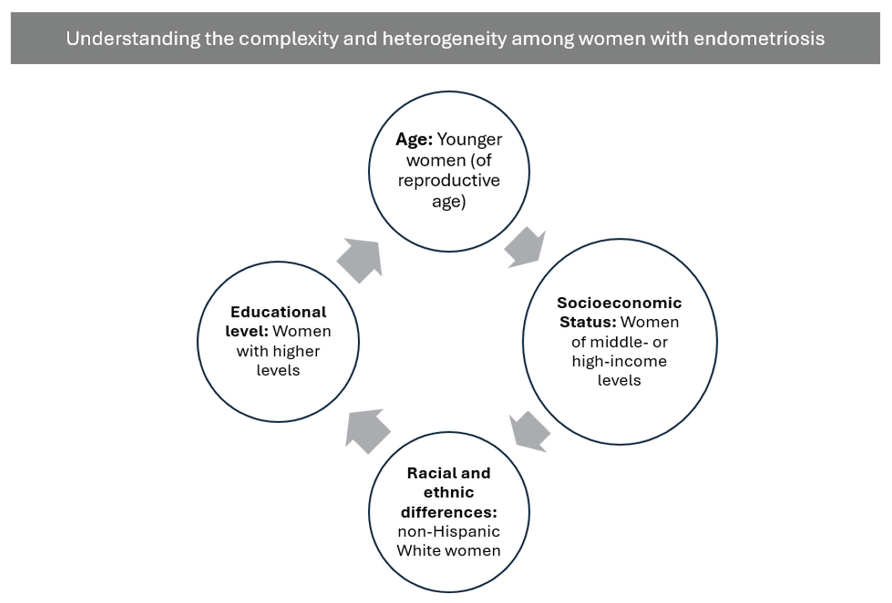

Table 1.
Adipose Tissue Dysregulation in Endometriosis: Inflammatory and Metabolic Evidence.
| Domain / Finding | Visceral Adipose Tissue (VAT) | Subcutaneous Adipose Tissue (SAT) | Serum / Systemic Markers |
|---|---|---|---|
| Adipocyte size | Smaller adipocytes (VAT showed the smallest adipocyte size overall) | Smaller adipocytes | — |
| Browning-related proteins (UCP-1, PGC-1α) | Upregulated | No significant changes | — |
| Pro-inflammatory markers (IL-6, MCP-1) | IL-6 ↑ (3.5-fold); MCP-1 ↑ (4.5-fold) | No significant changes; mild trend toward IL-6 ↑ and Galectin-3 ↑ | Serum IL-6 ↑ |
| Anti-inflammatory markers (CD206) | Reduced CD206 (indicating M1 predominance) | Mild trend toward reduced CD206 | — |
| Galectin-3 | Tendency to increase (not significant) | No significant differences | — |
| Browning-related gene expression | Trend toward decreased expression | Trend toward decreased expression; DIO2 significantly downregulated | — |
| Macrophage infiltration | Predominance of pro-inflammatory M1 macrophages | — | — |
| Perilesional adipose alterations | Increased fibrosis; increased angiogenesis; M1 macrophage infiltration; elevated FABP4, VEGF, IL-6, TNF-α, IL-1β | — | — |
| Overall interpretation | Marked localized inflammation; adipose tissue dysfunction; contribution to pathophysiology | Largely unaffected compared with VAT | Evidence of systemic inflammation (↑ serum IL-6) |
Disclaimer/Publisher’s Note: The statements, opinions and data contained in all publications are solely those of the individual author(s) and contributor(s) and not of MDPI and/or the editor(s). MDPI and/or the editor(s) disclaim responsibility for any injury to people or property resulting from any ideas, methods, instructions or products referred to in the content. |
© 2025 by the authors. Licensee MDPI, Basel, Switzerland. This article is an open access article distributed under the terms and conditions of the Creative Commons Attribution (CC BY) license (http://creativecommons.org/licenses/by/4.0/).
Copyright: This open access article is published under a Creative Commons CC BY 4.0 license, which permit the free download, distribution, and reuse, provided that the author and preprint are cited in any reuse.


