
Submitted:
17 November 2025
Posted:
18 November 2025
You are already at the latest version
Abstract
Docetaxel is a widely used chemotherapeutic agent effective against sol-id tumors, but its clinical use is limited by systemic toxicities, especially hepatotoxicity and nephrotoxicity. Curcuma longa, a phytochemical with antioxidant and anti-inflammatory properties, may offer organo-protective effects. Objective: To evaluate the protective effects of orally administered Curcuma longa against docetaxel-induced systemic toxicity in Wistar rats. Methods: Male Wistar rats were assigned to 5 subgroups (n=7/group) and treated for 7, 14, or 21 days with placebo, docetaxel (2.5 mg/kg, i.p.), or docetaxel combined with Curcuma longa (25, 50, or 500 mg/kg/day, oral). Serum biomarkers of hepatic function (Aspartate Aminotransferase (AST), Alanine Aminotransferase (ALT_, bilirubins) and renal function (urea, creatinine, electrolytes) were evaluated using summary measures, ANOVA, and Tukey’s post-hoc test, in addition to the relative weights of the liver, kidneys, heart, lungs, and small intes-tine. Results: Docetaxel induced significant elevations in hepatic and renal biomarkers and altered organ weights. Curcuma longa co-treatment attenuated these effects in a dose- and time-dependent man-ner. The 50 mg/kg dose consistently provided optimal protection. High-dose treatment was associated with marked splenic and intestinal hy-pertrophy. Conclusion: Curcuma longa demonstrates cytoprotective po-tential against docetaxel-induced toxicity, likely via Nrf2, NF-κB, and PI3K/Akt modulation. These findings support further translational re-search on Curcuma longa as an adjuvant in oncologic therapy.
Keywords:
chemotherapy-related toxicity
; phytotherapy
; hepatorenal function
; experimental toxicology
; organ-specific effects
1. Introduction
Docetaxel is a widely used antineoplastic agent belonging to the taxane class, with proven efficacy in the treatment of various solid tumors, including breast, prostate, and non-small cell lung cancers [1]. However, its clinical use is frequently limited by dose-dependent systemic toxicities, especially involving hepatic and renal function, and by off-target organ damage such as splenic and intestinal alterations. These toxic effects are often attributed to mechanisms including oxidative stress, inflammation, and impaired cellular metabolism [2,3,4,5,6].
In recent years, there has been growing scientific interest in the use of natural compounds with antioxidant and cytoprotective potential to reduce the adverse effects of chemotherapeutic agents. Curcuma longa L. (turmeric), a plant traditionally used in Ayurvedic and Chinese medicine, is recognized for its bioactive polyphenolic compounds, particularly curcumin [2,3,4,5]. These substances have been shown to exhibit anti-inflammatory, hepatoprotective, nephroprotective, and immunomodulatory activities [3,4,5,6,7,8]. Curcumin’s pharmacological profile also includes modulation of cellular signaling pathways, inhibition of pro-inflammatory cytokines, and attenuation of oxidative damage in various experimental models [9,10,11,12].
Despite encouraging evidence, the protective role of Curcuma longa in the context of docetaxel-induced systemic toxicity remains insufficiently explored, particularly regarding functional and structural outcomes in critical organs [13,14,15,16,17]. Additionally, dose–response relationships and organ-specific effects have not been consistently addressed in preclinical models.
The present study aimed to evaluate the potential protective effects of Curcuma longa extract, administered orally, on docetaxel-induced systemic toxicity in Wistar rats. The investigation focused on biochemical parameters of hepatorenal function, modulation of inflammatory cytokines, and physiopathological alterations in key target organs, including the liver, kidneys, spleen, and small intestine. This research seeks to contribute to the understanding of phytotherapeutic strategies for mitigating chemotherapy-associated organ toxicity.
2. Materials and Methods
This controlled case experimental study involved 105 male Wistar rats (n = 7 per subgroup; 8 weeks of age), obtained from the Animal Facility of Colombo, Paraná, Brazil. The animals were randomly allocated into three primary treatment groups based on duration: Group A (7 days), Group B (14 days), and Group C (21 days). Each primary group was subdivided into five treatment arms as follows:
Group A: 7-day treatment
Subgroups G1A to G5A received treatments as follows:
- G1A: oral placebo (water), daily for 7 days (control group).
- G2A: single intraperitoneal dose of docetaxel on Day 1.
- G3A: single intraperitoneal dose of docetaxel on Day 1 + Curcuma longa orally at 25 mg/kg/day.
- G4A: single intraperitoneal dose of docetaxel on Day 1 + Curcuma longa orally at 50 mg/kg/day.
- G5A: single intraperitoneal dose of docetaxel on Day 1 + Curcuma longa orally at 500 mg/kg/day.
Group B: 14-day treatment
Subgroups G1B to G5B received treatments as follows:
- G1B: oral placebo (water), daily for 14 days (control group).
- G2B: single intraperitoneal dose of docetaxel on Day 1.
- G3B: single intraperitoneal dose of docetaxel on Day 1 + Curcuma longa orally at 25 mg/kg/day.
- G4B: single intraperitoneal dose of docetaxel on Day 1 + Curcuma longa orally at 50 mg/kg/day.
- G5B: single intraperitoneal dose of docetaxel on Day 1 + Curcuma longa orally at 500 mg/kg/day.
Group C: 21-day treatment
Subgroups G1C to G5C received treatments as follows:
- G1C: oral placebo (water), daily for 21 days (control group).
- G2C: single intraperitoneal dose of docetaxel on Day 1.
- G3C: single intraperitoneal dose of docetaxel on Day 1 + Curcuma longa orally at 25 mg/kg/day.
- G4C: single intraperitoneal dose of docetaxel on Day 1 + Curcuma longa orally at 50 mg/kg/day.
- G5C: single intraperitoneal dose of docetaxel on Day 1 + Curcuma longa orally at 500 mg/kg/day.
The number of animals per subgroup (n = 7) was defined based on previous experimental studies employing similar designs, which demonstrated that this sample size is sufficient to detect significant differences in hematological, biochemical, and histopathological parameters. The selection also complied with international ethical guidelines for animal experimentation, adhering to the principles of the 3Rs (Replacement, Reduction, and Refinement), thereby ensuring the minimum number of animals required to obtain statistically reliable results
The dose of docetaxel was extrapolated for rats using allometric scaling from a human reference (70 kg), based on the methodology proposed by Pachaly (2006)43.
Calculations were based on specific metabolic rate (SMR) values:
- SMRref (human): 70 × 70⁻⁰·²⁵ = 24
- SMRrat (300 g): 70 × 0.3⁻⁰·²⁵ = 95
To reach the target dose in rats, the human equivalent dose (HED) was adjusted to 0.63 mg/kg. Thus:
- Rat dose = 0.63/24 × 95 = 2.5 mg/kg
Therefore, a final intraperitoneal dose of 2.5 mg/kg docetaxel was established, in a volume up to 1 mL/kg. Example for a 250 g rat: (2.5 mg – 1000 g; X mg – 250 g; X = 0.625 mg).
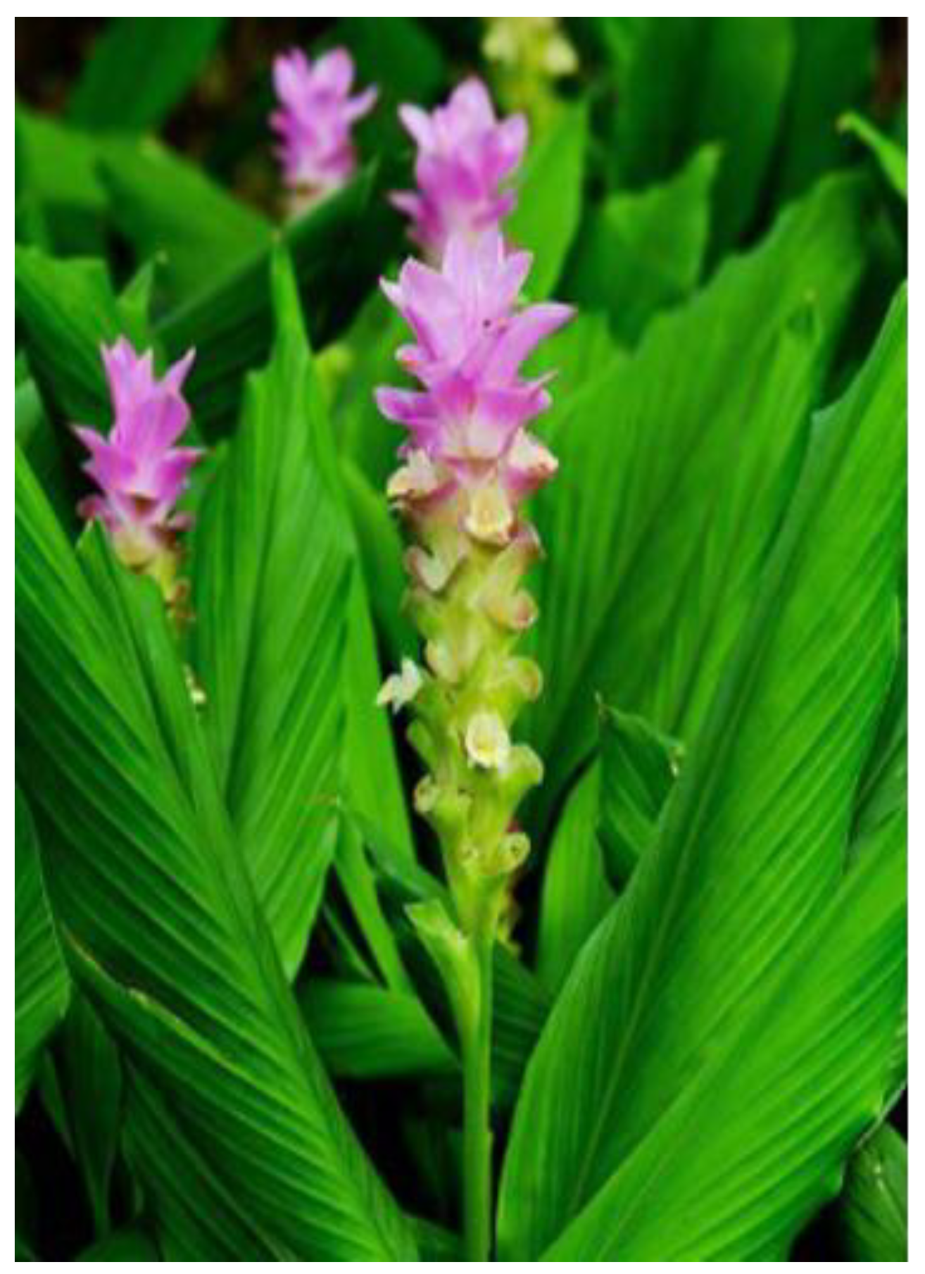
The Curcuma longa extract (95% curcuminoids) was obtained from Farmácia Farmavida (Umuarama, Paraná, Brazil). It was administered by gavage in doses of 25, 50, or 500 mg/kg/day, diluted in 1 mL/kg of water (Fig. A).
Figure A.
Curcuma Longa. Accessible at: https://www.easytogrowbulbs.com/products/ginger-curcuma-longa-turmeric.
Figure A.
Curcuma Longa. Accessible at: https://www.easytogrowbulbs.com/products/ginger-curcuma-longa-turmeric.
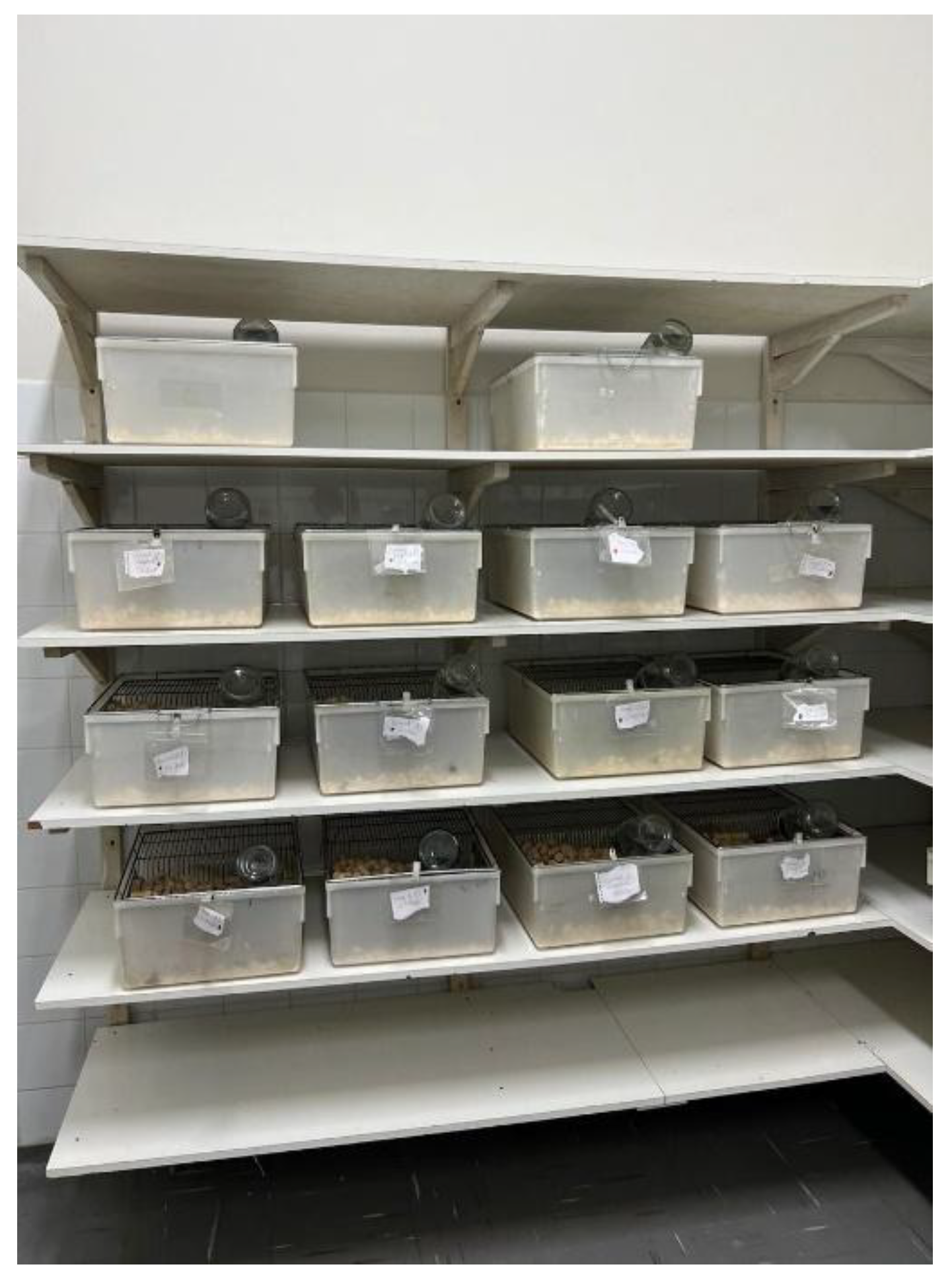
Animals were housed in polypropylene cages in a temperature-controlled room (22 ± 2 °C) with a 12-h light/dark cycle and had ad libitum access to standard chow and water throughout the experimental period (Fig. B). After administration, they were placed on a flat surface and observed continuously for one hour, then hourly for the first 6 hours, and subsequently once daily for 7, 14, or 21 days, according to the respective group, with the aim of analyzing their behavior. At the end of each experimental period, animals were fasted for 12 hours (with access to water), anesthetized using isoflurane, and euthanized via decapitation in accordance with the euthanasia guidelines of the Brazilian CONCEA.
Figure B.
Animals supervision at the Research Laboratory of Natural and Bioactive Products – UNIPAR.
Figure B.
Animals supervision at the Research Laboratory of Natural and Bioactive Products – UNIPAR.
Additional biological samples included blood for the assessment of liver function markers—namely aspartate aminotransferase (AST), alanine aminotransferase (ALT), and total bilirubin—and renal function parameters, including urea, creatinine, sodium, and potassium levels. Solid organs, including the liver, kidneys, small intestine, and heart—were surgically excised, cleaned of adherent tissue, and weighed with precision. Relative organ weight was subsequently calculated for each specimen as the ratio of the organ mass (g) to the final body weight (g) of the corresponding animal, and expressed as a percentage, according to the formula: Relative Weight (%) = (Organ Weight / Final Body Weight) × 100.
Carcasses were stored at −20 °C in labeled plastic bags until collected by a certified biological waste disposal company. Samples were homogenized and stored at 4 °C until analysis.
All procedures were approved by the Institutional Animal Care and Use Committee (IACUC) of University Paranaense under protocol number 40130 and conducted in compliance with the guidelines of the Brazilian CONCEA (2015).
Statistical analysis was performed using one-way analysis of variance (ANOVA), followed by Tukey’s post-hoc test for multiple comparisons. A p-value < 0.05 was considered statistically significant. All analyses were conducted using GraphPad Prism version 10.4.2.
Figure C.
Preclinical Research Laboratory of Natural and Bioactive Products – UNIPAR
3. Results
3.1. General Analysis
3.1.1. Biochemical Parameters
In the analysis of variance (ANOVA) conducted at a 5% significance level, differences among groups were observed for total bilirubin, direct bilirubin, indirect bilirubin, creatinine, sodium, Aspartate Aminotransferase (AST), and urea, whereas potassium and Alanine Aminotransferase (ALT) did not show significant variation. Tukey’s post-hoc test, also conducted at a 5% significance level, revealed that total bilirubin levels were higher in the 14-day group compared to the 7-day group, with the 21-day group showing intermediate values. For indirect bilirubin, the 14- and 21-day groups exhibited significantly higher values compared to the 7-day group. Regarding direct bilirubin, the 14-day group presented lower values than the 7-day group, while the 21-day group did not differ significantly from the others. Creatinine showed a marked increase in the 14-day group relative to the 7- and 21-day groups. Sodium levels were also higher in the 14- and 21-day groups compared to the 7-day group. For AST, the 14-day group had higher values than the 21-day group. Finally, urea levels were significantly higher in the 14-day group, whereas the 7- and 21-day groups did not differ significantly from each other (Table 1).
3.1.2. Organ Body Weight Parameters
The ANOVA performed at a 5% significance level indicated differences among groups for body weight, liver, right kidney, and left kidney, while no significant differences were observed for spleen, heart, lungs, or intestine. Tukey’s test showed that liver weight was higher in the 21-day group compared to the 7- and 14-day groups. For both the right and left kidneys, the 14- and 21-day groups exhibited higher mean values than the 7-day group. Regarding body weight, a tendency toward reduction was observed in the 14-day group, although no clear difference was detected relative to the other groups (Table 1).
3.2. Analysis by Period
3.2.1. Biochemical Parameters
Considering the ANOVA at a 5% significance level, no differences were detected among groups at 7 and 14 days. However, at 21 days, a significant effect was observed for potassium and urea. Tukey’s test showed that, for potassium, the CGC group presented higher values compared to the CG500 group, while the other groups did not differ. Regarding urea, the CGQ group exhibited higher values than the CG50 and CG500 groups, whereas CG25 and CGC remained in intermediate positions. These results indicate that, for biochemical parameters in animals treated for 21 days, there was a distinct response related to potassium and urea metabolism (Table 2).
3.2.2. Organ Body Weight Parameters
In the analysis of variance at a 5% significance level, no differences were observed among groups at 7 and 14 days. However, at 21 days, a significant effect was found for body weight, liver, right kidney, and left kidney. Tukey’s test showed that body weight was lower in the 500 mg/kg group compared to the 25 mg/kg group. For the liver, the 25 mg/kg group presented higher mean values than 500 mg/kg and Control. Regarding the kidneys, mean values were lower in 500 mg/kg, while 25 mg/kg exhibited higher means, highlighting differences related to exposure time (Table 2).
3.3. Correlation Between Organ Body Weight and Biochemical Parameters
In the 7-day group, no statistically significant correlations were found between the mean weight of the kidneys (right and left) and sodium, potassium, or urea, nor between liver weight and total bilirubin, direct bilirubin, indirect bilirubin, AST, or ALT (all with p ≥ 0.05) (Table 3).
In the 14-day group, significant positive associations were observed: (i) between the mean kidney weight and urea (ρ = 0.80; p < 0.01), indicating that higher urea concentrations were related to increased mean renal weight; (ii) between liver weight and total bilirubin (ρ = 0.47; p < 0.01); and (iii) between liver weight and indirect bilirubin (ρ = 0.36; p = 0.03). Correlations with sodium and potassium for the kidneys, as well as with direct bilirubin, AST, and ALT for the liver, were not significant (p ≥ 0.05) (Table 3).
In the 21-day group, no significant correlations were detected at the 5% level. Some relationships showed a tendency, such as mean kidney weight with potassium (ρ = 0.29; p = 0.10) and liver weight with ALT (ρ = 0.30; p = 0.08), but they remained below the adopted significance threshold (Table 3).
4. Discussion
Docetaxel, a widely used chemotherapeutic agent, is known for its potent antineoplastic activity against a range of solid tumors. However, its clinical utility is frequently hampered by dose-dependent systemic toxicities, notably hepatotoxicity and nephrotoxicity, which are major limiting factors in treatment adherence and overall patient outcomes [1]. In the present study, we demonstrated that docetaxel induced significant biochemical and morphophysiological disturbances in liver and kidney function, consistent with previous preclinical and clinical findings [6].
Importantly, our data show that Curcuma longa, particularly at intermediate and high doses, exerted a protective effect on key biochemical markers of hepatic function — especially AST and bilirubin fractions — and showed a non-significant tendency toward normalization of ALT levels, as well as on renal function markers (urea and creatinine). The time- and dose-dependent attenuation of these markers aligns with earlier evidence of curcumin’s hepatoprotective properties, which are mediated through its ability to modulate oxidative stress pathways, inhibit lipid peroxidation, and suppress pro-inflammatory cytokines such as TNF-α and IL-6 [2,3,4,5]. The reversal of elevated AST and ALT by Curcuma longa at 50 and 500 mg/kg corroborates its effect on stabilizing hepatocellular membranes and restoring hepatic enzymatic activity [3].
Elevations in total and direct bilirubin levels following docetaxel administration suggest an impairment in hepatobiliary excretion and potential cholestatic damage. The mitigation of bilirubin accumulation by curcumin may involve its influence on bile acid homeostasis and transporter regulation, as suggested in murine models of liver injury [4]. Curcumin has been shown to downregulate pro-apoptotic proteins and upregulate cytoprotective enzymes such as heme oxygenase-1 (HO-1), enhancing hepatocyte resilience against chemotoxic insults [7].
4.1. Hepatic Toxicity and Curcuma Longa’s Hepatoprotective Effects
Docetaxel exposure was associated with a tendency toward elevation of serum liver enzymes, particularly AST, and with a tendency toward increased bilirubin fractions, reaching statistical significance only in the overall analysis rather than at individual time points, indicating hepatocellular stress and possible cholestatic dysfunction. Although statistically significant differences in total and indirect bilirubin levels were observed primarily in the overall analysis rather than at individual time points, the upward trend suggests a potential impairment in hepatobiliary excretion and altered bile transport dynamics. Similar hepatotoxic profiles have been reported with taxane-based regimens, which can induce mitochondrial dysfunction, endoplasmic reticulum stress, and pro-inflammatory cytokine release in hepatocytes [8].
The observed normalization of liver enzyme levels and bilirubin concentrations following co-treatment with Curcuma longa, particularly at the 50 and 500 mg/kg doses, underscores its potential hepatoprotective action. These effects are consistent with previous reports demonstrating that curcumin, the principal bioactive constituent of Curcuma longa, exerts robust hepatoprotective properties through multiple pathways. Mechanistically, curcumin has been shown to attenuate hepatic oxidative stress by scavenging reactive oxygen species (ROS) and enhancing endogenous antioxidant defenses via activation of the nuclear factor erythroid 2-related factor 2 (Nrf2) signaling pathway [9,10]. Nrf2 translocation into the nucleus leads to upregulation of heme oxygenase-1 (HO-1), NAD(P)H:quinone oxidoreductase 1 (NQO1), and glutamate-cysteine ligase, which collectively restore redox balance and protect hepatocytes from apoptosis and necrosis [11].
Furthermore, curcumin downregulates nuclear factor-kappa B (NF-κB), a transcription factor implicated in inflammation-mediated liver damage. By inhibiting IκB kinase (IKK), curcumin prevents NF-κB nuclear translocation and transcription of pro-inflammatory mediators such as TNF-α, IL-1β, and IL-6 [12]. These anti-inflammatory effects reduce leukocyte infiltration and cytokine-induced hepatocellular apoptosis, mitigating liver injury under toxic insults.
Of note, curcumin also interferes with hepatic cytochrome P450 (CYP) enzymes, particularly CYP3A4 and CYP2E1, which are involved in the bioactivation of toxic intermediates during chemotherapeutic metabolism [13]. This enzymatic modulation may partially explain the reduction in docetaxel-induced hepatotoxicity in animals receiving Curcuma longa supplementation.
The dose-dependent nature of the hepatoprotective effect observed in this study aligns with data from other experimental models of drug-induced liver injury, including those induced by acetaminophen, isoniazid, and cisplatin [14]. In these studies, curcumin administration significantly attenuated transaminase elevation, lipid peroxidation, and histopathologic damage.
In the present model, the 500 mg/kg dose appeared most effective in restoring biochemical markers to near-baseline values by day 21. However, the 50 mg/kg dose also produced meaningful reductions in hepatotoxic markers, suggesting that intermediate dosing may offer an optimal balance between efficacy and safety.
Taken together, these findings reinforce the hepatoprotective profile of Curcuma longa and its potential utility as an adjuvant agent in chemotherapy protocols. Future investigations incorporating liver histopathology and molecular profiling will be essential to confirm the protective mechanisms and identify biomarkers predictive of response.
4.2.1. Renal Function Markers and Nephroprotection
Renal function parameters also showed marked alterations under docetaxel exposure. Serum creatinine and, to a lesser extent, urea levels showed elevations consistent with glomerular and tubular dysfunction. The most pronounced alterations occurred at day 14 (creatinine) and day 21 (urea), indicating a time-dependent pattern of renal involvement [6]. Co-treatment with Curcuma longa significantly normalized these parameters by day 21, particularly at 500 mg/kg, indicating nephroprotective activity. This effect is likely mediated by curcumin’s antioxidative modulation of Nrf2–ARE signaling, reduction of renal inflammation, and maintenance of mitochondrial function, as described in models of cisplatin- and gentamicin-induced renal injury [3,4].
Renal dysfunction is a well-established complication of taxane-based chemotherapy, with docetaxel exhibiting nephrotoxic potential primarily through mechanisms involving oxidative stress, mitochondrial dysfunction, and inflammatory injury to the renal tubular epithelium [15]. In the present study, the docetaxel-only groups demonstrated elevations in serum creatinine and urea levels, most pronounced at day 14, suggesting transient renal impairment. These alterations were less evident at days 7 and 21, indicating partial spontaneous recovery over time. These biochemical alterations agree with previously described patterns of taxane-induced nephrotoxicity in preclinical models [16].
Noteworthily, co-administration of Curcuma longa extract produced a dose-dependent nephroprotective effect, with the 500 mg/kg group demonstrating near-complete normalization of creatinine and urea values by day 21. This finding suggests that Curcuma longa may enhance renal clearance and mitigate chemotherapy-induced renal injury over time.
The nephroprotective mechanisms of curcumin have been extensively documented in various models of renal injury. One of its principal actions involves attenuation of oxidative stress via activation of the Nrf2–ARE (antioxidant response element) pathway, which upregulates endogenous antioxidant enzymes such as glutathione peroxidase, catalase, and superoxide dismutase [17,18]. By reducing reactive oxygen species (ROS) accumulation within renal tubular cells, curcumin preserves mitochondrial function and prevents lipid peroxidation—a key mediator of nephrotoxicity [19].
Additionally, curcumin has been shown to downregulate the expression of pro-apoptotic mediators (e.g., Bax, caspase-3) while enhancing anti-apoptotic pathways (e.g., Bcl-2), thereby preserving tubular epithelial cell integrity [20]. These anti-apoptotic effects have been correlated with improved histopathological scores in cisplatin-induced nephrotoxicity models, suggesting translational relevance across chemotherapeutic classes [21].
Moreover, curcumin’s ability to modulate inflammatory cytokine, such as TNF-α, IL-1β, and IL-6, contributes to reduced leukocyte infiltration and attenuation of glomerular inflammation [22]. Experimental studies in models of ischemia-reperfusion and diabetic nephropathy have shown that curcumin ameliorates renal injury by decreasing macrophage accumulation and improving microvascular perfusion [23].
In this study, the 500 mg/kg dose was the most effective in reversing biochemical markers of renal dysfunction, suggesting a threshold effect in achieving nephroprotection. However, the 50 mg/kg dose also showed partial efficacy, particularly at later timepoints, which may offer a clinically safer and equally effective window for translation.
Comparative studies with other nephrotoxic agents, such as cisplatin, reinforce the findings observed here. Al-Moundhri et al. demonstrated that post-treatment with curcumin significantly reduced cisplatin-induced elevations in serum creatinine and urea, preserved glomerular structure, and reduced tubular necrosis in rats [24]. Similarly, Jeong et al. reported that curcumin protected against cyclophosphamide-induced renal injury by restoring oxidative balance and preventing apoptosis in renal tissues [25].
Taken together, the data support the conclusion that Curcuma longa, particularly at higher doses, exerts a strong nephroprotective effect in the context of docetaxel-induced toxicity. These effects appear to be mediated by a combination of antioxidant, anti-inflammatory, and anti-apoptotic actions, with potential relevance for mitigating renal adverse effects in clinical oncology.
4.2.2. Electrolyte Balance and Systemic Homeostasis
Electrolyte disturbances, especially hypokalemia observed on day 14, are consistent with chemotherapy-related tubular injury. Although mild and transient, the partial reversal by Curcuma longa reflects a stabilizing effect on renal tubular integrity, which may preserve ionic transport systems. Interestingly, sodium levels remained stable across most groups, indicating a selective tubular impact.
Chemotherapy-induced disturbances in electrolyte homeostasis are clinically relevant manifestations of systemic toxicity, often reflecting underlying renal tubular dysfunction, gastrointestinal losses, or neurohormonal dysregulation. In the present study, docetaxel monotherapy induced a significant reduction in serum potassium levels at day 14 (p < 0.05 vs. control), consistent with hypokalemia. Although sodium levels remained relatively stable across treatment groups, mild hyponatremia was noted in the Curcuma longa 25 mg/kg group at day 7, but without sustained alterations thereafter.
Hypokalemia in the context of taxane-based chemotherapy may result from several mechanisms: increased urinary potassium excretion due to impaired tubular reabsorption, gastrointestinal potassium loss secondary to mucosal toxicity and diarrhea, or intracellular potassium shifts mediated by altered insulin or catecholamine activity [26,27]. In our model, the mid-phase (day 14) was particularly susceptible to hypokalemia, likely due to the cumulative nephrotoxic and enterotoxic effects of docetaxel. Notably, co-administration of Curcuma longa, especially at 50 and 500 mg/kg, attenuated this potassium loss and stabilized serum levels by day 21, indicating a potential role in restoring ionic equilibrium.
Curcumin’s contribution to electrolyte homeostasis may be linked to its protective effects on renal tubular integrity and its ability to suppress oxidative stress and proinflammatory cytokine production within the nephron microenvironment [28]. Oxidative damage to renal tubular cells, particularly in the thick ascending limb of Henle’s loop and distal tubules, is a known contributor to impaired sodium and potassium handling during systemic toxic insults [29]. By mitigating mitochondrial dysfunction and reducing ROS accumulation, curcumin preserves tubular transporter function and promotes ion reabsorption efficiency.
Additionally, curcumin has been shown to modulate key regulators of ion balance, including Na⁺/K⁺-ATPase and aquaporin channels, in experimental nephrotoxicity models [30]. It also exhibits indirect effects via its anti-inflammatory action, suppressing TNF-α and IL-1β, which are known to impair tubular handling of electrolytes and induce tubular apoptosis [28,29].
Clinically, maintaining electrolyte balance is paramount during chemotherapy to prevent arrhythmias, neuromuscular dysfunction, and acute kidney injury, particularly in regimens involving nephrotoxic agents such as cisplatin, cyclophosphamide, and taxanes [31]. The capacity of Curcuma longa to preserve potassium levels and avoid significant sodium fluctuations may enhance patient safety and reduce the need for corrective interventions such as electrolyte repletion or hospitalization.
These findings reinforce the relevance of phytotherapeutic adjuvants in systemic toxicity management, not only for organ-specific protection but also for preserving essential physiological parameters such as electrolyte balance.
4.2.3. Relative Organ Weight Changes: Functional Interpretation
Changes in relative organ weights are sensitive biomarkers of systemic toxicity and physiological stress induced by chemotherapeutic agents. In the present experimental model, docetaxel treatment led to distinct alterations in the relative mass of multiple organs, indicating multisystem involvement. Co-administration of Curcuma longa modulated several of these effects, demonstrating potential protective and regenerative actions in a dose- and time-dependent manner.
The organ weight analysis revealed systemic effects of docetaxel on several tissues. Liver hypertrophy, likely secondary to inflammation, congestion, and hepatocyte injury, was reversed with Curcuma longa, supporting its role in limiting hepatomegaly and tissue remodeling [4]. Conversely, renal hypotrophy, observed at day 14, may represent structural atrophy due to cytotoxic damage. Curcuma longa demonstrated only partial recovery, particularly at low doses, suggesting a more complex dose-response relationship requiring further exploration.
Cardiac and pulmonary weights were also affected. The reduction in heart weight under docetaxel treatment could reflect cardiomyocyte atrophy or loss, as observed in preclinical cardiotoxicity models with taxanes [6]. Curcuma longa preserved cardiac mass, aligning with previous reports of its cardioprotective effects via antioxidant and antiapoptotic mechanisms [2]. Lung weight increases, likely indicative of pulmonary edema or inflammation, were attenuated by Curcuma longa but not fully normalized, suggesting a partial protective effect. Although these changes in cardiac and pulmonary weights did not reach statistical significance, trends suggestive of cardiomyocyte alterations and pulmonary inflammatory processes were observed. These patterns, while not conclusive in this dataset, warrant further investigation with larger cohorts and detailed histopathological analysis
An unexpected finding was the increase in intestinal weight, notably in the group treated with 50 mg/kg Curcuma longa on day 14. This may reflect mucosal regeneration or enhanced epithelial turnover stimulated by curcumin, which has been previously associated with intestinal trophism and anti-ulcerogenic properties [5]. Further histopathological analysis would be necessary to clarify whether this finding denotes a beneficial or adverse response.
4.2.3.1. Liver
Hepatomegaly observed in docetaxel-treated groups, particularly at days 14 and 21, may reflect acute inflammatory response and/or hepatocellular regeneration following cytotoxic insult. Previous studies report hepatic hypertrophy linked to leukocyte infiltration, hepatocyte swelling, and proliferative activity in response to oxidative stress caused by chemotherapeutic agents [32].
In groups co-treated with Curcuma longa—notably at 50 and 500 mg/kg doses—this hypertrophy was significantly attenuated. This suggests that curcuminoids reduce hepatic oxidative damage and inflammation, while promoting redox homeostasis and controlled hepatocyte renewal without inducing reactive hyperplasia [33,34].
4.2.3.2. Kidneys
The observed reduction in kidney weight in docetaxel-only groups, especially at day 14, may be indicative of renal hypoperfusion, tubular necrosis, or apoptotic degeneration, which are well-documented outcomes of chemotherapy-induced nephrotoxicity [35]. In rats receiving Curcuma longa—particularly at 25 mg/kg—partial preservation of kidney mass was noted, supporting a renoprotective effect. This may be mediated through activation of endogenous antioxidant defenses (e.g., Nrf2–ARE pathway) and inhibition of tubulointerstitial fibrosis [36].
4.2.3.3. Heart
Docetaxel-treated animals showed a trend toward reduced cardiac mass at day 14, which may be attributed to mitochondrial oxidative stress and cardiomyocyte atrophy—a recognized complication of taxane therapy [37]. Notably, Curcuma longa-treated groups maintained near-normal heart weight throughout, suggesting cardioprotective properties likely driven by anti-apoptotic and mitochondrial stabilizing effects of curcumin [38].
4.2.3.4. Lungs
A non-significant increase in lung weight, particularly in the docetaxel-only group at day 21, suggests a possible trend toward pulmonary inflammation or edema. Taxane-induced pulmonary toxicity has been associated with interstitial inflammation, capillary leakage, and oxidative damage [39]. While Curcuma longa did not fully reverse these changes, it demonstrated a trend toward normalization of lung mass, supporting its potential anti-inflammatory effects at the alveolar–capillary interface [40].
4.2.3.5. Small Intestine
The most pronounced change in intestinal weight was observed in the group treated with Curcuma longa 50 mg/kg at day 14, showing a statistically significant increase. This may reflect a trophic effect of curcumin on intestinal mucosa, possibly through stimulation of crypt cell proliferation and enhancement of epithelial integrity. Curcumin has been shown to modulate intestinal growth factors such as EGF and TGF-β, supporting epithelial regeneration and mucosal barrier function [41,42]. Although this finding could represent a favorable adaptation, further histological investigation is warranted to rule out dysplastic or hyperplastic changes under prolonged exposure.
4.3. Dose-And-Time Dependent Protective Profile of Curcuma Longa
The present findings demonstrate a clear dose- and time-dependent profile for Curcuma Longa’s protective effects against docetaxel-induced systemic toxicity. Among the three tested doses (25, 50, and 500 mg/kg/day), the intermediate dose of 50 mg/kg emerged as the most consistently effective in mitigating functional alterations in hepatic and renal biomarkers, preserving organ weights, and supporting systemic homeostasis—particularly at the later timepoint (day 21).
This temporal dynamic aligns with the known pharmacodynamics of curcumin, which requires sustained exposure to accumulate intracellularly and exert long-lasting anti-inflammatory and antioxidative effects through pathways such as NF-κB inhibition, Nrf2 activation, and suppression of cytokine cascades (e.g., IL-6, TNF-α) [3,7,33,36].
Interestingly, while the highest dose (500 mg/kg) was effective in reversing elevations in bilirubin, urea, and creatinine, it also led to pronounced splenic and intestinal hypertrophy by day 21. These findings suggest a dual-edged response: enhanced detoxification and cytoprotection on one hand, and potential overstimulation of immune or regenerative pathways on the other. High-dose curcumin has previously been associated with immune cell recruitment, macrophage proliferation, and splenic architectural remodeling, which, if sustained, may predispose to organomegaly, fibrosis, or functional dysregulation [19,24,34].
This raises the possibility of a therapeutic efficacy plateau beyond which further dose escalation may not confer additional benefit and could instead trigger maladaptive responses. Such a biphasic pattern underscores the importance of optimizing dosing regimens to maximize therapeutic gain while avoiding overactivation of compensatory physiological mechanisms.
Moreover, the timing of protective effects is relevant. At earlier stages (days 7 and 14), modest biochemical improvements were observed, but the full extent of protection—particularly normalization of AST, ALT, creatinine, and bilirubin levels—was only evident by day 21. This lag suggests a cumulative protective action, possibly through gene expression modulation and restoration of metabolic homeostasis, consistent with previous in vivo models of phytochemical co-treatment during chemotherapy [2,4,41].
In summary, these results support the hypothesis that Curcuma longa exhibits a time-sensitive and dose-limited protective profile, with 50 mg/kg representing a potentially optimal therapeutic window in this preclinical setting. Future studies should further explore the long-term safety of high-dose administration and seek to determine the molecular thresholds beyond which curcumin’s beneficial effects may diminish or reverse.
4.4. Mechanism Insights: Curcumin and Molecular Pathways
The protective effects of Curcuma longa observed in this study—particularly the normalization of hepatic enzymes, improvement in renal function, stabilization of electrolyte balance, and modulation of organ weights—are consistent with curcumin’s multifaceted interaction with several intracellular signaling pathways. Among these, the Nrf2, NF-κB, MAPK, and PI3K/Akt pathways stand out as key molecular mediators of the cytoprotective and anti-inflammatory actions of curcumin.
The Nrf2 (nuclear factor erythroid 2-related factor 2) pathway plays a pivotal role in the cellular defense against oxidative stress. Upon activation, Nrf2 translocates to the nucleus and upregulates the transcription of antioxidant response elements (ARE), including heme oxygenase-1 (HO-1), NAD(P)H quinone dehydrogenase 1 (NQO1), and glutathione S-transferases [7,38,39]. In the current model, the time- and dose-dependent reductions in AST, ALT, bilirubins, creatinine, and urea suggest enhanced antioxidant capacity and restoration of redox homeostasis, likely mediated by Nrf2-driven transcriptional activity. This hypothesis is reinforced by the delayed, yet robust protective effects observed on day 21, consistent with the kinetics of Nrf2-dependent gene expression.
Conversely, curcumin has been widely recognized as a potent inhibitor of NF-κB, a transcription factor central to the propagation of inflammatory responses. Under chemotherapeutic stress—such as that induced by docetaxel—NF-κB is activated by cytokines like TNF-α and IL-1β, leading to increased expression of pro-inflammatory genes, including COX-2, iNOS, and various interleukins [3,33,40]. Curcumin attenuates this cascade by inhibiting IκB kinase (IKK), thereby preventing NF-κB nuclear translocation and downstream inflammatory amplification [3,5]. The reductions in hepatic and renal injury markers in the co-treated groups support this anti-inflammatory mechanism.
The MAPK (mitogen-activated protein kinase) signaling pathway, including ERK, JNK, and p38 kinases, has also been implicated in drug-induced organ injury via regulation of apoptosis, inflammation, and cell proliferation. Curcumin modulates MAPK signaling in a context-dependent manner: it can inhibit p38 and JNK in inflammatory settings while promoting ERK activation in regenerative and anti-apoptotic contexts [41]. These dual effects may account for the preserved organ weights and functional parameters seen in liver and kidney tissues, particularly at intermediate doses.
Moreover, the PI3K/Akt pathway, which is critically involved in cell survival, metabolism, and autophagy, is another target of curcumin modulation. In chemotherapy models, inhibition of PI3K/Akt has been associated with enhanced apoptosis and tissue damage. Curcumin has demonstrated the ability to restore PI3K/Akt signaling balance, enhancing cellular resilience to cytotoxic insults while preventing pathological proliferation [3,7,42]. In this context, the partial recovery of intestinal and splenic mass, alongside biochemical normalization, suggests Akt-mediated cell survival and regenerative cues.
At the cytokine and protein expression level, curcumin has been shown to downregulate TNF-α, IL-6, and IL-1β, while simultaneously upregulating anti-apoptotic proteins such as Bcl-2 and suppressing pro-apoptotic markers like Bax and caspase-3 [3,6,43]. These effects are likely instrumental in the mitigation of hepatocyte and tubular epithelial cell death observed in our study, particularly under high-dose Curcuma longa supplementation.
Taken together, these molecular insights provide a plausible mechanistic framework for the systemic cytoprotection observed in our experimental model. By orchestrating a multi-target regulatory network, spanning oxidative stress defense, inflammatory suppression, and apoptosis modulation, curcumin exerts comprehensive organoprotective effects that align with the observed attenuation of docetaxel-induced hepatotoxicity, nephrotoxicity, and systemic imbalance.
4.5. Translational Relevance and Future Directions
The findings of this study offer meaningful translational potential for improving the management of chemotherapy-associated toxicity in oncology patients. Docetaxel, while highly effective against various solid tumors, is often limited by its systemic side effects—particularly hepatotoxicity and nephrotoxicity—that compromise therapeutic intensity, continuity, and patient quality of life [1,6]. The demonstration that Curcuma longa extract, particularly at intermediate and high doses, attenuates these toxic effects without overt adverse outcomes supports its candidacy as an adjuvant cytoprotective agent during chemotherapeutic regimens.
From a clinical perspective, Curcuma longa could serve to preserve hepatic and renal function, reduce the incidence of treatment delays or dose reductions, and thereby maintain the planned chemotherapy dose-intensity—a well-established determinant of clinical efficacy in oncologic protocols [40]. This is especially relevant in settings where cumulative toxicity often dictates therapeutic limitations, such as in breast, prostate, and lung cancers treated with taxanes.
The results also lay the foundation for the design of future clinical trials, ideally randomized, placebo-controlled, and dose-escalated, to evaluate the safety and efficacy of Curcuma longa as an adjunctive intervention. Primary endpoints could include biochemical markers of liver and kidney function (AST, ALT, bilirubin, creatinine, urea), while secondary endpoints may assess patient-reported outcomes, quality of life, and treatment adherence. Additionally, the identification and validation of predictive biomarkers—such as cytokine profiles (e.g., IL-6, TNF-α), oxidative stress markers (e.g., malondialdehyde, glutathione), and apoptosis-related proteins (e.g., Bcl-2, Bax, caspase-3)—could optimize patient selection and therapeutic monitoring.
To further elucidate the mechanisms underlying the observed organoprotection, histopathological and immunohistochemical studies are warranted in future investigations. Such analyses should assess structural integrity, inflammatory infiltration, fibrosis, and vascular remodeling in hepatic, renal, and intestinal tissues. The integration of molecular pathway analysis, including the evaluation of Nrf2, NF-κB, MAPKs, and PI3K/Akt signaling cascades, will be instrumental in clarifying the intracellular targets of Curcuma longa in the context of chemotherapeutic injury.
Finally, emerging biotechnological tools, such as transcriptomic profiling, proteomics, and metabolomic screening, can provide a systems-level understanding of Curcuma longa’s pleiotropic actions and help to identify synergistic interactions when combined with cytotoxic agents. Such insights may ultimately guide the personalization of supportive care in oncology, aligning phytotherapeutic strategies with individual toxicity profiles and treatment regimens.
5. Conclusions
This study provides preclinical evidence supporting the adjuvant use of Curcuma longa in reducing docetaxel-induced systemic toxicity, paving the way for translational applications in oncologic care. Ongoing and future investigations will be critical to validate these findings, optimize dosing strategies, and ensure the safe integration of phytochemicals into standard chemotherapy protocols.
Overall, our findings support the hypothesis that Curcuma longa mitigates docetaxel-induced systemic toxicity through multifactorial mechanisms, including antioxidative defense, anti-inflammatory modulation, cellular protection, and restoration of physiological function in hepatic and renal systems. The dose- and time-dependent patterns observed highlight the importance of identifying optimal therapeutic windows for phytotherapeutic intervention.
Author Contributions
Isabella Morais Tavares Huber: Conceptualization, Funding acquisition, Writing - review & editing, Writing - original draft, Validation, Methodology, Investigation, Project administration, Data curation, Formal analysis, Resources; Emerson Luiz Botelho Lourenço: Conceptualization, Validation, Supervision, Project administration, Funding acquisition; Salviano Tramontin Bellettini: Investigation, Validation; Guilherme Donadel: Investigation; João Francisco Velasquez Matumoto: Investigation; Sandra Marisa Pelloso: Visualization; Maria Dalva de Barros Carvalho: Visualization; Stéfane Lele Rossoni: Data curation, Formal analysis; Mariana Morais Tavares Colferai: Visualization; Diego Ricardo Colferai: Visualization; Roberto Kenji Nakamura Cuman: Visualization; Leonardo Garcia Velasquez: Writing - original draft, Writing - review & editing, Project administration, Resources, Data curation, Software, Formal analysis, Validation, Methodology, Funding acquisition, Supervision.
Funding
Universidade Paranaense.
Institutional Review Board Statement
All procedures were approved by the Institutional Animal Care and Use Committee (CEPEA) of University Paranaense under protocol number 40130/2023 and conducted in compliance with the guidelines of the Brazilian CONCEA (2015).
Data Availability Statement
The raw data supporting the conclusions of this article will be made available by the authors on request.
Acknowledgments
The first author wishes to express her sincere appreciation to Professor Leonardo Garcia Velasquez for his thoughtful mentorship and constructive input throughout the development of this work. Gratitude is also extended to the technical staff at the animal research facility for their valuable assistance, and to the Universidade Paranaense for its support. Special thanks are owed to the laboratory team for their dedication and collaboration. The author acknowledges the essential role of the animals used in this research, recognizing their contribution with respect and ethical responsibility. Finally, heartfelt thanks to the author’s family for their steadfast support, and to all patients whose resilience continues to inspire the pursuit of meaningful scientific progress.
Conflicts of Interest
The authors declare no conflicts of interest.
References
- Crown J, O’Leary M, Ooi WS. Docetaxel and paclitaxel in the treatment of breast cancer: a review of clinical experience. Oncologist. 2004;9(Suppl 2):24–32. [CrossRef]
- Iqubal A, Iqubal MK, Sharma S, Najmi AK, Syed MA, Ali J. Neuroprotective effect of curcumin against cisplatin-induced neurotoxicity: behavioral and biochemical evidences. Neurochem Res. 2019;44(3):684–692. [CrossRef]
- Aggarwal BB, Harikumar KB. Potential therapeutic effects of curcumin, the anti-inflammatory agent, against neurodegenerative, cardiovascular, pulmonary, metabolic, autoimmune, and neoplastic diseases. Int J Biochem Cell Biol. 2009;41(1):40–59. [CrossRef]
- Al Moundhri MS, Al-Salam S, Al Mahrouqee A, Beegam S, Ali BH. Curcumin protects against cisplatin-induced nephrotoxicity: a post-treatment strategy. J Nephrol. 2013;26(3):469–476. [CrossRef]
- Chainani-Wu N. Safety and anti-inflammatory activity of curcumin: a component of turmeric (Curcuma longa). J Altern Complement Med. 2003;9(1):161–168. [CrossRef]
- Zwart SR, Jessup JM, Ji J, Smith SM. Chemotherapy-induced toxicity is associated with systemic inflammation and metabolic dysregulation. J Oncol Pharm Pract. 2020;26(7):1675–1683. [CrossRef]
- Sharma RA, Steward WP, Gescher AJ. Pharmacokinetics and pharmacodynamics of curcumin. In: Aggarwal BB, Surh YJ, Shishodia S, eds. The Molecular Targets and Therapeutic Uses of Curcumin in Health and Disease. New York: Springer; 2007:453–470. [CrossRef]
- Zhao Y, He X, Wang H, Zhang T, Zhang Y, Wang Y. Taxanes-induced hepatotoxicity: mechanisms and protective strategies. Front Pharmacol. 2021;12:676481. [CrossRef]
- Jaiswal AK. Nrf2 signaling in coordinated activation of antioxidant gene expression. Free Radic Biol Med. 2004;36(10):1199–1207. [CrossRef]
- Son YO, Pratheeshkumar P, Wang X, Roy RV, Hitron A, Wang L, et al. Activation of Nrf2 signaling by curcumin analog in human hepatoma cells. Biochem Biophys Res Commun. 2013;433(4):552–557. [CrossRef]
- Rushworth SA, MacEwan DJ, O’Connell MA. Lipopolysaccharide-induced expression of NAD(P)H:quinone oxidoreductase 1 and heme oxygenase-1 requires the Nrf2/ARE pathway in human monocytes and macrophages. Arch Biochem Biophys. 2008;451(2):108–114. [CrossRef]
- Jobin C, Bradham CA, Russo MP, Juma B, Narula AS, Brenner DA, Sartor RB. Curcumin blocks cytokine-mediated NF-κB activation and proinflammatory gene expression by inhibiting inhibitory factor I-κB kinase activity. J Immunol. 1999;163(6):3474–3483. PMID:1047761.
- Kim HG, Cho JH, Jeong EY, Lim JH, Lee SH, Son CG. Curcuma longa extract protects against liver injury by modulating the expression of hepatic genes involved in drug metabolism. Food Chem Toxicol. 2010;48(10):2771–2777. [CrossRef]
- Cinar SA, Kosar M, Akpinar D, Karadag CH, Ugan RA, Arslan M, et al. Protective effect of curcumin on acetaminophen-induced liver damage in rats. J Pharm Pharmacol. 2020;72(8):1108–1114. [CrossRef]
- Guo Y, Xu B, Wang Z, Zhang H, Yu W, Jin J, et al. Docetaxel-induced nephrotoxicity in cancer patients: a real-world analysis and review. Cancer Chemother Pharmacol. 2022;89(4):389–396. [CrossRef]
- Shrestha S, Yadav DK, Song DK. Docetaxel-induced mitochondrial dysfunction and oxidative stress in kidney tissues. Biomed Pharmacother. 2021;133:110951. [CrossRef]
- Soetikno V, Sari FR, Lakshmanan AP, Arumugam S, Harima M, Suzuki K, et al. Curcumin prevents diabetic nephropathy by inhibiting PKC-α and PKC-β1 activity in streptozotocin-induced type I diabetic rats. Mol Nutr Food Res. 2011;55(11):1655–1665. [CrossRef]
- Maheshwari RK, Singh AK, Gaddipati J, Srimal RC. Multiple biological activities of curcumin: a short review. Life Sci. 2006;78(18):2081–2087. [CrossRef]
- El-Shishtawy MM, Moustafa YM, El-Shishtawy KA, Al-Tel TH, Al-Khatib AJ. Curcumin attenuates oxidative stress and inflammation in cisplatin-induced nephrotoxicity. Biomed Pharmacother. 2020;128:110259. [CrossRef]
- Manikandan P, Vinothini G, Anandan R, Nagini S. Protective effects of curcumin and piperine against genotoxicity in mice treated with benzo[a]pyrene. Int J Cancer. 2010;126(4):770–775. [CrossRef]
- Sun Y, Chen D, Pan Y, Qu W, Hao X. Curcumin attenuates cisplatin-induced nephrotoxicity in rats by inhibiting inflammation. Biol Pharm Bull. 2011;34(9):1376–1381. [CrossRef]
- Ghosh SS, Massey HD, Krieg R, Fazelbhoy ZA, Ghosh S, Sica DA, et al. Curcumin ameliorates renal failure in 5/6 nephrectomized rats: role of inflammation. Am J Physiol Renal Physiol. 2009;296(5):F1146–F1157. [CrossRef]
- Kalantari H, Dashti F, Zakeri M, Zolfaghari B, Daraei B, Sabahi M. Curcumin attenuates renal ischemia/reperfusion injury through reduction of oxidative stress and inflammation. Ren Fail. 2016;38(10):1668–1675. [CrossRef]
- Al-Moundhri MS, Al-Salam S, Al Mahrouqee A, Beegam S, Ali BH. Curcumin protects against cisplatin-induced nephrotoxicity: a post-treatment strategy. J Nephrol. 2013;26(3):469–476. [CrossRef]
- Jeong JC, Jang SI, Moon YJ, Ha IH, Lee HJ, Jang JB, et al. Protective effect of curcumin against cyclophosphamide-induced myelosuppression and nephrotoxicity in mice. Food Chem Toxicol. 2016;89:129–135. [CrossRef]
- Muggia FM, Kudlowitz D. Taxane toxicity: preclinical and clinical observations. Semin Oncol. 2014;41 Suppl 2:S3–S8. [CrossRef]
- Santoso JT, Lucci JA III, Coleman RL. Chemotherapy-induced electrolyte abnormalities. Gynecol Oncol. 2003;90(3):529–534. [CrossRef]
- Soetikno V, Sari FR, Lakshmanan AP, Arumugam S, Harima M, Suzuki K, et al. Curcumin prevents diabetic nephropathy by inhibiting PKC-α and PKC-β1 activity in streptozotocin-induced diabetic rats. Mol Nutr Food Res. 2011;55(11):1655–1665. [CrossRef]
- Trujillo J, Chirino YI, Molina-Jijón E, Anderica-Romero AC, Tapia E, Pedraza-Chaverrí J. Renoprotective effect of curcumin in rats with 5/6 nephrectomy is associated with Nrf2 activation, decreased oxidative stress and reduced inflammation. PLoS One. 2013;8(6):e71593. [CrossRef]
- Ali BH, Al Za’abi M, Ramkumar A, Al Suleimani Y, Manoj P, Nemmar A. Effect of curcumin on cisplatin-induced nephrotoxicity: influence on renal oxidative stress and electrolyte handling. Biomed Pharmacother. 2018;98:587–595. [CrossRef]
- Kintzel PE. Anticancer drug-induced kidney disorders. Drug Saf. 2001;24(1):19–38. [CrossRef]
- Park EJ, Jeon CH, Ko G, Kim J, Sohn DH. Protective effect of curcumin in rat liver injury induced by carbon tetrachloride. J Pharm Pharmacol. 2000;52(4):437–440. [CrossRef]
- Al-Malki AL, Sayed AA. Thymoquinone attenuates cisplatin-induced hepatotoxicity via nuclear factor kappa-B. BMC Complement Altern Med. 2014;14:282. [CrossRef]
- Farombi EO, Adedara IA, Ajayi BO, Ayepola OR. Curcumin attenuates hepatotoxicity induced by acrylamide in rats. Food Chem Toxicol. 2015;76:185–195. [CrossRef]
- El-Sharaky AS, Newairy AA, Badreldeen MM, Eweda SM, Sheweita SA. Protective role of curcumin against lead-induced hepatic and renal toxicity in male rats. Food Chem Toxicol. 2007;45(5):805–812. [CrossRef]
- Trujillo J, Molina-Jijón E, Medina-Campos ON, Rodríguez-Muñoz R, Reyes JL, Sánchez-González DJ, et al. Curcumin prevents cisplatin-induced acute kidney injury by suppressing inflammation and oxidative stress in rats. Oxid Med Cell Longev. 2013;2013:501790. [CrossRef]
- Yeh ET, Tong AT, Lenihan DJ, Yusuf SW, Swafford J, Champion C, et al. Cardiovascular complications of cancer therapy: diagnosis, pathogenesis, and management. Circulation. 2004;109(25):3122–3131. [CrossRef]
- Srivastava S, Saksena S, Khanna VK. Curcumin modulates doxorubicin-induced cardiac apoptosis: an in vivo study. J Environ Pathol Toxicol Oncol. 2012;31(4):371–379. [CrossRef]
- Pietanza MC, Sima CS, Fiore JJ, Hayes ME, Kris MG, Rizvi NA, et al. Pulmonary toxicity in patients receiving taxane-based chemotherapy for lung cancer. J Thorac Oncol. 2012;7(4):537–543. [CrossRef]
- Lelli D, Sahebkar A, Johnston TP, Pedone C. Curcumin use in pulmonary diseases: state of the art and future perspectives. Pharmacol Res. 2017;115:133–148. [CrossRef]
- Demeule M, Brossard M, Pagé M, Gingras D, Béliveau R. Matrix metalloproteinase inhibition by curcumin: implication for cancer metastasis. Biochem Biophys Res Commun. 2005;337(1):461–465. [CrossRef]
- Suzuki R, Kohno H, Murakami A, Kinoshita T, Kimura S, Yano M, et al. Dietary curcumin inhibits azoxymethane-induced colorectal carcinogenesis in male F344 rats. Cancer Res. 2006;66(3):1982–1986. [CrossRef]
- Pachaly JR. Farmacologia Aplicada à Medicina Veterinária. São Paulo: Roca; 2006.

Table 1.
ANOVA and Tukey’s post-hoc test for multiple comparison of biochemical parameters and organ body weights.
Table 1.
ANOVA and Tukey’s post-hoc test for multiple comparison of biochemical parameters and organ body weights.
| Parameters | Variable | A - 7 Days | B - 14 Days | C - 21 Days | p value |
| Biochemical | Direct Bilirubin | 0.04±0.01 a | 0.03±0.02 b | 0.04±0.01 ab | 0.0303 |
| Indirect Bilirubin | 0.09±0.02 b | 0.12±0.04 a | 0.11±0.04 a | 0.0019 | |
| Total Bilirubin | 0.13±0.02 b | 0.15±0.03 a | 0.14±0.03 ab | 0.0247 | |
| Creatinine | 0.18±0.04 b | 0.41±0.17 a | 0.23±0.05 b | <0.001 | |
| Potassium | 5.13±0.59 a | 5.05±0.45 a | 5.07±0.48 a | 0.7943 | |
| Sodium | 133.71±5.15 b | 137.26±2.27 a | 136.69±2.4 a | <0.001 | |
| AST | 133.45±31 ab | 142.62±35.42 a | 125.15±18.89 b | 0.0486 | |
| ALT | 67.89±13.68 a | 72.93±15.23 a | 67.16±9.33 a | 0.1342 | |
| Urea | 39.52±4.18 b | 59.31±26.29 a | 44.34±5.86 b | <0.001 | |
| Organ Body Weight | Heart | 1.05±0.12 a | 1.03±0.14 a | 1.09±0.23 a | 0.3177 |
| Liver | 11.91±1.49 b | 11.65±2.58 b | 13.38±2.11 a | 0.0016 | |
| Intestine | 1.7±0.55 a | 1.19±1.73 a | 1.27±1.67 a | 0.2771 | |
| Body Weight | 348.91±40.84 a | 325.43±46.63 a | 349.83±50.07 a | 0.0465 | |
| Lung | 2.14±0.59 a | 2.05±0.3 a | 2.44±1.08 a | 0.072 | |
| Right Kidney | 1.02±0.12 b | 1.22±0.24 a | 1.16±0.17 a | <0.001 | |
| Left Kidney | 0.99±0.12 b | 1.21±0.25 a | 1.14±0.17 a | <0.001 |
Note: Values are expressed as mean ± standard deviation of the mean. Mean differences were evaluated using ANOVA followed by Tukey’s post-hoc test, in which means followed by the same letter in a row do not differ significantly at the 5% significance level.
Table 2.
ANOVA and Tukey’s post-hoc test for multiple comparison of biochemical parameters and organ body weight.
Table 2.
ANOVA and Tukey’s post-hoc test for multiple comparison of biochemical parameters and organ body weight.
| Period | Variable | 25 mg/kg* | 50 mg/kg* | 500 mg/kg* | Control | Chemotherapy | pvalue |
| Biochemical Parameters | |||||||
| 7 days | Direct Bilirubin | 0.04±0.01 a | 0.04±0.01 a | 0.04±0.01 a | 0.05±0.01 a | 0.04±0.01 a | 0.2516 |
| Indirect Bilirubin | 0.09±0.02 a | 0.09±0.02 a | 0.09±0.03 a | 0.08±0.03 a | 0.09±0.02 a | 0.7145 | |
| Total Bilirubin | 0.13±0.02 a | 0.13±0.02 a | 0.14±0.02 a | 0.13±0.02 a | 0.13±0.01 a | 0.9313 | |
| Creatinine | 0.2±0 a | 0.17±0.05 a | 0.19±0.04 a | 0.17±0.05 a | 0.19±0.04 a | 0.6271 | |
| Potassium | 5.14±0.36 a | 4.72±0.34 a | 4.99±0.42 a | 5.57±0.97 a | 5.22±0.39 a | 0.0885 | |
| Sodium | 136.71±2.29 a | 131.43±6.48 a | 135±3.37 a | 130.43±6.32 a | 135±4.32 a | 0.1034 | |
| AST | 156.86±15.98 a | 117.4±20 a | 120.44±30 a | 138.99±40.72 a | 133.56±32.55 a | 0.1113 | |
| ALT | 73.3±6.29 a | 67.43±17.96 a | 64.46±8.67 a | 66.03±19.95 a | 68.21±13.15 a | 0.8147 | |
| Urea | 40.85±2.43 a | 37.32±4.93 a | 37.99±3.05 a | 40.79±3.2 a | 40.67±5.96 a | 0.3228 | |
| 14 days | Direct Bilirubin | 0.04±0.02 a | 0.02±0.02 a | 0.03±0.02 a | 0.03±0.03 a | 0.03±0.02 a | 0.4294 |
| Indirect Bilirubin | 0.11±0.04 a | 0.14±0.03 a | 0.13±0.04 a | 0.09±0.03 a | 0.12±0.03 a | 0.1463 | |
| Total Bilirubin | 0.15±0.04 a | 0.16±0.02 a | 0.16±0.03 a | 0.13±0.04 a | 0.15±0.02 a | 0.2344 | |
| Creatinine | 0.51±0.29 a | 0.37±0.05 a | 0.36±0.05 a | 0.33±0.08 a | 0.5±0.2 a | 0.1327 | |
| Potassium | 5±0.21 a | 5.18±0.61 a | 4.97±0.39 a | 5.31±0.48 a | 4.77±0.42 a | 0.2152 | |
| Sodium | 137.43±1.4 a | 138±2.38 a | 135.14±2.54 a | 138±1.53 a | 137.71±2.43 a | 0.0842 | |
| AST | 138.34±19.83 a | 127.59±42.57 a | 137.2±49.33 a | 154.81±30.74 a | 155.16±29.15 a | 0.5421 | |
| ALT | 68.07±7.59 a | 74.97±21.63 a | 73.67±21.21 a | 69.61±6.64 a | 78.31±14.67 a | 0.7472 | |
| Urea | 80.74±48.99 a | 49.04±3.12 a | 48.68±5.74 a | 53.97±5 a | 62.66±22.74 a | 0.1221 | |
| 21 days | Direct Bilirubin | 0.03±0.01 a | 0.04±0.01 a | 0.04±0 a | 0.03±0.02 a | 0.04±0.01 a | 0.2743 |
| Indirect Bilirubin | 0.13±0.03 a | 0.09±0.01 a | 0.1±0.01 a | 0.12±0.06 a | 0.1±0.03 a | 0.2137 | |
| Total Bilirubin | 0.16±0.03 a | 0.13±0.02 a | 0.14±0.02 a | 0.15±0.06 a | 0.14±0.02 a | 0.4623 | |
| Creatinine | 0.23±0.05 a | 0.21±0.04 a | 0.21±0.04 a | 0.23±0.05 a | 0.26±0.05 a | 0.4115 | |
| Potassium | 4.95±0.29 ab | 5.03±0.46 ab | 4.8±0.23 b | 5.57±0.55 a | 5.02±0.47 ab | 0.0201 | |
| Sodium | 136.14±2.04 a | 136.14±2.12 a | 136.86±1.46 a | 136.14±4.14 a | 138.14±1.07 a | 0.4681 | |
| AST | 124.39±11.8 a | 123.3±16.76 a | 127.93±21.49 a | 129.73±21.59 a | 120.43±24.69 a | 0.9100 | |
| ALT | 75.53±6.69 a | 64.5±8.9 ab | 65.57±5.73 ab | 61.99±4.51 b | 68.21±13.81 ab | 0.0590 | |
| Urea | 43.75±3.55 ab | 40.66±4.42 b | 40.72±1.81 b | 46.05±4.43 ab | 49.99±8.21 a | 0.0078 | |
| Organ Body Weight Parameters | |||||||
| 7 days | Heart | 1.01±0.15 a | 1.06±0.12 a | 1.05±0.09 a | 1.05±0.18 a | 1.07±0.08 a | 0.9202 |
| Liver | 12.33±1.87 a | 11.4±0.92 a | 11.88±1.78 a | 11.87±1.4 a | 12.06±1.61 a | 0.8527 | |
| Intestine | 1.64±0.37 a | 2.19±0.57 a | 1.71±0.67 a | 1.5±0.58 a | 1.46±0.27 a | 0.0847 | |
| Body Weight | 360±33.1 a | 334.29±26.89 a | 349.43±38.5 a | 344.14±62.84 a | 356.71±41.36 a | 0.7994 | |
| Lung | 2.07±0.32 a | 2.19±0.42 a | 2±0.41 a | 2.51±1.11 a | 1.96±0.22 a | 0.4164 | |
| Right Kidney | 0.97±0.1 a | 1.02±0.14 a | 1.02±0.1 a | 1.06±0.13 a | 1.02±0.12 a | 0.6973 | |
| Left Kidney | 0.95±0.11 a | 0.99±0.15 a | 0.98±0.12 a | 1.01±0.11 a | 1±0.11 a | 0.8761 | |
| 14 days | Heart | 1.07±0.16 a | 1.01±0.2 a | 0.96±0.15 a | 1.03±0.05 a | 1.09±0.11 a | 0.4321 |
| Liver | 11.99±2.09 a | 11.83±1.35 a | 11.45±1.48 a | 9.67±3.96 a | 13.3±2.38 a | 0.1181 | |
| Intestine | 0.93±0.23 a | 2.42±3.82 a | 0.77±0.1 a | 0.94±0.28 a | 0.91±0.18 a | 0.3650 | |
| Body Weight | 338.71±54.44 a | 318.71±32.79 a | 300.14±38.53 a | 316.14±41.83 a | 353.43±55.01 a | 0.2357 | |
| Lung | 2.22±0.39 a | 2.08±0.12 a | 1.87±0.2 a | 2.07±0.34 a | 2.02±0.34 a | 0.3042 | |
| Right Kidney | 1.37±0.4 a | 1.16±0.09 ab | 1.03±0.1 b | 1.3±0.11 ab | 1.26±0.22 ab | 0.0523 | |
| Left Kidney | 1.39±0.42 a | 1.13±0.11 ab | 1.02±0.09 b | 1.26±0.13 ab | 1.23±0.25 ab | 0.0613 | |
| 21 days | Heart | 1.15±0.12 a | 1.15±0.23 a | 0.96±0.08 a | 0.99±0.15 a | 1.2±0.37 a | 0.1583 |
| Liver | 15.15±1.98 a | 13.7±2 ab | 12.05±1.25 b | 11.99±1.69 b | 14.01±2.09 ab | 0.0126 | |
| Intestine | 2.33±3.73 a | 0.95±0.19 a | 0.97±0.11 a | 0.86±0.18 a | 1.24±0.28 a | 0.4628 | |
| Body Weight | 392.29±42.28 a | 372.71±49.19 ab | 309.86±35.19 b | 328.57±31.87 ab | 345.71±51.2 ab | 0.0078 | |
| Lung | 2.55±0.45 a | 2.41±0.54 a | 1.83±0.28 a | 2.05±0.29 a | 3.36±2.08 a | 0.0696 | |
| Right Kidney | 1.26±0.09 a | 1.23±0.2 ab | 1.01±0.06 b | 1.1±0.12 ab | 1.19±0.21 ab | 0.0256 | |
| Left Kidney | 1.23±0.12 a | 1.23±0.21 a | 1±0.06 a | 1.08±0.13 a | 1.16±0.2 a | 0.0425 | |
Note: Values are expressed as mean ± standard deviation of the mean. Mean differences were evaluated using ANOVA followed by Tukey’s post-hoc test, in which means followed by the same letter in a row do not differ significantly at the 5% significance level. *All groups that received doses of Curcuma longa also received chemotherapy.
Table 3.
Correlation between variables (Pearson’s coefficient (ρ), p-value, and significance).
| Period | Variable X | Variable Y | Correlation () | p value |
| 7 days | Mean Kidney Weight | Sodium | 0.07 | 0.70 |
| Mean Kidney Weight | Potassium | -0.08 | 0.66 | |
| Mean Kidney Weight | Urea | 0.02 | 0.91 | |
| Liver | Total Bilirubin | 0.15 | 0.39 | |
| Liver | Direct Bilirubin | -0.24 | 0.16 | |
| Liver | Indirect Bilirubin | 0.23 | 0.18 | |
| Liver | AST | -0.23 | 0.19 | |
| Liver | ALT | -0.19 | 0.29 | |
| 14 days | Mean Kidney Weight | Sodium | 0.21 | 0.23 |
| Mean Kidney Weight | Potassium | -0.21 | 0.23 | |
| Mean Kidney Weight | Urea | 0.80 | <0.01 | |
| Liver | Total Bilirubin | 0.47 | <0.01 | |
| Liver | Direct Bilirubin | 0.10 | 0.55 | |
| Liver | Indirect Bilirubin | 0.36 | 0.03 | |
| Liver | AST | -0.04 | 0.83 | |
| Liver | ALT | 0.18 | 0.31 | |
| 21 days | Mean Kidney Weight | Sodium | 0.22 | 0.21 |
| Mean Kidney Weight | Potassium | 0.29 | 0.10 | |
| Mean Kidney Weight | Urea | 0.23 | 0.18 | |
| Liver | Total Bilirubin | 0.05 | 0.77 | |
| Liver | Direct Bilirubin | 0.08 | 0.67 | |
| Liver | Indirect Bilirubin | 0.02 | 0.92 | |
| Liver | AST | -0.1 | 0.58 | |
| Liver | ALT | 0.3 | 0.08 |
Note: Correlations were evaluated using Pearson’s coefficient at a 5% significance level (α < 0.05).
Disclaimer/Publisher’s Note: The statements, opinions and data contained in all publications are solely those of the individual author(s) and contributor(s) and not of MDPI and/or the editor(s). MDPI and/or the editor(s) disclaim responsibility for any injury to people or property resulting from any ideas, methods, instructions or products referred to in the content. |
© 2025 by the authors. Licensee MDPI, Basel, Switzerland. This article is an open access article distributed under the terms and conditions of the Creative Commons Attribution (CC BY) license (http://creativecommons.org/licenses/by/4.0/).
Copyright: This open access article is published under a Creative Commons CC BY 4.0 license, which permit the free download, distribution, and reuse, provided that the author and preprint are cited in any reuse.


