Preprint
Review

This version is not peer-reviewed.

Neutrophil Extracellular Traps in Systemic Lupus Erythematosus: Pathogenic Mechanisms, Crosstalk with Oxidative Stress, and Antioxidant Therapeutic Potential

Submitted:

13 November 2025

Posted:

13 November 2025

You are already at the latest version

Abstract

Systemic lupus erythematosus (SLE) is a complex autoimmune disorder marked by autoantibody production and immune complex (IC) formation, leading to widespread inflammation and tissue damage. Neutrophil extracellular traps (NETs) — web-like structures of DNA, histones, and antimicrobial proteins — support innate immunity but drive SLE pathogenesis when dysregulated. This review examines SLE-specific NET mechanisms, their crosstalk with oxidative stress, and their therapeutic potential as antioxidants. SLE patients exhibit excessive NET formation, driven by proinflammatory low-density granulocytes (LDG) and ICs, and impaired NET clearance (reduced DNase1/DNase1L3 activity or anti-nuclease autoantibodies), leading to circulating NET accumulation. These NETs act as autoantigen reservoirs, forming pathogenic NET–ICs that amplify autoimmunity. Oxidative stress (via NADPH oxidase) and various mitochondrial reactive oxygen species (ROS) promote NETosis; antioxidants (both enzymatic and non-enzymatic) can inhibit NET formation by scavenging ROS or blocking NADPH oxidase. Preclinical studies show that curcumin, resveratrol, and mitochondrial-targeted MitoQ reduce NETs and lupus nephritis; clinical trials confirm that curcumin and N-acetylcysteine (NAC) lower SLE activity and proteinuria, supporting their potential as safe adjuvant therapies. However, high-dose vitamin E may exacerbate autoimmunity. Future research should clarify NET mechanisms in SLE and optimize antioxidant therapies (e.g., bioavailability, safe dosage and long-term safety).

Keywords: 
;  ;  ;  ;  

1. Introduction

Systemic Lupus Erythematosus (SLE) is a chronic, multisystem autoimmune disease characterized by the production of autoantibodies and immune complexes (ICs) [1]. These can lead to inflammation and damage in multiple organs and tissues. SLE predominantly affects females, with a female-to-male ratio of approximately 9:1 [2,3,4]. It is more prevalent in individuals of African, Hispanic, and Asian descent compared to those of European ancestry, with reported prevalence rates up to 3–5 times higher [5]. The clinical manifestations of SLE are highly variable, ranging from mild symptoms such as fatigue and joint pain to severe, life-threatening complications involving the kidneys, cardiovascular system, lungs, and central nervous system [6,7]. Despite significant advances in understanding SLE pathogenesis and the development of new therapeutic strategies, the disease remains challenging to diagnose and manage due to its heterogeneous clinical presentation,environmental and immunological complexity.
Neutrophils, the most abundant type of white blood cells in humans, have long been recognized as the first line of defense against bacterial and fungal infections [8]. Traditionally, their role in immunity was primarily understood through phagocytosis, degranulation, and the production of reactive oxygen species (ROS) [9]. However, Brinkmann et al. [10] revealed a novel mechanism by which neutrophils combat pathogens: the formation of neutrophil extracellular traps (NETs). NETs are web-like structures composed of extracellular DNA, histones, and antimicrobial proteins released by neutrophils to trap and kill pathogens [11]. This discovery reshaped our understanding of neutrophil biology, revealing their multifaceted roles beyond phagocytosis — including pathogen trapping and modulation of immune responses.
NET formation, termed "NETosis," involves the release of chromatin from the neutrophil nucleus into the extracellular space [12]. This process is distinct from apoptosis and necrosis. It is regulated by a complex interplay of intracellular signaling pathways, including the nicotinamide adenine dinucleotide phosphate (NADPH) oxidase system and the translocation of neutrophil elastase and myeloperoxidase (MPO) from cytoplasmic granules to the nucleus [13,14]. The formation of NETs is triggered by various stimuli, including bacterial and fungal pathogens, as well as specific cytokines and ICs [15,16].
NETs have been involved in a wide range of inflammatory and autoimmune diseases [17]. Dysregulated NETs contribute to SLE, sepsis, and cancer. For instance, NETs have been implicated in the pathogenesis of SLE, where impaired NET degradation leads to the accumulation of autoantigens and the activation of the immune system [18]. Similarly, in sepsis, excessive NET formation has been associated with thrombosis and multiple organ failure [19]. Furthermore, NETs have been shown to play a role in cancer progression, contributing to tumor growth, metastasis, and immune evasion [20].
Given the diverse roles of NETs in health and disease, there is a growing interest in understanding the mechanisms underlying NET formation and their impact on immune responses. This review defines SLE-specific NET mechanisms, including low-density granulocytes (LDG)-driven NETosis and impaired clearance, their roles in autoantibody/tissue damage, bidirectional crosstalk between oxidative stress (such as NADPH oxidase and mitochondrial ROS) and NET formation in SLE pathogenesis. Furthermore, the review evaluates the therapeutic potential of antioxidants in SLE, including preclinical and clinical evidence for NET inhibition and symptom relief, and identifies gaps such as the low bioavailability of curcumin and dose-dependent risks of vitamin E. Finally, it proposes future directions, such as developing targeted antioxidants and investigating NET subtype heterogeneity, lytic versus non-lytic NETs in SLE.

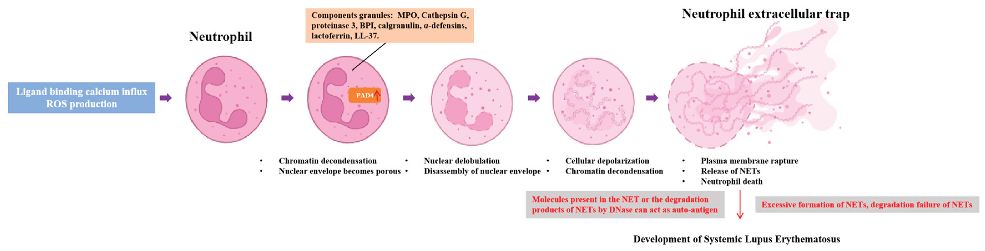
2. Formation Mechanisms of NETs

2.1. Definition and Structure of NETs

NETs are specialized extracellular effector structures generated by activated neutrophils, defined as interconnected networks of DNA fibers decorated with a diverse array of granule-derived proteins and nuclear components, evolved to entrap and eliminate invading microorganisms (e.g., bacteria, fungi, viruses) as a key component of innate host defense [21]. First identified by Brinkmann et al. [10] in 2004, NET formation primarily occurred via a lytic programmed cell death pathway. However, non-lytic release—preserving neutrophil membrane integrity and cellular viability—has also been observed under specific stimulatory conditions [22].
The structural core of NETs consists of both genomic DNA (derived from the neutrophil nucleus) and mitochondrial DNA (mtDNA) [23]. Notably, mtDNA within NETs exhibits enhanced immunostimulatory activity when oxidized during NET extrusion, a feature linked to amplified inflammatory responses in autoimmune contexts [24].
Embedded within this DNA scaffold are critical granule proteins that reinforce NET functionality, including MPO, neutrophil elastase, lysozyme, and matrix metalloproteinases (MMPs) [25]. These proteins not only contribute to microbial killing through enzymatic activity but also modulate tissue microenvironments by remodeling extracellular matrices [26].
Nuclear components, particularly histones, are another defining structural element of NETs, and their post-translational modifications—mediated by enzymes such as peptidylarginine deiminase 4 (PAD4)—play pivotal roles in shaping NET architecture and immunogenicity. PAD4 catalyzes the citrullination of arginine residues on histones, disrupting electrostatic interactions between histones and DNA to promote chromatin decondensation, a prerequisite for NET formation [27]. Additionally, ROS-dependent carbamylation of histones further alters their structure, increasing their pathogenic potential by enhancing autoantigenicity and tissue-damaging properties [28].
Collectively, the modular structure of NETs—integrating a flexible DNA backbone, functionally active granule proteins, and modified nuclear components—enables their dual role in host protection and, when dysregulated, in driving chronic inflammation and tissue injury [29]. This structural complexity also underpins their involvement in systemic autoimmune and autoinflammatory diseases, where NET-derived components (e.g., citrullinated histones, oxidized mtDNA) act as autoantigens to perpetuate aberrant immune responses [30].

2.2. Formation Process of NETs

2.2.1. ROS: Central Regulator of Chromatin Decondensation and NET Extrusion

ROS serve as a central regulator of NET formation, with their production (via NADPH oxidase and mitochondria) and downstream effects tightly coupled to chromatin decondensation, protease activation, and extracellular trap extrusion [31]. As documented in the literature, ROS are primarily generated through two key pathways during NET induction: the membrane-bound NADPH oxidase complex (which drives the "respiratory burst") and mitochondrial ROS release, both of which are essential for initiating and sustaining NET formation [32]. Upon stimulation by stimuli such as microorganisms, ICs, or autoantibodies, ROS first inhibit actin polymerization—this disruption of the neutrophil cytoskeleton removes the physical barrier that restricts nuclear material movement, laying the foundation for subsequent chromatin extrusion [33]. Additionally, ROS mediate the intracellular release of critical proteases (including neutrophil elastase and MPO) from cytoplasmic granules; these proteases then degrade histones and structural proteins like gasdermin D (GSDMD), reducing membrane stability and elevating cytoplasmic calcium (Ca²⁺) concentrations to further amplify chromatin decondensation signals [34]. Notably, ROS also contributes to post-translational modifications of NET components. For instance, ROS-driven oxidation of genomic and mtDNA within NETs not only impairs nuclease-mediated degradation of these nucleic acids but also enhances their recognition by innate immune sensors,such as the cGAS-STING pathway, linking ROS to both NET formation and subsequent immunostimulatory effects [35].

2.2.2. NADPH Oxidase: Rate-Limiting Step in ROS Production and NETosis

As the primary source of ROS, NADPH oxidase activation represents a rate-limiting step in NET formation [36]. This membrane-bound enzymatic complex is triggered by the engagement of diverse surface receptors on neutrophils, including cytokine receptors, Fcγ receptors (FcγRs), complement receptors (e.g., C5aR), and damage-associated molecular pattern (DAMP) receptors [37]. Ligand binding to these receptors converges on a standard downstream signal—elevated intracellular Ca²⁺ levels—which then activates protein kinase C (PKC); PKC subsequently mediates the assembly of the NADPH oxidase complex at the cell membrane. Once assembled, the complex catalyzes the production of superoxide free radicals (the primary ROS in this pathway), a process known as the "respiratory burst" [38]. This burst of ROS not only activates downstream proteases (as noted earlier) but also promotes the fusion of neutrophil granules with the nucleus, a key event that delivers granule proteins (e.g., MPO, elastase) to chromatin [39]. Importantly, NADPH oxidase activity is not confined to the plasma membrane; mitochondrial ROS production, which can be synergistically activated by NADPH oxidase-derived ROS, further reinforces NET formation by sustaining oxidative stress and amplifying chromatin decondensation [40]. Disruption of NADPH oxidase function—whether via genetic mutations or pharmacological inhibition—has been shown to abrogate NET formation in both murine models and human neutrophils, confirming its non-redundant role in this process [41].

2.2.3. MPO and Neutrophil Elastase: Synergistic Drivers of Chromatin Remodeling

MPO and neutrophil elastase, two abundant granule proteins in neutrophils, play complementary and indispensable roles in NET formation by modulating chromatin structure and degrading key cellular components [42]. Neutrophil elastase, a serine protease, is among the first granule proteins activated by ROS (via NADPH oxidase signaling); once released into the cytoplasm, it translocates to the nucleus, where it degrades core histones (e.g., H3, H4) and nuclear structural proteins—this degradation disrupts the compact organization of chromatin, initiating the decondensation process essential for NET extrusion [43]. Elastase also targets GSDMD, a pore-forming protein at the nuclear and plasma membranes; cleavage of GSDMD by elastase forms membrane pores, increasing membrane permeability, elevating cytoplasmic Ca²⁺ levels, and further facilitating the release of decondensed chromatin [44]. MPO, another granule-derived enzyme, contributes to NET formation through both enzymatic and oxidative mechanisms: it catalyzes the production of hypochlorous acid (HOCl) from hydrogen peroxide (a byproduct of NADPH oxidase activity), and this highly reactive oxidant enhances the degradation of histones and structural proteins by elastase, acting as a "co-factor" to amplify elastase-mediated chromatin remodeling [45]. Beyond its enzymatic role, MPO also participates in post-translational modifications of NET components—it mediates the carbamylation of histones (via ROS-dependent reactions), which disrupts histone-DNA interactions to promote chromatin decondensation and increases histone immunogenicity, making them more likely to act as autoantigens in autoimmune diseases[46]. Together, MPO and elastase synergistically promote efficient NET formation: MPO-derived oxidants prime chromatin by enhancing elastase’s ability to degrade histones; in turn, elastase cleaves nuclear structural proteins to disrupt membrane integrity, enabling MPO and other granule proteins to bind the NET DNA scaffold. This synergistic interaction ensures efficient NET formation and modulates NET pathogenicity [47]. This collaboration not only facilitates the mechanical release of DNA but also shapes the composition and pathogenic potential of NETs, linking their activity to both microbial clearance and, in dysregulated states, to tissue damage and autoimmunity [48].

2.3. Physiological Functions of NETs

NETs exert critical physiological functions tightly linked to maintaining host homeostasis, with their role in host defense among the most well-characterized and evolutionarily conserved [49]. Structurally composed of extracellular DNA fibers adorned with granule-derived proteins—including MPO, neutrophil elastase, and lysozyme—NETs act as a physical and enzymatic barrier to ensnare a broad spectrum of invading microorganisms, encompassing bacteria, fungi, and viruses [50]. Upon encountering microbial pathogens or their products (e.g., lipopolysaccharides, viral proteins), activated neutrophils deploy NETs to trap these invaders within the DNA mesh, preventing their dissemination to distant tissues; this physical sequestration is complemented by the enzymatic activity of granule proteins embedded in the NET scaffold, which directly degrade microbial cell walls, proteins, and nucleic acids to eliminate the pathogen [51]. For instance, MPO catalyzes the production of reactive oxidants that disrupt microbial membrane integrity, while neutrophil elastase cleaves key microbial virulence factors, synergistically enhancing killing efficacy [52]. Notably, NET formation can occur via both lytic and non-lytic pathways: the latter preserves neutrophil viability, allowing the same cell to contribute to sustained host defense through repeated NET release or phagocytosis, highlighting the adaptability of this mechanism across different infectious contexts [53]. This dual capacity for physical trapping and enzymatic clearance positions NETs as a pivotal component of the innate immune response, bridging the gap between immediate pathogen containment and subsequent adaptive immune activation [54].
Beyond their role in direct pathogen elimination, NETs also function as central modulators of inflammatory responses, orchestrating the activation and recruitment of other immune cells to fine-tune the body’s response to threats [55]. A key mechanism underlying this modulation is the ability of NET components to engage innate immune sensors on cells such as macrophages and plasmacytoid dendritic cells (pDCs): for example, NET-derived double-stranded DNA (both genomic and oxidized mtDNA) is recognized by the cGAS-STING pathway in macrophages, triggering the production of type I interferons (IFNs)—a family of cytokines that amplify antiviral immunity and prime adaptive immune responses [56]. Additionally, NETs are enriched with immunostimulatory molecules, including interleukin-33 (IL-33), high-mobility group box 1 (HMGB1), and the antimicrobial peptide cathelicidin LL-37; these molecules bind to receptors on immune cells (e.g., Toll-like receptors on DCs) to promote their maturation, enhance antigen presentation, and stimulate the secretion of pro-inflammatory cytokines [57]. NETs also contribute to inflammatory amplification by activating the complement system—via binding of complement component 1q (C1q) to NET components—and by recruiting additional neutrophils and monocytes to the site of injury or infection, creating a positive feedback loop that reinforces the inflammatory response [58]. Importantly, NETs are not solely pro-inflammatory: under specific conditions, aggregated NETs can exert anti-inflammatory effects by forming stable meshes that degrade pro-inflammatory cytokines and chemokines via embedded proteases, thereby limiting excessive immune cell infiltration and promoting the resolution of inflammation. This essential balance prevents tissue damage while ensuring adequate pathogen clearance [59].

3. Role of NETs in SLE

3.1. Immunopathological Mechanisms of SLE

In SLE, the production of autoantibodies arises from a multifaceted breakdown of immune tolerance, driven by intrinsic B cell dysregulation and extrinsic microenvironmental cues [60]. Beyond the well-characterized antinuclear antibodies (ANA) and anti-double-stranded DNA (anti-dsDNA) antibodies—where ANA serves as a sensitive yet nonspecific diagnostic marker and anti-dsDNA correlates with lupus nephritis activity—SLE patients also generate autoantibodies targeting ribonucleoprotein complexes, such as anti-Smith (anti-Sm), anti-Ro antibody/anti-Sjögren's Syndrome A antibody (anti-Ro/SSA), and anti-La antibody/anti-Sjögren's Syndrome B antibody (anti-La/SSB) [61]. Anti-Sm antibodies exhibit over 90% specificity for SLE, making them a definitive diagnostic tool, while anti-Ro/SSA antibodies are associated with cutaneous manifestations and neonatal lupus [62]. The cellular mechanisms underlying this autoantibody production are tightly linked to B-cell tolerance defects: genetic variants, including loss-of-function mutations in protein kinase C delta (PRKCD) and gain-of-function mutations in toll-like receptor 7 (TLR7), enable the survival and proliferation of autoreactive B cells [63]. Notably, escape from X-chromosome inactivation of TLR7 in some female patients further amplifies B-cell hyperactivation [64]. Additionally, aberrant extrafollicular B cell responses play a pivotal role: CD11c⁺T-bet⁺ age-associated B cells (ABCs), a proinflammatory B cell subset, are significantly expanded in SLE peripheral blood [65]. These ABCs differentiate into plasmablasts under the stimulation of interleukin-21 (IL-21) secreted by T peripheral helper cells (Tph), contributing to sustained autoantibody production [66]. NETs further fuel this process by acting as a reservoir of modified autoantigens—such as citrullinated histones and oxidized mtDNA—which not only directly engage autoreactive B cells but also activate pDCs to secrete type I IFNs, creating a feedforward loop that reinforces B cell activation [67]. Defects in nucleic acid clearance, including reduced activity of Deoxyribonuclease 1-like 3 (DNase1L3), prolong the persistence of extracellular DNA, providing a continuous source of autoantigens for antibody generation [68].
ICs, formed by the binding of autoantibodies to self-antigens, are central pathogenic mediators in SLE, driving tissue damage through complement activation, inflammatory cell recruitment, and direct tissue deposition [69]. The composition of these ICs is heterogeneous: they may include anti-dsDNA antibodies bound to NET-derived genomic DNA or nucleosomes, anti-Ro/SSA antibodies complexed with cytoplasmic RNA, or autoantibody-coated microparticles released from apoptotic cells—these microparticle-associated ICs exhibit enhanced tissue adhesion and complement-activating capacity [70]. A critical pathogenic step is impaired clearance of ICs: SLE patients often show deficiencies in early complement components (e.g., C1q, C4), which are essential for opsonizing ICs and facilitating their phagocytosis by macrophages [71]. Moreover, autoantibodies targeting DNase1 or DNase1L3 inhibit the degradation of DNA within ICs, preventing their dissolution and promoting their accumulation in the circulation [72]. Once formed and retained, ICs exert their pathogenic effects through two primary pathways: first, they activate the classical complement cascade, generating anaphylatoxins that recruit neutrophils and monocytes to sites of inflammation and enhance their proinflammatory activity; second, they engage FcγRs on innate immune cells—pDCs internalize ICs and trigger type I IFN production via TLR7/9 sensing of nucleic acids, while macrophages release tumor necrosis factor-α (TNF-α) and interleukin-1β (IL-1β) upon IC binding, amplifying local inflammation [73]. In target organs such as the kidney, ICs deposit in the glomerular basement membrane and mesangium, disrupting filtration function and inducing podocyte injury; in the skin, IC deposition in the dermal-epidermal junction contributes to erythematous lesions [74]. This cycle of IC formation, impaired clearance, and inflammatory activation underpins the chronic tissue damage observed in SLE [75].
The pathogenesis of SLE is further perpetuated by the activation of multiple inflammatory cell types, including neutrophils, macrophages, and DCs, which release proinflammatory cytokines and mediate tissue destruction [76]. Neutrophils exhibit distinct functional abnormalities in SLE: LDGs, a proinflammatory subset, are expanded in peripheral blood and exhibit an increased propensity for NETosis [77]. These NETs are enriched in oxidized mtDNA, which activates the cGAS-STING pathway in macrophages and DCs, driving type I IFN production. Additionally, SLE neutrophils are prone to ferroptosis—an iron-dependent form of cell death characterized by lipid peroxidation—releasing DAMPs, such as HMGB1 and heat shock protein 60 (HSP60), which further activate innate immune responses [78]. Macrophages undergo M1 polarization in SLE, a phenotype associated with enhanced secretion of TNF-α and IL-1β. M1 macrophages accumulate in inflamed tissues (e.g., renal interstitium, cutaneous lesions) and contribute to tissue injury by phagocytosing ICs and releasing MMPs that degrade the extracellular matrix. DCs, particularly pDCs and conventional dendritic cells (cDCs), play specialized roles: pDCs are the primary source of type I IFNs in SLE, activated by IC-derived nucleic acids via TLR7/9, and their secreted IFNs reinforce B cell activation and T cell differentiation; cDCs, upon uptake of autoantigens (e.g., nucleosomes), present these antigens to CD4⁺ T cells, promoting their differentiation into T follicular helper cells (Tfh) or Th17 cells [79]. Tfh cells secrete IL-21 to support B cell plasmablast differentiation, while Th17 cells produce interleukin-17 (IL-17) to exacerbate synovial inflammation and renal injury [80]. Importantly, the cytokines released by these activated cells (e.g., type I IFNs, TNF-α) create a proinflammatory microenvironment that further activates neighboring immune cells, establishing a positive feedback loop that sustains chronic inflammation and multi-organ damage in SLE [81].

3.2. Abnormalities of NETs in SLE: Overproduction, Impaired Clearance, and Autoantigen Function

3.2.1. Excessive NET Formation: LDG-Driven Pathogenesis

SLE patients exhibit expanded proinflammatory LDGs, which undergo spontaneous NETosis due to mitochondrial ROS overproduction and PAD4-mediated histone citrullination. SLE-derived ICs and autoantibodies further trigger NET formation via FcγRIIA/TLR engagement, even in healthy neutrophils exposed to SLE serum [82]. This heightened NETosis is not uniform across all neutrophil subsets: LDGs, a proinflammatory subset expanded in SLE peripheral blood, display a markedly increased propensity for spontaneous NET formation compared to normal-density neutrophils (HDNs) from both SLE patients and healthy controls [83]. LDGs in SLE are driven toward NETosis by multiple pathogenic cues, including mitochondrial reactive oxygen species (mtROS) overproduction [84]. Oxidized mtDNA released during this process not only constitutes a key component of NETs but also enhances their immunostimulatory capacity by activating the cGAS-STING pathway in myeloid cells [85]. Additionally, SLE-derived ICs (e.g., anti-dsDNA/dsDNA, anti-β2-glycoprotein I (anti-β2-GPI), and autoantibodies (e.g., anti-RNP, anti-LL37) directly trigger NET formation via engagement of FcγRIIA or Toll-like receptors on neutrophils [86]. Notably, even neutrophils from healthy individuals, when exposed to SLE serum or purified SLE autoantibodies, exhibit increased NET production, indicating that soluble factors in the SLE microenvironment prime neutrophils for NETosis [87]. The enzyme PAD4, which mediates histone citrullination and chromatin decondensation, also plays a pivotal role in SLE-associated excessive NETosis: PAD4 inhibition in murine lupus models reduces NET formation, autoantibody levels, and renal injury, though conflicting data exist regarding its protective vs. pathogenic role in specific contexts [88]. Collectively, this dysregulated NET production, driven by abnormal neutrophil subsets, proinflammatory stimuli, and altered intracellular signaling, creates a sustained pool of extracellular autoantigens that fuels SLE pathogenesis [89].

3.2.2. Impaired NET Clearance: DNase and Complement Defects

Compounding the issue of excessive NET formation, SLE patients exhibit profound impairment in NET clearance, a defect that perpetuates NET accumulation in tissues and the circulation and exacerbates autoimmune pathology [90]. Clearance of NETs is compromised by: (1) autoantibodies targeting DNase1/DNase1L3; (2) genetic mutations in DNase1L3; and (3) C1q-mediated inhibition of DNase1 activity. Reduced DNase activity correlates with higher circulating NET remnants (MPO-DNA complexes) and disease activity, particularly in lupus nephritis [91,92].
The primary mediators of NET degradation are DNase enzymes, particularly DNase1 and DNase1L3, whose activity is frequently compromised in SLE [91]. This impairment arises through three key mechanisms: (1) autoantibodies targeting DNase1 or DNase1L3 (e.g., anti-DNase1L3 IgG) in ~30–40% of SLE patients, which directly neutralize nuclease activity [93]; (2) genetic variants or loss-of-function mutations in DNase1/DNase1L3 (e.g., homozygous DNase1L3 null mutations) linked to early-onset SLE [94]; and (3) complement C1q binding to NET DNA, which inhibits DNase1 activity [92]. Clinically, impaired NET clearance correlates with disease activity: Lupus nephritis patients exhibit lower DNase1L3 activity compared to SLE patients without renal involvement, and reduced DNase activity is associated with higher levels of circulating NET remnants (e.g., MPO-DNA complexes), anti-dsDNA autoantibodies, and hypocomplementemia [95]. This failure to clear NETs not only prolongs exposure to autoantigens but also promotes the formation of stable NET-autoantibody complexes that resist degradation [96].

3.2.3. NETs as Autoantigen Reservoirs: NET–IC Formation

NETs serve as a critical "autoantigen reservoir" in SLE, as their components—including genomic DNA, oxidized mtDNA, citrullinated histones (e.g., citrullinated histone H3, CitH3), and granule proteins (e.g., MPO; neutrophil elastase)—directly interact with autoantibodies to form pathogenic ICs [97]. These NET-derived ICs exhibit unique immunostimulatory properties: for example, complexes of NET DNA (oxidized mtDNA or genomic DNA) with anti-dsDNA antibodies or the antimicrobial peptide LL37 activate pDCs via TLR9, triggering robust type I IFN secretion, triggering robust production of type I IFNs—a hallmark of SLE pathogenesis [98]. Similarly, ICs composed of citrullinated histones and anti-CitH3 antibodies engage B cells via B cell receptors (BCRs) and TLRs, driving B cell differentiation into autoantibody-secreting plasma cells and amplifying the autoimmune response [99]. NET-ICs also activate neutrophils and monocytes via FcγRIIA, inducing further NETosis and secretion of proinflammatory cytokines, creating a self-perpetuating "NET-IC-inflammatory loop" [100]. Significantly, these ICs deposit in target organs, such as the kidney: in lupus nephritis, NET-ICs accumulate in glomeruli, where they activate the complement system (via the classical pathway) and recruit inflammatory cells, leading to podocyte injury, mesangial proliferation, and renal fibrosis [101]. Additionally, NET components, such as NE and MPO, within these ICs degrade extracellular matrix proteins, exacerbating tissue damage [102]. Together, the interactions between NETs and autoantibodies are central to SLE progression, linking innate immune dysregulation to adaptive autoimmunity and end-organ injury [103].
In summary, the abnormality of NETs in SLE can be summarized as a 'triple disorder':
(1) Excessive NET Formation: Increased LDGs and spontaneous enhancement of NETosis, with further promotion of NET release by ICs in SLE serum; (2) Impaired NET Clearance: Decreased DNase activity (gene mutation/autoantibody inhibition) leads to accumulation of NETs; (3) NETs as Autoantigen Reservoirs: NET components form NET-ICs with autoantibodies, activating pDCs, B cells, and amplifying autoimmune responses.

3.3. Pathological Roles of NETs in SLE

NETs play a pivotal role as a central pathological mediator in SLE by orchestrating multifaceted proinflammatory effects that perpetuate autoimmune activation and tissue damage [104]. A key mechanism by which NETs amplify inflammatory responses is their ability to act as a "danger signal reservoir," activating a spectrum of innate and adaptive immune cells. For instance, NET-derived components—including oxidized mtDNA, CitH3, and HMGB1—engage pattern recognition receptors (PRRs) on pDCs, such as TLR9 and cGAS-STING [105]. This activation triggers robust secretion of type I IFNs, a hallmark of SLE pathogenesis that further primes neutrophils for NETosis and drives B cell differentiation into autoantibody-secreting plasma cells [106]. Macrophages, upon uptake of NET fragments via FcγRIIA, undergo M1 polarization and release proinflammatory cytokines, including TNF-α and IL-1β, which recruit additional neutrophils and monocytes to inflamed sites [107]. Moreover, NETs provide a scaffold for the formation of ICs with autoantibodies, which further activate neutrophils and B cells via FcγR and BCR cross-linking, creating a self-perpetuating inflammatory loop [108]. Notably, LDGs—an aberrant neutrophil subset expanded in SLE—synergize with NETs to enhance inflammation: LDGs not only exhibit increased spontaneous NET formation but also secrete higher levels of IL-8. This chemokine amplifies neutrophil recruitment and activates endothelial cells, further exacerbating the proinflammatory microenvironment [109].
Beyond amplifying inflammation, NETs directly mediate tissue damage in SLE through their cytotoxic components and by modulating cellular homeostasis in target organs [110]. The cationic histones (e.g., H3, H4) within NETs exert direct cytotoxicity toward vascular endothelial cells by disrupting membrane integrity, leading to increased permeability and endothelial dysfunction [111]. NET-associated enzymes exacerbate this: NE degrades extracellular matrix proteins (e.g., collagen, fibronectin), while MPO generates HOCl to oxidize endothelial proteins, thereby impairing barrier function [112]. In the kidney—a significant target of SLE—NETs deposit in glomeruli, where NE and MPO degrade podocyte proteins (e.g., nephrin), disrupting the glomerular filtration barrier and contributing to proteinuria [113]. Additionally, NET-derived MMP-9 activates endothelial MMP-2, promoting endothelial-to-mesenchymal transition (EndMT) of glomerular endothelial cells—a process linked to renal fibrosis and progressive lupus nephritis [114]. In the lungs, NETs disrupt the microvascular endothelial barrier, facilitating diffuse alveolar hemorrhage (DAH) in severe SLE; here, NE and cathepsin G cleave lung epithelial cell junctions, while NET-induced IL-8 recruitment of neutrophils amplifies tissue destruction [115]. Collectively, these effects underscore NETs as direct effectors of tissue injury across multiple organ systems in SLE.
NETs also contribute to SLE-associated thrombotic complications—a leading cause of mortality in SLE patients—by promoting platelet activation, activating the coagulation cascade, and stabilizing thrombi [116]. NETs bridge inflammation and thrombosis via three key mechanisms. First, platelet activation occurs as histones bind to platelet TLR2 and TLR4, while neutrophil elastase and cathepsin G activate platelet protease-activated receptor 4 (PAR4), inducing granule release and integrin αIIbβ3 activation [116,117]. The second coagulation cascade activation occurs because NETs express tissue factor, the initiator of the extrinsic pathway, and neutrophil elastase and cathepsin G degrade anticoagulants such as tissue factor pathway inhibitor (TFPI) and antithrombin III, shifting the balance toward a prothrombotic state [117]. Third thrombus stabilization occurs as the NET DNA scaffold concentrates platelet fibrinogen and von Willebrand factor, enhancing platelet aggregation and fibrin deposition [118]. Clinically elevated levels of CitH3 or MPO-DNA complexes correlate with increased risk of cardiovascular disease and pulmonary embolism in SLE [116]. Thus, NETs bridge inflammation and thrombosis in SLE, representing a critical link between autoimmune activation and organ damage.

4. Role of Oxidative Stress in NET Formation and SLE

4.1. Definition and Mechanisms of Oxidative Stress

Oxidative stress is a critical pathophysiological condition in SLE, in which the balance between reactive species production and the antioxidant defense system's capacity is disrupted, leading to an overproduction of reactive species [119]. At the core of oxidative stress are ROS and reactive nitrogen species (RNS), highly reactive molecules that can damage cellular components [120]. Under normal physiological conditions, ROS and RNS play essential roles in various cellular signaling pathways and regulatory processes [121]. However, in SLE, their production often exceeds the capacity of antioxidant defense mechanisms, leading to chronic oxidative stress [122].
The primary sources of ROS include the mitochondrial electron transport chain, cytochrome P450 enzymes, NADPH oxidases, and xanthine oxidase [123]. In eukaryotic cells, over 90% of ROS are generated by mitochondria, where electrons escaping from the electron transport chain interact with molecular oxygen to form superoxide anion radicals (•O2-) [124]. These radicals can further be converted into other ROS, such as hydrogen peroxide (H2O2) and hydroxyl radicals (•OH) [125]. RNS, on the other hand, are mainly derived from nitric oxide (NO) and its derivatives, such as peroxynitrite (ONOO) [126]. While these reactive species have critical physiological functions at low concentrations, their excessive accumulation in SLE can lead to significant oxidative damage [127].
Oxidative stress exerts detrimental effects on various cellular components, including lipids, proteins, and DNA [128]. Lipid peroxidation, a common consequence of oxidative stress, involves the oxidation of polyunsaturated fatty acids in cell membranes, leading to the formation of toxic aldehydes like malondialdehyde (MDA) and 4-hydroxynonenal (4-HNE) [129]. These aldehydes can disrupt membrane integrity and fluidity, impairing cellular functions [130]. Protein oxidation results in carbonylation, modification of amino acid residues, and protein cross-linking, which can alter protein structure and function, leading to enzyme inactivation and impaired cellular processes [131]. DNA damage caused by oxidative stress includes base modifications, strand breaks, and cross-linking, which can interfere with DNA replication and transcription, potentially leading to mutations and genomic instability [132]. Moreover, oxidative stress can activate various intracellular signaling pathways, such as the mitogen-activated protein kinase (MAPK) and nuclear factor-kappa B (NF-κB) pathways, further exacerbating cellular inflammation and apoptosis in SLE [133].
In summary, oxidative stress, characterized by an imbalance between ROS and RNS production and the antioxidant defense capacity, can cause extensive damage to cellular components and contribute to the pathogenesis of SLE [134]. Understanding the mechanisms underlying oxidative stress and its regulation in the context of SLE is crucial for elucidating the disease's etiology and developing effective therapeutic strategies [135].

4.2. SLE-Specific Oxidative Stress Amplification

In SLE, exacerbation of oxidative stress is a critical factor that significantly contributes to NET formation. Three key mechanisms primarily drive this process.
Firstly, LDGs exhibit a marked increase in mitochondrial ROS production. This heightened ROS generation drives spontaneous NETosis, leading to the release of oxidized mtDNA. This mtDNA, a potent immunostimulatory component of NETs, activates the cGAS-STING pathway in myeloid cells, further fueling inflammation and immune responses [84,88].
Secondly, SLE patients often suffer from impaired antioxidant defenses. Reduced serum levels of antioxidants, such as vitamin E and β-carotene, coupled with lower enzymatic antioxidant activities, lead to increased ROS accumulation. This imbalance further exacerbates oxidative stress and promotes NET formation [136].
Thirdly, a self-sustaining feedback loop between NETs and ICs further amplifies oxidative stress. Pathogenic NET–ICs activate neutrophils via FcγRIIA, which in turn boosts NADPH oxidase activity and ROS production. This creates a vicious cycle where oxidative stress and NET formation continuously reinforce each other, leading to chronic inflammation and tissue damage in SLE [108].
Oxidative stress plays a pivotal role in NET formation. The activation of the NADPH oxidase complex is a crucial step in ROS production, which, in turn, leads to the release of DNA and histones, culminating in NET formation [137,139]. Phorbol 12-myristate 13-acetate (PMA) has been shown to activate the NADPH oxidase complex in neutrophils, leading to ROS production and NET formation [138].
ROS are central to NETosis, while antioxidants(SOD, catalase, GSH, NAC) inhibit NETs by neutralizing ROS [140,144]. Fuchs et al. [141] reported that antioxidants like thiourea, dimethylthiourea, and N-acetyl-L-cysteine significantly attenuate PMA-induced NET formation. Studies also revealed mitochondrial ROS in Ca²⁺ ionophore-induced NET formation and demonstrated that mitochondrial ROS scavengers can inhibit NET formation [104,143].
Moreover, Gsr (glutathione reductase) is also found to be critical for neutrophil antioxidant capacity — Gsr-deficient neutrophils exhibit impaired NET formation under oxidative stress [136,142].
These findings underscore the significance of oxidative stress in NET formation and provide a foundation for developing therapeutic strategies targeting NET-related diseases.
Figure 1. The formation mechanism of NETs in SLE.
Preprints 184979 g001

5. Potential Applications of Antioxidants in NETs and SLE

5.1. Classification and Mechanisms of Action of Antioxidants

Antioxidants are a diverse group of compounds that play a crucial role in mitigating oxidative damage and maintaining cellular redox homeostasis [145]. They can be broadly categorized into three classes: enzymatic antioxidants, non-enzymatic antioxidants, and natural antioxidants, each with distinct mechanisms of action [146].
Enzymatic antioxidants, such as SOD, catalase, and glutathione peroxidase (GPx), are integral to the body's defense against ROS [147]. SOD catalyzes the dismutation of superoxide radicals into oxygen and hydrogen peroxide, thereby reducing their cytotoxic effects [148]. Catalase further decomposes hydrogen peroxide into water and oxygen, while GPx utilizes reduced GSH to reduce hydrogen peroxide and organic peroxides to water and corresponding alcohols, respectively [149]. These enzymes work synergistically to neutralize ROS and protect cells from oxidative damage [148,149].
Non-enzymatic antioxidants include vitamins C and E, and GSH [150]. Vitamin C, a water-soluble antioxidant, donates electrons to neutralize free radicals, such as the hydroxyl radical and the superoxide anion, thereby preventing oxidative damage to cellular components [151]. Vitamin E, a lipid-soluble antioxidant, is particularly effective in intercepting peroxyl radicals in cell membranes and lipoproteins, thereby interrupting the propagation of lipid peroxidation [152]. GSH, a tripeptide thiol, maintains the cellular redox balance by cycling between its reduced and oxidized forms and serves as a substrate for GPx in the reduction of peroxides [153].
Natural plant- and other-source antioxidants encompass a wide array of compounds with potent antioxidant properties. Resveratrol, a polyphenol found in grapes and red wine, exhibits antioxidant, anti-inflammatory, and anticancer activities by scavenging free radicals, inhibiting oxidative enzymes, and modulating intracellular signaling pathways[154]. Curcumin, another polyphenol from turmeric, possesses potent antioxidant properties and can suppress inflammation and apoptosis through multiple mechanisms [155]. Green tea polyphenols, primarily composed of catechins, exert antioxidant effects by directly quenching free radicals, inhibiting oxidative enzyme activities, and modulating the cellular antioxidant defense system [156].
In summary, antioxidants, through their diverse mechanisms of action, collectively neutralize ROS and RNS, thereby safeguarding cells from oxidative damage [157]. Enzymatic antioxidants catalyze the decomposition of ROS, non-enzymatic antioxidants directly scavenge free radicals, and natural antioxidants modulate cellular redox balance through various pathways [158]. The synergistic action of these antioxidants is essential for maintaining cellular integrity and preventing diseases associated with oxidative stress.

5.2. Effects of Antioxidants on NET Formation

NETs are a crucial component of the innate immune response, enabling neutrophils to capture and neutralize pathogens [159]. However, excessive or inappropriate NET formation has been implicated in various inflammatory and autoimmune diseases, including SLE, making NETs a potential therapeutic target [160]. Antioxidants have emerged as a promising strategy to modulate NET formation via three pathways: direct ROS scavenging, NADPH oxidase inhibition, and MPO/elastase inhibition.
Direct scavenging of ROS is one of the primary mechanisms by which antioxidants inhibit NET formation. ROS, particularly HOCl produced by MPO, play a pivotal role in triggering NETosis [161]. Studies have shown that thiocyanate (SCN-) and selenocyanate (SeCN-) can effectively neutralize HOCl, thereby reducing NET release in neutrophils stimulated with PMA or bacterial peptides such as nigericin [162]. Differentiated PLB-985 cells and primary human neutrophils treated with SCN- exhibited decreased NET release upon PMA stimulation, highlighting the efficacy of direct ROS scavenging in modulating NET formation [162].
Inhibition of NADPH oxidase is another mechanism by which antioxidants can suppress NET formation [163]. NADPH oxidase is essential for the production of ROS, which are necessary for NET release [164]. The NADPH oxidase inhibitor diphenylene iodonium (DPI) has been shown to reduce NET formation effectively in multiple studies. Hydrogen gas (H2) has been shown to inhibit PMA-induced NET formation in human neutrophils by neutralizing HOCl, a potent oxidant generated by NADPH oxidase, thereby suppressing DNA damage and NET release [164]. This suggests that targeting NADPH oxidase activity is a viable strategy for controlling NET formation.
Antioxidants can also inhibit the activity of MPO and elastase, enzymes involved in NET formation [165]. MPO-derived ROS, such as HOCl, contribute to the release of DNA and histones, key components of NETs [166]. Inhibiting MPO can reduce the formation of these ROS and, consequently, decrease NET release [167]. SeCN-, for example, has been shown to inhibit MPO activity, thereby reducing NET formation in neutrophils [162]. This highlights the importance of targeting MPO in modulating NET formation.
In conclusion, antioxidants offer a multifaceted approach to inhibit NET formation by directly scavenging ROS, inhibiting NADPH oxidase activity, and targeting MPO and elastase [162,163,164,167]. These strategies have shown promise in reducing NET formation in vitro and in vivo, suggesting that antioxidants could be a valuable therapeutic tool for managing NET-related diseases. Future research should focus on optimizing antioxidant formulations and exploring their efficacy in clinical settings to harness their potential in treating inflammatory and autoimmune conditions.

5.3. Potential Applications of Antioxidants in SLE Therapy

Preclinical Studies. SLE is a complex autoimmune disease characterized by widespread inflammation, organ involvement, and the production of autoantibodies[168]. Oxidative stress, resulting from an imbalance between ROS production and the body’s antioxidant defenses, has been implicated in the pathogenesis of SLE [169]. This imbalance is believed to contribute to NET formation, which is associated with increased inflammation and tissue damage in SLE patients [170]. Accumulating evidence from both in vitro and animal model studies has demonstrated that antioxidants can significantly reduce NET formation and alleviate the pathological symptoms of SLE.
Curcumin, a natural polyphenolic compound with anti-inflammatory and antioxidant properties, has been shown to significantly reduce proteinuria and renal inflammation in MRL/lpr mice, a commonly used SLE model [171]. This effect is attributed to its ability to inhibit the activation of the NLR family pyrin domain-containing 3 (NLRP3) inflammasome, a key player in the pathogenesis of lupus nephritis [172]. Curcumin has effectively alleviated lupus nephritis in both MRL/lpr and R848-treated mouse models by inhibiting neutrophil migration and inflammatory factor release, thereby regulating the PI3K/AKT/NF-κB signaling pathways [173].
Recent evidence suggests that resveratrol ameliorates immune disorders by inhibiting the overactivation of immune cells, making it a promising therapeutic option for SLE. Wang et al.[174] demonstrated that resveratrol could suppress the proliferation of B cells and induce apoptosis in CD4+ T cells, thereby reducing the production of autoantibodies and alleviating lupus-like symptoms. Additionally, resveratrol upregulated FcγRIIB expression in B cells via NF-κB activation, leading to reduced B cell numbers, decreased serum autoantibody levels, and improved lupus nephritis in MRL/lpr mice [175]. Similarly, the combination of resveratrol and piperine has been reported to mitigate renal, hepatic, and pulmonary manifestations in a pristane-induced SLE mouse model. Research by Pannu et al. [176] proved that resveratrol combined with piperine (especially RP-1 and Res-2) mitigated renal manifestations, limited NET-mediated tissue damage, alleviated oxidative stress, reduced inflammatory cytokines and lipogranuloma formation, but failed to abrogate autoantibody production or improve spleen/skin manifestations in a pristane-induced SLE murine model. They also demonstrated that resveratrol alone and in combination with piperine effectively mitigated oxidative stress and inflammation in a pristane-induced lupus-like murine model, reducing renal pathologies and ICs. However, neither treatment regulated autoantibody formation, and combining resveratrol with piperine did not enhance its efficacy compared with resveratrol alone [177].
Vitamins C and E, owing to their antioxidant properties, are advantageous in the treatment of SLE. A recent study showed that vitamin C contributed to treating SLE by acting as a hypoxia-inducible factor-1α (HIF-1α) inhibitor, which down-regulated the expression of the stress-response protein (regulated in development and DNA damage response 1, REDD1), suppressed autophagy induction in neutrophils, reduced the release of NETs decorated with TF and IL-17A, and thereby mitigated thromboinflammation and fibrosis associated with end-organ injury in SLE [178]. Mohammed et al. [179] found that vitamin C inhibits NETosis in polymorphonuclear neutrophils from healthy mice. In a hydralazine-induced lupus mouse model, vitamin E at a higher dose (50 mg/kg) significantly reduced lymphocyte hydrogen peroxide radicals compared to a lower dose (25 mg/kg), indicating its dose-dependent scavenging potential and suggesting it may be a promising therapeutic agent for SLE [180]. Another study demonstrated that low-dose vitamin E supplementation (250 mg/kg) extended lifespan, whereas high-dose supplementation (500 mg/kg) shortened it. High doses increased anti-dsDNA and anticardiolipin antibodies and suppressed IL-2 while boosting IL-4 and IL-10, suggesting that high vitamin E intake may exacerbate Th2-driven autoimmune diseases, such as lupus, in MRL/lpr mice [181].
Other antioxidants, such as coenzyme Q10 and N-acetylcysteine (NAC), have also been shown to inhibit NET formation by scavenging ROS and reducing oxidative stress. Blanco et al. [182] demonstrated that coenzyme Q10 analog Idebenone not only downregulated NET formation in neutrophils and inhibited neutrophil extracellular trap formation in lupus-prone mice, but also enhanced mitochondrial metabolism and ATP production, improved endothelium-dependent vasorelaxation, and reduced lipid peroxidation, indicating its potential as a therapeutic agent for SLE. In a parallel study, Fortner et al. [183] explored the therapeutic potential of the mitochondrial-targeted coenzyme Q10 (MitoQ) in lupus disease manifestations using MRL/lpr mice. In this study, lupus-prone MRL/lpr mice were treated with MitoQ (200 µM) for eleven weeks. After treatment, the mice exhibited reduced NETosis and ROS production, decreased serum IFN levels, and diminished immune complex formation in the kidneys. NAC has been demonstrated to decrease the expression of pro-inflammatory cytokines and chemokines, thereby mitigating the inflammatory response in SLE. In addition, in vitro experiments have indicated that NAC effectively reduced NET formation and oxidative stress in neutrophils by enhancing antioxidant capacity and decreasing ROS and lipid peroxidation levels [184].
Clinical Trials. Clinical trials evaluating the effects of antioxidants in SLE treatment have yielded promising results. Preliminary small-scale trials (n=24–70) have shown that curcumin (1,000–1,500 mg/day) improves select clinical and laboratory outcomes in SLE patients—including reduced proteinuria, anti-dsDNA levels, and IL-6 concentrations—with no severe adverse events. For example, a randomized, placebo-controlled study investigated the effects of oral curcumin supplementation in 24 patients with relapsing or refractory biopsy-proven lupus nephritis; results showed that the trial group had significant reductions in proteinuria), systolic blood pressure, and hematuria, with no adverse effects related to turmeric, leading to the conclusion that short-term turmeric supplementation can be a safe adjuvant therapy for patients with relapsing or refractory lupus nephritis [185]. In another randomized, triple-blinded, placebo-controlled trial, curcumin supplementation for 10 weeks significantly reduced anti-dsDNA and IL-6 levels in SLE patients, demonstrating its potential as an effective and safe adjuvant therapy to ameliorate autoimmune activity and inflammation [186]. However, a double-blind randomized controlled trial conducted in Saiful Anwar Hospital demonstrated that supplementing with curcuma (20 mg/day) in addition to vitamin D3 for three months did not significantly affect SLE Disease Activity Index (SLEDAI) scores, IL-6 levels, or transforming growth factor-β1 (TGF-β1) levels in SLE patients with hypovitamin D compared to vitamin D3 alone [187].
Several ongoing clinical trials are further investigating the potential benefits of NAC in SLE. For instance, a randomized, double-blind clinical trial involving SLE patients treated with NAC at a dose of 1800 mg/day for 3 months revealed a significant reduction in disease activity scores, as measured by SLEDAI and the British Isles Lupus Assessment Group (BILAG) criteria[188]. Another study by Li et al. [189] reported two cases of early-stage lupus nephritis treated with NAC. The patients received 1200 mg/day of NAC for 3 months, resulting in significant improvements in GSH levels, reductions in lipid peroxidation biomarkers, and overall clinical improvement. These findings suggest that NAC may be effective in modulating oxidative stress and improving disease outcomes in early-stage lupus nephritis. In addition to its antioxidant properties, NAC has been shown to block the mammalian target of rapamycin (mTOR) signaling pathway in T cells, which is implicated in SLE pathogenesis. Lai et al. [190] conducted a randomized, double-blind, placebo-controlled trial involving 36 SLE patients, demonstrating that NAC significantly reduced disease activity and fatigue by inhibiting mTOR. The study also revealed that NAC increased mitochondrial membrane potential and enhanced apoptosis in T cells, contributing to the overall therapeutic effect. Furthermore, comprehensive metabolome analyses by Perl et al. [191] identified significant changes in the metabolome of SLE patients, particularly in the pentose phosphate pathway. NAC treatment reduced kynurenine accumulation, a metabolite that activates mTOR, providing a metabolic basis for its therapeutic efficacy in SLE. In the context of neuropsychiatric manifestations, Garcia et al. [192] reported that NAC significantly improved attention deficit and hyperactivity disorder (ADHD) symptoms in SLE patients. The study used the ADHD Self-Report Scale (ASRS) to assess cognitive and hyperactivity symptoms and found that NAC treatment led to significant reductions in ASRS scores, suggesting its potential to address cognitive dysfunction in SLE. Finally, Doherty et al. [198] investigated the effects of NAC on mitochondrial function in SLE patients. The study found that NAC reduced oxygen consumption through mitochondrial electron transport chain (ETC) complex I, thereby decreasing oxidative stress. These findings further support NAC's role in modulating mitochondrial dysfunction and oxidative stress in SLE. In conclusion, the cumulative evidence from these studies indicates that NAC is a promising therapeutic agent for SLE, offering multiple benefits through its antioxidant properties, modulation of mitochondrial function, and inhibition of the mTOR signaling pathway.
In addition to these trials, several studies have also investigated the potential synergistic effects of vitamins C and E with conventional SLE therapies. Comstock et al. [193] conducted a prospective case-control study to investigate the serum concentrations of vitamin E, β-carotene, and retinol preceding the diagnosis of rheumatoid arthritis and SLE. The study found that individuals who later developed SLE had lower serum concentrations of these antioxidants than matched controls, suggesting that low antioxidant status may be a risk factor for SLE. This finding was supported by Bae et al. [194], who compared plasma antioxidant/oxidant status and dietary nutrient intake in SLE patients and healthy controls. They found that SLE patients had significantly lower plasma activities of SOD and GPx, higher levels of MDA, and lower dietary intake of vitamin A, β-carotene, and vitamin C. These results indicated that SLE patients have impaired antioxidant status and reduced dietary intake of antioxidants.
Maeshima et al.[195] conducted a preliminary study to evaluate the efficacy of vitamin E in reducing oxidative DNA damage and autoantibody production in SLE patients. The study measured urinary 8-hydroxydeoxyguanosine (8-OHdG) as an indicator of oxidative DNA damage and anti-dsDNA antibody levels as a predictor of disease activity. While urinary 8-OHdG levels did not differ significantly between patients receiving vitamin E and those who did not, anti-ds DNA antibody titers were significantly lower in the vitamin E group. This suggests that vitamin E may suppress autoantibody production in SLE, potentially through mechanisms independent of its antioxidant activity.
In conclusion, both preclinical and clinical studies have highlighted the potential therapeutic benefits of antioxidants in SLE. Further research is warranted to elucidate the precise mechanisms by which antioxidants exert their effects and to determine the optimal dosing and combinations for clinical use. Future studies should also focus on long-term outcomes and the potential for antioxidants to modify disease progression in SLE. Additionally, research should explore the synergistic effects of antioxidants with other therapeutic agents to develop more effective and safer treatment strategies for SLE patients.
Table 1. Characteristics of 21 Studies on Applications of Antioxidants in SLE Therapy.
Antioxidants Category Model Dosage Results Reference
Curcumin preclinical study female MRL/lpr mice 200 mg/kg/day Curcumin effectively reduces proteinuria, renal inflammation, serum anti-dsDNA levels, and spleen size, and inhibits NLRP3 inflammasome activation both in vivo and in vitro. Zhao et al.
,2019 [172]
preclinical study MRL/lpr mice and R848-treated mice 50 mg/kg/day Curcumin effectively reduces renal inflammation in lupus mouse models by inhibiting neutrophil migration and the release of inflammatory factors via the PI3K/AKT/NF-κB signaling pathway. Yang et al.,2024
[173]
Resveratrol preclinical study pristane-induced lupus mice 50 mg/kg/day;
75 mg/kg/day
Resveratrol significantly mitigates proteinuria, kidney immunoglobulin deposition, and glomerulonephritis in pristane-induced lupus mice, while also suppressing CD4+ T cell activation and proliferation, inducing CD4+ T cell apoptosis, and inhibiting B cell antibody production and proliferation in vitro. Wang et al.,2014
[174]
preclinical study MRL/lpr mice 20 mg /kg/day Resveratrol alleviates lupus symptoms in MRL/lpr mice by enhancing FcγRIIB expression in B cells via Sirt1 activation, reducing plasma cells and autoantibodies, and improving nephritis and survival. Jhou et al.,2017
[175]
preclinical study pristane-induced lupus mice 25 mg/kg/day;
50 mg/kg/day
In a pristane-induced SLE murine model, low-dose resveratrol combined with piperine and high-dose resveratrol reduced renal immunoglobulin deposition, hepatic lipogranuloma formation, and pulmonary inflammation, reduced oxidative stress, and improved lupus symptoms, but did not affect autoantibody formation or spleen/skin manifestations. Pannu et al.,2020
[176]
preclinical study pristane-induced lupus mice 25 mg/kg/day Resveratrol alone and in combination with piperine effectively mitigated oxidative stress and inflammation, improved renal function, and reduced histopathological manifestations in a pristane-induced lupus murine model. Still, neither treatment regulated autoantibody formation, and piperine did not enhance resveratrol's efficacy. Pannu et al.,2020
[177]
Vitamins C preclinical study peripheral blood neutrophils isolated from patients with active SLE 10 mM/day Vitamin C inhibits NETosis in SLE neutrophils by targeting the REDD1/autophagy/NET axis, reducing thromboinflammation and fibrosis. Frangou et al.,2018
[178]
preclinical study Gulo-/- mice 200 mg/kg/day Vitamin C reduces NETosis in sepsis by attenuating ER stress, autophagy, histone citrullination, and NFκB activation. Mohammed et al.,2013
[179]
Vitamins E preclinical study hydralazine-induced lupus mice 25 mg/kg/day;
50 mg/kg/day
Vitamin E, particularly at a higher dose (50 mg/kg), shows potential in reducing lymphocyte hydrogen peroxide radicals in a hydralazine-induced lupus mouse model. Githaiga et al.,2023 [180]
preclinical study MRL/lymphoproliferative lpr female mice 50 mg/kg/day; 250 mg/kg/day; 375 mg/kg/day; 500 mg/kg/day Low-dose vitamin E extends lifespan in MRL/lpr mice, whereas high-dose vitamin E increases Th2 cytokines and autoantibodies, potentially worsening Th2-driven autoimmune diseases like SLE. Hsieh et al.,2005 181]
Coenzyme Q10 preclinical study MRL/lpr mice 1 mg/kg; 1.5 mg/kg Coenzyme Q10 significantly reduces mortality, attenuates disease features, and improves mitochondrial function, renal function, and inflammation in lupus mouse models, supporting its potential therapeutic role in SLE. Blanco et al.,2020 [182]
preclinical study MRL/lpr mice MitoQ (200 µM) in drinking water/day MitoQ reduces neutrophil ROS and NET formation, MAVS oligomerisation, and serum IFN-I in lupus-prone MRL-lpr mice, highlighting the potential of targeting mROS as an adjunct therapy for lupus. Fortner et al.,2020 [183]
Curcumin clinical trial 24 patients with relapsing or refractory biopsy-proven lupus nephritis 1,500 mg/day Short-term curcumin supplementation significantly reduced proteinuria, hematuria, and systolic blood pressure in patients with relapsing or refractory lupus nephritis. Khajehdehi et al.,2012 [185]
clinical trial 70 SLE patients 1,000 mg/day Curcumin supplementation significantly reduced anti-dsDNA and IL-6 levels in SLE patients, with no significant changes in other variables. Sedighi et al.,2024 [186]
clinical trial SLE active (SLEDAI > 3) with levels of 25(OH)D3 ≤ 30 ng/ml SLE patients 1,200 IU/day Curcumin combined with vitamin D3 showed no significant effects on SLEDAI and serum levels of IL-6 and TGF-β1 in SLE patients with low vitamin D levels. However, decreased IL-6 levels correlated positively with reductions in SLEDAI. Singgih et al.,2017 [187]
N-acetylcysteine clinical trial 80 SLE patients 1,800 mg/day NAC (1800 mg/day) significantly reduced SLE disease activity and complications, as evidenced by lower BILAG and SLEDAI scores and improved CH50 levels after 3 months, with no adverse events reported. Abbasifard et al.,2023 [188]
clinical trial female SLE patients 1,200 mg/day NAC treatment in early-stage lupus nephritis led to increased glutathione levels, decreased lipid peroxidation biomarker levels, and significant improvements in routine blood counts, 24-h urine protein, erythrocyte sedimentation rate, and SLE disease activity index. Li et al.,2015 [189]
clinical trial 36 SLE patients 1.2 mg/day; 2.4 mg/day; 4.8 mg/day NAC at 2.4 and 4.8 mg/day significantly reduced SLE activity scores and fatigue levels, while improving mitochondrial function, reducing mTOR activity, enhancing apoptosis, and decreasing anti-dsDNA antibody production in SLE patients. Lai et al.,2012 [190]
clinical trial 49 SLE patients and 46 healthy control subjects 2.4 mg/day; 4.8 mg/day NAC treatment at dosages of 2.4 and 4.8 mg/day significantly reduced ADHD symptoms in SLE patients, as indicated by lower ASRS total and part A scores, demonstrating its efficacy in improving cognitive and inattentive aspects of ADHD in this patient group. Garcia et al.,2013 [192]
clinical trial 69 SLE patients and 37 healthy donors 3 mg/day NAC treatment effectively reduced oxygen consumption via mitochondrial ETC complex I and hydrogen peroxide levels in peripheral blood lymphocytes from SLE patients, indicating its potential therapeutic efficacy in reducing oxidative stress. Doherty et al.,2014 [196]
Vitamins E clinical trial 12 SLE patients 150-300 mg/day Vitamin E can suppress autoantibody production in SLE patients, as indicated by lower anti-dsDNA antibody titers, independent of its antioxidant activity. Maeshima et al.,2007 [195]

6. Conclusions

NETs are significant pathogenic factors in SLE, and their abnormal formation, accumulation, and oxidative stress are closely linked. Under normal physiological conditions, NETs help capture pathogens. In SLE, a pro-inflammatory subset of LDGs spontaneously forms NETs, releasing autoantigens such as oxidized mtDNA and citrullinated histones. These autoantigens can then form ICs with antibodies, such as anti-dsDNA.
These NET-ICs not only stimulate pDCs to secrete type I interferons and granular proteins (e.g., MPO) and neutrophil elastases but also lead to the deposition of interferons in organs such as the kidneys and skin, causing tissue damage. Patients with SLE exhibit impaired NET clearance due to reduced DNase activity and the presence of autoantibodies, thereby perpetuating autoimmune activation.
Oxidative stress (NADPH oxidase/mitochondrial ROS) drives NET chromatin decondensation and cellular damage. Antioxidants (enzymatic/non-enzymatic/natural) inhibit NETs by scavenging ROS or blocking NADPH oxidase/MPO. Preclinical (curcumin, resveratrol, MitoQ) and clinical (curcumin, NAC) data confirm they reduce SLE activity and proteinuria; low-dose vitamin E also shows promise.
Future research should: (1) define NET subtype heterogeneity in SLE, (2) improve antioxidant bioavailability/targeting, (3) develop combination therapies (NETs + oxidative stress + type I IFNs). Addressing formulation challenges and long-term safety will enable antioxidants to become effective SLE adjuvants.

Author Contributions

Conceptualization, Y.T., C.W., and H.H.; writing—original draft preparation, D.G. and X.C.; writing—review and editing, M.W. and L.W.; visualization, D.G. and X.C. All authors have read and agreed to the published version of the manuscript.”.

Funding

This study was supported by the National Natural Science Foundation of China (No.82305145) and the Zhejiang Provincial Natural Science Foundation of China (No. LQ23H270006).

Conflicts of Interest

The authors declare no conflicts of interest.

Abbreviations

The following abbreviations are used in this manuscript:
•O2− superoxide anion radicals
•OH hydroxyl radicals
4-HNE 4-hydroxynonenal
8-OHdG 8-hydroxydeoxyguanosine
ABCs age-associated B cells
ADHD attention deficit hyperactivity disorder
ANA antinuclear antibodies
anti-dsDNA anti-double-stranded DNA
anti-Sm anti-Smith
anti-β2-GPI anti-β2-glycoprotein I
ASRS ADHD Self-Report Scale
BCRs B-cell receptors
BILAG British Isles Lupus Assessment Group
C1q complement component 1q
Ca²⁺ calcium
cDCs conventional dendritic cells
CitH3 citrullinated histone H3
DAH diffuse alveolar hemorrhage
DAMP damage-associated molecular pattern
DCs dendritic cells
DNase1L3 Deoxyribonuclease 1-like 3
DPI diphenylene iodonium
EndMT endothelial-to-mesenchymal transition
FcγRs Fcγ receptors
GPx glutathione peroxidase
GSDMD gasdermin D
GSH glutathione
Gsr glutathione reductase
H2 hydrogen gas
H2O2 hydrogen peroxide
HDNs normal-density neutrophils
HIF-1α hypoxia-inducible factor-1α
HMGB1 high-mobility group box 1
HOCl hypochlorous acid
ICs immune complexes
IFNs interferons
IL-17 interleukin-17
IL-1β interleukin-1β
IL-21 interleukin-21
IL-33 interleukin-33
LDGs low-density granulocytes
MAPK mitogen-activated protein kinase
MDA malondialdehyde
MitoQ mitochondrial-targeted coenzyme Q10
MMPs matrix metalloproteinases
MPO myeloperoxidase
mtDNA mitochondrial DNA
mTOR mammalian target of rapamycin
mtROS mitochondrial reactive oxygen species
NAC N-acetylcysteine
NADPH nicotinamide adenine dinucleotide phosphate
NETs neutrophil extracellular traps
NF-κB nuclear factor-kappa B
NLRP3 NLR family pyrin domain containing 3
NO nitric oxide
ONOO− peroxynitrite
PAD4 peptidylarginine deiminase 4
PAR4 protease-activated receptor 4
pDCs plasmacytoid dendritic cells
PKC protein kinase C
PMA phorbol 12-myristate 13-acetate
PRKCD protein kinase C delta
PRRs pattern recognition receptors
REDD1 regulated in development and DNA damage response 1
RNS reactive nitrogen species
ROS reactive oxygen species
SCN- thiocyanate
SeCN- selenocyanate
SLE systemic lupus erythematosus
SLEDAI SLE Disease Activity Index
SOD superoxide dismutase
Tfh T follicular helper cells
TFPI tissue factor pathway inhibitor
TGF-β1 transforming growth factor-β1
TLR7 toll-like receptor 7
TLR9 toll-like receptor 9
TNF-α tumor necrosis factor-α
Tph T peripheral helper cells

References

  1. Shiozawa, S. Pathogenesis of Autoimmunity/Systemic Lupus Erythematosus (SLE). Cells 2025, 14. [Google Scholar] [CrossRef]
  2. Rekvig, O.P. SLE classification criteria: Science-based icons or algorithmic distractions - an intellectually demanding dilemma. Front Immunol 2022, 13, 1011591. [Google Scholar] [CrossRef]
  3. Cui, Y.; Sheng, Y.; Zhang, X. Genetic susceptibility to SLE: recent progress from GWAS. J Autoimmun 2013, 41, 25–33. [Google Scholar] [CrossRef] [PubMed]
  4. Robl, R.; Eudy, A.; Bachali, P.S.; Rogers, J.L.; Clowse, M.; Pisetsky, D.; Lipsky, P. Molecular endotypes of type 1 and type 2 SLE. Lupus Sci Med 2023, 10. [Google Scholar] [CrossRef]
  5. Yaniv, G.; Twig, G.; Shor, D.B.; Furer, A.; Sherer, Y.; Mozes, O.; Komisar, O.; Slonimsky, E.; Klang, E.; Lotan, E.; et al. A volcanic explosion of autoantibodies in systemic lupus erythematosus: a diversity of 180 different antibodies found in SLE patients. Autoimmun Rev 2015, 14, 75–79. [Google Scholar] [CrossRef]
  6. Love, P.E.; Santoro, S.A. Antiphospholipid antibodies: anticardiolipin and the lupus anticoagulant in systemic lupus erythematosus (SLE) and in non-SLE disorders. Prevalence and clinical significance. Ann Intern Med 1990, 112, 682–698. [Google Scholar] [CrossRef]
  7. Fan, Y.; Hao, Y.J.; Zhang, Z.L. Systemic lupus erythematosus: year in review 2019. Chin Med J (Engl) 2020, 133, 2189–2196. [Google Scholar] [CrossRef] [PubMed]
  8. Liew, P.X.; Kubes, P. The Neutrophil's Role During Health and Disease. Physiol Rev 2019, 99, 1223–1248. [Google Scholar] [CrossRef] [PubMed]
  9. Shao, S.; Xue, K.; Wang, G. Neutrophils in neutrophilic dermatoses: Emerging roles and promising targeted therapies. J Allergy Clin Immunol 2022, 149, 1203–1205. [Google Scholar] [CrossRef]
  10. Brinkmann, V.; Reichard, U.; Goosmann, C.; Fauler, B.; Uhlemann, Y.; Weiss, D.S.; Weinrauch, Y.; Zychlinsky, A. Neutrophil extracellular traps kill bacteria. Science 2004, 303, 1532–1535. [Google Scholar] [CrossRef]
  11. Wan, Y.; Shen, J.; Ouyang, J.; Dong, P.; Hong, Y.; Liang, L.; Liu, J. Bibliometric and visual analysis of neutrophil extracellular traps from 2004 to 2022. Front Immunol 2022, 13, 1025861. [Google Scholar] [CrossRef]
  12. Papayannopoulos, V. Neutrophil extracellular traps in immunity and disease. Nat Rev Immunol 2018, 18, 134–147. [Google Scholar] [CrossRef]
  13. Chu, C.; Wang, X.; Yang, C.; Chen, F.; Shi, L.; Xu, W.; Wang, K.; Liu, B.; Wang, C.; Sun, D.; et al. Neutrophil extracellular traps drive intestinal microvascular endothelial ferroptosis by impairing Fundc1-dependent mitophagy. Redox Biol 2023, 67, 102906. [Google Scholar] [CrossRef]
  14. Lee, K.H.; Kronbichler, A.; Park, D.D.; Park, Y.; Moon, H.; Kim, H.; Choi, J.H.; Choi, Y.; Shim, S.; Lyu, I.S.; et al. Neutrophil extracellular traps (NETs) in autoimmune diseases: A comprehensive review. Autoimmun Rev 2017, 16, 1160–1173. [Google Scholar] [CrossRef]
  15. Jo, A.; Kim, D.W. Neutrophil Extracellular Traps in Airway Diseases: Pathological Roles and Therapeutic Implications. Int J Mol Sci 2023, 24. [Google Scholar] [CrossRef] [PubMed]
  16. Fousert, E.; Toes, R.; Desai, J. Neutrophil Extracellular Traps (NETs) Take the Central Stage in Driving Autoimmune Responses. Cells 2020, 9. [Google Scholar] [CrossRef]
  17. Islam, M.M.; Takeyama, N. Role of Neutrophil Extracellular Traps in Health and Disease Pathophysiology: Recent Insights and Advances. Int J Mol Sci 2023, 24. [Google Scholar] [CrossRef]
  18. Tsokos, G.C.; Lo, M.S.; Costa Reis, P.; Sullivan, K.E. New insights into the immunopathogenesis of systemic lupus erythematosus. Nat Rev Rheumatol 2016, 12, 716–730. [Google Scholar] [CrossRef]
  19. Zhang, H.; Liu, J.; Zhou, Y.; Qu, M.; Wang, Y.; Guo, K.; Shen, R.; Sun, Z.; Cata, J.P.; Yang, S.; et al. Neutrophil extracellular traps mediate m(6)A modification and regulates sepsis-associated acute lung injury by activating ferroptosis in alveolar epithelial cells. Int J Biol Sci 2022, 18, 3337–3357. [Google Scholar] [CrossRef] [PubMed]
  20. Herre, M.; Cedervall, J.; Mackman, N.; Olsson, A.K. Neutrophil extracellular traps in the pathology of cancer and other inflammatory diseases. Physiol Rev 2023, 103, 277–312. [Google Scholar] [CrossRef] [PubMed]
  21. Xiang, J.; Cui, M.; Zhang, Y. Neutrophil extracellular traps and neutrophilic asthma. Respir Med 2025, 245, 108150. [Google Scholar] [CrossRef]
  22. Yang, D.; Liu, J. Neutrophil Extracellular Traps: A New Player in Cancer Metastasis and Therapeutic Target. J Exp Clin Cancer Res 2021, 40, 233. [Google Scholar] [CrossRef]
  23. Hidalgo, A.; Libby, P.; Soehnlein, O.; Aramburu, I.V.; Papayannopoulos, V.; Silvestre-Roig, C. Neutrophil extracellular traps: from physiology to pathology. Cardiovasc Res 2022, 118, 2737–2753. [Google Scholar] [CrossRef]
  24. Wang, Y.; Du, C.; Zhang, Y.; Zhu, L. Composition and Function of Neutrophil Extracellular Traps. Biomolecules 2024, 14. [Google Scholar] [CrossRef]
  25. Zhu, Z.; Zhou, S.; Li, S.; Gong, S.; Zhang, Q. Neutrophil extracellular traps in wound healing. Trends Pharmacol Sci 2024, 45, 1033–1045. [Google Scholar] [CrossRef]
  26. Li, X.; Xiao, S.; Filipczak, N.; Yalamarty, S.S.K.; Shang, H.; Zhang, J.; Zheng, Q. Role and Therapeutic Targeting Strategies of Neutrophil Extracellular Traps in Inflammation. Int J Nanomedicine 2023, 18, 5265–5287. [Google Scholar] [CrossRef]
  27. Mutua, V.; Gershwin, L.J. A Review of Neutrophil Extracellular Traps (NETs) in Disease: Potential Anti-NETs Therapeutics. Clin Rev Allergy Immunol 2021, 61, 194–211. [Google Scholar] [CrossRef] [PubMed]
  28. Kang, L.; Yu, H.; Yang, X.; Zhu, Y.; Bai, X.; Wang, R.; Cao, Y.; Xu, H.; Luo, H.; Lu, L.; et al. Neutrophil extracellular traps released by neutrophils impair revascularization and vascular remodeling after stroke. Nat Commun 2020, 11, 2488. [Google Scholar] [CrossRef] [PubMed]
  29. Yang, K.; Gao, R.; Chen, H.; Hu, J.; Zhang, P.; Wei, X.; Shi, J.; Chen, Y.; Zhang, L.; Chen, J.; et al. Myocardial reperfusion injury exacerbation due to ALDH2 deficiency is mediated by neutrophil extracellular traps and prevented by leukotriene C4 inhibition. Eur Heart J 2024, 45, 1662–1680. [Google Scholar] [CrossRef] [PubMed]
  30. Long, D.; Mao, C.; Xu, Y.; Zhu, Y. The emerging role of neutrophil extracellular traps in ulcerative colitis. Front Immunol 2024, 15, 1425251. [Google Scholar] [CrossRef]
  31. Azzouz, D.; Palaniyar, N. How Do ROS Induce NETosis? Oxidative DNA Damage, DNA Repair, and Chromatin Decondensation. Biomolecules 2024, 14. [Google Scholar] [CrossRef]
  32. Dan Dunn, J.; Alvarez, L.A.; Zhang, X.; Soldati, T. Reactive oxygen species and mitochondria: A nexus of cellular homeostasis. Redox Biol 2015, 6, 472–485. [Google Scholar] [CrossRef]
  33. Liu, Y.; Wang, R.; Song, C.; Ding, S.; Zuo, Y.; Yi, K.; Li, N.; Wang, B.; Geng, Q. Crosstalk between neutrophil extracellular traps and immune regulation: insights into pathobiology and therapeutic implications of transfusion-related acute lung injury. Front Immunol 2023, 14, 1324021. [Google Scholar] [CrossRef] [PubMed]
  34. Schönrich, G.; Raftery, M.J.; Samstag, Y. Devilishly radical NETwork in COVID-19: Oxidative stress, neutrophil extracellular traps (NETs), and T cell suppression. Adv Biol Regul 2020, 77, 100741. [Google Scholar] [CrossRef] [PubMed]
  35. Xia, L.; Yan, X.; Zhang, H. Mitochondrial DNA-activated cGAS-STING pathway in cancer: Mechanisms and therapeutic implications. Biochim Biophys Acta Rev Cancer 2025, 1880, 189249. [Google Scholar] [CrossRef] [PubMed]
  36. Huang, J.; Hong, W.; Wan, M.; Zheng, L. Molecular mechanisms and therapeutic target of NETosis in diseases. MedComm (2020) 2022, 3, e162. [Google Scholar] [CrossRef]
  37. Vorobjeva, N.V.; Chernyak, B.V. NETosis: Molecular Mechanisms, Role in Physiology and Pathology. Biochemistry (Mosc) 2020, 85, 1178–1190. [Google Scholar] [CrossRef]
  38. O'Brien, X.M.; Reichner, J.S. Neutrophil Integrins and Matrix Ligands and NET Release. Front Immunol 2016, 7, 363. [Google Scholar] [CrossRef]
  39. Miralda, I.; Uriarte, S.M.; McLeish, K.R. Multiple Phenotypic Changes Define Neutrophil Priming. Front Cell Infect Microbiol 2017, 7, 217. [Google Scholar] [CrossRef]
  40. Zeng, M.Y.; Miralda, I.; Armstrong, C.L.; Uriarte, S.M.; Bagaitkar, J. The roles of NADPH oxidase in modulating neutrophil effector responses. Mol Oral Microbiol 2019, 34, 27–38. [Google Scholar] [CrossRef]
  41. Nguyen, G.T.; Green, E.R.; Mecsas, J. Neutrophils to the ROScue: Mechanisms of NADPH Oxidase Activation and Bacterial Resistance. Front Cell Infect Microbiol 2017, 7, 373. [Google Scholar] [CrossRef]
  42. Masuda, S.; Nakazawa, D.; Shida, H.; Miyoshi, A.; Kusunoki, Y.; Tomaru, U.; Ishizu, A. NETosis markers: Quest for specific, objective, and quantitative markers. Clin Chim Acta 2016, 459, 89–93. [Google Scholar] [CrossRef] [PubMed]
  43. Demkow, U. Molecular Mechanisms of Neutrophil Extracellular Trap (NETs) Degradation. Int J Mol Sci 2023, 24. [Google Scholar] [CrossRef] [PubMed]
  44. Castaño, M.; Tomás-Pérez, S.; González-Cantó, E.; Aghababyan, C.; Mascarós-Martínez, A.; Santonja, N.; Herreros-Pomares, A.; Oto, J.; Medina, P.; Götte, M.; et al. Neutrophil Extracellular Traps and Cancer: Trapping Our Attention with Their Involvement in Ovarian Cancer. Int J Mol Sci 2023, 24. [Google Scholar] [CrossRef] [PubMed]
  45. Wang, H.; Kim, S.J.; Lei, Y.; Wang, S.; Wang, H.; Huang, H.; Zhang, H.; Tsung, A. Neutrophil extracellular traps in homeostasis and disease. Signal Transduct Target Ther 2024, 9, 235. [Google Scholar] [CrossRef]
  46. Döring, Y.; Soehnlein, O.; Weber, C. Neutrophil Extracellular Traps in Atherosclerosis and Atherothrombosis. Circ Res 2017, 120, 736–743. [Google Scholar] [CrossRef]
  47. Frade-Sosa, B.; Sanmartí, R. Neutrophils, neutrophil extracellular traps, and rheumatoid arthritis: An updated review for clinicians. Reumatol Clin (Engl Ed) 2023, 19, 515–526. [Google Scholar] [CrossRef]
  48. Fang, Q.; Stehr, A.M.; Naschberger, E.; Knopf, J.; Herrmann, M.; Stürzl, M. No NETs no TIME: Crosstalk between neutrophil extracellular traps and the tumor immune microenvironment. Front Immunol 2022, 13, 1075260. [Google Scholar] [CrossRef]
  49. Fukuchi, M.; Miyabe, Y.; Furutani, C.; Saga, T.; Moritoki, Y.; Yamada, T.; Weller, P.F.; Ueki, S. How to detect eosinophil ETosis (EETosis) and extracellular traps. Allergol Int 2021, 70, 19–29. [Google Scholar] [CrossRef]
  50. Delgado-Rizo, V.; Martínez-Guzmán, M.A.; Iñiguez-Gutierrez, L.; García-Orozco, A.; Alvarado-Navarro, A.; Fafutis-Morris, M. Neutrophil Extracellular Traps and Its Implications in Inflammation: An Overview. Front Immunol 2017, 8, 81. [Google Scholar] [CrossRef]
  51. Borregaard, N. Neutrophils, from marrow to microbes. Immunity 2010, 33, 657–670. [Google Scholar] [CrossRef]
  52. Honda, M.; Kubes, P. Neutrophils and neutrophil extracellular traps in the liver and gastrointestinal system. Nat Rev Gastroenterol Hepatol 2018, 15, 206–221. [Google Scholar] [CrossRef]
  53. Nakazawa, D.; Masuda, S.; Nishibata, Y.; Watanabe-Kusunoki, K.; Tomaru, U.; Ishizu, A. Neutrophils and NETs in kidney disease. Nat Rev Nephrol 2025, 21, 383–398. [Google Scholar] [CrossRef] [PubMed]
  54. Manda, A.; Pruchniak, M.P.; Araźna, M.; Demkow, U.A. Neutrophil extracellular traps in physiology and pathology. Cent Eur J Immunol 2014, 39, 116–121. [Google Scholar] [CrossRef]
  55. Granger, V.; Peyneau, M.; Chollet-Martin, S.; de Chaisemartin, L. Neutrophil Extracellular Traps in Autoimmunity and Allergy: Immune Complexes at Work. Front Immunol 2019, 10, 2824. [Google Scholar] [CrossRef]
  56. Poto, R.; Shamji, M.; Marone, G.; Durham, S.R.; Scadding, G.W.; Varricchi, G. Neutrophil Extracellular Traps in Asthma: Friends or Foes? Cells 2022, 11. [Google Scholar] [CrossRef]
  57. Nagaoka, I.; Tamura, H.; Reich, J. Therapeutic Potential of Cathelicidin Peptide LL-37, an Antimicrobial Agent, in a Murine Sepsis Model. Int J Mol Sci 2020, 21. [Google Scholar] [CrossRef] [PubMed]
  58. Hahn, J.; Knopf, J.; Maueröder, C.; Kienhöfer, D.; Leppkes, M.; Herrmann, M. Neutrophils and neutrophil extracellular traps orchestrate initiation and resolution of inflammation. Clin Exp Rheumatol 2016, 34, 6–8. [Google Scholar] [PubMed]
  59. Palomino-Segura, M.; Sicilia, J.; Ballesteros, I.; Hidalgo, A. Strategies of neutrophil diversification. Nat Immunol 2023, 24, 575–584. [Google Scholar] [CrossRef]
  60. Kiriakidou, M.; Ching, C.L. Systemic Lupus Erythematosus. Ann Intern Med 2020, 172, Itc81–itc96. [Google Scholar] [CrossRef]
  61. Yu, H.; Nagafuchi, Y.; Fujio, K. Clinical and Immunological Biomarkers for Systemic Lupus Erythematosus. Biomolecules 2021, 11. [Google Scholar] [CrossRef]
  62. Zucchi, D.; Elefante, E.; Schilirò, D.; Signorini, V.; Trentin, F.; Bortoluzzi, A.; Tani, C. One year in review 2022: systemic lupus erythematosus. Clin Exp Rheumatol 2022, 40, 4–14. [Google Scholar] [CrossRef]
  63. Piga, M.; Tselios, K.; Viveiros, L.; Chessa, E.; Neves, A.; Urowitz, M.B.; Isenberg, D. Clinical patterns of disease: From early systemic lupus erythematosus to late-onset disease. Best Pract Res Clin Rheumatol 2023, 37, 101938. [Google Scholar] [CrossRef]
  64. Fillatreau, S.; Manfroi, B.; Dörner, T. Toll-like receptor signalling in B cells during systemic lupus erythematosus. Nat Rev Rheumatol 2021, 17, 98–108. [Google Scholar] [CrossRef] [PubMed]
  65. Wen, L.; Zhang, B.; Wu, X.; Liu, R.; Fan, H.; Han, L.; Zhang, Z.; Ma, X.; Chu, C.Q.; Shi, X. Toll-like receptors 7 and 9 regulate the proliferation and differentiation of B cells in systemic lupus erythematosus. Front Immunol 2023, 14, 1093208. [Google Scholar] [CrossRef] [PubMed]
  66. Durcan, L.; O'Dwyer, T.; Petri, M. Management strategies and future directions for systemic lupus erythematosus in adults. Lancet 2019, 393, 2332–2343. [Google Scholar] [CrossRef]
  67. Dai, X.; Fan, Y.; Zhao, X. Systemic lupus erythematosus: updated insights on the pathogenesis, diagnosis, prevention and therapeutics. Signal Transduct Target Ther 2025, 10, 102. [Google Scholar] [CrossRef] [PubMed]
  68. Gergianaki, I.; Bortoluzzi, A.; Bertsias, G. Update on the epidemiology, risk factors, and disease outcomes of systemic lupus erythematosus. Best Pract Res Clin Rheumatol 2018, 32, 188–205. [Google Scholar] [CrossRef]
  69. Accapezzato, D.; Caccavale, R.; Paroli, M.P.; Gioia, C.; Nguyen, B.L.; Spadea, L.; Paroli, M. Advances in the Pathogenesis and Treatment of Systemic Lupus Erythematosus. Int J Mol Sci 2023, 24. [Google Scholar] [CrossRef]
  70. Pan, L.; Lu, M.P.; Wang, J.H.; Xu, M.; Yang, S.R. Immunological pathogenesis and treatment of systemic lupus erythematosus. World J Pediatr 2020, 16, 19–30. [Google Scholar] [CrossRef]
  71. Tusseau, M.; Khaldi-Plassart, S.; Cognard, J.; Viel, S.; Khoryati, L.; Benezech, S.; Mathieu, A.L.; Rieux-Laucat, F.; Bader-Meunier, B.; Belot, A. Mendelian Causes of Autoimmunity: the Lupus Phenotype. J Clin Immunol 2024, 44, 99. [Google Scholar] [CrossRef] [PubMed]
  72. Kuhn, A.; Wenzel, J.; Bijl, M. Lupus erythematosus revisited. Semin Immunopathol 2016, 38, 97–112. [Google Scholar] [CrossRef]
  73. Scherlinger, M.; Guillotin, V.; Truchetet, M.E.; Contin-Bordes, C.; Sisirak, V.; Duffau, P.; Lazaro, E.; Richez, C.; Blanco, P. Systemic lupus erythematosus and systemic sclerosis: All roads lead to platelets. Autoimmun Rev 2018, 17, 625–635. [Google Scholar] [CrossRef]
  74. Brilland, B.; Scherlinger, M.; Khoryati, L.; Goret, J.; Duffau, P.; Lazaro, E.; Charrier, M.; Guillotin, V.; Richez, C.; Blanco, P. Platelets and IgE: Shaping the Innate Immune Response in Systemic Lupus Erythematosus. Clin Rev Allergy Immunol 2020, 58, 194–212. [Google Scholar] [CrossRef]
  75. Kyttaris, V.C.; Juang, Y.T.; Tsokos, G.C. Immune cells and cytokines in systemic lupus erythematosus: an update. Curr Opin Rheumatol 2005, 17, 518–522. [Google Scholar] [CrossRef]
  76. Mistry, P.; Kaplan, M.J. Cell death in the pathogenesis of systemic lupus erythematosus and lupus nephritis. Clin Immunol 2017, 185, 59–73. [Google Scholar] [CrossRef] [PubMed]
  77. Marian, V.; Anolik, J.H. Treatment targets in systemic lupus erythematosus: biology and clinical perspective. Arthritis Res Ther 2012, 14 Suppl 4, S3. [Google Scholar] [CrossRef]
  78. Aringer, M. Inflammatory markers in systemic lupus erythematosus. J Autoimmun 2020, 110, 102374. [Google Scholar] [CrossRef] [PubMed]
  79. Gensous, N.; Schmitt, N.; Richez, C.; Ueno, H.; Blanco, P. T follicular helper cells, interleukin-21 and systemic lupus erythematosus. Rheumatology (Oxford) 2017, 56, 516–523. [Google Scholar] [CrossRef]
  80. Howe, H.S.; Leung, B.P.L. Anti-Cytokine Autoantibodies in Systemic Lupus Erythematosus. Cells 2019, 9. [Google Scholar] [CrossRef]
  81. Schroeder, J.O.; Euler, H.H. Recognition and management of systemic lupus erythematosus. Drugs 1997, 54, 422–434. [Google Scholar] [CrossRef]
  82. Gómez-Bañuelos, E.; Fava, A.; Andrade, F. An update on autoantibodies in systemic lupus erythematosus. Curr Opin Rheumatol 2023, 35, 61–67. [Google Scholar] [CrossRef]
  83. Fresneda Alarcon, M.; McLaren, Z.; Wright, H.L. Neutrophils in the Pathogenesis of Rheumatoid Arthritis and Systemic Lupus Erythematosus: Same Foe Different M.O. Front Immunol 2021, 12, 649693. [Google Scholar] [CrossRef]
  84. Koenig, A.; Buskiewicz-Koenig, I.A. Redox Activation of Mitochondrial DAMPs and the Metabolic Consequences for Development of Autoimmunity. Antioxid Redox Signal 2022, 36, 441–461. [Google Scholar] [CrossRef]
  85. Xu, Y.; Shen, J.; Ran, Z. Emerging views of mitophagy in immunity and autoimmune diseases. Autophagy 2020, 16, 3–17. [Google Scholar] [CrossRef]
  86. Lai, J.H.; Wu, D.W.; Huang, C.Y.; Hung, L.F.; Wu, C.H.; Ka, S.M.; Chen, A.; Huang, J.L.; Ho, L.J. Induction of LY6E regulates interleukin-1β production, potentially contributing to the immunopathogenesis of systemic lupus erythematosus. Cell Commun Signal 2025, 23, 146. [Google Scholar] [CrossRef] [PubMed]
  87. Lu, Z.; Tian, Y.; Bai, Z.; Liu, J.; Zhang, Y.; Qi, J.; Jin, M.; Zhu, J.; Li, X. Increased oxidative stress contributes to impaired peripheral CD56(dim)CD57(+) NK cells from patients with systemic lupus erythematosus. Arthritis Res Ther 2022, 24, 48. [Google Scholar] [CrossRef] [PubMed]
  88. Malamud, M.; Whitehead, L.; McIntosh, A.; Colella, F.; Roelofs, A.J.; Kusakabe, T.; Dambuza, I.M.; Phillips-Brookes, A.; Salazar, F.; Perez, F.; et al. Recognition and control of neutrophil extracellular trap formation by MICL. Nature 2024, 633, 442–450. [Google Scholar] [CrossRef] [PubMed]
  89. Krishnan, J.; Hennen, E.M.; Ao, M.; Kirabo, A.; Ahmad, T.; de la Visitación, N.; Patrick, D.M. NETosis Drives Blood Pressure Elevation and Vascular Dysfunction in Hypertension. Circ Res 2024, 134, 1483–1494. [Google Scholar] [CrossRef]
  90. Katarzyna, P.B.; Wiktor, S.; Ewa, D.; Piotr, L. Current treatment of systemic lupus erythematosus: a clinician's perspective. Rheumatol Int 2023, 43, 1395–1407. [Google Scholar] [CrossRef]
  91. Stabach, P.R.; Sims, D.; Gomez-Bañuelos, E.; Zehentmeier, S.; Dammen-Brower, K.; Bernhisel, A.; Kujawski, S.; Lopez, S.G.; Petri, M.; Goldman, D.W.; et al. A dual-acting DNASE1/DNASE1L3 biologic prevents autoimmunity and death in genetic and induced lupus models. JCI Insight 2024, 9. [Google Scholar] [CrossRef] [PubMed]
  92. Bruschi, M.; Bonanni, A.; Petretto, A.; Vaglio, A.; Pratesi, F.; Santucci, L.; Migliorini, P.; Bertelli, R.; Galetti, M.; Belletti, S.; et al. Neutrophil Extracellular Traps Profiles in Patients with Incident Systemic Lupus Erythematosus and Lupus Nephritis. J Rheumatol 2020, 47, 377–386. [Google Scholar] [CrossRef] [PubMed]
  93. Korn, M.A.; Steffensen, M.; Brandl, C.; Royzman, D.; Daniel, C.; Winkler, T.H.; Nitschke, L. Epistatic effects of Siglec-G and DNase1 or DNase1l3 deficiencies in the development of systemic lupus erythematosus. Front Immunol 2023, 14, 1095830. [Google Scholar] [CrossRef]
  94. Ding, S.C.; Chan, R.W.Y.; Peng, W.; Huang, L.; Zhou, Z.; Hu, X.; Volpi, S.; Hiraki, L.T.; Vaglio, A.; Fenaroli, P.; et al. Jagged Ends on Multinucleosomal Cell-Free DNA Serve as a Biomarker for Nuclease Activity and Systemic Lupus Erythematosus. Clin Chem 2022, 68, 917–926. [Google Scholar] [CrossRef]
  95. Engavale, M.; Hernandez, C.J.; Infante, A.; LeRoith, T.; Radovan, E.; Evans, L.; Villarreal, J.; Reilly, C.M.; Sutton, R.B.; Keyel, P.A. Deficiency of macrophage-derived Dnase1L3 causes lupus-like phenotypes in mice. J Leukoc Biol 2023, 114, 547–556. [Google Scholar] [CrossRef]
  96. Zhou, Z.; Ma, M.L.; Chan, R.W.Y.; Lam, W.K.J.; Peng, W.; Gai, W.; Hu, X.; Ding, S.C.; Ji, L.; Zhou, Q.; et al. Fragmentation landscape of cell-free DNA revealed by deconvolutional analysis of end motifs. Proc Natl Acad Sci U S A 2023, 120, e2220982120. [Google Scholar] [CrossRef]
  97. Dasdemir, S.; Yildiz, M.; Celebi, D.; Sahin, S.; Aliyeva, N.; Haslak, F.; Gunalp, A.; Adrovic, A.; Barut, K.; Artim Esen, B.; et al. Genetic screening of early-onset patients with systemic lupus erythematosus by a targeted next-generation sequencing gene panel. Lupus 2022, 31, 330–337. [Google Scholar] [CrossRef]
  98. Moreno-Angarita, A.; Aragón, C.C.; Tobón, G.J. Cathelicidin LL-37: A new important molecule in the pathophysiology of systemic lupus erythematosus. J Transl Autoimmun 2020, 3, 100029. [Google Scholar] [CrossRef]
  99. Priya, K.; Thacker, H.; Chaubey, M.; Rai, M.; Singh, S.; Rawat, S.; Giri, K.; Mohanty, S.; Rai, G. Dexamethasone and IFN-γ primed mesenchymal stem cells conditioned media immunomodulates aberrant NETosis in SLE via PGE2 and IDO. Front Immunol 2024, 15, 1461841. [Google Scholar] [CrossRef] [PubMed]
  100. Ameer, M.A.; Chaudhry, H.; Mushtaq, J.; Khan, O.S.; Babar, M.; Hashim, T.; Zeb, S.; Tariq, M.A.; Patlolla, S.R.; Ali, J.; et al. An Overview of Systemic Lupus Erythematosus (SLE) Pathogenesis, Classification, and Management. Cureus 2022, 14, e30330. [Google Scholar] [CrossRef]
  101. Zhang, F.; Xie, Y.; Dang, R.N.; Yu, J.; Tian, X.X.; Li, L.J.; Zhou, Q.M.; An, X.M.; Chen, P.L.; Luo, Y.Q.; et al. Activation of IRF2 signaling networks facilitates podocyte pyroptosis in lupus nephritis. Biochim Biophys Acta Mol Basis Dis 2025, 1871, 167990. [Google Scholar] [CrossRef]
  102. Sinico, R.A.; Bollini, B.; Sabadini, E.; Di Toma, L.; Radice, A. The use of laboratory tests in diagnosis and monitoring of systemic lupus erythematosus. J Nephrol 2002, 15 Suppl 6, S20–27. [Google Scholar]
  103. Knight, J.S.; Subramanian, V.; O'Dell, A.A.; Yalavarthi, S.; Zhao, W.; Smith, C.K.; Hodgin, J.B.; Thompson, P.R.; Kaplan, M.J. Peptidylarginine deiminase inhibition disrupts NET formation and protects against kidney, skin and vascular disease in lupus-prone MRL/lpr mice. Ann Rheum Dis 2015, 74, 2199–2206. [Google Scholar] [CrossRef]
  104. Dömer, D.; Walther, T.; Möller, S.; Behnen, M.; Laskay, T. Neutrophil Extracellular Traps Activate Proinflammatory Functions of Human Neutrophils. Front Immunol 2021, 12, 636954. [Google Scholar] [CrossRef]
  105. Sadeghi, M.; Dehnavi, S.; Jamialahmadi, T.; Johnston, T.P.; Sahebkar, A. Neutrophil extracellular trap: A key player in the pathogenesis of autoimmune diseases. Int Immunopharmacol 2023, 116, 109843. [Google Scholar] [CrossRef] [PubMed]
  106. Melbouci, D.; Haidar Ahmad, A.; Decker, P. Neutrophil extracellular traps (NET): not only antimicrobial but also modulators of innate and adaptive immunities in inflammatory autoimmune diseases. RMD Open 2023, 9. [Google Scholar] [CrossRef]
  107. Haider, P.; Kral-Pointner, J.B.; Mayer, J.; Richter, M.; Kaun, C.; Brostjan, C.; Eilenberg, W.; Fischer, M.B.; Speidl, W.S.; Hengstenberg, C.; et al. Neutrophil Extracellular Trap Degradation by Differently Polarized Macrophage Subsets. Arterioscler Thromb Vasc Biol 2020, 40, 2265–2278. [Google Scholar] [CrossRef] [PubMed]
  108. Arabi, T.Z.; Fawzy, N.A.; Abdul Rab, S.; Alabdul Razzak, G.; Sabbah, B.N.; Alkattan, K.; Tleyjeh, I.; Yaqinuddin, A. NETs, infections, and antimicrobials: a complex interplay. Eur Rev Med Pharmacol Sci 2023, 27, 9559–9568. [Google Scholar] [CrossRef]
  109. Mistry, P.; Nakabo, S.; O'Neil, L.; Goel, R.R.; Jiang, K.; Carmona-Rivera, C.; Gupta, S.; Chan, D.W.; Carlucci, P.M.; Wang, X.; et al. Transcriptomic, epigenetic, and functional analyses implicate neutrophil diversity in the pathogenesis of systemic lupus erythematosus. Proc Natl Acad Sci U S A 2019, 116, 25222–25228. [Google Scholar] [CrossRef] [PubMed]
  110. Ma, Y.; Yang, X.; Chatterjee, V.; Meegan, J.E.; Beard, R.S., Jr.; Yuan, S.Y. Role of Neutrophil Extracellular Traps and Vesicles in Regulating Vascular Endothelial Permeability. Front Immunol 2019, 10, 1037. [Google Scholar] [CrossRef]
  111. Meegan, J.E.; Yang, X.; Coleman, D.C.; Jannaway, M.; Yuan, S.Y. Neutrophil-mediated vascular barrier injury: Role of neutrophil extracellular traps. Microcirculation 2017, 24. [Google Scholar] [CrossRef] [PubMed]
  112. Liu, C.; Yalavarthi, S.; Tambralli, A.; Zeng, L.; Rysenga, C.E.; Alizadeh, N.; Hudgins, L.; Liang, W.; NaveenKumar, S.K.; Shi, H.; et al. Inhibition of neutrophil extracellular trap formation alleviates vascular dysfunction in type 1 diabetic mice. Sci Adv 2023, 9, eadj1019. [Google Scholar] [CrossRef]
  113. Liu, L.; de Leeuw, K.; van Goor, H.; Doornbos-van der Meer, B.; Arends, S.; Westra, J. Neutrophil extracellular traps and oxidative stress in systemic lupus erythematosus patients with and without renal involvement. Arthritis Res Ther 2024, 26, 220. [Google Scholar] [CrossRef]
  114. Juha, M.; Molnár, A.; Jakus, Z.; Ledó, N. NETosis: an emerging therapeutic target in renal diseases. Front Immunol 2023, 14, 1253667. [Google Scholar] [CrossRef]
  115. Jarrot, P.A.; Tellier, E.; Plantureux, L.; Crescence, L.; Robert, S.; Chareyre, C.; Daniel, L.; Secq, V.; Garcia, S.; Dignat-George, F.; et al. Neutrophil extracellular traps are associated with the pathogenesis of diffuse alveolar hemorrhage in murine lupus. J Autoimmun 2019, 100, 120–130. [Google Scholar] [CrossRef] [PubMed]
  116. Rao, A.N.; Kazzaz, N.M.; Knight, J.S. Do neutrophil extracellular traps contribute to the heightened risk of thrombosis in inflammatory diseases? World J Cardiol 2015, 7, 829–842. [Google Scholar] [CrossRef]
  117. Zucoloto, A.Z.; Jenne, C.N. Platelet-Neutrophil Interplay: Insights Into Neutrophil Extracellular Trap (NET)-Driven Coagulation in Infection. Front Cardiovasc Med 2019, 6, 85. [Google Scholar] [CrossRef]
  118. Jiao, Y.; Li, W.; Wang, W.; Tong, X.; Xia, R.; Fan, J.; Du, J.; Zhang, C.; Shi, X. Platelet-derived exosomes promote neutrophil extracellular trap formation during septic shock. Crit Care 2020, 24, 380. [Google Scholar] [CrossRef]
  119. Sies, H. Oxidative stress: a concept in redox biology and medicine. Redox Biol 2015, 4, 180–183. [Google Scholar] [CrossRef]
  120. Filomeni, G.; De Zio, D.; Cecconi, F. Oxidative stress and autophagy: the clash between damage and metabolic needs. Cell Death Differ 2015, 22, 377–388. [Google Scholar] [CrossRef] [PubMed]
  121. Jones, D.P. Radical-free biology of oxidative stress. Am J Physiol Cell Physiol 2008, 295, C849–868. [Google Scholar] [CrossRef]
  122. Hybertson, B.M.; Gao, B.; Bose, S.K.; McCord, J.M. Oxidative stress in health and disease: the therapeutic potential of Nrf2 activation. Mol Aspects Med 2011, 32, 234–246. [Google Scholar] [CrossRef]
  123. Gorrini, C.; Harris, I.S.; Mak, T.W. Modulation of oxidative stress as an anticancer strategy. Nat Rev Drug Discov 2013, 12, 931–947. [Google Scholar] [CrossRef]
  124. von Zglinicki, T. Oxidative stress and cell senescence as drivers of ageing: Chicken and egg. Ageing Res Rev 2024, 102, 102558. [Google Scholar] [CrossRef] [PubMed]
  125. Tang, Y.; Zhou, X.; Cao, T.; Chen, E.; Li, Y.; Lei, W.; Hu, Y.; He, B.; Liu, S. Endoplasmic Reticulum Stress and Oxidative Stress in Inflammatory Diseases. DNA Cell Biol 2022, 41, 924–934. [Google Scholar] [CrossRef]
  126. Khatri, N.; Thakur, M.; Pareek, V.; Kumar, S.; Sharma, S.; Datusalia, A.K. Oxidative Stress: Major Threat in Traumatic Brain Injury. CNS Neurol Disord Drug Targets 2018, 17, 689–695. [Google Scholar] [CrossRef]
  127. Cabello-Verrugio, C.; Simon, F.; Trollet, C.; Santibañez, J.F. Oxidative Stress in Disease and Aging: Mechanisms and Therapies 2016. Oxid Med Cell Longev 2017, 2017, 4310469. [Google Scholar] [CrossRef]
  128. Tan, B.L.; Norhaizan, M.E.; Liew, W.P. Nutrients and Oxidative Stress: Friend or Foe? Oxid Med Cell Longev 2018, 2018, 9719584. [Google Scholar] [CrossRef] [PubMed]
  129. Ma, C.; Hu, H.; Liu, H.; Zhong, C.; Wu, B.; Lv, C.; Tian, Y. Lipotoxicity, lipid peroxidation and ferroptosis: a dilemma in cancer therapy. Cell Biol Toxicol 2025, 41, 75. [Google Scholar] [CrossRef]
  130. Ramana, K.V.; Srivastava, S.; Singhal, S.S. Lipid peroxidation products in human health and disease 2014. Oxid Med Cell Longev 2014, 2014, 162414. [Google Scholar] [CrossRef] [PubMed]
  131. Stadtman, E.R.; Berlett, B.S. Reactive oxygen-mediated protein oxidation in aging and disease. Drug Metab Rev 1998, 30, 225–243. [Google Scholar] [CrossRef]
  132. Cooke, M.S.; Evans, M.D.; Dizdaroglu, M.; Lunec, J. Oxidative DNA damage: mechanisms, mutation, and disease. Faseb j 2003, 17, 1195–1214. [Google Scholar] [CrossRef]
  133. Steven, S.; Frenis, K.; Oelze, M.; Kalinovic, S.; Kuntic, M.; Bayo Jimenez, M.T.; Vujacic-Mirski, K.; Helmstädter, J.; Kröller-Schön, S.; Münzel, T.; et al. Vascular Inflammation and Oxidative Stress: Major Triggers for Cardiovascular Disease. Oxid Med Cell Longev 2019, 2019, 7092151. [Google Scholar] [CrossRef]
  134. Lupu, A.; Stoleriu, G.; Nedelcu, A.H.; Perju, S.N.; Gavrilovici, C.; Baciu, G.; Mihai, C.M.; Chisnoiu, T.; Morariu, I.D.; Grigore, E.; et al. Overview of Oxidative Stress in Systemic Lupus Erythematosus. Antioxidants (Basel) 2025, 14. [Google Scholar] [CrossRef]
  135. Yan, Z.; Chen, Q.; Xia, Y. Oxidative Stress Contributes to Inflammatory and Cellular Damage in Systemic Lupus Erythematosus: Cellular Markers and Molecular Mechanism. J Inflamm Res 2023, 16, 453–465. [Google Scholar] [CrossRef] [PubMed]
  136. Yan, J.; Meng, X.; Wancket, L.M.; Lintner, K.; Nelin, L.D.; Chen, B.; Francis, K.P.; Smith, C.V.; Rogers, L.K.; Liu, Y. Glutathione reductase facilitates host defense by sustaining phagocytic oxidative burst and promoting the development of neutrophil extracellular traps. J Immunol 2012, 188, 2316–2327. [Google Scholar] [CrossRef] [PubMed]
  137. Yazdani, H.O.; Roy, E.; Comerci, A.J.; van der Windt, D.J.; Zhang, H.; Huang, H.; Loughran, P.; Shiva, S.; Geller, D.A.; Bartlett, D.L.; et al. Neutrophil Extracellular Traps Drive Mitochondrial Homeostasis in Tumors to Augment Growth. Cancer Res 2019, 79, 5626–5639. [Google Scholar] [CrossRef]
  138. Ermert, D.; Urban, C.F.; Laube, B.; Goosmann, C.; Zychlinsky, A.; Brinkmann, V. Mouse neutrophil extracellular traps in microbial infections. J Innate Immun 2009, 1, 181–193. [Google Scholar] [CrossRef]
  139. Hakkim, A.; Fuchs, T.A.; Martinez, N.E.; Hess, S.; Prinz, H.; Zychlinsky, A.; Waldmann, H. Activation of the Raf-MEK-ERK pathway is required for neutrophil extracellular trap formation. Nat Chem Biol 2011, 7, 75–77. [Google Scholar] [CrossRef] [PubMed]
  140. Kim, J.; Gupta, R.; Blanco, L.P.; Yang, S.; Shteinfer-Kuzmine, A.; Wang, K.; Zhu, J.; Yoon, H.E.; Wang, X.; Kerkhofs, M.; et al. VDAC oligomers form mitochondrial pores to release mtDNA fragments and promote lupus-like disease. Science 2019, 366, 1531–1536. [Google Scholar] [CrossRef]
  141. Fuchs, T.A.; Abed, U.; Goosmann, C.; Hurwitz, R.; Schulze, I.; Wahn, V.; Weinrauch, Y.; Brinkmann, V.; Zychlinsky, A. Novel cell death program leads to neutrophil extracellular traps. J Cell Biol 2007, 176, 231–241. [Google Scholar] [CrossRef]
  142. Campillo-Navarro, M.; Leyva-Paredes, K.; Donis-Maturano, L.; Rodríguez-López, G.M.; Soria-Castro, R.; García-Pérez, B.E.; Puebla-Osorio, N.; Ullrich, S.E.; Luna-Herrera, J.; Flores-Romo, L.; et al. Mycobacterium tuberculosis Catalase Inhibits the Formation of Mast Cell Extracellular Traps. Front Immunol 2018, 9, 1161. [Google Scholar] [CrossRef] [PubMed]
  143. Douda, D.N.; Khan, M.A.; Grasemann, H.; Palaniyar, N. SK3 channel and mitochondrial ROS mediate NADPH oxidase-independent NETosis induced by calcium influx. Proc Natl Acad Sci U S A 2015, 112, 2817–2822. [Google Scholar] [CrossRef]
  144. Azzouz, D.; Palaniyar, N. ROS and DNA repair in spontaneous versus agonist-induced NETosis: Context matters. Front Immunol 2022, 13, 1033815. [Google Scholar] [CrossRef] [PubMed]
  145. Baek, J.; Lee, M.G. Oxidative stress and antioxidant strategies in dermatology. Redox Rep 2016, 21, 164–169. [Google Scholar] [CrossRef] [PubMed]
  146. Huang, D.; Ou, B.; Prior, R.L. The chemistry behind antioxidant capacity assays. J Agric Food Chem 2005, 53, 1841–1856. [Google Scholar] [CrossRef]
  147. Blokhina, O.; Virolainen, E.; Fagerstedt, K.V. Antioxidants, oxidative damage and oxygen deprivation stress: a review. Ann Bot 2003, 91 Spec No, 179–194. [Google Scholar] [CrossRef]
  148. He, L.; He, T.; Farrar, S.; Ji, L.; Liu, T.; Ma, X. Antioxidants Maintain Cellular Redox Homeostasis by Elimination of Reactive Oxygen Species. Cell Physiol Biochem 2017, 44, 532–553. [Google Scholar] [CrossRef]
  149. Galasso, M.; Gambino, S.; Romanelli, M.G.; Donadelli, M.; Scupoli, M.T. Browsing the oldest antioxidant enzyme: catalase and its multiple regulation in cancer. Free Radic Biol Med 2021, 172, 264–272. [Google Scholar] [CrossRef]
  150. Pisoschi, A.M.; Pop, A. The role of antioxidants in the chemistry of oxidative stress: A review. Eur J Med Chem 2015, 97, 55–74. [Google Scholar] [CrossRef]
  151. Bogacka, A.; Sobczak-Czynsz, A.; Balejko, E.; Heberlej, A.; Ciechanowski, K. Effect of Diet and Supplementation on Serum Vitamin C Concentration and Antioxidant Activity in Dialysis Patients. Nutrients 2022, 15. [Google Scholar] [CrossRef] [PubMed]
  152. Coskun, M.; Kayis, T.; Gulsu, E.; Alp, E. Effects of Selenium and Vitamin E on Enzymatic, Biochemical, and Immunological Biomarkers in Galleria mellonella L. Sci Rep 2020, 10, 9953. [Google Scholar] [CrossRef] [PubMed]
  153. Averill-Bates, D.A. The antioxidant glutathione. Vitam Horm 2023, 121, 109–141. [Google Scholar] [CrossRef]
  154. Huo, Y.; Yang, D.; Lai, K.; Tu, J.; Zhu, Y.; Ding, W.; Yang, S. Antioxidant Effects of Resveratrol in Intervertebral Disk. J Invest Surg 2022, 35, 1135–1144. [Google Scholar] [CrossRef] [PubMed]
  155. Menon, V.P.; Sudheer, A.R. Antioxidant and anti-inflammatory properties of curcumin. Adv Exp Med Biol 2007, 595, 105–125. [Google Scholar] [CrossRef]
  156. Lambert, J.D.; Elias, R.J. The antioxidant and pro-oxidant activities of green tea polyphenols: a role in cancer prevention. Arch Biochem Biophys 2010, 501, 65–72. [Google Scholar] [CrossRef]
  157. Li, S.; Tan, H.Y.; Wang, N.; Zhang, Z.J.; Lao, L.; Wong, C.W.; Feng, Y. The Role of Oxidative Stress and Antioxidants in Liver Diseases. Int J Mol Sci 2015, 16, 26087–26124. [Google Scholar] [CrossRef]
  158. Hassan, H.A.; Ahmed, H.S.; Hassan, D.F. Free radicals and oxidative stress: Mechanisms and therapeutic targets. Hum Antibodies 2024, 32, 151–167. [Google Scholar] [CrossRef]
  159. Beiter, T.; Fragasso, A.; Hartl, D.; Nieß, A.M. Neutrophil extracellular traps: a walk on the wild side of exercise immunology. Sports Med 2015, 45, 625–640. [Google Scholar] [CrossRef]
  160. Apel, F.; Zychlinsky, A.; Kenny, E.F. The role of neutrophil extracellular traps in rheumatic diseases. Nat Rev Rheumatol 2018, 14, 467–475. [Google Scholar] [CrossRef]
  161. Abonia, R.; Insuasty, D.; Castillo, J.C.; Laali, K.K. Recent Advances in the Synthesis of Organic Thiocyano (SCN) and Selenocyano (SeCN) Compounds, Their Chemical Transformations and Bioactivity. Molecules 2024, 29. [Google Scholar] [CrossRef] [PubMed]
  162. Ulfig, A.; Leichert, L.I. The effects of neutrophil-generated hypochlorous acid and other hypohalous acids on host and pathogens. Cell Mol Life Sci 2021, 78, 385–414. [Google Scholar] [CrossRef] [PubMed]
  163. Singel, K.L.; Segal, B.H. NOX2-dependent regulation of inflammation. Clin Sci (Lond) 2016, 130, 479–490. [Google Scholar] [CrossRef]
  164. Tabrizi, Z.A.; Khosrojerdi, A.; Aslani, S.; Hemmatzadeh, M.; Babaie, F.; Bairami, A.; Shomali, N.; Hosseinzadeh, R.; Safari, R.; Mohammadi, H. Multi-facets of neutrophil extracellular trap in infectious diseases: Moving beyond immunity. Microb Pathog 2021, 158, 105066. [Google Scholar] [CrossRef]
  165. Branzk, N.; Lubojemska, A.; Hardison, S.E.; Wang, Q.; Gutierrez, M.G.; Brown, G.D.; Papayannopoulos, V. Neutrophils sense microbe size and selectively release neutrophil extracellular traps in response to large pathogens. Nat Immunol 2014, 15, 1017–1025. [Google Scholar] [CrossRef]
  166. Rees, M.D.; Bottle, S.E.; Fairfull-Smith, K.E.; Malle, E.; Whitelock, J.M.; Davies, M.J. Inhibition of myeloperoxidase-mediated hypochlorous acid production by nitroxides. Biochem J 2009, 421, 79–86. [Google Scholar] [CrossRef]
  167. Chi, Q.; Zhang, Q.; Lu, Y.; Zhang, Y.; Xu, S.; Li, S. Roles of selenoprotein S in reactive oxygen species-dependent neutrophil extracellular trap formation induced by selenium-deficient arteritis. Redox Biol 2021, 44, 102003. [Google Scholar] [CrossRef]
  168. Zharkova, O.; Celhar, T.; Cravens, P.D.; Satterthwaite, A.B.; Fairhurst, A.M.; Davis, L.S. Pathways leading to an immunological disease: systemic lupus erythematosus. Rheumatology (Oxford) 2017, 56, i55–i66. [Google Scholar] [CrossRef]
  169. Poznyak, A.V.; Orekhov, N.A.; Churov, A.V.; Starodubtseva, I.A.; Beloyartsev, D.F.; Kovyanova, T.I.; Sukhorukov, V.N.; Orekhov, A.N. Mitochondrial Dysfunction in Systemic Lupus Erythematosus: Insights and Therapeutic Potential. Diseases 2024, 12. [Google Scholar] [CrossRef] [PubMed]
  170. Euler, M.; Hoffmann, M.H. The double-edged role of neutrophil extracellular traps in inflammation. Biochem Soc Trans 2019, 47, 1921–1930. [Google Scholar] [CrossRef]
  171. Zeng, L.; Yang, T.; Yang, K.; Yu, G.; Li, J.; Xiang, W.; Chen, H. Curcumin and Curcuma longa Extract in the Treatment of 10 Types of Autoimmune Diseases: A Systematic Review and Meta-Analysis of 31 Randomized Controlled Trials. Front Immunol 2022, 13, 896476. [Google Scholar] [CrossRef] [PubMed]
  172. Zhao, J.; Wang, J.; Zhou, M.; Li, M.; Li, M.; Tan, H. Curcumin attenuates murine lupus via inhibiting NLRP3 inflammasome. Int Immunopharmacol 2019, 69, 213–216. [Google Scholar] [CrossRef] [PubMed]
  173. Yang, H.; Zhang, H.; Tian, L.; Guo, P.; Liu, S.; Chen, H.; Sun, L. Curcumin attenuates lupus nephritis by inhibiting neutrophil migration via PI3K/AKT/NF-κB signalling pathway. Lupus Sci Med 2024, 11. [Google Scholar] [CrossRef]
  174. Wang, Z.L.; Luo, X.F.; Li, M.T.; Xu, D.; Zhou, S.; Chen, H.Z.; Gao, N.; Chen, Z.; Zhang, L.L.; Zeng, X.F. Resveratrol possesses protective effects in a pristane-induced lupus mouse model. PLoS One 2014, 9, e114792. [Google Scholar] [CrossRef]
  175. Jhou, J.P.; Chen, S.J.; Huang, H.Y.; Lin, W.W.; Huang, D.Y.; Tzeng, S.J. Upregulation of FcγRIIB by resveratrol via NF-κB activation reduces B-cell numbers and ameliorates lupus. Exp Mol Med 2017, 49, e381. [Google Scholar] [CrossRef] [PubMed]
  176. Pannu, N.; Bhatnagar, A. Combinatorial therapeutic effect of resveratrol and piperine on murine model of systemic lupus erythematosus. Inflammopharmacology 2020, 28, 401–424. [Google Scholar] [CrossRef]
  177. Pannu, N.; Bhatnagar, A. Prophylactic effect of resveratrol and piperine on pristane-induced murine model of lupus-like disease. Inflammopharmacology 2020, 28, 719–735. [Google Scholar] [CrossRef]
  178. Frangou, E.; Chrysanthopoulou, A.; Mitsios, A.; Kambas, K.; Arelaki, S.; Angelidou, I.; Arampatzioglou, A.; Gakiopoulou, H.; Bertsias, G.K.; Verginis, P.; et al. REDD1/autophagy pathway promotes thromboinflammation and fibrosis in human systemic lupus erythematosus (SLE) through NETs decorated with tissue factor (TF) and interleukin-17A (IL-17A). Ann Rheum Dis 2019, 78, 238–248. [Google Scholar] [CrossRef]
  179. Mohammed, B.M.; Fisher, B.J.; Kraskauskas, D.; Farkas, D.; Brophy, D.F.; Fowler, A.A., 3rd; Natarajan, R. Vitamin C: a novel regulator of neutrophil extracellular trap formation. Nutrients 2013, 5, 3131–3151. [Google Scholar] [CrossRef]
  180. Githaiga, F.M.; Omwenga, G.I.; Ngugi, M.P. In vivo ameliorative effects of vitamin E against hydralazine-induced lupus. Lupus Sci Med 2023, 10. [Google Scholar] [CrossRef]
  181. Hsieh, C.C.; Lin, B.F. Opposite effects of low and high dose supplementation of vitamin E on survival of MRL/lpr mice. Nutrition 2005, 21, 940–948. [Google Scholar] [CrossRef]
  182. Blanco, L.P.; Pedersen, H.L.; Wang, X.; Lightfoot, Y.L.; Seto, N.; Carmona-Rivera, C.; Yu, Z.X.; Hoffmann, V.; Yuen, P.S.T.; Kaplan, M.J. Improved Mitochondrial Metabolism and Reduced Inflammation Following Attenuation of Murine Lupus With Coenzyme Q10 Analog Idebenone. Arthritis Rheumatol 2020, 72, 454–464. [Google Scholar] [CrossRef]
  183. Fortner, K.A.; Blanco, L.P.; Buskiewicz, I.; Huang, N.; Gibson, P.C.; Cook, D.L.; Pedersen, H.L.; Yuen, P.S.T.; Murphy, M.P.; Perl, A.; et al. Targeting mitochondrial oxidative stress with MitoQ reduces NET formation and kidney disease in lupus-prone MRL-lpr mice. Lupus Sci Med 2020, 7. [Google Scholar] [CrossRef]
  184. Sainglers, W.; Khamwong, M.; Chareonsudjai, S. N-acetylcysteine inhibits NETs, exhibits antibacterial and antibiofilm properties and enhances neutrophil function against Burkholderia pseudomallei. Sci Rep 2025, 15, 29943. [Google Scholar] [CrossRef]
  185. Khajehdehi, P.; Zanjaninejad, B.; Aflaki, E.; Nazarinia, M.; Azad, F.; Malekmakan, L.; Dehghanzadeh, G.R. Oral supplementation of turmeric decreases proteinuria, hematuria, and systolic blood pressure in patients suffering from relapsing or refractory lupus nephritis: a randomized and placebo-controlled study. J Ren Nutr 2012, 22, 50–57. [Google Scholar] [CrossRef] [PubMed]
  186. Sedighi, S.; Faramarzipalangar, Z.; Mohammadi, E.; Aghamohammadi, V.; Bahnemiri, M.G.; Mohammadi, K. The effects of curcumin supplementation on inflammatory markers in systemic lupus erythematosus patients: a randomized placebo-controlled trial. Eur J Nutr 2024, 64, 8. [Google Scholar] [CrossRef] [PubMed]
  187. Singgih Wahono, C.; Diah Setyorini, C.; Kalim, H.; Nurdiana, N.; Handono, K. Effect of Curcuma xanthorrhiza Supplementation on Systemic Lupus Erythematosus Patients with Hypovitamin D Which Were Given Vitamin D(3) towards Disease Activity (SLEDAI), IL-6, and TGF-β1 Serum. Int J Rheumatol 2017, 2017, 7687053. [Google Scholar] [CrossRef] [PubMed]
  188. Abbasifard, M.; Khorramdelazad, H.; Rostamian, A.; Rezaian, M.; Askari, P.S.; Sharifi, G.T.K.; Parizi, M.K.; Sharifi, M.T.K.; Najafizadeh, S.R. Effects of N-acetylcysteine on systemic lupus erythematosus disease activity and its associated complications: a randomized double-blind clinical trial study. Trials 2023, 24, 129. [Google Scholar] [CrossRef]
  189. Li, M.; Gao, W.; Ma, J.; Zhu, Y.; Li, X. Early-stage lupus nephritis treated with N-acetylcysteine: A report of two cases. Exp Ther Med 2015, 10, 689–692. [Google Scholar] [CrossRef]
  190. Lai, Z.W.; Hanczko, R.; Bonilla, E.; Caza, T.N.; Clair, B.; Bartos, A.; Miklossy, G.; Jimah, J.; Doherty, E.; Tily, H.; et al. N-acetylcysteine reduces disease activity by blocking mammalian target of rapamycin in T cells from systemic lupus erythematosus patients: a randomized, double-blind, placebo-controlled trial. Arthritis Rheum 2012, 64, 2937–2946. [Google Scholar] [CrossRef]
  191. Perl, A.; Hanczko, R.; Lai, Z.W.; Oaks, Z.; Kelly, R.; Borsuk, R.; Asara, J.M.; Phillips, P.E. Comprehensive metabolome analyses reveal N-acetylcysteine-responsive accumulation of kynurenine in systemic lupus erythematosus: implications for activation of the mechanistic target of rapamycin. Metabolomics 2015, 11, 1157–1174. [Google Scholar] [CrossRef]
  192. Garcia, R.J.; Francis, L.; Dawood, M.; Lai, Z.W.; Faraone, S.V.; Perl, A. Attention deficit and hyperactivity disorder scores are elevated and respond to N-acetylcysteine treatment in patients with systemic lupus erythematosus. Arthritis Rheum 2013, 65, 1313–1318. [Google Scholar] [CrossRef] [PubMed]
  193. Comstock, G.W.; Burke, A.E.; Hoffman, S.C.; Helzlsouer, K.J.; Bendich, A.; Masi, A.T.; Norkus, E.P.; Malamet, R.L.; Gershwin, M.E. Serum concentrations of alpha tocopherol, beta carotene, and retinol preceding the diagnosis of rheumatoid arthritis and systemic lupus erythematosus. Ann Rheum Dis 1997, 56, 323–325. [Google Scholar] [CrossRef] [PubMed]
  194. Bae, S.C.; Kim, S.J.; Sung, M.K. Impaired antioxidant status and decreased dietary intake of antioxidants in patients with systemic lupus erythematosus. Rheumatol Int 2002, 22, 238–243. [Google Scholar] [CrossRef]
  195. Maeshima, E.; Liang, X.M.; Goda, M.; Otani, H.; Mune, M. The efficacy of vitamin E against oxidative damage and autoantibody production in systemic lupus erythematosus: a preliminary study. Clin Rheumatol 2007, 26, 401–404. [Google Scholar] [CrossRef] [PubMed]
  196. Doherty, E.; Oaks, Z.; Perl, A. Increased mitochondrial electron transport chain activity at complex I is regulated by N-acetylcysteine in lymphocytes of patients with systemic lupus erythematosus. Antioxid Redox Signal 2014, 21, 56–65. [Google Scholar] [CrossRef]
Disclaimer/Publisher’s Note: The statements, opinions and data contained in all publications are solely those of the individual author(s) and contributor(s) and not of MDPI and/or the editor(s). MDPI and/or the editor(s) disclaim responsibility for any injury to people or property resulting from any ideas, methods, instructions or products referred to in the content.
Copyright: This open access article is published under a Creative Commons CC BY 4.0 license, which permit the free download, distribution, and reuse, provided that the author and preprint are cited in any reuse.
Prerpints.org logo

Preprints.org is a free preprint server supported by MDPI in Basel, Switzerland.

Subscribe

Disclaimer

Terms of Use

Privacy Policy

Privacy Settings

© 2025 MDPI (Basel, Switzerland) unless otherwise stated