
Submitted:
04 November 2025
Posted:
06 November 2025
You are already at the latest version
Abstract
Proteins are the building blocks of all living beings. They are composed of amino acid sequences which are folded into sheets, helixes and loops which ultimately gives them complex tertiary structure with domains and scaffolds. The quaternary structure is gained by combination of two or more subunits. The structural organization of these protein gives them spatial arrangement that allows for specific interactions with other molecules. These interaction sites are of utmost interest to biologists for their use as drug targets. The visualization of these sites has been traditionally done by pictorial representation which may or may not have some 3D detailing. Software platforms help in visualization of these interactive site. However, such images are mere projections of the actual structure where users can shuffle through various angles of protein view. The virtual reality platform allows the user to visualize actual 3D structure of protein models in an immersive environment that gives better details of the interaction sites. This in turn helps in selecting binding sites for docking ligands and designing molecules of interest. The present study summarizes the advances in protein structure visualization as well the advantages of VR platforms in this aspect.
Keywords:
3D view
; protein structure
; binding sites
; docking
; ligand
Introduction
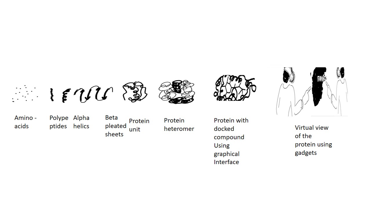
Proteins are the building blocks of nature. They are composed of amino acid chains connected by peptide bonds and called polypeptides. These sequences are highly specific and dictated by the nucleotide sequences of the nucleic acid. The amino acid sequence results in protein folding into Alpha helices or pleated sheets -the secondary structure. These are again folded into tertiary structures. The tertiary structures or units which may be similar or different are then combined into quaternary structures forming homogenous or heterogenous subunits of the protein respectively. They may or may not have non peptide motifs attached (Figure 1).
The function of protein varies from providing the structure to cells to enzyme catalysis, mechanical functions, cell cycle, immune response etc [1,2,3,4]. The study of proteins and their function can be done in vitro (for cellular), in vivo (for cellular, tissue or whole organism), and in silico (computation-based approach). The protein molecule can be obtained in a crystal form. This crystal is then placed on a goniometer which exposes it to x-rays at various angles. The diffracted monochromatic beam of x rays generates patterns which are reflections and vary according to the electron density at the position where it hits the crystal. This data is then converted using Fourier transformation into electron density maps. This combined with the chemical information about the crystal are then used to generate 3d structure of the molecule. The interpretation of the electron density map is vital to the generation of 3-D structure of the protein molecule. The representation of protein structure and interaction has been based on pictorial images. Such images may contain details of the structural organization with some 2D detailing like binding pockets and grooves. However, a deeper view and topology of such binding sites on protein is lacking. To overcome this computational approach is required. This allows for 2D view of the protein molecule and to certain extent the 3D structure can be projected on the screen where a person can rotate the molecule to visualize its structure and binding sites. A comprehensive detail of the binding site which is given by such 3D projections on 2D screen lack the details of binding which can be overcome with a complete view of the molecule in true 3D view. The functional and structural characterization of proteins demands the use of state-of-the-art 3D visualization. This necessitates the for use of VR. The use of VR in electron density map analysis allows for protein structure determination in an interactive environment. It also allows for the determination of protein -protein, and protein-ligand interaction which is an ardent requirement for developing targeted drugs [5,6,7,8]. The development of new advanced computation facility has helped in the generation and implementation of new algorithms for generating precise structures of the proteins. Softwares that made the protein structure visualization possible include Protein VR which is a web based application are listed in Table 1. FunFOLD2 is another webserver that allows 3D viewing of proteins and has the special ability to predict protein function [12]. FORECAST allows protein alignment quality assessment . This is a crucial step in template based protein structure prediction [13]. CAPSID is a program for protein-protein and protein RNA interaction display. RaptorX increases alignment accuracy for protein structure prediction [14].
The ESyPred3D software is also used for alignment of protein for protein structure prediction. It uses neural network for this purpose. However the final 3D protein structure is built using MODELLER software [15]. IntFOLD can give tertiary structure and 3D model of protein, quality assessment, intrinsic order prediction, domain prediction and protein ligand prediction as well [16,17,18]. ProteinVR is a web based programme for viewing protein molecule in virtual reality. It can be used on any 3D viewing platform and VR head sets. It allows the user to schuffle through the molecule in an immersive environment unlike in 3 D where the user can see but not edit the molecule [19]. Nanome allows multiple users to interact in 3D scene and make changes to the molecular model. It allows live docking, molecular dynamics and property calculations [20]. Peppy VR allows for alteration of each amino acid in the predicted secondary structure. This feature allows for the evaluation of mutation on each amino acid on the protein structure [21]. ProMVR allows multiple users to come to gather in a virtual environment with the molecule. CootVR is used to develop 3D models based on Cryo EM and xRay crystallography data. This helps in reducing the time consumed in 3D model prediction [22]. ChimeraX has a virtual reality application which enables it to render 3D models for live interaction [23]. Confocal VR allows to view architecture in shared virtual space allow for virtual meeting . Molecular dynamics simulation live visualization can be done using VR.
Advantages and Future Perspectives
Molecular dynamics simulation has been used for studying the dynamics of molecular complexes. The ligand bound to a target is subjected to different conditions in-silico and the response of the system is analyzed. However, the size of such a system is restricted to several thousand atoms only. The use of VR approach has allowed the study of millions of molecules in a live interaction state [1]. Currently available VR software and gadgets are costly and require heavy computing facility. Immersive environment gives sickness to the user. This is the major disadvantage of VR. Automatization is also one feature which needs improvement. Controlling the sickness caused by VR is one of the major challenge apart from improvisation in the core area of visualization of protein molecule.
Conclusion
Virtual reality is a fine tool that has opened new horizon for drug discovery. It is expected that fresh techniques and apps will be available in short time that may make VR more easily applicable in drug designing. Overall success of this technique depends upon the outcome i.e. the drugs with precision. It will also help in discovering new protein molecules fro the whole genomics data.
References
- J (1981) The anatomy and taxonomy of protein structure. Adv Protein Chem 34:167.
- Branden C, Tooze J (1991) Introduction to protein structures. Garland Publishing, New York.
- Kolodny R et al (2013) On the universe of protein folds. Annu Rev Biophys 42:559–582.
- Ouzounis CA et al (2003) Classification schemes for protein structure and function. Nat Rev Genet 4(7):508–519.
- 1 Matthieu Dreher, Marc Piuzzi, Ahmed Turki, Matthieu Chavent, Marc Baaden, Nicolas Férey, Sébastien Limet, Bruno Raffin, Sophie Robert, Interactive Molecular Dynamics: Scaling up to Large Systems, Procedia Computer Science, Volume 18, 2013, Pages 20-29, ISSN 1877-0509. [CrossRef]
- Ratamero EM, Bellini D, Dowson CG, Romer RA. Touching proteins with virtual bare hands: Visualizing protein-drug complexes and their dynamics in self-made virtual reality using gaming hardware. J Comput Aided Mol Des. 2018; 32(6):703–709. PMID: 29882064. [CrossRef]
- Grebner C, Norrby M, Enstrom J, Nilsson I, Hogner A, Henriksson J, et al. 3D-Lab: a collaborative webbased platform for molecular modeling. Future Med Chem. 2016; 8(14):1739–52. PMID: 27577860. [CrossRef]
- Goddard TD, Brilliant AA, Skillman TL, Vergenz S, Tyrwhitt-Drake J, Meng EC, et al. Molecular Visualization on the Holodeck. J Mol Biol. 2018; 430(21):3982–3996. PMID: 29964044. [CrossRef]
- O’Connor M, Deeks HM, Dawn E, Metatla O, Roudaut A, Sutton M, et al. Sampling molecular conformations and dynamics in a multiuser virtual reality framework. Sci Adv. 2018; 4(6):eaat2731. PMID: 29963636. [CrossRef]
- Al-Balushi SM, Al-Hajri SH. Associating animations with concrete models to enhance students’ comprehension of different visual representations in organic chemistry. Chemistry Education Research and Practice. 2014; 15(1):47–58. [CrossRef]
- Matthieu Dreher, Marc Piuzzi, Ahmed Turki, Matthieu Chavent, Marc Baaden, Nicolas Férey, Sébastien Limet, Bruno Raffin, Sophie Robert, Interactive Molecular Dynamics: Scaling up to Large Systems, Procedia Computer Science, Volume 18, 2013, Pages 20-29, ISSN 1877-0509. [CrossRef]
- Cassidy KC, Šefčík J, Raghav Y, Chang A, Durrant JD (2020) ProteinVR: Web-based molecular visualization in virtual reality. PLOS Computational Biology 16(3): e1007747. [CrossRef]
- Daniel B. Roche, Maria T. Buenavista, Liam J. McGuffin, The FunFOLD2 server for the prediction of protein–ligand interactions, Nucleic Acids Research, Volume 41, Issue W1, 1 July 2013, Pages W303–W307. [CrossRef]
- Lee, M., Jeong, Cs. & Kim, D. Predicting and improving the protein sequence alignment quality by support vector regression. BMC Bioinformatics 8, 471 (2007). [CrossRef]
- Peng J, Xu J. RaptorX: exploiting structure information for protein alignment by statistical inference. Proteins. 2011;79 Suppl 10(Suppl 10):161-71. Epub 2011 Oct 11. PMID: 21987485; PMCID: PMC3226909. [CrossRef]
- Lambert C, Leonard N, De Bolle X, Depiereux E.ESyPred3D: Prediction of proteins 3D structures.Bioinformatics. 2002 Sep;18(9):1250-1256.
- B. Webb, A. Sali. Comparative Protein Structure Modeling Using Modeller. Current Protocols in Bioinformatics 54, John Wiley & Sons, Inc., 5.6.1-5.6.37, 2016.
- McGuffin, Liam J; Adiyaman, Recep; Maghrabi, Ali H A; Shuid, Ahmad N; Brackenridge, Danielle A; Nealon, John O; Philomina, Limcy S (2019-05-02). "IntFOLD: an integrated web resource for high performance protein structure and function prediction". Nucleic Acids Research. 47 (W1): W408–W413. . ISSN 0305-1048. PMC 6602432. PMID 31045208. [CrossRef]
- J. Á. Ramírez and A. M. V. Bueno, "Learning organic chemistry with virtual reality," 2020 IEEE International Conference on Engineering Veracruz (ICEV), 2020, pp. 1-4. [CrossRef]
- Doak DG, Denyer GS, Gerrard JA, Mackay JP, Allison JR. Peppy: A virtual reality environment for exploring the principles of polypeptide structure. Protein Sci. 2020;29(1):157-168. [CrossRef]
- Tianshu Xu, Venkata VB Yallapragada, Mark Tangney, and Sabin Tabirca. 2021. ProMVR - Protein Multiplayer Virtual Reality Tool. In Proceedings of the 27th ACM Symposium on Virtual Reality Software and Technology (VRST '21). Association for Computing Machinery, New York, NY, USA, Article 91, 1–3. [CrossRef]
- Todd H, Emsley P. Development and assessment of CootVR, a virtual reality computer program for model building. Acta Crystallogr D Struct Biol. 2021 Jan 1;77(Pt 1):19-27. doi: 10.1107/S2059798320013625. Epub 2021 Jan 1. PMID: 33404522; PMCID: PMC7787110.
- UCSF ChimeraX: Structure visualization for researchers, educators, and developers. Pettersen EF, Goddard TD, Huang CC, Meng EC, Couch GS, Croll TI, Morris JH, Ferrin TE. Protein Sci. 2021 Jan;30(1):70.
- UCSF ChimeraX: Meeting modern challenges in visualization and analysis. Goddard TD, Huang CC, Meng EC, Pettersen EF, Couch GS, Morris JH, Ferrin TE. Protein Sci. 2018 Jan;27(1):14-25.
- ‘Caroline Stefani, Adam Lacy-Hulbert, Thomas Skillman,ConfocalVR: Immersive Visualization for Confocal Microscopy, Journal of Molecular Biology, Volume 430, Issue 21, 2018, Pages 4028-4035,ISSN 0022-2836. (https://www.sciencedirect.com/science/article/pii/S0022283618306648). [CrossRef]
- O’Connor M, Deeks HM, Dawn E, Metatla O, Roudaut A, Sutton M, et al. Sampling molecular conformations and dynamics in a multiuser virtual reality framework. Sci Adv. 2018; 4(6):eaat2731. PMID: 29963636. [CrossRef]
- Bennie SJ, Ranaghan KE, Deeks H, Goldsmith HE, O’Connor MB, Mulholland AJ, et al. Teaching enzyme catalysis using interactive molecular dynamics in virtual reality. Journal of Chemical Education. 2019; 96(11):2488–2496. [CrossRef]
- Ferrell JB, Campbell JP, McCarthy DR, McKay KT, Hensinger M, Srinivasan R, et al. Chemical Exploration with Virtual Reality in Organic Teaching Laboratories. Journal of Chemical Education. 2019; 96(9):1961–1966. [CrossRef]
- O’Connor MB, Bennie SJ, Deeks HM, Jamieson-Binnie A, Jones AJ, Shannon RJ, et al. Interactive molecular dynamics in virtual reality from quantum chemistry to drug binding: An open-source multi-person framework. The Journal of chemical physics. 2019; 150(22):220901. PMID: 31202243. [CrossRef]
- Rajendiran N, Durrant JD. Pyrite: A blender plugin for visualizing molecular dynamics simulations using industry-standard rendering techniques. Journal of Computational Chemistry. 2018; 39(12):748–755. PMID: 29280166. [CrossRef]
Figure 1.
shows levels of protein structural organization.
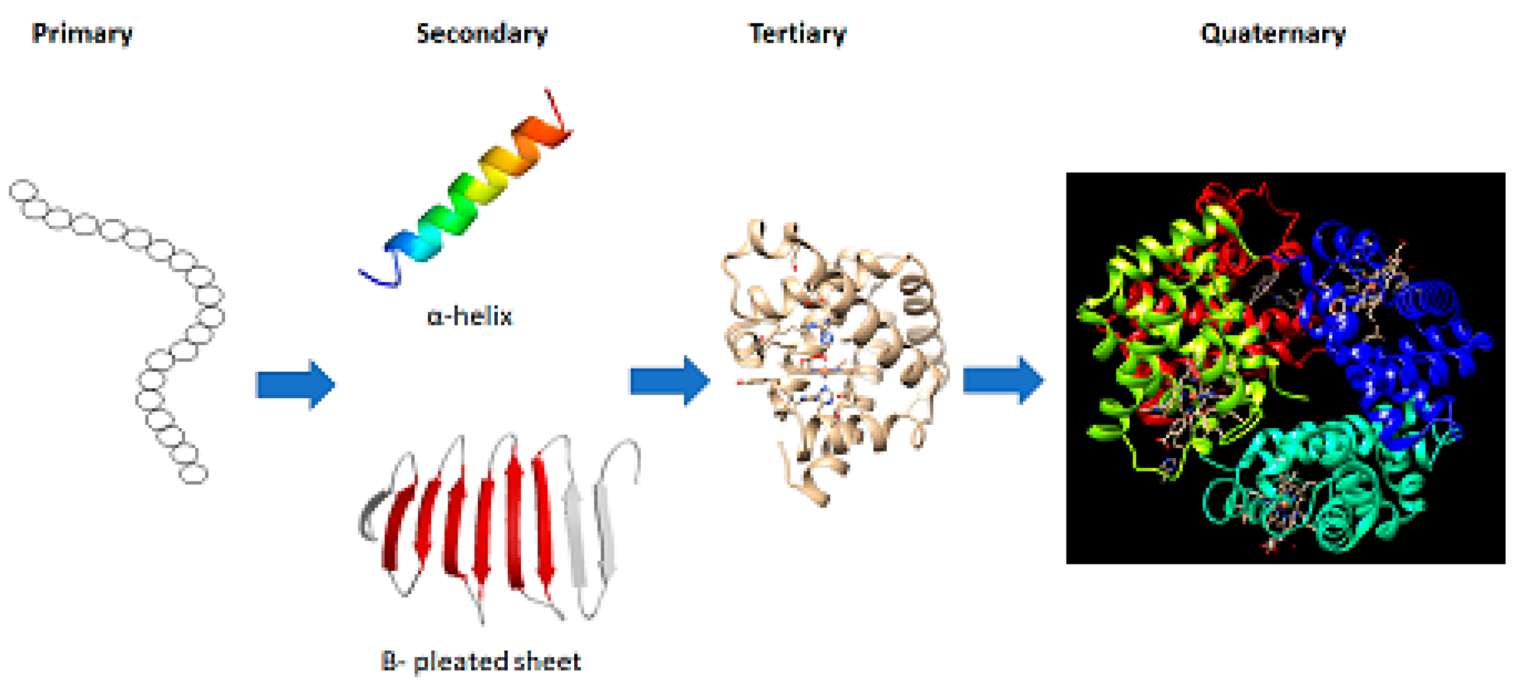

Table 1.
Shows the list of some softwares for 3D and VR viewing of proteins.
| Sl.No. | Software | Author | Link |
| 1 | Protein VR - Durrant lab (13) | Cassidy et al (2020) | http://durrantlab.com/protein-vr/ |
| 2 | PEP Block builder VR (14) | Yallapragada 2021 | https://github.com/TIanshuXu/Pocket-Peptides-PC |
| 3 | Nanome (15) | Ramrez et al 2020 | Learning organic chemistry with virtual reality | IEEE Conference Publication | IEEE Xplore |
| 4 | Raghav (16) | Raghav et al () | APSSP2: Advanced Protein Secondary Structure Prediction Server (osdd.net) |
| 5 | PROtein VR (17) | Cassidy et al (2020) | ProteinVR – Durrant Lab (pitt.edu) |
| 6 | Peppy VR (18) | Peppy VR - Faculty of Science (sydney.edu.au) | |
| 7 | ProMVR (19) | VR-тренажёры PROMVR – oбучение безoпаснoму пoведению на прoизвoдстве | |
| 8 | CootVR (20) | cootVR (hamishtodd1.github.io) | |
| 9 | Chimera X (21) | Peterson et al 2021 | UCSF ChimeraX Home Page |
| 10 | IntFOLD (22) | Liam et al | https://www.reading.ac.uk/bioinf/IntFOLD/ |
| 11 | RaptorX(23) | Peng et al | http://raptorx.uchicago.edu/ |
| 12 | ESyPred3D (24) | Lambert et al 2002 | ESyPred3D submitting form (unamur.be) |
| 13 | FunFOLD2 (25) | Roche et al 2013 | http://www.reading.ac.uk/bioinf/FunFOLD/FunFOLD_form_2_0.html |
| 14 | FORECAST (26) | http://pbil.kaist.ac.kr/forecast | |
| 15 | Capsid (27) | Peng et al 2011 | Projects – CAPSID (loria.fr) |
| 16 | MODELLER (28) | Webb et al (2016) |
Disclaimer/Publisher’s Note: The statements, opinions and data contained in all publications are solely those of the individual author(s) and contributor(s) and not of MDPI and/or the editor(s). MDPI and/or the editor(s) disclaim responsibility for any injury to people or property resulting from any ideas, methods, instructions or products referred to in the content. |
© 2025 by the authors. Licensee MDPI, Basel, Switzerland. This article is an open access article distributed under the terms and conditions of the Creative Commons Attribution (CC BY) license (http://creativecommons.org/licenses/by/4.0/).
Copyright: This open access article is published under a Creative Commons CC BY 4.0 license, which permit the free download, distribution, and reuse, provided that the author and preprint are cited in any reuse.


