Preprint
Article

This version is not peer-reviewed.

Role of MicroRNA-21 and MicroRNA-210 Expression in the Diagnosis, Prognosis, and Metastatic Potential of Renal Cell Carcinoma Subtypes

Submitted:

27 October 2025

Posted:

28 October 2025

You are already at the latest version

Abstract

Background and Objectives: Differentiating between subtypes of renal cell carcinoma (RCC) can be challenging due to overlapping immunohistochemical and histomorphological features. Furthermore, despite the evaluation of numerous histopathologic parameters, predicting metastasis and prognosis remains unclear. In this study, we aimed to evaluate the potential of microRNA (miRNA) expression levels in establishing definitive diagnoses, assessing metastatic potential, and determining patient prognosis. Materials and Methods: A total of 35 clear cell RCC (cc-RCC), 11 chromophobe RCC (ch-RCC), 9 papillary RCC (p-RCC) cases, and 9 cc-RCC metastases were retrospectively analyzed. MiRNA-21 and miRNA-210 expression profiles were assessed using non-parametric tests, receiver operating characteristic (ROC) analysis, and Cox regression models. Results: The expression levels of both miRNA-21 and miRNA-210 were significantly elevated in cc-RCC and p-RCC cases. In contrast, miRNA-210 expression levels were significantly reduced in all ch-RCC cases, whereas miRNA-21 expression showed no significant change. Although miRNA-21 expression levels were higher in metastases compared to primary cc-RCC tumors, the difference was not statistically significant (p=0.053). No significant difference was observed in miRNA-210 expression between metastatic and primary cc-RCC tumors (p=0.237). Regardless of histological subtype, an increase in miRNA-210 expression was associated with a significant reduction in overall survival (p=0.03, HR=1.729, 95% CI: 1.043-2.864). Conclusion: The findings suggest that miRNA-21 and miRNA-210 expression levels can help distinguish ch-RCC from cc-RCC and p-RCC. Additionally, miRNA-210, but not miRNA-21, may differentiate cc-RCC from p-RCC. Higher miRNA-210 expression levels were associated with worse overall survival in RCC patients. While miRNA-21 levels were increased in cc-RCC metastases, further studies with larger sample sizes are needed to establish its role in predicting metastasis.

Keywords: 
;  ;  ;  ;  ;  

1. Introduction

Renal cell carcinoma (RCC) is one of the few carcinomas with increasing incidence and high lethality [1]. Among RCC subtypes, clear cell renal cell carcinoma (cc-RCC) is the most common and aggressive variant, while chromophobe renal cell carcinoma (ch-RCC) and papillary renal cell carcinoma (p-RCC) are other frequently diagnosed kidney tumor subtypes [2,3].
MicroRNAs (miRNAs) are small, non-coding, single-stranded RNA molecules consisting of approximately 20–22 nucleotides. MiRNAs regulate biological processes at the RNA level by epigenetically influencing nearly 30% of human genes [4]. Changes in miRNA levels in tissue samples, blood, and other body fluids have been observed in various tumor types [5]. Their potential as biomarkers in tumor diagnosis, classification, postoperative follow-up, and prognosis assessment has been demonstrated in previous studies [6,7,8,9]. Therapeutic approaches targeting miRNAs involved in carcinogenesis are currently being developed, aiming to suppress oncogenic miRNAs and activate tumor-suppressor miRNAs [10]. Therefore, identifying tumor-associated miRNA profiles may contribute significantly to personalized targeted therapies.
MiRNA-21 plays a crucial role in embryogenesis, carcinogenesis, epithelial-mesenchymal transition, tumor invasiveness, recurrence, and drug resistance [11]. Recent evidence demonstrated that miR-21 promotes proliferation, invasion, migration, angiogenic abilitity and epithelial-mesenchymal transition in RCC by directly targeting the Programmed Cell Death Protein 4 / c-Jun (PCDP-4/c-Jun) and Phospholysine Phosphohistidine Inorganic Pyrophosphate Phosphatase(LHPP) and its overexpression is significantly associated with poor differentiation, higher stage, and worse prognosis in patients [12,13]. Functioning as an oncogene (oncomiR), miRNA-21 inhibits pro-apoptotic genes and targets the tumor suppressor gene, PTEN. The PTEN gene and the PI3K/Akt signaling pathway are known to play essential roles in RCC development [14,15].
MiRNA-210 is associated with angiogenesis, cell proliferation and differentiation, DNA damage response, apoptosis, tumor invasion, and metastasis [16]. It is the most highly upregulated miRNA in hypoxic cells. Hypoxia-inducible factors (HIFs) are key regulators of the cellular response to hypoxia. Increased miRNA-210 expression in response to hypoxia has been found to regulate HIF-1 and other target genes involved in RCC carcinogenesis [8,17].
In this study, we aimed to determine the diagnostic and prognostic significance of miRNA-21 and miRNA-210 expression in tissue samples from three different RCC subtypes. Although the roles of miRNA-21 and miRNA-210 in cc-RCC and p-RCC have been previously studied, limited data exist for ch-RCC. We selected miRNA-21 and miRNA-210 for this study due to their reported involvement in RCC carcinogenesis [8,11,14]. Additionally, we investigated their potential as indicators of metastatic disease and prognostic markers.

2. Materials and Methods

2.1. Case Selection and Preparation

Patients who underwent total or partial nephrectomy or metastasectomy for RCC between 2012 and 2018 at our institution were identified for this study. Cases with significant tumor necrosis or a history of a different primary malignancy were excluded. For each patient, healthy cortical kidney tissue was used as the control sample.
Histological grading was performed according to the World Health Organization (WHO)/International Society of Urological Pathology (ISUP) grading system. Cases with grade 1 and 2 tumors were classified as “low grade,” while those with grade 3 and 4 tumors were classified as “high grade.” Among 14 patients with metastatic cc-RCC, metastatic tissue paraffin blocks were available for 9 cases, forming the “metastasis group.”

2.2. Total RNA Isolation

Formalin-fixed paraffin-embedded (FFPE) tissue blocks from selected hematoxylin and eosin (H&E)-stained slides were used for RNA extraction. Total RNA was isolated using the RecoverAll™ Total Nucleic Acid Isolation Kit for FFPE (Thermo Fisher Scientific Inc., Foster City, CA, USA; catalog no: AM1975) following the manufacturer’s protocol.

2.3. Complementary DNA Synthesis from Total RNA

Complementary DNA (cDNA) synthesis was performed via reverse transcription using the TaqMan™ miRNA Reverse Transcription Kit (Thermo Fisher Scientific Inc., USA; catalog no: 4366596) according to the manufacturer’s instructions. cDNA synthesis was conducted separately for miRNA-21, miRNA-210, and the endogenous control RNU6b for each sample.

2.4. Evaluation of MiRNA Expression by Quantitative Real-Time PCR

Quantitative real-time polymerase chain reaction (qRT-PCR) was performed using the 7500 Real-Time PCR System (Applied Biosystems, USA). For each patient’s paired tumor and non-tumor samples, specific PCR mixtures for miRNA-21, miRNA-210, and RNU6b, along with RNA-free contamination controls, were loaded into the system for amplification. Fluorescence levels were recorded over 40 cycles.

2.5. Normalization of MiRNA Expression and Determination of Expression Profiles

Cycle threshold (CT) values were determined for each sample. RNU6b, selected as an endogenous control due to its stable expression across all cell types, was used for normalization. Expression data were quantitatively analyzed using the ΔΔCT method.
The relative change in miRNA expression, known as “fold change,” was calculated using the formula 2^−ΔΔCT, comparing tumor or metastatic tissue expression levels to healthy kidney tissue [18].

2.6. Statistical Analysis

All statistical analyses were performed using SPSS v15.0 (SPSS Inc., Chicago, IL, USA).
  • The normality of miRNA-21 and miRNA-210 expression levels was assessed using the Kolmogorov-Smirnov test, and homogeneity of variances was evaluated by the Levene test.
  • Differences in miRNA expression levels among histological subtypes and between primary tumors and metastases were analyzed using one-way ANOVA, Tukey’s HSD test, and paired t-tests, respectively.
  • Correlations in parametric data were assessed with the Pearson correlation test.
  • The diagnostic performance of miRNA expression levels for histological subtyping and metastatic potential was evaluated using receiver operating characteristic (ROC) curve analysis.
  • The association between overall survival and miRNA expression levels was determined using the Cox regression model.
Statistical significance was set at a 95% confidence level (α=0.05).

2.7. Ethical Approval

Our study’s ethics committee approval was obtained from Health Sciences University Istanbul Education and Research Hospital, non-interventional clinical research ethics committee chairmanship. The study was conducted following the Declaration of Helsinki, and the ethical standards of the institutional research committee.

3. Results

3.1. Clinicopathological Features

Seventeen of the patients (30.9%) were female, and thirty-eight (69.1%) were male. The median age of the patients was 65 years, and the mean tumor size was 5.5 ± 2.5 cm. Pathological features are summarized in Table 1.

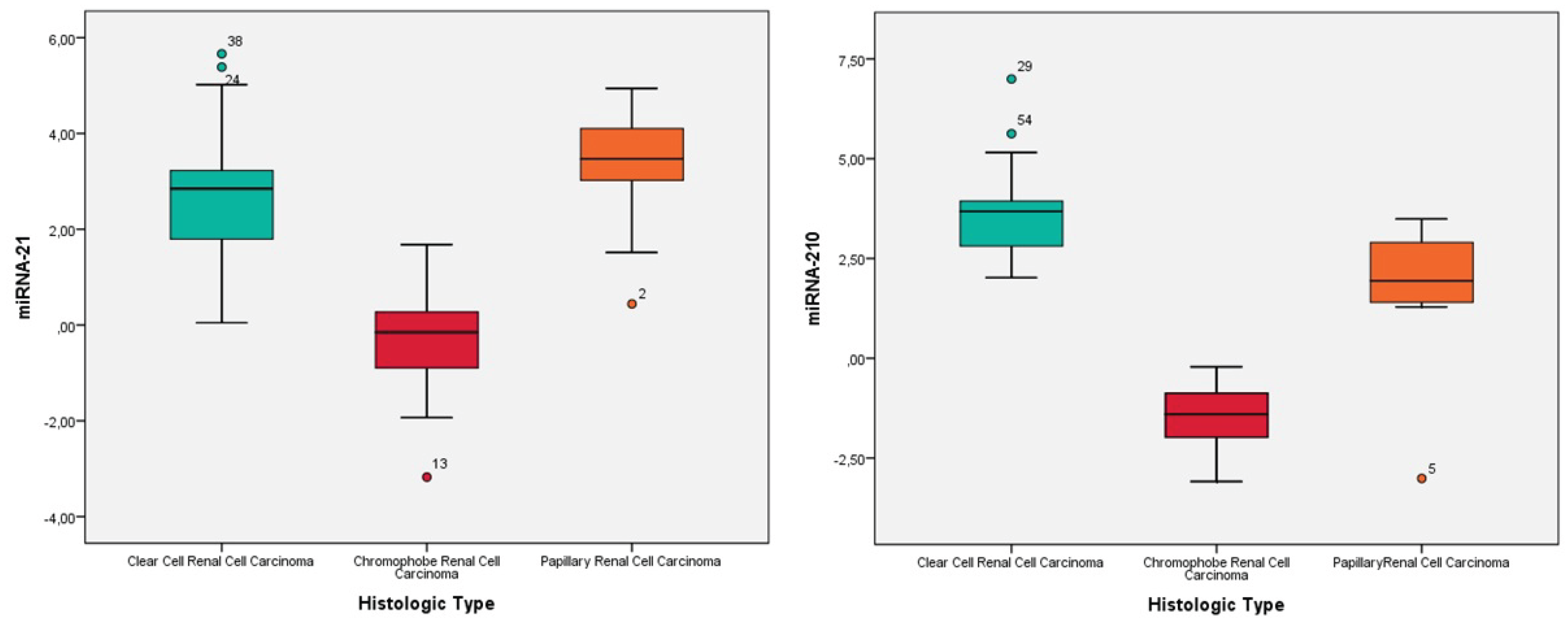
3.2. Expression Levels of MiRNA-21 and MiRNA-210 in Renal Cell Carcinomas

The expression levels of miRNA-21 and miRNA-210 in RCCs are presented in Figure 1.
Accordingly, miRNA-21 expression levels were significantly increased in cc-RCC cases compared to non-tumoral renal tissues (p<0.001). In p-RCCs, miRNA-21 expression levels were elevated in all cases. In ch-RCC cases, miRNA-21 expression levels increased in five cases and decreased in six cases; therefore, the expression change was not statistically significant (p = 0.76) (Table 2).
MiRNA-210 expression levels were elevated in all cc-RCC cases but decreased in all ch-RCC cases. In p-RCCs, miRNA-210 expression increased in eight cases and decreased in one case, with a statistically significant increase (p = 0.02) (Table 2).
There was no statistically significant correlation between miRNA-21 and miRNA-210 expression levels and clinicopathological parameters such as age, gender, tumor diameter, WHO/ISUP grade, lymphovascular invasion, or necrosis (p>0.05). (Supplementary S2).

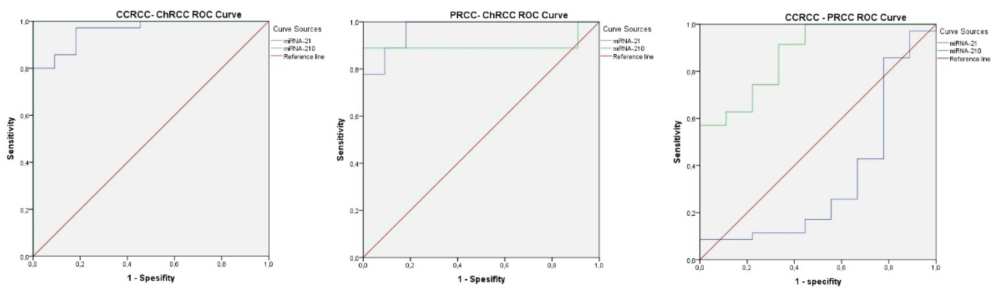
3.3. Histological Subtype Determination Based on MiRNA-21 and MiRNA-210 Expression Levels

A statistically significant difference was found between cc-RCC and ch-RCC, and between p-RCC and ch-RCC, in terms of both miRNA-21 and miRNA-210 expression levels (p < 0.001). For miRNA-210 expression levels, a significant difference was also observed between cc-RCC and p-RCC cases (p < 0.001), whereas miRNA-21 expression did not show a significant difference (p = 0.53) (see Supplementary S3).
Among the three histological subtypes, cutoff values that can be used to distinguish RCC types were analyzed.
  • To differentiate cc-RCC from ch-RCC, the cutoff values were 1.72 for miRNA-21 (AUROC = 0.961, 95% CI: 0.909–1.000, p < 0.001) and 0.90 for miRNA-210 (AUROC = 1.000, 95% CI: 1.000–1.000, p < 0.001). Sensitivity and specificity for miRNA-21 and miRNA-210 were 80% and 100%; and 100% and 100%, respectively. (see Figure 2A, Supplementary S4).
  • To distinguish p-RCC from ch-RCC, the cutoff values were 0.44 for miRNA-21 (AUROC = 0.970, 95% CI: 0.906–1.000, p < 0.001) and 0.54 for miRNA-210 (AUROC = 0.899, 95% CI: 0.711–1.000, p = 0.003). Sensitivity and specificity for miRNA-21 and miRNA-210 were 100% and 82%, and 88% and 100%, respectively. (see Figure 2B, Supplementary S4).
  • To differentiate cc-RCC from p-RCC, the cutoff value for miRNA-210 was 0.54 (AUROC = 0.873, 95% CI: 0.749–0.997, p = 0.001). Sensitivity and specificity were 91% and 67%, respectively. MiRNA-21 did not show a statistically significant distinction (p = 0.150). (see Figure 2C, Supplementary S4).

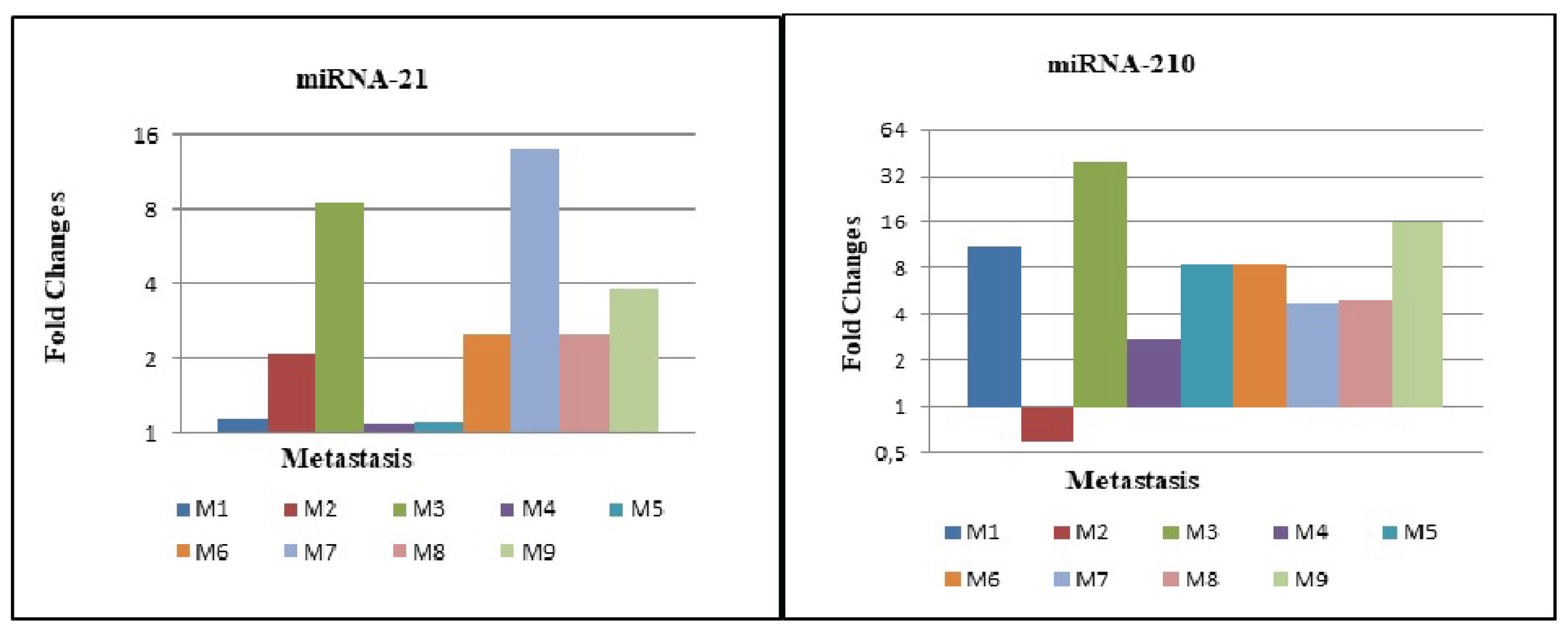
3.4. Expression Levels of MiRNA-21 and MiRNA-210 in Metastatic cc-RCC

The fold changes for miRNA-21 and miRNA-210 in metastatic samples are shown in Figure 3.
In nine metastatic cc-RCC cases where both primary tumor and metastasis tissues were available, miRNA-21 expression levels were higher in metastatic tissues compared to primary tumors, but this difference was not statistically significant (p = 0.053). Similarly, no significant difference was observed for miRNA-210 expression levels between metastatic and primary cc-RCC tissues (p = 0.237). (see Supplementary S5).
Comparison of primary tumor tissues between metastatic and non-metastatic cc-RCC cases revealed no statistically significant differences in miRNA-21 (p = 0.875) or miRNA-210 (p = 0.236) expression levels. (see Supplementary S5).

3.5. Relationship Between MiRNA-21 and MiRNA-210 Expression Levels and Survival Time

Patient survival times were analyzed in relation to miRNA-21 and miRNA-210 expression levels. A total of 55 patients were included in this study.
  • Clear Cell Renal Cell Carcinoma (ccRCC): 35 patients were included in this group. The mean follow-up time was 87.3 ± 69.7 months, with a mortality rate of 37.1%.
  • Chromophobe Renal Cell Carcinoma (chRCC): 11 patients were analyzed in this group. The mean follow-up time was 101.5 ± 30.1 months, with a mortality rate of 9.1%.
  • Papillary Renal Cell Carcinoma (pRCC): 9 patients were included in this group. The mean follow-up time was 81.1 ± 51.1 months, with a mortality rate of 33.3%.
To better evaluate the relationship between miRNA levels and patient survival, Kaplan-Meier survival analysis and Chi-Square tests were conducted (see supplementary 7)
Relationship Between miRNA Expression and Survival in Renal Cell Carcinoma Subtypes
The association between miRNA expression levels and patient survival was evaluated across papillary, clear cell, and chromophobe RCC subtypes.
For miRNA-21, higher expression appeared to be associated with lower mortality rates in both papillary (28.6% vs. 50.0%) and clear cell RCC (30.0% vs. 46.7%). However, these differences did not reach statistical significance (Chi-Square test: p = 1.000 and p = 0.512, respectively).
In contrast, miRNA-210 showed an opposite trend, with higher expression corresponding to higher mortality rates in papillary (50.0% vs. 28.6%) and clear cell RCC (40.0% vs. 30.0%). Nonetheless, the observed differences were not statistically significant (Chi-Square test: p = 1.000 and p = 0.868, respectively).
Due to limited case numbers, chromophobe RCC could not be subjected to meaningful statistical analysis. There is no statistically significant relationship between miRNA-21 or miRNA-210 levels and survival. While some numerical differences in mortality rates were observed between high and low miRNA groups, these were not statistically significant. A larger sample size may be necessary to determine potential prognostic value. Further studies with larger patient cohorts and more robust statistical power are needed to confirm these findings.

4. Discussion

Differentiating histological subtypes of RCC can be challenging in certain cases due to overlapping morphological and immunohistochemical features. In such situations, analyzing miRNA expression profiles is recommended as a valuable method to support differential diagnosis [7,19].
The upregulation of miRNA-21 and miRNA-210 in cc-RCC and p-RCC is consistent with previous studies indicating their involvement in tumor progression, angiogenesis, and hypoxia-related pathways. miRNA-21, often considered an oncogenic miRNA, has been reported to promote cell proliferation and inhibit apoptosis by targeting tumor suppressor genes such as PTEN and PDCD4. Similarly, miRNA-210, widely recognized as a hypoxia-inducible miRNA, contributes to cellular adaptation under low-oxygen conditions by modulating mitochondrial metabolism and DNA repair mechanisms.
The markedly lower miRNA-210 expression in chromophobe RCC observed in our cohort may therefore underline the relatively indolent nature of this subtype and its distinct pathogenesis, which is less dependent on hypoxia-driven molecular mechanisms. Collectively, these findings highlight the subtype-specific heterogeneity of miRNA expression patterns in RCC and suggest that miRNA-21 and miRNA-210 could serve as complementary biomarkers for diagnostic and prognostic stratification. Further studies with larger sample sizes and functional analyses are warranted to clarify the underlying biological mechanisms and validate their clinical applicability.
Regarding metastases, although miRNA-21 expression levels were higher in metastatic tissues compared with primary cc-RCC tumors, this difference did not reach statistical significance (p = 0.053). Likewise, miRNA-210 expression levels showed no significant variation between metastatic and primary tumor tissues (p = 0.237). Moreover, when comparing primary tumors of metastatic and non-metastatic cc-RCC cases, no statistically significant differences were observed in either miRNA-21 or miRNA-210 expression (p = 0.875 and p = 0.236, respectively).
In contrast to our results, several previous studies have reported significant alterations in miRNA expression profiles between metastatic and non-metastatic RCC cases, suggesting that specific miRNAs may contribute to metastatic potential and could serve as diagnostic or prognostic biomarkers [20,21,22]The absence of statistical significance in our cohort might be explained by the limited sample size, intratumoral heterogeneity, or methodological differences, such as variations in RNA extraction protocols, normalization strategies, and tissue preservation conditions [23]. Nevertheless, the trend toward increased miRNA-21 expression in metastatic tissues observed in our study is in line with previous report linking this miRNA to tumor invasion, epithelial–mesenchymal transition, and angiogenesis [24]. This observation supports the hypothesis that miRNA-21 may still play a role in promoting tumor progression and metastatic behavior, warranting further investigation in larger, well-characterized cohorts.
MiRNA-21 is one of the most extensively studied oncogenic miRNAs, known for its role in tumorigenesis by targeting tumor suppressor genes such as PTEN and influencing the PI3K/Akt signaling pathway. Elevated miRNA-21 levels have been reported in various tumor types, including RCC [25,26,27,28,29]. Our study supports previous findings by demonstrating significantly higher miRNA-21 expression in cc-RCC and p-RCC, although ch-RCC cases showed variable expression patterns. These variations in miRNA-21 expression among ch-RCC cases might be attributed to chromosomal losses commonly observed in this tumor subtype [30].
MiRNA-210, recognized for its role in hypoxia adaptation, has been linked to tumor proliferation, apoptosis inhibition, and angiogenesis [16]. Consistent with previous literature, we found that miRNA-210 expression was significantly elevated in cc-RCC and p-RCC cases but was significantly decreased in ch-RCC. In support of our findings, a recent high-throughout study identified a panel of dysregulated miRNAs—including miR-210, miR-200c, miR-455-3p, and miR-483-5p—that were associated with ccRCC stage and progression, emphasizing their diagnostic and prognostic value. [31] This decrease might be associated with the chromosomal losses frequently observed in ch-RCC, potentially leading to the downregulation of miRNA-210 expression [30,32].
Survival analysis demonstrated that increased miRNA-210 expression was significantly associated with decreased overall survival (p = 0.03, HR = 1.729, 95% CI: 1.043–2.864), regardless of histological subtype. However, miRNA-21 expression levels were not significantly associated with survival (p = 0.49). Consistent with our findings, Vitruk et al. demonstrated that miR-210 expression was significantly associated with resistance to sunitinib in RCC patients, suggesting its potential utility not only as a diagnostic biomarker but also as a predictor of treatment response and prognosis in targeted therapy [33]. These findings align with several studies indicating miRNA-210 as a potential prognostic biomarker in RCC, highlighting its role in tumor aggressiveness and hypoxia-driven progression [34,35,36].
While some studies have reported an association between miRNA-21 and miRNA-210 expression levels with tumor grade, pathological stage, and lymphovascular invasion, our findings did not reveal any statistically significant correlation with clinicopathological parameters such as age, gender, tumor diameter, WHO/ISUP grade, lymphovascular invasion, or necrosis (p > 0.05) [37]. The discrepancies observed among studies might be attributed to differences in sample size, patient demographics, or technical methodologies used in miRNA quantification.
Our results indicate that miRNA-21 and miRNA-210 expression levels can effectively distinguish between cc-RCC and ch-RCC, as well as p-RCC and ch-RCC subtypes, with high sensitivity and specificity. Specifically, miRNA-210 had a cutoff value of 0.90 to differentiate cc-RCC from ch-RCC (AUROC = 1.000, 95% CI: 1.000–1.000, p < 0.001), with a sensitivity and specificity of 100%. Similarly, the differentiation of p-RCC from ch-RCC was achieved with a cutoff of 0.54 for miRNA-210 (AUROC = 0.899, 95% CI: 0.711–1.000, p = 0.003). A recent meta-analysis of high-throughput RCC miRNA datasets identified a panel of subtype-specific microRNAs, including miR-21-5p, miR-155-5p, and miR-210-3p as upregulated in clear cell RCC, supporting their potential utility in molecular classification of RCC subtypes and aligning with our findings regarding their differential expression[38]However, miRNA-21 did not provide statistically significant differentiation between cc-RCC and p-RCC (p = 0.150), consistent with their shared proximal tubule origin [35].
Although our study provides valuable insights into the diagnostic and prognostic roles of miRNA-21 and miRNA-210 in RCC, it has certain limitations. The relatively small sample size, particularly in the metastatic subgroup, may limit the generalizability of our findings. Further large-scale studies with more extensive cohorts are required to confirm our results and evaluate the potential of miRNA-based biomarkers in clinical practice.

5. Conclusions

Our findings suggest that miRNA-21 and miRNA-210 expression levels are valuable in distinguishing RCC subtypes, particularly in differentiating cc-RCC and p-RCC from ch-RCC. Moreover, increased miRNA-210 expression is a potential prognostic biomarker for poor overall survival in RCC patients, regardless of histological subtype. However, additional studies are necessary to further elucidate the role of miRNA-21 in metastatic progression.

Supplementary Materials

The following supporting information can be downloaded at the website of this paper posted on Preprints.org, Supplementary S1: Oneway ANOVA, Supplementary S2: Correlations Test, Supplementary S3: Post Hoc Tests- Tukey HSD, Supplementary S4: ROC (Receiver Operating Charecteristic) curve analysis, Supplementary S5: Paired Samples Test, Supplementary S6: Cox regression Test, Supplementary S7: Kaplan-Meier survival analysis and Chi-Square tests.

Author Contributions

Conceptualization, T.C.S and S.B.K.; methodology, T.C.S and I.Y.; validation, T.C.S and I.Y..; formal analysis, E.T. and G.N ; investigation, T.C.S, E.C.K.T.,S.B.K. and H.K; data curation, T.C.S. and I.Y.; writing—original draft preparation, T.C.S..; writing—review and editing, T.C.S, E.C.K.T, S.B.K and T.B.S; visualization, T.C.S., G.N, T.B.S; supervision, E.C.K.T and I.Y..; project administration, T.C.S. and E.C.K.T.; funding acquisition, E.C.K.T. All authors have read and agreed to the published version of the manuscript.

Funding

This research was funded by University of Health Sciences, Scientific Research Projects Unit, Istanbul, Turkey.

Institutional Review Board Statement

The study was conducted in accordance with the Declaration of Helsinki and approved by the Institutional Review Board (or Ethics Committee) of Istanbul Training and Research Hospital (approval No. 07.12.2018/1555).

Informed Consent Statement

Informed consent was obtained from all subjects involved in the study.

Data Availability Statement

The original contributions presented in this study are included in the article/Supplementary Materials. Further inquiries can be directed to the corresponding author.

Conflicts of Interest

The authors declare no conflicts of interest. The funders had no role in the design of the study; in the collection, analyses, or interpretation of data; in the writing of the manuscript; or in the decision to publish the results.

References

  1. Moch H, Hoon Tan P, Amin B M, Turajlic S. Renal cell tumours, Introduction. In: Who Classification of Tumors Editorial Board. WHO Classification of Tumours of the Urinary System and Male Genital Organs. 5th Edition ed. Lyon, France: IARC; 2022.
  2. Zhou M, Hattab MA, Eble JN, Cheng L. Neoplasms of the Kidney. In: Zhou M, Magi-Galluzzi C, editors. Genitourinary Pathology Foundations in Diagnostic Pathology. 2nd Edition ed. Philadephia,PA: Saunders, Elsevier; 2015. p. 306-77.
  3. Delahunt B, Algaba F, Eble J, Cheville J, Amin MB, Argani P, et al. Papillary renal cell carcinoma. In: Moch H, Humphrey PA, Ulbright TM, Reuter VE, editors. WHO Classification of Tumours of the Urinary System and Male Genital Organs. Lyon: IARC; 2016. p. 23-5.
  4. Wang J, Zhao J, Shi M, Ding Y, Sun H, Yuan F, et al. Elevated expression of miR-210 predicts poor survival of cancer patients: a systematic review and meta-analysis. PLoS One. 2014;9(2):e89223. [CrossRef]
  5. Calin GA, Sevignani C, Dumitru CD, Hyslop T, Noch E, Yendamuri S, et al. Human microRNA genes are frequently located at fragile sites and genomic regions involved in cancers. Proc Natl Acad Sci U S A. 2004;101(9):2999-3004. [CrossRef]
  6. Heinzelmann J, Unrein A, Wickmann U, Baumgart S, Stapf M, Szendroi A, et al. MicroRNAs with prognostic potential for metastasis in clear cell renal cell carcinoma: a comparison of primary tumors and distant metastases. Ann Surg Oncol. 2014;21(3):1046-54. [CrossRef]
  7. Lu J, Getz G, Miska EA, Alvarez-Saavedra E, Lamb J, Peck D, et al. MicroRNA expression profiles classify human cancers. Nature. 2005;435(7043):834-8. [CrossRef]
  8. McCormick RI, Blick C, Ragoussis J, Schoedel J, Mole DR, Young AC, et al. miR-210 is a target of hypoxia-inducible factors 1 and 2 in renal cancer, regulates ISCU and correlates with good prognosis. Br J Cancer. 2013;108(5):1133-42. [CrossRef]
  9. Jung M, Mollenkopf HJ, Grimm C, Wagner I, Albrecht M, Waller T, et al. MicroRNA profiling of clear cell renal cell cancer identifies a robust signature to define renal malignancy. J Cell Mol Med. 2009;13(9B):3918-28. [CrossRef]
  10. Gordanpour A, Nam RK, Sugar L, Seth A. MicroRNAs in prostate cancer: from biomarkers to molecularly-based therapeutics. Prostate Cancer Prostatic Dis. 2012;15(4):314-9. [CrossRef]
  11. Hong L, Han Y, Zhang Y, Zhang H, Zhao Q, Wu K, et al. MicroRNA-21: a therapeutic target for reversing drug resistance in cancer. Expert Opin Ther Targets. 2013;17(9):1073-80. [CrossRef]
  12. Meng K, Li Z, Cui X. Three LHPP gene-targeting co-expressed microRNAs (microRNA-765, microRNA-21, and microRNA-144) promote proliferation, epithelial-mesenchymal transition, invasion, and are independent prognostic biomarkers in renal cell carcinomas patients. J Clin Lab Anal. 2021;35:e24077. [CrossRef]
  13. Fan B, Jin Y, Zhang H, Zhao R, Sun M, Sun M, et al. MicroRNA-21 contributes to renal cell carcinoma cell invasiveness and angiogenesis via the PDCD4/c-Jun (AP-1) signalling pathway. Int J Oncol. 2020;56(1):178-192. [CrossRef]
  14. Vergho DC, Kneitz S, Kalogirou C, Burger M, Krebs M, Rosenwald A, et al. Impact of miR-21, miR-126 and miR-221 as prognostic factors of clear cell renal cell carcinoma with tumor thrombus of the inferior vena cava. PLoS One. 2014;9(10):e109877. [CrossRef]
  15. Meng F, Henson R, Wehbe-Janek H, Ghoshal K, Jacob ST, Patel T. MicroRNA-21 regulates expression of the PTEN tumor suppressor gene in human hepatocellular cancer. Gastroenterology. 2007;133(2):647-58. [CrossRef]
  16. Bavelloni A, Ramazzotti G, Poli A, Piazzi M, Focaccia E, Blalock W, et al. MiRNA-210: A Current Overview. Anticancer Res. 2017;37(12):6511-21. [CrossRef]
  17. Valera VA, Walter BA, Linehan WM, Merino MJ. Regulatory Effects of microRNA-92 (miR-92) on VHL Gene Expression and the Hypoxic Activation of miR-210 in Clear Cell Renal Cell Carcinoma. J Cancer. 2011;2:515-26. [CrossRef]
  18. Schmittgen TD, Livak KJ. Analyzing real-time PCR data by the comparative C(T) method. Nat Protoc. 2008;3(6):1101-8.
  19. Silva-Santos RM, Costa-Pinheiro P, Luis A, Antunes L, Lobo F, Oliveira J, et al. MicroRNA profile: a promising ancillary tool for accurate renal cell tumour diagnosis. Br J Cancer. 2013;109(10):2646-53. [CrossRef]
  20. Gu L, Li H, Chen L, Ma X, Gao Y, Li X, Zhang Y. MicroRNAs as prognostic molecular signatures in renal cell carcinoma. Oncotarget. 2015;6(6):4536–4549. [CrossRef]
  21. Fan Y, Ma X, Li H, Gao Y, Huang Q, Zhang Y, Shen D. MicroRNA-21 contributes to renal cell carcinoma progression via PTEN/PI3K/AKT pathway modulation. Oncol Rep. 2019;41(1):89–98.
  22. Petrozza V, et al. Emerging role of secreted miR-210-3p as potential biomarker for clear cell renal cell carcinoma metastasis. Cancer Biomark. 2020;28(4):441–449.
  23. Jones J, Otu H, Spentzos D, Kolia S, Inan M, Beecken WD, et al. Gene signatures of progression and metastasis in renal cell cancer. Clin Cancer Res. 2019;11(16):5730–5739. [CrossRef]
  24. Wang Z, et al. miRNA-21 promotes renal carcinoma cell invasion by targeting TIMP3 and regulating the MMP2 pathway. RSC Adv. 2017;7(43):27086–27095.
  25. Lee EJ, Gusev Y, Jiang J, Nuovo GJ, Lerner MR, Frankel WL, et al. Expression profiling identifies microRNA signature in pancreatic cancer. Int J Cancer. 2007;120(5):1046-54. [CrossRef]
  26. Faragalla H, Youssef YM, Scorilas A, Khalil B, White NM, Mejia-Guerrero S, et al. The clinical utility of miR-21 as a diagnostic and prognostic marker for renal cell carcinoma. J Mol Diagn. 2012;14(4):385-92. [CrossRef]
  27. Zaman MS, Shahryari V, Deng G, Thamminana S, Saini S, Majid S, et al. Up-regulation of microRNA-21 correlates with lower kidney cancer survival. PLoS One. 2012;7(2):e31060. [CrossRef]
  28. Vergho D, Kneitz S, Rosenwald A, Scherer C, Spahn M, Burger M, et al. Combination of expression levels of miR-21 and miR-126 is associated with cancer-specific survival in clear-cell renal cell carcinoma. BMC Cancer. 2014;14:25. [CrossRef]
  29. Gunawan RR, Astuti I, Danarto HR. miRNA-21 as High Potential Prostate Cancer Biomarker in Prostate Cancer Patients in Indonesia. Asian Pac J Cancer Prev. 2023;24(3):1095-1099. [CrossRef]
  30. Speicher MR, Schoell B, du Manoir S, Schrock E, Ried T, Cremer T, et al. Specific loss of chromosomes 1, 2, 6, 10, 13, 17, and 21 in chromophobe renal cell carcinomas revealed by comparative genomic hybridization. Am J Pathol. 1994;145(2):356-64.
  31. Gilyazova I, Ivanova E, Izmailov A, Sharifgaliev I, Karunas A, Pudova E, et al. MicroRNA Expression Signatures in Clear Cell Renal Cell Carcinoma: High-Throughput Searching for Key miRNA Markers in Patients from the Volga-Ural Region of Eurasian Continent. Int J Mol Sci. 2023;24(8):6909. [CrossRef]
  32. Ge YZ, Xin H, Lu TZ, Xu Z, Yu P, Zhao YC, et al. MicroRNA expression profiles predict clinical phenotypes and prognosis in chromophobe renal cell carcinoma. Sci Rep. 2015;5:10328. [CrossRef]
  33. Vitruk YV, Semko SL, Voylenko OA, Pikul MV, Borikun TV, Zadvornyi TV, et al. Evaluation of response to tyrosine kinase inhibitors in renal cell carcinoma patients based on expression of miR-99b, -144, -210, -222, -302a and -377 in tumor tissue. Exp Oncol. 2021;43(2):98–103. [CrossRef]
  34. Samaan S, Khella HW, Girgis A, Scorilas A, Lianidou E, Gabril M, et al. miR-210 is a prognostic marker in clear cell renal cell carcinoma. J Mol Diagn. 2015;17(2):136-44. [CrossRef]
  35. Fridman E, Dotan Z, Barshack I, David MB, Dov A, Tabak S, et al. Accurate molecular classification of renal tumors using microRNA expression. J Mol Diagn. 2010;12(5):687-96. [CrossRef]
  36. Lokeshwar SD, Talukder A, Yates TJ, Hennig MJP, Garcia-Roig M, Lahorewala SS, et al. Molecular Characterization of Renal Cell Carcinoma: A Potential Three-MicroRNA Prognostic Signature. Cancer Epidemiol Biomarkers Prev. 2018;27(4):464-72. [CrossRef]
  37. Gowrishankar B, Ibragimova I, Zhou Y, Slifker MJ, Devarajan K, Al-Saleem T, et al. MicroRNA expression signatures of stage, grade, and progression in clear cell RCC. Cancer Biol Ther. 2014;15(3):329–341. [CrossRef]
  38. Kajdasz A, Majer W, Kluzek K, Sobkowiak J, Milecki T, Derebecka N, et al. Identification of RCC Subtype-Specific microRNAs–Meta-Analysis of High-Throughput RCC Tumor microRNA Expression Data. Cancers (Basel). 2021;13(3):548. [CrossRef]
Figure 1. A) MiRNA-21 and B) miRNA-210 distributions according to histological types.
Preprints 182481 g001
Figure 2. ROC curves. The cut-off values for miRNA-21 and miRNA-210 that can be used to differentiate A) cc-RCC from ch-RCC B) p-RCC from ch-RCC C) cc-RCC from p-RCC.
Preprints 182481 g002
Figure 3. The fold changes for A) miRNA-21 and B) miRNA-210 of metastatic foci.
Preprints 182481 g003
Table 1. Pathologic stage and WHO/ISUP grade distribution in all Renal Cell Carcinomas.
HISTOLOGIC TYPE WHO/ ISUP GRADE TOTAL
1 2 3 4
Clear Cell RCC* Pathologic Stage T1a 2 5 6 0 13 (37,1%)
T1b 0 3 5 0 8 (22,9%)
T2a 0 0 1 0 1 (2,9%)
T3a 0 1 5 5 11 (31,4%)
T4 0 0 0 2 2 (5,7%)
Total 2 (5,7%) 9 (25,7%) 17 (48,6%) 7 (20%) 35 (100%)
PapillaryRCC* Pathologic Stage T1b 4 1 0 5 (55,6%)
T2a 0 2 1 3 (33,3%)
T3a 1 0 1 (11,1%)
Total 4 (44,4%) 4 (44,4%) 1 (11,2%) 9 (100%)
Chromophobe RCC* Pathologic Stage T1a
T1b
T2a
T3a
3
2
5
1
3 (27,3%)
2 (18,2%)
5 (45,4%)
1 (9,1%)
Total 11 11 (100%)
Total Pathologic Stage T1a 2 5 6 0 13 (29,5%)
T1b 0 7 6 0 13 (29,5%)
T2a 0 0 3 1 4 (9,1%)
T3a 0 1 6 5 12 (27,3%)
T4 0 0 0 2 2 (4,6%)
Total 2 (4,6%) 13 (29,5%) 21 (47,7%) 8 (18,2%) 44 (100%)
*RCC: Renal Cell Carcinoma.
Table 2. miRNA21 and miRNA-210 expression status in tumor histological subtypes compared to healthy kidney tissue.
Histological Type miRNA-21 expression N (%) P value miRNA-210 expression N(%) P value
Clear Cell RCC* Increased expression 34(97.1%) <0,001 Increased expression 35(100%)
Unvarying expresison 1(2.9%)
Papillary RCC* Increased expression 9 (100%) Decreased expression 8(88.9%) 0.02
Decresased expression 1(11.1%)
Chromofobe RCC* Increased expression 5(45.5%) 0.763 Decreased expression 11 (%100)
Decreased expression 6(55.5%)
*RCC: Renal Cell Carcinoma.
Disclaimer/Publisher’s Note: The statements, opinions and data contained in all publications are solely those of the individual author(s) and contributor(s) and not of MDPI and/or the editor(s). MDPI and/or the editor(s) disclaim responsibility for any injury to people or property resulting from any ideas, methods, instructions or products referred to in the content.
Copyright: This open access article is published under a Creative Commons CC BY 4.0 license, which permit the free download, distribution, and reuse, provided that the author and preprint are cited in any reuse.
Prerpints.org logo

Preprints.org is a free preprint server supported by MDPI in Basel, Switzerland.

Subscribe

Disclaimer

Terms of Use

Privacy Policy

Privacy Settings

© 2025 MDPI (Basel, Switzerland) unless otherwise stated