Preprint
Communication

This version is not peer-reviewed.

First Histological Study of the Gastrointestinal Tract and Associated Lymphoid Structures of Harbour Porpoise (Phocoena phocoena)

A peer-reviewed article of this preprint also exists.

Submitted:

16 October 2025

Posted:

21 October 2025

You are already at the latest version

Abstract

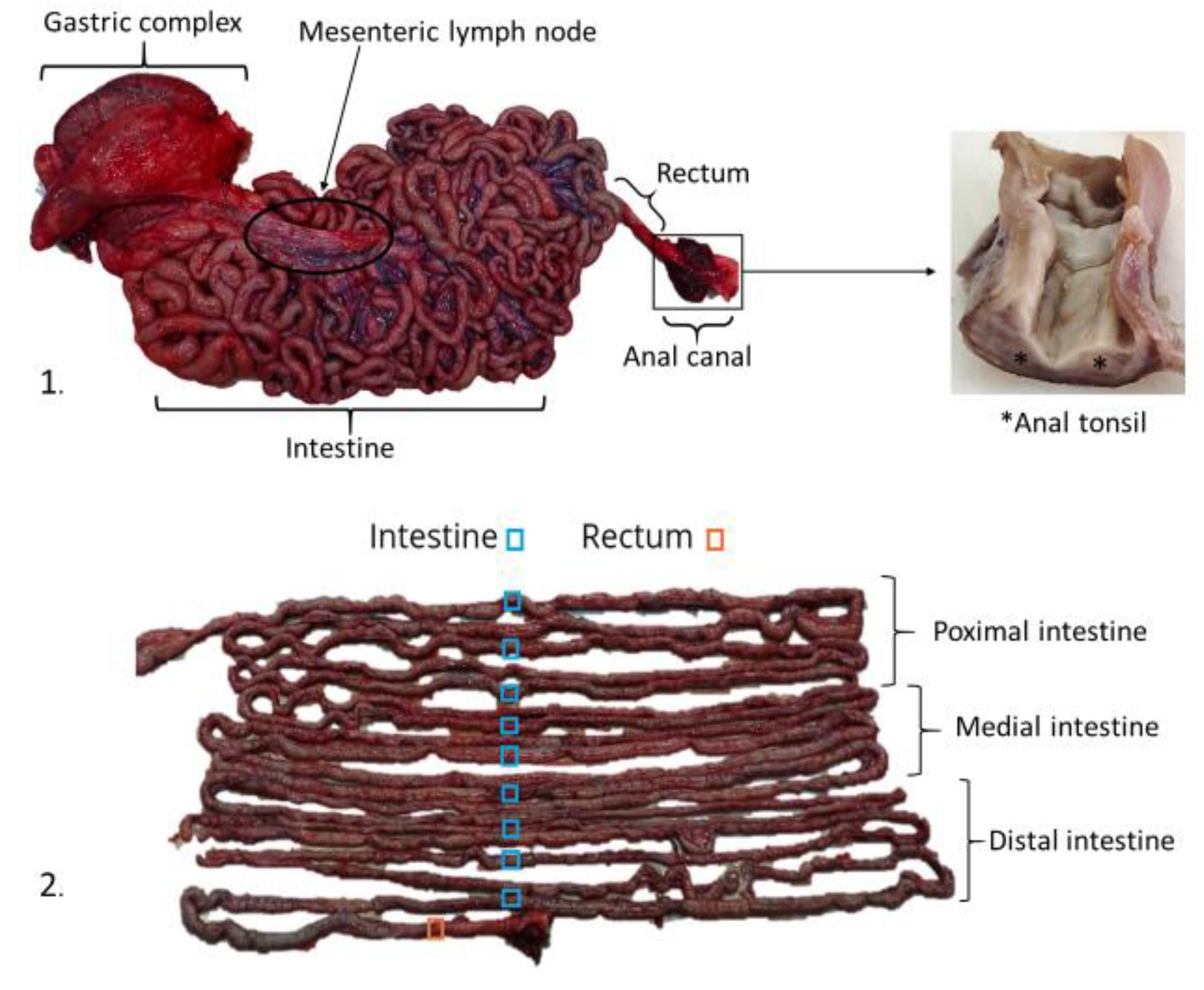
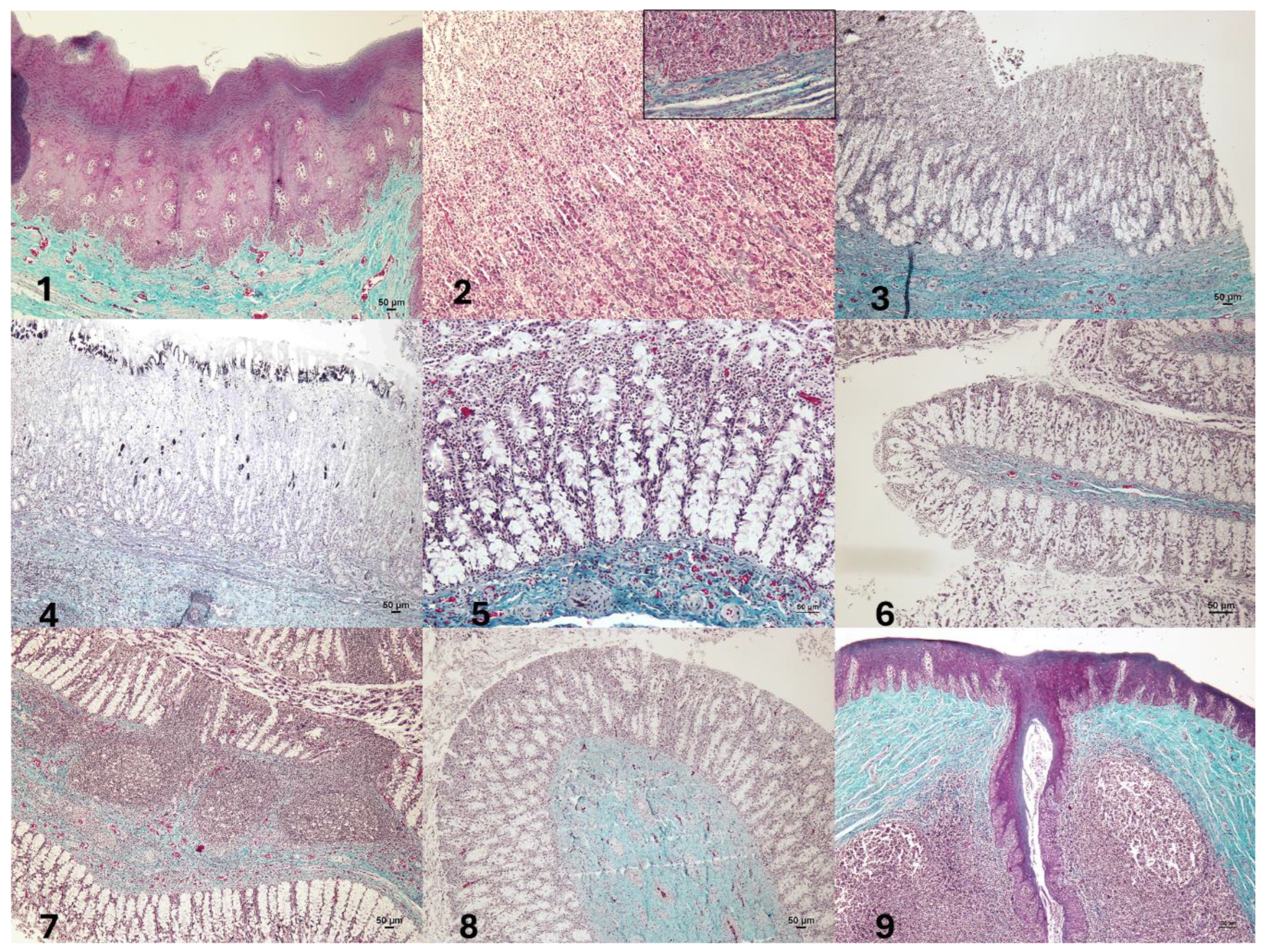
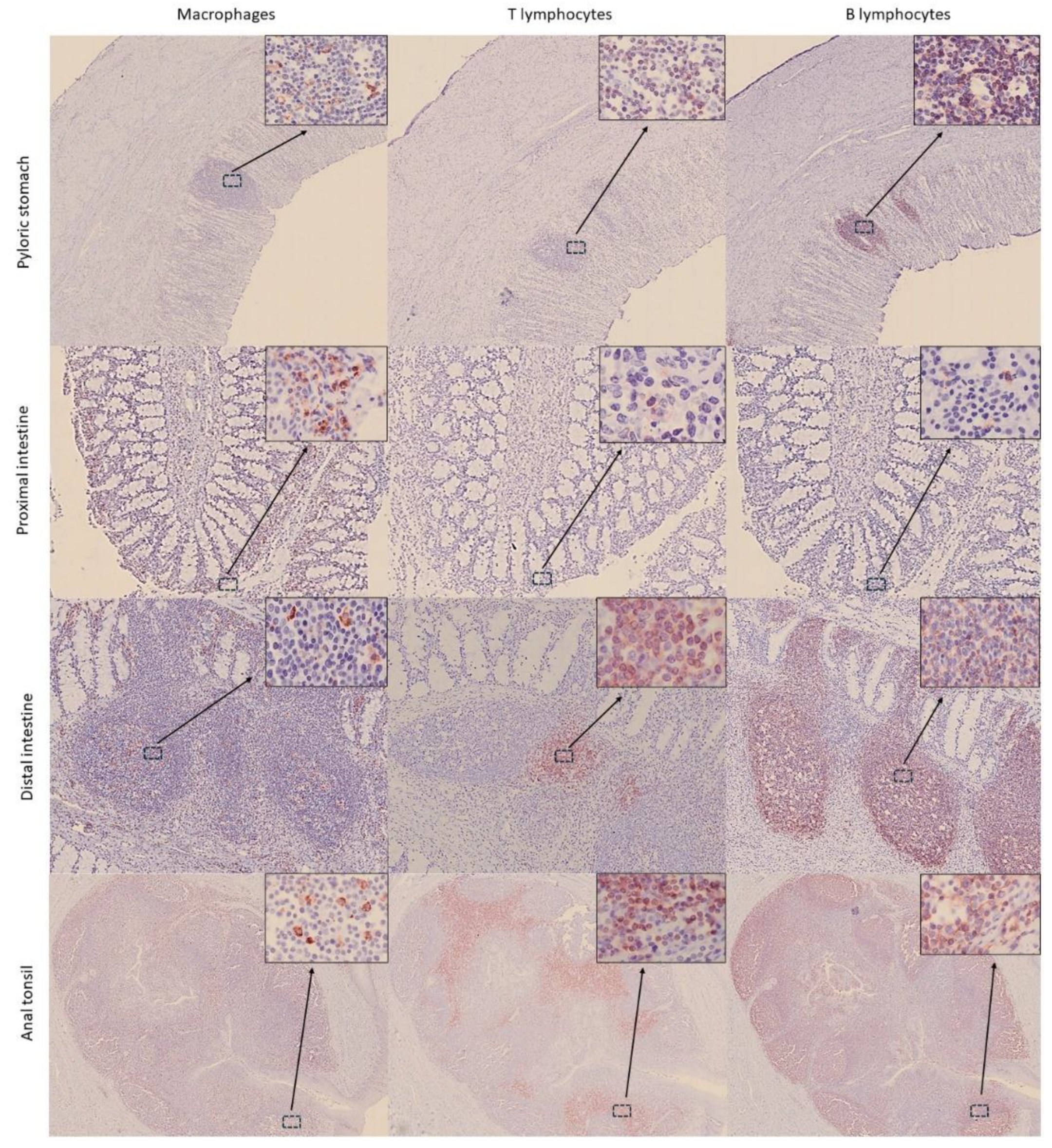
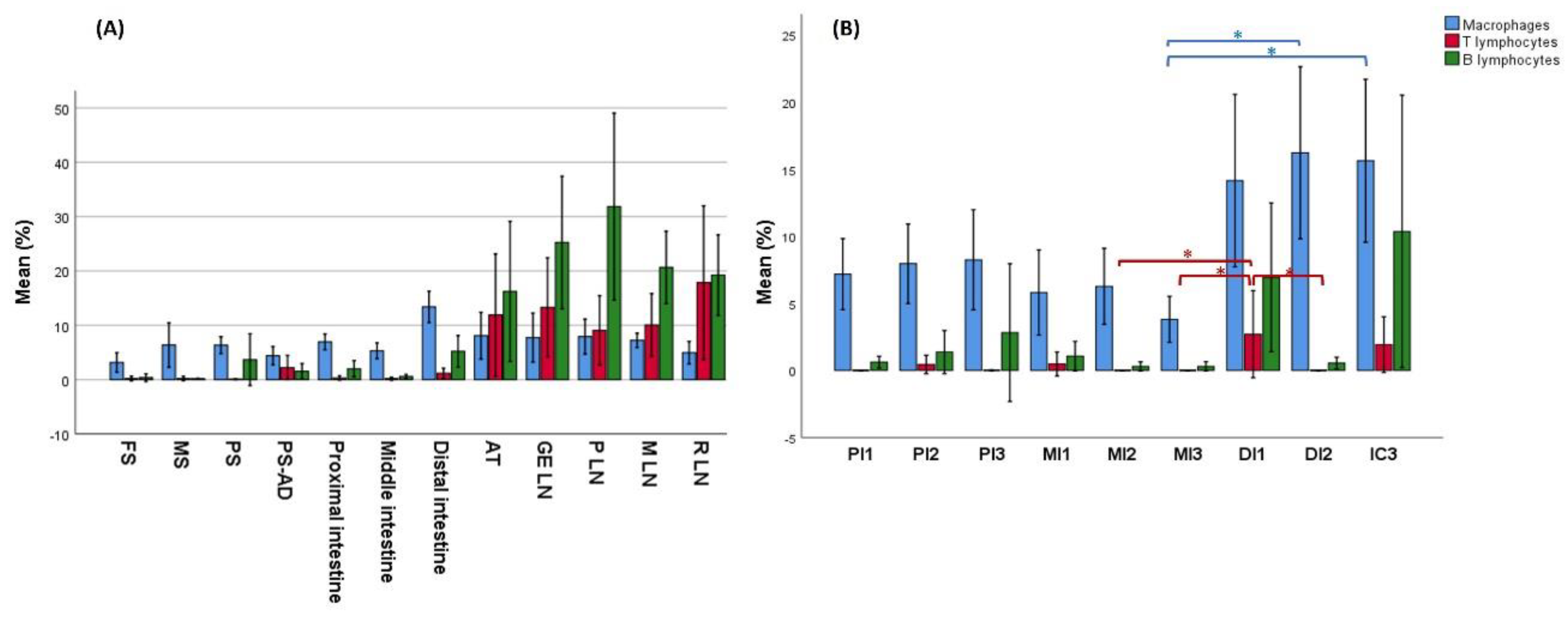
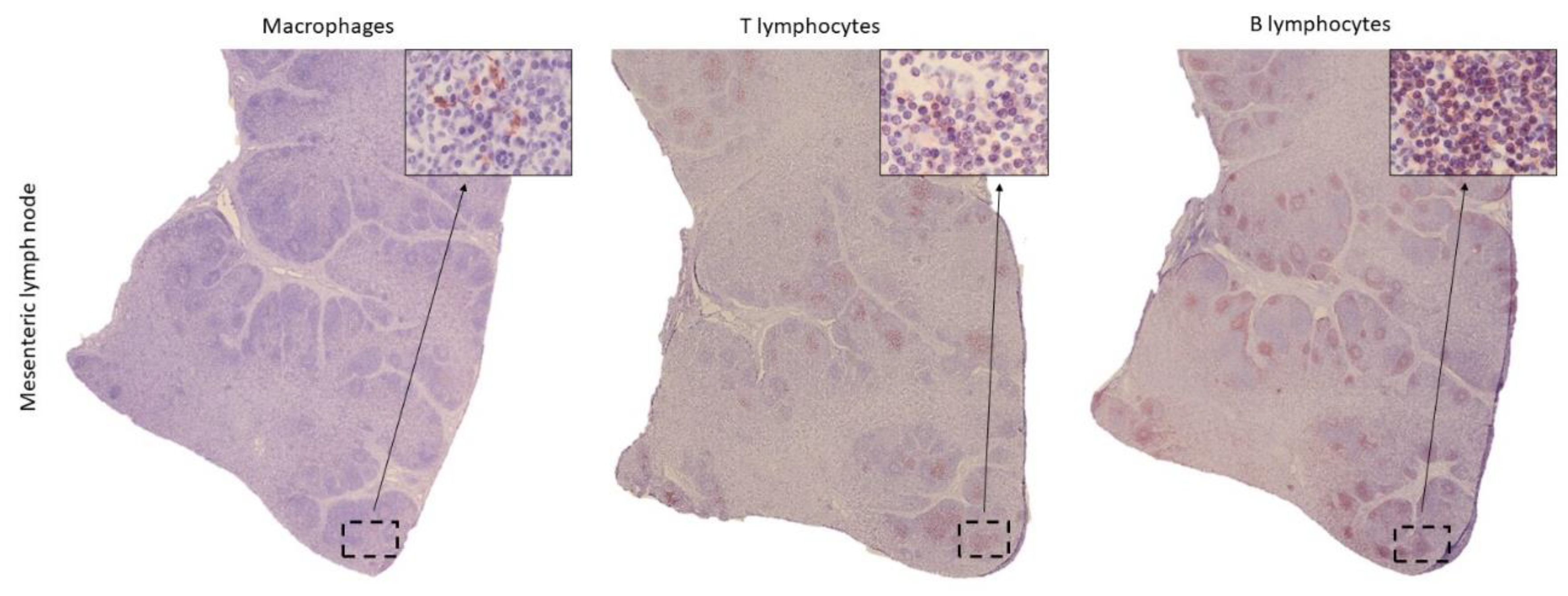
The current knowledge on the histological structure of the gastrointestinal tract (GIT) in cetaceans is based on general descriptions, which do not reflect the diversity of these marine mammals. The aim of this study was to characterize the histology and expression of immune cell markers in samples from the GIT and lymph nodes (LNs) in a harbour porpoise (Phocoena phocoena), which died due to bycatch in the Bay of Biscay, Cantabrian Sea. After hematoxylin-eosin and Masson's trichrome staining, the thickness of the histological layers of the GIT was measured, being greater in the stomachs and anal canal, although no significant differences were found among any intestinal segment (p = 0.448). LNs exhibited a well-differentiated cortical region, containing lymphoid follicles, and a medullary area. In addition, immunohistochemical techniques were performed to identify the following markers: IBA1 for macrophages, CD3 for T lymphocytes, and CD20 for B lymphocytes. The distribution of immune cells varied significantly along the GIT, with higher percentages of all three cell types in the distal intestine and the anal tonsil. Variation in thickness, morphology of the folds, and the presence of Peyer's patches allowed to distinguish the duodenal ampulla and the distal segments from the rest of the intestine. Within the LNs, B lymphocytes represented the predominant cell population. This study establishes a reliable methodology for the characterization of the histological structure of the GIT and associated lymphoid tissue in cetaceans and provides a description of the histological structure of those organs in harbour porpoise, useful for future research studies.

Keywords: 
;  ;  ;  ;  ;  

1. Introduction

Cetacea represent one of the most diverse order of mammals with two distinct suborders: Mysticeti which possess baleen plates in the oral cavity, and Odontoceti which have teeth instead [1].
The anatomy and histology of the gastrointestinal tract (GIT) of cetaceans can be found in various publications [1,2]; however, those are general descriptions in which numerous aspects remain unclear, especially considering the wide diversity of cetacean species. In addition, the study of the normal morphology of the GIT and the cell populations of associated lymphoid tissue in wild cetaceans faces significant methodological limitations, i.e., although stranded individuals provide valuable data, they often present underlying diseases or advanced deterioration, which can significantly alter the morphology of the GIT, compromising the representativeness of the findings. In contrast, individuals obtained through bycatch could be considered a suitable source for the histological characterization of the GIT, since the ultimate cause of death is attributed to fishery interaction, rather than to any underlying pathological condition.
Odontocetes exhibit anatomical peculiarities in their GIT. First, these animals possess a gastric complex consisted of three distinct chambers [3]. The first chamber, the forestomach, continues the oesophagus and performs a mechanical digestion function. Histologically, it is characterized by a stratified keratinized lining epithelium, reflecting the adaptation of this chamber to mechanical digestion, as odontocetes do not chew their food [4]. The main stomach follows the forestomach and serves as the principal chamber for enzymatic and chemical digestion, displaying a histological structure similar to that of terrestrial mammals [1]. Finally, the pyloric stomach is the last chamber and presents a mucosa containing specialized pyloric glands involved in chemical and enzymatic digestion [5]. At the beginning of the intestine, a dilation of the gut, namely the duodenal ampulla, is present, where the hepato-pancreatic duct empties in most species. Although it may be mistaken for a gastric compartment, it represents the most proximal portion of the intestine [1]. One of the most remarkable features in cetaceans of the Delphinidae and Phocoenidae families is the absence of macroscopic differentiation between the small and large intestine [6], with some authors referring to the entire gut as the small intestine [7]. In addition, while mysticetes possess a cecum and a vermiform appendix, these structures are absent in the odontocetes [3]. The intestine continues towards the rectum and the anal canal, where the anal tonsil, a relevant lymphoid tissue, is located [8].
Gut-associated lymphoid tissue (GALT) comprises lymphoid structures in the mucosa and submucosa of the GIT, playing a central role in immune tolerance and pathogen defence [9]. GALT includes Peyer’s patches (organized aggregates mainly found in the ileum), diffuse lymphoid follicles along the intestine, and isolated immune cells such as intraepithelial lymphocytes [10]. In cetaceans, studies in belugas reported diffuse lymphocytes and follicles but no true Peyer’s patches [11]. In young individuals, the distal rectum contains prominent lymphoid structures, that disappear just before the anal canal (“pigskin appearance”), which regress with age, remaining only scattered lymphocytes [8]. More recent studies have confirmed the presence of Peyer’s patches in juvenile cetaceans, especially in the mid to distal gut [12] and highlights age-related involution of GALT in bottlenose dolphins [13].
In addition, GALT includes the anal tonsil, which refers to a macroscopically visible lymphoid structure located in the anal canal, found in some cetacean species [6,11]. The anal tonsil may function in antigen presentation, reacting to waterborne antigens entering during diving [8]. It consists of lymphoid cell clusters with epithelial crypts and occasional mucous glands. In advanced involution stages, glandular structures suggest a shift from immune to mechanical functions, such as lubrication [13].
In contrast to the GIT, the histological structure of the lymph nodes (LNs) has been described in several studies conducted on cetaceans [8,14], and a morphology similar to that of terrestrial mammals’ LNs has been observed. However, in the odontocetes harbour porpoise (Phocoena Phocoena) and common dolphin (Delphinus delphis), it has been reported that a pig-like structure may occasionally be present [15], in which the cortical and medullary regions exhibit an inverted pattern.
Taking into account that lack of information, the aims of this study were to (i) histologically characterize the GIT and associated lymphoid tissue of a harbour porpoise which died due to bycatch, (ii) quantify and describe the distribution of macrophages, T lymphocytes (TL) and B lymphocytes (BL) in the lymphoid tissue present in the GIT and regional LNs and (iii) to establish a reliable method for further histological structure studies of GIT and LNs in cetaceans.

2. Materials and Methods

2.1. Sampling

One subadult male harbour porpoise, which died because of bycatch in the Bay of Biscay, was used in this study. Necropsy was performed in 48 hours and samples from the GIT and associated lymphoid tissue were collected and fixed in 10% buffer formalin for further histological and immunohistochemical (IHC) studies. GIT samples included all three chambers from the gastric complex (i.e., forestomach, main stomach and pyloric stomach), the transition between gastric compartment and intestine, duodenal ampulla, intestine and rectum. The intestine was divided into three regions: proximal, medial and distal, with a length of six meters each. Three samples were taken from equidistant points from each region, making a total of nine samples from the intestine (Figure 1). Intestinal samples were collected in a closed state to maintain the tubular shape, and formalin was carefully injected into the lumen to attempt to preserve the mucosal integrity. LNs were taken from the gastro-splenic and pancreatic regions. Additionally, the mesenteric LN was sampled and subdivided into proximal, middle, and distal portions. The rectal LN and the anal canal were also collected (with its associated anal tonsil). Pathological evaluation confirmed the absence of any disease.

2.2. Histology and Immunohistochemistry

For each sample (n=16), serial paraffin-embedded sections (3 µm) were used for histological and IHC studies. The staining techniques included the hematoxylin and eosin standard stain and Masson’s trichrome stain, for specific visualization of the connective tissue.
IHC consisted of the detection of three cell populations using different primary antibodies: ionized calcium-binding adaptor molecule 1 (IBA1) for macrophages, CD3 for TL and CD20 for BL. After being deparaffinized, antigen retrieval was carried out with sodium citrate buffer (10 mmol/L, pH 6.0) with heat induction by microwave for 20 min. Endogenous peroxidase activity was subsequently blocked by incubation in hydrogen peroxide (0.5%) solution in distilled water for 30 min at room temperature. The tissue sections were then incubated overnight at 4°C in a humidified chamber with commercial monoclonal or polyclonal antibodies diluted in TBS+BSA 0.1% (Table 1), washed with TBS 1x, and incubated with a secondary antibody (Vector Laboratories, California, USA), diluted 1:200 in TBS+BSA 0.1% (Table 1), followed by incubation with the Avidin-biotin-peroxidase complex reagent-method (ABC Standard, Vector Laboratories, California, USA) in TBS 1x for 30 min. Labeling was visualized using the Vector® NovaRed™ peroxidase substrate kit (Vector Laboratories, California, USA) as chromogen substrate. Slides were counterstained with Mayer´s haematoxylin, dehydrated and mounted with DPX (Fluka, Sigma, St. Louis, MO, USA). The negative control consisted of an additional slide without the primary antibody. Lymph node tissue from badger was used as a positive control for the three antibodies.

2.3. Measurement of Histological Layer Thickness in the GIT

Samples were examined using a Nikon E600 microscope and a Nikon DS-FI1 camera. Image analysis software (Nikon NIS-Elements Br, Nikon, Japan) was used to measure the thickness of the different layers of the GIT. Five measurements were taken for each sample: mucosa or mucosa-submucosa layers (when muscularis mucosae was absent), submucosa, and internal and external muscularis layers. A total of 350 measurements were taken.

2.4. Evaluation and Quantification of Cell Types in the Gastrointestinal Tract (GIT) and Lymph Nodes (LNs)

Immunostained tissue sections were scanned at the Microscopy Service of the University of León. An Olympus BX51 microscope (Olympus, Tokyo, Japan) and an Olympus XC10 camera (Olympus, Tokyo, Japan) were used. The digitally scanned samples were then analyzed using the QuPath v. 0.5.1 image analysis software [16] (QuPath, University of Edinburgh, Scotland).
For quantification of cells in the GIT, five fields were randomly sampled from mucosa, submucosa, and muscularis layers (a total of 15 counts/sample). Regarding LNs, five fields were randomly sampled from the diffuse lymphoreticular tissue of the cortex, lymphoid follicles, and medulla, obtaining 15 cell counts/LN.
For cell counting in each field, an area of ​​62,000 μm2 was delimited, yielding the following results: total number of cells detected, number of positive and negative cells, percentage of positive cells detected in the area, and number of positive cells per mm2. A total of 745 positive cell count fields were obtained.

2.5. Statistical Analysis

Statistical analysis was performed using IBM SPSS Statistics v. 22.0 software (IBM SPSS Statistics, IBM, USA). Since the values did not meet the assumptions of normality (Kolmogorov–Smirnov and Shapiro–Wilk tests), nonparametric tests were used. Statistical significance between categories was assessed using the Mann–Whitney U test and the Kruskal–Wallis test to compare two or more independent groups, respectively. When statistical differences were found, a pairwise comparisons were conducted. The level of statistical significance was set at p = 0.05.

3. Results and Discussion

3.1. Histological Structure of the Gastrointestinal Tract (GIT)

Main histological findings in the GIT are shown in Table 2. In the histological study of the gastric complex, the adaptation of forestomach to mechanical digestion [1], was reflected in the robust lining epithelium (Figure 2), the absence of glands, and the marked development of the muscularis (Table 2). In turn, the pronounced development of the mucosa (Table 2) and the high number of secretory cells in the main stomach highlighted the relevance of this chamber in chemical and enzymatic digestion. Although the literature considers that the duodenal ampulla and the remaining segments of the duodenum share the same structure [1], in this study the ampulla exhibited a greater development of the muscularis mucosae and lacked folds (Figure 2), which were present in the subsequent intestinal sections. Furthermore, the thickness of all histological layers was notably greater in the duodenal ampulla compared to the other intestinal regions (see Table 2). Consistent with previous descriptions, folds were observed in the intestinal mucosa [1,3,17], being more abundant in the proximal sections and gradually decreasing towards the more distal regions of the intestine.
The presence of glands in the submucosa has been reported to vary among different cetacean species [3]. In this study, no glands were observed in any of the gastrointestinal segments examined. Regarding villi, while some authors reported the absence of well-defined villi [7], others described short and thin villi [1,17], or even their complete absence in the most distal regions of the intestine [18]. In this porpoise, no villi were observed. Given these discrepancies, it is important to consider the challenges associated with cetacean sample collection and the rapid degeneration of intestinal mucosa after death. In the same line, no muscularis mucosae was observed in the rectum, which contrasts with its presence in other terrestrial mammalian species [19,20]. Also, numerous lymphoid follicles have been identified in the mucosa of this segment in other studies [4], although they were not observed in the harbour porpoise studied.
Finally, the statistical analysis of the thickness of the nine intestinal samples, taken from the duodenal ampulla to the rectum, showed no significant differences, making impossible to distinguish between the small and large intestine based solely on wall thickness.

3.2. Distribution of Immune Cells Within Gastrointestinal Tract (GIT) and Associated Lymphoid Tissue

In the GIT, the most frequently detected cells were macrophages, followed by BL and TL. Likewise, the submucosa represented the layer with the highest density of immunopositive cells, followed by the mucosa and muscularis.
Within the gastric chambers, the pyloric stomach exhibited the highest percentage of immunopositive cells, with an average of 6.73% of IBA1-stained cells in the mucosa, 8.83% in the submucosa, and 3.44% in the muscularis layer. However, the higher cellular positivity in this chamber, at its transition with the intestine and in the duodenal ampulla itself, is likely associated with the presence of lymphoid cell clusters (Figure 3).
In the intestine, significant differences were observed in the distribution of the three cell populations across the nine collected segments (p = 0.005, p = 0.000, and p = 0.034 for macrophages, TL, and BL, respectively) (Figure 4B). Macrophages were mainly located in the apical region of the mucosa, forming an almost continuous layer of cells (Figure 3). TL and BL were diffusely distributed near the lamina propria of the mucosa layer and predominated in the lymphoid aggregates observed in the submucosa, with BL being the most abundant cell (Figure 3). While immune cells in the proximal and medial intestine samples appeared only occasionally as aggregates, in the distal segment, aggregates were more frequently observed, forming well-defined lymphoid follicles in the submucosa and infiltrating the mucosa layer (Figure 3). This was reflected in the marked increase in immune cell detection in this last segment (Figure 4). Furthermore, the follicles found in the distal portions were structures similar to the Peyer’s patches of terrestrial mammals [20]. These structures appeared as well-defined, circular lymphoid follicles in the submucosa layer, either isolated or in contact with nearby follicles, sometimes infiltrating the entire thickness of the mucosa (Figure 3). They were frequently detected in mucosal folds, and no well-defined germinal centers were observed. Also, macrophages were diffusely distributed within these Peyer’s patches, BL were the most abundant cells predominating in all follicles and mucosal infiltrations, and TL were diffusely distributed, mainly in the interfollicular areas. Although these lymphoid structures are consistent with those described by Silva et al. (2016) [12], the authors reported their presence in the medial region and final third of the intestine, whereas in this study they were exclusively observed in the final third. In this line, Cowan and Smith (1999) [8] described the presence of a continuous layer of lymphoid structures in the lamina propria of the distal intestinal segments beyond the splenic flexure, without providing a detailed description of the tissue morphology. Moreover, authors suggested that this segment was analogous to the vermiform appendix. In comparison to other terrestrial mammals, where Peyer’s patches are most frequently found in the ileum [19,21], our findings suggest that, in the harbour porpoise, these lymphoid structures are located further distally in the intestine, where the colon would typically be expected. However, it should be considered that the porpoise studied in this work was a subadult, in which Peyer’s patches were likely undergoing involution [13].
In addition, immune cells were identified in the anal canal, diffusely distributed within the keratinized stratified epithelium and the mucosa-submucosa (Figure 3 and Figure 4). However, most immune cells were concentrated in large aggregates of lymphoid tissue surrounding the epithelial crypts, corresponding to the anal tonsil. Although the histological structure observed was consistent with previous descriptions [8,12], the use of IHC techniques in the present study provided deeper insights into the cellular composition of this organ. In this regard, while macrophages were the most frequently detected immune cells throughout the GIT, in the anal tonsil they were less abundant than TL and BL, with the latter being the most prevalent (average percentage of positive cells in the mucosa-submucosa: 10.7%, 17.8%, and 24.3%, respectively) (Figure 4). The higher proportion of BL may be related to a stronger local humoral immune response at this level [9]. Regarding their distribution, BL and macrophages were observed diffusely in the epithelium and lamina propria-submucosa, as well as within the lymphoid aggregates, whereas TL were detected exclusively between these clusters. Those aggregates showed well-defined follicles, with macrophages diffusely distributed, TL predominating in the interfollicular regions, and BL distributed both diffusely or associated with the mantle and germinal center of the lymphoid follicles. Interestingly, the cellular detection levels of macrophages, TL, and BL were comparable to those observed in the LN analyzed in this study, as no significant differences were found. Therefore, based on its cellular composition, the anal tonsil might be considered a lymphoid organ analogous to the LNs.
Although some publications have described the LNs of the harbour porpoise as having an inverted structure similar to that of pigs [15], in the present study they exhibited a typical structure, with a well-differentiated cortex containing lymphoid follicles and a medullary region (Figure 5), consistent with descriptions in other cetacean species [17,22]. Overall, BL were the most frequently detected cells, followed by lower proportions of TL and macrophages (Figure 4). As expected, the cortical lymphoid follicles represented the region with the highest density of immunopositive cells, compared to the diffuse lymphoreticular tissue of both cortical and medullary regions (Figure 5). The percentage of macrophages did not show significant differences between the different LNs. In contrast, BL showed higher positivity in the cranial LNs, with the main detection in the pancreatic LN, followed by a gradual decrease in caudal LNs (Figure 4). In contrast, TL displayed an opposite pattern to that of BL, increasing in the caudal regions and reaching maximum positivity in the rectal LN (Figure 4).
A limitation of this work is the study of a single specimen; however, the inherent challenges in obtaining cetacean samples, particularly from healthy individuals, must be considered. In addition, IHC resulted an useful tool to detect immune cells in GALT, thus opening up the possibility of future immune response studies in cetaceans.

5. Conclusions

This study provides the first detailed histological and immunochemical characterization of the GIT and associated lymphoid tissue in harbour porpoise. The results highlight structural adaptations in the GIT for digestion and regional variation in immune cell distribution, which enables microscopic identification of the distal intestinal segment, even though distinguishing between the small and large intestines remains unfeasible. Lymphoid follicles resembling Peyer’s patches were restricted to the distal intestine, and the anal tonsil showed features comparable to LNs, supporting its relevant role as a major lymphoid organ. LNs showed typical mammalian organization, with B lymphocytes predominating and displaying cranio-caudal variation opposite to that of T cells. This study establishes a preliminary information for future research on cetacean health.

Author Contributions

Conceptualization, A.B. and N.G-A.; methodology, D.P-M., A.B., P.B., I.M., A.F., J.F.G.M. and N.G-A.; software, D.P-M.; validation, A.B., A.F. and N.G-A.; formal analysis, D.P-M., A.B. and N.G-A.; investigation, D.P-M., A.B., P.B., I.M., A.F., J.F.G.M. and N.G-A.; resources, A.B., J.F.G.M. and A.F.; writing—original draft preparation, D.P-M., N.G.A. and A.B.; writing—review and editing, all authors; funding acquisition, A.F., J.F.G.M. and A.B. All authors have read and agreed to the published version of the manuscript.

Funding

This research was funded by Convenio entre el Ministerio de Agricultura, Pesca y Alimentación, la Universidad de Las Palmas de Gran Canaria y la Universidad de León, sobre investigación de cetáceos para una pesca ambientalmente sostenible con fondos del Plan de Recuperación, Transformación y Resiliencia (PRTR).

Institutional Review Board Statement

Ethical review and approval were waived for this study due to the animal died due to by-catch.

Informed Consent Statement

Not applicable.

Data Availability Statement

All data can be found in the Manuscript.

Acknowledgments

The authors thank AZTI, Centro de Investigación Marina y Alimentaria and Grupo TRAGSA for helping in the sampling.

Conflicts of Interest

The authors declare no conflicts of interest.

Abbreviations

The following abbreviations are used in this manuscript:
BL
TL
GALT
GIT
IBA1
IHC
LN
B lymphocytes
T lymphocytes
Gut associated lymphoid tissue
Gastrointestinal tract
Ionized calcium-binding molecule 1
Immunohistochemistry
Lymph node

References

  1. Cozzi, B.; Huggenberger, S.; Oelschläger, H. Anatomy of dolphins: insights into body structure and function, 1st ed.; Academic press: London, UK, 2017; pp. 344–359. [Google Scholar]
  2. Fiorucci, L. Determinación de los principales datos de referencia para el examen del tracto gastrointestinal en delfines mulares (Tursiops truncatus) clínicamente sanos bajo el cuidado humano. Doctoral Dissertation, Universidad de Las Palmas de Gran Canaria, Spain, 2016. [Google Scholar]
  3. Rommel, S.A.; Lowenstine, L.J. Gross and microscopic anatomy. In CRC Handbook of marine mammal medicine, 1st ed.; Dierauf, L.A., Gulland, F.M.D., Eds.; CRC Press: Boca Ratón, USA, 2001; pp. 129–164. [Google Scholar]
  4. Huggenberger, S. : Oelschläger, H.; Cozzi, B. Atlas of the anatomy of dolphins and whales, 1st ed.; Academic press: London, UK, 2019; pp. 363–479. [Google Scholar]
  5. Caballero Cansino, M.J.; Jaber Mohamad, J.R.; Fernández Rodríguez, A. Atlas de histología de peces y cetáceos, 1st ed.; Vicerrectorado de Planificación y Calidad de la Universidad de Las Palmas de Gran Canaria: Las Palmas de Gran Canaria, España, 2005; pp. 87–94. [Google Scholar]
  6. Mead, J.G. Gastrointestinal tract. In Encyclopedia of marine mammals., 2nd ed.; Perrin, W.F., Würsig, B., Thewissen, J.G.M., Eds.; Academic Press: London, UK, 2009; pp. 472–477. [Google Scholar]
  7. Harrison, R.J.; Johnson, F.R.; Young, B.A. The small intestine of dolphins (Tursiops, Delphinus, Stenella). In Functional Anatomy of Marine Mammals., 1st ed.; Harrison, R.J., Ed.; Academic Press: New York, USA, 1977; pp. 297–331. [Google Scholar]
  8. Cowan, D. F.; Smith, T. L. Morphology of the lymphoid organs of the bottlenose dolphin, Tursiops truncates. J Anat 1999, 194, 505–517. [Google Scholar] [CrossRef] [PubMed]
  9. Bemark, M.; Pitcher, M. J.; Dionisi, C.; Spencer, J. Gut-associated lymphoid tissue: a microbiota-driven hub of B cell immunity. Trends Immunol 2024, 45, 211–223. [Google Scholar] [CrossRef] [PubMed]
  10. Olivares-Villagómez, D.; Van Kaer, L. Intestinal intraepithelial lymphocytes: sentinels of the mucosal barrier. Trends Immunol 2018, 39, 264–275. [Google Scholar] [CrossRef] [PubMed]
  11. Romano, T.A.; Felten, S.Y.; Olschowka, J.A.; Felten, D.L. A microscopic investigation of the lymphoid organs of the beluga, Delphinapterus leucas. J Morphol 1993, 215, 261–287. [Google Scholar] [CrossRef] [PubMed]
  12. Silva, F. M. O.; Guimarães, J. P.; Vergara-Parente, J. E.; Carvalho, V. L.; Carolina, A.; Meirelles, O.; Marmontel, M.; Oliveira, B. S. S. P.; Santos, S. M.; Becegato, E. Z.; Evangelista, J. S. A. M.; Miglino, M. A. Morphology of mucosa-associated lymphoid tissue in odontocetes. Micros Res Tech 2016, 79, 845–855. [Google Scholar] [CrossRef]
  13. Clark, L.S.; Turner, J.P.; Cowan, D.F. Involution of lymphoid organs in bottlenose dolphins (Tursiops truncatus) from the western Gulf of Mexico: implications for life in an aquatic environment. Anat Rec 2005, 282A, 67–73. [Google Scholar] [CrossRef] [PubMed]
  14. Vuković, S.; Lucić, H.; Gomerčić, H.; Duras-Gomerčić, M.; Gomerčić, T.; Škrtić, D.; Vurković, S. Morphology of the lymph nodes in bottlenose dolphin (Tursiops truncatus) and striped dolphin (Stenella coeruleoalba) from the Adriatic Sea. Acta Vet Hung 2005, 53, 1–11. [Google Scholar] [CrossRef]
  15. Moskov, M.; Schiwatschewa, T.; Bonev, S. Comparative histological study of lymph nodes in mammals. Lymph nodes of the dolphin. Anat Anz 1969, 124, 49–67. [Google Scholar] [PubMed]
  16. Bankhead, P.; Loughrey, M.B.; Fernández, J.A.; Dombrowski, Y.; McArt, D. G.; Dunne, P.D.; McQuaid, S.; Gray, R.T.; Murray, L.J.; Coleman, H.G.; James, J.A.; Salto-Tellez, M.; Hamilton, P.W. QuPath: Open source software for digital pathology image analysis. Sci Rep 2017, 7, 1–7. [Google Scholar]
  17. Simpson, J. C.; Gardner, M.D. Comparative microscopic anatomy of selected marine mammals. In Mammals of the sea: biology and medicine., 1st ed.; Ridgway, S.H., Ed.; Charles C Thomas: Springfield, USA, 1972; pp. 298–418. [Google Scholar]
  18. Russo, F.; Gatta, C.; De Girolamo, P.; Cozzi, B.; Giurisato, M.; Lucini, C.; Varricchio, E. Expression and immunohistochemical detection of leptinlike peptide in the gastrointestinal tract of the South American sea lion (Otaria flavescens) and the bottlenose dolphin (Tursiops truncatus). Anat Rec 2012, 295, 1482–1493. [Google Scholar] [CrossRef] [PubMed]
  19. Frappier, B.L. Digestive system. In Dellmann’s textbook of veterinary histology., 6th ed.; Eurell, J.A., Frappier, B.L., Eds.; Blackwell Publishing: Ames, USA, 2006; pp. 170–211. [Google Scholar]
  20. Junqueira, L.C.; Carneiro, J. Junqueira & Carneiro. Histología básica. Texto y atlas, 13th ed.; Editorial Médica Panamericana: City of Mexico, Mexico, 2022; pp. 308–329. [Google Scholar]
  21. Buendía Martín, A.J.; Durán Flórez, E.; Gázquez Ortiz, A.; Gómez Cabrera, S.; Méndez Sánchez, A.; Navarro Cámara, J.A. Aparato digestivo. In Tratado de histología veterinaria., 1st ed.; Gázquez Ortiz, A., Blanco Rodríguez, A., Eds.; Masson: Barcelona, Spain, 2004; pp. 239–280. [Google Scholar]
  22. Silva, F. M. D. O. E.; Guimarães, J. P.; Vergara-parente, J. E.; Carvalho, V. L.; De Meirelles, A. C. O.; Marmontel, M. , Ferrão, J. S. P.; Miglino, M. A. Morphological análisis of lymph nodes in odontocetes from northand northeast coast of Brazil. Anat Rec 2014, 297, 939–948. [Google Scholar] [CrossRef] [PubMed]
Figure 1. Macroscopical structure of the gastrointestinal tract of harbour porpoise (Phocoena phocoena). 1. Gastrointestinal segments are shown. 2. The nine intestinal samples collected are highlighted.
Preprints 181218 g001
Figure 2. Microphotographs of the different segments of the gastrointestinal tract in harbour porpoise (Phocoena phocoena). Masson’s trichrome staining. 1. Forestomach (40x); 2. Main stomach (100x); 3. Pyloric stomach (40x); 4. Duodenal ampulla (40x); 5. Proximal intestine (100x); 6. Middle intestine (40x); 7. Distal intestine (40x); 8. Rectum (40x); 9. Anal canal (40x).
Preprints 181218 g002
Figure 3. Distribution of immune cells (macrophages—IBA1, B lymphocytes—CD20, and T lymphocytes—CD3) in the gastrointestinal tract of harbour porpoise (Phocoena phocoena). Microphotographs at 25x magnification; insets at 400x magnification.
Preprints 181218 g003
Figure 4. Percentage of immune cells along the gastrointestinal tract and lymph nodes in harbour porpoise (Phocoena phocoena). (A) Mean percentage of immune cells (macrophages—IBA1, B lymphocytes—CD20, and T lymphocytes—CD3). FS, forestomach; MS, main stomach; PS, pyloric stomach; DA, duodenal ampulla; AT, anal tonsil; GE LN, gastrosplenic lymph node; P LN, pancreatic lymph node; M LN, mesenteric lymph node; R LN, rectal lymph node. (B) Samples collected from cranial to distal segments of proximal intestine, PI1-PI2-PI3, middle intestine, MI1-MI2-MI3, and distal intestine, DI1, DI2, DI3. When significant differences were found, a pairwise comparisons were conducted (*). Error bars represent 95% confidence intervals.
Preprints 181218 g004
Figure 5. Distribution of immune cells (macrophages—IBA1, B lymphocytes—CD20, and T lymphocytes—CD3) in the proximal mesenteric lymph node of harbour porpoise (Phocoena phocoena). Microphotographs at 25x magnification; insets at 400x magnification.
Preprints 181218 g005
Table 1. Primary and secondary antibodies used for cellular type characterization.
Primary antibody
(dilution)
Cell type
detected
Clone nº Source Secondary antibody
(dilution)
IBA11
(1:1000)

Macrophages
Polyclonal
019-19741
FLUJIFILM-Wako Chemicals Eu-rope GmbH, Neuss, Germany Goat anti-rabbit
(1:200)
CD3
(1:500)

T lymphocytes
Monoclonal
NCL-L-CD3-565
Novacastra, Leica Biosystem, Newcastle, UK Horse anti-mouse
(1:200)
CD20
(1:400)

B lymphocytes
Polyclonal
PA5-16701
ThermoFisher, Massachusetts, USA Goat anti-rabbit
(1:200)
1 Ionized calcium-binding adapter molecule 1.
Table 2. Main histological findings in the gastrointestinal tract of harbour porpoise (Phocoena phocoena).
Structure Mucosa Submucosa Muscularis
Forestomach (FS) Stratified keratinized epithelium. Muscularis mucosae highly developed. Absence of glands. Thickness: 937 microns (µm). No glands. Submucosal plexus. Thickness: 252 µm. Layers difficult to differentiate. Myenteric plexus. Thickness: 1430 µm.
Main stomach (MS) Simple columnar epithelium. Gastric pits with secretory cells. Muscularis mucosae less developed than in E1. Thickness: 3180 µm. No glands. Submucosal plexus. Thickness: 403 µm. Inner and outer layers easily distinguishable. Myenteric plexus. Thickness: 913 µm (inner) and 432 µm (outer).
Pyloric stomach (PS) Simple columnar epithelium. Pyloric glands. Muscularis mucosae. Thickness: 1260 µm. No glands. Submucosal plexus. Thickness: 292 µm. Inner and outer layers easily distinguishable. Myenteric plexus. Thickness: 774 µm (inner) and 212 µm (outer).
Duodenal ampulla (DA) Simple columnar epithelium. Mucous glands. Muscularis mucosae poorly developed. No folds. Thickness: 1140 µm. No glands. No submucosal plexus. Thickness: 639 µm. Inner and outer layers easily distinguishable. Myenteric plexus. Thickness: 712 µm (inner) and 512 µm (outer).
Proximal intestine 1 (PI1) Simple columnar epithelium. Mucous glands. Muscularis mucosae very poorly developed. No villi. Numerous folds. Thickness: 272 µm. No glands. No submucosal plexus. Thickness: 135 µm. Inner and outer layers easily distinguishable. Myenteric plexus. Thickness: 245 µm (inner) and 61.8 µm (outer).
Proximal intestine 2 (PI2) Simple columnar epithelium. Mucous glands. Muscularis mucosae very poorly developed. No villi. Numerous folds. Thickness: 461 µm. No glands. No submucosal plexus. Thickness: 97.6 µm. Inner and outer layers easily distinguishable. Myenteric plexus. Thickness: 629 µm (inner) and 162 µm (outer).
Proximal intestine 3 (PI3) Simple columnar epithelium. Mucous glands. Muscularis mucosae very poorly developed. No villi. Numerous folds. Thickness: 361 µm. No glands. No submucosal plexus. Thickness: 96.8 µm. Inner and outer layers easily distinguishable. Myenteric plexus. Thickness: 629 µm (inner) and 191 µm (outer).
Middle intestine 1 (MI1) Simple columnar epithelium. Mucous glands. Muscularis mucosae very poorly developed. No villi. Numerous folds. Thickness: 351 µm. No glands. No submucosal plexus. Thickness: 63.9 µm. Inner and outer layers easily distinguishable. Myenteric plexus. Thickness: 522.464 µm (inner) and 130 µm (outer).
Middle intestine 2 (MI2) Simple columnar epithelium. Mucous glands. Muscularis mucosae very poorly developed. No villi. Few folds. Thickness: 423 µm. No glands. No submucosal plexus. Thickness: 96.3 µm. Inner and outer layers easily distinguishable. Myenteric plexus. Thickness: 456 µm (inner) and 91.4 µm (outer).
Middle intestine 3 (MI3) Simple columnar epithelium. Mucous glands. Muscularis mucosae very poorly developed. No villi. Few folds. Thickness: 373 µm. No glands. No submucosal plexus. Thickness: 111 µm. Inner and outer layers easily distinguishable. Myenteric plexus. Thickness: 408 µm (inner) and 124 µm (outer).
Distal intestine 1 (DI1) Simple columnar epithelium. Mucous glands. Muscularis mucosae very poorly developed. No villi. Few folds. Thickness: 328 µm. No glands. No submucosal plexus. Thickness: 142 µm. Inner and outer layers easily distinguishable. Myenteric plexus. Thickness: 359 µm (inner) and 112 µm (outer).
Distal intestine 2 (DI2) Simple columnar epithelium. Mucous glands. Muscularis mucosae very poorly developed. No villi. Few folds. Thickness: 364 µm. No glands. No submucosal plexus. Thickness: 142 µm. Inner and outer layers easily distinguishable. Myenteric plexus. Thickness: 373 µm (inner) and 90.3 µm (outer).
Distal intestine 3 (DI3) Simple columnar epithelium. Mucous glands. Muscularis mucosae very poorly developed. No villi. Few folds. Thickness: 344 µm. No glands. No submucosal plexus. Thickness: 143 µm. Inner and outer layers easily distinguishable. Myenteric plexus. Thickness: 518 µm (inner) and 103 µm (outer).
Rectum (R) Simple columnar epithelium. Mucous glands. No muscularis mucosae. Wide folds. Thickness: 737 µm. No glands. Submucosal plexus present. Inner and outer layers easily distinguishable. Myenteric plexus. Thickness: 808 µm (inner) and 353 µm (outer).
Anal canal (AC) Stratified keratinized epithelium. Crypts with stratified keratinized epithelium. No glands. No muscularis mucosae. Thickness: 3400 µm. No glands. No submucosal plexus. Large lymphoid aggregates (anal tonsil). Inner and outer layers easily distinguishable. Myenteric plexus. Thickness: 854 µm (inner) and 544 µm (outer).
Disclaimer/Publisher’s Note: The statements, opinions and data contained in all publications are solely those of the individual author(s) and contributor(s) and not of MDPI and/or the editor(s). MDPI and/or the editor(s) disclaim responsibility for any injury to people or property resulting from any ideas, methods, instructions or products referred to in the content.
Copyright: This open access article is published under a Creative Commons CC BY 4.0 license, which permit the free download, distribution, and reuse, provided that the author and preprint are cited in any reuse.
Prerpints.org logo

Preprints.org is a free preprint server supported by MDPI in Basel, Switzerland.

Subscribe

Disclaimer

Terms of Use

Privacy Policy

Privacy Settings

© 2026 MDPI (Basel, Switzerland) unless otherwise stated