Preprint
Article

This version is not peer-reviewed.

Comparative Analysis of Embryonic Development and Mitochondrial Genome of a New Intergeneric Hybrid Grouper (Epinephelus fasciatus ♀ × Plectropomus leopardus ♂)

A peer-reviewed article of this preprint also exists.

Submitted:

13 October 2025

Posted:

14 October 2025

You are already at the latest version

Abstract

To investigate the early developmental characteristics and mitochondrial inheritance patterns of the hybrid offspring of a paternal Plectropomus leopardus and maternal Epinephelus fasciatus, the study systematically mapped the embryonic developmental trajectory and mitochondrial genome of the new germplasm. Results revealed that the fertilized hybrid eggs completed embryonic development within 28h55min, with the newly hatched larvae measuring 2.05 ± 0.37 mm in total length. The mitochondrial genome length of the hybrid was 16,570 bp, preserving 13 protein-coding genes (PCGs), two ribosomal RNA (rRNA) genes, and 22 transfer RNA (tRNA) genes. The hybrid's mitochondrial gene composition and arrangement showed high consistency with that of the maternal E. fasciatus. Concurrently, the co-linearity, Ka/Ks, and phylogenetic tree analyses collectively indicate that the hybrid progeny has a closer genetic relationship with the maternal parent, supporting the mitochondrial maternal inheritance of this species. This study details the embryonic development and mitochondrial inheritance of an intergeneric hybrid grouper germplasm, providing significant molecular biological evidence for grouper hybrid breeding, germplasm resource identification, and genetic diversity conservation.

Keywords: 
;  ;  ;  ;  

1. Introduction

Within the Serranidae family, the black-edged grouper (Epinephelus fasciatus) and leopard coral grouper (Plectropomus leopardus) both inhabit tropical and subtropical coral reefs. With their vivid orange-red coloration, they are prized for their delicious flesh, making them important aquaculture and ornamental fish. Morphologically, E. fasciatus has an orange-red body, with the upper part of the head and back being reddish-brown. Its sides are marked with 5-6 wide transverse bands, and the tips of the dorsal fin spines are black. It is primarily found in the eastern coastal waters of the Indian Ocean to the central Pacific Ocean, and the waters of Taiwan, China, and the South China Sea [1]. P. leopardus has a bright red body, with small blue spots across its trunk and head [2]. It is primarily distributed in the southwestern Pacific Ocean, northern Australia, and the South China Sea [3]. E. fasciatus is a newly developed and valuable aquaculture species, with fry (5-8 cm) priced at 40 Yuan per individual at ex-factory price and supply falling short of demand [4]. However, E. fasciatus is a slow-growing, medium-to-small fish species, with adult fish only reaching a length of 50 cm [5]. Under artificial farming conditions, one-year-old fish weigh only 58.89 ± 18.78 g, which significantly restricts the large-scale farming and industrial promotion of this species. On the other hand, P. leopardus is also a valuable aquaculture species, attaining a maximum adult body length of 120 cm [6] and a one-year-old weight of 525 g. However, with the expansion of aquaculture, problems such as miniaturization of breeding stock, low fry survival rates, poor stress resistance, and frequent diseases have emerged, severely constraining the sustainable development of grouper farming. Addressing these industry bottlenecks, the development of fast-growing and high-quality aquaculture germplasm through efficient genetic improvement is of great significance for advancing the upgrading of the grouper seed industry and grouper aquaculture as a whole.
Distant hybridization breeding technology enables the integration of genomes from species with distant genetic relationships, yielding hybrid offspring with highly variable genotypes and phenotypes. With over 160 species, groupers represent a rich genetic resource for hybrid breeding research. China's grouper aquaculture sector yields 267,488 tons of fish per annum, with an output value of 30 billion Yuan, and hybrid varieties constitute more than 70% of the production. Among the currently approved new grouper varieties, three hybrids of E. fuscoguttatus ♀ × E. lanceolatus ♂, E. moara ♀ × E. lanceolatus ♂, and E. fuscoguttatus ♀ × E. tukula ♂ have been successfully developed. These hybrid varieties demonstrate significant hybrid growth vigor, stress tolerance, and survival rates. For instance, one-year-old E. moara ♀ × E. lanceolatus ♂ shows 100% faster growth compared to their maternal E. moara [7]. Research on E. fuscoguttatus ♀ × E. tukula ♂ revealed that 15-month-old offspring grew 103% faster than the maternal E. fuscoguttatus, while displaying superior cold tolerance through significantly lower feeding cessation at semi-lethal temperatures than parental strains [8].
Currently, interspecific hybridization dominates grouper crossbreeding efforts, while intergeneric hybridization studies are rare. For example, the hybrid offspring of Cromileptes altivelis ♀ × E. tukula ♂ exhibits significant growth heterosis. At 330 days of age, their body weight reaches 220.50 ± 25.30 g, 1.55 times that of the maternal C. altivelis, and their total length measures 23.57 ± 0.94 cm, 1.28 times that of the maternal parent [9]. The intergeneric hybrid offspring of C. altivelis ♀ × E. lanceolatus ♂ grows significantly faster than the maternal C. altivelis, with an absolute weight growth rate 1.6 times higher, and a 4.70% increase in meat yield compared to the paternal E. lanceolatus [10]. In addition, embryos of the intergeneric hybrid E. fuscoguttatus ♀ × P. leopardus ♂ develop normally. Here, comparative transcriptomics of dynamic embryonic development between hybrid and parental embryos identified that the Wnt signaling pathway plays a crucial role in regulating embryonic dorsal-ventral axis formation [11].
Earlier studies established that optimal growth performance in grouper hybrids requires selection of large-bodied, fast-growing males as paternal contributors [12]. Therefore, the large and vividly colored P. leopardus was first selected as the paternal parent to improve the growth rate of E. fasciatus through distant hybridization breeding, successfully producing viable hybrid offspring. Through early embryonic development and mitochondrial genome comparative analysis, the feasibility of intergeneric hybridization in groupers was evaluated to develop new grouper hybrid germplasm with accelerated growth, superior quality, and enhanced appearance. This research provides theoretical and technical guidance for distant hybridization breeding and new variety development in groupers.

2. Materials and Methods

2.1. Sampling and Observation

The experimental site and broodstock were supplied by Laizhou Ming Bo Aquaculture Co., Ltd. (Laizhou, China). Sexually mature female E. fasciatus were selected, and mature eggs were collected by gently pressing the abdomen into a dry plastic basin. Concurrently, high-motility sperm from the male P. leopardus were collected for artificial insemination. After a 5-minute settling period, high-quality floating fertilized eggs were placed in micro-flow incubation barrels at 24.8 ± 0.5°C, with salinity 28–30‰, and dissolved oxygen > 6.0 mg/L.
Buoyant eggs were regularly taken from the hatching barrel, and an optical microscope (Olympus CX43, Yijingtong Optics Technology Co., Ltd., China) was used to photograph and measure the embryonic development of the new hybrid groupers. The time and developmental characteristics of each developmental stage were recorded.

2.2. DNA Extraction and Sequencing

Genomic DNA was extracted from the parental lines and hybrid using the DNA kit (OMEGA, Guangzhou, China), and the concentration and quality of the DNA were detected and analyzed with a Qubit 4.0 fluorometer and 1.0% agarose gel electrophoresis, respectively. Genomic libraries were sequenced using paired-end reads on the Illumina NovaSeq 6000 platform, and the sequencing data were processed with Trimmomatic v0.39. The primary workflow included removing adapter sequences from reads, trimming 5’-end bases containing non-AGCT nucleotides before clipping, trimming low-quality read ends (sequencing quality value < Q20), filtering out reads with N content ≥ 10%, and discarding short fragments < 75 bp after adapter removal and quality trimming.

2.3. Mitochondrial Genome Assembly and Gene Annotation

The parental and hybrid mitochondrial genomes were assembled using GetOrganelle v1.7.5. Based on the parental reference genome, the starting position and orientation of the assembled mitochondrial sequence were determined to generate the final mitochondrial genome sequence. The MITOS2 software was used to predict protein-coding genes (PCGs), tRNAs, and rRNAs within the mitochondrial genome. After redundancy removal and manual correction of the predicted genes, the start and stop codon positions of the mitochondrial genes were confirmed, yielding a highly accurate set of conserved genes. Finally, CGView visualized the genome compositions through a circular map.
The base composition and gene distribution of animal mitochondria were statistically analyzed and summarized, with the coding genes, rRNA, and tRNA arranged in genomic coordinate order. The mitochondrial genome distribution was visually displayed by identifying information such as gene length, gene interval, and codon composition. The cusp software (EMBOSS v6.6.0.0) was used to calculate the Relative Synonymous Codon Usage (RSCU) to assess codon preference.

2.4. Genome Collinearity Analysis

Blastp alignment (e-value < 1e-5) was performed against the protein sequences of the parental and hybrid mitochondrial genomes. For genes with multiple alignments in the database, only the best alignment result for each gene was retained. AliTV (https://alitvteam.github.io/AliTV/d3/AliTV.html) was used to analyze the colinearity of the mitochondrial genomes.

2.5. Ka/Ks Analysis

To understand natural selection pressure during the evolution of P. leopardus, E. fasciatus, and their hybrids, gene sequences were compared using the MUSCLE v3.8.31, and then the KaKs Calculator 2.0 was used to calculate the non-synonymous (Ka) synonymous (Ks) ratio (Ka/Ks) with -c 11 (codon table), -m=MS (model selection based on AICc index).

2.6. Phylogenetic Analysis

To evaluate the genomic evolutionary relationship of the hybrid germplasm, mitochondrial genome sequences of known pure and hybrid groupers were downloaded from the NCBI database. Nucleotide sequences of 13 coding genes from 19 grouper genomes were selected for multiple sequence alignment of shared single-copy genes using MUSCLE v3.8.31. Maximum Likelihood and Bayesian phylogenetic trees were constructed in PlyML v3.0 and MrBayes v3.2.6, respectively, and the final evolutionary tree was displayed using iTOL 3.4.3.

3. Results

3.1. Embryonic Development Observation and Mitochondrial Group Comparative Analysis

The fertilized eggs of the hybrid germplasm were transparent, spherical, and buoyant. At water temperatures of 24.8 ± 0.5°C, the fertilized egg completed embryonic development in 28h55min. Early embryonic development comprises six major stages: cleavage, blastula, gastrula, neurula, organogenesis, and hatching. The developmental characteristics of each stage are summarized in Table 1, and the main morphological features are illustrated in Figure 1.
The cleavage pattern of the hybrid embryo was incomplete discoid cleavage, with an egg diameter of 0.90 ± 0.02 mm. At 00:41 h after fertilization (HAF) (Figure 1a), a curved disc-shaped blastoderm began to form at the animal pole (Figure 1b). At 00:48 HAF, the embryo reached the cleavage stage, which included eight phases: the 2-cell, 4-cell, 8-cell, 16-cell, 32-cell, 64-cell, multicellular, and morula stages (Figure 1c–j). The blastomeres were divided into equal-sized cells from the 2-cell stage to the 32-cell stage. However, the dividing cells were irregular and unequal in size and began to overlap. At 03:05 HAF, the embryo entered the morula stage, with the appearance of a mulberry-shaped mass of cells. At 03:41 HAF, the embryos progressed to the blastula stage (Figure 1k–l), and at 05:53 HAF, the embryo’s development reached the gastrula stage (Figure 1m–o). At 10:50 HAF, the embryos entered the neurula stage, followed by the organogenesis stage at 13:13 HAF (Figure 1p–w) and the hatching stage at 27:14 HAF (Figure 1z1). Embryonic development ended when the number of hatched larvae exceeded half at 28:55 HAF (Figure 1z2). The newly hatched hybrid larvae had plump yolk sacs on their abdomens, with no pigment deposition on the body surface, and the notochord showed the physiological S-shaped curve. The total length of the newly hatched larvae was 2.05 ± 0.37 mm.

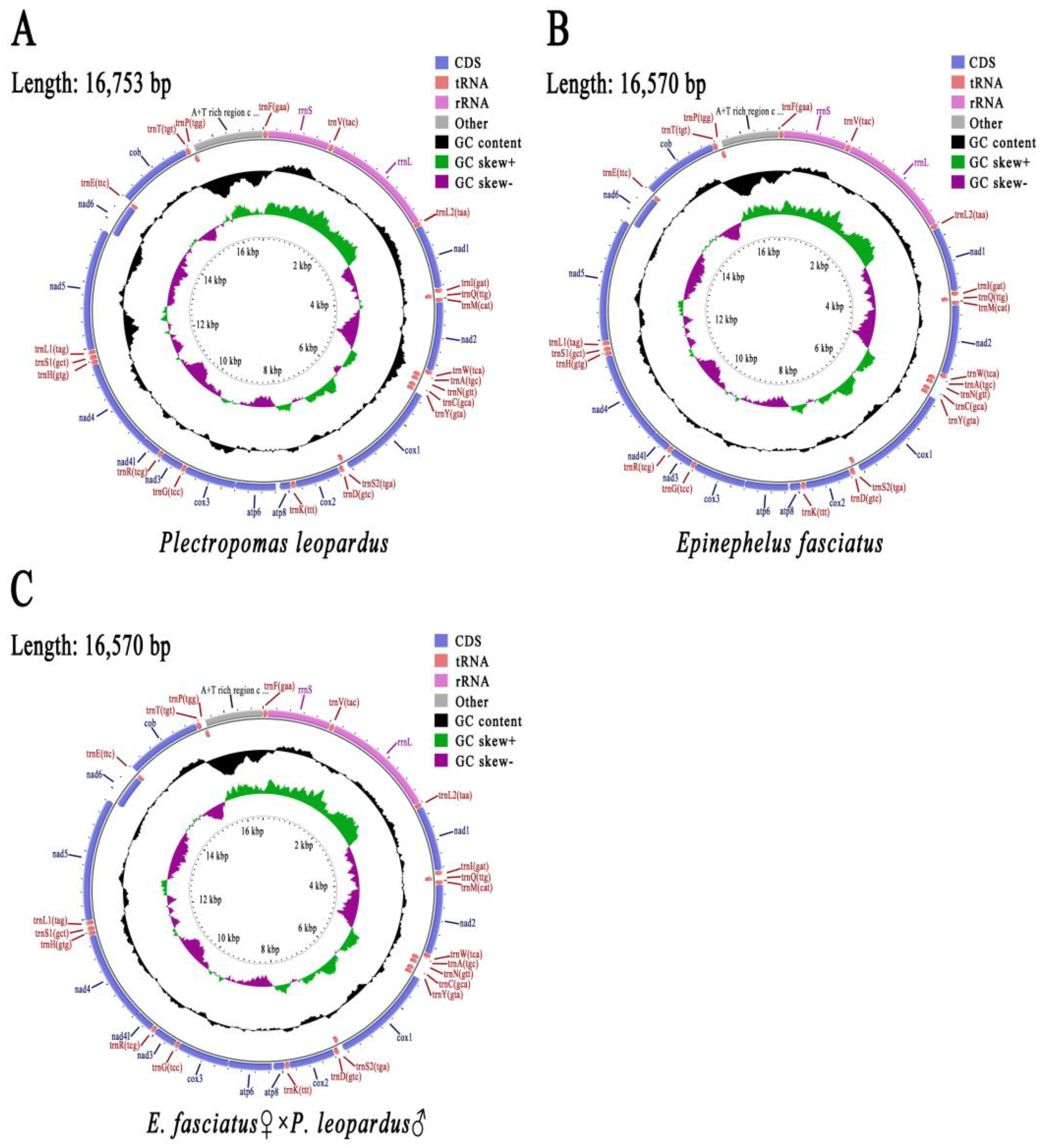
3.2. Mitochondrial Genome Composition

The mitochondrial genome sizes of the paternal P. leopardus, the maternal E. fasciatus, and their hybrid offspring were 16,753 bp, 16,570 bp, and 16,570 bp, respectively (Figure 2). A total of 37 known genes were separately identified in their mitochondrial genomes, including 13 PCGs, 22 tRNA genes, and two rRNA genes (Figure 2, Table 1).
Among the 13 PCGs of the paternal genome, all genes started with ATG except for the start codon of cox1, which began with GTG. There were three stop codons for its PCGs, namely TAG, TAA, and T. For the maternal E. fasciatus, three start codons were found in the mitochondrial genes, where the atp6 gene started with ATA, while cox1 started with GTG, and all the other genes started with ATG. For its protein stop codons, only TAA and T were found. In the hybrid offspring, only three start codons (GTG, ATA, and ATG) and two stop codons (TAA and T) were found, and the mitochondrial composition was consistent with that of the maternal E. fasciatus. The shortest tRNA of 67 bp, trnC (GCA), and the longest tRNA of 78 bp, trnS1 (GCT), were also identified in the paternal mitochondrial genome. The shortest tRNA of E. fasciatus was 67 bp (trnC (GCA)), while the longest tRNA was 75 bp (trnL2 (TAA)). The corresponding results of the hybrid offspring were consistent with those of the maternal E. fasciatus (Table 2).
The mitochondrial base compositions of the three groupers are shown in Table 3. The ratios of P. leopardus were 29.11% A, 27.92% T, 26.75% C, 16.22% G, with an AT bias of 57.03%, and a GC bias of 42.97%. The nucleotide composition of E. fasciatus revealed 28.73% A, 26.81% T, 28.32% C, 16.14% G, an AT bias of 55.55%, and a GC bias of 44.45%. The hybrid genome included 28.73% A, 26.82% T, 28.32% C, 16.13% G, an AT bias of 55.55%, and a GC bias of 44.45%. The base composition of the hybrid germplasm was largely consistent with that of E. fasciatus, with only slight differences in the T and G base content.

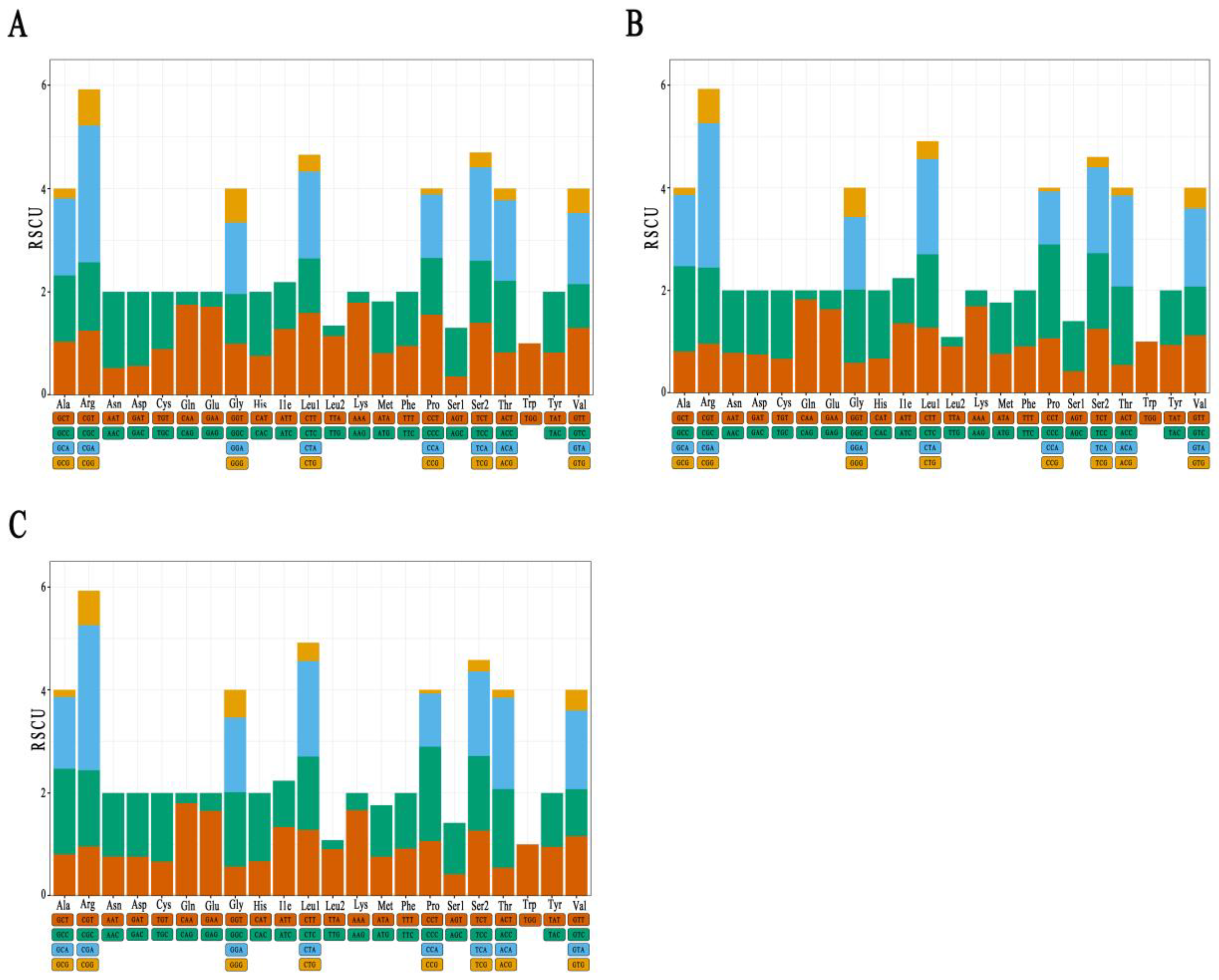
3.3. Analysis of Mitochondrial PCGs

The RSCU values of the PCGs in three groupers can be seen in Figure 3. Among them, there were 31 codons with an RSCU > 1 in the paternal P. leopardus, including the codons ending with the A base (12), with the C base (12), and seven codons ending with T. There were 29 codons with an RSCU > 1 in the maternal E. fasciatus, including 11 codons ending with the A base, 13 codons ending with C and five codons ending with T. There were 29 codons with an RSCU > 1 in the hybrid offspring and their codon composition was consistent with that of the maternal genome. In addition, compared with other amino acids, Ala, Arg, Gly, Leu1, Pro, Ser2, Thr, and Val exhibited the highest codon abundance, while Trp was encoded by only 1 codon, with the lowest codon abundance in all three species.

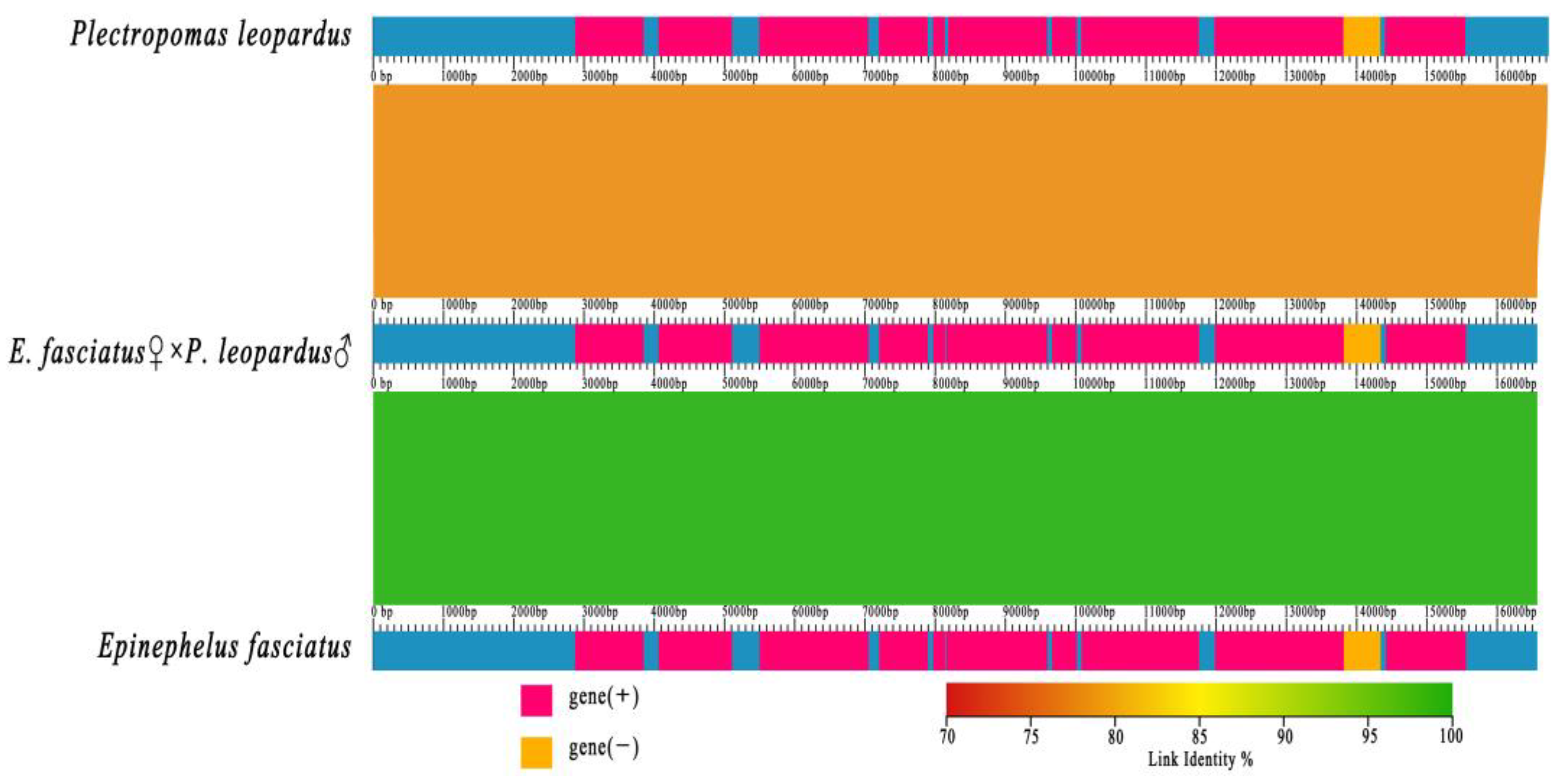
3.4. Collinearity Analysis
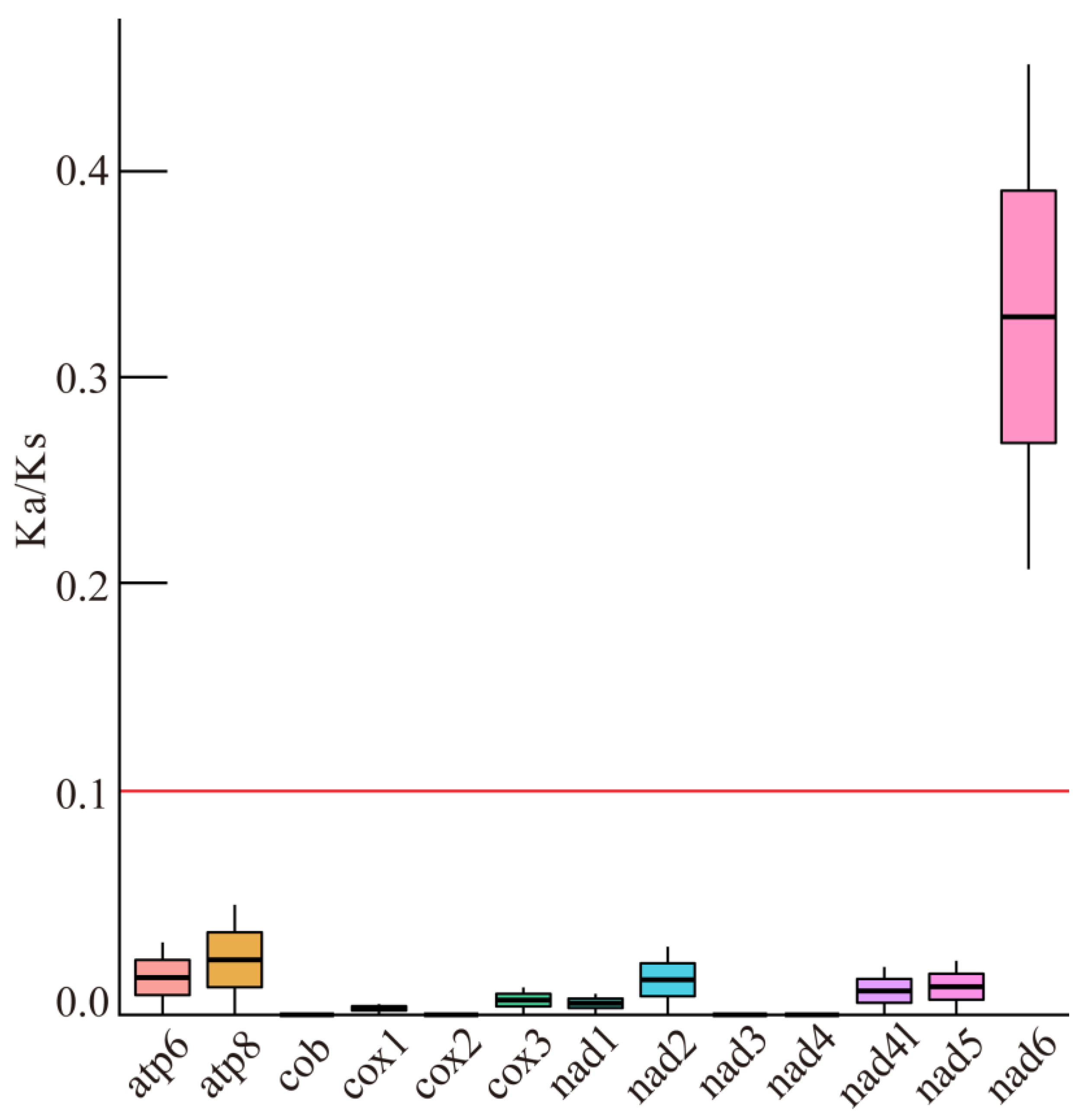
A collinearity comparison of the mitochondrial genomes of the hybrid and its parents was conducted based on their mitochondrial gene linkage relationships (Figure 4). The mitochondrial genomes of the three species were found to be largely consistent in both gene composition and arrangement. However, the hybrid was genetically closely related to the maternal E. fasciatus, which is consistent with the maternal inheritance pattern of vertebrates.

3.5. Ka/Ks Analysis

To assess the selective pressure on PCGs within the mitochondrial genome of the hybrid, the non-synonymous (Ka) and synonymous (Ks) substitution rates of each mitochondrial gene were determined (Figure 5). The results showed that the Ka/Ks ratio of all PCGs was less than 1, indicating that the mitochondrial genome underwent purifying selection. Excluding the nad6 gene, which exhibited a higher Ka/Ks ratio of 0.34, the Ka/Ks ratios of the other PCGs were all less than 0.10, suggesting that each gene experienced considerable negative selection pressure with high amino acid sequence conservation.

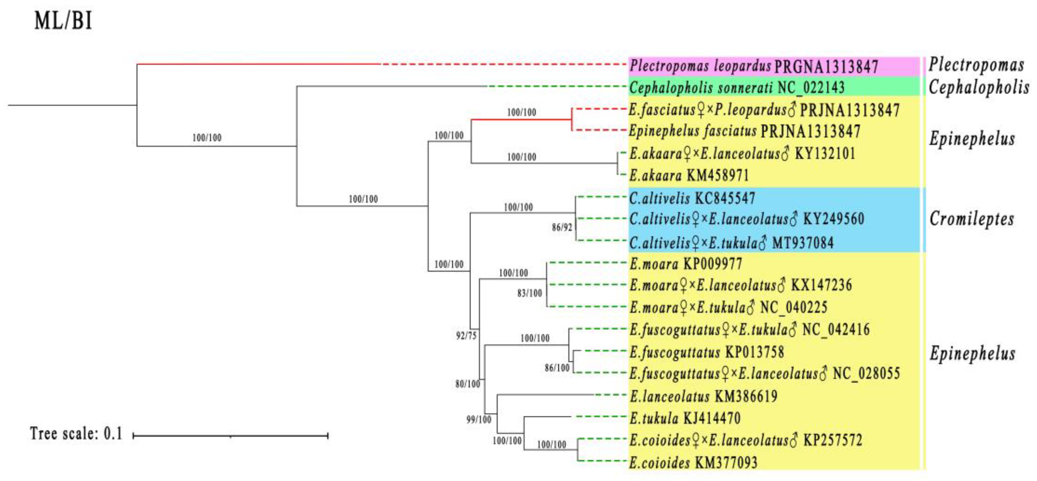
3.6. Phylogenetic Analysis

A phylogenetic analysis was conducted using the 13 PCGs sequences from 10 purebred grouper species (Epinephelus, Chromileptes, Plectropomus, and Cephalopholis), and their nine hybrid offspring (Figure 6). The analysis revealed that each hybrid offspring had a closer genetic relationship to its maternal parent, consistent with maternal inheritance patterns. As expected, the hybrid germplasm clustered with the maternal E. fasciatus, even though the genetic distance between Chromileptes and Epinephelus is relatively close, and the taxonomic boundary is not obvious.

4. Discussion

This study successfully bred intergeneric hybrid offspring between P. leopardus (♂) and E. fasciatus (♀) using hybridization breeding technology. At a water temperature of 24 ± 0.8℃, the hybrid germplasm completed embryo development in 28h55min, with a development rate that is faster than the maternal E. fasciatus (31h12min) [13]. The total length of the newly hatched larvae was 2.05 ± 0.37 mm, significantly longer than that of the paternal P. leopardus (1.50 ± 0.03 mm) and the maternal E. fasciatus (1.44 ± 0.06 mm) [13], indicating that the hybrid germplasm exhibited growth heterosis in its early development, and providing theoretical support for the subsequent cultivation and promotion of this new hybrid germplasm. Similarly, the hybrid F1 of E. moara ♀ × E. septemfasciatus ♂ exhibited a greater body length than its parents E. moara ♀ and E. septemfasciatus ♂ [14]. Furthermore, the total length of the newly hatched larvae of E. moara ♀ × E. lanceolatus ♂ was 1.95 ± 0.06 mm, which was significantly longer than that of E. moara (1.64 ± 1.42 mm) or E. lanceolatus (1.53 ± 0.12 mm) [15]. The growth heterosis in response to hybridization breeding is crucial for the genetic improvement of the growth of E. fasciatus.
The mitochondrial genomes of P. leopardus, E. fasciatus, and their hybrid offspring were composed of 13 PCGs, 22 tRNA genes, and 2 rRNA genes. Most PCGs in the three mitochondrial genomes started with the typical ATG, which is common to many bony fish [16]. However, the GTG start codon of the cox1 gene has been reported in a few groupers [17]. In addition, the atp6 gene of most groupers usually starts with CTG or ATG. For example, in the hybrid Hyporthodus septemfasciatus ♀ × E. moara ♂, except for the cox1 and atp6, which use GTG and CTG as the start codons, respectively, all other genes use ATG as the start codon [18]. In the hybrid E. moara ♀ × E. tukula ♂, except for cox1 and nd4, which use GTG, atp6 that uses CTG, and nd3 that uses ATA, the remaining genes all use ATG [19]. Interestingly, the atp6 gene of the current hybrid germplasm and the maternal E. fasciatus uses ATA as the start codon, which is uncommon in most bony fish.
Stop codon usage varies among genes. In the new hybrid germplasm, only two types of stop codons, TAA and the incomplete stop codon T, were detected in the mitochondrial genome, which is consistent with that of the maternal E. fasciatus. However, the paternal P. leopardus had three types of stop codons, including TAA, TAG, and T. The presence of incomplete stop codons is relatively common in fish mitochondrial genomes and can be completed as TAA during post-transcriptional polyadenylation of the mRNA [20]. This is a "correction mechanism" in gene expression that ensures that even if the stop codon on the DNA is incomplete, it can still be correctly identified, avoiding translational read-through.
The mitochondrial genomes of P. leopardus, E. fasciatus, and their hybrid offspring all showed a higher AT than GC bias. The G content of the three groupers was lower than 17%, indicating a strong anti-G bias [21]. Most of the base composition of the hybrid largely corresponds to that of the maternal species, further supporting the theory of mitochondrial maternal inheritance. However, differences in the composition of T and G between the hybrid offspring and the maternal E. fasciatus can serve as the basis for discrimination between the two.
The relative probability of a specific codon in the synonymous codons encoding the corresponding amino acid can reflect the degree of codon preference, where the RSCU value is typically used to evaluate the usage bias of synonymous codons [22, 23]. The RSCU results of P. leopardus, E. fasciatus and their hybrid offspring all indicate that Ala (GCT, GCC, GCA, and GCG), Arg (CGT, CGC, CGA, and CGG), Gly (GGT, GGC, GGA, and GGG), Leu1 (CTT, CTC, CTA, and CTG), Pro (CCT, CCC, CCA, and CCG), Ser2 (TCT, TCC, and T), Thr (ACT, ACC, ACA, and ACG), and Val (GTT, GTC, GTA, and GTG) are the most abundant amino acids, while Trp (TGG) is rarely used. The PCGs tend to use A and C rather than T and G on the third codon, similar to other bony fish. In E. bilobatus, E. maculatus, and E. longispinis, the PCG codons ending with A and C are the most common, while those ending with T and G are the fewest [23]. In addition, the hybrid offspring were highly consistent with the maternal E. fasciatus in codon preference, suggesting that maternal inheritance has a robust impact on the offspring’s mitochondrial coding sequence. Moreover, the RSCU values of the three species were not all equal to 1, indicating that there are varying degrees of bias in their utilization of amino acids. The frequency of codon usage is linked to how strongly a gene is expressed; genes that are expressed very efficiently show a stronger bias in codon usage compared to those expressed less efficiently, and they typically favor specific synonymous codons [24].
Genomic collinearity refers to the genetic linkage relationship, which assesses the evolutionary scale and phylogenetic distance between different species. In molecular evolution, Ka/Ks is a commonly used metric for assessing selection pressure and evolutionary relationships between species [25]. In this study, the Ka/Ks ratio for all PCGs was less than 1, signifying that the mitochondrial genomes of both the hybrid and parents underwent purifying selection. Among the 13 PCGs, the nad6 and atp8 genes exhibited relatively high Ka/Ks values, suggesting they experienced less negative selection pressure, aligning with findings from studies on vertebrate and mollusk mitochondrial genomes [26]
Phylogenetic analyses support other analyses indicating that the new intergeneric hybrid germplasm has a closer genetic relationship with the maternal E. fasciatus and exhibits a classical mitochondrial maternal inheritance pattern. Within the family Epinephelidae, hybrid grouper mitochondrial genomes all follow maternal inheritance [27-29]. Furthermore, the closer genetic relatedness of Chromileptes and Epinephelus render them suitable for distant hybridization breeding. In aquaculture, the hybrid offspring from these two genera have a relatively high survival rate and exhibit prominent growth heterosis. For example, the body weight of C. altivelis ♀ × E. tukula ♂ at 330 days of age was 220.50 ± 25.30 g, which was 1.55 times that of the maternal C. altivelis [9]. Furthermore, the growth rate of C. altivelis ♀ × E. lanceolatus ♂ was significantly faster than that of the maternal C. altivelis, at the absolute value of 1.6 times higher [10].

5. Conclusions

A systematic study was conducted on the embryonic development and mitochondrial genome evolution of a new intergeneric hybrid grouper, E. fasciatus ♀ × P. leopardus ♂. The fertilized eggs of the new germplasm completed their embryonic development within 28h55min, with a newly hatched total length of 2.05 ± 0.37 mm. The mitochondrial genome of the hybrid was 16,570 bp, and collinearity, Ka/Ks, and phylogenetic analyses collectively showed that the hybrid germplasm was more closely related to its maternal E. fasciatus, with a maternal inheritance pattern. The results from this study can further enrich the mitochondrial genome database of groupers and provide theoretical guidance for the creation of new grouper germplasm.

Author Contributions

Y.L. Y.G., and Y.T. co-conceived this study and supervised the experiments; X.J., T.D., Y.W., S.W., C.Z., and F.Y. performed the experiments, and created the figures; L.W., Z.L., L.L., and Y.X. conducted the data analysis, and provided the funding support. X.J. wrote the manuscript. All authors have read and agreed to the published version of the manuscript.

Funding

This research was funded by Hainan Province Science and Technology Special Fund (ZDYF2025SXLH002); the Key Research and Development Project of Shandong Province (2022LZGC016); the National Key Research and Development Program of China (2022YFD2400502; 2022YFD2400103); China Agriculture Research System of MOF and MARA (CARS-47-G31); Qingdao Natural Science Foundation (23-2-1-53-zyyd-jch; 24-4-4-zrjj-39-jch); Qingdao Science and Technology Benefiting the People Demonstration Project (24-1-8-xdny-3-nsh); the Central Public- interest Scientific Institute Basal Research Fund, CAFS (2025CG02; 2020TD19); and the Yellow Sea Fisheries Research Institute Research Fees(20603022025002; 20603022021010).

Institutional Review Board Statement

The research in this manuscript has been conducted under the Experimental Animal Care, Ethics and Safety Inspection Form Yellow Sea Fisheries Research Institute, CAFS (approval code: YSFRI-2025072 and approval date: 4 February 2025).

Data Availability Statement

The mitochondrial genome data presented in the study are deposited in the NCBI SRA repository (https://www.ncbi.nlm.nih.gov/, accession number PRGNA1313847).

Conflicts of Interest

The author declares no conflicts of interest.

References

  1. Kuriiwa, K.; Chiba, S.N.; Motomura, H.; Matsuura, K. Phylogeography of Blacktip Grouper, Epinephelus fasciatus (Perciformes: Serranidae), and influence of the Kuroshio Current on cryptic lineages and genetic population structure. Ichthyol. Res. 2014, 61, 361–374. [Google Scholar] [CrossRef]
  2. Zhu, X.; Hao, R.; Tian, C.; Zhang, J.; Zhu, C.; Li, G. Integrative Transcriptomics and Metabolomics Analysis of Body Color Formation in the Leopard Coral Grouper (Plectropomus leopardus). Front. Mar. Sci. 2021, 8. [Google Scholar] [CrossRef]
  3. Pratchett, M.S.; Caballes, C.F.; Hobbs, J.-P.A.; DiBattista, J.D.; Bergseth, B.; Waldie, P.; Champion, C.; Mc Cormack, S.P.; Hoey, A.S. Variation in the Physiological Condition of Common Coral Trout (Plectropomus leopardus) Unrelated to Coral Cover on the Great Barrier Reef, Australia. Fishes 2023, 8, 497. [Google Scholar] [CrossRef]
  4. THàNH P N, TAKAFUMI A. Growth, sexual transition, and maturation of blacktip grouper Epinephelus fasciatus under long-term artificial rearing: Puberty and its associated physiological and endocrine changes. 2022.
  5. Kawabe, K.; Kohno, H. Morphological development of larval and juvenile blacktip grouper, Epinephelus fasciatus. Fish. Sci. 2009, 75, 1239–1251. [Google Scholar] [CrossRef]
  6. Sun, Z.; Xia, S.; Feng, S.; Zhang, Z.; Rahman, M.M.; Rajkumar, M.; Jiang, S. Effects of water temperature on survival, growth, digestive enzyme activities, and body composition of the leopard coral grouper Plectropomus leopardus. Fish. Sci. 2014, 81, 107–112. [Google Scholar] [CrossRef]
  7. TIAN Y S, CHEN Z F, DUAN H M. The family line establishment of the hybrid Epinephelus moara(♀)× E. lanceolatus(♂) by using cryopreserved sperm and the related genetic effect analysis. J. Fish. 1: China 2017, 41(12), 2017.
  8. CHEN S, TIAN Y. Heterosis in growth and low temperature tolerance in Jinhu grouper (Epinephelus fuscoguttatus♀× Epinephelus tukula♂). 7: Aquaculture 2023, 562, 2023.
  9. LI Z T, TIAN Y S, CHENG M L. Comparison of development and growth of hybrid Chromileptes altivelis (♀) × Epinephelus tukula(♂). 1: South China Fisheries Science 2020, 16(1), 2020.
  10. GONG S, WANG T. Development and characterization of a novel intergeneric hybrid grouper (Cromileptes altivelis♀× Epinephelus lanceolatus♂). 7: Aquaculture 2025, 595, 2025.
  11. Yan, F.; Wang, C.; Zhu, Y.; Wu, J.; Yang, J.; Gan, S.; Lin, H.R.; Zhang, Y.; Li, S. Transcriptome analysis reveals the dynamics of gene expression during early embryonic development of hybrid groupers. Aquaculture 2024, 596. [Google Scholar] [CrossRef]
  12. Liu, Y.; Tian, Y.; Wang, L.; Li, Z.; Zhang, J.; Wu, Y.; Chen, S.; Li, L.; Li, W.; Ma, W.; et al. Construction of high-density linkage maps and QTL mapping for growth-related traits in F1 hybrid Yunlong grouper (Epinephelus moara♀ × E. lanceolatus♂). Aquaculture 2022, 548. [Google Scholar] [CrossRef]
  13. DING T T, TIAN Y S, BAI D Q. Observation on Embryonic and Post-Embryonic Development, and Morphological Changes of Larval, Juvenile and Young Epinephelus fasciatus. Progress in Fishery Sciences 2025: 1-11.10.19663/j.issn2095-9869. 2024.
  14. L L Y, C C, Y W Q. Comparative analysis of growth characteristics between hybrid F1 by Epinephelus moara(♀)×Epinephelus septemfasciatus(♂) and the offspring of their parents. 4: Progress in Fishery Sciences 2015, 36(03), 2015.
  15. CHEN Z F, TIAN Y S. Embryonic and larval development of a hybrid between kelp grouper Epinephelus moara♀× giant grouper E. lanceolatus♂ using cryopreserved sperm. Aquacult. Res. 1: 2018, 49(4), 2018.
  16. Lü, Z.; Zhu, K.; Jiang, H.; Lu, X.; Liu, B.; Ye, Y.; Jiang, L.; Liu, L.; Gong, L. Complete mitochondrial genome of Ophichthus brevicaudatus reveals novel gene order and phylogenetic relationships of Anguilliformes. Int. J. Biol. Macromol. 2019, 135, 609–618. [Google Scholar] [CrossRef] [PubMed]
  17. Zhu, K.; Huang, G.; Zhang, D.; Guo, Y.; Yu, D. The complete nucleotide sequence of Malabar grouper (Epinephelus malabaricus) mitochondrial genome. Mitochondrial DNA 2014, 27, 1–2. [Google Scholar] [CrossRef] [PubMed]
  18. KIM Y H, PARK J Y. Complete mitochondrial genome of the hybrid grouper Hyporthodus septemfasciatus (♀)× Epinephelus moara (♂)(Perciformes, Serranidae) and results of a phylogenetic analysis. 7: Mitochondrial DNA Part B 2021, 6(3), 2021.
  19. Li, Z.; Tian, Y.; Cheng, M.; Wang, L.; Zhang, J.; Wu, Y.; Pang, Z.; Ma, W.; Zhai, J. The complete mitochondrial genome of the hybrid grouper Epinephelus moara (♀)×Epinephelus tukula (♂), and phylogenetic analysis in subfamily Epinephelinae. Acta Oceanol. Sin. 2020, 39, 65–75. [Google Scholar] [CrossRef]
  20. He, H.; Gao, Z.; Hu, Z.; Cai, L.; Huang, Y.; Zhou, M.; Liang, R. Comparative characterization and phylogenetic analysis of complete mitogenome of three taxonomic confused groupers and the insight to the novel gene tandem duplication in Epinephelus. Front. Mar. Sci. 2024, 11, 1450003. [Google Scholar] [CrossRef]
  21. Zhuang, X.; Qu, M.; Zhang, X.; Ding, S. A Comprehensive Description and Evolutionary Analysis of 22 Grouper (Perciformes, Epinephelidae) Mitochondrial Genomes with Emphasis on Two Novel Genome Organizations. PLOS ONE 2013, 8, e73561. [Google Scholar] [CrossRef] [PubMed]
  22. Wang, C.; Ye, P.; Liu, M.; Zhang, Y.; Feng, H.; Liu, J.; Zhou, H.; Wang, J.; Chen, X. Comparative Analysis of Four Complete Mitochondrial Genomes of Epinephelidae (Perciformes). Genes 2022, 13, 660. [Google Scholar] [CrossRef] [PubMed]
  23. Wen, J. Complete Mitochondrial Genome of King Threadfin, Polydactylus macrochir (Günther, 1867): Genome Characterization and Phylogenetic Analysis. Genes 2025, 16, 88. [Google Scholar] [CrossRef] [PubMed]
  24. Liao, X.; Shih, Y.; Jia, C.; Gao, T. Complete Mitochondrial Genome of Four Peristediidae Fish Species: Genome Characterization and Phylogenetic Analysis. Genes 2024, 15, 557. [Google Scholar] [CrossRef] [PubMed]
  25. Zhang, D.; Zou, H.; Wu, S.G.; Li, M.; Jakovlić, I.; Zhang, J.; Chen, R.; Li, W.X.; Wang, G.T. Three new Diplozoidae mitogenomes expose unusual compositional biases within the Monogenea class: implications for phylogenetic studies. BMC Evol. Biol. 2018, 18, 1–17. [Google Scholar] [CrossRef] [PubMed]
  26. Monnens, M.; Thijs, S.; Briscoe, A.G.; Clark, M.; Frost, E.J.; Littlewood, D.T.J.; Sewell, M.; Smeets, K.; Artois, T.; Vanhove, M.P. The first mitochondrial genomes of endosymbiotic rhabdocoels illustrate evolutionary relaxation of atp8 and genome plasticity in flatworms. Int. J. Biol. Macromol. 2020, 162, 454–469. [Google Scholar] [CrossRef] [PubMed]
  27. Chen, J.; Ye, Z.; Yu, Z.; Wang, J.; Li, P.; Chen, X.; Liu, Y.; Zhang, Y. The complete mitochondrial genome of the hybrid grouper (Cromileptes altivelis♀ × Epinephelus lanceolatus♂) with phylogenetic consideration. Mitochondrial DNA Part B 2017, 2, 171–172. [Google Scholar] [CrossRef] [PubMed]
  28. TANG Z, CHEN J. The complete mitochondrial genome of the hybrid grouper Epinephelus coioides♀× Epinephelus akaara♂ with phylogenetic consideration. 3: Mitochondrial DNA Part B 2017, 2(1), 2017.
  29. LI L, TIAN Y. Characterization of the complete mitochondrial genome of the hybrid grouper (Cromileptes altivelis♀× Epinephelus tukula♂) with phylogenetic consideration. 1: Mitochondrial DNA Part B 2021, 6(3), 2021.
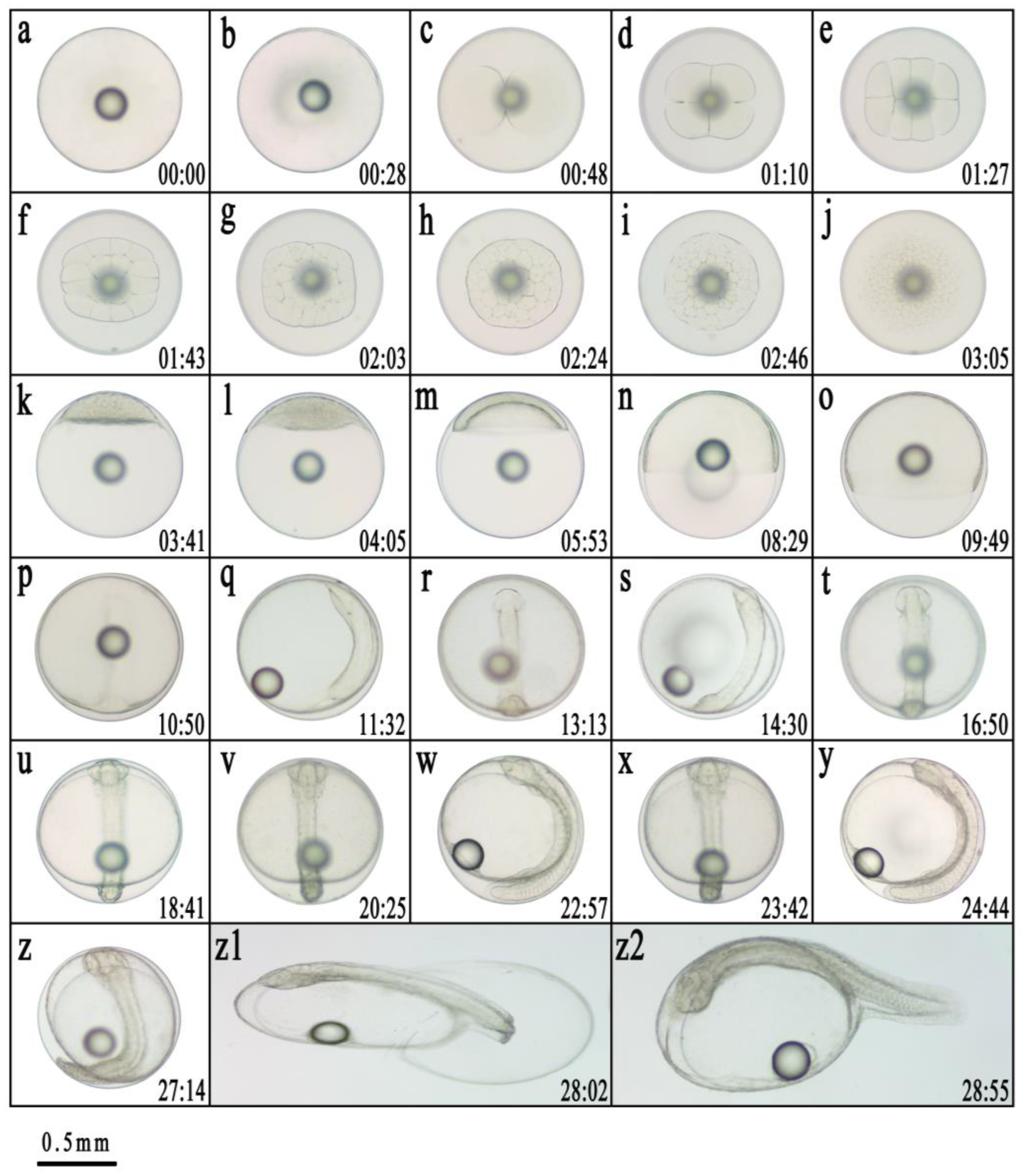
Figure 1. Embryonic development of Epinephelus fasciatus ♀ × Plectropomus leopardus ♂. (a): Fertilized egg; (b): Blastodisc formation; (c): 2-cell stage; (d): 4-cell stage; (e): 8-cell stage; (f): 16-cell stage; (g): 32-cell stage; (h): 64-cell stage; (i): Multi-cell stage; (j): morula stage; (k): High blastula stage; (l): Low blastula stage; (m): Early gastrula stage; (n): Middle gastrula stage; (o): Late gastrula stage; (p): Embryo body stage; (q): Closure of blastopore stage; (r): Optic capsule stage; (s): Muscle burl stage; (t): Otocyst stage; (u): Brain vesicle stage; (v): Heart stage; (w): Tail-bud stage; (x): Crystal stage; (y): Heart-beating stage; (z): Pre incubation stage; (z1): Hatching stage; (z2): Newly hatched larvae. The number in the lower right corner represents the time in hours and minutes after fertilization. The scale is 0.5 mm.
Preprints 180624 g001
Figure 2. Mitochondrial genome maps of Plectropomas leopardus (A), Epinephelus fasciatus (B), and E. fasciatus ♀ × P. leopardus ♂ (C).
Preprints 180624 g002
Figure 3. Relative synonymous codon usage (RSCU) of Plectropomas leopardus (A), Epinephelus fasciatus (B), and E. fasciatus ♀ × P. leopardus ♂ (C).
Preprints 180624 g003
Figure 4. Mitochondrial whole genome alignment of Plectropomas leopardus, Epinephelus fasciatus, and E. fasciatus ♀ × P. leopardus ♂.
Preprints 180624 g004
Figure 5. Ka/Ks values for 13 PCGs of E. fasciatus ♀ × P. leopardus ♂ compared to P. leopardus and E. fasciatus.
Preprints 180624 g005
Figure 6. The combined maximum likelihood (ML) and Bayesian inference (BI) tree of 19 grouper species using 13 protein-coding genes (PCGs). Bootstrap values are shown at the base of each node. GenBank accession numbers follow scientific names. The species from the current study are highlighted with red dashed lines.
Preprints 180624 g006
Table 1. Hybrid embryo development schedule of Epinephelus fasciatus ♀ × Plectropomus leopardus ♂.
Developmental
stage
Developmental stage of
embryonic
Main developmental
characteristics
Time after fertilization
One-cell fertilized egg Spherical, with one oil ball 0

Cleavage
blastodisc formation stage The blastoderm has formed, and when viewed from the side, it can be seen that the blastoderm protrudes like a cap 28min
2-cell stage The first cleavage forms two cells 48min
4-cell stage The second cleavage forms four cells 1h10min
8-cell stage The third cleavage forms eight cells 1h27min
16-cell stage The fourth cleavage forms 16 cells 1h43min
32-cell stage The fifth cleavage forms 32 cells 2h03min
64-cell stage The sixth cleavage formed 64 cells, and the division surface was rather disordered 2h24min
Multicellular stage Continuous division leads to smaller cells and an increase in their number 2h46min
Morula stage The cells are piled up in multiple layers, resembling mulberries in appearance 3h05min
Blastula high blastula stage The blastocyst is tall and concentrated, and when viewed from the side, it appears as a high cap 3h41min
low blastula stage The blastocyst becomes lower, and the cells are preparing to wrap towards the lower pole of the plant 4h05min
Gastrula early gastrula stage One-third of the yolk is encapsulated under the germ layer 5h53min
middle gastrula stage The embryo layer is subtracted from half of the yolk 8h29min
late gastrula stage The embryo layer is subtracted to three-quarters of the yolk, the blastocyst becomes slender, and the embryo body is in the process of formation 9h49min
Neurula Embryonic formation stage The embryonic body is formed with distinct contours 10h50min
Embryonic hole closure stage The embryo layer is subcapsulated, and the embryo pore is completely closed 11h32min
Organogenesis Optic capsule stage A pair of visual sacs appeared at the head of the embryo 13h13min
Muscle burl stage Muscle segments appear in the middle of the embryo 14h30min
Otocyst stage A pair of auditory sacs appeared at the posterior position of the visual sac in the head 16h50min
Brain vesicle stage Brain vesicles appear between the two visual sacs 18h41min
Heart stage The heart is formed on the ventral side with a clear outline 20h25min
Tail-bud stage The tail of the embryo begins to separate from the yolk sac 22h57min
Crystal stage Crystals appear in the eye of the embryo 23h42min
Heart-beating stage The heart began to beat slightly and then gradually stabilized 24h44min
Hatching Early incubation stage The embryo is twitching violently 27h14min
Incubation stage The head emerges from the membrane first 28h02min
Newly hatched fry The larvae hatch from the membrane 28h55min
Incubate for 48h The yolk sac shrinks and becomes smaller, the notochord becomes more obvious, the dorsal fin folds, the ventral fin folds, and the caudal fin membranes widen, pigmentation begins to form in the eyes, and the mouth can be slightly opened and closed Total length is 2.35 ± 0.05 mm
Table 2. Mitochondrial genome characteristics of Plectropomas leopardus, Epinephelus fasciatus, and E. fasciatus ♀ × P. leopardus ♂.
Gene Strand Plectropomus leopardus Epinephelus fasciatus E. Fasciatus♀ × P. leopardus Start_coden Stop_coden
Position
(Start-End)
Length(bp) Intergenic_spacer Position
(Start-End)
Length(bp) Intergenic_spacer Position
(Start-End)
Length(bp) Intergenic_spacer
trnF(gaa) F 1-69 69 - 1-70 70 - 1-70 70 - - -
rrnS F 70-1018 949 0 71-1022 952 0 71-1022 952 0 - -
trnV(tac) F 1022-1092 71 3 1024-1094 71 1 1024-1094 71 1 - -
rrnL F 1096-2802 1707 3 1098-2798 1701 3 1098-2798 1701 3 - -
trnL2(taa) F 2800-2872 73 -3 2799-2873 75 0 2799-2873 75 0 - -
nad1 F 2873-3847 975 0 2874-3848 975 0 2874-3848 975 0 ATG/ATG/ATG TAA/TAA/TAA
trnI(gat) F 3852-3921 70 4 3853-3922 70 4 3853-3922 70 4 - -
trnQ(ttg) R 3921-3991 71 -1 3922-3992 71 -1 3922-3992 71 -1 - -
trnM(cat) F 3992-4060 69 0 3993-4061 69 0 3993-4061 69 0 - -
nad2 F 4061-5107 1047 0 4062-5108 1047 0 4062-5108 1047 0 ATG/ATG/ATG TAA/TAA/TAA
trnW(tca) F 5107-5177 71 -1 5108-5178 71 -1 5108-5178 71 -1 - -
TrnA(tgc) R 5179-5247 69 1 5180-5248 69 1 5180-5248 69 1 - -
trnN(gtt) R 5249-5321 73 1 5249-5321 73 0 5249-5321 73 0 - -
trnC(gca) R 5358-5424 67 36 5362-5428 67 40 5362-5428 67 40 - -
trnY(gta) R 5425-5494 70 0 5429-5499 71 0 5429-5499 71 0 - -
cox1 F 5496-7046 1551 1 5501-7051 1551 1 5501-7051 1551 1 GTG/GTG/GTG TAA/TAA/TAA
trnS2(tga) R 7047-7117 71 0 7053-7123 71 1 7053-7123 71 1 - -
trnD(gtc) F 7120-7193 74 2 7124-7197 74 0 7124-7197 74 0 - -
cox2 F 7200->7890 691 6 7205->7895 691 7 7205->7895 691 7 ATG/ATG/ATG T/T/T
trnK(ttt) F 7891-7965 75 0 7896-7968 73 0 7896-7968 73 0 - -
atp8 F 7967-8134 168 1 7970-8137 168 1 7970-8137 168 1 ATG/ATG/ATG TAA/TAA/TAA
atp6 F 8188-8808 621 53 8155-8811 657 17 8155-8811 657 17 ATG/ATA/ATA TAA/TAA/TAA
cox3 F 8808-9593 786 -1 8811-9596 786 -1 8811-9596 786 -1 ATG/ATG/ATG TAA/TAA/TAA
trnG(tcc) F 9593-9662 70 -1 9596-9666 71 -1 9596-9666 71 -1 - -
nad3 F 9663-10013 351 0 9667->10015 349 0 9667->10015 349 0 ATG/ATG/ATG TAG/T/T
trnR(tcg) F 10012-10080 69 -2 10016-10084 69 0 10016-10084 69 0 - -
nad4l F 10081-10377 297 0 10085-10381 297 0 10085-10381 297 0 ATG/ATG/ATG TAA/TAA/TAA
nad4 F 10371->11751 1381 -7 10375->11755 1381 -7 10375->11755 1381 -7 ATG/ATG/ATG T/T/T
trnH(gtg) F 11752-11820 69 0 11756-11825 70 0 11756-11825 70 0 - -
trnS1(gct) F 11821-11898 78 0 11826-11895 70 0 11826-11895 70 0 - -
trnL1(tag) F 11905-11977 73 6 11911-11983 73 15 11911-11983 73 15 - -
nad5 F 11978-13816 1839 0 11984-13822 1839 0 11984-13822 1839 0 ATG/ATG/ATG TAA/TAA/TAA
nad6 R 13813-14334 522 -4 13819-14340 522 -4 13819-14340 522 -4 ATG/ATG/ATG TAG/TAA/TAA
trnE(ttc) R 14335-14403 69 0 14341-14409 69 0 14341-14409 69 0 - -
cob F 14406->15546 1141 2 14417->15557 1141 7 14417->15557 1141 7 ATG/ATG/ATG T/T/T
trnT(tgt) F 15547-15618 72 0 15558-15630 73 0 15558-15630 73 0 - -
trnP(tgg) R 15618-15688 71 -1 15630-15699 70 -1 15630-15699 70 -1 - -
Table 3. Mitochondrial base composition of Plectropomas leopardus, Epinephelus fasciatus, and E. fasciatus ♀ × P. leopardus ♂.
Species Region Lengthbp T% C% A% G% AT% GC%
Plectropomus leopardus Genome 16753 27.92 26.75 29.11 16.22 57.03 42.97
Protein_coding_genes 11370 30.30 27.86 26.40 15.44 56.70 43.30
First position 3790 28.71 28.23 26.70 16.36 55.41 44.59
Second position 3790 31.58 28.39 24.85 15.17 56.44 43.56
Third position 3790 30.61 26.97 27.65 14.78 58.26 41.74
tRNA 1564 27.69 20.65 28.32 23.34 56.01 43.99
rRNA 2656 22.06 23.43 33.43 20.97 55.50 44.50
Epinephelus fasciatus Genome 16570 26.81 28.32 28.73 16.14 55.55 44.45
Protein_coding_genes 11404 28.64 29.69 26.25 15.42 54.88 45.12
First position 3801 24.49 26.99 26.57 21.94 51.06 48.93
Second position 3801 33.88 29.83 21.91 14.36 55.8 44.2
Third position 3801 27.52 32.25 30.25 9.97 57.77 42.22
tRNA 1560 27.44 20.96 28.97 22.63 56.41 43.59
rRNA 2653 21.56 25.52 32.42 20.51 53.98 46.02
E.fasciatus ♀ × P.leopardus ♂ Genome 16570 26.82 28.32 28.73 16.13 55.55 44.45
Protein_coding_genes 11404 28.65 29.68 26.25 15.42 54.89 45.11
First position 3801 24.52 26.96 26.6 21.91 51.11 48.88
Second position 3801 33.83 29.88 21.91 14.36 55.74 44.25
Third position 3801 27.57 32.2 30.23 10 57.8 42.2
tRNA 1560 27.44 20.96 28.97 22.63 56.41 43.59
rRNA 2653 21.56 25.52 32.42 20.51 53.98 46.02
Disclaimer/Publisher’s Note: The statements, opinions and data contained in all publications are solely those of the individual author(s) and contributor(s) and not of MDPI and/or the editor(s). MDPI and/or the editor(s) disclaim responsibility for any injury to people or property resulting from any ideas, methods, instructions or products referred to in the content.
Copyright: This open access article is published under a Creative Commons CC BY 4.0 license, which permit the free download, distribution, and reuse, provided that the author and preprint are cited in any reuse.
Prerpints.org logo

Preprints.org is a free preprint server supported by MDPI in Basel, Switzerland.

Subscribe

Disclaimer

Terms of Use

Privacy Policy

Privacy Settings

© 2025 MDPI (Basel, Switzerland) unless otherwise stated