Submitted:
08 October 2025
Posted:
09 October 2025
You are already at the latest version
Abstract
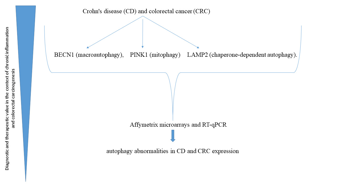
Crohn’s disease (CD) and colorectal cancer (CRC) are clinically distinct but pathogenetically related conditions in which significant abnormalities in autophagy are observed. The aim of the study was to evaluate the expression of three key autophagy-related genes, i.e., BECN1 (macroautophagy), PINK1 (mitophagy) and LAMP2 (chaperone-mediated autophagy) in tissue samples from patients with CD and CRC. The study material included samples from 48 patients with CD (n = 96 biopsy samples) and 87 patients with CRC (n = 87 tumors; n = 87 normal paired controls). Transcriptomic analyses were performed using Affymetrix HG-U133A microarrays. They were confirmed by RT-qPCR. The Kruskal-Wallis test with Dunn’s post hoc analysis (α = 0.05) and Spearman’s correlation coefficients were used for statistical evaluation. Expression of BECN1 and LAMP2 was significantly decreased in both CD and CRC compared to the controls (p = 0.009; p = 0.023, respectively). However, PINK1 showed significantly higher expression levels in CD compared to CRC and the controls (p < 0.001). The clinical stages of CRC (I-IV) did not significantly affect the expression of the analyzed genes. The study findings confirm the presence of common abnormalities in autophagy in CD and CRC with decreased macroautophagy and chaperone-mediated autophagy with the compensatory activation of mitophagy. BECN1, PINK1 and LAMP2 expressions may have a diagnostic and therapeutic value in the context of chronic inflammation and colorectal carcinogenesis.
Keywords:
1. Introduction
2. Results and Discussion
3. Materials and Methods
3.1. Patients and Methods
3.1.1. Study Design
3.1.2. Statistical Analysis
3.1.3. Bioethical Consent
3.1.4. Limitations of the Study
4. Conclusions
Author Contributions
Funding
Conflicts of Interest
Abbreviations
| (CD) | Crohn’s disease |
| (UC) | ulcerative colitis |
| (IBD) | inflammatory bowel disease |
| (CRC) | colorectal cancer |
| BECN1 | the human gene encoding the Beclin-1 protein |
| PINK1—PTEN | induced kinase 1 |
| LAMP2 | Lysosome-associated membrane protein 2 |
| (CMA) | chaperone-mediated autophagy |
| (CSI, CSII, CSIII and CSIV) | different stages of colon cancer |
| (mTOR) | major cell proliferation pathways |
| CD107b) | Cluster of Differentiation 107b |
References
- Verdier, J.; Breunig, I.R.; Ohse, M.C.; et al. Faecal Micro- RNAs in Inflammatory Bowel Diseases. J Crohns Colitis. 2020, 14, 110–117. [Google Scholar] [CrossRef]
- Shawki, S.; Ashburn, J.; Signs, S.A.; Huang, E. Colon Cancer: Inflammation-Associated Cancer. Surg Oncol Clin N Am. 2018, 27, 269–287. [Google Scholar] [CrossRef]
- Wanders, L.K.; Dekker, E.; Pullens, B.; et al. Cancer risk after resection of polypoid dysplasia in patients with longstanding ulcerative colitis: a meta-analysis. Clin Gastroenterol Hepatol. 2014, 12, 756–764. [Google Scholar] [CrossRef]
- Follin-Arbelet, B.; Cvancarova Småstuen, M.; Hovde, Ø.; et al. Risk of Cancer in Patients With Crohn Disease 30 Years After Diagnosis (the IBSEN Study). Crohns Colitis 2023, 5, otad057. [Google Scholar] [CrossRef]
- Hatami, B.; Pasharavesh, L.; Sharifian, A.; Zali, M.R. Concurrent inflammatory bowel disease and primary sclerosing cholangitis: a review of pre- and post-transplant outcomes and treatment options. Gastroenterol Hepatol Bed Bench. 2023, 16, 259–269. [Google Scholar]
- Puca, P.; Del Vecchio, L.E.; Ainora, M.E.; et al. Role of Multiparametric Intestinal Ultrasound in the Evaluation of Response to Biologic Therapy in Adults with Crohn’s Disease. Diagnostics (Basel). 2022, 12, 1991. [Google Scholar] [CrossRef]
- Choi, C.H.R.; Rutter, M.D.; Askari, A.; Lee, G.H.; et al. Forty-Year Analysis of Colonoscopic Surveillance Program for Neoplasia in Ulcerative Colitis: An Updated Overview. Am J Gastroenterol. 2015, 110, 1022–1034. [Google Scholar] [CrossRef]
- Gil, J.; Karpińsk, P.; Sąsiadek, M.M. Transcriptomic Profiling for the Autophagy Pathway in Colorectal Cancer. Int. J Mol Sci. 2020, 21, 7101. [Google Scholar] [CrossRef]
- He, M.M.; Lo, C.H.; Wang, K.; et al. Immune-Mediated Diseases Associated With Cancer Risks. JAMA Oncol. 2022, 8, 209–219. [Google Scholar] [CrossRef]
- Lo, B.; Zhao, M.; Vind, I.; Burisch, J. The Risk of Extraintestinal Cancer in Inflammatory Bowel Disease: A Systematic Review and Meta-analysis of Population-based Cohort Studies. Clin Gastroenterol Hepatol. 2021, 19, 1117–1138. [Google Scholar] [CrossRef]
- Levine, B.; Kroemer, G. Biological Functions of Autophagy Genes: A Disease Perspective. Cell. 2019, 176, 11–42. [Google Scholar] [CrossRef]
- Mizushima, N. A brief history of autophagy from cell biology to physiology and disease. Nat Cell Biol. 2018, 20, 521–527. [Google Scholar] [CrossRef]
- Bednarczyk, M.; Zmarzły, N.; Grabarek, B.; et al. Genes involved in the regulation of different types of autophagy and their participation in cancer pathogenesis. Oncotarget. 2018, 9, 34413–34428. [Google Scholar] [CrossRef]
- Lin, C.; Jia, S.N.; Yang, F.; et al. The transcription factor p8 regulates autophagy during diapause embryo formation in Artemia parthenogenetica. Cell Stress Chaperones. 2016, 21, 665–675. [Google Scholar] [CrossRef]
- Zhang, B.; Liu, L. Autophagy is a double-edged sword in the therapy of colorectal cancer. Oncol Lett. 2021, 21, 378. [Google Scholar] [CrossRef]
- Choi, C.H.R.; Al Bakir, I.; Ding, N.S.J.; Lee, G.H.; Askari, A.; Warusavitarne, J.; Moorghen, M.; Humphries, A.; Ignjatovic-Wilson, A.; Thomas-Gibson, S.; et al. Cumulative burden of inflammation predicts colorectal neoplasia risk in ulcerative colitis: a large single-centre study. Gut. 2019, 68, 414–422. [Google Scholar] [CrossRef]
- Xu, Y.; Shen, J.; Ran, Z. Emerging views of mitophagy in immunity and autoimmune diseases. Autophagy. 2020, 16, 3–17. [Google Scholar] [CrossRef]
- Okai, N.; Watanabe, T.; Minaga, K.; et al. Alterations of autophagic and innate immune responses by the Crohn’s disease-associated ATG16L1 mutation. World J Gastroenterol. 2022, 28, 3063–70. [Google Scholar] [CrossRef] [PubMed]
- Kaplan, G.G. The global burden of IBD: from 2015 to Nat Rev Gastroenterol Hepatol. 2015, 12, 720–727. [Google Scholar]
- Levy, J.M.M.; Towers, C.G.; Thorburn, A. Targeting autophagy in cancer. Nat Rev Cancer. 2017, 9, 528–542. [Google Scholar] [CrossRef]
- Yuan, Z.; Wang, S.; Tan, X.; Wang, D. New Insights into the Mechanisms of Chaperon- Mediated Autophagy and Implications for Kidney Diseases. Cells 2022, 11. [Google Scholar] [CrossRef] [PubMed]
- Ramjeet, M.; Hussey, S.; Philpott, D.J.; Travassos, L.H. ' Nodophagy': New crossroads in Crohn disease pathogenesis. Gut Microbes. 2010, 1, 307. [Google Scholar] [CrossRef]

| Parameter |
CD group N = 48 |
CRC group N = 87 |
p | |||||||
| Median | Range | Q1 | Q3 | Median | Range | Q1 | Q3 | |||
| Age [y] | 43.5 | 22-78 | 31.0 | 58.5 | 68.0 | 41-82 | 59.0 | 73.0 | 0.0001 | |
| Height [m] | 1.70 | 1.54-1.94 | 1.63 | 1.77 | 1.66 | 1.54-1.88 | 1.56 | 1.76 | n.s | |
| Body mass [kg] | 62.0 | 35-107 | 56.5 | 72.5 | 76.0 | 45-105 | 63.0 | 87.0 | 0.0001 | |
| BMI (kg/m2) | 21.85 | 13.5-28.43 | 19.42 | 24.20 | 26.45 | 18.17-36.73 | 24.45 | 29.50 | 0.0001 | |
| Ht [%] | 36.1 | 25.1-47.4 | 30.4 | 40.5 | 37.9 | 27.2-47.8 | 36.2 | 40.1 | n.s | |
| WBC [M] | 7.12 | 3.60 -25.1 | 4.84 | 9.67 | 6.56 | 2.90-15.76 | 5.09 | 8.37 | n.s | |
| Gender | CD group | CRC group | p | ||
| N | % | N | % | ||
| Female | 28 | 58.0 | 36 | 41.4 | n.s. |
| Male | 20 | 42.0 | 51 | 58.6 | n.s. |
| Cancer stage | Number of cases | % |
| Stage I | 22 | 25.3 |
| Stage II | 20 | 23.0 |
| Stage III | 33 | 37.9 |
| Stage IV | 12 | 13.8 |
| Total | 87 | 100.0 |
| Gene/group patients | N | Mean rank | Median | Q1 | Q3 | H | P | η2 | post hoc a | |
| mRNA copy/μg RNA | ||||||||||
| BECN1 | CD | 96 | 121.511 | 4062.000 | 280.500 | 16890.000 | 9.421 | 0.01 | 0.030 | CD vs CT |
| CRC | 87 | 132.823 | 6388.000 | 1571.000 | 13100.000 | |||||
| CT | 87 | 156.433 | 10560.000 | 3853.000 | 20385.000 | |||||
| PINK1 | CD | 96 | 197.411 | 17380.000 | 5929.000 | 21952.000 | 89.920 | <0.001 | 0.320 | CD vs CRC CD vs CT |
| CRC | 87 | 119.332 | 194.000 | 0.571 | 7199.000 | |||||
| CT | 87 | 91.701 | 13.201 | 0.073 | 1292.000 | |||||
| LAMP2 | CD | 96 | 126.900 | 19215.000 | 4596.500 | 24650.000 | 7.540 | 0.02 | 0.020 | CRC vs CT |
| CRC | 87 | 122.500 | 18250.000 | 10770.000 | 29310.000 | |||||
| CT | 87 | 151.702 | 34135.000 | 14357.500 | 51742.500 | |||||
Disclaimer/Publisher’s Note: The statements, opinions and data contained in all publications are solely those of the individual author(s) and contributor(s) and not of MDPI and/or the editor(s). MDPI and/or the editor(s) disclaim responsibility for any injury to people or property resulting from any ideas, methods, instructions or products referred to in the content. |
© 2025 by the authors. Licensee MDPI, Basel, Switzerland. This article is an open access article distributed under the terms and conditions of the Creative Commons Attribution (CC BY) license (http://creativecommons.org/licenses/by/4.0/).


