
Submitted:
06 October 2025
Posted:
07 October 2025
You are already at the latest version
Abstract
Gold nanoparticles (AuNPs) are of significant interest due to their unique properties and applications in biomedicine. While hyaluronic acid (HA) has been used to modify pre-formed AuNPs, its thiolated derivative (HA-SH) has been less explored for the direct synthesis and stabilization of AuNPs. This study investigates the use of thiolated hyaluronic acid as a key component in the synthesis of AuNPs. A series of HA-AuNPs (HA-AuNP1-4) were synthesized by reacting HA-SH with HAuCl₄ at different mass ratios. The resulting nanoparticles were characterized using UV-Vis spectroscopy, scanning/transmission electron microscopy (SEM/STEM), X-ray photoelectron spectroscopy (XPS), photon cross-correlation spectroscopy (PCCS), and zeta potential measurements. The chemical transformations of the thiol ligand were studied using NMR spectroscopy. The morphologies and sizes of AuNPs depended on the HA-SH to HAuCl₄ ratio, ranging from icosahedral and triangular particles (≥146 nm) to quasi-spherical particles with a bimodal distribution (6–7 nm and 45–60 nm). XPS confirmed the presence of metallic gold (Au⁰) and Au-S bond, while NMR and XPS revealed the partial oxidation of thiol groups to sulfonic acid. Zeta potential measurements showed that lower HAuCl₄ concentrations resulted in higher negative charge (up to -41.5 mV), enhancing colloidal stability. This work demonstrates a versatile approach to the synthesis hyaluronic acid-based gold nanomaterials with tunable properties for potential biomedical applications.
Keywords:
Au nanoparticles
; Thiolated hyaluronic acid
; Scanning transmission electron microscopy
; Photon cross-correlation spectroscopy
; X-ray photoelectron spectroscopy
1. Introduction
In recent decades, gold nanoparticles (AuNPs) have become the subject of a rapidly and exponentially growing number of studies. Due to their numerous beneficial properties, gold nanoparticles have secured a strong position in the global market. Gold nanoparticles are produced in a variety of forms and are characterized by large surface area, high electrical conductivity, bioinertness, stability, and excellent solubility. AuNPs are used in fields such as catalysis, optics, sensors, delivery of therapeutic agents, photodynamic therapy, and electronics [1,2,3,4,5].
Thus, the unique properties and high demand for AuNPs determine significant interest in these materials in scientific and practical fields. Fine-tuning the physical and chemical parameters of metal nanoparticles is possible due to advances in synthesis methods, which allow for variation in their composition, size, morphology, and stability.
The following three main reasons for the success of AuNPs are noted: (1) high chemical and physical stability, which also ensures biocompatibility, (2) ease of surface functionalization with organic and biological molecules, and (3) a variety of optical properties related to the presence of the surface plasmon resonance effect [6].
There are many methods for synthesizing AuNPs, which can be divided into two main classes: “top-down” or “bottom-up” [7]. “Bottom-up” transformations are of great interest because, by varying the nature of the precursor, reducing agent, stabilizer, and reaction conditions, it becomes possible to control the physical and chemical properties of the resulting nanoparticles.
The literature contains a substantial number of studies on the synthesis or use of AuNPs in the presence of various polysaccharides or the decoration of nanoparticles with polysaccharides [8,9,10,11]. Modification of inorganic components with polysaccharide—hyaluronic acid is used to develop materials employed in disease diagnostics, theranostics, drug delivery, and gene therapy [12]. Hyaluronic acid can be utilized as a reducing agent, as well as a stabilizing, non-fouling (antifouling), and biomolecule-targeting (biotargeting) ligand for nanoparticles [12]. For example, gold nanoparticles decorated with hyaluronic acid (HA) can serve as CD44-targeted anticancer agents [13]. AuNPs and thiol-modified biomacromonomers derived from hyaluronic acid (HA) and gelatin have been applied to form printable semisynthetic extracellular matrix (sECM) hydrogels [14]. The synthesis of AuNPs is known by the reaction of HAuCl4 with hyaluronic acid (HA) [15], or in the presence of HA [16,17,18,19]. Dicarboxylated hyaluronate (DCH) and cellulose (DCC) with controlled composition and molecular weight have been used as reducing and stabilizing agents in an environmentally friendly one-step synthesis of AuNPs [20]. Thiolated hyaluronic acid has only been utilised as a modifier of pre-formed gold nanoparticles (AuNPs) [13,14,21,22].
The aim of this study is to explore the potential use of thiolated hyaluronic acid for the synthesis and stabilization of gold nanoparticles. The study investigated the transformation of HA-SH in reaction with HAuCl4, the morphology, composition, size, and charge of the resulting gold particles.
2. Results and Discussion
2.1. HA-AuNP Synthesis
To obtain AuNPs, in the first stage, the synthesis of –S–S– and –SH derivatives of hyaluronic acid 1 and 2, respectively, was carried out according to the method described in Refs. [23,24]. Using the polysaccharide, dithiodihydrazide, and the water-soluble dehydrating agent 1-ethyl-3-[3-(dimethylamino)propyl]carbodiimide (EDC) in a ratio of 2:1:1.5, the HA conjugate 1 was obtained with a substitution degree of approximately 40%. To obtain the HA derivative with terminal SH groups, the reducing agent DTT (dithiothreitol, Cleland’s reagent) was added to the reaction mixture (Scheme 1). Compounds 1 (HA–S–S–HA) and 2 (HA–SH) were purified by dialysis. Next, compound 2 was reacted with HAuCl4 at mass ratios of 1:(1–0.1) in bidistilled water, heated to 90 °C for 2 hours. Then, the mixture was left to stir at room temperature for 24 hours, resulting in a series of HA-AuNPs: [HA]:[HAuCl4] = 1:0.25 (HA-AuNP1), 1:0.225 (HA-AuNP2), 1:0.20 (HA-AuNP3), 1:0.175 (HA-AuNP4).
As shown in Figure 1, the color of the AuNP solutions gradually changed from yellow to violet-brown during the reaction, and from light violet to brown with increasing concentration of HAuCl4.
2.2. HA-AuNP Characterization
The UV-visible absorption spectra of the HA-AuNP1 to HA-AuNP4 samples, shown in Figure 2, exhibited peaks in the range of 550–570 nm. This is characteristic of the surface plasmon resonance (SPR) of gold nanoparticles (AuNPs), which depends on their size, shape, and aggregation state [25,26,27,28].
The presence of a small peak in the UV spectrum for AuNP4, along with the pale violet color of the solution, may indicate a low concentration of particles, which is reflected in the decreased intensity.
For further investigation of the morphology and structure of the samples, both scanning electron microscopy (SEM) and bright-field scanning transmission electron microscopy (BF-STEM) were used. These studies demonstrated that the shape and size of the particles depend on the synthesis conditions.
Figure 3 presents micrographs of HA-AuNP1, HA-AuNP2, and HA-AuNP3. SEM analysis confirmed that the synthesized particles are within the nanometer range and exhibit different morphologies depending on the concentration of HAuCl4. The dark brown HA-AuNP1 solution, as revealed by BF-STEM analysis, contains nanodisperse icosahedral particles measuring within the nanoscale (Figure 3b, Figure S4a–f), as well as equilateral triangular particles (Figure S4g–i) with sizes exceeding the nanometer range (diameter 146–172 nm). The particle size of HA-AuNP1, determined by PCCS, ranged from 65 to 90 nm (Figure S1). The icosahedral gold nanoparticles are surrounded by a modified hyaluronic acid shell with a thickness varying between 12 and 18 nm (Figure S4e).
In contrast, the HA-AuNP2 solution consists exclusively of nanodisperse particles with heterogeneous shapes ranging from 40 to 70 nm (Figure 3c and Figure S5a–f). These AuNPs exhibit icosahedral, pentagonal, prismatic, trapezoidal, and triangular morphologies. Notably, hyaluronic acid forms a shell approximately 15 nm thick around smaller gold particles (~40 nm), while larger particles (60–70 nm) are coated with a shell measuring 15–20 nm in thickness (Figure S5d–f). PCCS size analysis showed particle diameters between 55 and 70 nm (Figure S2).
Unlike the previous two, the light violet HA-AuNP3 solution contains only nanodisperse quasi-spherical particles enveloped by a polysaccharide shell, exhibiting a bimodal size distribution of 8–13 nm and around 50 nm (Figure 3e,f, Figure S6a–f). PCCS measurements also revealed a bimodal distribution for HA-AuNP3 particles, with size populations centered at approximately 6–7 nm and 45–60 nm, respectively (Figure S3).
During the XPS analysis, the results of which are shown in Figure 4, it was demonstrated that the spectrum of the obtained HA-AuNPs (using HA-AuNP3 as an example) is characterized by two main peaks Au 4f7/2 and Au 4f5/2 spin-orbit-splitting values of about 3.67 eV and a peak intensity ratio of 1.33, which is typical for gold [29,30,31,32]. The peak of Au at approximately 83.3 eV indicates the presence of Au0, which is typical for nanoparticles with a preserved metallic structure [29,30,31,32]. The presence of the Au 4f(7/2) peak at a higher binding energy (83.8 eV) indicates the presence of an oxidized form of gold (Au(I)) [33].
The oxygen O1s is represented as three components: O1 532.3 (47.3%), O2 531.4 (27.4%), O3 529.6 (25.3%). The O1 peak at 532.3 eV can be attributed to the hydroxyl group [34,35]. The O2 component (531.4 eV) apparently corresponds to oxygen atoms of the amide bond, carboxylic group, or titanium hydroxide of the substrate. The O3 signal may belong to titanium oxide present on the metal substrate [36].
The XPS spectra contain two sulfur S2p signals at 168.1 eV and 162.9 eV. The peak at 162.9 eV corresponds to sulfide sulfur bound to the gold surface [37]. The peak at 168.1 eV may indicate a higher oxidation state of sulfur –SOx–, which may point to the oxidation of disulfide/thiol ligands [38,39,40,41].
Thus, as follows from the XPS spectra, the thiol group in the polysaccharide was partially oxidized to a sulfoxide, whose structure was clarified by us using NMR spectroscopy.
In the 1H NMR spectrum of compound 1, there is a broadened signal in the region δ 2.74 ppm, which corresponds to the protons of the methylene group at the -S-S- bond (-CH2-S-), and in the region δH 2.43 ppm related to the protons of the methylene group at C=O (Figure 5a). 1H NMR spectrum of compound 2 exhibits a signal at δH 2.55 ppm, corresponding to the protons of the methylene group at the -SH bond (-CH2-SH), and δH 2.44 ppm, which relates to the methylene group at C=O (Figure 5b), while the resonance lines of hydrogen atoms at the C2 atom in both cases overlapped with the N-acetylmethyl protons of HA. The spectral data of compound 2 were consistent to the literature [42].
As indicated by the 1H and 13C NMR spectra of the reaction mixture obtained after the reaction of 2 with HAuCl4, the thiol group undergoes significant changes, the result of which is apparently the formation of sulfonic acid. This is evidenced by the appearance of a new set of signals of –(CH2)3– fragments at δH 2.02 ppm, 2.43 ppm, and 2.89 ppm in the 1H NMR spectra shifted to the downfield relative to the corresponding signals of compound 2 (Figure 5c). The intensity of these signals raised as the amount of HAuCl4 introduced increased (Figure 5d). The multiplicity of the downfield signal of the protons of the CH2 fragment at δH 2.89 corresponded to the pattern AA’BB’ of the CH2-CH2 group (JAB= 6.5, JAB’= 9.0 Hz) with a predominance of the gauche conformation over the anti-conformation [43], which may be a consequence of the appearance of a symmetrical sulfonate group, limiting the conformational mobility of the hydrocarbon chain. The appearance of the sulfonate fragment is also indicated by the shift of the signal of the carbon atom at the sulfur atom from δС 23.03 ppm in compound 2 to δС 49.90 ppm (Figure S12) [44,45].
Thus, it can be assumed that along with the formation of AuNPs decorated with -S-HA, oxidation of the thiol group to sulfonic acid takes place, as represented in Scheme 1. The process apparently occurs with the participation of H2O2, the formation of which is possible as a result of the spontaneous reduction of Au3+ ions in HAuCl4 solutions to Au+ ions and Au nanoparticles [33], or with O2. The interaction of HA-SH with Au3+ and hydrogen peroxide or oxygen can lead to cross-linking of the SH bonds with their transformation into HA-S-S-HA [46,47,48], and/or to the formation of sulfonic acid (Scheme 1) [49,50,51].
To assess the electrostatic stability of colloidal systems, the ζ-potential was measured. The samples were analyzed in an aqueous medium at pH ~5-6 and room temperature. The obtained negative values of ζ-potential, presented in Table 1, indicate that AuNPs were coated with anionic HA [17]. AuNPs obtained at HA to HAuCl4 ratios of 1:0.25 and 1:0.225 showed ζ-potential of –27.6 and –28.6 mV, respectively, whereas at lower Au concentrations of 1:0.20 and 1:0.175, a significant increase in negative charge to −41.0 and −41.5 mV, respectively, was observed, which indicates an increase in system stability [52,53]. The resulting sulfonic acid derivative of HA can also participate in the stabilization of gold nanoparticles.
These data indicate that the use of different concentrations of HAuCl4 affects the surface properties of the obtained AuNPs. Higher values of negative ζ-potential with decreasing amounts of HAuCl4 may indicate more effective coating or stabilization of the nanoparticles by hyaluronic acid, which leads to an increase density of negatively charged functional groups on the surface. This contributes to an increase in electrostatic repulsion between particles, which in turn improves the stability of the system. On the other hand, at higher HAuCl4 content (1:0.25 and 1:0.225), less negative values of ζ-potential are observed, which may be related to the partial neutralizing effect of gold ions or to a decrease in the availability of negatively charged HA groups on the nanoparticle surface. This can lead to a reduction in electrostatic stabilization and an increase in the probability of aggregation.
Thus, changing the ratio of thiol-modified HA to HAuCl4 has a significant effect on the charge characteristics of the nanoparticle surface and, consequently, on their colloidal stability. Optimization of this parameter is a key factor for obtaining stable and functional nanomaterials based on hyaluronic acid and gold.
3. Materials and Methods
General Information. The following reagents were used for the synthesis: the low molecular weight (LMW) HA (<0.1 MDa), 4,4’-Dithiodibutyric acid (95%, Sigma Aldrich, Germany), 1-methyl-3-(3-dimethylaminopropyl)carbodiimide hydrochloride (EDC*HCl, 98%, ABCR, Germany), Cleland’s Reagent (DTT, 98%, ABCR, Germany). 4,4’-Dithiodibutyric acid dihydrazide (DTDBA) was obtained by the method [54], SH-derivative of HA (2) was obtained according to the known procedure [42].
Spectroscopic studies were carried out by the means of 1H and 13C on an AVANCE-500 spectrometer (Bruker, operating frequency 500.17 MHz (1H) and 125.78 MHz (13C)). D2O and CDCl3 were used as internal standards and solvents. Samples were prepared in a standard ampoule with a diameter of 5 mm. The chemical shifts of hydrogen atoms are given in the scale δ (ppm) relative to tetramethylsilane (TMS). One- and two-dimensional NMR spectra (COSY 1Н–1Н, HSQC, HMBC, NOESY, DOSY) were recorded using standard Bruker pulse sequences.
Preparation of HA-AuNPs.
AuNPs were synthesized by the reaction of HA-SH (2) with HAuCl4. Into an aqueous solution of HA-SH (2) with a concentration of 4.6 mg/ml under intensive stirring, an aqueous solution of HAuCl4 (at a concentration of 10 mg/ml) was added at mass ratios of HA to HAuCl4 from 1:1 to 1:0.1, respectively. The reaction mixtures were heated and stirred at 90 °C for 2 hours. Then, they were cooled to room temperature and stirred at room temperature for 12 hours.
Characterization of AuNPs
The microstructure of the samples was studied using scanning electron microscopy (SEM) and bright-field scanning transmission electron microscopy (BF-STEM) on a Hitachi Regulus 8220 electron microscope (Japan). Before measurements the samples were deposited on the 3 mm carbon-coated copper grids from water suspension. Images were acquired in transmitted electron mode at 30 kV accelerating voltage.
XPS spectra were obtained using a JEOL JPS 9010MX spectrometer (Japan) equipped with an X-ray source (Mg Kα). The pressure in the analytical chamber during spectrum acquisition was less than 7·10−8 Pa. The samples of AuNP solutions were deposited on a titanium plates (Grade 4), and then dried from the solvent. Spectra were collected from 0 to 1100 eV with a pass energy of 50 eV and a step size of 0.5 eV. Binding energies (BE) were corrected by adjusting the position of the C1s peak to 284.7 eV. The JEOL SpecSurf software was used to determine peak areas, calculate elemental composition from peaks, and fit peaks to high-resolution spectra. Deconvolution of the spectra was performed using the Voigt function with the JEOL SpecSurf v. 1.9.0 software.
Optical properties of AuNPs solutions were determined by scanning all samples at 250 nm to 800 nm using UV–Vis spectrophotometer UV-1800 (Shimadzu, Japan).
The particle size distribution was studied by the means of Photon Cross-correlation Spectroscopy (PCCS) implemented in the NanoPhox (Sympatec, Germany). Particle size analysis was performed using the PAQXOS 4.2 program. Each sample was measured three times at 25 °C. NanosphereTM Size Standards (ThermoFisher Scientific) with particle sizes 23±2, 100 and 510±7 nm were examined prior to analysis to verify the accuracy.
The SMH ζ-potential was measured by the optical heterodyning technique, using Zetasizer Nano ZS (Malvern Instruments Ltd., UK). In this technique, laser radiation scattering is used to measure the velocity of charged particles in an electric field applied to a cell through a pair of electrodes.
4. Conclusions
In this study, we have demonstrated the synthesis of gold nanoparticles using thiolated hyaluronic acid (HA-SH) as a synthetic and stabilization agent. A series of gold nanoparticles (HA-AuNP1-4) were synthesized by varying the mass ratio of HA-SH to HAuCl4. The concentration of the gold precursor was found to be a critical parameter, directly influencing the size, morphology, and optical properties of the resulting AuNPs, as evidenced by UV-Vis, PCCS, SEM, and STEM analyses.
The investigation revealed that the reaction involves not only the reduction of Au3+ to form nanoparticles but also a concurrent chemical transformation of the thiol group. Comprehensive NMR and XPS analyses confirmed the oxidation of the thiol (-SH) groups to sulfonic acid (-SO3H), suggesting a complex redox process alongside nanoparticle formation. Nevertheless, XPS analysis confirmed the metallic nature of the gold core (Au0) and the presence of a Au-S bond, proving the direct involvement of the thiolated polymer in capping the nanoparticles.
The synthesized AuNPs were effectively stabilized by the anionic hyaluronic acid shell, as indicated by negative zeta potential values and SEM images.
In conclusion, thiolated hyaluronic acid serves as an effective platform for the one-pot synthesis and stabilization of gold nanoparticles with controllable characteristics. The unveiled oxidation pathway adds a new dimension to the understanding of the synthesis mechanism. The resulting HA-AuNPs, with their tunable properties represent promising candidates for further development in targeted drug delivery, theranostics, and other biomedical applications.
Supplementary Materials
The following supporting information can be downloaded at the website of this paper posted on Preprints.org, Figure S1: PCCS of HA-AuNP1 obtained in mass ratio system [2]:[HAuCl4] = 1:0.25; Figure S2: PCCS of HA-AuNP2 obtained in mass ratio system [2]:[HAuCl4] = 1:0.25; Figure S3: PCCS of HA-AuNP3 obtained in mass ratio system [2]:[HAuCl4] = 1:0.225; Figure S4: BF-STEM and SEM images of HA-AuNP1; Figure S5: BF-STEM and SEM images of HA-AuNP2; Figure S6: BF-STEM and SEM images of HA-AuNP3; Figure S7: NMR 1H spectra of compound 2; Figure S8: NMR 13C spectra of compound 2; Figure S9: NMR 2D COSY HH spectra of compound 2; Figure S10: NMR 2D HSQC spectra of compound 2; Figure S11: NMR 1H spectra of the reaction mixture of 2 with HAuCl4 at a ratio of [2]:[HAuCl4] = 1:1; Figure S12: NMR 13C spectra of the reaction mixture of 2 with HAuCl4 at a ratio of [2]:[HAuCl4] = 1:1; Figure S13: NMR 2D COSY HH spectra the reaction mixture of 2 with HAuCl4 at a ratio of [2]:[HAuCl4] = 1:1; Figure S14: NMR 2D HSQC spectra the reaction mixture of 2 with HAuCl4 at a ratio of [2]:[HAuCl4] = 1:1.
Author Contributions
Conceptualization, L.V.P.; methodology, Z.R.G., S.N.S. and C.S.; formal analysis, Z.R.G. and N.V.P.; investigation, E.I.A., G.U.G., Z.R.G., E.S.M., S.N.S. and N.V.P.; resources, L.V.P., L.M.K. and C.S.; data curation, E.I.A., G.U.G., Z.R.G., S.N.S. and N.V.P.; writing—original draft preparation, E.I.A., G.U.G. and E.S.M.; writing—review and editing, L.V.P. and C.S.; visualization, E.I.A., G.U.G. and E.S.M.; supervision, L.V.P.; project administration, L.M.K.; funding acquisition, L.M.K. All authors have read and agreed to the published version of the manuscript.
Funding
This work was financially supported by the Russian Science Foundation, grant number 23-73-00024, https://rscf.ru/project/23-73-00024.
Institutional Review Board Statement
Not applicable.
Informed Consent Statement
Not applicable.
Data Availability Statement
The datasets generated during and/or analyzed during the current study are available from the corresponding author on reasonable request.
Conflicts of Interest
The authors declare no competing interests.
References
- Hossain, A.; Rayhan, M.T.; Mobarak, M.H.; Rimon, M.I.H.; Hossain, N.; Islam, S.; Kafi, S.M.A.A. Advances and significances of gold nanoparticles in cancer treatment: A comprehensive review. Results in Chemistry 2024, 8, 101559. [Google Scholar] [CrossRef]
- Shevtsov, M.; Zhou, Y.; Khachatryan, W.; Multhoff, G.; Gao, H. Recent Advances in Gold Nanoformulations for Cancer Therapy. Curr Drug Metab 2018, 19, 768–780. [Google Scholar] [CrossRef]
- Abu-Dief, A.; Salaheldeen, M.; El-Dabea, T. Recent Advances in Development of Gold Nanoparticles for Drug Delivery Systems. Journal of Modern Nanotechnology 2021, 1. [Google Scholar] [CrossRef]
- Love, J.C.; Estroff, L.A.; Kriebel, J.K.; Nuzzo, R.G.; Whitesides, G.M. Self-Assembled Monolayers of Thiolates on Metals as a Form of Nanotechnology. Chemical Reviews 2005, 105, 1103–1170. [Google Scholar] [CrossRef]
- Corma, A.; Garcia, H. Supported gold nanoparticles as catalysts for organic reactions. Chemical Society Reviews 2008, 37, 2096–2126. [Google Scholar] [CrossRef]
- Amendola, V.; Pilot, R.; Frasconi, M.; Maragò, O.M.; Iatì, M.A. Surface plasmon resonance in gold nanoparticles: a review. J Phys Condens Matter 2017, 29, 203002. [Google Scholar] [CrossRef]
- Sergievskaya, A.; Chauvin, A.; Konstantinidis, S. Sputtering onto liquids: a critical review. Beilstein J Nanotechnol 2022, 13, 10–53. [Google Scholar] [CrossRef]
- Yang, X.; Shi, X.; D’Arcy, R.; Tirelli, N.; Zhai, G. Amphiphilic polysaccharides as building blocks for self-assembled nanosystems: molecular design and application in cancer and inflammatory diseases. Journal of Controlled Release 2018, 272, 114–144. [Google Scholar] [CrossRef] [PubMed]
- Facchi, D.P.; da Cruz, J.A.; Bonafé, E.G.; Pereira, A.G.B.; Fajardo, A.R.; Venter, S.A.S.; Monteiro, J.P.; Muniz, E.C.; Martins, A.F. Polysaccharide-Based Materials Associated with or Coordinated to Gold Nanoparticles: Synthesis and Medical Application. Curr Med Chem 2017, 24, 2701–2735. [Google Scholar] [CrossRef] [PubMed]
- Thodikayil, A.T.; Sharma, S.; Saha, S. Engineering Carbohydrate-Based Particles for Biomedical Applications: Strategies to Construct and Modify. ACS Applied Bio Materials 2021, 4, 2907–2940. [Google Scholar] [CrossRef] [PubMed]
- Liu, H.; Zhang, M.; Meng, F.; Su, C.; Li, J. Polysaccharide-based gold nanomaterials: Synthesis mechanism, polysaccharide structure-effect, and anticancer activity. Carbohydrate Polymers 2023, 321, 121284. [Google Scholar] [CrossRef]
- Cai, Z.; Zhang, H.; Wei, Y.; Cong, F. Hyaluronan-Inorganic Nanohybrid Materials for Biomedical Applications. Biomacromolecules 2017, 18, 1677–1696. [Google Scholar] [CrossRef] [PubMed]
- Cao, F.; Yan, M.; Liu, Y.; Liu, L.; Ma, G. Photothermally Controlled MHC Class I Restricted CD8+ T-Cell Responses Elicited by Hyaluronic Acid Decorated Gold Nanoparticles as a Vaccine for Cancer Immunotherapy. Advanced Healthcare Materials 2018, 7, 1701439. [Google Scholar] [CrossRef] [PubMed]
- Skardal, A.; Zhang, J.; McCoard, L.; Oottamasathien, S.; Prestwich, G.D. Dynamically Crosslinked Gold Nanoparticle—Hyaluronan Hydrogels. Advanced Materials 2010, 22, 4736–4740. [Google Scholar] [CrossRef]
- Lin, C.-M.; Kao, W.-C.; Yeh, C.-A.; Chen, H.-J.; Lin, S.-Z.; Hsieh, H.-H.; Sun, W.-S.; Chang, C.-H.; Hung, H.-S. Hyaluronic acid-fabricated nanogold delivery of the inhibitor of apoptosis protein-2 siRNAs inhibits benzo[a]pyrene-induced oncogenic properties of lung cancer A549 cells. Nanotechnology 2015, 26, 105101. [Google Scholar] [CrossRef]
- Sanfilippo, V.; Caruso, V.C.; Cucci, L.M.; Inturri, R.; Vaccaro, S.; Satriano, C. Hyaluronan-Metal Gold Nanoparticle Hybrids for Targeted Tumor Cell Therapy. International Journal of Molecular Sciences 2020, 21, 3085. [Google Scholar] [CrossRef]
- Kumar, C.S.; Raja, M.D.; Sundar, D.S.; Gover Antoniraj, M.; Ruckmani, K. Hyaluronic acid co-functionalized gold nanoparticle complex for the targeted delivery of metformin in the treatment of liver cancer (HepG2 cells). Carbohydrate Polymers 2015, 128, 63–74. [Google Scholar] [CrossRef]
- Wang, J.; Liu, N.; Su, Q.; Lv, Y.; Yang, C.; Zhan, H. Green Synthesis of Gold Nanoparticles and Study of Their Inhibitory Effect on Bulk Cancer Cells and Cancer Stem Cells in Breast Carcinoma. Nanomaterials 2022, 12, 3324. [Google Scholar] [CrossRef]
- Wang, W.; Li, D.; Zhang, Y.; Zhang, W.; Ma, P.; Wang, X.; Song, D.; Sun, Y. One-pot synthesis of hyaluronic acid–coated gold nanoparticles as SERS substrate for the determination of hyaluronidase activity. Microchimica Acta 2020, 187, 604. [Google Scholar] [CrossRef]
- Vávrová, A.; Čapková, T.; Kuřitka, I.; Vícha, J.; Münster, L. One-step synthesis of gold nanoparticles for catalysis and SERS applications using selectively dicarboxylated cellulose and hyaluronate. International Journal of Biological Macromolecules 2022, 206, 927–938. [Google Scholar] [CrossRef] [PubMed]
- Lee, M.Y.; Yang, J.A.; Jung, H.S.; Beack, S.; Choi, J.E.; Hur, W.; Koo, H.; Kim, K.; Yoon, S.K.; Hahn, S.K. Hyaluronic acid-gold nanoparticle/interferon α complex for targeted treatment of hepatitis C virus infection. ACS Nano 2012, 6, 9522–9531. [Google Scholar] [CrossRef] [PubMed]
- Kang, S.H.; Nafiujjaman, M.; Nurunnabi, M.; Li, L.; Khan, H.A.; Cho, K.J.; Huh, K.M.; Lee, Y.-k. Hybrid photoactive nanomaterial composed of gold nanoparticles, pheophorbide-A and hyaluronic acid as a targeted bimodal phototherapy. Macromolecular Research 2015, 23, 474–484. [Google Scholar] [CrossRef]
- Pouyani, T.; Prestwich, G.D. Functionalized Derivatives of Hyaluronic Acid Oligosaccharides: Drug Carriers and Novel Biomaterials. Bioconjugate Chemistry 1994, 5, 339–347. [Google Scholar] [CrossRef]
- Varghese, O.P.; Sun, W.; Hilborn, J.; Ossipov, D.A. In Situ Cross-Linkable High Molecular Weight Hyaluronan−Bisphosphonate Conjugate for Localized Delivery and Cell-Specific Targeting: A Hydrogel Linked Prodrug Approach. Journal of the American Chemical Society 2009, 131, 8781–8783. [Google Scholar] [CrossRef]
- Henglein, A. Radiolytic Preparation of Ultrafine Colloidal Gold Particles in Aqueous Solution: Optical Spectrum, Controlled Growth, and Some Chemical Reactions. Langmuir 1999, 15, 6738–6744. [Google Scholar] [CrossRef]
- Boisselier, E.; Astruc, D. Gold nanoparticles in nanomedicine: preparations, imaging, diagnostics, therapies and toxicity. Chemical Society Reviews 2009, 38, 1759–1782. [Google Scholar] [CrossRef] [PubMed]
- Hien, N.Q.; Van Phu, D.; Duy, N.N.; Quoc, L.A. Radiation synthesis and characterization of hyaluronan capped gold nanoparticles. Carbohydrate Polymers 2012, 89, 537–541. [Google Scholar] [CrossRef]
- Meen, T.-H.; Tsai, J.-K.; Chao, S.-M.; Lin, Y.-C.; Wu, T.-C.; Chang, T.-Y.; Ji, L.-W.; Water, W.; Chen, W.-R.; Tang, I.T.; et al. Surface plasma resonant effect of gold nanoparticles on the photoelectrodes of dye-sensitized solar cells. Nanoscale Research Letters 2013, 8, 450. [Google Scholar] [CrossRef] [PubMed]
- Nyholm, R.; Berndtsson, A.; Martensson, N. Core level binding energies for the elements Hf to Bi (Z=72-83). Journal of Physics C: Solid State Physics 1980, 13, L1091. [Google Scholar] [CrossRef]
- Moulder, J.F.; Chastain, J.; King, R.C. Handbook of X-ray Photoelectron Spectroscopy: A Reference Book of Standard Spectra for Identification and Interpretation of XPS Data; Physical Electronics: 1995.
- Casaletto, M.P.; Longo, A.; Martorana, A.; Prestianni, A.; Venezia, A.M. XPS study of supported gold catalysts: the role of Au0 and Au+δ species as active sites. Surface and Interface Analysis 2006, 38, 215–218. [Google Scholar] [CrossRef]
- Sahoo, S.R.; Ke, S.-C. Spin-Orbit Coupling Effects in Au 4f Core-Level Electronic Structures in Supported Low-Dimensional Gold Nanoparticles. Nanomaterials 2021, 11, 554. [Google Scholar] [CrossRef]
- Eatoo, M.A.; Wehbe, N.; Kharbatia, N.; Guo, X.; Mishra, H. Why do some metal ions spontaneously form nanoparticles in water microdroplets? Disentangling the contributions of the air–water interface and bulk redox chemistry. Chemical Science 2025, 16, 1115–1125. [Google Scholar] [CrossRef]
- Wang, K.; Liu, Q. Adsorption of phosphorylated chitosan on mineral surfaces. Colloids and Surfaces A: Physicochemical and Engineering Aspects 2013, 436, 656–663. [Google Scholar] [CrossRef]
- Rouxhet, P.G.; Genet, M.J. XPS analysis of bio-organic systems. Surface and Interface Analysis 2011, 43, 1453–1470. [Google Scholar] [CrossRef]
- Fan, C.; Chen, C.; Wang, J.; Fu, X.; Ren, Z.; Qian, G.; Wang, Z. Black Hydroxylated Titanium Dioxide Prepared via Ultrasonication with Enhanced Photocatalytic Activity. Scientific Reports 2015, 5, 11712. [Google Scholar] [CrossRef]
- Castner, D.G.; Hinds, K.; Grainger, D.W. X-ray Photoelectron Spectroscopy Sulfur 2p Study of Organic Thiol and Disulfide Binding Interactions with Gold Surfaces. Langmuir 1996, 12, 5083–5086. [Google Scholar] [CrossRef]
- Lindberg, B.J.; Hamrin, K.; Johansson, G.; Gelius, U.; Fahlman, A.; Nordling, C.; Siegbahn, K. Molecular Spectroscopy by Means of ESCA II. Sulfur compounds. Correlation of electron binding energy with structure. Physica Scripta 1970, 1, 286. [Google Scholar] [CrossRef]
- Shanthi, P.M.; Hanumantha, P.J.; Ramalinga, K.; Gattu, B.; Datta, M.K.; Kumta, P.N. Sulfonic Acid Based Complex Framework Materials (CFM): Nanostructured Polysulfide Immobilization Systems for Rechargeable Lithium–Sulfur Battery. Journal of The Electrochemical Society 2019, 166, A1827. [Google Scholar] [CrossRef]
- Zhang, Q.; Tao, Q.; He, H.; Liu, H.; Komarneni, S. An efficient SO2-adsorbent from calcination of natural magnesite. Ceramics International 2017, 43, 12557–12562. [Google Scholar] [CrossRef]
- Tudino, T.C.; Nunes, R.S.; Mandelli, D.; Carvalho, W.A. Influence of Dimethylsulfoxide and Dioxygen in the Fructose Conversion to 5-Hydroxymethylfurfural Mediated by Glycerol’s Acidic Carbon. Frontiers in Chemistry 2020, 8, 263. [Google Scholar] [CrossRef]
- Shu, X.Z.; Liu, Y.; Luo, Y.; Roberts, M.C.; Prestwich, G.D. Disulfide Cross-Linked Hyaluronan Hydrogels. Biomacromolecules 2002, 3, 1304–1311. [Google Scholar] [CrossRef] [PubMed]
- Reich, H.J. Organic Chemistry Data Collection. Available online: https://organicchemistrydata.org/hansreich/resources/nmr/?page=05-hmr-15-aabb%2F (accessed on 1 September 2020).
- Herke, R.; Rasheed, K. Addition of bisulfite to α-olefins: Synthesis of n-alkane sulfonates and characterization of intermediates. Journal of the American Oil Chemists Society 1992, 69, 47–51. [Google Scholar] [CrossRef]
- He, R.; Wang, J.; Yu, Z.-H.; Moyers, J.S.; Michael, M.D.; Durham, T.B.; Cramer, J.W.; Qian, Y.; Lin, A.; Wu, L.; et al. Structure-Based Design of Active-Site-Directed, Highly Potent, Selective, and Orally Bioavailable Low-Molecular-Weight Protein Tyrosine Phosphatase Inhibitors. Journal of Medicinal Chemistry 2022, 65, 13892–13909. [Google Scholar] [CrossRef]
- Witt, D. Recent Developments in Disulfide Bond Formation. Synthesis 2008, 2008, 2491–2509. [Google Scholar] [CrossRef]
- McNeil, N.M.R.; McDonnell, C.; Hambrook, M.; Back, T.G. Oxidation of Disulfides to Thiolsulfinates with Hydrogen Peroxide and a Cyclic Seleninate Ester Catalyst. Molecules 2015, 20, 10748–10762. [Google Scholar] [CrossRef]
- Spiliopoulou, N.; Kokotos, C.G. Photochemical metal-free aerobic oxidation of thiols to disulfides. Green Chemistry 2021, 23, 546–551. [Google Scholar] [CrossRef]
- Koval, I.V. The chemistry of disulfides. Russian Chemical Reviews 1994, 63, 735. [Google Scholar] [CrossRef]
- Bagiyan, G.A.; Koroleva, I.K.; Soroka, N.V.; Ufimtsev, A.V. Oxidation of thiol compounds by molecular oxygen in aqueous solutions. Russian Chemical Bulletin 2003, 52, 1135–1141. [Google Scholar] [CrossRef]
- van Bergen, L.A.H.; Roos, G.; De Proft, F. From Thiol to Sulfonic Acid: Modeling the Oxidation Pathway of Protein Thiols by Hydrogen Peroxide. The Journal of Physical Chemistry A 2014, 118, 6078–6084. [Google Scholar] [CrossRef]
- Lunardi, C.N.; Gomes, A.J.; Rocha, F.S.; De Tommaso, J.; Patience, G.S. Experimental methods in chemical engineering: Zeta potential. The Canadian Journal of Chemical Engineering 2021, 99, 627–639. [Google Scholar] [CrossRef]
- Chauvin, J.-P.R.; Pratt, D.A. On the Reactions of Thiols, Sulfenic Acids, and Sulfinic Acids with Hydrogen Peroxide. Angewandte Chemie International Edition 2017, 56, 6255–6259. [Google Scholar] [CrossRef] [PubMed]
- Vercruysse, K.P.; Marecak, D.M.; Marecek, J.F.; Prestwich, G.D. Synthesis and in Vitro Degradation of New Polyvalent Hydrazide Cross-Linked Hydrogels of Hyaluronic Acid. Bioconjugate Chemistry 1997, 8, 686–694. [Google Scholar] [CrossRef] [PubMed]
Scheme 1.
Synthesis of HA-AuNP1-4.

Figure 1.
The color of solutions depending on time and the ratio of reagents HA-AuNP1-4.
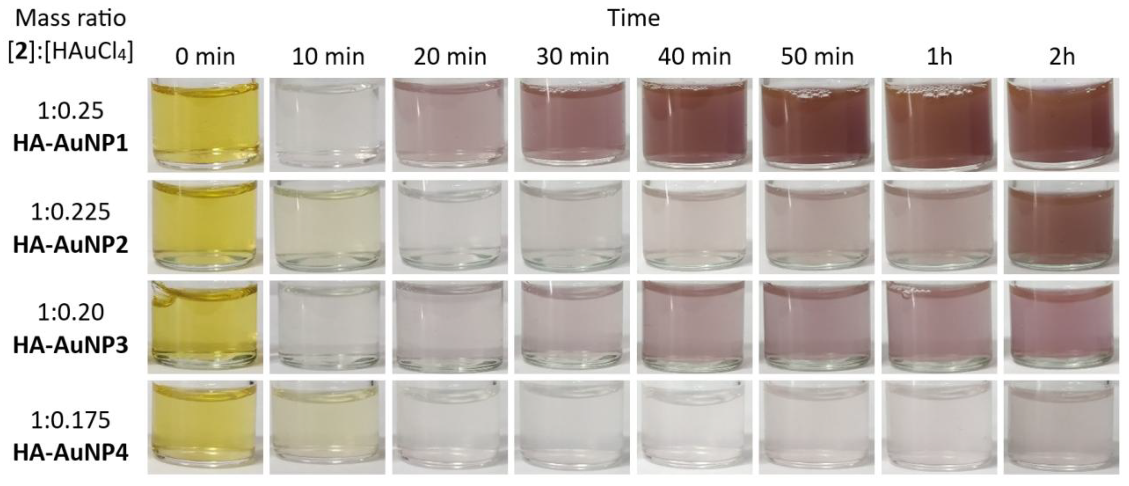
Figure 2.
UV-Vis spectra of HA-AuNPs.
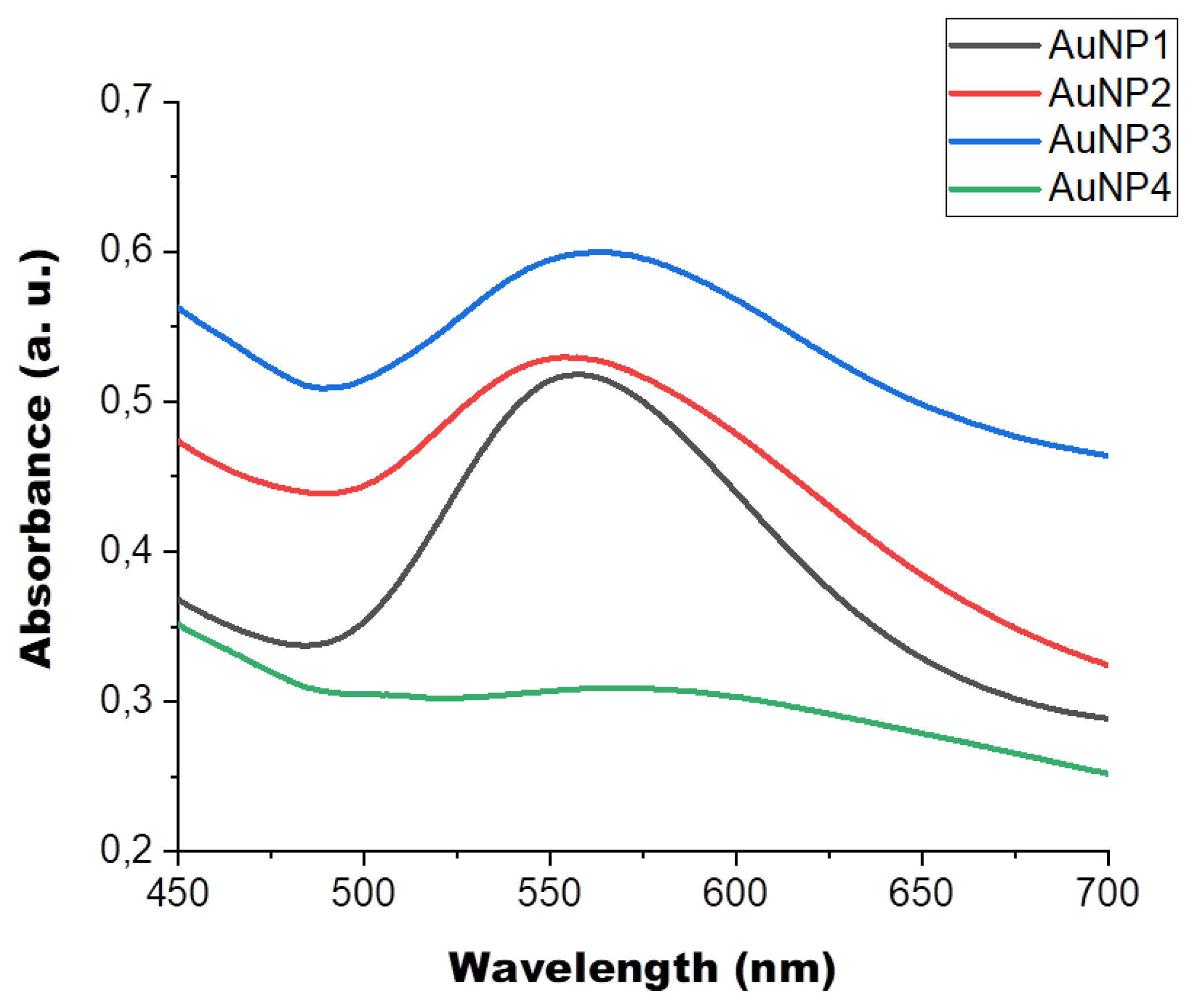
Figure 3.
BF-STEM and SEM images of AuNPs; microscopy characterization of precipitates at ×35 000 (a, HA-AuNP1), ×350 000 (b, HA-AuNP1), ×90 000 (c, HA-AuNP2), ×350 000 (d, HA-AuNP2), ×130 000 (e, HA-AuNP3), and ×200 000 (f, HA-AuNP3) magnifications (corresponding micrometer and nanometer scale bars shown on each image).
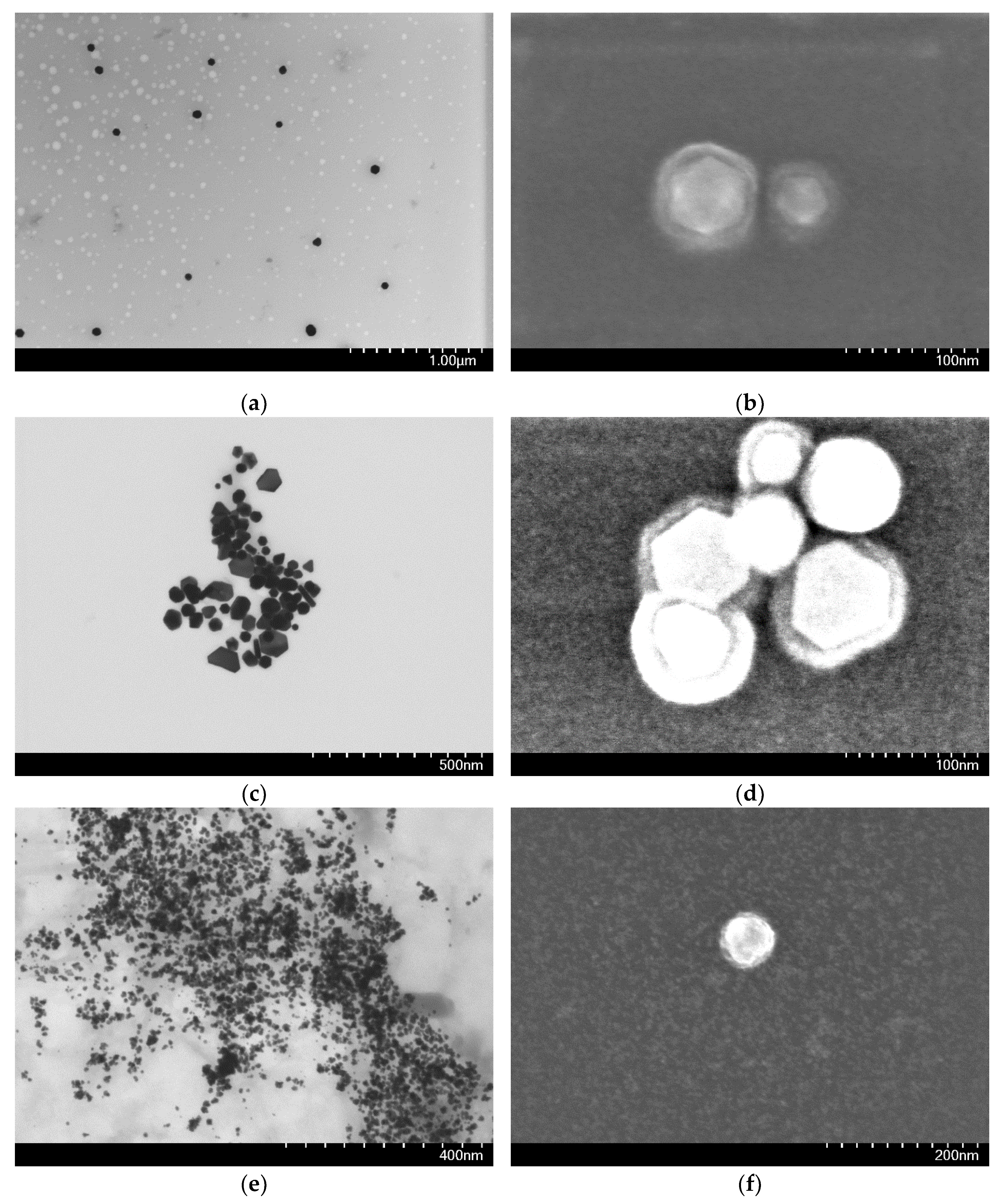
Figure 4.
XPS spectra of HA-AuNP3.
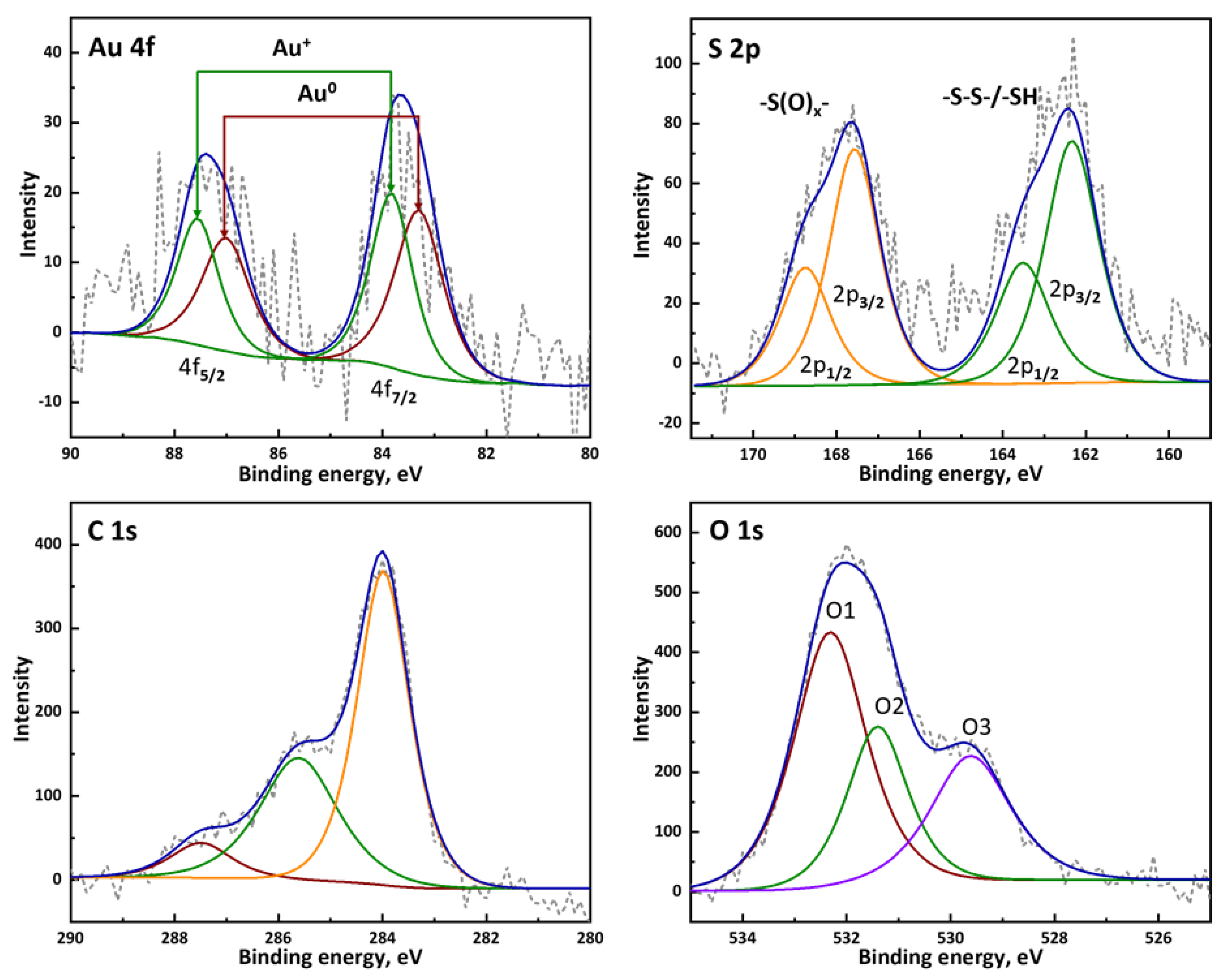
Figure 5.
1H NMR spectra of compounds in D2O: a) HA-S-S-HA (1); b) HA-SH (2); c) result of the reaction of 2 with HAuCl4 at a ratio of [2]:[HAuCl4] = 1:0.20; d) result of the reaction of 2 with HAuCl4 at a ratio of [2]:[HAuCl4] = 1:1.
Figure 5.
1H NMR spectra of compounds in D2O: a) HA-S-S-HA (1); b) HA-SH (2); c) result of the reaction of 2 with HAuCl4 at a ratio of [2]:[HAuCl4] = 1:0.20; d) result of the reaction of 2 with HAuCl4 at a ratio of [2]:[HAuCl4] = 1:1.
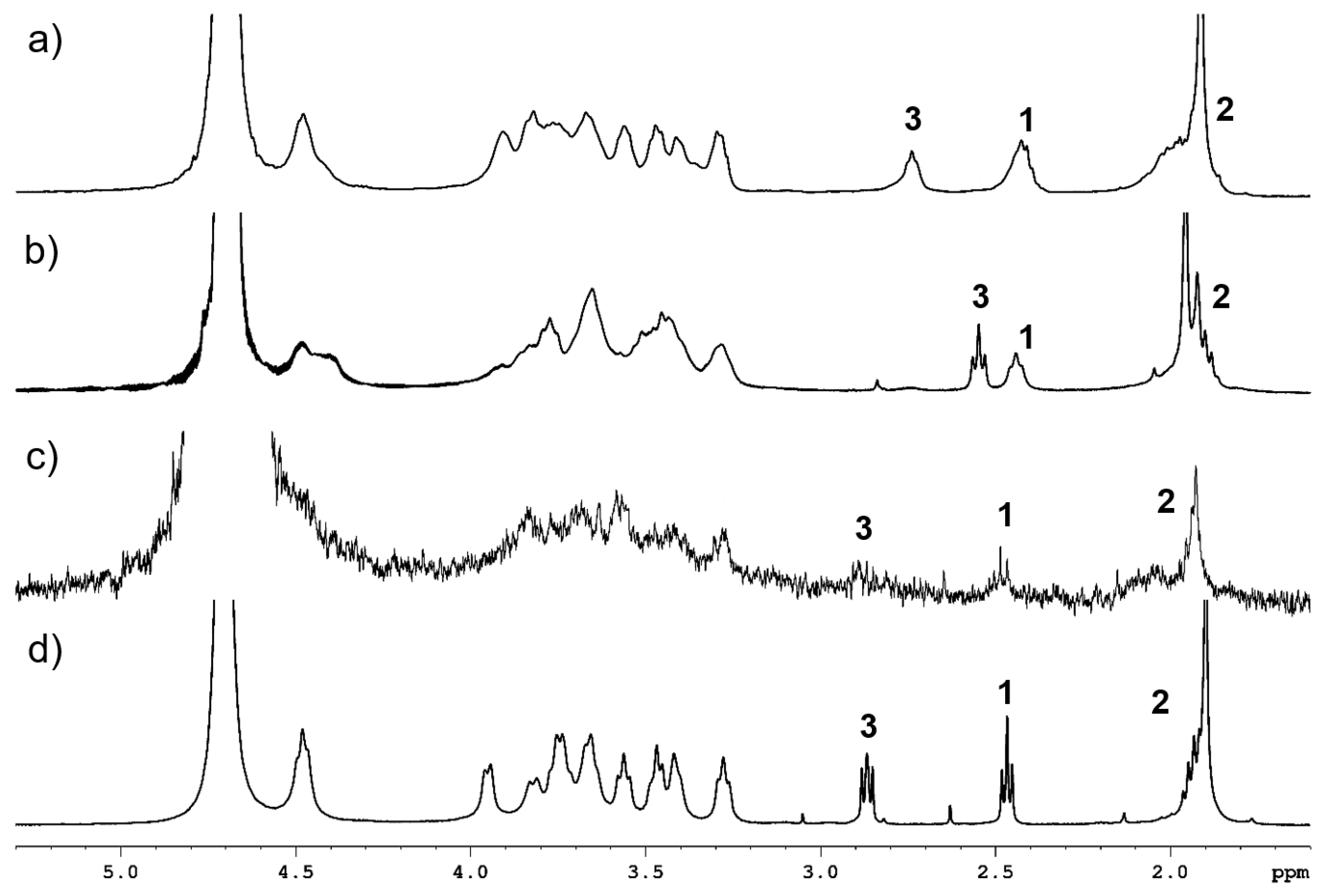

Table 1.
Values of ζ-potentials of samples HA-AuNP1-4.
| HA-AuNP1 | HA-AuNP2 | HA-AuNP3 | HA-AuNP4 | |
|---|---|---|---|---|
| Zeta-potential (mV) | -27.6±3.4 | -28.6±1.7 | -41.0±4.0 | -41.5±1.3 |
Disclaimer/Publisher’s Note: The statements, opinions and data contained in all publications are solely those of the individual author(s) and contributor(s) and not of MDPI and/or the editor(s). MDPI and/or the editor(s) disclaim responsibility for any injury to people or property resulting from any ideas, methods, instructions or products referred to in the content. |
© 2025 by the authors. Licensee MDPI, Basel, Switzerland. This article is an open access article distributed under the terms and conditions of the Creative Commons Attribution (CC BY) license (http://creativecommons.org/licenses/by/4.0/).
Copyright: This open access article is published under a Creative Commons CC BY 4.0 license, which permit the free download, distribution, and reuse, provided that the author and preprint are cited in any reuse.


