
Submitted:
03 October 2025
Posted:
06 October 2025
You are already at the latest version
Abstract
This work presents the synthesis, characterization, and multifunctional properties of bismuth vanadate (BiVO₄) nanoparticles prepared by a rapid and eco-friendly microwave-assisted method. X-ray diffraction (XRD) confirmed the formation of monoclinic scheelite-type BiVO₄ with an average crystallite size of ~19 nm. Transmission electron microscopy (TEM) revealed nearly uniform nanoscale morphology, while Fourier-transform infrared spectroscopy (FTIR) verified the presence of characteristic Bi–O and V–O vibrational modes. Nitrogen adsorption–desorption analysis (BET) indicated a specific surface area of 7.5 m²/g, consistent with a non-porous or weakly porous material. UV–Vis diffuse reflectance spectroscopy (DRS) determined a band gap of 2.55 eV, confirming visible-light activity. Photocatalytic performance was evaluated through the degradation of Acid Orange 7 (AO7) under visible-light irradiation. Effects of catalyst dosage and the initial concentration of pollutant Acid Orange 7 on photocatalytic degradation efficiency, were explained in details. Catalyst loading and initial dye concentration strongly influenced efficiency, achieving up to 77% removal within 120 minutes and well fitting to pseudo-first-order kinetics. In addition, the BiVO₄ nanoparticles exhibited notable antibacterial activity against Escherichia coli, attributed to synergistic effects of reactive oxygen species generation and direct surface interactions with bacterial membranes. These findings demonstrate that microwave-synthesized BiVO₄ is a multifunctional material with strong potential for integrated wastewater purification and disinfection applications.
Keywords:
Bismuth vanadate (BiVO₄)
; Microwave-assisted synthesis
; Energy band gap
; Visible-light photocatalysis
; Acid Orange 7
; Antibacterial properties
; Wastewater treatment
1. Introduction
Water is a fundamental natural resource, indispensable for sustaining life, ensuring human well-being, and maintaining ecosystem functionality. However, rising living standards are often accompanied by increased industrial production and consumption, which lead to excessive pollution of water, air, and soil [1]. The presence of pollutants such as pesticides, heavy metals, and pharmaceuticals in the environment can cause severe ecological and public health risks [2].
Among various contaminants in aquatic ecosystems, organic dyes represent a particularly concerning group due to their extensive use in industries such as textiles, leather, paper, food processing, and cosmetics [3,4]. Within this group, azo dyes are the most widely used, accounting for more than 60% of all synthetic dyes produced worldwide [5]. They are characterized by one or more azo bonds, which provide high chemical stability, strong color intensity, and remarkable resistance to light, temperature, and microbial degradation [5,6]. While these properties are desirable in industrial applications, they also make azo dyes extremely persistent in the environment. Moreover, their degradation products, including aromatic amines, are often toxic, mutagenic, or carcinogenic to humans and aquatic life [7]. A well-known representative of this group is AO7, an anionic azo dye widely used in textile dyeing and paper printing. Its complex aromatic structure makes AO7 highly resistant to conventional wastewater treatment methods, which poses a major environmental challenge [8].
Conventional water purification approaches, including coagulation, activated carbon adsorption, reverse osmosis, ultrafiltration, and biological treatments, are frequently insufficient for the effective elimination of these pollutants [9]. As a result, advanced oxidation processes (AOPs) have attracted considerable attention for the degradation of azo dyes, particularly AO7 [8,10]. These processes generate reactive oxygen species capable of mineralizing pollutants into non-toxic end products such as CO₂, water, and inorganic ions [11]. Within this context, heterogeneous photocatalytic processes have achieved significant progress, distinguished by their environmental compatibility and demonstrated efficiency in wastewater treatment and air purification [12,13].
The most widely studied photocatalyst is titanium dioxide (TiO₂), valued for its stability and low toxicity. However, its relatively wide band gap (3.0–3.2 eV for rutile and anatase) restricts its activity to UV irradiation, which accounts for only about 5% of sunlight [14,15]. Accordingly, recent studies have increasingly focused on visible-light-responsive photocatalysts, which are capable of harvesting a larger fraction of solar energy [16]. Among these, bismuth vanadate (BiVO₄) has emerged as a particularly promising candidate due to its relatively narrow band gap (~2.4 eV), high photostability, chemical inertness, non-toxicity, and facile synthesis methods [17,18,19]. Its electronic structure, involving O 2p and Bi 6s hybrid orbitals, facilitates visible-light absorption more efficiently than TiO₂, whose valence band consists only of O 2p orbitals [20]. The monoclinic scheelite phase of BiVO₄ is recognized for superior photocatalytic activity compared to tetragonal forms, mainly due to its narrower band gap and favorable charge transport properties. This has been repeatedly demonstrated in azo dye degradation under visible-light irradiation [21,22].
Further modifications enhance BiVO₄ performance by improving light absorption and reducing charge recombination. Europium doping, for instance, extended the lifetime of photoexcited electrons and enabled 90% degradation of Methyl Orange within 180 minutes under visible light [23]. Heterojunction formation has also proven highly effective. BiVO₄/TiO₂ composite achieved complete degradation of Acid Blue 113 in just 20 minutes [24], while AgBr-decorated BiVO₄ reached ~93% removal of AO7 in 80 minutes under visible-light irradiation [25].
In addition to its well-established photocatalytic performance, BiVO₄ has also been increasingly recognized as an effective antimicrobial agent against both Gram-positive and Gram-negative bacteria, primarily through mechanisms involving the generation of reactive oxygen species (ROS) and the resulting oxidative stress that compromises cellular membranes and essential biomolecules [17,26]. Microwave-synthesized BiVO₄ nanoparticles have been shown to significantly inhibit the growth of Escherichia coli and Staphylococcus aureus, thereby confirming their combined photocatalytic and antibacterial functionality [26,27,28]. Furthermore, ultra-small tetragonal zircon-type BiVO₄ nanoparticles (2–8 nm) obtained via an ethylene glycol-assisted method were reported to induce severe bacterial cell wall damage, which was attributed to the synergistic effect of ROS formation and electrostatic interactions between positively charged Bi³⁺ surface sites and negatively charged bacterial membranes [29,30] . Collectively, these results underscore the potential of BiVO₄ as a multifunctional material capable of simultaneously enabling pollutant degradation and efficient microbial inactivation, which is of particular importance for integrated water treatment and disinfection applications.
Based on these advantages, the present study aims to synthesize, characterize, and evaluate the photocatalytic and antibacterial properties of BiVO₄ nanoparticles. The material was obtained using a rapid, cost-effective, and eco-friendly microwave-assisted method, which ensures uniform heating, shorter reaction times, and improved energy efficiency. The obtained BiVO₄ nanoparticles were characterized by XRD to determine the crystal structure and crystallite size, TEM to examine morphology and particle size, BET analysis to assess porosity and specific surface area, and FTIR to identify functional groups. Photocatalytic activity was evaluated through the degradation of AO7 under visible-light irradiation at the natural pH of the solution, with emphasis on the effects of catalyst dosage and initial dye concentration. Furthermore, the antibacterial properties of BiVO₄ nanoparticles were investigated against representative bacterial strain, highlighting their potential as a dual-action material for integrated wastewater remediation and disinfection applications.
2. Materials and Methods
2.1. Materials
All reagents were of analytical grade and used without further purification. Table 1 summarizes the main chemicals applied in the synthesis and photocatalytic experiments.

Table 1.
The main chemical reagents used in the experiments.
| Reagent Name | Chemical formula | Purity | CAS No. |
|---|---|---|---|
| Bismuth nitrate pentahydrate | Bi(NO3)3 x 5H2O | ≥99% (AR) | 10035-06-0 |
| Ammonium metavanadate | NH4VO3 | 99 % | 7803-55-6 |
| Nitric acid | HNO3 | ≥99% (AR) | 7697-37-2 |
| Acid Orange 7(AO7) | C₁₆H₁₁N₂NaO₄S | 95% (dye grade) | 633-96-5 |
2.2. Synthesis of BiVO4
BiVO₄ was synthesized via a microwave-assisted process. An aqueous solution of Bi(NO₃)₃·5H₂O (5 mL, 0.05 M) was combined with NH₄VO₃ (5.5 mL, 0.05 M) in a G30 process vial equipped with a magnetic stir bar. The mixture was magnetically stirred for 5 minutes to achieve homogeneity and subsequently sealed with a septum and standard reactor cap. The reaction was carried out in a closed-vessel microwave reactor under temperature-control mode at 170 °C for 10 min. After cooling to room temperature, the resulting suspension was transferred to centrifuge tubes and subjected to centrifugation at 12,000 rpm for 20 minutes. The obtained precipitate was washed three times with deionized water and ethanol to remove residual ions and then dried in a vacuum oven at 70 °C for 180 minutes. A light-yellow powder of monoclinic BiVO₄ was obtained with a yield of approximately 98%.
2.3. Characterization
XRD measurements were carried out using a BRUKER AXS GMBH, A24A10 diffractometer equipped with Cu Kα radiation (λ = 0.15406 nm), operated at 40 kV and 30 mA. Diffraction data were recorded at room temperature over a 2θ range of 10–70°, with a scanning speed of 3°/min and a 0.5 mm divergent slit. The crystallite size was determined from the most intense diffraction peak by applying the Scherrer equation (Eq. (1)):
where D represents the crystallite size, k is the shape factor, λ the X-ray wavelength, β the full width at half maximum (FWHM), and θ the Bragg angle.
TEM measurements were performed using a FEI Tecnai F20 at 200 kV electron acceleration voltage after drop- casting of sample material on lacey carbon TEM grids.
The textural properties of the material were examined by nitrogen adsorption–desorption isotherms recorded at 77 K using a Quantachrome Autosorb iQ3 analyzer. Prior to measurement, the sample was degassed under vacuum by stepwise heating at 90 °C for 1 h and subsequently at 200 °C, until the pressure rise rate was below 50 millitorr min⁻¹, indicating complete removal of adsorbed gases and moisture. The specific surface area was calculated by the Brunauer–Emmett–Teller (BET) method in the relative pressure range of 0.05–0.30, and pore characteristics were derived from the shape of the isotherm.
FTIR spectroscopy was carried out on a Thermo Scientific Nicolet iS50 spectrometer equipped with a built-in all-reflective ATR diamond accessory. Spectra were recorded in the range of 400–4000 cm⁻¹ with a spectral resolution of 4 cm⁻¹ at room temperature.
The optical properties were evaluated by UV–Vis DRS using a Shimadzu 1800 spectrophotometer equipped with an integrating sphere in the wavelength range of 300–800 nm. The reflectance data were converted into absorbance using the Kubelka–Munk function (Eq. (2)):
where R is the observed reflectance.
The optical band gap (Eg) was then estimated according to the Tauc relation (Eq. (3)):
where FKM(R) is the Kubelka–Munk function; R is the observed reflectance in the UV-Vis spectrum; hν is the photon energy; A is a proportionality constant, and n characterizes the electronic transition type. Since BiVO₄ is a direct-allowed semiconductor, the value n=1/2 was applied in the Tauc’s relation.
2.4. Photocatalytic Activity Evaluation
The photocatalytic activity of BiVO₄ was evaluated through the degradation of AO7 aqueous solution under visible light irradiation. A 500 W xenon lamp was used as the illumination source, positioned at a distance of 30 cm from the surface of the reaction mixture.
In a typical experiment, a given mass of BiVO₄ (10, 15, or 20 mg, depending on the run) was dispersed in 50 mL of AO7 solution with an initial concentration of 15, 20, or 25 ppm. The suspension was first treated in an ultrasonic bath for 15 min and subsequently stirred in the dark for 60 min to ensure adsorption–desorption equilibrium between AO7 and BiVO₄.
During irradiation, aliquots of 1.5 mL were withdrawn at predetermined time intervals (up to 120 min), centrifuged at 12,000 rpm for 20 min to remove the catalyst, and the supernatant was analyzed by UV–Vis spectroscopy. The concentration of AO7 was monitored at its absorption maximum (λmax ≈ 484 nm).
The degradation efficiency (D) was calculated using Equation (4):
where C0 is the initial concentration of AO7 and Ct the concentration after irradiation time.
The photocatalytic degradation kinetics were analyzed according to the Langmuir–Hinshelwood pseudo-first-order model (Eq. (5)):
where Kt is the apparent pseudo-first-order rate constant.
2.5. Antibacterial Properties
Antimicrobial activity of the BiVO4 nanoparticles was examined against the Gram-negative bacterium Escherichia coli ATCC 25922. An overnight culture of E. coli was prepared in tryptic soy broth (Torlak, Serbia) supplemented with 0.6% yeast extract (Torlak, Serbia). The culture was centrifuged, washed twice with sterile physiological saline (0.9% NaCl), and resuspended to a final concentration of approximately 10⁷ CFU/mL. Afterwards, 100 µL of the bacterial suspension was added to 9.9 mL of a BiVO₄ nanoparticle suspension (1 mg/mL), which had been previously irradiated with UV light for 30 minutes to enhance photocatalytic activity. The sample was incubated at 37 °C with shaking at 120 rpm for 4 hours, in parallel with control (E. coli without BiVO4). Following incubation, serial decimal dilutions were prepared and plated on sterile tryptic soy agar (TSA) plates. The plates were incubated for 24 hours at 37 °C, after which colony-forming units (CFU) were counted. Percent of bacterial growth inhibition was calculated as follows (Eq. (6)):
where C0 is the number of CFU from the control sample and C is the number of CFU from the treated sample.
The experiment was performed in triplicate and statistical analysis of the results was performed using OriginPro (OriginLab Corporation, Northampton, MA, USA). One-way analysis of variance (ANOVA), followed by Tukey’s test, was used to determine statistical significance. A p-value of p<0.05 was considered statistically significant.
3. Results and Discussion
3.1. Structural and Morphological Properties of the Microwave-Synthesized BiVO4
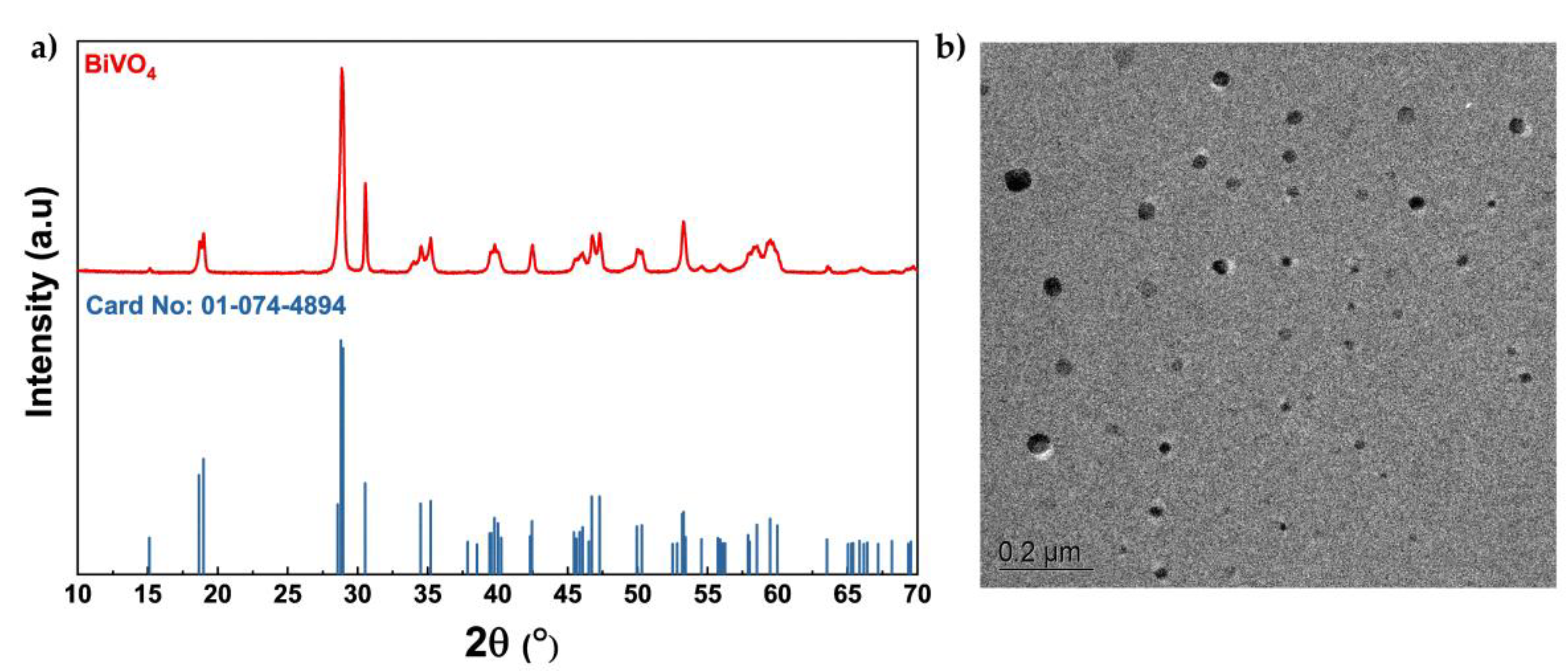
Figure 1 illustrates the structural and morphological characterization of the synthesized BiVO₄ powder. The XRD pattern (Figure 1a) reveals diffraction peaks that are in excellent agreement with the reference standard (JCPDS card No. 01-074-4894), confirming the formation of a single-phase monoclinic scheelite-type structure (clinobisvanite, space group I2/b). The main diffraction peaks observed at approximately 18.9°, 28.9°, 30.5°, 35.2°, and 39.7° were indexed to the (011), (112), (004), (020), and (211) planes of monoclinic BiVO₄, respectively, confirming that the microwave-assisted synthesis yielded a highly crystalline and phase-pure material. The average crystallite size, estimated from the most intense reflection at 2θ ≈ 28.9° using the Scherrer approach, was found to be around 19 nm.
Complementary TEM observations (Figure 1b) provide further insight into the microscopic morphology of the BiVO₄ nanoparticles. The TEM micrograph reveals well-defined nanosized particles with relatively uniform distribution with size approximately of 20 nm, which is in full agreement with the crystallite size calculated from XRD (~19 nm). Most of the nanoparticles exhibit near-spherical to slightly irregular shapes, which is characteristic of microwave-assisted synthesis due to rapid nucleation and growth [17,20]. The image also indicates that the majority of the particles are discrete, although some degree of agglomeration can be observed, which is commonly reported for BiVO₄ nanoparticles owing to their high surface energy [21,26].
Figure 1.
a) XRD pattern of the synthesized BiVO₄ sample together with vertical bars from card reference(No: 01-0074-4894); b) TEM image of BiVO₄ nanoparticles.
Figure 1.
a) XRD pattern of the synthesized BiVO₄ sample together with vertical bars from card reference(No: 01-0074-4894); b) TEM image of BiVO₄ nanoparticles.
3.2. Textural, Vibrational, and Optical Properties of the Microwave-Synthesized BiVO₄
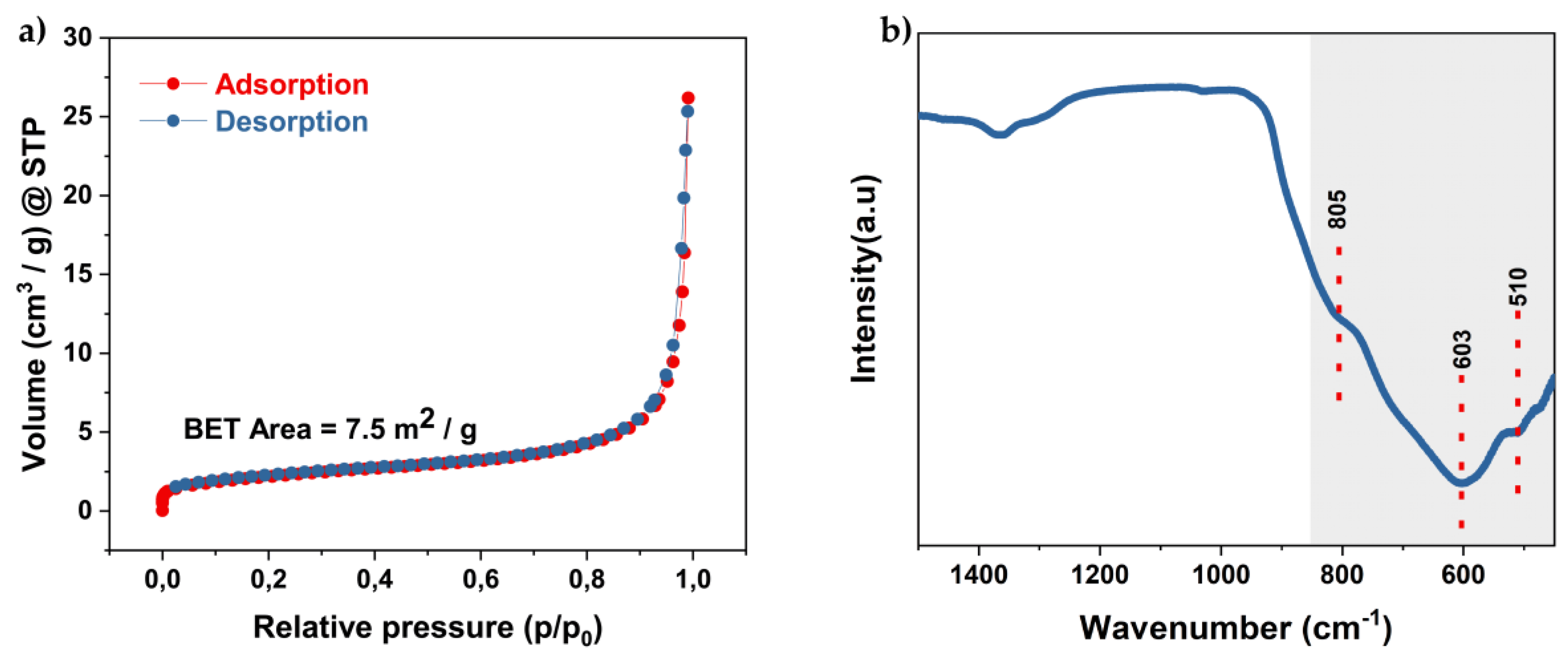
Furthermore, the textural features and vibrational modes of the synthesized BiVO₄ were characterized by nitrogen adsorption–desorption and FTIR spectroscopy (Figure 2). The N₂ adsorption–desorption isotherm (Figure 2a) is best described as Type II without a discernible hysteresis loop, typical of non-porous or macroporous solids. The BET surface area of the sample was calculated to be 7.5 m²/g. The FTIR spectrum (Figure 2b) reveals several distinct absorption features between 500 and 800 cm⁻¹, which are characteristic of BiVO₄. A pronounced band centered at 603 cm⁻¹ together with a shoulder at 805 cm⁻¹ corresponds to the asymmetric and symmetric stretching vibrations of VO₄³⁻ units, while the band at 510 cm⁻¹ is assigned to Bi–O stretching. In addition, a weak absorption at 1380 cm⁻¹ is attributed to residual nitrate species originating from the precursors used during synthesis.
Figure 2.
Textural and vibrational characterization of BiVO₄: a) N₂ adsorption–desorption isotherm; b) FTIR spectrum.
Figure 2.
Textural and vibrational characterization of BiVO₄: a) N₂ adsorption–desorption isotherm; b) FTIR spectrum.
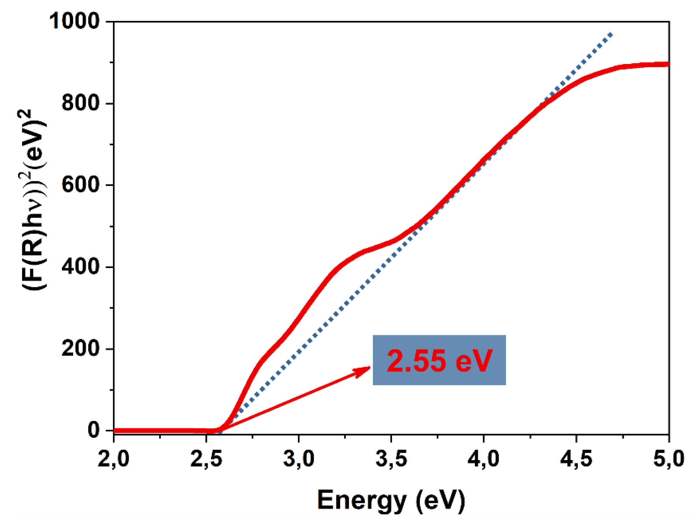
The optical band gap of the synthesized BiVO₄ was determined from the Tauc plot (using Eq. 2) and Eq. 3) assuming a direct allowed transition, yielding a value of 2.55 eV (Figure 3). This finding is in excellent agreement with previously reported values for monoclinic scheelite-type BiVO₄, which typically fall in the range of 2.4–2.6 eV [17]. The consistency of the obtained result with the literature further corroborates the formation of a pure monoclinic scheelite phase, as confirmed by XRD, and underlines the semiconductor’s suitability for visible-light-driven photocatalytic applications. The band gap position ensures efficient utilization of the solar spectrum while maintaining sufficient oxidative potential, thus providing a favorable balance between light absorption and redox activity.
Figure 3.
Tauc`s plot of BiVO₄ derived from Kubelka–Munk transformed UV–Vis diffuse reflectance data.
Figure 3.
Tauc`s plot of BiVO₄ derived from Kubelka–Munk transformed UV–Vis diffuse reflectance data.
3.3. Photocatalytic Performance of the Microwave-Synthesized BiVO₄
AO7 in aqueous medium was used as a model pollutant to assess the visible-light photocatalytic activity of BiVO₄. Prior to irradiation, dye catalyst suspensions were stirred in the dark for 60 min to establish adsorption–desorption equilibrium. The influence of catalyst dosage and initial dye concentration on performance is evaluated below; pH was not systematically varied and remained near its native value (~4.5).
3.3.1. The Effect of Catalyst Dosage on the Degradation of Acid Orange 7
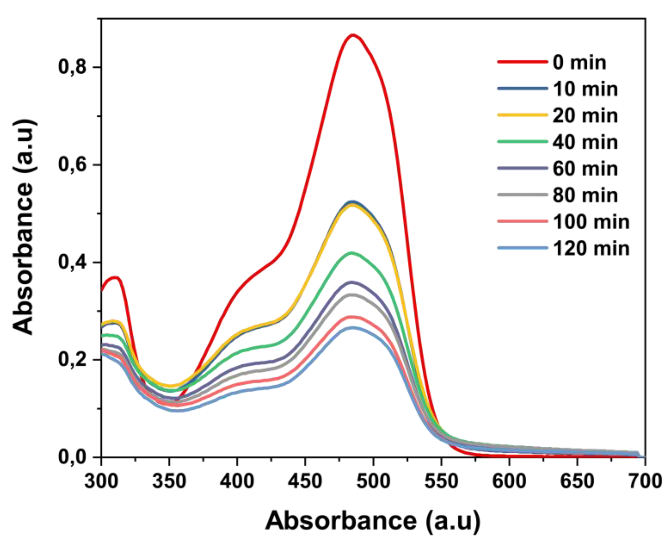
Figure 4 presents the time-resolved UV–Vis spectra of AO7 under visible-light irradiation in the presence of 15 mg BiVO₄. The principal visible band centered at λmax = 484 nm decays steadily over 0–120 min, accompanied by a slight hypsochromic shift within the visible band, remaining centered at 484 nm. The shoulder within 440–520 nm progressively collapses, while the near-UV features around 250–320 nm attenuate more slowly, consistent with stepwise loss of conjugation in the dye chromophore [31].
Figure 4.
UV-Vis absorption spectra of the AO7 solution (20 ppm) for 15 mg of BiVO4.
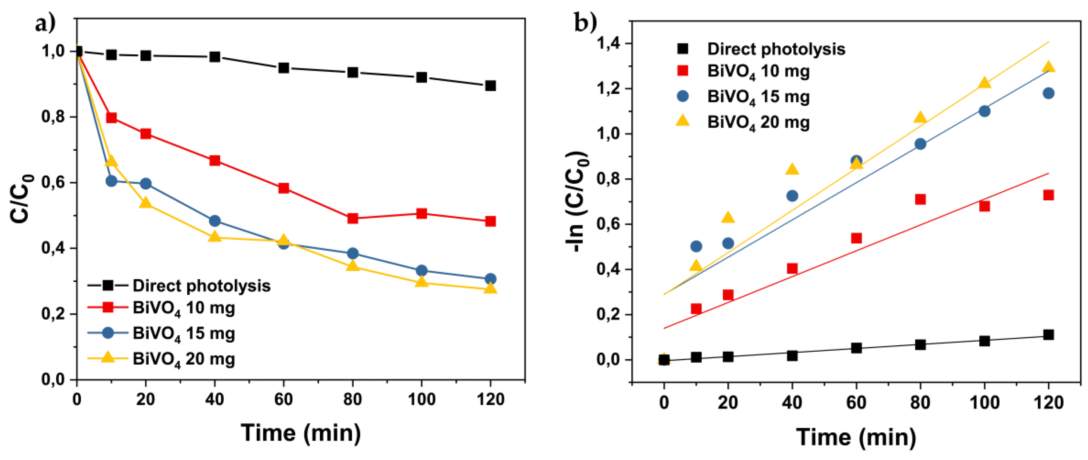
As shown in Figure 5, the influence of BiVO₄ dosage on the photocatalytic degradation of AO7 (20 ppm) under visible-light irradiation was investigated. Figure 5a illustrates the temporal decay of the normalized concentration (C/C₀) at the absorption maximum of AO7 (λmax ≈ 484 nm). In the absence of BiVO₄, only about 10% of AO7 was removed after 120 minutes, confirming that the degradation is primarily photocatalyst-driven. Increasing the catalyst dosage from 10 to 15 mg markedly enhanced degradation efficiency, with ~68% removal compared to ~55% for the lower loading. A further increase to 20 mg resulted in only a slight additional improvement, reaching ~72% after 120 minutes, indicating that the system operates close to its optimum catalyst loading.
The corresponding pseudo-first-order kinetic plots (Figure 5b) exhibit good linearity, consistent with the Langmuir–Hinshelwood model. The calculated rate constants increased with catalyst dosage, being lowest for 10 mg and highest for 20 mg, although the difference between 15 and 20 mg remained relatively small. This trend reflects a balance between the number of active surface sites and the efficiency of light utilization. At higher dosages, more reactive sites are available; however, excessive amounts can lead to light scattering, shielding effects, and partial particle aggregation, which ultimately limit further improvements in photocatalytic activity [32,33].
Based on these results, a dosage of 15 mg BiVO₄ was used for the investigation of the effect of the initial AO7 concentration, as it ensures nearly maximal efficiency while avoiding the drawbacks associated with higher loadings.
Figure 5.
Effect of BiVO₄ catalyst dosage on the photocatalytic degradation of AO7 (20 ppm) under visible light irradiation, including direct photolysis: a) temporal decay of normalized concentration (C/C₀) with time; b) pseudo-first-order kinetic plots.
Figure 5.
Effect of BiVO₄ catalyst dosage on the photocatalytic degradation of AO7 (20 ppm) under visible light irradiation, including direct photolysis: a) temporal decay of normalized concentration (C/C₀) with time; b) pseudo-first-order kinetic plots.
3.3.2. Effect of the Initial Concentration of Pollutant Acid Orange 7 on Photocatalytic Degradation Efficiency
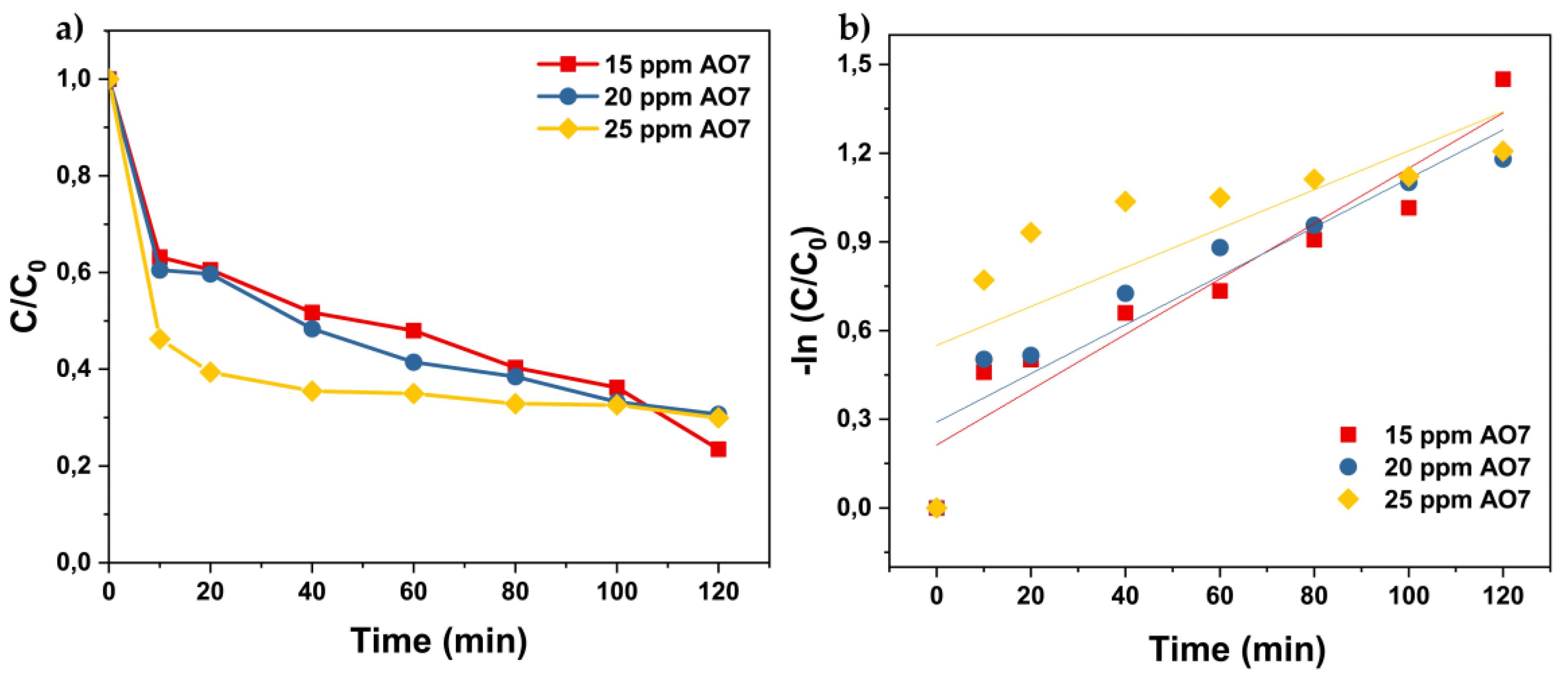
After establishing 15 mg BiVO₄ as the optimal catalyst dosage, the effect of the initial AO7 concentration was investigated. Figure 6a shows that the degradation efficiency decreases with increasing dye concentration. At 15 ppm, about 77% of AO7 was removed within 120 minutes, while the efficiency declined to ~69% at 20 ppm and ~70% at 25 ppm. This reduction is associated with stronger coloration of the solution at higher concentrations, which limits light penetration, as well as the saturation of active sites by excess dye molecules, reducing the availability of reactive oxygen species for photocatalytic reactions [34,35,36].
The kinetic plots (Figure 6b) confirm that the process follows pseudo-first-order kinetics, with linear fits yielding apparent rate constants of 0.0121 min⁻¹ for 15 ppm, 0.0098 min⁻¹ for 20 ppm, and 0.0100 min⁻¹ for 25 ppm. The higher value obtained at 15 ppm indicates that the reaction proceeds faster at lower pollutant loads, while the decrease in k with increasing concentration reflects competition among dye molecules for both photons and active sites. Correlation coefficients (R² > 0.95) further support the applicability of the Langmuir–Hinshelwood kinetic model [37,38].
Figure 6.
Effect of initial AO7 concentration (15 ppm, 20 ppm and 25 ppm) on the photocatalytic degradation over BiVO₄ (15 mg) under visible light irradiation: a) temporal decay of normalized concentration (C/C₀) with time; b) pseudo-first-order kinetic plots.
Figure 6.
Effect of initial AO7 concentration (15 ppm, 20 ppm and 25 ppm) on the photocatalytic degradation over BiVO₄ (15 mg) under visible light irradiation: a) temporal decay of normalized concentration (C/C₀) with time; b) pseudo-first-order kinetic plots.
3.4. Possible Photocatalytic Pathway for the Degradation of Acid Orange 7
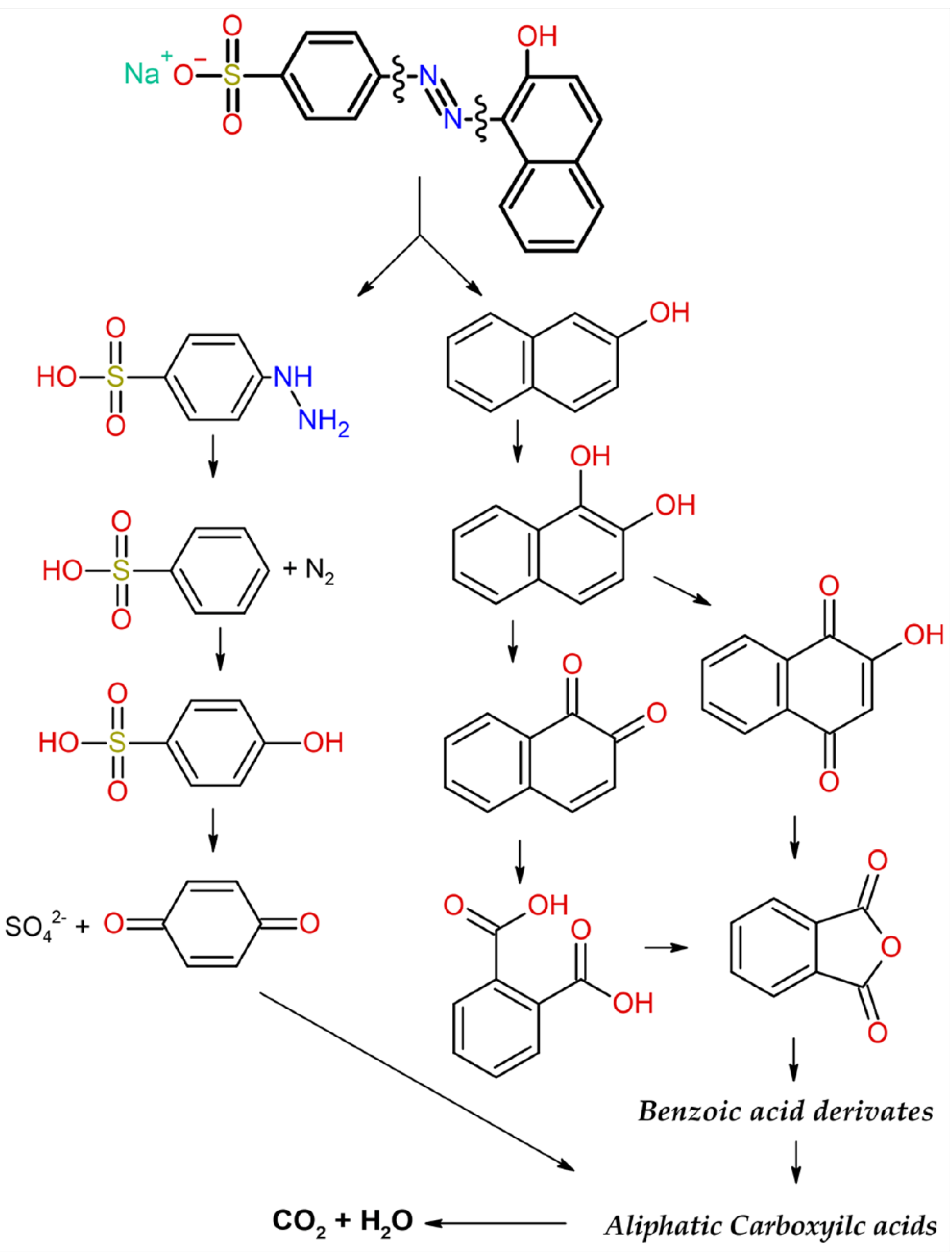
Based on the obtained photocatalytic results and supporting evidence from previously reported studies, the degradation pathway of AO7 under visible-light HBiVO₄ generates electron–hole pairs that initiate oxidative reactions mediated by ROS, mainly hydroxyl radicals (●OH) and superoxide anions (●O₂⁻), in addition to direct hole oxidation (h⁺). The degradation process starts with cleavage of the azo bond, giving rise to two main pathways. In the left pathway, sulfonated aromatic amines are formed and further oxidized to sulfonated hydroxybenzenes and benzoic acid–type derivatives. In the right pathway, hydroxylated naphthalene intermediates are produced, which undergo sequential transformations into hydroxylated naphthalenes, quinone derivatives, and benzoic acid–type compounds. Both branches subsequently converge toward low-molecular-weight aliphatic carboxylic acids, and the overall process ultimately leads to complete mineralization into CO₂ and H₂O.
The stepwise degradation route illustrated in Figure 7 is consistent with the photocatalytic performance observed in this study and aligns with previously reported mechanisms of structurally similar azo dyes [39].
Figure 7.
Proposed degradation pathway of AO7 under visible-light photocatalysis (Modified from [39]).
Figure 7.
Proposed degradation pathway of AO7 under visible-light photocatalysis (Modified from [39]).
3.4. Antibacterial Properties
The results showed that BiVO₄ nanoparticles exhibited an antibacterial effect on Escherichia coli as a model indicator of pathogenic bacteria. After 4 hours of incubation with BiVO₄, the number of viable bacterial cells was significantly reduced compared to the control (Figure 8). On average, the inhibition of bacterial growth was 67.21 ± 3.15.
Figure 8.
Photography of the TSA plate with serial dilutions of untreated E. coli (left) and the sample treated with UV-activated BiVO₄ nanoparticles (right).
Figure 8.
Photography of the TSA plate with serial dilutions of untreated E. coli (left) and the sample treated with UV-activated BiVO₄ nanoparticles (right).
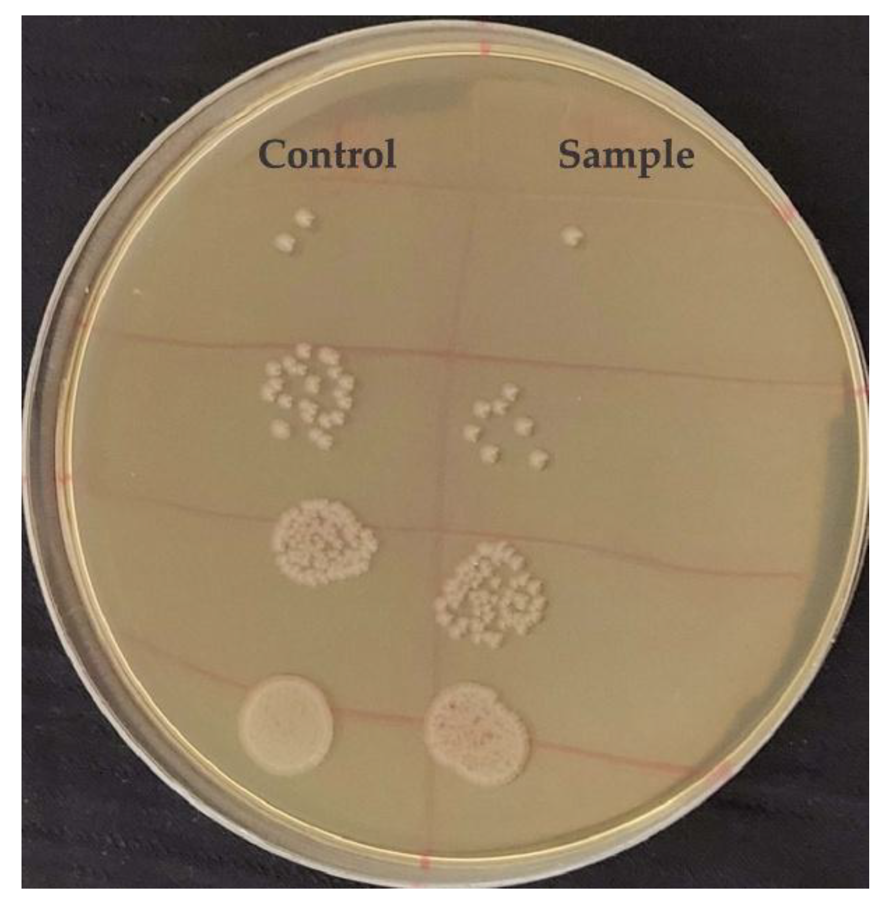
Statistical analysis confirmed that this reduction was significant (p < 0.05), based on one-way ANOVA followed by Tukey’s test. These findings indicate that BiVO₄ nanoparticles can effectively reduce the number of viable E. coli cells under the tested conditions.
The antibacterial effect of BiVO₄ nanoparticles against Escherichia coli can be attributed to a combination of multiple mechanisms. Due to their small crystalline size and high surface area-to-volume ratio, BiVO₄ nanoparticles can effectively penetrate the bacterial cell wall [26]. The positively charged metal ions on the nanoparticle surface interact electrostatically with the negatively charged bacterial cell wall components, leading to structural disruption and increased membrane permeability. This initial damage compromises the integrity of the bacterial cell and facilitates further nanoparticle entry. Furthermore, BiVO₄ could generate the formation of ROS, which induce oxidative stress in bacterial cells by damaging essential biomolecules such as lipids, proteins, and DNA. The oxidative damage results in inhibition of protein synthesis and interference with DNA replication processes, ultimately leading to bacterial cell death.
4. Conclusions
In this study, BiVO₄ nanoparticles were successfully synthesized by a rapid and eco-friendly microwave-assisted method. Structural and morphological analyses confirmed the formation of a pure monoclinic scheelite phase with nanoscale particle size and a specific surface area of 7.5 m²/g. The optical band gap of 2.55 eV indicated visible-light responsiveness, making the material suitable for photocatalytic applications. Photocatalytic experiments demonstrated that the optimal catalyst dosage was 15 mg, which achieved nearly 80% degradation of AO7 (20 ppm) after 120 min under visible-light irradiation, while higher loadings provided only marginal improvement due to optical shielding and particle aggregation. Variation of the initial AO7 concentration further revealed that degradation efficiency and apparent rate constants decreased with increasing pollutant load, confirming the negative influence of excess dye molecules on photon penetration and active site availability. The reaction followed pseudo-first-order kinetics with R² values above 0.95, consistent with the Langmuir–Hinshelwood model. In addition, the BiVO₄ nanoparticles exhibited notable antibacterial activity, inhibiting the growth of E. coli by ~67%. Collectively, these findings demonstrate that microwave-synthesized BiVO₄ is a multifunctional material with strong potential for integrated wastewater remediation and disinfection under irradiation.
Author Contributions
Conceptualization, Dragana Marinković; investigation, Nataša Tot and Bojana Vasiljević; characterization, Anđela Pustak and Ivan Marić; antibacterial experiments, Slađana Davidović; data curation, Jovana Prekodravac Filipović; writing—original draft preparation, Nataša Tot and Bojana Vasiljević; writing—review and editing, Dragana Marinković; supervision, Dragana Marinković. All authors have read and agreed to the published version of the manuscript. All authors have read and agreed to the published version of the manuscript.
Funding
This research was funded by the Ministry of Science, Technological Development, and Innovation of the Republic of Serbia, grant number 451-03-136/2025-03/200017
Data Availability Statement
The raw data supporting the conclusions of this article will be made available by the authors on request
Acknowledgments
The authors are thankful to Dr. Yehezkel Yogev, Dr. Tanja Barudžija, and Prof. Dušan Mijin for providing TEM, XRD, and FTIR measurements.
Conflicts of Interest
The authors declare no conflicts of interest.
References
- Moghimi Dehkordi, M.; Pournuroz Nodeh, Z.; Soleimani Dehkordi, K.; Salmanvandi, H.; Rasouli Khorjestan, R.; Ghaffarzadeh, M. Soil, Air, and Water Pollution from Mining and Industrial Activities: Sources of Pollution, Environmental Impacts, and Prevention and Control Methods. Results Eng. 2024, 23, 102729. [Google Scholar] [CrossRef]
- Shetty, S.S.; Deepthi, D.; Harshitha, S.; Sonkusare, S.; Naik, P.B.; Kumari, N.S.; Madhyastha, H. Environmental Pollutants and Their Effects on Human Health. Heliyon 2023, 9, e19496. [Google Scholar] [CrossRef]
- Tkaczyk, A.; Mitrowska, K.; Posyniak, A. Synthetic Organic Dyes as Contaminants of the Aquatic Environment and Their Implications for Ecosystems: A Review. Sci. Total Environ. 2020, 717, 137222. [Google Scholar] [CrossRef]
- Slama, H.B.; Chenari Bouket, A.; Pourhassan, Z.; Alenezi, F.N.; Silini, A.; Cherif-Silini, H.; Oszako, T.; Luptakova, L.; Golińska, P.; Belbahri, L. Diversity of Synthetic Dyes from Textile Industries, Discharge Impacts and Treatment Methods. Appl. Sci. 2021, 11, 6255. [Google Scholar] [CrossRef]
- Benkhaya, S.; M’rabet, S.; El Harfi, A. Classifications, Properties, Recent Synthesis and Applications of Azo Dyes. Heliyon 2020, 6, e03271. [Google Scholar] [CrossRef] [PubMed]
- Kusumlata, *!!! REPLACE !!!*; Ambade, B.; Kumar, A.; Gautam, S. Sustainable Solutions: Reviewing the Future of Textile Dye Contaminant Removal with Emerging Biological Treatments. Limnol. Rev. 2024, 24, 126–149. [Google Scholar] [CrossRef]
- Dutta, S.; Adhikary, S.; Bhattacharya, S.; Roy, D.; Chatterjee, S.; Chakraborty, A.; Banerjee, D.; Ganguly, A.; Nanda, S.; Rajak, P. Contamination of Textile Dyes in Aquatic Environment: Adverse Impacts on Aquatic Ecosystem and Human Health, and Its Management Using Bioremediation. J. Environ. Manage. 2024, 353, 120103. [Google Scholar] [CrossRef]
- Daneshvar, N.; Aber, S.; Hosseinzadeh, F. Study of C.I. Acid Orange 7 Removal in Contaminated Water by Photo Oxidation Processes. Glob. NEST J. 2008, 10, 16–23. [Google Scholar] [CrossRef]
- Kathi, S.; El Din Mahmoud, A. Trends in Effective Removal of Emerging Contaminants from Wastewater: A Comprehensive Review. Desalination Water Treat. 2024, 317, 100258. [Google Scholar] [CrossRef]
- Iervolino, G.; Vaiano, V.; Pepe, G.; Campiglia, P.; Palma, V. Degradation of Acid Orange 7 Azo Dye in Aqueous Solution by a Catalytic-Assisted, Non-Thermal Plasma Process. Catalysts 2020, 10, 888. [Google Scholar] [CrossRef]
- Tanos, F.; Razzouk, A.; Lesage, G.; Cretin, M.; Bechelany, M. A Comprehensive Review on Modification of Titanium Dioxide-Based Catalysts in Advanced Oxidation Processes for Water Treatment. ChemSusChem 2024, 17, e202301139. [Google Scholar] [CrossRef]
- Pham, H.-C.; Kim, K.-S. Effect of TiO2 Thin Film Thickness on NO and SO2 Removals by Dielectric Barrier Discharge-Photocatalyst Hybrid Process. Ind. Eng. Chem. Res. 2013, 52, 5296–5301. [Google Scholar] [CrossRef]
- Guo, Q.; Ma, Z.; Zhou, C.; Ren, Z.; Yang, X. Single Molecule Photocatalysis on TiO2 Surfaces: Focus Review. Chem. Rev. 2019, 119, 11020–11041. [Google Scholar] [CrossRef]
- Etacheri, V.; Di Valentin, C.; Schneider, J.; Bahnemann, D.; Pillai, S.C. Visible-Light Activation of TiO2 Photocatalysts: Advances in Theory and Experiments. J. Photochem. Photobiol. C Photochem. Rev. 2015, 25, 1–29. [Google Scholar] [CrossRef]
- Kaiba, A.; Alansi, A.M.; Oubelkacem, A.; Chabri, I.; Hameed, S.T.; Afzal, N.; Rafique, M.; Qahtan, T.F. Sunlight-Driven Synthesis of TiO2/(MA)2SnCl4 Nanocomposite Films for Enhanced Photocatalytic Degradation of Organic Pollutants. Catalysts 2025, 15, 214. [Google Scholar] [CrossRef]
- Ishigaki, T.; Nakada, Y.; Tarutani, N.; Uchikoshi, T.; Tsujimoto, Y.; Isobe, M.; Ogata, H.; Zhang, C.; Hao, D. Enhanced Visible-Light Photocatalytic Activity of Anatase-Rutile Mixed-Phase Nano-Size Powder Given by High-Temperature Heat Treatment. R. Soc. Open Sci. 2020, 7, 191539. [Google Scholar] [CrossRef] [PubMed]
- Kamble, G.S.; Natarajan, T.S.; Patil, S.S.; Thomas, M.; Chougale, R.K.; Sanadi, P.D.; Siddharth, U.S.; Ling, Y.-C. BiVO4 As a Sustainable and Emerging Photocatalyst: Synthesis Methodologies, Engineering Properties, and Its Volatile Organic Compounds Degradation Efficiency. Nanomaterials 2023, 13, 1528. [Google Scholar] [CrossRef]
- Dolić, S.D.; Jovanović, D.J.; Štrbac, D.; Far, L.Đ.; Dramićanin, M.D. Improved Coloristic Properties and High NIR Reflectance of Environment-Friendly Yellow Pigments Based on Bismuth Vanadate. Ceram. Int. 2018, 44, 22731–22737. [Google Scholar] [CrossRef]
- Dolić, S.D.; Jovanović, D.J.; Smits, K.; Babić, B.; Marinović-Cincović, M.; Porobić, S.; Dramićanin, M.D. A Comparative Study of Photocatalytically Active Nanocrystalline Tetragonal Zyrcon-Type and Monoclinic Scheelite-Type Bismuth Vanadate. Ceram. Int. 2018, 44, 17953–17961. [Google Scholar] [CrossRef]
- Jeong, S.Y.; Choi, K.S.; Shin, H.-M.; Kim, T.L.; Song, J.; Yoon, S.; Jang, H.W.; Yoon, M.-H.; Jeon, C.; Lee, J.; Lee, J.; Lee, S. Enhanced Photocatalytic Performance Depending on Morphology of Bismuth Vanadate Thin Film Synthesized by Pulsed Laser Deposition. ACS Appl. Mater. Interfaces 2017, 9, 505–512. [Google Scholar] [CrossRef]
- Jelić, S.T.; Ćirković, J.; Jovanović, J.; Novaković, T.; Podlogar, M.; Mitrić, J.; Branković, G.; Branković, Z. High Efficiency Solar Light Photocatalytic Degradation of Mordant Blue 9 by Monoclinic BiVO4 Nanopowder. Mater. Chem. Phys. 2025, 333, 130341. [Google Scholar] [CrossRef]
- Regmi, C.; Dhakal, D.; Lee, S.W. Visible-Light-Induced Ag/BiVO4 Semiconductor with Enhanced Photocatalytic and Antibacterial Performance. Nanotechnology 2018, 29, 064001. [Google Scholar] [CrossRef]
- Zhang, A.; Zhang, J. Effects of Europium Doping on the Photocatalytic Behavior of BiVO4. J. Hazard. Mater. 2010, 173, 265–272. [Google Scholar] [CrossRef]
- Drisya, K.T.; Solís-López, M.; Ríos-Ramírez, J.J.; Durán-Álvarez, J.C.; Rousseau, A.; Velumani, S.; Asomoza, R.; Kassiba, A.; Jantrania, A.; Castaneda, H. Electronic and Optical Competence of TiO2/BiVO4 Nanocomposites in the Photocatalytic Processes. Sci. Rep. 2020, 10, 13507. [Google Scholar] [CrossRef]
- Ran, J.-H.; Fei, X.; Ni, L.; Telegin, F. Enhanced Photocatalytic Degradation of Acid Orange 7 by AgBr/BiVO4 under Visible Light. J. Fiber Bioeng. Inform. 2018, 11, 151–161. [Google Scholar] [CrossRef]
- Bulut, D.T. Exploring the Dual Role of BiVO4 Nanoparticles: Unveiling Enhanced Antimicrobial Efficacy and Photocatalytic Performance. J. Sol-Gel Sci. Technol. 2025, 114, 198–222. [Google Scholar] [CrossRef]
- Pramila, S.; Nagaraju, G.; Mallikarjunaswamy, C.; Latha, K.C.; Chandan, S.; Ramu, R.; Rashmi, V.; Lakshmi Ranganatha, V. Green Synthesis of BiVO4 Nanoparticles by Microwave Method Using Aegle marmelos Juice as a Fuel: Photocatalytic and Antimicrobial Study. Anal. Chem. Lett. 2020, 10, 298–306. [Google Scholar] [CrossRef]
- Marinković, D.; Righini, G.C.; Ferrari, M. Advances in Synthesis and Applications of Bismuth Vanadate-Based Structures. Inorganics 2025, 13, 268. [Google Scholar] [CrossRef]
- Marinković, D.; Righini, G.C.; Ferrari, M. Synthesis, Optical, and Photocatalytic Properties of the BiVO4 Semiconductor Nanoparticles with Tetragonal Zircon-Type Structure. Photonics 2025, 12, 438. [Google Scholar] [CrossRef]
- Li, B.; Gao, X.; Qu, J.; Xiong, F.; Xuan, H.; Jin, Y.; Yuan, H. Visible-Light-Driven Antimicrobial Activity and Mechanism of Polydopamine-Reduced Graphene Oxide/BiVO4 Composite. Int. J. Mol. Sci. 2022, 23, 7712. [Google Scholar] [CrossRef]
- Zhang, S.-J.; Yu, H.-Q.; Li, Q.-R. Radiolytic Degradation of Acid Orange 7: A Mechanistic Study. Chemosphere 2005, 61, 1003–1011. [Google Scholar] [CrossRef] [PubMed]
- Iqbal, N.; Huang, X.; Mohamedali Hamid, K.; Yuan, H.; Batool, I.; Yang, Y. Efficient Visible-Light-Driven Photocatalysis of BiVO4@Diatomite for Degradation of Methoxychlor. Catalysts 2025, 15, 672. [Google Scholar] [CrossRef]
- Maran, M.A.; Zheng, A.L.T.; Tan, H.Y.; Sarbini, S.R.; Tan, K.B.; Boonyuen, S.; Wong, K.K.S.; Chung, E.L.T.; Lease, J.; Andou, Y. Assessing the Photocatalytic Performance of Hydrothermally Synthesized Fe-Doped BiVO4 Under Low-Intensity UV Irradiation. Arab. J. Sci. Eng. 2025. [Google Scholar] [CrossRef]
- Nunes, M.J.; Lopes, A.; Pacheco, M.J.; Ciríaco, L. Visible-Light-Driven AO7 Photocatalytic Degradation and Toxicity Removal at Bi-Doped SrTiO3. Materials 2022, 15, 2465. [Google Scholar] [CrossRef]
- Reza, K.M.; Kurny, A.; Gulshan, F. Parameters Affecting the Photocatalytic Degradation of Dyes Using TiO2: A Review. Appl. Water Sci. 2017, 7, 1569–1578. [Google Scholar] [CrossRef]
- Khan, S.; Noor, T.; Iqbal, N.; Yaqoob, L. Photocatalytic Dye Degradation from Textile Wastewater: A Review. ACS Omega 2024, 9, 21751–21767. [Google Scholar] [CrossRef]
- Asenjo, N.G.; Santamaría, R.; Blanco, C.; Granda, M.; Álvarez, P.; Menéndez, R. Correct Use of the Langmuir–Hinshelwood Equation for Proving the Absence of a Synergy Effect in the Photocatalytic Degradation of Phenol on a Suspended Mixture of Titania and Activated Carbon. Carbon 2013, 55, 62–69. [Google Scholar] [CrossRef]
- Phanichphant, S.; Nakaruk, A.; Chansaenpak, K.; Channei, D. Evaluating the Photocatalytic Efficiency of the BiVO4/rGO Photocatalyst. Sci. Rep. 2019, 9, 16091. [Google Scholar] [CrossRef]
- Viswanathan, B. Photocatalytic Degradation of Dyes: An Overview. Curr. Catal. 2018, 7, 99–121. [Google Scholar] [CrossRef]
Disclaimer/Publisher’s Note: The statements, opinions and data contained in all publications are solely those of the individual author(s) and contributor(s) and not of MDPI and/or the editor(s). MDPI and/or the editor(s) disclaim responsibility for any injury to people or property resulting from any ideas, methods, instructions or products referred to in the content. |
© 2025 by the authors. Licensee MDPI, Basel, Switzerland. This article is an open access article distributed under the terms and conditions of the Creative Commons Attribution (CC BY) license (https://creativecommons.org/licenses/by/4.0/).
Copyright: This open access article is published under a Creative Commons CC BY 4.0 license, which permit the free download, distribution, and reuse, provided that the author and preprint are cited in any reuse.


