Preprint
Article

This version is not peer-reviewed.

Clinical Characteristics and Risk Factors of Tuberculosis in Children and Adolescents in Xinjiang, China: A Retrospective Analysis

  † These authors contributed equally to this article.

A peer-reviewed article of this preprint also exists.

Submitted:

16 September 2025

Posted:

26 September 2025

You are already at the latest version

Abstract

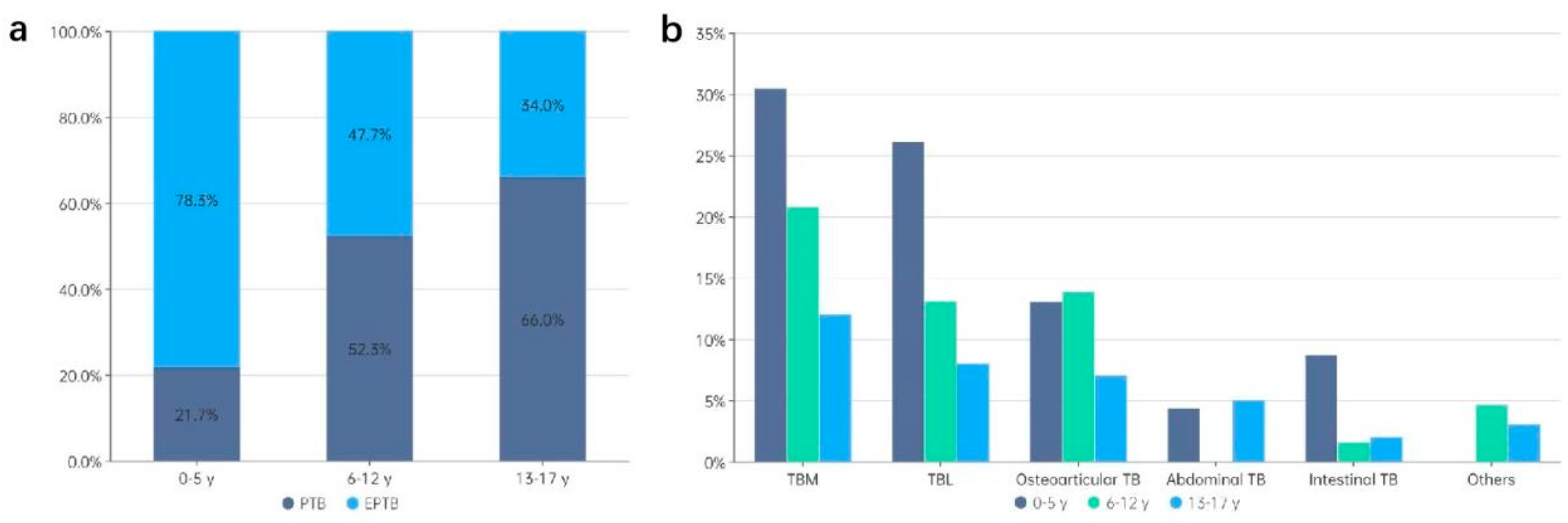
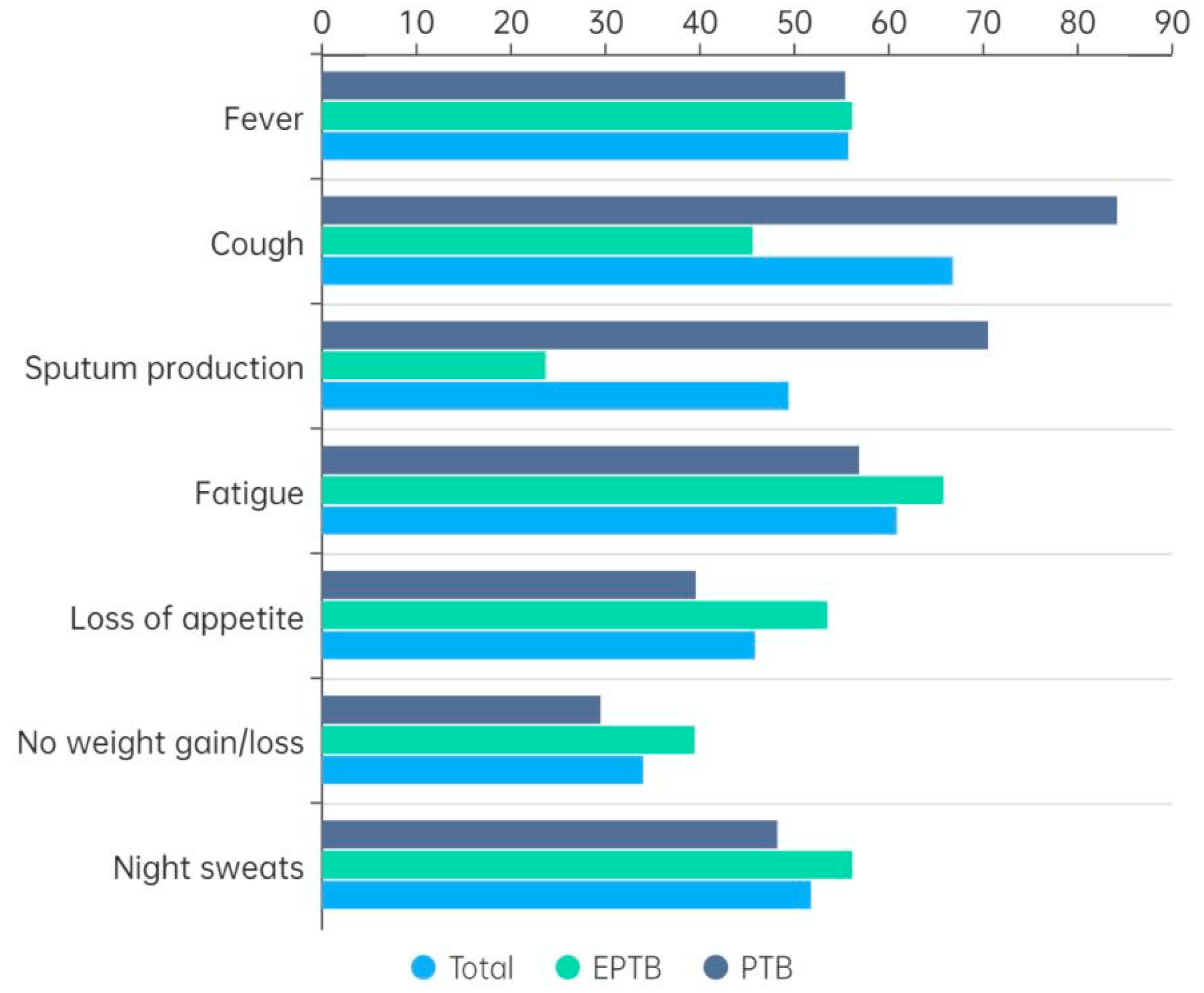
Background: Tuberculosis (TB) continues to be a significant public health concern among children and adolescents in high-burden countries, including China. However, there is a paucity of literature concerning the clinical features and epidemiological characteristics of childhood TB in Xinjiang, the region with the highest TB burden in China. Methods: A retrospective analysis was conducted of children and adolescents aged 0-17 years who were hospitalized with TB between January 2020 and December 2022. A comprehensive analysis of demographic, clinical and laboratory data on different types of TB was conducted, and risk factors for extrapulmonary tuberculosis (EPTB) and severe TB were explored. Results: Among the 253 children and adolescents, 55.3% had pulmonary TB (PTB) and 45.1% had EPTB. The younger children (0-5 years) were more affected by EPTB (78.3%). The most prevalent clinical symptoms were fever (82.2%), cough (79.4%), fatigue (66.4%), and night sweats (52.6%). Tuberculous meningitis (TBM) was the predominant form of EPTB, accounting for 40.4% of cases. Younger age and rural residence were significant risk factors for both EPTB and severe TB. Laboratory results demonstrated high positivity rates for tuberculin skin tests (96.1%) and interferon-γ release assays (84.5%) in all patients, with lower rates of positive smear microscopy and GeneXpert results in EPTB cases. Conclusion: The epidemiology of childhood TB in Xinjiang is characterized by a high incidence of EPTB, with a particularly high prevalence of TBM among younger children. The improvement of early diagnosis of TB in children and adolescents is of critical importance for the enhancement of disease outcomes.

Keywords: 
;  ;  ;  

1. Introduction

Tuberculosis (TB), caused by Mycobacterium tuberculosis (MTB), remains a major global health threat, significantly affecting the lives and development of millions of children and adolescents. In 2023, TB regained its status as the leading cause of death from a single infectious agent, with 10.8 million cases developing worldwide. Children and adolescents under 15 years of age accounted for approximately 12% of all TB cases [1]. TB in children and adolescents accounts for a significant proportion of the total estimated number of cases in low-income countries compared to high-income countries. This has resulted in a significant number of underdiagnoses of childhood TB in low-income areas. The underlying factors contributing to this phenomenon include inadequate staffing levels, limited expertise in the diagnosis of childhood TB, and the suboptimal quality of diagnostic tools for TB [2,3]. In instances where the healthcare team lacks expertise in TB or encounters a disease that can be easily confused with other illnesses, the diagnosis and treatment of TB may be delayed [4]. Moreover, unregulated treatment frequently results in the emergence of drug-resistant TB. This has been identified as a significant contributing factor to the physical sequelae and long-term financial burden experienced by families of sick children [5].
China is one of the countries with a high burden of TB, accounting for approximately 6.8% of the global total. The Xinjiang Uygur Autonomous Region, situated in the northwestern region of China, has been identified as a region with a high prevalence of TB. As demonstrated by the reported incidence data for pulmonary tuberculosis from the Chinese Tuberculosis Information Management System between 2009 and 2018, the Xinjiang Uygur Autonomous Region exhibited the highest average reported incidence rate of pulmonary tuberculosis (155 per 100,000) [6]. However, data concerning the prevalence of TB in children and adolescents within this region remains limited. Spatial clustering of childhood TB cases serves as a practical indicator of localized uncontrolled TB transmission [7]. Furthermore, it plays an important role in the future epidemiological landscape of TB.
The present study employed a retrospective design to investigate the clinical characteristics of pediatric TB patients admitted to a regional referral hospital of Xinjiang Uygur Autonomous Region for infectious diseases. The aim of this study was to provide a comprehensive description of the clinical features of children and adolescents diagnosed with TB, with a particular focus on identifying risk factors associated with severe forms of the disease.

2. Methods

2.1. Study Population

This study was conducted between January 2020 and December 2022 at the Infectious Disease Hospital of the Xinjiang Uygur Autonomous Region. The study population comprised children and adolescents aged 0-17 years who had been diagnosed with TB and admitted to the pediatric TB wards. For patients who have been hospitalized multiple times, only the information from the first admission is included in the analysis. The demographic and clinical data of the patients were retrieved from their respective medical records. A clinical information Survey was completed for each patient (Table S1). The study was conducted in accordance with the principles of the Declaration of Helsinki. The research protocol was reviewed and approved by the The Infectious Disease Hospital of Xinjiang Uygur Autonomous Region (No. 2024-018). All patient data were anonymized and de-identified to protect patient privacy. The study adhered to the relevant national and institutional guidelines for the ethical conduct of research involving human participants.

2.2. Diagnosis of TB

The diagnosis of TB was made in accordance with the Chinese Pulmonary Diagnosis Criteria WS288-2017 [8]. TB cases were categorized as follows: (1) Confirmed case: A biological specimen that has been identified as positive by smear microscopy, culture, molecular methods, or histopathological examination. (2) Clinically diagnosed case: Radiographic evidence was found to be consistent with TB, and at least one of the following was present: TB symptoms, positive tuberculin skin test (TST) or interferon-γ release assay (IGRA) results, histopathological findings, or bronchoscopy evidence consistent with TB. (3) Non-TB: An alternative diagnosis is established, with clinical resolution without anti-TB treatment.

2.3. Laboratory Tests

The TB-related laboratory tests performed included TST, IGRA, smear microscopy, culture, and molecular detection. The commonly used specimens for pathogen detection (smear microscopy, MTB culture, and Gene Xpert) included sputum (including induced sputum), lavage fluid, and gastric aspirate in most patients with PTB. Patients with EPTB were provided with tissue biopsy specimens.

2.4. TB Classification

TB classification followed Chinese criteria WS196-2017 [9]:
Pulmonary TB (PTB): TB confined to the lungs, trachea, bronchi, or pleura.
Extrapulmonary TB (EPTB): TB affecting organs or tissues other than the lungs (e.g., lymph nodes, abdomen, genitourinary tract, skin, joints and bones, meninges). Patients presenting with both PTB and EPTB were exclusively categorized in the EPTB group.
Disseminated TB (DTB): TB involving ≥2 noncontiguous sites [10].
Severe TB: Includes DTB, or tuberculosis meningitis (TBM) [10].

2.5. Statistical Analysis

Descriptive statistics were employed, including frequency analysis for categorical variables. Subsequently, comparative analyses between subgroups were conducted, with consideration given to factors such as gender, age, residence, Time from onset of symptoms to hospital visit and contact history. The patients were divided into three age groups for the purposes of the study: 0–5 years (pre-school age), 6–12 years (school age), and 13–17 years (adolescence). The time interval from the onset of symptoms to the patient’s hospital visit was categorized into the following intervals: 0-30, 31-60, 61-90, and <90.
The Modified Poisson Regression was employed to calculate crude and adjusted relative risk (RR) using univariable and multivariable analysis. Statistical analyses were conducted utilizing SPSS 27.0, with a two-sided P value of less than 0.05 being considered statistically significant. E value was calculated using online E-value calculator (https://www.evalue-calculator.com/evalue/) [11,12]. Population Attributable Fraction (PAF) was calculated using the formula: PAF=Pcases*(RR-1)/RR, while Pcases indicates the prevalence of exposure among cases [32].

3. Results

3.1. Demographic and Clinical Data

A total of 253 children and adolescents diagnosed with TB were enrolled in the study between January 2020 and December 2022. As demonstrated in Table 1, 39.9% (101/253) of the participants were male. The distribution by age and gender revealed a higher proportion of males in the 0-5 years age group (56.5%, 13/23), which decreased to 40.0% (52/130) in the 6-12 years group and 36.0% (36/100) in the 13-17 years group. In other words, the proportion of male patients decreased with increasing age.
The median age of the enrolled children and adolescents was 12 years (interquartile range 8-14 years), with an age range of 7 months to 17 years. The majority of subjects were in the 6-12 years age group (51.4%, 130/253). Most patients (56.5%, 143/253) were from rural areas. The Bacillus Calmette-Guérin (BCG) vaccination rate was found to be comparatively low, with 42.7% (108/253) of children and adolescents having confirmed vaccination status, due to incomplete vaccination records. For more than half of the patients (54.5%, 138/253), an accurate diagnosis was made within 30 days of the onset of symptoms.
Close contact with active TB patients was reported by 19.4% (49/253) of children and adolescents. Of those with known exposure, 30.4% (7/23) were aged 0-5 years, 18.5% (24/130) were aged 6-12 years, and 18.0% (18/100) were aged 13-17 years.
The incidence of PTB was found to be higher among the enrolled children and adolescents (55.3%, 140/253). Among EPTB cases, the most prevalent form was TBM (40.4%, 46/114), followed by lymphatic TB (TBL) (27.2%, 31/114) and osteoarticular TB (24.6%, 28/114). Severe TB was observed in 31.2% (79/253) of cases.

3.2. Distribution of TB Types by Age

The distribution of TB types across different age groups was analyzed. As shown in Figure 1a, the 0-5 years age group was predominantly affected by EPTB, with the proportion of PTB cases increasing with age. In the 13-17 years age group, the most prevalent form was PTB. Further analysis of the EPTB types revealed that TBM was the most prevalent, followed by TBL and osteoarticular TB (Figure 1b).

3.3. Clinical Symptoms by TB Type

The most frequently observed clinical manifestations in the pediatric TB patients were fever, cough, fatigue, and night sweats. Figure 2 compares the clinical symptoms observed in patients with PTB and EPTB. Patients with PTB were more likely to present with a significant cough (84.2% vs. 45.6%, P < 0.001) and sputum production (70.5% vs. 23.7%, P < 0.001). In contrast, patients with EPTB were more likely to present with loss of appetite (53.5% vs. 39.6%, P = 0.027). Furthermore, a higher prevalence of no weight gain or weight loss (39.5% vs. 29.5%) and night sweats (56.1% vs. 48.2%) was observed in EPTB cases, though these differences were not statistically significant.

3.4. Risk Factors Associated with EPTB or Severe TB

Subsequent analysis identified risk factors associated with EPTB and severe TB. As demonstrated in Table 2 and S2, a higher prevalence of EPTB and severe TB was observed in younger children (aged 0-5 years). The relative risk for these conditions demonstrated a decline with increasing age. Furthermore, children and adolescents residing in urban areas exhibited a reduced likelihood of developing EPTB or severe TB in comparison to those residing in rural or town areas, thereby indicating a heightened risk in the latter group.
A stratified analysis was conducted by age and gender. However, due to limitations in the sample size, we conducted EPTB risk factor-related analyses exclusively for the 6-12 age group (Table S3) and girls (Table S4). The risk factors for EPTB in girls were found to be comparable to those observed in the overall cases, and the 6-12 age group exhibited a similar trend; however, the majority of the observed differences were not statistically significant.

3.5. Laboratory Results

As demonstrated in Table 3, laboratory testing revealed that 96.1% (99/103) of children and adolescents tested positive for the TST, while 84.5% (196/232) tested positive for the IGRA. The prevalence of positive TST (92.3% vs. 98.4%) and IGRA (85.6% vs. 83.6%) results was similar between EPTB and PTB patients. Of the diagnostic tests performed, smear microscopy yielded a positive result in 35 children and adolescents, MTB culture in 59 children and adolescents, and GeneXpert in 82 children and adolescents. Patients with EPTB exhibited a significantly lower proportion of positive smear microscopy (6.3% vs. 22.3%, P = 0.001) and GeneXpert tests (31.4% vs. 53.1%, P = 0.004) compared to patients with PTB.

4. Discussion

This study aimed to examine the clinical and epidemiological characteristics of TB in children and adolescents from Xinjiang, a region of China that has highest burden of TB [6]. To the best of our knowledge, this is the first report focusing on pediatric TB in this region, which underscores the significance of understanding local TB trends, particularly among children and adolescents. The findings provide valuable insights into the distribution, clinical manifestations, and risk factors associated with TB, offering a foundation for improving early detection and management strategies for pediatric TB in the area.
The findings of the present study indicated that the majority of children and adolescents hospitalized with TB were from rural areas (56.5%), thus highlighting a significant urban-rural disparity in terms of TB burden. This finding is consistent with those from other regions, where rural areas often face challenges such as limited access to healthcare, higher rates of malnutrition, and poor living conditions. These factors contribute to increased TB transmission and disease progression [13,14,15]. This finding emphasizes the need for targeted TB control programs in rural regions to mitigate the growing burden of childhood TB.
The gender distribution in our study showed a predominance of females (60.1%), which is in contrast to the male-dominated prevalence observed in some previous studies [10,16,17]. Notably, we observed that the proportion of males was higher among children aged 0-5 years (56.5%), whereas it decreased to 38.3% in those older than five years. This shift in gender disparity aligns with findings from studies in South Africa, Uganda, and India, where adolescent girls were found to be more susceptible to TB [18,19,20]. This discrepancy may be attributable to a number of factors. Firstly, the influence of hormonal levels. Hormonal changes during puberty have been hypothesized to affect the incidence of active tuberculosis in children and adolescents. It has been demonstrated that, in the case of female subjects, androgen levels increase prior to estrogen levels during the process of puberty. Estrogen has been demonstrated to promote TH1-mediated immune responses, while androgens have been shown to inhibit these responses [21]. Secondly, the nutritional status. Surveys have indicated that the level of 25-hydroxyvitamin D in female subjects aged between 12 and 19 years is significantly lower than in male subjects [22]. Furthermore, the incidence of anemia is higher in female subjects aged between 12 and 15 years [23]. These factors might contribute to the higher prevalence of tuberculosis in adolescent females. Consequently, in the prevention and control of tuberculosis among children and adolescents, there is a need to strengthen screening of adolescents and to pay particular attention to improving the nutritional status of adolescents, especially females.
In the present study, EPTB was identified as a prevalent form in pediatric TB patients, accounting for 45.1% of the observed cases. This prevalence is lower than that reported by Beijing Children’s Hospital (54%) [10] and Syria (73.6%) [24], but higher than in adults (37.4%) [25]. The high incidence of EPTB in younger children, particularly those under five years old, can be attributed to their immature immune systems, which render them more susceptible to extrapulmonary spread. In the present study, the incidence of EPTB in children under five years was 78.3%, in comparison to 34% in those aged 13-17 years. The decline in the prevalence of EPTB with increasing age, as evidenced by both this study and a previous investigation [10], lends further support to the notion that age constitutes a significant risk factor for EPTB.
Among the EPTB cases, TBM was the most prevalent form, accounting for 40.4%, followed by lymphatic TB (27.2%) and osteoarticular TB (24.6%). These results are consistent with those from other studies, including those conducted at Beijing Children’s Hospital (38.8%) and across mainland China (34.18%) [10,26]. However, some studies have reported lymphatic TB as the most common form of EPTB [24,27]. This discrepancy may be explained by the fact that the patients included in our study were primarily hospitalized with severe cases of TB, which may have delayed their diagnosis, leading to a higher prevalence of severe forms such as TBM. The necessity for early recognition and prompt intervention to prevent complications is emphasized.
The most prevalent clinical symptoms in this cohort were fever, cough, fatigue, and night sweats. Furthermore, the presence of cough and sputum production was observed in cases of EPTB, with 51.8% of cases exhibiting concurrent PTB. Patients with PTB were more likely to exhibit significant coughing and sputum production compared to EPTB cases, who were more likely to present with loss of appetite and no weight gain or loss. These findings are consistent with the extant literature, which suggests that weight loss and loss of appetite are more pronounced in EPTB cases, particularly in TBM and other severe forms of EPTB [27,28]. The association of TB with wasting syndrome, particularly in cases of EPTB, calls attention to the need for comprehensive nutritional support and careful monitoring of weight changes in pediatric TB patients.
The present study identified several risk factors that have been demonstrated to be associated with an increased likelihood of developing EPTB or severe forms of TB. The findings of this study indicate that younger age and residing in a rural area were significant independent risk factors for both EPTB and severe TB. It is noteworthy that 66.7% of the EPTB cases were classified as severe, indicating an overlap between the factors that predispose children and adolescents to EPTB and those that result in more severe forms of TB. It is evident that younger children, particularly those in the 0-5 years age group, were at an elevated risk of both EPTB and severe TB. The result aligns with previous studies, which have indicated that children under five years old are particularly vulnerable to progressing to severe forms of TB, including TBM and disseminated TB, due to their underdeveloped immune systems [10]. Additionally, living in rural areas was found to be a significant risk factor for both EPTB and severe TB, further emphasizing the need for targeted interventions in these regions. Children and adolescents residing in rural areas frequently encounter delayed diagnosis, a consequence of constrained access to healthcare. This predicament can lead to the manifestation of more severe forms of TB by the time these children and adolescents are identified.
In order to assess the potential impact of unmeasured confounders, the E-value was calculated. The E-values for all potential risk factors with significant differences were above 2.5. For instance, the risk of EPTB in children aged 13-17 years was found to be significantly reduced (RR = 0.413, 95% CI: 0.298–0.571), with a corresponding E-value of 4.28. This finding suggests that a high degree of unmeasured confounding would be necessary to fully explain the observed results, thereby indicating the reliability of the study’s conclusions. Furthermore, the PAF of risk factors was calculated (Table 2, Tables S2–S4). The findings of the present study indicated that 27.6% (95% CI: 21.3%–34.1%) of EPTB cases within the study population could be attributed to rural residence. This suggests that, in the hypothetical scenario of the complete eradication of the excess risk associated with rural living, approximately one in four EPTB cases could be averted. It is therefore vital that the national strategy to end TB prioritizes the reduction of the urban-rural gap.
The BCG vaccine is extensively utilized for the prevention of TB, particularly in pediatric populations. In the present study, data pertaining to the BCG vaccination status of children and adolescents was accessible for only 46.2% of the sample. Despite this, it is well-established that BCG vaccination significantly reduces the risk of developing severe forms of TB, including EPTB [29,30]. The findings of this study suggest that BCG vaccination plays a crucial role in preventing severe TB in children and adolescents, underscoring the importance of ensuring full vaccination coverage.
Contact history with TB patients is a well-documented risk factor for developing severe TB [10]. In the present study, 19.3% of children and adolescents had close contact with active TB patients, a figure similar to the 20.9% observed in Liao Q’s study. While contact history was associated with a higher proportion of severe TB cases, the difference was not statistically significant. This observation underscores the complex interplay of factors that contribute to TB transmission and disease severity in children and adolescents.
The challenges associated with diagnosing pediatric TB have been extensively documented, and this study further corroborates the difficulties involved in diagnosing TB in children and adolescents, particularly in the context of EPTB. While immunological diagnostic methods, such as the TST and IGRA, demonstrated high positivity rates in both PTB and EPTB patients, the sensitivity of pathogenetic diagnostic methods, including smear microscopy, culture, and GeneXpert, was lower than anticipated. EPTB patients exhibited significantly lower rates of positive smear microscopy and GeneXpert testing in comparison to PTB patients, thereby underscoring the diagnostic limitations inherent to extrapulmonary disease, where MTB is frequently present in low numbers and in inaccessible body sites.
Furthermore, previous studies have noted the high false-positive rates of IGRA in non-TB populations, which may limit its utility in active TB diagnosis in regions with a high TB burden [31]. The relatively low sensitivity of smear microscopy and MTB culture underscores the need for improved diagnostic techniques, including the use of advanced molecular assays, to facilitate early and accurate diagnosis in children and adolescents, particularly those with EPTB. Additionally, the utilization of the GeneXpert test has demonstrated potential in the diagnosis of TB in pediatric cases, however, its sensitivity remains a constraining factor, particularly in the context of non-pulmonary forms of the disease.

5. Limitations

It is imperative to acknowledge the limitations of this study when interpreting the results. Firstly, the retrospective nature of the study meant that data collection was dependent on the accuracy and completeness of medical records, which may have introduced bias. Secondly, some risk factors (such as BCG vaccination, contact history, etc.) were defined based on self-reported data, and misclassification of risk factors may impact the performance metrics (either RR or PAF). Thirdly, while this study provides important insights into the epidemiology of pediatric TB in Xinjiang, it is important to note that the findings may not be generalizable to other parts of China or low-income countries. It is evident that further multicenter prospective studies are required in order to both confirm these findings and explore additional risk factors.

6. Conclusions

The present study draws attention to the burden of both pulmonary and extrapulmonary TB among hospitalized children and adolescents in Xinjiang. The findings demonstrated a higher prevalence of EPTB in younger children, particularly those under five years old, with TBM being the most prevalent form. Key risk factors for EPTB and severe TB included younger age and residence in rural areas, both of which have been shown to be associated with delayed diagnosis and advanced disease. These findings emphasized the necessity for early detection, enhanced diagnostic capacity, and targeted TB control interventions, particularly in rural areas and among younger children. In view of the fact that cases of TBM in children under five years of age are so prevalent, it is recommended that lumbar puncture and CNS imaging be considered in cases of febrile children exposed to TB or who are unresponsive to antibiotics in rural clinics. The access for molecular detection assays (such as GeneXpert) should be expanded to township hospitals, paired with specimen transport networks. Further research into the molecular and immunological mechanisms underlying pediatric TB in this high-burden region is needed to improve outcomes and reduce the overall burden of disease.

Supplementary Materials

The following supporting information can be downloaded at: https://www.mdpi.com/article/doi/s1.

Author Contributions

B. Z. and J. E. were involved in investigation. They conducted surveys, or other investigative activities. B. Z. collected the field data through extensive surveys, and J. E. did data analysis. T. X. and G. S. was the main author of the original draft. They wrote the first version of the manuscript, integrating the research findings and ideas into a coherent narrative. T. X. and G. S. structured the paper, drafted the introduction, methods, results, and discussion sections, and ensured that the content was consistent with the research objectives. Q. L. and W. J. participated in writing - review & edit. They reviewed the original draft, provided critical feedback, and made suggestions for improvement. Q. L. focused on the clarity and flow of the arguments, Q. L. and W. J. checked for scientific accuracy and consistency, and W. J. edited the language and grammar to enhance the readability of the manuscript. Q.L. and W. J. was involved in funding acquisition. He/She was responsible for securing the financial resources for the research project through grant applications, sponsorships, or other funding sources. Q. L. and W. J. identified potential funding agencies, prepared the funding proposals, and managed the financial aspects of the project once the funds were obtained.

Funding

This study was supported by the Key R&D Program of Xinjiang (2024B03021-2), Tianshan Talent Training Program of Xinjiang Uygur Autonomous Region (2022TSYCCX0105), Training Plan for High level Public Health Technical Talents (No. 2022-3-041).

Institutional Review Board Statement

The study was conducted in accordance with the principles of the Declaration of Helsinki. The research protocol was reviewed and approved by the The Infectious Disease Hospital of Xinjiang Uygur Autonomous Region (No. 2024-018). Given the retrospective nature of the study, which involved the analysis of existing clinical data, the need for individual patient consent was waived by the IRB. This decision was based on the determination that the study posed minimal risk to patient privacy and confidentiality, and that obtaining informed consent was not feasible due to the retrospective collection of data. All patient data were anonymized and de-identified to protect patient privacy. The study adhered to the relevant national and institutional guidelines for the ethical conduct of research involving human participants.

Clinical Trial

Not applicable.

Informed Consent Statement

All authors have read and approved the final manuscript.

Data Availability Statement

The datasets generated and or analyzed during the current study are available from the corresponding author on reasonable request.

Conflicts of Interest

The authors declare no conflicts of interest.

Abbreviations

TB Tuberculosis
EPTB Extrapulmonary tuberculosis
PTB Pulmonary tuberculosis
TBM Tuberculous meningitis
MTB Mycobacterium tuberculosis
TBL Lymphatic tuberculosis
DTB Disseminated tuberculosis
TST Tuberculin skin test
IGRA Interferon-γ release assay
BCG Bacillus Calmette-Guérin
OR Odds ratios
WHO World Health Organization

References

  1. World Health Organization, Global tuberculosis report 2024. Geneva: World Health Organization, 2024. Licence: CC BY-NC-SA 3.0 IGO.
  2. Yom An, Alvin Kuo Jing Teo, Chan Yuda Huot, Sivanna Tieng, Kim Eam Khun, Sok Heng Pheng, Chhenglay Leng, Serongkea Deng, Ngak Song, Daisuke Nonaka, Siyan Yi. Barriers to childhood tuberculosis case detection and management in Cambodia: the perspectives of healthcare providers and caregivers. BMC infectious diseases. 2023; 23(1): 80. [CrossRef]
  3. Bonaventure Michael Ukoaka, Faithful Miebaka Daniel, Precious Miracle Wagwula, Mohamed Mustaf Ahmed, Ntishor Gabriel Udam, Olalekan John Okesanya, Adetola Babalola, Tajuddeen Adam Wali, Samson Afolabi, Raphael Augustine Udoh, Iniubong Godswill Peter, Lina Abdulhameed Maaji. Prevalence, clinical characteristics, and treatment outcomes of childhood tuberculosis in Nigeria: a systematic review and meta-analysis. BMC infectious diseases. 2024; 24(1):1447. [CrossRef]
  4. Jing Tong, Mengqiu Gao, Yu Chen, Jie Wang. A case report about a child with drug-resistant tuberculous meningitis. BMC infectious diseases. 2023; 23(1):83. [CrossRef]
  5. Temesgen Yihunie Akalu, Archie C A Clements, Adhanom Gebreegziabher Baraki, Kefyalew Addis Alene. Protocol for a systematic review of long-term physical sequelae and financial burden of multidrug-resistant and extensively drug-resistant tuberculosis. PloS one. 2023; 18(5):e0285404. [CrossRef]
  6. Maogui Hu, Yuqing Feng, Tao Li, Yanlin Zhao, Jinfeng Wang, Chengdong Xu, Wei Chen. Unbalanced Risk of Pulmonary Tuberculosis in China at the Subnational Scale: Spatiotemporal Analysis. JMIR public health and surveillance. 2022; 8(7):e36242. [CrossRef]
  7. Gunasekera KS, Zelner J, Becerra MC, et al. Children as sentinels of tuberculosis transmission: disease mapping of programmatic data. BMC medicine, 2020, 18(1): 234. [CrossRef]
  8. National Health and Family Planning commission of the People’s Republic of China. Diagnosis for Pulmonary tuberculosis (WS288-2017). Released on 2017-11-09. Implemented on 2018-05-01.
  9. National Health and Family Planning commission of the People’s Republic of China. Classification of tuberculosis (WS196-2017). Released on 2017-11-09. Implemented on 2018-05-01.
  10. Xi-Rong Wu, Qing-Qin Yin, An-Xia Jiao, Bao-Ping Xu, Lin Sun, Wei-Wei Jiao, Jing Xiao, Qing Miao, Chen Shen, Fang Liu, Dan Shen, Adong Shen. Pediatric tuberculosis at Beijing Children’s Hospital: 2002-2010. Pediatrics. 2012; 130(6): e1433-40.
  11. Mathur MB, Ding P, Riddell CA, VanderWeele TJ. Website and R package for computing E-values. Epidemiology, 2018, 29(5), e45-e47. [CrossRef]
  12. VanderWeele TJ & Ding P. Sensitivity analysis in observational research: introducing the E-value. Annals of Internal Medicine, 2017, 167(4), 268-274.
  13. Meng Li, Mingcheng Guo, Ying Peng, Qi Jiang, Lan Xia, Sheng Zhong, Yong Qiu, Xin Su, Shu Zhang, Chongguang Yang, Peierdun Mijiti, Qizhi Mao, Howard Takiff, Fabin Li, Chuang Chen, Qian Gao. High proportion of tuberculosis transmission among social contacts in rural China: a 12-year prospective population-based genomic epidemiological study. Emerging microbes & infections. 2022;11(1):2102-2111. [CrossRef]
  14. J. J. Liu, H. Y. Yao, E. Y. Liu. Analysis of factors affecting the epidemiology of tuberculosis in China. Int J Tuberc Lung Dis. 2005;9(4):450-454.
  15. Baazeem, M, Kruger, E, Tennant, M. Current Status of Tertiary Healthcare Services and Its Accessibility in Rural and Remote Australia: A Systematic Review. Health Sci Rev (Oxf). 2024-03-01; 100158. [CrossRef]
  16. Katherine C Horton, Peter MacPherson, Rein M G J Houben, Richard G White, Elizabeth L Corbett. Sex Differences in Tuberculosis Burden and Notifications in Low- and Middle-Income Countries: A Systematic Review and Meta-analysis. PLoS medicine. 2016;13(9): e1002119. [CrossRef]
  17. Shang WJ, Liu M. Epidemic trend of tuberculosis in adolescents in China. Chin J Epidemiol. 2024;45(1): 78-86.
  18. Kaitlyn M Berry, Carly A Rodriguez, Rebecca H Berhanu, Nazir Ismail, Lindiwe Mvusi, Lawrence Long, Denise Evans. Treatment outcomes among children, adolescents, and adults on treatment for tuberculosis in two metropolitan municipalities in Gauteng Province, South Africa. BMC public health. 2019;19(1):973. [CrossRef]
  19. Samson Omongot, Winters Muttamba, Irene Najjingo, Joseph Baruch Baluku, Sabrina Kitaka, Stavia Turyahabwe, Bruce Kirenga. Strategies to resolve the gap in adolescent tuberculosis care at four health facilities in Uganda: The teenager’s TB pilot project. PloS one. 2024; 19(4):e0286894. [CrossRef]
  20. Suman Thakur, Vivek Chauhan, Ravinder Kumar, Gopal Beri. Adolescent Females are More Susceptible than Males for Tuberculosis. Journal of global infectious diseases. 2021;13(1):3-6. [CrossRef]
  21. Olivier Neyrolles, Lluis Quintana-Murci. Sexual inequality in tuberculosis. PLoS Med, 2009, 6(12): e1000199. [CrossRef]
  22. Fugang Wang, Wenxiu Zhu, Xiaomei Wang, Chao Luo, Fan Yu. Status of vitamin D levels in Karamay residents of Xinjiang province. Chin J Osteoporosis & Bone Miner Res. 2019, 12(2): 132-135.
  23. Huiling Wu, Maimaitiming Tuerxunjiang, Xianhua Wang, Jimeng Li. Survey on the health status of 1008 junior high school students in Changji City, Xinjiang. Journal of Medical Pest Control. 2019, 35(7):697-699.
  24. Hussein Hamdar, Ali Alakbar Nahle, Jamal Ataya, Ali Jawad, Hadi Salame, Rida Jaber, Mohammad Kassir, Hala Wannous. Comparative analysis of pediatric pulmonary and extrapulmonary tuberculosis: A single-center retrospective cohort study in Syria. Heliyon. 2024;10(17):e36779. [CrossRef]
  25. Yu Pang, Jun An, Wei Shu, Fengmin Huo, Naihui Chu, Mengqiu Gao, Shibing Qin, Hairong Huang, Xiaoyou Chen, Shaofa Xu. Epidemiology of Extrapulmonary Tuberculosis among Inpatients, China, 2008-2017. Emerging infectious diseases. 2019;25(3):457-464. [CrossRef]
  26. Ping Chu, Yan Chang, Xuan Zhang, Shujing Han, Yaqiong Jin, Yongbo Yu, Yeran Yang, Guoshuang Feng, Xinyu Wang, Ying Shen, Xin Ni, Yongli Guo, Jie Lu. Epidemiology of extrapulmonary tuberculosis among pediatric inpatients in mainland China: a descriptive, multicenter study. Emerging microbes & infections. 2022;11(1):1090-1102. [CrossRef]
  27. Melanie M Dubois, Meredith B Brooks, Amyn A Malik, Sara Siddiqui, Junaid F Ahmed, Maria Jaswal, Farhana Amanullah, Mercedes C Becerra, Hamidah Hussain. Age-specific Clinical Presentation and Risk Factors for Extrapulmonary Tuberculosis Disease in Children. The Pediatric infectious disease journal. 2022;41(8):620-625. [CrossRef]
  28. Sadhna B Lal, Rishi Bolia, Jagadeesh V Menon, Vybhav Venkatesh, Anmol Bhatia, Kim Vaiphei, Rakesh Yadav, Sunil Sethi. Abdominal tuberculosis in children: A real-world experience of 218 cases from an endemic region. JGH open. 2020;4(2):215-220. [CrossRef]
  29. Elena Bonifachich, Monica Chort, Ana Astigarraga, Nora Diaz, Beatriz Brunet, Stella Maris Pezzotto, Oscar Bottasso. Protective effect of Bacillus Calmette-Guerin (BCG) vaccination in children with extra-pulmonary tuberculosis, but not the pulmonary disease. A case-control study in Rosario, Argentina. Vaccine. 2006; 24(15):2894-9. [CrossRef]
  30. Qiong Liao, Yangming Zheng, Yanchun Wang, Leping Ye, Xiaomei Liu, Weiwei Jiao, Yang Liu, Yu Zhu, Jihang Jia, Lin Sun, Adong Shen, Chaomin Wan. Effectiveness of Bacillus Calmette-Guérin vaccination against severe childhood tuberculosis in China: a case-based, multicenter retrospective study. International journal of infectious diseases. 2022; 121:113-119. [CrossRef]
  31. Wan-Li Kang, Gui-Rong Wang, Mei-Ying Wu, Kun-Yun Yang, A Er-Tai, Shu-Cai Wu, Shu-Jun Geng, Zhi-Hui Li, Ming-Wu Li, Liang Li, Shen-Jie Tang. Interferon-Gamma Release Assay is Not Appropriate for the Diagnosis of Active Tuberculosis in High-Burden Tuberculosis Settings: A Retrospective Multicenter Investigation. Chinese medical journal. 2018; 131(3):268-275. [CrossRef]
  32. Miettinen OS. Proportion of disease caused or prevented by a given exposure, trait or intervention. Am J Epidemiol, 1974; 99:325-32. [CrossRef]
Figure 1. Classification of TB types stratified by age group. (a) The prevalence of pulmonary and extrapulmonary tuberculosis in different age groups. (b)The prevalence of distinct forms of extrapulmonary tuberculosis across age groups.
Preprints 176933 g001
Figure 2. Comparison of clinical symptoms in PTB and EPTB cases.
Preprints 176933 g002
Table 1. The demographic characteristics of pediatric patients with TB.
Characteristics n(%)
Gender
  Male 101 (39.9)
  Female 152 (60.1)
Age group, y
  0-5 23 (9.1)
  6-12 130 (51.4)
  13-17 100 (39.5)
Residence
  City 58 (22.9)
  County 52 (20.6)
  Rural 143 (56.5)
BCG vaccination
  Yes 108 (42.7)
  No
  Unknown
9 (3.6)
136 (53.7)
Time from onset of symptoms to hospital visit, d
  0-30 138 (54.5)
  31-60 35 (13.8)
  61-90 12 (4.7)
  >90 68 (26.9)
TB type
  PTB 139 (54.9)
  EPTB 114 (45.1)
    Tuberculous meningitis 46/114 (40.4)
    Lymphatic TB 31/114 (27.2)
    Osteoarticular TB 28/114 (24.6)
    Abdominal TB 6/114 (5.3)
    Intestinal TB 6/114 (5.3)
Contact history
  Yes 49 (19.4)
  No 93 (36.7)
  Unknown 111 (43.9)
Severity of TB
  Severe 79 (31.2)
  Non-severe 174 (68.8)
Clinical manifestations
  Fever 141 (55.7)
  Cough 169 (66.8)
  Sputum production 125 (49.4)
  Fatigue 154 (60.9)
  Loss of appetite 116 (45.8)
  No weight gain or loss 86 (34.0)
  Night sweats 131 (51.8)
Note: TB: Tuberculosis; PTB: Pulmonary tuberculosis; EPTB: Extrapulmonary tuberculosis.
Table 2. Risk factors for extrapulmonary tuberculosis in this study.
Characteristics Total (n=253) PTB (n=139) EPTB (n=114) Crude RR (95% CI) P Value Adjusted RR (95% CI) P Value E value PAF
Age groups, y
  0-5 23 5 (21.7) 18 (78.3) 1.00 Ref. 1.00 Ref.
  6-12 130 68 (52.3) 62 (47.7) 0.609 (0.460-0.807) <0.001 0.557 (0.429-0.724) <0.001 2.99 -40.9
  13-17 100 66 (66.0) 34 (34.0) 0.434 (0.307-0.615) <0.001 0.413 (0.298-0.571) <0.001 4.28 -56.2
Gender
  Male 101 57 (56.4) 44 (43.6) 1.00 Ref.
  Female 152 82 (53.9) 70 (46.1) 1.057 (0.798-1.400) 0.698
Residence
  City 58 40 (69.0) 18 (31.0) 1.00 Ref. 1.00 Ref.
  County 52 25 (48.1) 27 (51.9) 1.673 (1.052-2.662) 0.030 1.732 (1.115-2.691) 0.014 2.86 8.7
  Rural 143 74 (51.7) 69 (48.3) 1.555 (1.022-2.365) 0.039 1.666 (1.102-2.519) 0.016 2.72 22.6
BCG vaccinated
  Yes 108 63 (58.3) 45 (41.7) 1.00 Ref. 1.00 Ref.
  No 9 2 (22.2) 7 (77.8) 1.867 (1.233-2.825) 0.003 1.727 (1.164-2.561) 0.007 2.85 1.5
  Unknown 136 74 (54.4) 62 (45.6) 1.094 (0.820-1.461) 0.542 1.115 (0.843-1.477) 0.446 1.47 5.5
Time from onset of symptoms to hospital visit, d
  0-30 138 77 (55.8) 61 (44.2) 1.00 Ref.
  31-60 35 20 (57.1) 15 (42.9) 0.970 (0.633-1.485) 0.887
  61-90 12 7 (58.3) 5 (41.7) 0.943 (0.470-1.889) 0.868
  >90 68 35 (51.5) 33 (48.5) 1.098 (0.807-1.494) 0.553
Contact history
  Yes 49 26 (53.1) 23 (46.9) 1.00 Ref.
  No 93 51 (54.8) 42 (45.2) 0.962 (0.663-1.396) 0.839
  Unknown 111 62 (55.9) 49 (44.1) 0.940 (0.654-1.353) 0.741
Note: PTB: Pulmonary tuberculosis; EPTB: Extrapulmonary tuberculosis; BCG: Bacillus Calmette-Guérin; RR: relative risk; PAF: population attributed fraction.
Table 3. Laboratory test results for PTB and EPTB cases.
Diagnostic Test Total (n=253) PTB (n=139) EPTB (n=114) P value
TST (positive) 99/103 (96.1%) 63/64 (98.4%) 36/39 (92.3%) 0.151
IGRA (positive) 196/232 (84.5%) 107/128 (83.6%) 89/104 (85.6%) 0.678
Smear microscopy (positive) 35/225 (15.6%) 29/130 (22.3%) 6/95 (6.3%) 0.001
MTB culture (positive) 59/176 (33.5%) 38/100 (38%) 21/76 (27.6%) 0.149
Gene Xpert (positive) 82/183 (44.8%) 60/113 (53.1%) 22/70 (31.4%) 0.004
Note: PTB: Pulmonary tuberculosis; EPTB: Extrapulmonary tuberculosis; IGRA: Interferon-γ release assay; TST: Positive tuberculin skin test; MTB: Mycobacterium tuberculosis.
Disclaimer/Publisher’s Note: The statements, opinions and data contained in all publications are solely those of the individual author(s) and contributor(s) and not of MDPI and/or the editor(s). MDPI and/or the editor(s) disclaim responsibility for any injury to people or property resulting from any ideas, methods, instructions or products referred to in the content.
Copyright: This open access article is published under a Creative Commons CC BY 4.0 license, which permit the free download, distribution, and reuse, provided that the author and preprint are cited in any reuse.
Prerpints.org logo

Preprints.org is a free preprint server supported by MDPI in Basel, Switzerland.

Subscribe

Disclaimer

Terms of Use

Privacy Policy

Privacy Settings

© 2025 MDPI (Basel, Switzerland) unless otherwise stated