Preprint
Article

This version is not peer-reviewed.

Isolation, Identification, and Antimicrobial Susceptibility of Exiguobacterium mexicanum from a Giraffe

A peer-reviewed article of this preprint also exists.

Submitted:

23 September 2025

Posted:

24 September 2025

You are already at the latest version

Abstract

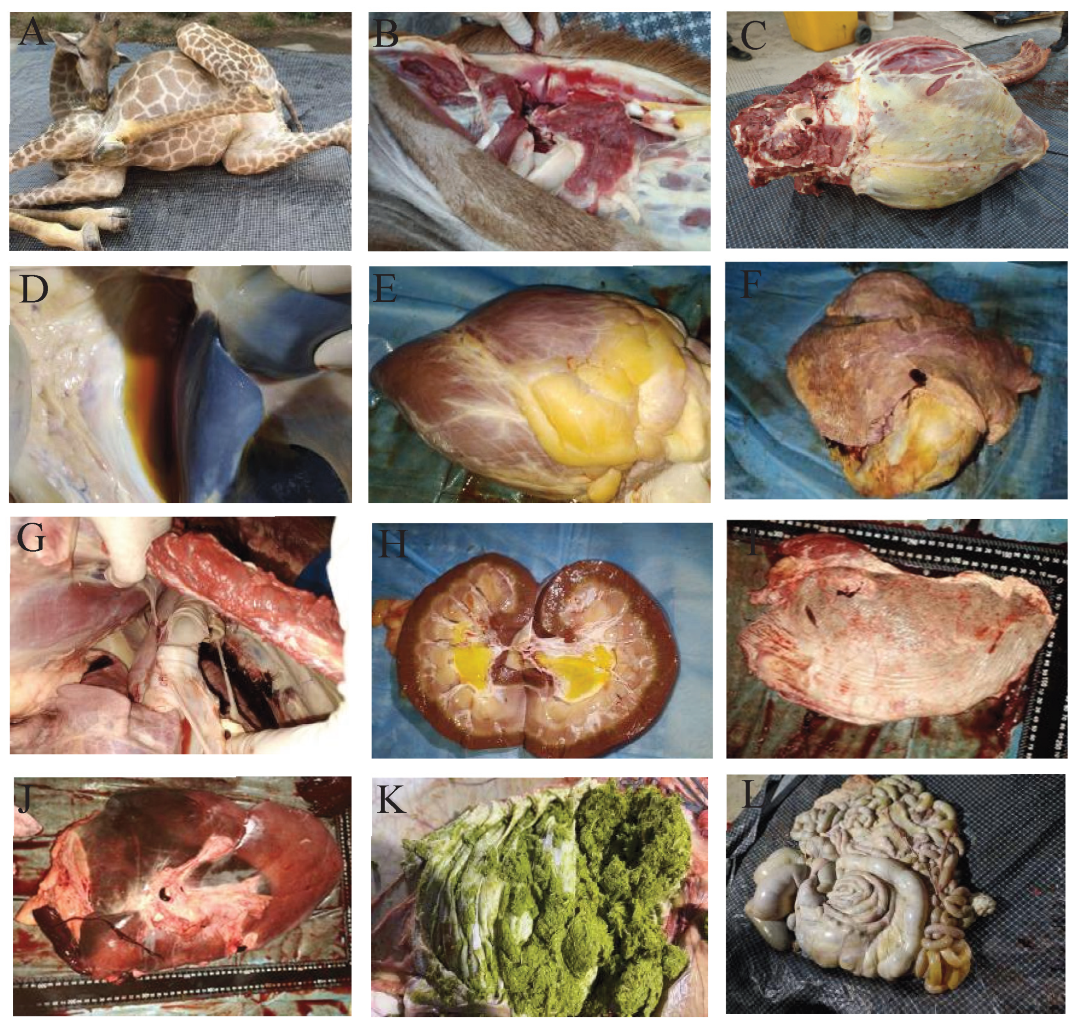
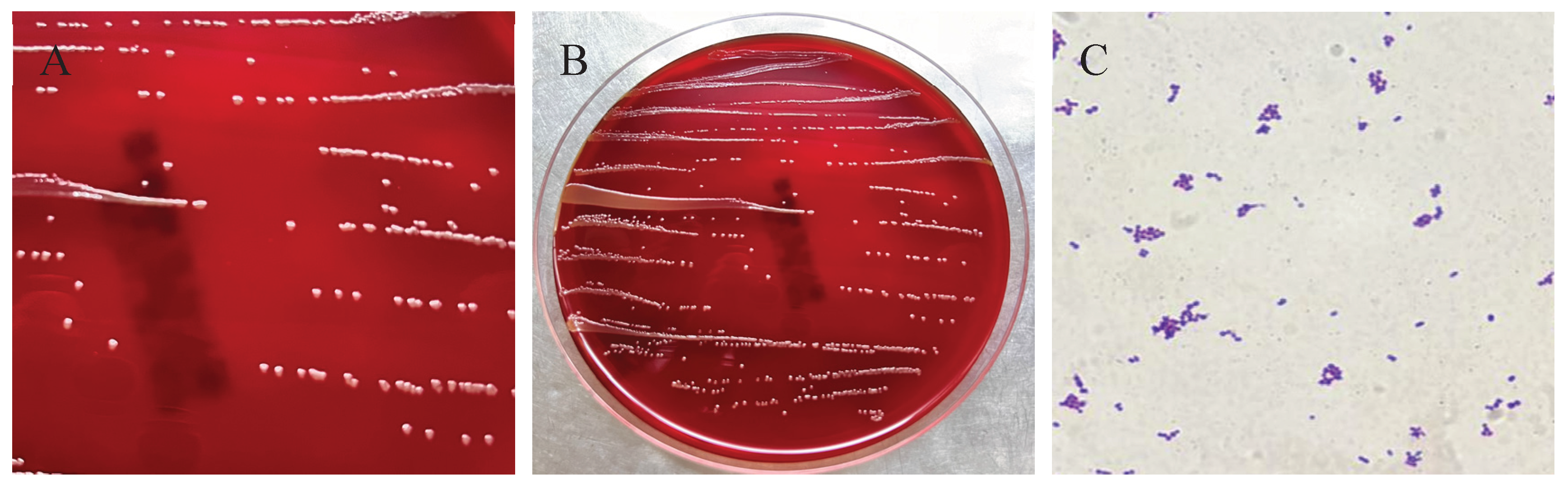
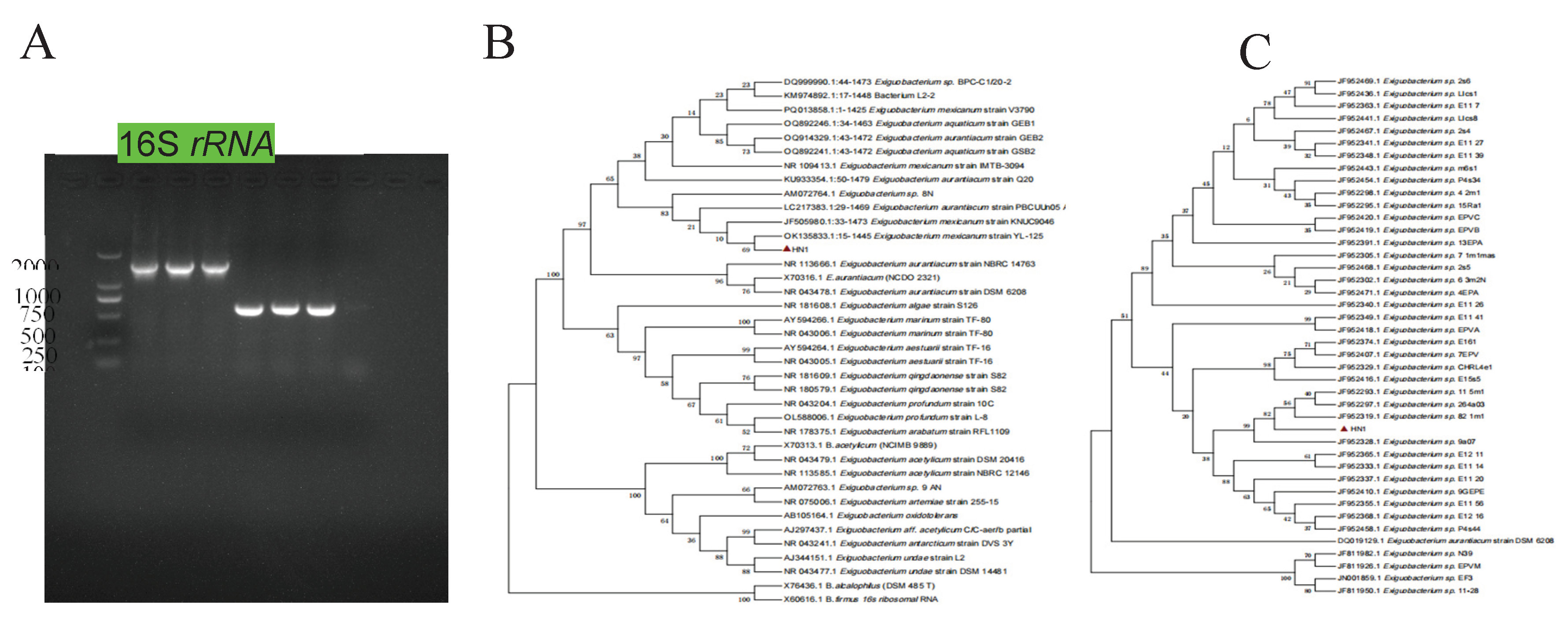
In May 2025, a female giraffe in poor body condition died unexpectedly at a zoo in Henan Province, China. A bacterial strain, designated HN-1, was isolated from the heart, liver, spleen, lungs, and kidneys of the deceased animal. After 24 hours of incubation at 37°C on Luria–Bertani (LB) agar, the colonies appeared round, smooth, pale yellow, translucent, and raised. Gram staining revealed that the isolate was a Gram-positive, rod-shaped, and non-spore-forming bacterium. Based on 16S rRNA gene sequencing, the strain showed more than 99.7% homology with reference sequences of E. mexicanum from various sources in GenBank. The results of the susceptibility test showed that E. mexicanum was susceptible to levofloxacin, clindamycin, chloramphenicol, trimethoprim, rifampicin, tetracycline, minocycline, gentamicin, erythromycin, and doxycycline, but resistant to oxacillin, penicillin, ciprofloxacin, and linezolid. These findings provide valuable insights for the diagnosis and treatment of infections caused by E. mexicanum in giraffes.

Keywords: 
;  ;  ;  ;  

1. Introduction

Giraffes (Giraffa camelopardalis), the world’s tallest terrestrial animals and largest ruminants [1], face a variety of health challenges in both wild and captive environments stemming from variations in diet [2], environmental conditions, climate [3], and susceptibility to infectious diseases. Captive giraffes are particularly vulnerable to bacterial infections [4,5,6], as exemplified by cases of Mycobacterium avium subsp. paratuberculosis identified in wild ruminants at a Mexican zoo [7], Fusobacterium necrophorum causing severe bilateral necrotic lesions near the tongue base and oral commissures in giraffes at Ordos Zoo [8], and Mycobacterium bovis associated with tuberculosis in giraffes at Greater Kruger National Park [9]. Given the dramatic decline in giraffe populations in recent decades, effective conservation strategies are crucial for ensuring their long-term survival [10].
E. mexicanum is a metabolically versatile bacterium exhibiting noteworthy biological characteristics and potential applications [11,12]. As a member of the Exiguobacterium genus, known for its ability to thrive across broad temperature and pH ranges [9], E. mexicanum was initially isolated and identified as one of two Gram-positive strains from brine shrimp (Artemia) cysts [13]. Subsequent isolations include strain A-EM from Atlantic deep-sea hydrothermal vents [14] and strain HA2 from Ilam Mountain in Iran, which demonstrates growth at 0-25 °C and tolerance up to 5% NaCl [15]. The genus has also been identified in the hyperarid Atacama Desert of Chile [16] and petroleum-contaminated permafrost [17], highlighting E. mexicanum’s adaptability to extreme environmental conditions. While direct evidence of E. mexicanum pathogenicity in animals or humans remains limited, other Exiguobacterium species are recognized as opportunistic pathogens with the potential to infect immunocompromised individuals [18]. Reported cases have linked the genus to community-acquired pneumonia and bacteremia, including a 2017 report of Exiguobacterium sp. isolated from both the blood and bronchoalveolar lavage fluid of a pneumonia patient [11]. Although the specific pathogenic potential of E. mexicanum requires further elucidation, the established pathogenicity of related species warrants continued investigation into its potential health implications.

2. Materials and Methods

2.1. Case Reports and Pathological Examination

In May 2025, a 605 kg female giraffe (ID: 41) in poor body condition died unexpectedly at a zoo in Zhengzhou, Henan Province, China. On the evening of May 18 (after 19:00), the giraffe showed reduced standing time, frequent recumbency, obvious weakness during ambulation, and an increased respiratory rate. It retained its appetite, consuming the provided foliage, but refused to drink water. On May 19, the giraffe attempted to stand twice, each attempt lasting approximately two minutes, presenting with an unsteady gait, deep and rapid respiration, lethargy, dull eyes, and excretion of dark brown, tea-colored urine. At 11:35, it exhibited limb paddling and neck weakness, and unfortunately died despite emergency treatment. A comprehensive necropsy was subsequently performed, including a thorough pathological examination of all organs. Fresh tissue samples were sent to the Veterinary Diagnostic Laboratory of Henan Agricultural University for further analysis. Samples were collected from visceral organs (heart, liver, spleen, lungs, and kidneys) using sterile scalpels, scissors, and inoculating loops for subsequent histopathological and laboratory studies.

2.2. Bacterial Isolation and Morphological Observation

Under sterile conditions in a biosafety cabinet, samples from the heart, liver, spleen, and kidney were inoculated onto LB agar and blood agar plates (Biocell, Zhengzhou, China) using a sterile inoculating loop.The plates were then incubated at 37 °C for 16-24 hours to facilitate colony observation. Single colonies exhibiting uniform morphology were selected and repeatedly streaked to ensure pure cultures displaying consistent colony size and morphology. These purified single colonies were subsequently inoculated into LB liquid medium for culture expansion and preserved on agar plates at 4 °C. Gram staining was performed on the purified bacterial strains, followed by microscopic examination to confirm cell morphology and Gram reaction.

2.3. Molecular Identification

Genomic DNA was extracted from pure bacterial colonies using a DNA purification kit (Tiangen Biotech, Beijing, China), with the extracted DNA stored at -20 °C until use as PCR templates. Molecular identification was performed through sequencing and phylogenetic analysis of the 16S rRNA and recA genes. The 16SrRNA gene was amplified using universal primers 27F (5ʹ-AGAGTTTGATCCTGGCTCAG-3ʹ) and 1492R (5ʹ-GGCTACCTTGTTACGACTT-3ʹ) [19]. The recA gene was amplified with primers recA-F (TATCGACTTCTGCCGTCTTGAAC) and recA-R(GTCATCGAAGTGTACGGACCTG).
PCR reactions were performed in a 25 µL volume, comprising 12.5 µL Premix TaqTM, 1 µL each of the forward and reverse primers, 2 µL of DNA template, and 8.5 µL of sterile ddH2O. The amplification protocol consisted of an initial denaturation step at 95 °C for 5 min, followed by 30 cycles of denaturation at 95 °C for 30 s, annealing at 59 °C for 30 s, and extension at 72 °C for 20 s, with a final extension at 72 °C for 10 min. PCR products were verified by agarose gel electrophoresis and subsequently submitted to Shangya Biotechnology Co., Ltd. (Henan, China) for sequencing. The resulting sequences were compared to sequences in GenBank using the BLAST algorithm (National Center for Biotechnology Information, NCBI). Phylogenetic trees, based on both 16S rRNA and recA gene sequences, were constructed using the neighbor-joining method implemented in MEGA 7.0 software, with bootstrap analysis performed using 1000 replicates.

2.4. Antibiotic Susceptibility Testing

A 100 uL bacterial suspension with a concentration of 0.5 McFarland standard was uniformly spread on Mueller-Hinton agar plates (HOPEBIO, Qingdao, China) using a spreader. Antimicrobial susceptibility test discs (OXOID) were picked up with sterile forceps and attached to the surface of the plates, with 3 parallel groups set for each type of disc. After standing for 3 minutes, the plates were inverted and incubated in a 37 °C constant-temperature incubator for 24 hours. The diameter of the inhibition zone was measured using a vernier caliper, and the measured diameter of the inhibition zone was expressed as the mean value. Since the Clinical and Laboratory Standards Institute (CLSI) has not established interpretive criteria for Exiguobacterium spp., the CLSI criteria for staphylococci were adopted in this study, with Staphylococcus aureus ATCC 25923 as the reference strain, in accordance with the Clinical and Laboratory Standards Institute (CLSI M100).

3. Results

3.1. Clinical and Pathological Findings in the Deceased Giraffe

Clinically, the affected giraffe exhibited marked lethargy and emaciation. Postmortem examination revealed multi-organ pathological changes: hemorrhage in the cervical lymph nodes (Figure 1B); gelatinous subcutaneous edema in the inguinal region and axilla of the right forelimb; and yellow discoloration of the sternal subcutaneous musculature (Figure 1C). Additionally, the ribs were fragile, with a small amount of brownish-yellow effusion in the peritoneal cavity (Figure 1D). The heart (6.5 kg) showed pale myocardium, yellow gelatinous deposits on the epicardium, scattered petechiae, and extensive thrombi in the cardiac chambers (Figure 1E). The lungs (6.5 kg) presented a marbled appearance (Figure 1F), with widened interlobular septa, localized congestion, pale margins, white tracheobronchial froth, and enlarged hilar lymph nodes (Figure 1G). The kidneys (left: 0.8 kg; right: 1.1 kg) displayed uneven coloration, with yellow gelatinous material in the renal pelvis (Figure 1H). The spleen (0.9 kg) was notably pale (Figure 1I), while the liver (6.15 kg) showed mottled discoloration, accompanied by congestion, firm texture, and generalized atrophy (Figure 1J). Other lesions included varying degrees of enlargement of pancreatic lymph nodes; gastric tympany with mucosal sloughing and greenish frothy contents (Figure 1K); abomasal hemorrhage with rice-sized ulcers; orange-yellow fluid in the intestinal lumen (Figure 1L); petechiae in the cecum and colon; and mucoid rectal feces. Collectively, these pathological findings are consistent with a diagnosis of acute bacterial septicemia complicated by multi-organ failure.

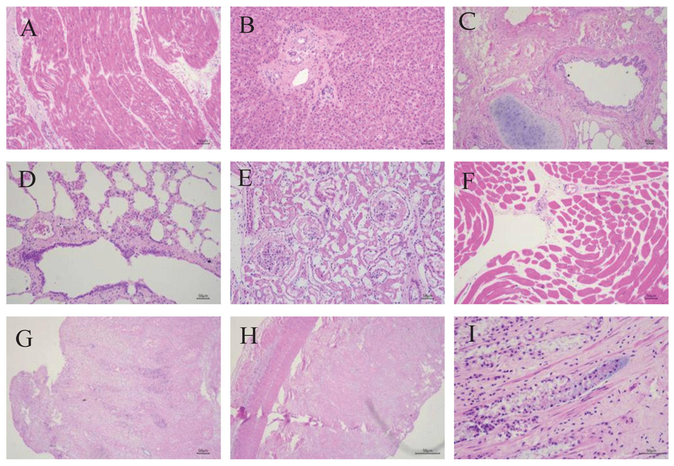
3.2. Histopathological Findings in the Deceased Giraffe

Histopathological examination of the deceased giraffe revealed significant pathological changes, characterized by multi-organ atrophy, extensive necrosis, and inflammatory cell infiltration in multiple tissues. Myocardial fibers showed dissolution and disorganization, with partial cardiomyocyte degeneration and necrosis manifested as pyknosis or loss of nuclei (Figure 2A). The liver exhibited atrophied hepatocyte cords with minimal bile pigment deposition, collagen fiber proliferation in the portal triads, and sparse lymphocytic infiltration (Figure 2B). Bronchial mucosal epithelium was desquamated with loss of cilia, accompanied by peribronchial accumulation of lymphocytes and plasma cells (Figure 2C). Scattered erythrocytes, neutrophils, desquamated epithelial cells, and exudates were observed in pulmonary vessels, along with perivascular fibrin deposition, alveolar macrophages, dust cells, and squamous metaplasia of bronchiolar mucosa (Figure 2D). Renal lesions included degenerative and necrotic tubular epithelial cells with fibrinous casts in the lumens, atrophied glomeruli with capsular degeneration and exudate accumulation, and interstitial inflammatory cell infiltration (Figure 2E). Skeletal muscles displayed atrophy and structural disorganization (Figure 2F). Transmural necrosis was present in the gastric wall, with abundant rod-shaped bacteria observed in the mucosal glands (Figure 2G). Intestinal tissues showed transmural necrosis with disintegration of mucosal epithelium and glandular architecture, blurred cellular boundaries, and nuclear loss (Figure 2H), along with dense aggregates of rod-shaped bacteria in the intestinal glands accompanied by peripheral tissue necrosis (Figure 2I). These histopathological changes indicate severe pyogranulomatous inflammation and extensive tissue necrosis, suggesting that the pathogen is likely an invasive pathogenic bacterium capable of inducing a fatal septic shock-like syndrome.

3.3. Bacterium Isolation and Microscopic Examination

Sterilely collected tissue samples were inoculated onto blood agar plate (Biocell, Zhengzhou, China) and LB agar plates for bacterial isolation and culture. A total of 18 bacterial isolates were isolated from heart, spleen, lung, kidney, and liver tissues. One strain, found across multiple tissues, was designated as HN-1. HN-1 colonies were observed to be round, smooth, pale yellow, and raised in morphology (Figure 3A,B). Gram staining identified the newly isolated HN-1 strain as Gram-positive, displaying a short, rod-shaped morphology with blunt ends, and arranged singly or in pairs (Figure 3C). These characteristics, indicative of systemic infection, suggest that strain HN-1 possesses biological properties consistent with septicemia pathogens.

3.4. Strain HN-1 Was Identified as E. mexicanum

HN-1 strain underwent further characterization through 16S rRNA and recA gene sequence analysis. Amplification yielded 16S rRNA and recA gene fragments of 1446 bp (GenBank accession number: PX248038) and 575 bp (GenBank accession number: PX255259), respectively. The agarose gel electrophoresis of the PCR products (Figure 4A) shows three lanes for both 16S rRNA and recA gene amplicons, corresponding to bacterial colonies isolated from three independent culture plates, confirming the consistency of the amplification results across different isolates of the same strain. Subsequent BLAST analysis against the NCBI database revealed high sequence similarity between strain HN-1 and various Exiguobacterium strains. Specifically, the 16S rRNA sequence exhibited 99.73–99.86% similarity, while the recA gene sequence showed 98.26–100% similarity. Furthermore, mass spectrometry confirmed that the bacterial strains isolated from different samples were all identified as E. mexicanum. Phylogenetic trees, constructed using both 16S rRNA and recA gene sequences from HN-1 and relevant reference strains, provided further insight into its taxonomic position. The 16S rRNA phylogenetic tree (Figure 4B) demonstrated that HN-1 clustered exclusively with E. mexicanum OK135833. Likewise, the recA phylogenetic tree (Figure 4C) indicated that strain HN-1 formed a distinct clade encompassing all Exiguobacterium species included in the analysis. Integrating morphological characteristics with the results of 16S rRNA and recA gene sequence analyses, as well as confirmation by mass spectrometry, the bacterial strain HN-1 was conclusively identified as E. mexicanum.

3.5. Antimicrobial Susceptibility Detection

To investigate the antimicrobial susceptibility profile of E. mexicanum HN-1, the disk diffusion method was used to determine the susceptibility of this strain to 14 antibiotics. The results showed that strain HN-1 exhibited resistance to oxacillin, penicillin, ciprofloxacin, and linezolid, which was characterized by small inhibition zones. In contrast, the strain was susceptible to levofloxacin, clindamycin, chloramphenicol, trimethoprim, rifampicin, tetracycline, minocycline, gentamicin, erythromycin, and doxycycline (Table 1). These findings not only clarify the resistance characteristics of this strain to certain antibiotics but also provide potential therapeutic options for clinical infections caused by Exiguobacterium mexicanum HN-1.

4. Discussion

Global outbreaks of zoonotic diseases have become increasingly frequent in recent decades, posing escalating threats to public health and wildlife conservation [20]. Advances in diagnostic technologies have facilitated the identification of emerging zoonoses—infections caused by bacteria, parasites, viruses, and fungi that originate in animals and are transmitted to humans from wild or domestic reservoirs [18,21]. Within this context, the genus Exiguobacterium, a group of Gram-positive bacteria renowned for their exceptional adaptability (including thermotolerance, psychrotolerance, alkaliphily, and halotolerance) [22,23], has garnered increasing attention due to its underrecognized pathogenic potential.Although reports of Exiguobacterium-related infections remain scarce, existing evidence underscores their clinical significance. For instance, E. aurantiacum has been isolated from blood cultures of bacteremia patients, including those with multiple myeloma, highlighting the heightened susceptibility of immunocompromised individuals [24]. In China, Exiguobacterium sp. A1b/GX59 has been associated with community-acquired pneumonia and bacteremia, with the pathogen detected in both blood and bronchoalveolar lavage fluid [11]. These cases align with our findings but also reveal a critical knowledge gap: prior to this study, no cases of E. mexicanum infection had been reported in giraffes, nor had systemic infection leading to fatal septicemia been documented. This novelty establishes our work as a pivotal step in expanding the known host range of E. mexicanum—previously limited to environmental niches and human clinical cases—to include large ruminants, particularly captive wildlife.
E. mexicanum, an opportunistic pathogen ubiquitous in soil and water [25,26], likely infects giraffes through multiple transmission routes such as ingestion of contaminated feed or water, skin wounds, or respiratory exposure [27,28]. This is consistent with the systemic distribution of strain HN-1 across the heart, liver, spleen, lungs, and kidneys observed in this case. Captive giraffes, already vulnerable to stress-induced immune impairment due to overcrowding or poor sanitation [29], may face elevated risks, as evidenced by the severe pathological lesions—including multi-organ necrosis, thrombus formation, and inflammatory infiltration—identified in our study. Notably, this contrasts with prior research on E. mexicanum, which has focused primarily on its role in environmental bioremediation (e.g., nitrogen removal in saline wastewater) [30,31,32] rather than its pathogenicity, underscoring the need to reevaluate its ecological and clinical importance. Currently, antibiotic therapy remains the primary treatment option due to its broad applicability and rapid efficacy [33]. In terms of antimicrobial susceptibility, our findings reveal a distinct profile compared to related species. While six E. aurantiacum strains isolated from human blood cultures showed susceptibility to all the tested antibiotics. [34], whereas E. mexicanum HN-1 showed resistance to oxacillin, penicillin, ciprofloxacin, and linezolid, and susceptibility to levofloxacin, clindamycin, chloramphenicol, trimethoprim, rifampicin, tetracycline, minocycline, gentamicin, erythromycin, and doxycycline. This resistance pattern may reflect local antibiotic usage practices [35] and also emphasizes the importance of conducting species-specific antimicrobial susceptibility testing to avoid ineffective treatments. For the management of captive wildlife, these data provide actionable guidance: prioritizing the use of susceptible antibiotics such as levofloxacin, clindamycin, and gentamicin, while avoiding resistant drugs like penicillin and oxacillin, may improve the cure rate of future cases.
Collectively, this study addresses key gaps in our understanding of E. mexicanum pathogenicity. By documenting the first case of E. mexicanum infection in giraffes, we confirm that this bacterium can infect large herbivores, thereby expanding its known host range. The observed antibiotic resistance further highlights the need for targeted surveillance in captive settings, where the risk of zoonotic spillover may be amplified. Moving forward, investigations into the bacterium’s virulence factors (e.g., adhesion proteins or toxin production) and the links between environmental reservoirs and animal or human infections will be critical. For zoos and wildlife facilities, our work underscores the urgency of enhancing biosecurity measures—including pathogen monitoring, stress reduction, and prudent antibiotic use—to mitigate risks posed by this emerging pathogen, ultimately safeguarding both animal welfare and public health [36,37,38].

5. Conclusions

This study reports the first isolation and identification of E. mexicanum from a deceased giraffe, with strain HN-1 exhibiting resistance to penicillin, oxacillin, ciprofloxacin, and linezolid while being susceptible to most other tested antibiotics. The systemic distribution of HN-1 in multiple visceral organs and the severe pathological changes observed suggest its potential role in the giraffe’s fatal septicemia. However, it is important to acknowledge that 18 bacterial strains were isolated from the animal, and only HN-1 was characterized. Thus, we cannot rule out the possibility of a mixed infection, where other unstudied strains may have contributed to the pathogenesis. Future research should focus on comprehensive identification of all isolates, assessment of their pathogenicity in combination, and exploration of virulence factors to clarify the exact etiology of such infections in giraffes. These findings provide a foundation for understanding E. mexicanum infections in large ruminants and offer guidance for clinical treatment in captive wildlife settings.

Author Contributions

F.G.: Investigation, Writing - original draft, Methodology, Formal analysis; Q.L.: Conceptualization, Investigation, Resources, Project administration; R.Z.: Methodology, Formal analysis, Visualization; Y.X.: Investigation, Writing - original draft; C.Z.: Investigation, Resources; Y.Y.: Formal analysis, Visualization; L.Y.: Methodology; D.L.: Writing - review & editing; H.D.: Writing - review & editing; W.D.: Conceptualization, Writing - review & editing, Supervision, Project administration; Y.L.: Conceptualization, Formal analysis, Visualization, Supervision, Project administration, Funding acquisition.

References

  1. Bashaw, M.J.; Tarou, L.R.; Maki, T.S.; Maple, T.L. A survey assessment of variables related to stereotypy in captive giraffe and okapi. Appl. Anim. Behav. Sci. 2001, 73, 235–247. [Google Scholar] [CrossRef] [PubMed]
  2. Kearney, C.C.; Ball, R.L.; Hall, M.B. Effects of altering diet carbohydrate profile and physical form on zoo-housed giraffe Giraffa camelopardalis reticulata. J. Anim. Physiol. Anim. Nutr. 2024, 108, 1119–1133. [Google Scholar] [CrossRef] [PubMed]
  3. Saito, M.; Matsunaga, M.; Fukuizumi, H.; Nakamichi, M.; Kinoshita, K. Factors affecting captive female giraffe stress response: Male presence, small enclosure, and low temperature. Zoo Biol. 2023, 42, 632–643. [Google Scholar] [CrossRef]
  4. Passos, L.F.; Garcia, G.; Young, R.J. Comparing the bacterial communities of wild and captive golden mantella frogs: Implications for amphibian conservation. PLOS ONE 2018, 13, e0205652. [Google Scholar] [CrossRef] [PubMed]
  5. Diaz, J.; Reese, A.T. Possibilities and limits for using the gut microbiome to improve captive animal health. Anim. Microbiome 2021, 3, 1–14. [Google Scholar] [CrossRef]
  6. Schmidt, D.A.; Barbiers, R.B.; Ellersieck, M.R.; Ball, R.L.; Koutsos, E.A.; Griffin, M.E.; Grobler, D.; Citino, S.B.; Bush, M. Serum Chemistry Comparisons Between Captive and Free-ranging Giraffes (Giraffa camelopardalis). J. Zoo Wildl. Med. 2011, 42, 33–39. [Google Scholar] [CrossRef]
  7. Hernández-Reyes, A.L.; Chávez-Gris, G.; Maldonado-Castro, E.; Alcaraz-Sosa, L.E.; Díaz-Negrete, M.T. First identification of Mycobacterium avium subsp. paratuberculosis in wild ruminants in a zoo in Mexico. Veter- World 2022, 15, 655–661. [Google Scholar] [CrossRef]
  8. Wang, J.; Wang, Y.; Ding, Y.; Suljid, J.; Wang, W. Oral and pulmonary necrobacillosis in a juvenile reticulated giraffe. J. Veter- Diagn. Investig. 2021, 33, 345–347. [Google Scholar] [CrossRef]
  9. Hlokwe, T.M.; Michel, A.L.; Mitchel, E.; Gcebe, N.; Reininghaus, B. First detection ofMycobacterium bovisinfection in Giraffe (Giraffa camelopardalis) in the Greater Kruger National Park Complex: Role and implications. Transbound. Emerg. Dis. 2019, 66, 2264–2270. [Google Scholar] [CrossRef]
  10. Armstrong, D.; Seddon, P. Directions in reintroduction biology. Trends Ecol. Evol. 2008, 23, 20–25. [Google Scholar] [CrossRef]
  11. Chen, X.; Wang, L.; Zhou, J.; Wu, H.; Li, D.; Cui, Y.; Lu, B. Exiguobacterium sp. A1b/GX59 isolated from a patient with community-acquired pneumonia and bacteremia: genomic characterization and literature review. BMC Infect. Dis. 2017, 17, 1–7. [Google Scholar] [CrossRef]
  12. Shen, L.; Liu, Y.; Chen, L.; Lei, T.; Ren, P.; Ji, M.; Song, W.; Lin, H.; Su, W.; Wang, S.; et al. Genomic basis of environmental adaptation in the widespread poly-extremophilic Exiguobacterium group. ISME J. 2024, 18. [Google Scholar] [CrossRef]
  13. López-Cortés, A.; Schumann, P.; Pukall, R.; Stackebrandt, E. Exiguobacterium mexicanum sp. nov. and Exiguobacterium artemiae sp. nov., isolated from the brine shrimp Artemia franciscana. Syst. Appl. Microbiol. 2006, 29, 183–190. [Google Scholar] [CrossRef] [PubMed]
  14. Lei, J.; Zheng, M.; Wang, L.; Yin, G.; Lou, Y.; Shi, L. Complete genome sequence of Exiguobacterium mexicanum A-EM, isolated from seafloor hydrothermal vents in Atlantic Ocean. Mar. Genom. 2021, 55, 100801. [Google Scholar] [CrossRef] [PubMed]
  15. Exiguobacterium, Sp. HA2, isolated from the Ilam Mountains of IranReza Heidari, Mostafa Akbariqomi, Gholamreza Tavoosidana, Garshasb RigibioRxiv 2021. 03.12.43 5112. [CrossRef]
  16. Remonsellez, F.; Castro-Severyn, J.; Pardo-Esté, C.; Aguilar, P.; Fortt, J.; Salinas, C.; Barahona, S.; León, J.; Fuentes, B.; Areche, C.; et al. Characterization and Salt Response in Recurrent Halotolerant Exiguobacterium sp. SH31 Isolated From Sediments of Salar de Huasco, Chilean Altiplano. Front. Microbiol. 2018, 9, 2228. [Google Scholar] [CrossRef]
  17. Erofeevskaia, L. A. , Popova, L. L., & Saltycova, A. L. (2016). PROPERTIES OF STRAIN OF EXIGUOBACTERIUM MEXICANUM. [CrossRef]
  18. Abdulkarem, A.T.; Hasan, A.A.; Khudhur, H.R.; Abed, S.M. Overview of Opportunistic Bacteria. Karbala J. Pharm. Sci. 2024, 14, 11–21. [Google Scholar] [CrossRef]
  19. Li, H.; Qian, Z.; Yan, Y.; Wang, H. Isolation, Identification, and Drug Sensitivity Test of Pseudomonas aeruginosa from Cynomolgus Monkey (Macaca fascicularis). Veter- Sci. 2025, 12, 636. [Google Scholar] [CrossRef]
  20. Khalafalla, A.I. Zoonotic diseases transmitted from the camels. Front. Veter- Sci. 2023, 10, 1244833. [Google Scholar] [CrossRef]
  21. Zhao, C.; Qin, P.; Li, S.; Chen, Z.; Wang, T.; Liang, Q.; He, W.; Peng, Z.; Yang, Y.; Peng, Z.; et al. Exploring Aeromonas dhakensis in Aldabra giant tortoises: a debut report and genetic characterization. BMC Microbiol. 2024, 24, 1–10. [Google Scholar] [CrossRef]
  22. Shen, L.; Liu, Y.; Chen, L.; Lei, T.; Ren, P.; Ji, M.; Song, W.; Lin, H.; Su, W.; Wang, S.; et al. Genomic basis of environmental adaptation in the widespread poly-extremophilic Exiguobacterium group. ISME J. 2024, 18. [Google Scholar] [CrossRef]
  23. Zhang, D.; Zhu, Z.; Li, Y.; Li, X.; Guan, Z.; Zheng, J. Comparative Genomics of Exiguobacterium Reveals What Makes a Cosmopolitan Bacterium. mSystems 2021, 6, e0038321. [Google Scholar] [CrossRef] [PubMed]
  24. Pitt, T.; Malnick, H.; Shah, J.; Chattaway, M.; Keys, C.; Cooke, F.; Shah, H. Characterisation of Exiguobacterium aurantiacum isolates from blood cultures of six patients. Clin. Microbiol. Infect. 2007, 13, 946–948. [Google Scholar] [CrossRef]
  25. Ed-Dra, A.; Abdallah, E.M.; Sulieman, A.M.E.; Anarghou, H. Harnessing medicinal plant compounds for the control of Campylobacter in foods: a comprehensive review. Veter- Res. Commun. 2024, 48, 2877–2900. [Google Scholar] [CrossRef]
  26. Stephenson, M.M.; Coleman, M.E.; Azzolina, N.A. Trends in Burdens of Disease by Transmission Source (USA, 2005–2020) and Hazard Identification for Foods: Focus on Milkborne Disease. J. Epidemiology Glob. Heal. 2024, 14, 787–816. [Google Scholar] [CrossRef]
  27. Kamel, M.S.; Davidson, J.L.; Verma, M.S. Strategies for Bovine Respiratory Disease (BRD) Diagnosis and Prognosis: A Comprehensive Overview. Animals 2024, 14, 627. [Google Scholar] [CrossRef]
  28. Kamel, M.S.; Davidson, J.L.; Verma, M.S. Strategies for Bovine Respiratory Disease (BRD) Diagnosis and Prognosis: A Comprehensive Overview. Animals 2024, 14, 627. [Google Scholar] [CrossRef]
  29. Nakov, D.; Hristov, S.; Stankovic, B.; Pol, F.; Dimitrov, I.; Ilieski, V.; Mormede, P.; Hervé, J.; Terenina, E.; Lieubeau, B.; et al. Methodologies for Assessing Disease Tolerance in Pigs. Front. Veter- Sci. 2019, 5, 329. [Google Scholar] [CrossRef] [PubMed]
  30. Kasana, R.C.; Pandey, C.B. Exiguobacterium: an overview of a versatile genus with potential in industry and agriculture. Crit. Rev. Biotechnol. 2017, 38, 141–156. [Google Scholar] [CrossRef]
  31. Cui, Y.; Cui, Y.-W.; Huang, J.-L. A novel halophilic Exiguobacterium mexicanum strain removes nitrogen from saline wastewater via heterotrophic nitrification and aerobic denitrification. Bioresour. Technol. 2021, 333, 125189. [Google Scholar] [CrossRef] [PubMed]
  32. Iyer, R.; Damania, A. Draft Genome Sequence of Exiguobacterium sp. KKBO11, Isolated Downstream of a Wastewater Treatment Plant in Houston, Texas. Genome Announc. 2016, 4, e00681–16. [Google Scholar] [CrossRef]
  33. Sabnis, A.; Hagart, K.L.; Klöckner, A.; Becce, M.; E Evans, L.; Furniss, R.C.D.; Mavridou, D.A.; Murphy, R.; Stevens, M.M.; Davies, J.C.; et al. Colistin kills bacteria by targeting lipopolysaccharide in the cytoplasmic membrane. eLife 2021, 10, e65836. [Google Scholar] [CrossRef]
  34. Yuan, X.; Lv, Z.; Zhang, Z.; Han, Y.; Liu, Z.; Zhang, H. A Review of Antibiotics, Antibiotic Resistant Bacteria, and Resistance Genes in Aquaculture: Occurrence, Contamination, and Transmission. Toxics 2023, 11, 420. [Google Scholar] [CrossRef]
  35. Fastl, C.; Ferreira, H.C.D.C.; Martins, S.B.; Afonso, J.S.; di Bari, C.; Venkateswaran, N.; Pires, S.M.; Mughini-Gras, L.; Huntington, B.; Rushton, J.; et al. Animal sources of antimicrobial-resistant bacterial infections in humans: a systematic review. Epidemiology Infect. 2023, 151, 1–31. [Google Scholar] [CrossRef]
  36. Bai, Y.; Wang, Q.; Liu, M.; Bian, L.; Liu, J.; Gao, F.; Mao, Q.; Wang, Z.; Wu, X.; Xu, M.; et al. The next major emergent infectious disease: reflections on vaccine emergency development strategies. Expert Rev. Vaccines 2022, 21, 471–481. [Google Scholar] [CrossRef] [PubMed]
  37. Levings, R.L. Emerging and Exotic Zoonotic Disease Preparedness and Response in the United States – Coordination of the Animal Health Component. Zoonoses Public Heal. 2012, 59, 80–94. [Google Scholar] [CrossRef] [PubMed]
  38. Cohn, J.; Mendelson, M.; Kanj, S.S.; Shafiq, N.; Boszczowski, I.; Laxminarayan, R. Accelerating antibiotic access and stewardship: a new model to safeguard public health. Lancet Infect. Dis. 2024, 24, e584–e590. [Google Scholar] [CrossRef] [PubMed]
Figure 1. Gross lesions observed in the deceased giraffe.(A) Overall view of the giraffe carcass. (B) Hemorrhage in cervical lymph nodes. (C) Yellow discoloration of subcutaneous muscles. (D) Accumulation of fluid in abdominal cavity. (E) Yellow gelatinous material on epicardium. (F) Marbled appearance of lung surface. (G) White froth in tracheobronchial tree. (H) Yellow gelatinous material in renal pelvis. (I) Pale coloration of spleen. (J) Congested liver. (K) Greenish gastric contents. (L) Yellow intestinal contents.
Preprints 177938 g001
Figure 2. Histopathological lesions in the deceased giraffe (H&E staining). (A) Dissolution of myocardial fibers with partial cardiomyocyte degeneration and necrosis. (B) Atrophy of hepatic cell cords in the liver. (C) Desquamation of bronchial mucosal epithelium with peribronchial lymphocytes and plasma cells. (D) Fibrin exudation around blood vessels and presence of macrophages and dust cells in alveoli. (E) Degeneration and necrosis of renal tubular epithelial cells with glomerular degeneration and atrophy. (F) Skeletal muscle atrophy with disorganized tissue arrangement. (G) Transmural necrosis of the gastric wall. (H) Transmural intestinal necrosis with nuclear disappearance. (I) Dense aggregates of rod-shaped bacteria in intestinal mucosal glands.
Preprints 177938 g002
Figure 3. Colonial morphology and microscopic observations of bacterial isolate HN-1.(A, B) The bacteria grew well on sheep blood agar plates without hemolytic rings. (C) Under oil immersion microscopy, Gram-positive bacilli of varying sizes with inconsistent staining intensity were observed.
Preprints 177938 g003
Figure 4. Molecular analysis of HN-1 gene amplicons.(A) Agarose gel electrophoresis of 16S rRNA and recA PCR products from bacterial isolate HN-1. The three lanes for each gene represent amplification products from colonies isolated from three independent culture plates, verifying the reproducibility of the results. (B) Phylogenetic tree based on 16S rRNA gene sequences. (C) Phylogenetic tree based on recA gene sequences. Numbers at nodes represent bootstrap support values from 1000 replicate samplings.
Preprints 177938 g004
Table 1. Antimicrobial sensitivity analysis results of isolated strain.
Antibiotics Medication Dose
(ug/disc)
IZD (mm) Sensitivity Judgment Standard of Inhibition Zone Diameter (mm)
Resistant Intermediate Sensitive
Gentamicin 10 21.1 S ≤12 13~14 ≥15
Ciprofloxacin 5 12.4 R ≤15 16~20 ≥21
Chloramphenicol 30 24 S ≤12 13~17 ≥18
Erythromycin 15 20.5 I ≤13 14~22 ≥23
Doxycycline 30 21 S ≤12 13~15 ≥16
Levofloxacin 5 20 S ≤15 16~18 ≥19
Tetracycline 30 26 S ≤14 15~18 ≥19
Minocycline 30 27 S ≤14 15~18 ≥19
Trimethoprim 5 10.5 I ≤10 11~15 ≥16
Clindamycin 2 22 S ≤14 15~20 ≥21
Linezolid 30 15.2 R ≤22 23~25 ≥26
Rifampicin 5 17 I ≤16 17~19 ≥20
Oxacillin 1 9.7 R ≤24 ≥25
Penicillin 10 16.6 R ≤28 ≥29
Note: S: susceptible, I: intermediate, R: resistant.
Disclaimer/Publisher’s Note: The statements, opinions and data contained in all publications are solely those of the individual author(s) and contributor(s) and not of MDPI and/or the editor(s). MDPI and/or the editor(s) disclaim responsibility for any injury to people or property resulting from any ideas, methods, instructions or products referred to in the content.
Copyright: This open access article is published under a Creative Commons CC BY 4.0 license, which permit the free download, distribution, and reuse, provided that the author and preprint are cited in any reuse.
Prerpints.org logo

Preprints.org is a free preprint server supported by MDPI in Basel, Switzerland.

Subscribe

Disclaimer

Terms of Use

Privacy Policy

Privacy Settings

© 2026 MDPI (Basel, Switzerland) unless otherwise stated