Preprint
Review

This version is not peer-reviewed.

Advancing Liver Cancer Treatment through Dynamic Genomics and Systems Biology: A Path Toward Personalized Oncology

Submitted:

12 September 2025

Posted:

16 September 2025

You are already at the latest version

Abstract

This review examines the transformative potential of dynamic genomics and systems biology in modern healthcare, focusing on their roles in precision oncology for liver cancer (Hepatocellular Carcinoma, HCC). It provides an integrated overview of how multi-omics technologies combine to help understand the complex biological landscape of tumors, including genomics, transcriptomics, proteomics, interactomics, metabolomics, and spatial transcriptomics. These advancements enable detailed patient stratification based on molecular, spatial, and functional tumor features, supporting personalized treatment strategies. The review emphasizes the significance of regulatory networks and cell-specific pathways in influencing tumor behavior and immune interactions. By mapping these networks with multi-omics data, clinicians can expect resistance mechanisms, identify the best therapeutic targets, and customize interventions. The approach shifts from traditional one-size-fits-all methods to dynamic, adaptable treatment plans guided by real-time monitoring, including liquid biopsies and wearable biosensors. A practical case study illustrates how a patient with HCC benefits from a personalized therapy plan involving epigenetic therapy, checkpoint inhibitors, and continuous multi-omics monitoring. This highlights the move toward healthcare that anticipates problems, considers the entire body, and adapts quickly to changes in a tumor. Looking ahead, the review discusses innovations such as cloud-based genomic ecosystems, federated learning for data privacy, and AI-driven interpretations that analyze complex multi-layered data. These advancements aim to improve decision-making, enhance clinical results, and change the disease management model, from reactive to predictive and preventative. The review also covers some important ongoing or completed clinical trials targeting HCC that use advanced molecular and immunological techniques. Overall, the review advocates adopting a systems-level, technological, and spatial approach to cancer treatment, stressing the importance of integrating data-driven insights into clinical workflows to advance personalized medicine.

Keywords: 
;  ;  ;  ;  ;  ;  ;  ;  ;  ;  ;  

1. Introduction

Living systems are more than just physical entities; they are complex information-processing systems, informational systems [1]. This information provides the blueprint for constructing and maintaining every part of an organism, from single cells to entire bodies. Its role in creating complexity highlights the importance of the stored biological information. Genes do not store information passively. They actively express, read, and interpret it through various molecular mechanisms, contributing to the complexity of individuals and species [2]. How living things use, turn on or off, combine, or modify these genes explains their diversity and adaptability.
Biological relationships, such as those between proteins and DNA or different cells and tissues, further organize and manage information. These relationships also interpret genetic data, helping determine how genes function in different situations. This organization contributes to the characteristics of individual organisms and the diversity seen within and between species. Because each biological event, whether it is the activation of a gene, folding a protein, or the communication between cells, involves the use and transmission of information, nature considers each biological event informational. The process begins with the DNA or RNA. It plays out through many interconnected physical and molecular interactions that drive life processes, building up the incredible complexity seen in living organisms.
Therefore, information is fundamental to biology: it explains how life maintains order, adapts, evolves, and passes traits from one generation to the next. Information plays its role in several ways.
In eukaryotes, DNA stores genetic information, which is transcribed into RNA to guide protein synthesis. DNA serves as the static basis of biological information, encoding instructions for protein synthesis. Genetic information influences organism characteristics as it passes from one generation to the next [3].
  • 1.1. Information dictates which genes activate or deactivate in response to environmental stimuli, regulating biological processes. Proteins represent the dynamic aspect, which is vital for maintaining homeostasis and regulating gene expression [4].
  • 1.2. Molecular Interaction - Biological functions result from the combined activity of multiple biomolecules, rather than from a single molecule. Physical interactions between biomolecules, which are sequences of elementary acts, or “bits” of communication, allow data to be exchanged through sequential molecular processes and can be viewed as elementary information events.
  • 1.3. Functional Modules - A complex interactive network transmits the functional information, which comes from elementary actions, through digital communication, processes it, and generates specific biological functions, which is the processing product [5]. Relational activity creates emergent properties that characterize specific informational and functional modules (or subgraphs) within a biological system [6]. Therefore, biological function arises from the joint processing of information via elementary events, which can be both long-lasting (e.g., protein complexes) and momentary.
  • 1.4. Interactive networks - Elementary interactions between molecules form complex networks that process information, generating particular biological functions.
  • 1.5. Cellular Interaction - Cells communicate through chemical signals, which are forms of information that regulate cellular functions. This communication through signaling pathways, transmits information through molecules like hormones, neurotransmitters, and cytokines. These signaling events coordinate activities among cells and tissues, ensuring appropriate responses to environmental changes or developmental signals. Cellular and metabolic relationships are interconnected, enabling informed decisions at the tissue or organ level [7].
  • 1.6. Complex systems - Interactions among proteins, genes, and metabolites form complex networks (interaction networks or graphs) that are essential for organism functioning through information distribution. Biological complexity emerges from combining information at different levels (molecular, cellular, ecological), leading to new properties [8].
  • 1.7. Biotechnology and research - Understanding biological information facilitates genetic engineering and developing innovative therapies. Analyzing biological informational data is crucial for medical and environmental research [9].
The framework discussed highlights two key aspects of biological information. First, it serves as a crucial link that connects the structure, function, and evolution of living organisms, deepening our understanding of life itself. We can understand each biological event as information that acts upon, interprets, and converts into new states or behaviors. These processes lead to emerging complex traits, behaviors, and even consciousness. Second, it asserts that simply analyzing individual molecules will not allow us to gain complete knowledge of biology. Instead, it requires a systemic view that considers how biomolecules interact and relate to each other. The relational model resulting from this holistic approach is vital for understanding the complexity and functions of life, with DNA being the fundamental element from which this understanding starts [10]. Thus, biological information is not static; it is dynamic and central to all processes in living systems. The way living things manage, interpret, and use this information shapes everything from molecular interactions to evolving species.

2. The Role of DNA

DNA and its functions form the foundation of genomics and functional genomics [11]. Our current knowledge still provides only a snapshot of the phenomena we study. DNA segments contain ambiguous information that requires contextual editing and rearrangement before being used. However, genomics is an evolving, not static, process [12]. Much remains to be understood about many aspects of gene activity, such as where, what, who, and which. “Where” refers to the context of gene expression, “what” is the action that a gene’s product does, “who” is the expression of different cell types, and “which” is where systems biology and network analysis come in, describing the mechanisms that drive gene expression processes. Therefore, we should view genomics as a dynamic and multifaceted process when applying this gene expression approach. The questions “where,” “what,” “who,” and “which” are crucial for understanding not only the static model of DNA but also the insights that enable us to explore its role in the complex choreography of cellular function and its dynamic nature. The coordinated activity of many genes and their products allows the functioning a living cell. We divide this activity into regulatory modules and co-regulated genes with common functions [13] to organize it. Operating multi-protein complexes and most metabolic pathways depends on this vital structuralization. Some additional dimensions could enrich this framework.

2.1. What Do They Mean?

a) Some genes are expressed in specific contexts such as developmental stages, particular tissues, environmental stimuli, or disease states, and understanding the “where” of gene expression is essential in these situations. This leads us to epigenetics, which studies how external factors influence gene activity without changing the DNA sequence [14].
b) The “what” of functional output indicates we need to understand what a gene’s product does, beyond knowing the gene’s expression. Therefore, functional genomics links gene expression to phenotypic outcomes, such as protein function, cellular behavior, or organismal traits [15].
c) The “who” addresses cellular and tissue specificity [16]. Different cell types express distinct sets of genes; understanding which expresses what adds another layer of detail. As a result, technologies like single-cell RNA sequencing are helping us map this genomic aspect with increasing accuracy [17].
d) The “which” question involves regulatory networks [18]. Genes do not act alone but use regulatory elements, transcription factors, and noncoding RNAs to control their expression. This is where systems biology and network analysis come into play [19].
This is important because genomics is shifting from just cataloging to becoming a dynamic, systems-level science. We are progressing toward understanding the components and their interactions, timing, and effects. Capturing the full complexity of biological regulation through epigenetics or single-cell analysis helps us see how gene expression is a coordinated process that converts genetic information into functional products like proteins [20].

2.2. The Core Mechanisms That Drive Gene Expression

1. Transcription. RNA polymerase transcribes DNA into messenger RNA (mRNA) [21]. Promoters and transcription factors regulate where and when transcription begins. An example is the lac operon in bacteria, which uses a repressor protein to block transcription without lactose.
2. RNA processing (eukaryotes only). The initial mRNA (pre-mRNA) undergoes modifications [22]:
- 5’ capping: adds a modified guanine to the beginning.
- Polyadenylation: adds a poly-A tail to the end.
- Splicing: removes introns and joins exons.
An example is the alternative splicing of the DSCAM gene, which produces thousands of protein variants in fruit flies [23].
3. Translation. Ribosomes decode mRNA to synthesize proteins, while tRNA provides amino acids corresponding to the mRNA codons [24]. An example is the synthesis of haemoglobin, which is regulated at the translational level in red blood cells.
4. Post-translational modifications. Various chemical reactions, characterized by covalent modifications such as phosphorylation, glycosylation, and cleavage, chemically modify proteins after synthesis [25]. An important example is insulin, which is produced as a precursor and activated by peptide cleavage [26].

2.3. Regulatory Mechanisms

5. Epigenetic modifications. DNA methylation and histone modifications are chemical modifications that regulate gene accessibility [27]. DNA methylation, for example, silences tumor suppressor genes in cancer [28].
6. Enhancers and repressors. These DNA sequences increase or decrease transcription at a distance [29]. An example is the β-globin gene, which is regulated by a locus control region (LCR) that acts as an enhancer [30].
7. RNA interference (RNAi). This is where small RNAs (such as siRNA or miRNA) come into play [31]. These types of RNA, other than mRNA, bind specifically to mRNA, preventing its translation or promoting its degradation. A specific example is miRNA21, which regulates cell proliferation and apoptosis genes [32].
Gene expression is like a molecular symphony [33]; the mechanisms that regulate it are the instruments and conductors that shape the performance. Here are a few more complex mechanisms.

2.4. Additional Gene Expression Mechanisms

Each of these mechanisms adds a layer of control, enabling cells to respond to internal and external cues.
1. Chromatin Remodeling. Specialized protein complexes reposition or restructure nucleosomes to make DNA more or less accessible [34]. For example, the SWI/SNF complex opens up chromatin to allow transcription machinery to bind [35].
2. DNA Methylation. Adding methyl groups to cytosine bases silences gene expression by tightening chromatin [36]. Extensive methylation drives X-chromosome inactivation in females [37].
3. Histone Modification. Enzymes that acetylate, methylate, phosphorylate, etc., histones affect the tightness of DNA wrapping [38]. For example, histone acetylation promotes transcription by loosening chromatin.
4. Non-coding RNAs. These RNAs do not code for proteins but regulate gene expression [39]:
- miRNAs bind to mRNA and block translation or trigger degradation.
- lncRNAs can scaffold chromatin modifiers or interfere with transcription. A typical example is the XIST lncRNA, which coats the X chromosome to silence it.
5. Riboswitches. Riboswitches in bacteria are RNA parts that modify their shape according to metabolites and manage translation [40]. For example, the thiamine pyrophosphate riboswitch halts thiamine synthesis when levels are sufficient.
6. Feedback Loops. Genes regulate their expression or that of others in a loop [41]. For example, the p53 tumor suppressor regulates its inhibitors, creating a feedback system for stress response [42].
7. Signal Transduction Pathways. External signals like hormones or growth factors activate intracellular cascades that alter gene expression [43]. The MAPK pathway activates transcription factors in response to stress or mitogens [44].
8. RNA Editing. Post-transcription processes alter mRNA sequences, changing the protein product [45]. The ADAR enzymes convert adenosine to inosine in mRNA, affecting neural receptor function [46].

2.5. The RNA Editing

RNA editing is a fascinating subject that warrants deeper exploration, especially considering its biological significance and unique roles in living organisms [47]. It is one of the most intriguing yet least understood processes after transcription, when our cells create RNA based on DNA. Nature modifies the genetic message after writing but before translating it into proteins, which is fascinating.
RNA editing involves making chemical modifications to RNA molecules after transcription from DNA [48]. These modifications can change the nucleotide sequence of the RNA, leading to the production of proteins different from those expected based on the original DNA sequence [49]. This opens up a world of possibilities.
There are several reasons RNA editing is so essential. First, it contributes to protein diversity. Editing a single gene can give rise to multiple protein variants, increasing our proteome’s complexity. This is vital because different proteins can perform various functions within cells and tissues.
Another critical aspect is tissue specificity. Some RNA editing changes occur only in specific tissues, such as the brain or the intestine. This means that expressing the same gene can lead to different outcomes depending on its location in the body, which is crucial to function specialized organs [50].
There is a growing recognition of the role that abnormal RNA editing plays in various diseases. Research has linked alterations in RNA editing to conditions such as cancers, neurological disorders, and autoimmune diseases [51]. This connection underscores the potential significance of understanding RNA editing better and how it might inform disease treatment and prevention [52]. The therapeutic potential of RNA editing is an area gaining traction. Innovative techniques, such as CRISPR-Cas13, are being explored for their ability to edit RNA [53]. This approach offers the possibility of reversible and precise gene regulation, which could lead to new avenues for treating diseases at the genetic level.
RNA editing is a complex and crucial aspect of molecular biology that deserves more attention. From creating protein diversity to its implications in health and disease and its therapeutic promise, ongoing research could enhance our understanding of genetics and its medical applications. Table 1 shows significant RNA modifications.
In a few words, RNA editing enables a single gene to produce multiple protein variants, increasing the proteome’s protein diversity [54]. Some modifications occur only in specific tissues, such as the brain or intestine, creating tissue specificity [55]. However, editing also plays an essential role in disease since abnormal RNA editing has been associated with cancer, neurological disorders, and autoimmune diseases.

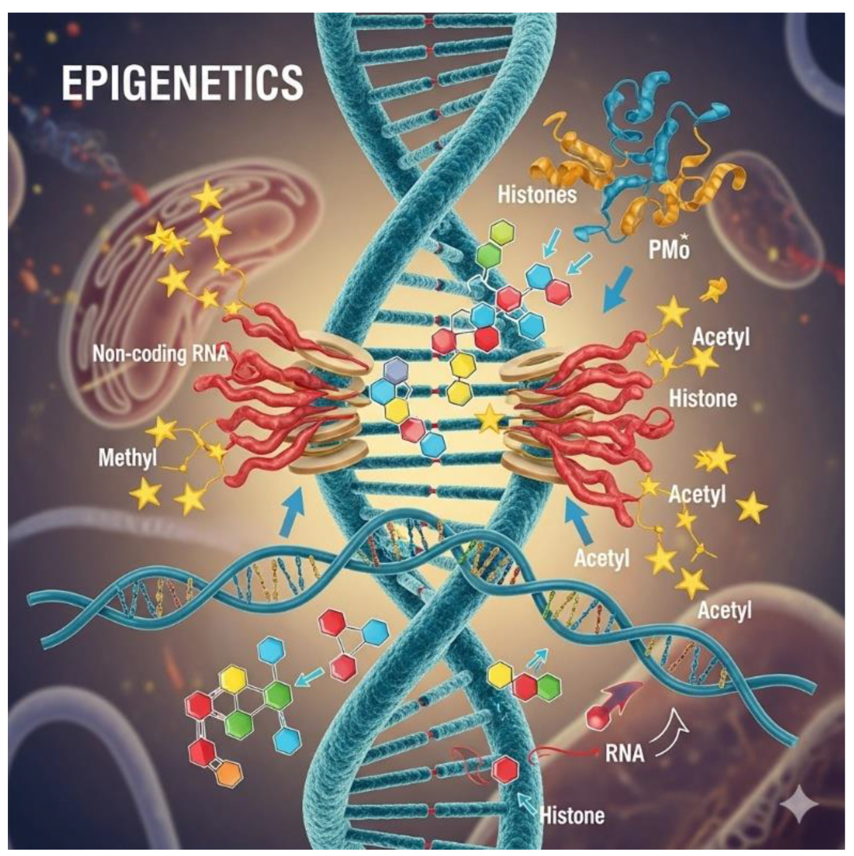
3. Epigenetic Effects and “The Why”

Gene expression is a complex process controlled by various mechanisms ranging from transcription to translation. These mechanisms are determined by the inherited genetic makeup. However, epigenetics is crucial in determining which genes are expressed and when by influencing gene activity in each individual and tissue without altering DNA [56]. This component, therefore, depends on individual experiences and environment, contributing to the formation of the particular phenotype [57]. Transcription, RNA processing, translation, post-translational modifications, enhancers, silencers, and RNA interference are all genetically regulated processes, inherited control mechanisms that determine which genes are expressed and how intensely [58].
Epigenetics is a level of regulation that goes beyond the genetic code. It concerns the “Where” of gene expression. Epigenetics involves specific chemical modifications of DNA or associated proteins, such as histone modifications or DNA methylation, which can influence DNA accessibility to the transcriptional machinery and thus gene expression. Epigenetic modifications occur in specific cells, not in all cells of an organism concurrently. This specificity characterizes the differential expression of genes: which genes are active or inactive, and “Where”. Epigenetic modifications are the primary mechanism controlling this process.
The environment, age, lifestyle, and other external factors, which can change gene activity without altering the DNA sequence, influence epigenetics [59]. This means that epigenetics contributes to individual variability and determines one’s phenotype, or the observable characteristics of an organism. In short, while genetic inheritance provides the basis for gene expression, epigenetics is a key factor in individual specificity and phenotype manifestation, with its close connection to the environment [60].
Epigenetics is where biology becomes poetic. It explores how something turns our genes on or off without altering the DNA sequence. Epigenetics functions like a dimmer switch for our genetic code: the blueprint remains the same, but the brightness (expression) varies depending on the environment, lifestyle, and experiences. Figure 1 and Table 2 present the key mechanisms of epigenetics.
Preprints 176459 i001
Epigenetic effects are changes in gene expression that can be reversible, heritable, and influenced by external factors such as diet, stress, toxins, and aging [50]. Epigenetics explains why identical twins can develop different diseases, how lifestyle choices influence future generations, and why some cancers occur without genetic mutations. It’s a powerful reminder that genes are not a fixed destiny but a potential, shaped by the context [61].

3.1. Real-Life Examples

  • Cell differentiation: All cells share the same DNA, but epigenetics determines whether a cell becomes a neuron, a muscle cell, or a skin cell [62].
  • Environmental influences: Exposure to pollution or poor nutrition can increase DNA methylation, potentially silencing protective genes [63].
  • Mental health: Stress and trauma can change epigenetic markers, impacting genes related to mood and cognition [64].
Epigenetic relationships influence gene expression, illustrating biology’s subtlety and control. Epigenetics determines a gene’s expression in terms of how, when, and where, without changes to the DNA sequence, but through modifications to the chromatin structure and regulatory signals. These changes decide whether a gene is accessible to the transcription machinery or remains silent. Table 3 outlines the essential epigenetic mechanisms and their effects on gene expression.
Preprints 176459 i002

3.2. A Closer Look at Dynamic Relationships

1. Histone Code Hypothesis - Combinations result in modified histones (acetylation, methylation, phosphorylation). These combinations form a “code” that proteins read to determine transcriptional outcomes [65]. An example is H3K4me3 (trimethylation of histone H3 at lysine 4), a mark of active promoters [66].
2. DNA Methylation and Gene Silencing - Methylation near gene promoters blocks transcription factor binding [67,68]. For example, hypermethylation in cancers often silences tumor suppressor genes like p16 [69].
3. Chromatin Accessibility - Open chromatin (euchromatin) allows transcription; closed chromatin (heterochromatin) represses it. Remodeling complexes shift nucleosomes to regulate this accessibility [70].
4. Non-coding RNAs as Epigenetic Architects - lncRNAs like XIST recruit silencing complexes to the X chromosome. miRNAs bind mRNA transcripts to prevent translation or promote degradation [71,72].
5. Feedback Loops and Crosstalk - Epigenetic marks influence transcription factor binding and vice versa [73]. For example, a transcription factor may recruit a histone acetyltransferase to activate its target gene, producing another factor that modifies chromatin elsewhere.

3.3. Environmental and Developmental Influences on the Epigenome

Nutrition, stress, toxins, and aging can reshape the epigenome [74]. These changes can be transient or heritable and influence gene expression across generations.
Nutrition: Nutritional intake influences the shaping of the epigenome [75]. Specific nutrients, such as folate, B vitamins, and specific amino acids, can affect the addition of methyl groups to DNA, a process known as DNA methylation. For instance, a diet high in these nutrients can encourage beneficial epigenetic modifications. In contrast, a poor diet may cause harmful changes that could contribute to health problems like obesity, diabetes, and cardiovascular diseases [75].
Stress: Psychological and physical stressors can activate pathways leading to epigenetic modifications. Chronic stress has been associated with changes in gene expression related to stress response, metabolism, and even mental health disorders[76]. Offspring can sometimes inherit these epigenetic changes, influencing their stress responses and overall health.
Toxins: Exposure to environmental toxins, such as heavy metals, pollutants, and endocrine disruptors, can negatively affect the epigenome [77]. These substances might disrupt the mechanisms that control epigenetic changes or directly alter DNA or histones, resulting in gene expression changes that could lead to various diseases, including cancer.
Aging: As organisms age, their epigenomes change [78]. These changes often include modifications in DNA methylation patterns and histone alterations, which can affect regulating genes involved in cellular aging and age-related diseases. Studying how aging affects the epigenome can offer insights into potential approaches for promoting healthy aging.
Intergenerational Effects: Epigenetic changes triggered by environmental factors can be temporary, yet they also have the potential to be inherited across generations [79]. For example, a mother’s nutritional status during pregnancy can modify DNA methylation patterns in her children, affecting their metabolic health and disease risk [80]. This shows that the effects of environmental exposures can extend beyond an individual’s lifetime to influence the health and development of future generations.
Overall, the interaction between environmental factors and the epigenome highlights the importance of considering genetic and non-genetic influences in understanding health and disease. Epigenetic research offers insights into interventions that could reduce negative environmental impacts and enhance health outcomes across generations. Recent studies have emphasized the importance of chromatin loops and nuclear architecture as key epigenetic regulators [81]. Chromatin loops involve physical interactions between distant genomic regions that can bring enhancers and promoters closer together, facilitating gene activation. The spatial organization within the nucleus is essential for proper gene expression and cellular function, as it affects how the transcriptional machinery accesses genes. The idea of phase-separated condensates has become a significant mechanism in gene regulation [82]. These are fluid-like compartments inside the nucleus, where various proteins, RNAs, and other molecules concentrate to boost transcriptional processes at active genomic sites [83]. This concentration enables the efficient assembly and operation of the transcriptional machinery, resulting in stronger and more precise gene activation. Advances in single-cell epigenomics have also provided more profound insights into cell-type-specific expression patterns [84]. By examining epigenetic modifications and transcriptional profiles at the single-cell level, researchers can discover how different cell types preserve their unique gene expression programs [85]. This approach reveals tissue heterogeneity and helps identify specific regulatory elements that determine cell fate and identity. These findings highlight the complex interplay among chromatin structure, nuclear organization, and gene regulation, offering a new understanding of how cells maintain their identity and respond to various signals.

3.4. Epigenetic Mechanisms Driving Aging

Epigenetic mechanisms that drive aging include DNA methylation changes, histone modifications, and non-coding RNA activity [86], called epigenetic drift. The drift is the gradual and often irreversible deregulation of the epigenetic machinery that occurs with aging, leading to changes in gene expression and a loss of cellular plasticity [87]. This process involves variable, bidirectional changes in DNA methylation and other epigenetic marks, occurring in a cell- and tissue-specific manner. Researchers hypothesize epigenetic drift contributes to biological aging, increases the risk of age-related diseases like cancer, and may even influence an organism’s maximum lifespan, while the functional implications are still being researched. However, changes in histone proteins that package DNA affect chromatin structure and gene accessibility. Histone acetylation and deacetylation, for example, regulate transcription [88]. ncRNAs, particularly microRNAs, play a significant role in genome regulation and can contribute to age-related decline [89].
Ongoing DNA damage triggers persistent changes in chromatin structure and epigenetic marks, leading to epigenetic heterogeneity and functional decline [89]. These changes are a primary cause of aging, caused by factors like DNA damage and environmental exposures, which interfere with normal gene expression and cell function. Understanding these changes is vital for creating healthy aging strategies, such as epigenetic rejuvenation therapies and lifestyle adjustments.
Unseen changes in gene activity also connect to the visible effects of aging on our bodies. As we age, the epigenetic process of DNA methylation becomes less accurate. This results in changes in gene expression associated with decreased organ function and increased susceptibility to disease as we age. Today, we can analyze genomic DNA methylation patterns to create biological clocks of aging, tools that allow us to measure an individual’s biological age. The epigenetic process affects gene expression and can vary throughout life. The idea of a “clock” that can quantify biological degradation relative to chronological age offers new insights for biology and preventive medicine research [90].
A fundamental unresolved question is how different tissue types share these aging signatures. Some cell types exhibit distinctive methylation patterns, influenced by development, lifestyle, and environment [91]. This variability presents challenges in standardizing biological age measurements. Understanding how these signatures intersect across different tissues could improve our understanding of the mechanisms of aging and inform therapeutic strategies for age-related conditions.
By analyzing the individual gene profiles of each tissue, we can find several genes with methylation alterations that are also potent biological markers of aging. For example, HDAC4 and HOX are developmental regulators correlating with senescence and age-related decline [92]. MEST, associated with obesity and diabetes, also speeds up aging [93]. High methylation of the protocadherin gamma (PCDHG) family has emerged as a key determinant of aging in several organs. Other studies show that PCDHG hypermethylation reduces white matter in the brain, a marker of rapid cognitive decline.
A recent preprint meta-analysis has made significant strides in understanding the role of DNA methylation as a potential target for anti-aging interventions [94]. The study investigated epigenetic changes across 17 different human tissue types, aiming to unveil patterns of DNA methylation that correlate with aging. One of the study’s key findings is that specific tissues, including the retina and stomach, show a more rapid accumulation of age-related DNA methylation changes than others. This implies that the tissues could be susceptible to the effects of aging. The observed methylation changes could show underlying functional changes rather than acting as a passive biological clock. In exploring the general trends, the study noted that most human tissues exhibit an overall increase in DNA methylation levels as individuals age [94]. However, the analysis highlighted exceptions to this norm, in skeletal and lung tissues, which do not follow the expected pattern of increased methylation with age [94]. This deviation raises intriguing questions about the mechanisms at play in these specific tissues and their unique aging processes. The implications of this research are profound, as understanding the specific patterns of DNA methylation changes could pave the way for targeted anti-aging therapies. By identifying key sites of methylation that are altered with age, researchers may develop interventions aimed at reversing or mitigating these changes, leading to healthier aging and prolonged lifespan.

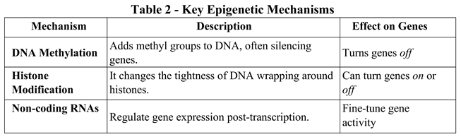
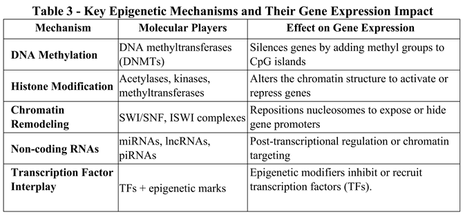
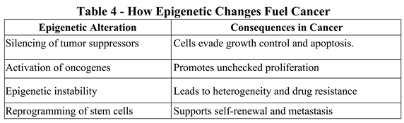
3.5. Key Epigenetic Mechanisms Driving Cancer

Epigenetics is a deep and fascinating field. Its modifications act as molecular switches that can change gene behavior without altering the genetic code. Epigenetic changes impact cancer development [95,96], and environmental factors like diet or stress can also reshape gene expression across generations. In cancer, these switches often get stuck in the wrong position, leading to uncontrolled cell growth, evasion of death signals, and resistance to treatment. Here is how it unfolds:
1. DNA Methylation - DNA methylation involves adding a methyl group to the cytosine base in DNA, at CpG sites. In cancer, hypermethylation of the promoters of tumor suppressor genes, like p16 and BRCA1, causes these protective genes to be silenced, stopping them from regulating cell growth and repair [97]. Hypomethylation across the genome can activate oncogenes, which leads to uncontrolled cell division, and can also cause chromosomal instability, raising the risk of mutations that can lead to cancer [98].
2. Histone Modifications - Histones are proteins around which DNA wraps to form chromatin, and their post-translational modifications influence gene expression. Acetylation relaxes the chromatin structure, making DNA more accessible and increasing gene expression. At the same time, methylation can either activate or repress gene expression depending on the specific histone and locating the modification. For instance, removing the H3K27me3 mark, a repressive histone modification, can activate genes that promote cell proliferation, contributing to tumor development [99].
3. Non-coding RNAs - Non-coding RNAs, including microRNAs (miRNAs) and long non-coding RNAs (lncRNAs), are essential in regulating gene expression after transcription. miRNAs are small RNA molecules that can bind to messenger RNAs (mRNAs) and block their translation or cause their degradation. In cancer, some miRNAs, such as miR-124a, are often silenced, removing their protective effects against tumor growth [100]. Some miRNAs may be overexpressed, suppressing tumor suppressor genes and promoting cancer progression [101]. lncRNAs can also influence gene expression by interacting with chromatin and transcription factors, further adding to the complexity of regulation [102].
4. Chromatin Remodeling - Chromatin remodeling involves the dynamic structural changes of chromatin that enable transcriptional machinery to access DNA [103]. Proteins called chromatin remodelers (e.g., SWI/SNF complexes) can reposition, remove, or restructure nucleosomes [104]. Mutations or functional changes in these remodelers can disrupt regular gene expression, leading to abnormal oncogene activation or tumor suppressor gene silencing. These alterations can promote the growth and survival of cancer cells by increasing their proliferation or resistance to cell death.
These epigenetic alterations lead to the aberrant expression of genes critical for cell cycle regulation, cell death, and growth. Accumulating these epigenetic changes contributes to transforming normal cells into cancer cells with hallmarks like uncontrolled proliferation and metastasis [105]. Epigenetic mechanisms and genetic mutations often cooperate, with epigenetic alterations sometimes causing genetic mutations and vice versa, creating a complex intertwining of events that drives cancer progression [106]. Table 4 shows how epigenetic changes fuel cancers.
Preprints 176459 i003

4. Therapeutic Implications

Epi-drugs such as DNA methyltransferase inhibitors (e.g., azacitidine) and histone deacetylase inhibitors (e.g., vorinostat) reverse abnormal epigenetic markers [107]. They show how we can exploit reversible epigenetic changes to fight cancer. These drugs can reactivate silenced genes, sensitize tumors to chemotherapy, and enhance immune responses. Let’s consider a case study to understand how epigenetic changes influence cancer development.

4.1. Case Study: Epigenetics in Colorectal Cancer

Colorectal cancer (CRC) is a complex disease marked by a series of genetic and epigenetic changes that drive tumor development [108]. We can break down the sequence of epigenetic events in CRC into several key stages.
1. Initial Epigenetic Changes: The process often starts with disruptions in key epigenetic mechanisms, such as DNA methylation and histone modifications. DNA methylation can silence tumor suppressor genes. In colorectal cancer, promoter hypermethylation silences genes like MLH1 (involved in DNA repair), leading to microsatellite instability, a hallmark of some colorectal cancers [109]. In normal colonic cells, these mechanisms regulate specific genes. As cells accumulate epigenetic changes, the promoter regions of tumor suppressor genes may become hypermethylated, silencing these genes. For example, colorectal tumors often silence the CDKN2A gene [100], essential for cell cycle regulation.
2. Histone modifications can promote oncogene activation [111]: loss of repressive histone marks (e.g., H3K27me3) can turn on genes like c-MYC, which drives cell proliferation. Overexpression of histone deacetylases (HDACs) is common, resulting in more condensed chromatin and silencing of protective genes [112].
3. Disruption of chromatin structure: The changes in histone modifications can make chromatin more accessible, promoting the expression of oncogenes [113]. Histone acetylation is associated with active gene expression, and modifications here can lead to expressing genes that promote cell growth, such as MYC.
4. Non-coding RNAs: Another layer of regulation involves non-coding RNAs, including microRNAs (miRNAs) and long non-coding RNAs (lncRNAs) [114]. These molecules influence gene regulation. In colorectal cancer, miRNAs like miR-21 are often upregulated [114]. miR21 suppresses tumor suppressor genes such as PTEN, shifting the balance toward unchecked growth. These RNAs can either inhibit tumor suppressors or promote oncogenes, aiding the malignant transformation of colonic cells. In particular, miR-21 is linked to a poor prognosis.
5. Tumor Microenvironment Effects (TME): The tumor microenvironment, involving interactions with stromal cells, immune cells, and the extracellular matrix, can further influence epigenetic changes [115]. Signaling molecules from surrounding cells activate pathways that lead to epigenetic modifications supporting tumor growth and spread [116].
6. Somatic Mutations and Epigenetic Interactions: Often, somatic mutations work together with epigenetic changes. For example, mutations in the APC gene, a key event in familial adenomatous polyposis, can trigger further epigenetic alterations, such as changes in the Wnt signaling pathway, helping tumors form and grow [117].
7. Tumor Evolution and Diversity: As tumors develop, they acquire diverse epigenetic traits, resulting in varied treatment responses. Some cells may become resistant because of specific epigenetic modifications that activate drug resistance mechanisms or alter cell death pathways [118].
8. Therapeutic Targets: Understanding the epigenetic landscape of colorectal cancer offers opportunities for targeted therapies. Drugs that inhibit DNA methyltransferases or histone deacetylases are being researched as treatments to reverse abnormal epigenetic states in CRC.
9. Epigenetic Therapy Examples: Drugs like azacitidine (a DNA methylation inhibitor) and vorinostat (an HDAC inhibitor) are undergoing testing to reactivate silenced genes [119,120]. These treatments aim to restore regular gene expression, making cancer cells more susceptible to chemotherapy or immune responses. Breast cancer, leukemia, glioblastoma, and other cancers show similar epigenetic alterations. Because these changes are reversible, they provide a promising avenue for innovative treatments [121].
In summary, the sequence of epigenetic events in colorectal cancer is a dynamic process involving multiple molecular changes that contribute to tumor onset, progression, and diversity, ultimately guiding new therapeutic strategies [122]. Epigenetic therapies aim to reverse inappropriate gene silencing or activation. Moreover, a medical expert can tailor personalized epigenetic treatments based on an individual’s specific profile [123]. Table 5 provides examples of such personalized therapies for certain diseases.

5. Epigenetic Biomarkers in Cancer Detection

Epigenetic biomarkers and personalized therapies are transforming cancer care [124]. This is where advanced science connects with personalized healthcare [125]. Epigenetic biomarkers are chemical tags on DNA or histones that signal abnormal gene regulation. We can detect them in blood, saliva, or tissue samples, often before symptoms appear.
Some examples include:
  • sEPT9 DNA methylation, used in blood tests to detect early-stage colorectal cancer [126].
  • gSTP1 hypermethylation in prostate cancer helps distinguish malignant from benign tumors [127].
  • miRNA signatures: Unique patterns of microRNAs in the blood can indicate breast, lung, or pancreatic cancer [128].
These biomarkers are noninvasive and, therefore, ideal for screening and monitoring. Their ability to find cancer early is due to their extreme accuracy. They can also reflect changes in tumor behavior over time.

5.1. The Future: Precision Epigenomics

Liquid Biopsies and Epigenetic Markers: Noninvasive tests analyzing components found in bodily fluids such as blood or saliva are revolutionizing early disease detection [129]. By focusing on epigenetic markers and chemical modifications on DNA that do not alter the underlying genetic sequence, these tests can identify disease-related changes at earlier stages than traditional methods. For example, researchers are honing in on specific methylation patterns in circulating tumor DNA (ctDNA) that can signal existing tumors, enabling more timely and less invasive diagnostics [130].
AI-Driven Epigenomic Profiling: Integrating artificial intelligence in healthcare is creating powerful tools for personalized medicine. AI algorithms are being developed to analyze large datasets of epigenomic information, identifying patterns that connect specific epigenetic modifications to patient responses to various therapies [131]. This allows clinicians to match patients with treatments most likely to be effective based on their unique epigenomic profiles [132]. This improves therapy effectiveness and reduces adverse effects by avoiding ineffective treatments.
CRISPR-Based Epigenetic Editing: Advances in CRISPR technology extend beyond gene editing; researchers are also exploring CRISPR’s potential for epigenetic editing [133]. Using CRISPR-based systems, scientists can target specific epigenetic marks, such as methylation or histone modifications, without permanently altering the DNA sequence. This precision enables reversible modulation of gene expression. Such capabilities could lead to innovative treatments for various conditions, including genetic disorders and cancers, by turning genes on or off with high specificity and minimal off-target effects.

6. From Epigenetics to Proteins: The Molecular Cascade.

Modern biology and genomics are heading toward connecting epigenetics, gene expression, and the human proteome [134] to answer the “What?” Epigenetic changes, such as DNA methylation, histone modifications, and regulating noncoding RNA, don’t just alter gene expression. They dictate protein production, timing, and quantities, reshaping the entire proteomic landscape.
Here’s how the flow works:
1. Epigenetic marks (e.g., methylation)
2. Gene Expression Regulation (transcription on/off)
3. mRNA Production and Processing
4. Protein Translation
5. Post-Translational Modifications
6. Functional Proteins in the Proteome
So, epigenetics acts as the gatekeeper of the proteome.

6.1. Real-World Implications in Molecular Processes

1. Protein Diversity. Epigenetic regulation enables alternative splicing and isoform variation, expanding the proteome beyond the number of genes [135]. For example, one gene can produce multiple protein variants depending on the cell type and epigenetic context.
2. Cellular Identity. Different cell types express different proteins because of epigenetic programming [136]. Example: Neurons and liver cells have identical DNA but vastly different proteomes because of epigenetic control [137].
3. Disease Mechanisms. Aberrant epigenetic marks can lead to misfolded proteins, overactive enzymes, or missing structural proteins [138]. For example, silencing DNA repair genes in cancer leads to accumulating mutated proteins [139].
4. Proteomic Signatures. Epigenetic changes leave protein-level fingerprints detectable in blood or tissue [140]. We can use these as biomarkers for diagnosis, prognosis, and treatment monitoring.

6.2. When Epigenetics and Proteomics Collide

Recent studies show that DNA methylation patterns correlate with circulating protein levels in blood [141]. These relationships help uncover hidden disease pathways and guide personalized therapies. Epigenome-wide association studies (EWAS) are now paired with proteomics to map complex disease networks. In short, the epigenome is the conductor, and the proteome is the orchestra [142]. Together, they perform the molecular symphony of life, and when something goes off-key, disease can emerge. This interplay is being used in drug development and personalized medicine. Everything we’ve discussed comes together to form an interesting vision for personalized medicine, a field transforming healthcare from a one-size-fits-all approach to one tailored to each individual.
Here is how epigenetics and proteomics drive personalized medicine:
1. Epigenetics: The Personal Blueprint. Epigenetic marks like DNA methylation, histone modifications, and non-coding RNAs reflect our unique life history, diet, stress, environment, and even prenatal conditions [143]. These marks influence which genes are active or silent, shaping our risk for diseases, drug response, and aging trajectory. In personalized medicine, epigenetic profiling helps:
- Predict disease susceptibility (e.g., cancer, diabetes, neurodegeneration).
- Tailor treatments based on gene activity.
- Monitor therapy effectiveness in real time.
2. Proteomics: The Functional Fingerprint. The proteome is the dynamic output of our genome, which our body does at the molecular level [144]. Proteomic analysis reveals:
- Which proteins are present, modified, or malfunctioning.
- How cells communicate and respond to therapies.
- Biomarkers for early detection and prognosis.
Together, epigenetics tells us what might happen, and proteomics shows us what is happening [145]. Table 6 shows some interesting applications of the epigenetics-proteomics approach.
To sum up, through a Multi-Omics Synergy, modern personalized medicine integrates data from:
•Genomics (our DNA code), DNA mutations, and variants
•Epigenomics (how our DNA is regulated), methylation, histone marks, chromatin accessibility.
•Transcriptomics (which RNAs are expressed), RNA expression levels.
•Proteomics (which proteins are active), protein abundance, and modifications.
•Metabolomics (what biochemical reactions are occurring), cellular metabolic activity.
This multi-omics approach provides a 360° view of our health [146], enabling:
•Precise diagnosis.
•Tailored treatment plans
•Predictive prevention strategies.
We analyze these layers to build a molecular fingerprint of each patient’s disease. Integrating such a mass of information causes it to grow, requiring optimization, which is often done using artificial intelligence.
AI algorithms process massive multi-omics datasets to identify biomarkers for early diagnosis and prognosis [147]. They can also predict drug response based on molecular signatures and stratify patients into subgroups for personalized therapies. Monitor disease progression and therapy resistance in real time [148]. For example, with an HCC patient, AI could:
•Detect subtle epigenetic changes linked to tumor aggressiveness.
•Match patients to immunotherapies based on PD-L1 expression and miRNA profiles.
•Predict recurrence risk using proteomic variations in circulating biomarkers.

7. The Near Future: Digital Twins And Virtual Trials

Digital twins and virtual trials are no longer science fiction [149]. Leading cancer centers and research institutions worldwide have already adopted them. Digital twins involve AI in creating a virtual model of the patient to simulate treatment outcomes. Virtual trials match patients to therapies based on molecular profiles, reducing the trial-and-error process.

7.1. Digital Twins

Digital twins in healthcare are dynamic virtual models of patients, organs, or systems based on artificial intelligence (AI), which use real-time data to simulate health status, predict outcomes, and optimize treatments [150]. This enables personalized medicine, virtual clinical trials, efficient system operations, and improved patient monitoring and engagement. They offer advantages such as customized treatment trials and medical device optimization, but face challenges such as data privacy, system integration, high costs, and regulatory hurdles.

7.1.1. How Does the Virtual Digital Twin System in Healthcare Work?

A digital twin is a constantly updated virtual replica of a physical entity, such as a patient’s body or a specific organ [151]. Unlike static models, it integrates real-time data from various sources, including electronic health records, genetic information, and wearable sensors. Artificial intelligence (AI) and advanced analytics are used to build and update these virtual models, creating a personalized simulation of a person’s health status.
Doctors can use a patient’s digital twin to test different treatments and procedures virtually before applying them to the patient, reducing risks and improving outcomes [152]. For chronic conditions, digital twins enable remote monitoring of disease progression and treatment effectiveness, enabling timely therapy adjustments [153].

7.1.2. Development, Discovery, and Planning Activities

Digital twins can accelerate in silico clinical trials, supporting biomarker research and drug discovery. They facilitate the design and optimization of new medical devices and can help configure existing devices based on specific patient needs [154]. Digital twins enable virtual surgical planning and simulations to assess risk and plan complex procedures [152,154,155]. They offer patients easy-to-understand health information, which helps them stay healthy and manage their conditions.

7.1.3. Future Challenges and Considerations

  • Privacy and Data Security: Ensuring the secure handling of large amounts of sensitive patient data is a significant concern [156].
  • Integration: Integrating digital twin platforms with existing healthcare systems and workflows is complex [157].
  • Cost: The initial investment in technology, infrastructure, and data management can be substantial [158].
  • Technical complexity: developing accurate, reliable, scalable digital twin models requires advanced technical expertise [159].
  • Regulation: Establishing an appropriate regulatory framework for digital twin technologies is challenging [160].

7.2. Virtual Trials

Virtual clinical trials use digital technologies like mobile apps and wearable devices to enable participants to submit data and interact with the study remotely [161]. This reduces the need for on-site visits to healthcare facilities. This participant-focused approach enhances convenience and accessibility, leading to quicker recruitment and greater patient diversity. It also offers potential cost savings and improved data accuracy through automated, real-time data collection [162]. However, clinicians face challenges such as maintaining participant engagement, ensuring technology reliability, and navigating regulatory requirements; hybrid models that combine virtual and traditional methods are often helpful [163].

7.2.1. How Do Virtual Clinical Trials Work?

Remote data collection: Participants record and transmit health data using digital tools such as wearable sensors, mobile health apps, and electronic patient-reported outcomes (ePRO) devices [164].
  • Telemedicine: Virtual visits with research staff via videoconferencing replace in-person site visits for activities like drug assessments or reviews [165].
  • Remote monitoring and verification: Techniques like remote data verification help maintain the quality and accuracy of collected data [166].

7.2.2. Benefits of Virtual Clinical Trials

  • Greater patient access and diversity: By removing the need to travel to clinical sites, virtual trials allow patients in rural areas or with mobility challenges to take part, broadening the participant pool and enhancing diversity [165].
  • Enhanced convenience and engagement: Participants can complete study activities at home, saving time and minimizing disruption to daily routines [165].
  • Cost and time savings: Virtual models can cut overhead costs associated with traditional sites and speed up recruitment and study timelines [167].
  • Continuous data collection: Real-time information from wearable devices offers researchers more detailed and frequent insights into participant health and study results [168].

7.2.3. Challenges of Virtual Clinical Trials

  • Participant engagement: Keeping participants engaged can be challenging without regular in-person contact and interaction with study staff [165,166].
  • Technology and data security: Dependence on participants’ devices and the internet can lead to data loss because of power outages or device failures [168].
  • Regulatory and oversight challenges: Ensuring compliance and managing remote activities can complicate matters [169].
  • Patient-staff interaction: The absence of direct contact makes it harder to ensure participants understand study requirements [165,166].

7.2.4. The future of Virtual Clinical Trials

  • Hybrid models: Many trials now combine virtual components with traditional onsite visits to capitalize on the advantages of both approaches [170].
  • Increased adoption: The COVID-19 pandemic has sped up the widespread adoption and standardization of virtual trial models in the life sciences sector [170].
  • Transformative shift: Virtual clinical trials mark a significant change toward more patient-centered, accessible, and efficient research [171].

8. The “Who”, Cellular and Tissue Specificity

Cell and tissue specificity, a crucial aspect of functional genomics, addresses the “who” of gene expression. It answers the questions: Which cells or tissues express which genes, and why? This specificity allows a single genome to give rise to the diverse array of cell types and functions in the human body [172]. Although every cell (with few exceptions) contains the same DNA, gene expression is highly selective. Different cell types activate distinct sets of genes based on their function, location, and developmental stage [172].
Cell specificity refers to gene expression patterns unique to a particular cell type (e.g., neurons versus hepatocytes). In contrast, tissue specificity pertains to genes expressed in one tissue (e.g., insulin in pancreatic β-cells) [173]. Various mechanisms regulate this specificity, including cell-type-specific transcription factors, epigenetic signals that differ among tissues, chromatin accessibility, enhancer landscapes, and non-coding RNAs that influence gene expression.
Some genes have ubiquitous expression (housekeeping genes), while others have conditional expression, becoming active only in response to specific stimuli or during particular phases such as development or regeneration [174]. The interplay between these categories adds further complexity to the “who” of gene expression.
The importance of tissue-specific expression is in defining organ function and reflects their functional identity. For example, liver cells, not neurons, express albumin because it is vital for liver function. Failure to express tissue-specific genes can lead to disease; understanding these genes is essential to clarify pathological mechanisms [175]. For example, MYH7, expressed in cardiac muscle, can cause skeletal muscle disorders if misregulated [176]. Understanding tissue specificity aids in designing targeted therapies with fewer side effects, improving drug precision. For example, anticancer drugs that target only proteins expressed in tumor cells spare healthy tissue [177]. Tissue-specific proteins in the blood can signal disease in a particular organ, acting as biomarkers [178]. Elevated troponin indicates cardiac damage. Modern tools have enhanced our understanding of these specificities. Single-cell RNA sequencing reveals gene expression at the level of individual cells, while spatial transcriptomics (ST) maps the locations where genes are expressed within tissues [17,179]. Epigenomics profiling uncovers differences in chromatin structure across cell types [180]. These technologies allow to construe cellular atlases and detailed gene expression maps across various tissues and developmental stages [181].

9. Precision Oncology Reimagined

The merging of spatial and multilayered molecular data transforms cancer treatment, shifting from a one-size-fits-all approach to a precision-focused strategy [182]. Instead of viewing tumors as uniform masses, researchers now see them as complex ecosystems, where location, cell type, and molecular activity are all essential factors.
Spatial transcriptomics enables scientists to map the tumor landscape, showing where genes are active within the tumor and its surrounding area. This spatial understanding uncovers immune landscapes versus immune hotspots and pinpoints invasive fronts filled with stem or resistant cells [17,179,180,181,182]. It monitors how the tumor microenvironment (TME) changes after therapy. This helps doctors target treatments more accurately by selecting drugs that work on specific regions or cell types within the tumor.
Multi-omics decoding reveals the layers. Indeed, multi-omics platforms integrate genetic mutations (e.g., TP53, CTNNB1), epigenetic silencing (e.g., tumor suppressor methylation), transcriptomic changes (e.g., immune exhaustion markers), and proteomic and metabolic reorganization [183]. Together, these levels offer a 360° view of tumor biology, enabling patient stratification by molecular subtype and real-time monitoring of resistance and recurrence. This approach leads to adaptive therapy tailored to the tumor’s evolving dynamics.
This allows us to expand the “What” and the “Which” concerning the tumor. The “What” refers to identifying what to target, including specific pathways, cell types, and spatial zones that promote tumor growth or immune evasion. The “Which” helps us determine which patients will benefit, based on their tumor’s unique molecular and spatial signature. This dual clarity transforms clinical trials into dynamic learning systems, where experts test treatments and continually refine them [184]. AI-based dashboards then come into play, simulating tumor progression to guide real-time therapeutic decisions. Digital pathology offers spatial maps for physicians, which makes complex data accessible; furthermore, mutation status, spatial immune context, and metabolic profile will determine how physicians personalize combination therapies [185]. Overall, these combined approaches promise more effective treatments, fewer side effects, and improved outcomes, treating the right patient with the right drug at the right time and place.
The ability to track genomic evolution over time has fostered dynamic genomics. This involves the real-time and contextual use of genomic data: not just static sequencing, but also ongoing monitoring, interpretation, and response [186]. This genomics adapts to the patient, the disease, and the environment. Its future will be revolutionary. Genomic data now actively guides, adapts, and predicts biology in real time, not just describes it [187].

9.1. The Key Trends Already Shaping The Future Are:

1. Real-time genomic monitoring. Wearable biosensors and liquid biopsies will monitor genomic and epigenomic changes. As a result, continuous monitoring could detect tumor recurrence, drug resistance, or immune activation before symptoms appear [188].
2. Artificial intelligence-driven genomic interpretation [189]. Machine learning models will predict disease trajectories, drug responses, and even suggest interventions. Tools like DeepVariant (a deep learning algorithm used to analyze DNA sequences and identify genetic variants associated with diseases, including cancer, with extremely high accuracy, aiding diagnosis and treatment) already outperform traditional variant analysis tools [190]. This deep sequencing technology is essential for identifying specific mutations that can influence prognosis and treatment options in cancer, and undoubtedly, researchers will extend it to whole-genome simulations.
3. Multi-omics integration. Researchers will combine genomics with transcriptomics, proteomics, metabolomics, and spatial data to develop dynamic models of human biology [191]. These models will simulate treatment outcomes, optimize dosing, and personalize care in real time.
4. Cloud-based genomic ecosystems. Global platforms will store, share, and analyze genomic data, enabling collaborative diagnosis and research [192]. Privacy-preserving federated learning will allow AI models to learn from data without compromising patient confidentiality [193].
5. Genomic decision support in clinics. Doctors will use genomic dashboards to guide real-time treatment, choosing therapies based on the patient’s evolving molecular profile [194]. This will be revolutionary in oncology, rare diseases, and transplant medicine.
Dynamic genomics transforms medicine from reactive to proactive, from static to adaptive. Instead of waiting for the disease to manifest, we will anticipate and prevent it. Instead of fixed treatment plans, we will have adaptive protocols that evolve with the patient. It’s the genomic equivalent of moving from a snapshot to a live video.

9.2. Regulatory Networks: The Logic of Cellular Decision-Making

Regulatory networks describe how genes and proteins regulate each other, like a circuit of switches and feedback loops [195]. Transcription factors activate or repress gene expression, while signaling cascades transmit external signals to the nucleus, and feedback loops maintain homeostasis or drive disease states. Examples include liver cancer. A patient with active Wnt/β-catenin signaling might resist immunotherapy, while another patient with TGF-β-induced immunosuppression might benefit from TGF-β inhibitors [196]. These insights come from mapping regulatory networks using multiomics data, revealing each tumor’s functional architecture. Feedback loops can explain how dual signaling pathways, such as VEGF and TGF-β, develop compensatory mechanisms that reduce the effectiveness of therapy, requiring multi-targeted strategies.

10. Systems Biology: The Whole Is Greater Than the Sum

The cell functions as a complex, interconnected system, following the holistic principles of Systems Biology. This approach stresses that biological systems are complete entities, rather than just the sum of their parts [197].
In this framework, each cell component, such as genes, proteins, lipids, and metabolites, interacts with others. These interactions can lead to emergent properties, where the entire system’s behavior cannot be predicted by examining individual parts [198]. For instance, how signaling pathways connect and respond to environmental cues can affect cellular processes like metabolism, growth, and apoptosis.
Systems Biology employs computational models and high-throughput experimental techniques to analyze data from various levels of biological organization, from molecules to cells to tissues [199]. By combining data from genomics, proteomics, and metabolomics, researchers can better understand cellular functions and their roles in health and disease.
In conclusion, the cell operates within a complex network of interactions driven by Systems Biology principles, emphasizing the need for a holistic perspective when studying cellular behavior and biological systems [200]. This theory views the cell as a dynamic, interconnected system that integrates key functional activities, such as
  • Genomics, transcriptomics, proteomics, metabolomics
  • Spatial data and temporal dynamics
  • Environmental and therapeutic inputs
This holistic approach allows us to simulate a patient’s tumor response to various perturbations, such as drugs, immune activation, or metabolic stress. For “The Which,” systems biology helps to:
  • Predict therapy response pathways
  • Identify compensatory pathways that influence resistance
  • Design combination therapies tailored to system-level vulnerabilities
Interactomics is a key tool in network analysis, which maps the molecular web. Network analysis uses graph theory to study interactions among proteins, genes, metabolites [201]. Interactomics focuses on protein-protein interactions (PPIs), revealing functional modules [5,6]. Centrality measures identify key hubs (e.g., MYC, TP53) that control network behavior, while pathway enrichment links molecular profiles to biological processes [202]. A tumor with a disrupted immune interactome (e.g., low CD8-MHC-I interaction) may evade immune surveillance in patient stratification [203]. Or, a network dominated by stemness factors (e.g., PROM1, SOX2) may predict recurrence [204]. These patterns help define molecular subtypes, guiding the “Which One” [205].
Now, let’s try to put it all together and imagine a patient with liver cancer.
- Multi-omics reveals elevated PD-L1 expression, a CTNNB1 mutation, and suppressed interferon signaling [206].
- Network analysis shows a compromised immune interactome and active Wnt signaling [207].
- Systems biology simulation predicts a poor response to checkpoint inhibitors alone [208].
So, the “Which” becomes clear. This patient might benefit from Wnt inhibitors combined with immunotherapy, not just a monotherapy.
The rise of Systems Biology, with its holistic, indeterministic, and non-reductionist approach, has affected our understanding of both genomics and proteomics. Many genes, as discovered in DNA sequences, encode proteins. Unfortunately, something almost always changes the protein that we once called native [209,210]. Proteins undergo various post-translational modifications, producing products tailored to specific cellular environments, where they interact with particular targets, usually other proteins, to perform specialized functions in collaboration with an extensive network of proteins [210].
Over the past 30 years, this perspective has transformed our biological and biomedical knowledge. Systems Biology enables dynamic tumor modeling, allowing us to understand better how computational models simulate tumor progression and resistance. These models offer deeper insights by integrating data from multi-omics studies, spatial heterogeneity, and immune system interactions. For instance, simulations of Wnt signaling and its interactions with immune pathways can help predict patient responses to combination therapies. Network-informed decision trees [211] using algorithms that leverage network topology and patient-specific molecular data can adapt in real time as new omics and spatial information are added. This adaptability enables more accurate predictions of therapeutic responses.

10.1. The Which”: Patient Stratification Through Network Insight

Up to this point, we have considered the proteome in our analysis. Now we’ll explore how “The Which,” the question of which patients benefit, intersects with regulatory networks, systems biology, and network analysis, such as interactomics [212]. These fields form the analytical backbone of precision medicine, especially in complex diseases like cancer.
“The Which,” patient stratification through network understanding [213], asks which patients will respond to a therapy based on their molecular and cellular profiles. To answer this, we need to understand isolated biomarkers and how genes, proteins, and pathways interact. These interactions vary from individual to individual, leveraging the entire body of genomic knowledge used thus far.

10.2. Dynamic Decision Tree: Network-Informed Stratification in HCC

Let’s now explore how this framework could apply to a patient profile through a dynamic decision tree based on network-informed stratification. We analyze a conceptual dynamic decision tree that uses network-informed stratification to guide personalized treatment decisions in liver cancer (HCC) [214]. This tree adapts based on regulatory networks, systems biology, and interactomics data, enabling clinicians to make real-time, biology-based decisions [215]. Let’s examine the dynamic decision tree based on network-informed stratification. This tree is not static: it evolves as new data emerge from multi-omics platforms, spatial transcriptomics, and clinical monitoring [215]. Each branch represents a decision point informed by the behavior of the network, not just isolated biomarkers.

10.3. Tumor Molecular Profile: Root Node

Input: Multi-omics data (genomics, transcriptomics, proteomics, epigenomics).
Events:
-
If CTNNB1 mutation → Branch to Wnt/βcatenin pathway analysis [216].
-
If TP53 mutation + high PDL1 → Branch to immune checkpoint evaluation [217].
-
If PROM1/CD47 expression → Branch to stemness and immune evasion module [218].
Branch 1: Regulatory Network Activation.
Input: Pathway enrichment + transcription factor activity.
-
If the Wnt pathway is active, → Consider Wnt inhibitors; immunotherapy is likely ineffective [219].
-
If TGFβ signaling is dominant, → Add TGFβ blockade to restore immune infiltration [220].
-
If the interferon response is suppressed → Evaluate viral etiology (HBV/HCV) and consider TCRT cell therapy [221].
Branch 2: Interactome Disruption [222].
Input: Protein–protein interaction maps.
  • If the immune interactome is disrupted (e.g., low CD8–MHC-I interaction) → predict immune escape [223]; consider priming strategies.
  • If the angiogenic interactome is active (e.g., VEGF hub centrality) → add antiangiogenic agents [224].
  • If the fibrotic interactome is dense → consider stromal remodeling agents (e.g., FAP-targeted therapies) [225.
Branch 3: Systems Biology Simulation
Input: AI model simulating tumor evolution under therapy [226].
  • If we predict adaptive resistance → Preemptively adjust drug combinations [226].
  • If we forecast immune cell exhaustion → Add IL-2 agonists or checkpoint boosters [227].
  • If we detect metabolic reprogramming → Tailor diet or add metabolic inhibitors [228].
Leaf Nodes: Personalized Therapy Recommendations.
Each leaf represents a tailored treatment plan, such as
  • Anti-PD-1 + TGF-β inhibitor for immune-excluded tumors [229].
  • Wnt inhibitor + metabolic modulator for CTNNB1-mutant HCC [230].
  • TCR-T cell therapy + antiviral for HBV-HCC with high viral antigen load [231].
The decision tree is dynamic, so new biopsy or liquid biopsy data can redirect the patient to a distinct branch. Therapy response feeds back into the system, updating the interactome and regulatory network maps, while AI continuously improves predictions, making the tree smarter.

11. A Case Study of a Middle-Aged Patient with Hepatocellular Carcinoma (HCC)

Despite ongoing research efforts, the fundamental biology of hepatocellular carcinoma (HCC) remains understood. HCC’s high heterogeneity characterizes its ability to evolve throughout its progression. As highlighted, intricate regulatory networks can describe the cellular dynamics underpinning HCC. These networks illustrate the complex interactions between genes and proteins, showcasing how they influence and regulate each other via protein-protein interactions.
These interactions are not only multifaceted but also vary in terms of their duration and stability. The interfaces formed between interacting proteins and the temporal aspects of these interactions are crucial, when substantial conformational changes occur during the binding process. This complexity is fundamental in predicting the three-dimensional structure of protein complexes. However, many current AI-driven methodologies encounter significant difficulties in predicting multi-chain assemblies [232]. They frequently don’t have key information about the ligands, ions, and cofactors needed for proteins to work. Capturing the dynamic characteristics of protein-protein interactions, especially the conformational changes that transpire during binding, presents a substantial hurdle for computational modeling [233]. The inherent structural flexibility of proteins is vital for their biological function and contradicts the often-static perspective that conventional models adopt. Understanding the dynamics of protein-protein interactions is essential for unraveling the complexities of biological processes.
Digital systems can extract and analyze scientific information to support research endeavors or validate existing theories. Information is the conduit for transmitting concepts or updates with immediate or practical significance [234]. While this indirect approach is valuable for advancing human knowledge, it is essential to note that research relies on observations presented as data [235]. Data are concrete, quantitative, and verifiable, derived from coded reference quantities that yield consistent results upon replication [236,237]. In stark contrast to digital systems, the scientific method employs a direct experimental approach to problem-solving by adhering to established scientific principles for data collection [238]. Careful observation, thorough experimentation, precise measurement, and results generation through generalization (induction) form its foundation. Then, scientists confirm these results through multiple tests.
It is imperative that all research findings, those associated with hepatocellular carcinoma, align with this logical and methodological framework, emphasizing the need for experimental validation of all results and hypotheses. Recent quantitative estimates show that a mere 2% of the protein-protein interactions within the HCC network have undergone experimental validation [239,240,241]. This dramatic gap in validated interactions negatively contributes to the challenges in understanding and treating this complex form of cancer.
We now analyze a clinical case after examining and considering systems biology and integrating various omics disciplines to determine the different phases through which we could develop a personalized medicine protocol. Let’s imagine that a 58-year-old patient suffers from chronic hepatitis B and cirrhosis, two major risk factors for HCC. A routine scan reveals a suspicious liver lesion. Here’s how we would develop the personalized medicine protocol for him.
Phase 1: Epigenetic Profiling
The patient’s biopsy undergoes epigenomic analysis.
  • We observe hypermethylation of tumor suppressor genes such as CDKN2A and RASSF1A, indicating aggressive tumor behavior [242].
  • Histone modification patterns show a loss of H3K27me3, suggesting active oncogene expression [243].
  • Non-coding RNA signatures reveal elevated levels of miR-21, suppressing apoptosis and promoting cell proliferation [244].
Further information: These epigenetic markers help classify the patient’s tumor as high-risk and help guide the selection of targeted therapies.
Phase 2: Proteomic Analysis
Mass spectrometry and protein microarrays allow physicians to analyze a patient’s tumor.
  • Overexpression of AFP (alpha-fetoprotein) and GPC3 (glypican-3) confirms hepatocellular carcinoma (HCC) [245].
  • Increased levels of VEGF and PD-L1 suggest processes related to angiogenesis and immune evasion [246].
  • Unique protein signatures indicate pathways involved in drug resistance.
Insight: Proteomic data aids in predicting a patient’s response to immunotherapy and antiangiogenic treatments.
Phase 3: Personalized Treatment Plan
Based on the patient’s molecular profile, he receives epigenetic therapy with a DNA methyltransferase inhibitor to reactivate silenced tumor suppressors [247]. Physicians add checkpoint inhibitors (such as anti-PD-1) to counteract immune evasion, and a multiomics control panel monitors his response in real time, allowing therapy adjustments as needed.
Phase 4: Monitoring and Prognosis
  • Liquid biopsies detect circulating tumor DNA and protein biomarkers [108,125,188].
  • Tracking epigenetic changes helps identify recurrence early [248].
  • Proteomic alterations inform drug dosing and combination strategies. This approach transforms patient care from reactive to proactive and precise, enhancing survival rates and quality of life.

11.1. Cell and Tissue Specificity in Liver Cancer

Liver cancer is not just a mass of malignant hepatocytes but a complex ecosystem of different cell types interacting within a unique tissue environment [249]. This is how specificity manifests:
  • Malignant Hepatocytes
    • They are the primary tumor cells in HCC.
    • They display altered gene expression patterns, often driven by mutations and epigenetic changes.
    • In HBV-related HCC, viral integration into hepatocyte DNA can activate oncogenes [250].
  • Tumor-Associated Macrophages (TAMs)
In a healthy liver, Kupffer cells maintain immune balance. In HCC, TAMs with distinct profiles replace them; TREM2+ and UBE2C+ TAMs dominate primary tumors, and SPP1+ and WDR45B+ TAMs are more common in metastatic liver tumors and are associated with poor prognosis [251].
3.
T Cells and B Cells
Effector T cells and memory B cells are active in the non-tumorous liver. In HCC, these shift toward exhausted T and inhibitory B cells, especially in metastases [252]. A unique population of TCF7+ CD8+ memory T cells in metastatic tumors can undergo exhaustion via the p38 MAPK signaling pathway [253].
4.
Cancer-Associated Fibroblasts (CAFs)
These cells remodel the extracellular matrix and support tumor growth. CAFs in HCC exhibit plasticity, adapting to tumor needs and influencing immune evasion and angiogenesis [254].
5.
Endothelial Cells
In HCC, they acquire immune tolerance and angiogenic capacity, promoting blood vessel formation and tumor survival.

11.2. The Importance of Tissue Specificity in Hepatocellular Carcinoma (HCC)

Hepatocellular carcinoma (HCC) is the most common type of liver cancer, and understanding its tissue specificity is crucial for developing effective therapeutic strategies. Here’s a deeper look into the key features that significantly impact liver cancer progression:
  • Cell-Type Composition
Composing different cell types within the liver tumor microenvironment plays a vital role in shaping the immune landscape [255]. Various immune cell populations, including macrophages, T cells, and natural killer cells, can promote or help eradicate tumor growth. The balance between pro-inflammatory and anti-inflammatory signals is essential, as tumor-associated macrophages, for example, may support tumor progression and contribute to therapy resistance [256]. Variations in cellular composition can influence how patients respond to immunotherapy and targeted treatments.
2.
Spatial Organization
The arrangement of cells within the tumor affects treatment success and the chance of metastasis. This organization can create gradients of oxygen, nutrients, and drugs, which are critical in controlling how a tumor acts and reacts to treatment.. For instance, cells located farther from blood vessels may adapt to hypoxic conditions, resulting in more aggressive tumor types and increased resistance to treatment. Understanding spatial organization better can help optimize drug delivery systems and improve therapeutic outcomes by enabling the development of approaches targeting specific tumor regions.
3.
Gene Expression Profiles
The gene expression profile of HCC cells is vital for identifying biomarkers that predict disease outcomes and guide treatment options. Specific gene signatures can reveal a tumor’s aggressiveness, potential for metastasis, or likelihood of responding to particular therapies. Profiling these genes facilitates the discovery of new therapeutic targets and supports the development of more personalized treatment strategies. Differences in gene expression between tumor cells and surrounding liver tissue can provide insights into tumor development and resistance mechanisms, paving the way for innovative therapies.
4.
Microenvironment Dynamics
The tumor microenvironment is more than just a setting for tumor cells; it is a dynamic network of interactions among cancer cells, stromal cells, and extracellular matrix components. These interactions influence tumor growth, progression, and the ability to evade immune detection [257]. For example, cytokines and growth factors secreted by the tumor and stroma can establish an immunosuppressive microenvironment, aiding immune escape. Understanding these interactions can help design combination therapies that disrupt communication pathways, boost immune responses, and improve treatment effectiveness.
Recognizing prizing tissue-specific characteristics in HCC is essential for advancing personalized medicine and creating more effective therapies tailored to each tumor. Table 7 summarizes the main points, showing prizing tissue specificity in hepatocellular carcinoma (HCC).

11.4. Clinical Relevance in Liver Cancer.

Single-cell transcriptomics and spatial mapping reveal the behavior of different cell types in primary versus metastatic liver tumors. This information helps identify prognostic markers and therapeutic targets, such as WDR45B+ TAMs or TCF7+ T cells [251]. Understanding tissue-specific interactions enables precision immunotherapy, antiangiogenic strategies, and treatments targeting CAFs [258]. In studying liver tumor heterogeneity, we can explore the entire cellular landscape or delve deeper into the plasticity of the tumor microenvironment.
Why It Matters: Understanding the “Who” in liver cancer, which cells are active, suppressed, or transformed, allows physicians to predict treatment responses, design combination therapies, and monitor disease progression with cell-specific biomarkers. This represents the frontier of precision oncology, where the tumor’s cellular identity guides every clinical decision. Understanding how this information is used in liver cancer clinical trials and how spatial transcriptomics maps these differences in real time is crucial. Let’s complete the picture by exploring how we apply spatial transcriptomics and multi-omics platforms in liver cancer research and clinical trials, especially to comprehend cellular and tissue specificity.

11.5. Spatial Transcriptomics in Liver Cancer

Spatial transcriptomics (ST) enables researchers to map gene expression within the physical context of tissue architecture. This is vital in liver cancer, where the tumor microenvironment (TME) exhibits high heterogeneity.
Key insights of ST in HCC:
  • Reveals distinct cellular neighborhoods: tumor clusters, immune niches, and stromal zones.
  • Identifies PROM1+ and CD47+ cancer stem cell niches linked to metastasis and immune evasion [259].
  • Maps tertiary lymphoid structures (TLS) and their proximity to tumor cells, influencing immune infiltration and therapy response.
  • Detects spatial gradients of gene expression from non-tumor to tumor regions, illustrating how the tumor capsule affects transcriptome diversity.
Niche formation involves details about spatial gradients of gene expression that determine local microenvironmental conditions, such as hypoxia or acidity, which influence tumor growth and immunogenicity. These spatial data assist clinicians in pinpointing therapy targets and identifying which cell types contribute to resistance or progression. They explain how the spatial proximity of immune cells (e.g., exhausted T cells expressing PD-1 and LAG3) to tumor cells, through cell-to-cell interactions, influences immune suppression. For example, the proximity of PROM1+ cancer stem cell niches to immune-excluded zones suggests mechanisms of immune evasion and tumor recurrence.
In summary, ST’s role in HCC maps the tumor microenvironment, showing how tumor cells interact with immune cells, fibroblasts, and blood vessels. It locates immune cells such as exhausted T cells expressing PD-1 and LAG3, which may cluster near tumor cells, indicating immune evasion [260]. It also shows where immune cells infiltrate or retreat, aiding in assessing drug efficacy and response areas to therapy. This spatial data helps researchers stratify patients: those with “hot” tumors (rich in immune cells) may respond better to immunotherapy than those with “cold” tumors. Multi-omics integrates multiple layers of biological data, as shown in Table 8.

11.6. Multi-Omics in Clinical Trials

Network topology and critical nodes (e.g., hubs) explore how combining genomics, transcriptomics, and proteomics helps identify key network nodes such as PD-L1, CTNNB1, or TGF-β, which can serve as therapeutic leverage points. These nodes often have high network centrality, meaning their modulation results in widespread effects. Recent studies integrate genomics, epigenomics, transcriptomics, proteomics, and metabolomics to create a comprehensive molecular profile of each patient’s tumor. HCC trials focus on various aspects, including drug development, treatment efficacy, and patient outcomes [261]. Here are some key applications:
  • Predictive biomarkers: multi-omics helps identify which patients will respond to checkpoint inhibitors or TCR-mediated T-cell therapy.
  • Resistance mechanisms: Studies reveal why only about 30% of HCC patients respond to immunotherapy, often because of tumor-intrinsic and extrinsic factors [262,263].
  • Cellular immunotherapy: Clinical trials evaluate engineered T cells, DC vaccines, and macrophage modulation based on the patient’s immune profile.
These methods effectively distinguish HBV-related HCC from nonviral HCC, highlighting viral antigen expression in HBV-related HCC, facilitating TCR-based therapies. Metabolic and fibrotic signatures in nonviral HCC guide antiangiogenic and anti-fibrotic strategies.

11.7. The Future: AI-Driven Precision Oncology.

The future of liver cancer treatment looks promising with integrating AI-powered precision oncology [264]. This method uses advanced AI platforms that combine spatial and multi-omics data, information from genomics, proteomics, metabolomics, and other biological layers, to improve treatment strategies. AI platforms now integrate spatial and multi-omics data to simulate tumor evolution and therapy response, identify spatiotemporal recurrence niches, regions likely to relapse post-treatment, guiding adaptive therapy regimens that evolve with the tumor’s molecular shifts. This is transforming liver cancer care from reactive to anticipatory and personalized. Here’s a more detailed overview of ST in clinical trials.
1. Tumor Evolution Simulation: Using machine learning algorithms, these AI platforms can simulate how tumors evolve in response to different therapies [265]. This helps clinicians understand tumor biology and adaptive mechanisms and allows them to predict how a tumor might change when they apply other treatments.
2. Spatiotemporal Recurrence Niche Identification: A significant challenge in liver cancer treatment is the risk of recurrence after therapy. AI tools can analyze spatial and temporal data to identify specific areas within the liver (or the tumor) that are more likely to recur [266]. By pinpointing these “niches,” healthcare providers can carry out more targeted monitoring and interventions to address residual disease.
3. Guided Adaptive Therapeutic Regimens: As liver tumors change, their molecular profiles shift. AI can monitor these changes in real time, enabling treatment plans to be adjusted [267,268]. This flexibility ensures therapies stay effective over time, tailored to the patient’s tumor characteristics rather than using a one-size-fits-all approach.
These advancements represent a transformative shift in liver cancer management, from a reactive strategy focused on treating recurrence to a proactive approach emphasizing prevention and personalization. By forecasting tumor behavior and customizing interventions, the aim is to improve patient outcomes and increase survival rates in those diagnosed with liver cancer. AI technology can revolutionize cancer care and provide a roadmap for future oncology treatments across various cancer types.
Figure 2. Schematic representation of AI-Driven Precision Oncology.
Preprints 176459 g002

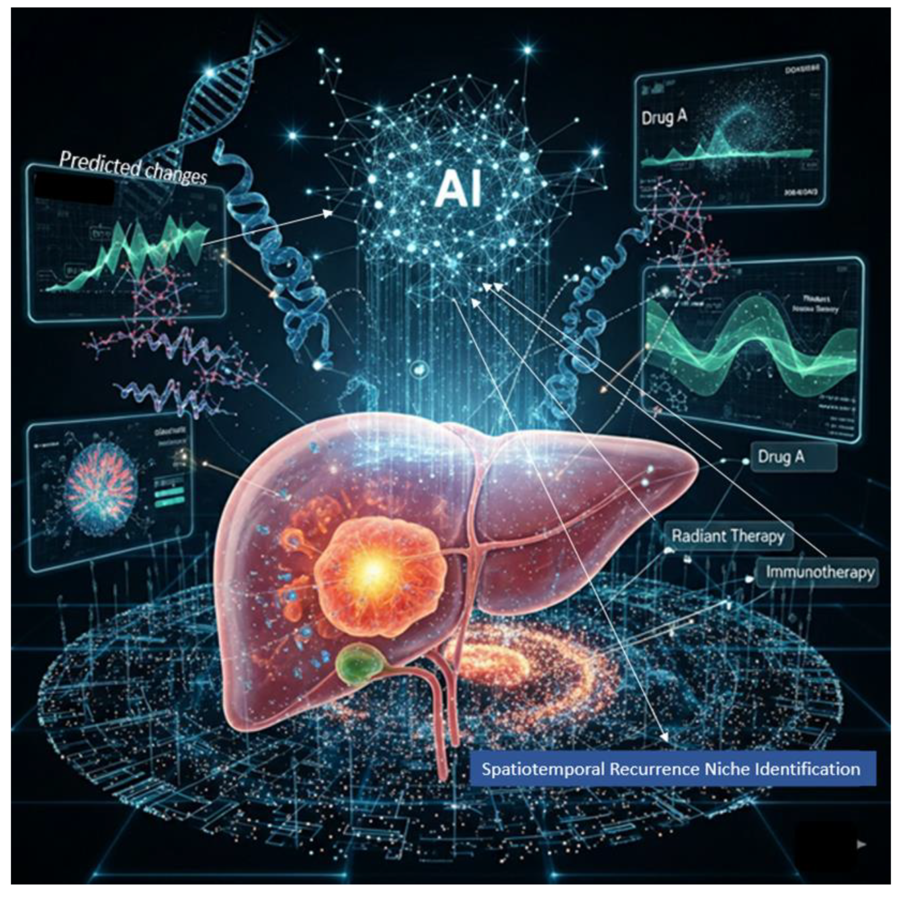
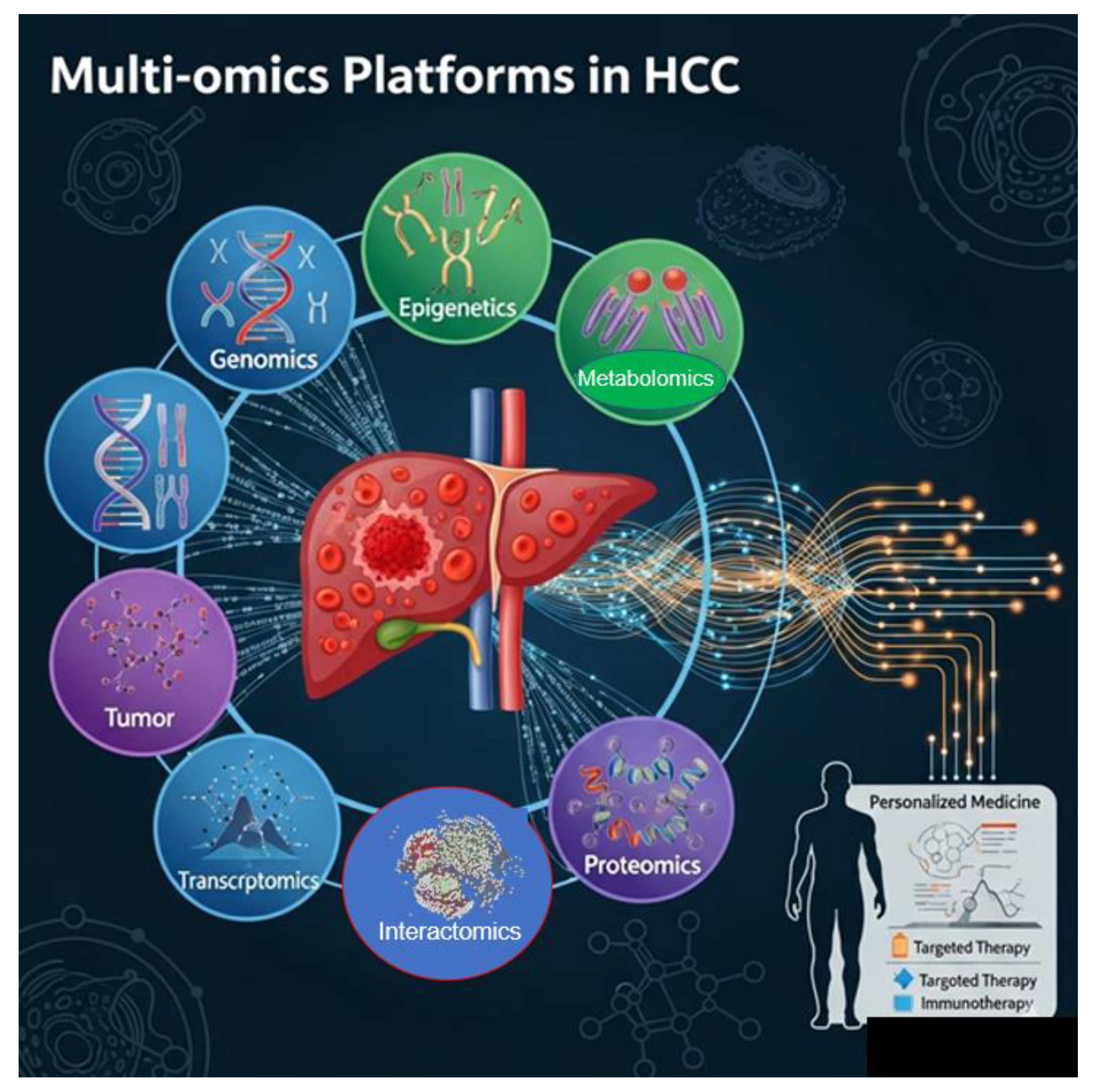
11.8. Multi-Omics Platforms in HCC

HCC-specific multi-omics platforms integrate:
  • Genomic mutations, such as TP53 and CTNNB1 [216,217].
  • Epigenetic marks, like RASSF1A methylation [242].
  • Transcriptomic profiles (e.g., miR-21, TCF7+ T cells) [115,231].
  • Proteomic signatures (e.g., AFP, VEGF, PD-L1) [241].
  • Metabolomic changes, such as lipid metabolism in NAFLD-HCC [269].
Clinical trials use these data to match patients to therapies. For example, patients with HBV-HCC showing high expression of PD-L1 and viral antigens may receive immune checkpoint inhibitors combined with TCR T-cell therapy [270]. Clinicians also employ them to monitor resistance because proteomic changes can signal the onset of resistance before imaging detects its progression. Epigenetic reprogramming may require flexible adjustments in drug combinations and dosing strategies. Understanding its regulation illuminates mechanisms such as DNA methylation of tumor suppressor genes (e.g., RASSF1A), histone modifications, and noncoding RNAs (like miR-21) that drive oncogenic phenotypes and resistance, emphasizing its dynamic and reversible nature. Some clinical trials use real-time AI-based dashboards to simulate tumor evolution or predict recurrence zones based on spatial and molecular data [271]. This enables real-time therapy adjustments based on biomarker changes. These are useful in differentiating HBV-related HCC from non-viral HCC, where the tumor microenvironment and immune dynamics differ [272]. Gene regulation and signaling cascades help describe how transcription factors (e.g., β-catenin, STAT3, TGF-β) participate in feedback mechanisms that can stabilize or promote oncogenic states. For example, in Wnt/β-catenin activation, stabilized β-catenin translocate to the nucleus, promoting cell proliferation and immune evasion by influencing expressing immune checkpoints.
In liver cancer (HCC), research concentrates on targeting various biological features and mechanisms driving the disease, including molecular drivers, immune landscapes, and tumor vulnerabilities. Key therapeutic targets in HCC trials include immune checkpoints such as PD-1, PD-L1, and CTLA-4, often upregulated in immune-excluded tumors, alongside oncogenic pathways like Wnt/β-catenin (with CTNNB1 mutations), TGF-β signaling, and VEGF-mediated angiogenesis. Cancer stem cells (like PROM1, EpCAM, and CD47) link to recurrence and resistance, while metabolic reprogramming, lipid metabolism in NAFLD-HCC and glutamine addiction, plays a vital role in aggressive subtypes. Epigenetic silencing through methylation of tumor suppressors such as RASSF1A and SOCS1 is also essential. Spatial transcriptomics allows precise identification of where these targets are active within tumor regions, and multi-omics approaches validate their behavior across various biological layers. Patient stratification, which considers factors such as viral versus nonviral HCC, with HBV-induced HCC often exhibiting immune exhaustion, opens the way to checkpoint inhibitors and TCR-T cell therapy. Tumors are also categorized as “hot” or “cold,” with treatment strategies differing; “hot” tumors may benefit from direct immune engagement, while “cold” tumors could require priming with oncolytic viruses or TGFβ blockade. Mutation-driven subtypes, such as CTNNB1-mutant tumors resistant to immunotherapy, may respond to Wnt inhibitors, and fibrotic versus non-fibrotic microenvironments might cause stromal remodeling agents to improve immune cell access. Multi-omics platforms integrate these layers to develop predictive models, with some clinical trials using AI dashboards to simulate responses and adapt therapies in real-time, promoting personalized treatment approaches.
Figure 3. Schematic representation of the Multi-omics Platforms in HCC.
Preprints 176459 g003

11.9. Tailored Solutions Are Only One Element of the Total Scope

Precision oncology combined with advanced artificial intelligence platforms may not be enough. It represents only the last step after patients discover they have the disease. HCC is a subtle disease that doesn’t show symptoms until it is very advanced. However, it’s not just its subtle nature that creates complex clinical situations; the difficulties associated with early diagnosis lead to detection at an advanced stage, when symptoms become clear or patients present in an emergency setting. This highlights a significant gap in current strategies focused on diagnosis and prevention.
Integrating advancements in imaging and AI into preventive strategies beyond symptomatic patients is crucial [273]. They should be part of routine screening programs to identify pre-symptomatic or asymptomatic cases, which can improve outcomes through earlier intervention.
Primary prevention strategies, such as vaccination against hepatitis B, lifestyle modifications to prevent non-alcoholic fatty liver disease, and public health initiatives to raise awareness about HCC’s risk factors, are necessary. Without these preventative measures, reliance on advanced diagnostics may not address the fundamental issues of late-stage presentations. Health policymakers should outline guidelines for screening high-risk populations using advanced technologies and biomarkers [274,275,276,277]. A multifaceted approach that combines advanced diagnostic capabilities with proactive public health initiatives will be vital to reducing the incidence and mortality associated with HCC [278,279,280]. Raising awareness among patients and healthcare providers about the risks associated with liver disease and the potential for HCC formation can drive proactive health behaviors and encourage earlier consultations, thus facilitating earlier detection.

12. Advanced International Trials on HCC

Here are some noteworthy ongoing or completed advanced clinical trials targeting HCC that incorporate innovative molecular and immunological strategies. Trial govID provided by National Library of Medicine NIH-NLM (https://clinicaltrials.gov/):
IMbrave150 Trial - A Study of Atezolizumab in Combination with Bevacizumab Compared With Sorafenib in Patients With Untreated Locally Advanced or Metastatic Hepatocellular Carcinoma (IMbrave150) - Hoffmann-La Roche (Responsible Party) – NIH, USA, ClinicalTrials.gov ID: NCT03434379. This study will evaluate the efficacy and safety of atezolizumab (anti-PD-L1) in combination with bevacizumab (anti-VEGF) compared with sorafenib in participants with locally advanced or metastatic Hepatocellular Carcinoma (HCC) who have received no prior systemic treatment. The study showed significant improvement in overall survival and progression-free survival compared to sorafenib. It incorporates immunotherapy combined with anti-angiogenic therapy; it exemplifies a modern approach integrating immune modulation and vascular targeting.
HIMALAYA Trial – This study - AstraZeneca (Responsible Party) - NIH, USA, ClinicalTrials.gov ID NCT03298451 - is a global, randomized, open-label, Phase III trial evaluating the efficacy of the dual checkpoint inhibitor “STRIDE” (Single Tremelimumab Regular Interval Durvalumab) combination regimen and durvalumab (anti-PD-L1) monotherapy, with or without tremelimumab (anti-CTLA-4), compared to standard sorafenib in patients with advanced hepatocellular carcinoma in the first-line setting. The study enrolled 1,171 patients with advanced hepatocellular carcinoma (HCC) who did not have refractory ascites, portal vein invasion, or hepatitis B or C virus co-infection. The updated four-year overall survival results from the HIMALAYA study underscore the efficacy of the STRIDE immuno-immunotherapy combination as a first-line treatment for hepatocellular carcinoma and confirm its role as a practice-changing treatment. The announced approval by the Italian Medicines Agency (AIFA) for the durvalumab plus tremelimumab combination is consistent with these results and provides access to this important therapeutic option.
LEAP-002 Trial - Safety and Efficacy of Lenvatinib (E7080/MK-7902) in Combination With Pembrolizumab (MK-3475) Versus Lenvatinib as First-line Therapy in Participants With Advanced Hepatocellular Carcinoma (MK-7902-002/E7080-G000-311/LEAP-002) - Merck Sharp & Dohme LLC (Responsible Party) – NIH, USA, ClinicalTrials.gov ID NCT03713593. This study evaluated the safety and efficacy of lenvatinib (E7080/MK-7902) in combination with pembrolizumab (MK-3745) versus lenvatinib in combination with placebo as first-line therapy to treat advanced hepatocellular carcinoma in adult participants. This study’s core assumptions are that lenvatinib combined with pembrolizumab is better than lenvatinib with a placebo, looking at how long people live without the disease getting worse and overall survival. Details: Evaluates combination immunotherapy targeted at both immune evasion and angiogenesis pathways.
HCC-SYS-QC Trial - Study on Recurrence Monitoring of Hepatocellular Carcinoma With 5-Hydroxymethylcytosine Test - Shanghai Zhongshan Hospital (Responsible Party) – NIH, USA, ClinicalTrials.gov ID NCT03493763. In this study, investigators aim to determine how plasma 5hmC level changes in hepatocellular carcinoma patients after liver resection and whether 5hmC can be used as a biomarker for HCC recurrence monitoring. Using systems biology and network modeling to individualize therapy is in the early phases, but promising for therapeutic decision algorithms.

References

  1. Tkačik, G., & Bialek, W. (2016). Information processing in living systems. Annual Review of Condensed Matter Physics, 7(1), 89-117.
  2. Godfrey-Smith, P. (2007). Information in biology. The Cambridge Guide to the Philosophy of Biology, ed. David L. Hull and Michael Ruse, 103-119.
  3. Portin, P., & Wilkins, A. (2017). The evolving definition of the term “gene”. Genetics, 205(4), 1353-1364.
  4. Rué, P., & Martinez Arias, A. (2015). Cell dynamics and gene expression control in tissue homeostasis and development. Molecular systems biology, 11(2), 792.
  5. Cevallos, Y., Molina, L., Santillán, A., De Rango, F., Rushdi, A., & Alonso, J. B. (2017). A digital communication analysis of gene expression of proteins in biological systems: A layered network model view. Cognitive Computation, 9(1), 43-67.
  6. Emmert-Streib, F., & Dehmer, M. (2011). Networks for systems biology: conceptual connection of data and function. IET systems biology, 5(3), 185-207.
  7. Su, J., Song, Y., Zhu, Z., Huang, X., Fan, J., Qiao, J., & Mao, F. (2024). Cell–cell communication: new insights and clinical implications. Signal transduction and targeted therapy, 9(1), 196.
  8. Wolf, Y. I., Katsnelson, M. I., & Koonin, E. V. (2018). Physical foundations of biological complexity. Proceedings of the National Academy of Sciences, 115(37), E8678-E8687.
  9. Iyengar, G. V. (2024). Elemental Analysis of Biological Systems: Biological, Medical, Environmental, Compositional, and Methodological Aspects, Volume I. CRC Press (Taylor & Francis Group). [CrossRef]
  10. Gaiseanu, F. (2021). Evolution and development of the information concept in biological systems: From empirical description to informational modeling of the living structures. Philosophy Study, 11(7), 501-516.
  11. Kellis, M., Wold, B., Snyder, M. P., Bernstein, B. E., Kundaje, A., Marinov, G. K., ... & Hardison, R. C. (2014). Defining functional DNA elements in the human genome. Proceedings of the National Academy of Sciences, 111(17), 6131-6138.
  12. Heng, H. H., Liu, G., Stevens, J. B., Bremer, S. W., Ye, K. J., Abdallah, B. Y., ... & Ye, C. J. (2011). Decoding the genome beyond sequencing: the new phase of genomic research. Genomics, 98(4), 242-252.
  13. Segal, E., Shapira, M., Regev, A., Pe'er, D., Botstein, D., Koller, D., & Friedman, N. (2003). Module networks: identifying regulatory modules and their condition-specific regulators from gene expression data. Nature genetics, 34(2), 166-176.
  14. Turner, B. M. (2009). Epigenetic responses to environmental change and their evolutionary implications. Philosophical Transactions of the Royal Society B: Biological Sciences, 364(1534), 3403-3418.
  15. Buccitelli, C., & Selbach, M. (2020). mRNAs, proteins and the emerging principles of gene expression control. Nature Reviews Genetics, 21(10), 630-644.
  16. Shen-Orr, S. S., Tibshirani, R., Khatri, P., Bodian, D. L., Staedtler, F., Perry, N. M., ... & Butte, A. J. (2010). Cell type–specific gene expression differences in complex tissues. Nature methods, 7(4), 287-289.
  17. Kolodziejczyk, A. A., Kim, J. K., Svensson, V., Marioni, J. C., & Teichmann, S. A. (2015). The technology and biology of single-cell RNA sequencing. Molecular cell, 58(4), 610-620.
  18. Barrett, L. W., Fletcher, S., & Wilton, S. D. (2012). Regulation of eukaryotic gene expression by the untranslated gene regions and other non-coding elements. Cellular and molecular life sciences, 69(21), 3613-3634.
  19. Ma’ayan, A. (2011). Introduction to network analysis in systems biology. Science signaling, 4(190), tr5-tr5.
  20. Stuart, T., & Satija, R. (2019). Integrative single-cell analysis. Nature reviews genetics, 20(5), 257-272.
  21. Buratowski, S. (1994). The basics of basal transcription by RNA polymerase II. Cell, 77(1), 1-3.
  22. Henras, A. K., Plisson-Chastang, C., O'Donohue, M. F., Chakraborty, A., & Gleizes, P. E. (2015). An overview of pre-ribosomal RNA processing in eukaryotes. Wiley Interdisciplinary Reviews: RNA, 6(2), 225-242.
  23. Celotto, A. M., & Graveley, B. R. (2001). Alternative splicing of the Drosophila Dscam pre-mRNA is both temporally and spatially regulated. Genetics, 159(2), 599-608.
  24. Meyuhas, O., Avni, D., & Shama, S. (1996). Translational control of ribosomal protein mRNAs in eukaryotes. Cold spring harbor monograph series, 30, 363-388.
  25. Ramazi, S., & Zahiri, J. (2021). Post-translational modifications in proteins: resources, tools and prediction methods. Database, 2021, baab012.
  26. Steiner, D. F., Park, S. Y., Støy, J., Philipson, L. H., & Bell, G. I. (2009). A brief perspective on insulin production. Diabetes, Obesity and Metabolism, 11, 189-196.
  27. Vaissière, T., Sawan, C., & Herceg, Z. (2008). Epigenetic interplay between histone modifications and DNA methylation in gene silencing. Mutation Research/Reviews in Mutation Research, 659(1-2), 40-48.
  28. Baylin, S. B. (2005). DNA methylation and gene silencing in cancer. Nature clinical practice Oncology, 2(Suppl 1), S4-S11.
  29. Belotserkovskii, B. P., Mirkin, S. M., & Hanawalt, P. C. (2013). DNA sequences that interfere with transcription: implications for genome function and stability. Chemical reviews, 113(11), 8620-8637.
  30. Liang, S., Moghimi, B., Yang, T. P., Strouboulis, J., & Bungert, J. (2008). Locus control region mediated regulation of adult β-globin gene expression. Journal of cellular biochemistry, 105(1), 9-16.
  31. Chitwood, D. H., & Timmermans, M. C. (2010). Small RNAs are on the move. Nature, 467(7314), 415-419.
  32. Buscaglia, L. E. B., & Li, Y. (2011). Apoptosis and the target genes of microRNA-21. Chinese journal of cancer, 30(6), 371.
  33. Mary Brittoa, S., Jayarajanb, D. (2023). Unravelling the Molecular Symphony: Investigating Gene Expression at the Biochemical Level. Eur. Chem. Bull. 2023,12(Special Issue 8),2799-2810. ISSN 2063-5346.
  34. Clapier, C. R., & Cairns, B. R. (2009). The biology of chromatin remodeling complexes. Annual review of biochemistry, 78(1), 273-304.
  35. Roberts, C. W., & Orkin, S. H. (2004). The SWI/SNF complex, chromatin and cancer. Nature Reviews Cancer, 4(2), 133-142.
  36. Chen, K., Zhao, B. S., & He, C. (2016). Nucleic acid modifications in regulation of gene expression. Cell chemical biology, 23(1), 74-85.
  37. Youness, A., Miquel, C. H., & Guéry, J. C. (2021). Escape from X chromosome inactivation and the female predominance in autoimmune diseases. International journal of molecular sciences, 22(3), 1114.
  38. Zhao, P., & Malik, S. (2022). The phosphorylation to acetylation/methylation cascade in transcriptional regulation: how kinases regulate transcriptional activities of DNA/histone-modifying enzymes. Cell & Bioscience, 12(1), 83.
  39. Buccitelli, C., & Selbach, M. (2020). mRNAs, proteins and the emerging principles of gene expression control. Nature Reviews Genetics, 21(10), 630-644.
  40. Breaker, R. R. (2018). Riboswitches and translation control. Cold Spring Harbor perspectives in biology, 10(11), a032797.
  41. Kadauke, S., & Blobel, G. A. (2009). Chromatin loops in gene regulation. Biochimica et Biophysica Acta (BBA)-Gene Regulatory Mechanisms, 1789(1), 17-25.
  42. Ak, P., & Levine, A. J. (2010). p53 and NF-κB: different strategies for responding to stress lead to a functional antagonism. The FASEB Journal, 24(10), 3643-3652.
  43. Lu, N., & Malemud, C. J. (2019). Extracellular signal-regulated kinase: a regulator of cell growth, inflammation, chondrocyte and bone cell receptor-mediated gene expression. International journal of molecular sciences, 20(15), 3792.
  44. Cowan, K. J., & Storey, K. B. (2003). Mitogen-activated protein kinases: new signaling pathways functioning in cellular responses to environmental stress. Journal of Experimental Biology, 206(7), 1107-1115.
  45. Sanchez-Diaz, P., & Penalva, L. O. (2006). Post-transcription meets post-genomic: the saga of RNA binding proteins in a new era. RNA biology, 3(3), 101-109. [CrossRef]
  46. Yang, Y., Okada, S., & Sakurai, M. (2021). Adenosine-to-inosine RNA editing in neurological development and disease. RNA biology, 18(7), 999-1013.
  47. Rees, H. A., & Liu, D. R. (2018). Base editing: precision chemistry on the genome and transcriptome of living cells. Nature reviews genetics, 19(12), 770-788.
  48. Ontiveros, R. J., Stoute, J., & Liu, K. F. (2019). The chemical diversity of RNA modifications. Biochemical Journal, 476(8), 1227-1245.
  49. Zhang, D., Zhu, L., Gao, Y., Wang, Y., & Li, P. (2024). RNA editing enzymes: structure, biological functions and applications. Cell & Bioscience, 14(1), 34. [CrossRef]
  50. Picardi, E., Manzari, C., Mastropasqua, F., Aiello, I., D’Erchia, A. M., & Pesole, G. (2015). Profiling RNA editing in human tissues: towards the inosinome Atlas. Scientific reports, 5(1), 14941.
  51. Kung, C. P., Maggi Jr, L. B., & Weber, J. D. (2018). The role of RNA editing in cancer development and metabolic disorders. Frontiers in Endocrinology, 9, 762.
  52. Doudna, J. A. (2020). The promise and challenge of therapeutic genome editing. Nature, 578(7794), 229-236.
  53. Cox, D. B., Gootenberg, J. S., Abudayyeh, O. O., Franklin, B., Kellner, M. J., Joung, J., & Zhang, F. (2017). RNA editing with CRISPR-Cas13. Science, 358(6366), 1019-1027.
  54. Eisenberg, E. (2020). Proteome diversification by RNA editing. RNA Editing: Methods and Protocols, 229-251.
  55. Hwang, T., Park, C. K., Leung, A. K., Gao, Y., Hyde, T. M., Kleinman, J. E., ... & Weinberger, D. R. (2016). Dynamic regulation of RNA editing in human brain development and disease. Nature neuroscience, 19(8), 1093-1099.
  56. Reik, W. (2007). Stability and flexibility of epigenetic gene regulation in mammalian development. Nature, 447(7143), 425-432.
  57. Jaenisch, R., & Bird, A. (2003). Epigenetic regulation of gene expression: how the genome integrates intrinsic and environmental signals. Nature genetics, 33(3), 245-254.
  58. Carlberg, C., & Molnár, F. (2016). Mechanisms of gene regulation (pp. 57-73). Dordrecht: Springer.
  59. Abdul, Q. A., Yu, B. P., Chung, H. Y., Jung, H. A., & Choi, J. S. (2017). Epigenetic modifications of gene expression by lifestyle and environment. Archives of pharmacal research, 40(11), 1219-1237. [CrossRef]
  60. Boyko, A., & Kovalchuk, I. (2011). Genome instability and epigenetic modification, heritable responses to environmental stress?. Current opinion in plant biology, 14(3), 260-266.
  61. Heine, S. J. (2017). DNA is not destiny: The remarkable, completely misunderstood relationship between you and your genes. WW Norton & Company.
  62. Huang, B., Jiang, C., & Zhang, R. (2014). Epigenetics: the language of the cell?. Epigenomics, 6(1), 73-88.
  63. Li, S., Chen, M., Li, Y., & Tollefsbol, T. O. (2019). Prenatal epigenetics diets play protective roles against environmental pollution. Clinical epigenetics, 11(1), 82. [CrossRef]
  64. Argentieri, M. A., Nagarajan, S., Seddighzadeh, B., Baccarelli, A. A., & Shields, A. E. (2017). Epigenetic pathways in human disease: the impact of DNA methylation on stress-related pathogenesis and current challenges in biomarker development. EBioMedicine, 18, 327-350. [CrossRef]
  65. Prakash, K., & Fournier, D. (2017). Histone code and higher-order chromatin folding: a hypothesis. Genomics and computational biology, 3(2), e41.
  66. Guillemette, B., Drogaris, P., Lin, H. H. S., Armstrong, H., Hiragami-Hamada, K., Imhof, A., ... & Festenstein, R. J. (2011). H3 lysine 4 is acetylated at active gene promoters and is regulated by H3 lysine 4 methylation. PLoS genetics, 7(3), e1001354. [CrossRef]
  67. Yin, Y., Morgunova, E., Jolma, A., Kaasinen, E., Sahu, B., Khund-Sayeed, S., ... & Taipale, J. (2017). Impact of cytosine methylation on DNA binding specificities of human transcription factors. Science, 356(6337), eaaj2239.
  68. Tirado-Magallanes, R., Rebbani, K., Lim, R., Pradhan, S., & Benoukraf, T. (2016). Whole genome DNA methylation: beyond genes silencing. Oncotarget, 8(3), 5629.
  69. Auerkari, E. I. (2006). Methylation of tumor suppressor genes p16 (INK4a), p27 (Kip1) and E-cadherin in carcinogenesis. Oral oncology, 42(1), 4-12. [CrossRef]
  70. Clapier, C. R., & Cairns, B. R. (2009). The biology of chromatin remodeling complexes. Annual review of biochemistry, 78(1), 273-304.
  71. Loda, A., & Heard, E. (2019). Xist RNA in action: Past, present, and future. PLoS genetics, 15(9), e1008333.
  72. Dalmay, T. (2013). Mechanism of miRNA-mediated repression of mRNA translation. Essays in biochemistry, 54, 29-38. [CrossRef]
  73. Kan, R. L., Chen, J., & Sallam, T. (2022). Crosstalk between epitranscriptomic and epigenetic mechanisms in gene regulation. Trends in Genetics, 38(2), 182-193.
  74. Tiffon, C. (2018). The impact of nutrition and environmental epigenetics on human health and disease. International journal of molecular sciences, 19(11), 3425.
  75. Tiffon, C. (2018). The impact of nutrition and environmental epigenetics on human health and disease. International journal of molecular sciences, 19(11), 3425. [CrossRef]
  76. Reul, J. M. (2014). Making memories of stressful events: a journey along epigenetic, gene transcription, and signaling pathways. Frontiers in psychiatry, 5, 5.
  77. Bommarito, P. A., Martin, E., & Fry, R. C. (2017). Effects of prenatal exposure to endocrine disruptors and toxic metals on the fetal epigenome. Epigenomics, 9(3), 333-350.
  78. Booth, L. N., & Brunet, A. (2016). The aging epigenome. Molecular cell, 62(5), 728-744.
  79. Skinner, M. K. (2011). Environmental epigenetic transgenerational inheritance and somatic epigenetic mitotic stability. Epigenetics, 6(7), 838. [CrossRef]
  80. Dominguez-Salas, P., Cox, S. E., Prentice, A. M., Hennig, B. J., & Moore, S. E. (2012). Maternal nutritional status, C1 metabolism and offspring DNA methylation: a review of current evidence in human subjects. Proceedings of the Nutrition Society, 71(1), 154-165.
  81. Schneider, R., & Grosschedl, R. (2007). Dynamics and interplay of nuclear architecture, genome organization, and gene expression. Genes & development, 21(23), 3027-3043.
  82. Li, X., Liu, C., Lei, Z., Chen, H., & Wang, L. (2024). Phase-separated chromatin compartments: Orchestrating gene expression through condensation. Cell insight, 3(6), 100213.
  83. Ashwin, S. S., Maeshima, K., & Sasai, M. (2020). Heterogeneous fluid-like movements of chromatin and their implications to transcription. Biophysical Reviews, 12(2), 461-468. [CrossRef]
  84. Li, C., Li, Z., Wu, Z., & Lu, H. (2023). Phase separation in gene transcription control: Phase separation in gene transcription control. Acta Biochimica et Biophysica Sinica, 55(7), 1052.
  85. Hu, Y., Shen, F., Yang, X., Han, T., Long, Z., Wen, J., ... & Guo, Q. (2023). Single-cell sequencing technology applied to epigenetics for the study of tumor heterogeneity. Clinical Epigenetics, 15(1), 161.
  86. Kamies, R., & Martinez-Jimenez, C. P. (2020). Advances of single-cell genomics and epigenomics in human disease: where are we now?. Mammalian Genome, 31(5), 170-180.
  87. Wang, K., Liu, H., Hu, Q. et al. Epigenetic regulation of aging: implications for interventions of aging and diseases. Sig Transduct Target Ther 7, 374 (2022). [CrossRef]
  88. Bertucci-Richter, E.M., Parrott, B.B. The rate of epigenetic drift scales with maximum lifespan across mammals. Nat Commun 14, 7731 (2023). [CrossRef]
  89. Gonzalo S. Epigenetic alterations in aging. J Appl Physiol (1985). 2010 Aug;109(2):586-97. Epub 2010 May 6. PMID: 20448029; PMCID: PMC2928596. [CrossRef]
  90. Li, Y., Tian, X., Luo, J. et al. Molecular mechanisms of aging and anti-aging strategies. Cell Commun Signal 22, 285 (2024). [CrossRef]
  91. Pinel, C., Green, S., & Svendsen, M. N. (2023). Slowing down decay: biological clocks in personalized medicine. Frontiers in Sociology, 8, 1111071. [CrossRef]
  92. Szyf, M. (2012). The early-life social environment and DNA methylation. Clinical genetics, 81(4), 341-349.
  93. Di Giorgio, E., Paluvai, H., Dalla, E., Ranzino, L., Renzini, A., Moresi, V., ... & Brancolini, C. (2021). HDAC4 degradation during senescence unleashes an epigenetic program driven by AP-1/p300 at selected enhancers and super-enhancers. Genome Biology, 22(1), 129. [CrossRef]
  94. Reifsnyder, P. C., Flurkey, K., Doty, R., Calcutt, N. A., Koza, R. A., & Harrison, D. E. (2022). Rapamycin/metformin co-treatment normalizes insulin sensitivity and reduces complications of metabolic syndrome in type 2 diabetic mice. Aging Cell, 21(9), e13666.
  95. Nir Eynon, Macsue Jacques, Kirsten Seale et al. DNA Methylation Ageing Atlas Across 17 Human Tissues, 07 August 2025, PREPRINT (Version 1, Under Review at Nature Portfolio) available at Research Square. [CrossRef]
  96. Dandoti, S. (2021). Mechanisms adopted by cancer cells to escape apoptosis–A review. Biocell, 45(4), 863.
  97. von Manstein, V., Min Yang, C., Richter, D., Delis, N., Vafaizadeh, V., & Groner, B. (2013). Resistance of cancer cells to targeted therapies through the activation of compensating signaling loops. Current signal transduction therapy, 8(3), 193-202. [CrossRef]
  98. Garinis, G. A., Patrinos, G. P., Spanakis, N. E., & Menounos, P. G. (2002). DNA hypermethylation: when tumour suppressor genes go silent. Human genetics, 111(2), 115-127.
  99. Hoffmann, M. J., & Schulz, W. A. (2005). Causes and consequences of DNA hypomethylation in human cancer. Biochemistry and cell biology, 83(3), 296-321.
  100. Wiles, E. T., & Selker, E. U. (2017). H3K27 methylation: a promiscuous repressive chromatin mark. Current opinion in genetics & development, 43, 31-37. [CrossRef]
  101. Jia, X., Wang, X., Guo, X., Ji, J., Lou, G., Zhao, J., ... & Yu, S. (2019). MicroRNA-124: an emerging therapeutic target in cancer. Cancer medicine, 8(12), 5638-5650.
  102. Chen, Y., Fu, L. L., Wen, X., Liu, B., Huang, J., Wang, J. H., & Wei, Y. Q. (2014). Oncogenic and tumor suppressive roles of microRNAs in apoptosis and autophagy. Apoptosis, 19(8), 1177-1189.
  103. Long, Y., Wang, X., Youmans, D. T., & Cech, T. R. (2017). How do lncRNAs regulate transcription?. Science advances, 3(9), eaao2110.
  104. Singh, A., Modak, S. B., Chaturvedi, M. M., & Purohit, J. S. (2023). SWI/SNF chromatin remodelers: structural, functional and mechanistic implications. Cell Biochemistry and Biophysics, 81(2), 167-187.
  105. Singh, A., Modak, S. B., Chaturvedi, M. M., & Purohit, J. S. (2023). SWI/SNF chromatin remodelers: structural, functional and mechanistic implications. Cell Biochemistry and Biophysics, 81(2), 167-187.
  106. Pathak, A., Tomar, S., & Pathak, S. (2023). Epigenetics and cancer: a comprehensive review. Asian Pacific Journal of Cancer Biology, 8(1), 75-89. [CrossRef]
  107. You JS, Jones PA. Cancer genetics and epigenetics: two sides of the same coin? Cancer Cell. 2012 Jul 10;22(1):9-20. PMID: 22789535; PMCID: PMC3396881. [CrossRef]
  108. Yang, T., Yang, Y., & Wang, Y. (2021). Predictive biomarkers and potential drug combinations of epi-drugs in cancer therapy. Clinical Epigenetics, 13(1), 113.
  109. Hong, S. N. (2018). Genetic and epigenetic alterations of colorectal cancer. Intestinal research, 16(3), 327.
  110. Niv, Y. (2007). Microsatellite instability and MLH1 promoter hypermethylation in colorectal cancer. World journal of gastroenterology: WJG, 13(12), 1767. [CrossRef]
  111. Shi, W. K., Li, Y. H., Bai, X. S., & Lin, G. L. (2022). The cell cycle-associated protein CDKN2A may promotes colorectal cancer cell metastasis by inducing epithelial-mesenchymal transition. Frontiers in Oncology, 12, 834235.
  112. Chen, H. P., Zhao, Y. T., & Zhao, T. C. (2015). Histone deacetylases and mechanisms of regulation of gene expression. Critical Reviews™ in Oncogenesis, 20(1-2). [CrossRef]
  113. Füllgrabe, J., Kavanagh, E., & Joseph, B. (2011). Histone onco-modifications. Oncogene, 30(31), 3391-3403.
  114. Chrun, E. S., Modolo, F., & Daniel, F. I. (2017). Histone modifications: A review about the presence of this epigenetic phenomenon in carcinogenesis. Pathology-Research and Practice, 213(11), 1329-1339.
  115. Slaby, O., Svoboda, M., Fabian, P., Smerdova, T., Knoflickova, D., Bednarikova, M., ... & Vyzula, R. (2008). Altered expression of miR-21, miR-31, miR-143 and miR-145 is related to clinicopathologic features of colorectal cancer. Oncology, 72(5-6), 397-402. [CrossRef]
  116. Garcia-Gomez, A., Rodríguez-Ubreva, J., & Ballestar, E. (2018). Epigenetic interplay between immune, stromal and cancer cells in the tumor microenvironment. Clinical Immunology, 196, 64-71.
  117. Yang, J., Xu, J., Wang, W., Zhang, B., Yu, X., & Shi, S. (2023). Epigenetic regulation in the tumor microenvironment: molecular mechanisms and therapeutic targets. Signal transduction and targeted therapy, 8(1), 210. [CrossRef]
  118. Segditsas, S., & Tomlinson, I. (2006). Colorectal cancer and genetic alterations in the Wnt pathway. Oncogene, 25(57), 7531-7537.
  119. Wilting, R. H., & Dannenberg, J. H. (2012). Epigenetic mechanisms in tumorigenesis, tumor cell heterogeneity and drug resistance. Drug Resistance Updates, 15(1-2), 21-38. [CrossRef]
  120. Wang, N., Ma, T., & Yu, B. (2023). Targeting epigenetic regulators to overcome drug resistance in cancers. Signal transduction and targeted therapy, 8(1), 69. [CrossRef]
  121. Patnaik, S., & Anupriya. (2019). Drugs targeting epigenetic modifications and plausible therapeutic strategies against colorectal cancer. Frontiers in Pharmacology, 10, 588. [CrossRef]
  122. Sahafnejad, Z., Ramazi, S., & Allahverdi, A. (2023). An update of epigenetic drugs for the treatment of cancers and brain diseases: a comprehensive review. Genes, 14(4), 873. [CrossRef]
  123. Zoratto, F., Rossi, L., Verrico, M., Papa, A., Basso, E., Zullo, A., ... & Tomao, S. (2014). Focus on genetic and epigenetic events of colorectal cancer pathogenesis: implications for molecular diagnosis. Tumor Biology, 35(7), 6195-6206. [CrossRef]
  124. Rasool, M., Malik, A., Naseer, M. I., Manan, A., Ansari, S. A., Begum, I., ... & Gan, S. H. (2015). The role of epigenetics in personalized medicine: challenges and opportunities. BMC medical genomics, 8(Suppl 1), S5. [CrossRef]
  125. La Thangue, N. B., & Kerr, D. J. (2011). Predictive biomarkers: a paradigm shift towards personalized cancer medicine. Nature reviews Clinical oncology, 8(10), 587-596.
  126. Rituraj, Pal, R. S., Wahlang, J., Pal, Y., Chaitanya, M. V. N. L., & Saxena, S. (2025). Precision oncology: transforming cancer care through personalized medicine. Medical Oncology, 42(7), 246. [CrossRef]
  127. Payne, S. R. (2010). From discovery to the clinic: the novel DNA methylation biomarker m SEPT9 for the detection of colorectal cancer in blood. Epigenomics, 2(4), 575-585.
  128. Henrique, R., & Jerónimo, C. (2004). Molecular detection of prostate cancer: a role for GSTP1 hypermethylation. European urology, 46(5), 660-669. [CrossRef]
  129. Schultz, N. A., Dehlendorff, C., Jensen, B. V., Bjerregaard, J. K., Nielsen, K. R., Bojesen, S. E., ... & Johansen, J. S. (2014). MicroRNA biomarkers in whole blood for detection of pancreatic cancer. Jama, 311(4), 392-404.
  130. Liao, C., Chen, X., & Fu, Y. (2023). Salivary analysis: An emerging paradigm for non-invasive healthcare diagnosis and monitoring. Interdisciplinary Medicine, 1(3), e20230009.
  131. Hamamoto, R., Komatsu, M., Takasawa, K., Asada, K., & Kaneko, S. (2019). Epigenetics analysis and integrated analysis of multiomics data, including epigenetic data, using artificial intelligence in the era of precision medicine. Biomolecules, 10(1), 62.
  132. Wen, X., Pu, H., Liu, Q., Guo, Z., & Luo, D. (2022). Circulating tumor DNA, a novel biomarker of tumor progression and its favorable detection techniques. Cancers, 14(24), 6025.
  133. Goell, J. H., & Hilton, I. B. (2021). CRISPR/Cas-based epigenome editing: advances, applications, and clinical utility. Trends in Biotechnology, 39(7), 678-691.
  134. Corella, D., & Ordovas, J. M. (2017). Basic concepts in molecular biology related to genetics and epigenetics. Revista Española de Cardiología (English Edition), 70(9), 744-753. [CrossRef]
  135. Wang, N., Hu, Y., & Wang, Z. (2024). Regulation of alternative splicing: Functional interplay with epigenetic modifications and its implication to cancer. Wiley Interdisciplinary Reviews: RNA, 15(1), e1815.
  136. Cantone, I., & Fisher, A. G. (2013). Epigenetic programming and reprogramming during development. Nature structural & molecular biology, 20(3), 282-289.
  137. Weng, M. K., Natarajan, K., Scholz, D., Ivanova, V. N., Sachinidis, A., Hengstler, J. G., ... & Leist, M. (2014). Lineage-specific regulation of epigenetic modifier genes in human liver and brain. PLoS One, 9(7), e102035.
  138. Robusti, G., Vai, A., Bonaldi, T., & Noberini, R. (2022). Investigating pathological epigenetic aberrations by epi-proteomics. Clinical Epigenetics, 14(1), 145. [CrossRef]
  139. Lahtz, C., & Pfeifer, G. P. (2011). Epigenetic changes of DNA repair genes in cancer. Journal of molecular cell biology, 3(1), 51-58.
  140. Constantin, N., Sina, A. A. I., Korbie, D., & Trau, M. (2022). Opportunities for early cancer detection: the rise of ctDNA methylation-based pan-cancer screening technologies. Epigenomes, 6(1), 6. [CrossRef]
  141. Myte, R., Sundkvist, A., Van Guelpen, B., & Harlid, S. (2019). Circulating levels of inflammatory markers and DNA methylation, an analysis of repeated samples from a population based cohort. Epigenetics, 14(7), 649-659.
  142. Suhre, K., & Zaghlool, S. (2021). Connecting the epigenome, metabolome and proteome for a deeper understanding of disease. Journal of internal medicine, 290(3), 527-548. [CrossRef]
  143. Moore, D. S. (2015). The developing genome: An introduction to behavioral epigenetics. Oxford University Press. ISBN 978-0-19-992234-5.
  144. Aebersold, R., & Mann, M. (2016). Mass-spectrometric exploration of proteome structure and function. Nature, 537(7620), 347-355.
  145. Noberini, R., Sigismondo, G., & Bonaldi, T. (2016). The contribution of mass spectrometry-based proteomics to understanding epigenetics. Epigenomics, 8(3), 429-445.
  146. Mohr, A. E., Ortega-Santos, C. P., Whisner, C. M., Klein-Seetharaman, J., & Jasbi, P. (2024). Navigating challenges and opportunities in multi-omics integration for personalized healthcare. Biomedicines, 12(7), 1496.
  147. Ali, H. (2023). Artificial intelligence in multi-omics data integration: Advancing precision medicine, biomarker discovery and genomic-driven disease interventions. Int J Sci Res Arch, 8(1), 1012-30.
  148. Mao, Y., Shangguan, D., Huang, Q., Xiao, L., Cao, D., Zhou, H., & Wang, Y. K. (2025). Emerging artificial intelligence-driven precision therapies in tumor drug resistance: recent advances, opportunities, and challenges. Molecular Cancer, 24(1), 123.
  149. An, G. (2022). Specialty grand challenge: What it will take to cross the valley of death: Translational systems biology,“true” precision medicine, medical digital twins, artificial intelligence and in silico clinical trials. Frontiers in Systems Biology, 2, 901159.
  150. Łukaniszyn, M., Majka, Ł., Grochowicz, B., Mikołajewski, D., & Kawala-Sterniuk, A. (2024). Digital twins generated by artificial intelligence in personalized healthcare. Applied Sciences, 14(20), 9404.
  151. Sun, T., He, X., & Li, Z. (2023). Digital twin in healthcare: Recent updates and challenges. Digital health, 9, 20552076221149651. [CrossRef]
  152. Faisal, S. M., Ishrat, M., & Khan, W. (2025). Digital Twins in Healthcare: Revolutionizing Patient Care and Medical Operations. In Digital Twins for Smart Cities and Urban Planning (pp. 69-89). CRC Press.
  153. Vallée, A. (2024). Envisioning the future of personalized medicine: role and realities of digital twins. Journal of medical Internet research, 26, e50204. [CrossRef]
  154. Yang, H., & Jiang, Z. (2024). Decision support for personalized therapy in implantable medical devices: A digital twin approach. Expert Systems with Applications, 243, 122883. [CrossRef]
  155. Abd Elaziz, M., Al-qaness, M. A., Dahou, A., Al-Betar, M. A., Mohamed, M. M., El-Shinawi, M., ... & Ewees, A. A. (2024). Digital twins in healthcare: Applications, technologies, simulations, and future trends. Wiley Interdisciplinary Reviews: Data Mining and Knowledge Discovery, 14(6), e1559.
  156. Abouelmehdi, K., Beni-Hessane, A., & Khaloufi, H. (2018). Big healthcare data: preserving security and privacy. Journal of big data, 5(1), 1-18. [CrossRef]
  157. Roopa, M. S., & Venugopal, K. R. (2025). Digital Twins for Cyber-Physical Healthcare Systems: Architecture, Requirements, Systematic Analysis and Future Prospects. IEEE Access. [CrossRef]
  158. Moro-Visconti, R., & Quirici, M. C. (2020). Digital technology and efficiency gains in healthcare infrastructural investments. Healthcare Research and Public Safety Journal, 2(1), 1-8.
  159. Jia, W., Wang, W., & Zhang, Z. (2022). From simple digital twin to complex digital twin Part I: A novel modeling method for multi-scale and multi-scenario digital twin. Advanced Engineering Informatics, 53, 101706. [CrossRef]
  160. Zoltick, M. M., & Maisel, J. B. (2023). Societal impacts: Legal, regulatory and ethical considerations for the digital twin. In The digital twin (pp. 1167-1200). Cham: Springer International Publishing.
  161. Rosa, C., Marsch, L. A., Winstanley, E. L., Brunner, M., & Campbell, A. N. (2021). Using digital technologies in clinical trials: current and future applications. Contemporary clinical trials, 100, 106219.
  162. Gomase, V. S., Ghatule, A. P., Sharma, R., Sardana, S., & Dhamane, S. P. (2025). Cloud Computing Facilitating Data Storage, Collaboration, and Analysis in Global Healthcare Clinical Trials. Reviews on Recent Clinical Trials.
  163. Reddy, K. J. (2025). Integrating Technology into Clinical Practice. In Innovations in Neurocognitive Rehabilitation: Harnessing Technology for Effective Therapy (pp. 329-350). Cham: Springer Nature Switzerland.
  164. Pritchett, J. C., Patt, D., Thanarajasingam, G., Schuster, A., & Snyder, C. (2023). Patient-reported outcomes, digital health, and the quest to improve health equity. American Society of Clinical Oncology Educational Book, 43, e390678.
  165. Cummings, S. R. (2021). Clinical trials without clinical sites. JAMA Internal Medicine, 181(5), 680-684.
  166. Yamada, O., Chiu, S. W., Takata, M., Abe, M., Shoji, M., Kyotani, E., ... & Yamaguchi, T. (2021). Clinical trial monitoring effectiveness: remote risk-based monitoring versus on-site monitoring with 100% source data verification. Clinical trials, 18(2), 158-167. [CrossRef]
  167. Arman, M. (2023). The advantages of online recruitment and selection: A systematic review of cost and time efficiency. Business Management and Strategy, 14(2), 220-240. [CrossRef]
  168. Azodo, I., Williams, R., Sheikh, A., & Cresswell, K. (2020). Opportunities and challenges surrounding the use of data from wearable sensor devices in health care: qualitative interview study. Journal of Medical Internet Research, 22(10), e19542.
  169. Apostolaros, M., Babaian, D., Corneli, A., Forrest, A., Hamre, G., Hewett, J., ... & Randall, P. (2020). Legal, regulatory, and practical issues to consider when adopting decentralized clinical trials: recommendations from the clinical trials transformation initiative. Therapeutic innovation & regulatory science, 54(4), 779-787.
  170. Bhat, A., Su, W. C., Cleffi, C., & Srinivasan, S. (2021). A hybrid clinical trial delivery model in the COVID-19 era. Physical Therapy, 101(8), pzab116. [CrossRef]
  171. Yaakov, R. A., Güler, Ö., Mayhugh, T., & Serena, T. E. (2021). Enhancing patient centricity and advancing innovation in clinical research with virtual randomized clinical trials (vRCTs). Diagnostics, 11(2), 151.
  172. Arendt, D., Musser, J. M., Baker, C. V., Bergman, A., Cepko, C., Erwin, D. H., ... & Wagner, G. P. (2016). The origin and evolution of cell types. Nature Reviews Genetics, 17(12), 744-757.
  173. Perelis, M., Marcheva, B., Moynihan Ramsey, K., Schipma, M. J., Hutchison, A. L., Taguchi, A., ... & Bass, J. (2015). Pancreatic β cell enhancers regulate rhythmic transcription of genes controlling insulin secretion. Science, 350(6261), aac4250.
  174. Kouadjo, K. E., Nishida, Y., Cadrin-Girard, J. F., Yoshioka, M., & St-Amand, J. (2007). Housekeeping and tissue-specific genes in mouse tissues. BMC genomics, 8(1), 127.
  175. Hekselman, I., & Yeger-Lotem, E. (2020). Mechanisms of tissue and cell-type specificity in heritable traits and diseases. Nature Reviews Genetics, 21(3), 137-150.
  176. Gao, Y., Peng, L., & Zhao, C. (2024). MYH7 in cardiomyopathy and skeletal muscle myopathy. Molecular and Cellular Biochemistry, 479(2), 393-417.
  177. Dobbelstein, M., & Moll, U. (2014). Targeting tumour-supportive cellular machineries in anticancer drug development. Nature reviews Drug discovery, 13(3), 179-196. [CrossRef]
  178. Mohammed, A., Biegert, G., Adamec, J., & Helikar, T. (2017). Identification of potential tissue-specific cancer biomarkers and development of cancer versus normal genomic classifiers. Oncotarget, 8(49), 85692. [CrossRef]
  179. Ståhl, P. L., Salmén, F., Vickovic, S., Lundmark, A., Navarro, J. F., Magnusson, J., ... & Frisén, J. (2016). Visualization and analysis of gene expression in tissue sections by spatial transcriptomics. Science, 353(6294), 78-82.
  180. Cheung, P., Vallania, F., Warsinske, H. C., Donato, M., Schaffert, S., Chang, S. E., ... & Kuo, A. J. (2018). Single-cell chromatin modification profiling reveals increased epigenetic variations with aging. Cell, 173(6), 1385-1397.
  181. Wilbrey-Clark, A., Roberts, K., & Teichmann, S. A. (2020). Cell Atlas technologies and insights into tissue architecture. Biochemical Journal, 477(8), 1427-1442.
  182. Zuo, C., Zhu, J., Zou, J., & Chen, L. (2025). Unravelling tumour spatiotemporal heterogeneity using spatial multimodal data. Clinical and Translational Medicine, 15(5), e70331. [CrossRef]
  183. Pettini, F., Visibelli, A., Cicaloni, V., Iovinelli, D., & Spiga, O. (2021). Multi-omics model applied to cancer genetics. International journal of molecular sciences, 22(11), 5751.
  184. Fountzilas, E., Pearce, T., Baysal, M. A., Chakraborty, A., & Tsimberidou, A. M. (2025). Convergence of evolving artificial intelligence and machine learning techniques in precision oncology. NPJ Digital Medicine, 8(1), 75. [CrossRef]
  185. Huss, R., Raffler, J., & Märkl, B. (2023). Artificial intelligence and digital biomarker in precision pathology guiding immune therapy selection and precision oncology. Cancer Reports, 6(7), e1796. [CrossRef]
  186. Gardy, J. L., & Loman, N. J. (2018). Towards a genomics-informed, real-time, global pathogen surveillance system. Nature Reviews Genetics, 19(1), 9-20.
  187. Leung, M. K., Delong, A., Alipanahi, B., & Frey, B. J. (2015). Machine learning in genomic medicine: a review of computational problems and data sets. Proceedings of the IEEE, 104(1), 176-197.
  188. Heidrich, I., Deitert, B., Werner, S., & Pantel, K. (2023). Liquid biopsy for monitoring of tumor dormancy and early detection of disease recurrence in solid tumors. Cancer and Metastasis Reviews, 42(1), 161-182.
  189. Alagarswamy, K., Shi, W., Boini, A., Messaoudi, N., Grasso, V., Cattabiani, T., ... & Gumbs, A. (2024). Should AI-Powered Whole-Genome Sequencing Be Used Routinely for Personalized Decision Support in Surgical Oncology, A Scoping Review. BioMedInformatics, 4(3), 1757-1772.
  190. Huang, P. J., Chang, J. H., Lin, H. H., Li, Y. X., Lee, C. C., Su, C. T., ... & Tang, P. (2020). DeepVariant-on-Spark: Small-Scale Genome Analysis Using a Cloud-Based Computing Framework. Computational and Mathematical Methods in Medicine, 2020(1), 7231205. [CrossRef]
  191. Kuo, T. C., Tian, T. F., & Tseng, Y. J. (2013). 3Omics: a web-based systems biology tool for analysis, integration and visualization of human transcriptomic, proteomic and metabolomic data. BMC systems biology, 7(1), 64.
  192. Langmead, B., & Nellore, A. (2018). Cloud computing for genomic data analysis and collaboration. Nature Reviews Genetics, 19(4), 208-219.
  193. Ali, M., Naeem, F., Tariq, M., & Kaddoum, G. (2022). Federated learning for privacy preservation in smart healthcare systems: A comprehensive survey. IEEE journal of biomedical and health informatics, 27(2), 778-789. [CrossRef]
  194. Suura, S. R. (2025). Integrating Artificial Intelligence, Machine Learning, and Big Data with Genetic Testing and Genomic Medicine to Enable Earlier, Personalized Health Interventions. Deep Science Publishing.
  195. Schlitt, T., & Brazma, A. (2007). Current approaches to gene regulatory network modelling. BMC bioinformatics, 8(Suppl 6), S9.
  196. Singh, S., Gouri, V., & Samant, M. (2023). TGF-β in correlation with tumor progression, immunosuppression and targeted therapy in colorectal cancer. Medical Oncology, 40(11), 335. [CrossRef]
  197. Gatherer, D. (2010). So what do we really mean when we say that systems biology is holistic?. BMC systems biology, 4(1), 22.
  198. O’Connor, T. (2020). Emergent properties. Available from Stanford Encyclopedia of Philosophy (https://plato.stanford.edu/entries/properties-emergent/?utm_source=chatgpt.com).
  199. Tavassoly, I., Goldfarb, J., & Iyengar, R. (2018). Systems biology primer: the basic methods and approaches. Essays in biochemistry, 62(4), 487-500.
  200. Ma'ayan, A. (2017). Complex systems biology. Journal of the Royal Society Interface, 14(134), 20170391.
  201. Panditrao, G., Bhowmick, R., Meena, C., & Sarkar, R. R. (2022). Emerging landscape of molecular interaction networks: Opportunities, challenges and prospects. Journal of Biosciences, 47(2), 24. [CrossRef]
  202. Criscuolo, C., & Timmis, J. (2018). GVCs and centrality: Mapping key hubs, spokes and the periphery.
  203. Ávila, A. Á. (2023). Characterization of the anti-leukemic properties of anti-CD3 antibodies in T-cell acute lymphoblastic leukemia (T-ALL) (Doctoral dissertation, Université Paris Cité).
  204. Mamun, M. A., Mannoor, K., Cao, J., Qadri, F., & Song, X. (2020). SOX2 in cancer stemness: tumor malignancy and therapeutic potentials. Journal of molecular cell biology, 12(2), 85-98. [CrossRef]
  205. Zhao, L., Lee, V. H., Ng, M. K., Yan, H., & Bijlsma, M. F. (2019). Molecular subtyping of cancer: current status and moving toward clinical applications. Briefings in bioinformatics, 20(2), 572-584.
  206. Shen, Q., He, Y., Qian, J., & Wang, X. (2022). Identifying tumor immunity-associated molecular features in liver hepatocellular carcinoma by multi-omics analysis. Frontiers in Molecular Biosciences, 9, 960457.
  207. Ljungberg, J. K., Kling, J. C., Tran, T. T., & Blumenthal, A. (2019). Functions of the WNT signaling network in shaping host responses to infection. Frontiers in Immunology, 10, 2521.
  208. Przedborski, M., Smalley, M., Thiyagarajan, S., Goldman, A., & Kohandel, M. (2021). Systems biology informed neural networks (SBINN) predict response and novel combinations for PD-1 checkpoint blockade. Communications biology, 4(1), 877.
  209. Wrinch, D. (1947). The native protein. Science, 106(2743), 73-76.
  210. Smejkal, G. B. (2013). The meaning of ‘native’. Expert Review of Proteomics, 10(5), 407-409.
  211. Mastropietro, A. (2024). Toward explainable biomedical deep learning. Training and Explaining Neural Networks in Bioinformatics and Medicinal Chemistry. Doctoral Thesis in Data Science. Available at UNIROMA1: https://iris.uniroma1.it/retrieve/cc782790-8122-4446-8c9f-7bf3827ab4da/Tesi_dottorato_Mastropietro.pdf.
  212. Karunakaran, K. B. (2024). Interactome-based framework to translate disease genetic data into biological and clinical insights (Doctoral dissertation, University of Reading).
  213. Petti, M., & Farina, L. (2023). Network medicine for patients' stratification: From single-layer to multi-omics. WIREs Mechanisms of Disease, 15(6), e1623.
  214. Yavuz, B. R., Jang, H., & Nussinov, R. (2024). Anticancer Target Combinations: Network-Informed Signaling-Based Approach to Discovery. bioRxiv, 2024-10.
  215. Mukherjee, A., Abraham, S., Singh, A., Balaji, S., & Mukunthan, K. S. (2025). From data to cure: A comprehensive exploration of multi-omics data analysis for targeted therapies. Molecular biotechnology, 67(4), 1269-1289. [CrossRef]
  216. Bonnet, S., Gaujoux, S., Launay, P., Baudry, C., Chokri, I., Ragazzon, B., ... & Tissier, F. (2011). Wnt/β-catenin pathway activation in adrenocortical adenomas is frequently due to somatic CTNNB1-activating mutations, which are associated with larger and nonsecreting tumors: a study in cortisol-secreting and-nonsecreting tumors. The Journal of Clinical Endocrinology & Metabolism, 96(2), E419-E426.
  217. Assoun, S., Theou-Anton, N., Nguenang, M., Cazes, A., Danel, C., Abbar, B., ... & Zalcman, G. (2019). Association of TP53 mutations with response and longer survival under immune checkpoint inhibitors in advanced non-small-cell lung cancer. Lung cancer, 132, 65-71. [CrossRef]
  218. Sarkar, H., Lee, E., Lopez-Darwin, S. L., & Kang, Y. (2025). Deciphering normal and cancer stem cell niches by spatial transcriptomics: opportunities and challenges. Genes & Development, 39(1-2), 64-85.
  219. Takeuchi, Y., Tanegashima, T., Sato, E., Irie, T., Sai, A., Itahashi, K., ... & Nishikawa, H. (2021). Highly immunogenic cancer cells require activation of the WNT pathway for immunological escape. Science immunology, 6(65), eabc6424. [CrossRef]
  220. Bai, X., Yi, M., Jiao, Y., Chu, Q., & Wu, K. (2019). Blocking TGF-β signaling to enhance the efficacy of immune checkpoint inhibitor. OncoTargets and therapy, 9527-9538.
  221. Randall, R. E., & Goodbourn, S. (2008). Interferons and viruses: an interplay between induction, signalling, antiviral responses and virus countermeasures. Journal of general virology, 89(1), 1-47.
  222. Chen, S., Fragoza, R., Klei, L., Liu, Y., Wang, J., Roeder, K., ... & Yu, H. (2018). An interactome perturbation framework prioritizes damaging missense mutations for developmental disorders. Nature genetics, 50(7), 1032-1040.
  223. Li, X., Ruan, Z., Yang, S., Yang, Q., Li, J., & Hu, M. (2024). Bioinformatic-Experimental Screening Uncovers Multiple Targets for Increase of MHC-I Expression through Activating the Interferon Response in Breast Cancer. International Journal of Molecular Sciences, 25(19), 10546.
  224. Lungu, C. N., Mangalagiu, I. I., Romila, A., Nechita, A., Putz, M. V., & Mehedinti, M. C. (2025). Molecular Mediated Angiogenesis and Vasculogenesis Networks. International Journal of Molecular Sciences, 26(13), 6316. [CrossRef]
  225. Li, Y., Liu, Q., Jing, X., Wang, Y., Jia, X., Yang, X., & Chen, K. (2025). Cancer-Associated Fibroblasts: Heterogeneity, Cancer Pathogenesis, and Therapeutic Targets. MedComm, 6(7), e70292.
  226. Ho, D. (2020). Artificial intelligence in cancer therapy. Science, 367(6481), 982-983.
  227. Yong, C. S., Telarovic, I., Gregor, L., Raeber, M. E., Pruschy, M., & Boyman, O. (2024). IL-2 immunotherapy rescues irradiation-induced T cell exhaustion in vivo. bioRxiv, 2024-01.
  228. Faubert, B., Solmonson, A., & DeBerardinis, R. J. (2020). Metabolic reprogramming and cancer progression. Science, 368(6487), eaaw5473.
  229. Karami, Z., Mortezaee, K., & Majidpoor, J. (2023). Dual anti-PD-(L) 1/TGF-β inhibitors in cancer immunotherapy–updated. International Immunopharmacology, 122, 110648.
  230. Matsumoto, S., & Kikuchi, A. (2024). Wnt/β-catenin signaling pathway in liver biology and tumorigenesis. In Vitro Cellular & Developmental Biology-Animal, 60(5), 466-481. [CrossRef]
  231. Meng, F., Zhao, J., Tan, A. T., Hu, W., Wang, S. Y., Jin, J., ... & Wang, F. S. (2021). Immunotherapy of HBV-related advanced hepatocellular carcinoma with short-term HBV-specific TCR expressed T cells: results of dose escalation, phase I trial. Hepatology international, 15(6), 1402-1412.
  232. Zhan, Z. H., Hong, J., Li, J. Y., Wang, C., He, L., Xu, Z., & Zhang, J. (2025). Artificial intelligence-based methods for protein structure prediction: a survey. Artificial Intelligence Review, 58(10), 1-36. [CrossRef]
  233. Jastrzębski, M. K. (2024). Computational Methods to Target Protein-Protein Interactions. In Protein-Protein Docking: Methods and Protocols (pp. 327-343). New York, NY: Springer US.
  234. Anderson, J. B., & Johnnesson, R. (2006). Understanding information transmission. John Wiley & Sons.
  235. Morgan, S. J., Pullon, S. R., Macdonald, L. M., McKinlay, E. M., & Gray, B. V. (2017). Case study observational research: A framework for conducting case study research where observation data are the focus. Qualitative health research, 27(7), 1060-1068. [CrossRef]
  236. Uher, J. (2022). Functions of units, scales and quantitative data: Fundamental differences in numerical traceability between sciences. Quality & Quantity, 56(4), 2519-2548. [CrossRef]
  237. Barba, L. A. (2018). Terminologies for reproducible research. arXiv preprint arXiv:1802.03311.
  238. Hepburn, B., & Andersen, H. (2015). Scientific method. Available at Stanford Encyclopedia of Philosophy (https://plato.stanford.edu/entries/scientific-method/?utm_source=webtekno).
  239. Colonna, G. (2025). Protein biophysics: current limitations and prospects. AIMS Biophysics, 12(3), 333-374.
  240. Sharma A, Colonna G. System-Wide Pollution of Biomedical Data: Consequence of the Search for Hub Genes of Hepatocellular Carcinoma Without Spatiotemporal Consideration. Mol Diagn Ther. 2021 Jan;25(1):9-27. https://doi.org/10.1007/s40291-020-00505-3. Epub 2021 Jan 21. Erratum in: Mol Diagn Ther. 2021 May;25(3):387. PMID: 33475988; PMCID: PMC7847983. [CrossRef]
  241. Sharma A, Colonna G. Correction to: System-Wide Pollution of Biomedical Data: Consequence of the Search for Hub Genes of Hepatocellular Carcinoma Without Spatiotemporal Consideration. Mol Diagn Ther. 2021 May;25(3):387. https://doi.org/10.1007/s40291-021-00527-5. Epub 2021 May 5. Erratum for: Mol Diagn Ther. 2021 Jan;25(1):9-27. PMID: 33950420; PMCID: PMC8276865. [CrossRef]
  242. Arya, A. K., Bhadada, S. K., Singh, P., Sachdeva, N., Saikia, U. N., Dahiya, D., ... & Rao, S. D. (2017). Promoter hypermethylation inactivates CDKN2A, CDKN2B and RASSF1A genes in sporadic parathyroid adenomas. Scientific Reports, 7(1), 3123.
  243. Nagaraja, S., Quezada, M. A., Gillespie, S. M., Arzt, M., Lennon, J. J., Woo, P. J., ... & Monje, M. (2019). Histone variant and cell context determine H3K27M reprogramming of the enhancer landscape and oncogenic state. Molecular cell, 76(6), 965-980. [CrossRef]
  244. Wu, H., Ng, R., Chen, X., Steer, C. J., & Song, G. (2016). MicroRNA-21 is a potential link between non-alcoholic fatty liver disease and hepatocellular carcinoma via modulation of the HBP1-p53-Srebp1c pathway. Gut, 65(11), 1850-1860.
  245. Xu, C., Yan, Z., Zhou, L., & Wang, Y. (2013). A comparison of glypican-3 with alpha-fetoprotein as a serum marker for hepatocellular carcinoma: a meta-analysis. Journal of cancer research and clinical oncology, 139(8), 1417-1424.
  246. Hack, S. P., Zhu, A. X., & Wang, Y. (2020). Augmenting anticancer immunity through combined targeting of angiogenic and PD-1/PD-L1 pathways: challenges and opportunities. Frontiers in immunology, 11, 598877.
  247. Mund, C., Brueckner, B., & Lyko, F. (2006). Reactivation of epigenetically silenced genes by DNA methyltransferase inhibitors: basic concepts and clinical applications. Epigenetics, 1(1), 8-14.
  248. Thomas, M. L., & Marcato, P. (2018). Epigenetic modifications as biomarkers of tumor development, therapy response, and recurrence across the cancer care continuum. Cancers, 10(4), 101.
  249. Li, X., Ramadori, P., Pfister, D., Seehawer, M., Zender, L., & Heikenwalder, M. (2021). The immunological and metabolic landscape in primary and metastatic liver cancer. Nature Reviews Cancer, 21(9), 541-557. [CrossRef]
  250. Michalak, T. I. (2023). The initial hepatitis B virus-hepatocyte genomic integrations and their role in hepatocellular oncogenesis. International Journal of Molecular Sciences, 24(19), 14849. [CrossRef]
  251. Gui, M., Huang, S., Li, S., Chen, Y., Cheng, F., Liu, Y., ... & Zhou, G. (2024). Integrative single-cell transcriptomic analyses reveal the cellular ontological and functional heterogeneities of primary and metastatic liver tumors. Journal of Translational Medicine, 22(1), 206.
  252. Ye, B., Liu, X., Li, X., Kong, H., Tian, L., & Chen, Y. (2015). T-cell exhaustion in chronic hepatitis B infection: current knowledge and clinical significance. Cell death & disease, 6(3), e1694-e1694. [CrossRef]
  253. Zhang, J., Lyu, T., Cao, Y., & Feng, H. (2021). Role of TCF-1 in differentiation, exhaustion, and memory of CD8+ T cells: A review. The FASEB Journal, 35(5), e21549.
  254. Akkız, H. (2023). Emerging role of cancer-associated fibroblasts in progression and treatment of hepatocellular carcinoma. International Journal of Molecular Sciences, 24(4), 3941. [CrossRef]
  255. Li, X., Ramadori, P., Pfister, D., Seehawer, M., Zender, L., & Heikenwalder, M. (2021). The immunological and metabolic landscape in primary and metastatic liver cancer. Nature Reviews Cancer, 21(9), 541-557.
  256. Yuan, Y., Wu, D., Li, J., Huang, D., Zhao, Y., Gao, T., ... & Tang, Y. (2023). Mechanisms of tumor-associated macrophages affecting the progression of hepatocellular carcinoma. Frontiers in pharmacology, 14, 1217400.
  257. Sükei, T., Palma, E., & Urbani, L. (2021). Interplay between cellular and non-cellular components of the tumour microenvironment in hepatocellular carcinoma. Cancers, 13(21), 5586. [CrossRef]
  258. Lin, A., Xiong, M., Tang, B., Jiang, A., Shen, J., Liu, Z., ... & Luo, P. (2025). Decoding the hepatic fibrosis-hepatocellular carcinoma axis: from mechanisms to therapeutic opportunities. Hepatology International, 1-28.
  259. Saw, P. E., Liu, Q., Wong, P. P., & Song, E. (2024). Cancer stem cell mimicry for immune evasion and therapeutic resistance. Cell Stem Cell, 31(8), 1101-1112.
  260. Andrews, L. P., Butler, S. C., Cui, J., Cillo, A. R., Cardello, C., Liu, C., ... & Vignali, D. A. (2024). LAG-3 and PD-1 synergize on CD8+ T cells to drive T cell exhaustion and hinder autocrine IFN-γ-dependent anti-tumor immunity. Cell, 187(16), 4355-4372. [CrossRef]
  261. Luo, X. Y., Wu, K. M., & He, X. X. (2021). Advances in drug development for hepatocellular carcinoma: clinical trials and potential therapeutic targets. Journal of Experimental & Clinical Cancer Research, 40(1), 172. [CrossRef]
  262. Hedrich, V., Breitenecker, K., Djerlek, L., Ortmayr, G., & Mikulits, W. (2021). Intrinsic and extrinsic control of hepatocellular carcinoma by TAM receptors. Cancers, 13(21), 5448.
  263. Lindblad, K. E., Ruiz de Galarreta, M., & Lujambio, A. (2021). Tumor-intrinsic mechanisms regulating immune exclusion in liver cancers. Frontiers in immunology, 12, 642958.
  264. Bhange, M., & Telange, D. (2025). Convergence of nanotechnology and artificial intelligence in the fight against liver cancer: a comprehensive review. Discover Oncology, 16(1), 77. [CrossRef]
  265. Bhange, M., & Telange, D. (2025). Convergence of nanotechnology and artificial intelligence in the fight against liver cancer: a comprehensive review. Discover Oncology, 16(1), 77.
  266. Calderaro, J., Žigutytė, L., Truhn, D., Jaffe, A., & Kather, J. N. (2024). Artificial intelligence in liver cancer, new tools for research and patient management. Nature Reviews Gastroenterology & Hepatology, 21(8), 585-599. [CrossRef]
  267. Romeo, M., Dallio, M., Napolitano, C., Basile, C., Di Nardo, F., Vaia, P., ... & Federico, A. (2025). Clinical Applications of Artificial Intelligence (AI) in Human Cancer: Is It Time to Update the Diagnostic and Predictive Models in Managing Hepatocellular Carcinoma (HCC)?. Diagnostics, 15(3), 252. [CrossRef]
  268. Mansur, A., Vrionis, A., Charles, J. P., Hancel, K., Panagides, J. C., Moloudi, F., ... & Daye, D. (2023). The role of artificial intelligence in the detection and implementation of biomarkers for hepatocellular carcinoma: outlook and opportunities. Cancers, 15(11), 2928.
  269. Ahmed, E. A., El-Derany, M. O., Anwar, A. M., Saied, E. M., & Magdeldin, S. (2022). Metabolomics and lipidomics screening reveal reprogrammed signaling pathways toward cancer development in non-alcoholic steatohepatitis. International Journal of Molecular Sciences, 24(1), 210. [CrossRef]
  270. Li, B., Yan, C., Zhu, J., Chen, X., Fu, Q., Zhang, H., ... & Fang, W. (2020). Anti–PD-1/PD-L1 blockade immunotherapy employed in treating hepatitis B virus infection–related advanced hepatocellular carcinoma: a literature review. Frontiers in Immunology, 11, 1037.
  271. Tiwari, A., Mishra, S., & Kuo, T. R. (2025). Current AI technologies in cancer diagnostics and treatment. Molecular Cancer, 24(1), 159. [CrossRef]
  272. Sun, R., Li, J., Lin, X., Yang, Y., Liu, B., Lan, T., ... & Wu, B. (2023). Peripheral immune characteristics of hepatitis B virus-related hepatocellular carcinoma. Frontiers in Immunology, 14, 1079495. [CrossRef]
  273. Sharma, N., & Kaushik, P. (2025). Integration of AI in healthcare systems, A discussion of the challenges and opportunities of integrating AI in healthcare systems for disease detection and diagnosis. AI in Disease Detection: Advancements and Applications, 239-263.
  274. Ferraro, S., Biganzoli, E. M., Castaldi, S., & Plebani, M. (2022). Health technology assessment to assess value of biomarkers in the decision-making process. Clinical Chemistry and Laboratory Medicine (CCLM), 60(5), 647-654. [CrossRef]
  275. Horgan, D., Ciliberto, G., Conte, P., Baldwin, D., Seijo, L., Montuenga, L. M., ... & Curigliano, G. (2020). Bringing greater accuracy to europe’s healthcare systems: The unexploited potential of biomarker testing in oncology. Biomedicine hub, 5(3), 1-42. [CrossRef]
  276. Graig, L. A., Phillips, J. K., Moses, H. L., Committee on Policy Issues in the Clinical Development and Use of Biomarkers for Molecularly Targeted Therapies, & National Academies of Sciences, Engineering, and Medicine. (2016). Supportive Policy Environment for Biomarker Tests for Molecularly Targeted Therapies. In Biomarker Tests for Molecularly Targeted Therapies: Key to Unlocking Precision Medicine. National Academies Press (US).
  277. Van Poppel, H., Roobol, M. J., Chapple, C. R., Catto, J. W., N’Dow, J., Sønksen, J., ... & Wirth, M. (2021). Prostate-specific antigen testing as part of a risk-adapted early detection strategy for prostate cancer: European Association of Urology position and recommendations for 2021. European Urology, 80(6), 703-711. [CrossRef]
  278. Kudo, M., Oanh, B. T., Chen, C. J., Ngat, D. T., George, J., Kim, D. Y., ... & Sarno, R. (2025). Roadmap for HCC Surveillance and Management in the Asia Pacific. Cancers, 17(12), 1928. [CrossRef]
  279. Kinsey, E., & Lee, H. M. (2024). Management of hepatocellular carcinoma in 2024: the multidisciplinary paradigm in an evolving treatment landscape. Cancers, 16(3), 666. [CrossRef]
  280. Albini, A., Trapani, D., Bertolini, F., Noonan, D. M., Orecchia, R., & Corso, G. (2025). From combination early detection to multicancer testing: shifting cancer care toward proactive prevention and interception. Cancer Prevention Research, OF1-OF20. [CrossRef]
Figure 1. In the image, "PMo" is a protein modifier (Protein MOdifier). It represents one of many proteins (enzymes) responsible for adding or removing epigenetic "markers," such as acetyl or methyl groups, from histones or DNA. It is the worker who carries out the modifications. The yellow stars represent acetyl or methyl groups. For example, histone acetylation opens the DNA wrapped around histones, making genes more accessible and therefore easier to activate. The colored chemical structures represent the nitrogenous bases in DNA, to which methyl groups can bind. These are the molecules that, by binding to a sugar and a phosphate group, form nucleotides. DNA methylation, another key epigenetic modification, occurs by adding a methyl group (CH₃) to one of these bases (cytosine). This modification tends to "turn off" or silence genes.
Preprints 176459 g001
Table 1. Types of RNA Editing.
Type Mechanisms Example
A-to-I Editing Adenosine is converted to inosine by ADAR enzymes. Inosine is read as guanosine during translation. Common in the brain; affects neurotransmitter receptors.
C-to-U Editing Cytidine is deaminated to uridine by cytidine deaminases. Alters the ApoB gene in the intestine, producing a shorter protein.
U Insertion/Deletion Uridines are inserted or deleted, often in mitochondrial RNA. Found in trypanosomes (parasitic protozoa).
Table 5. Some tailored approaches to epigenetic therapies.
Therapy Type Mechanism Example Use Case
DNMT Inhibitors Block DNA methylation Azacitidine for leukemia
HDAC Inhibitors Loosen chromatin to reactivate genes Vorinostat for cutaneous T-cell lymphoma
miRNA Modulators Restore normal miRNA levels Experimental in lung and breast cancers
Combination Therapies Pair epi-drugs with chemo or immunotherapy Enhances response and reduces resistance
Table 6. Applications of Epigenetics-Proteomics.
Area Epigenetic Role Proteomic Role Personalized Impact
Cancer Therapy Identifies silenced tumor suppressors Tracks oncogenic protein activity Matches patients to targeted treatments.
Autoimmune
Disorders
Reveals immune gene dysregulation Measures inflammatory protein levels Adjusts immunosuppressive regimens
Mental Health Links stress to gene expression changes Detects neurotransmitter-related proteins Guides psychiatric drug selection
Cardiovascular Risk Shows methylation of lipid metabolism genes Profiles heart-related enzymes and markers Predicts heart attack risk and drug response
Table 7. Importance of tissue specificity in HCC.
Feature Impact on Liver Cancer Progression
Cell-type composition Determines immune response and therapy resistance
Spatial organization Influences drug delivery and metastatic potential
Gene expression profiles Guide biomarker discovery and targeted therapies
Microenvironment dynamics Shape tumor evolution and immune escape
Table 8. Integrated multi-omics layers in HCC.
Omics Type What It Measures Example in HCC Trials
Genomics DNA mutations TP53, CTNNB1 mutations
Transcriptomics RNA expression miR-21, TCF7+ T cells
Proteomics Protein levels AFP, VEGF, PD-L1
Epigenomics DNA methylation, histone modifications RASSF1A methylation
Metabolomics Metabolic changes Lipid metabolism shifts
Disclaimer/Publisher’s Note: The statements, opinions and data contained in all publications are solely those of the individual author(s) and contributor(s) and not of MDPI and/or the editor(s). MDPI and/or the editor(s) disclaim responsibility for any injury to people or property resulting from any ideas, methods, instructions or products referred to in the content.
Copyright: This open access article is published under a Creative Commons CC BY 4.0 license, which permit the free download, distribution, and reuse, provided that the author and preprint are cited in any reuse.
Prerpints.org logo

Preprints.org is a free preprint server supported by MDPI in Basel, Switzerland.

Subscribe

Disclaimer

Terms of Use

Privacy Policy

Privacy Settings

© 2026 MDPI (Basel, Switzerland) unless otherwise stated