Preprint
Article

This version is not peer-reviewed.

Clinical Evaluation of PolyDeep, a Computer Aided Detection system: A Multicenter Randomized Tandem Colonoscopy Trial

A peer-reviewed article of this preprint also exists.

Submitted:

11 September 2025

Posted:

12 September 2025

You are already at the latest version

Abstract

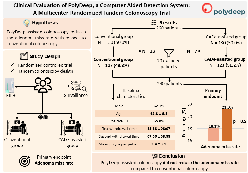
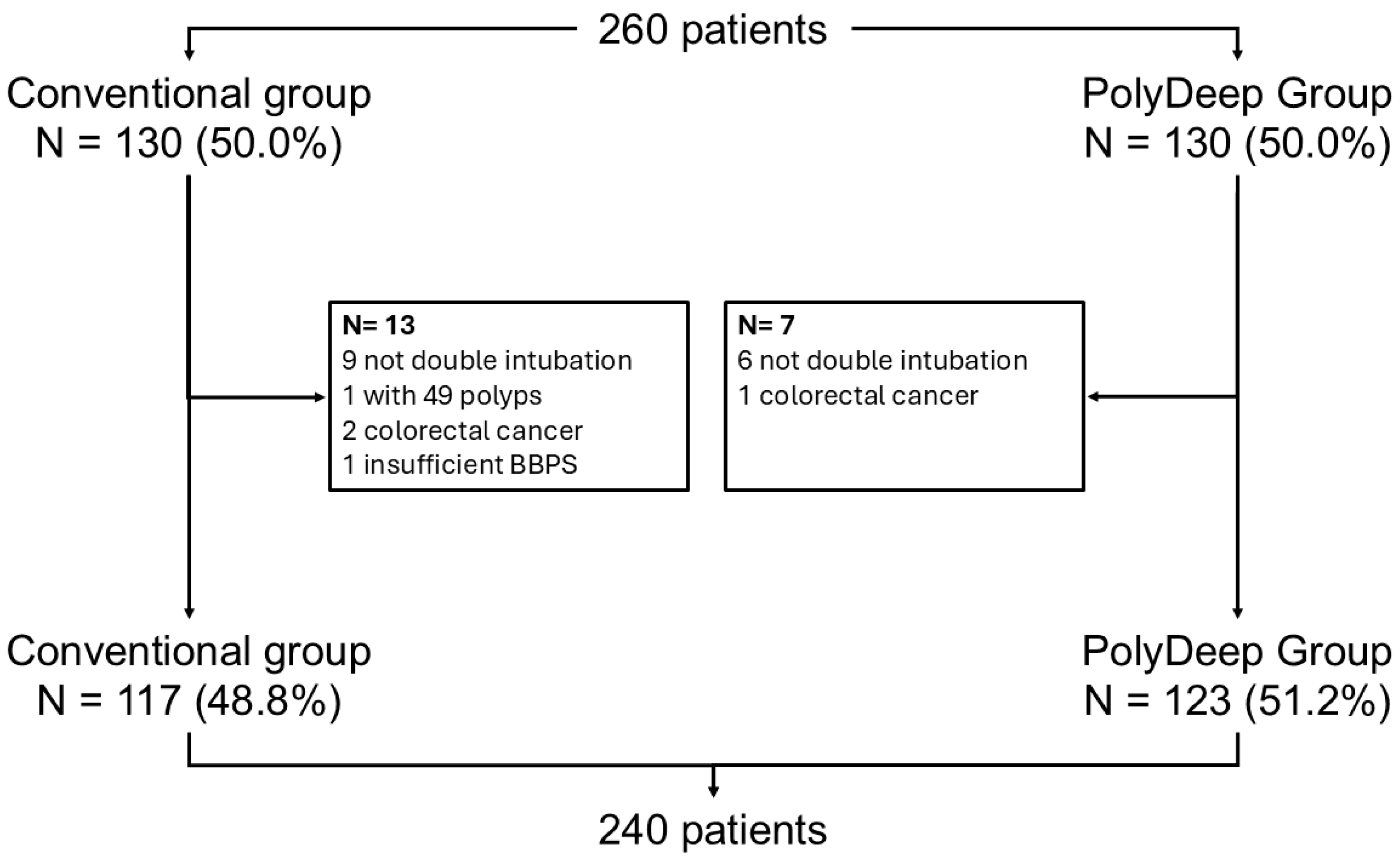
Background/Objectives: Computer-aided detection (CADe) systems are increasingly used in endoscopy to enhance lesion recognition. PolyDeep is a CADe/x tool previously assessed in an observational study. The aim of our study is to determine if PolyDeep-assisted colonoscopy reduces the adenoma miss rate (AMR) compared with conventional colonoscopy. Methods: We carried out a multicenter randomized controlled trial with a tandem colonoscopy design in participants from a colorectal cancer screening program (positive fecal immunochemical test-FIT or surveillance). Expert endoscopists performed all colonoscopies, and patients were allocated to groups by a computer-generated sequence. The primary endpoint was AMR; secondary endpoints included polyp miss rate (PMR), serrated lesion miss rate (SLMR) and advanced polyp miss rate (APMR). Results: From May to November 2023, we recruited 260 patients and excluded 20, leaving 240 for analysis. Baseline characteristics were balanced between groups (62.1% male; mean age 62.3 ± 6.5 years; 65.8% FIT-positive; mean first withdrawal time 13:38 ± 08:07 minutes; mean second withdrawal time 07:50 ± 03:38 minutes; lesion detection rate 76.6%; mean polyps per patient 3.4 ± 3.1). We did not find statistically significant differences between PolyDeep-assisted and conventional colonoscopy groups in AMR (21.3% vs 18.1%, p = 0.5), PMR (21.8% vs 20.3%, p = 0.7), SLMR (23.4% vs 25.6%, p = 0.9) or APMR (7.3% vs 11.3%, p = 0.5). In the subgroup analysis according to indication, we did not find any statistically significant differences. Conclusions: In the context of a CRC screening program, PolyDeep-assisted colonoscopy did not reduce AMR.

Keywords: 
;  ;  ;  ;  ;  ;  ;  ;  ;  

1. Introduction

Colorectal cancer (CRC) is the third most commonly diagnosed malignancy and the second leading cause of cancer-related mortality worldwide, with the highest incidence and mortality rates observed globally [1]. CRC typically develops over a number of years from precancerous lesions, such as adenomas and serrated polyps [2]. Organised CRC screening programmes are offered to individuals at average risk (generally those aged between 50 and 75 years) reducing both the incidence and mortality of the disease [2,3,4]. The primary screening modalities employed to detect precancerous lesions are the faecal immunochemical occult blood test (FIT) and colonoscopy [3,5]. In several countries, including Spain, FIT is the initial test offered; individuals with a positive result are subsequently referred for diagnostic colonoscopy, which remains the gold standard for lesion detection [3,5].
Colonoscopy remains the only test able of detecting precancerous lesions, namely adenomas and serrated lesions, within the colonic mucosa [3,5]. A proportion of these lesions present challenges owing to their small size (i.e., <5 mm) or morphology (e.g., flat or depressed), which can hinder their detection [6,7]. In addition, factors related to endoscopist performance such as fatigue, suboptimal mucosal exposure, or limited experience may further compromise lesion identification [6,7]. Collectively, these factors contribute to the risk of missed lesions during colonoscopy, which may subsequently lead to the development of post-colonoscopy colorectal cancer (PCCRC) [4]. The adenoma miss rate (AMR), defined as the proportion of adenomas detected during a second withdrawal relative to the total number of adenomas, ranges from 17% to 48%, with two meta-analyses estimating average miss rates of 22% and 26%, respectively [8,9].
In recent years, various computer-aided diagnosis (CAD) systems based on artificial intelligence (AI) and deep learning (DL) have been applied to medical image analysis, including radiography, mammography, and cardiovascular imaging [10,11,12]. In the context of colonoscopy, computer-aided detection (CADe) systems have been evaluated in randomized clinical trials employing a range of designs (i.e., comparisons between CADe-assisted and conventional colonoscopy, or tandem colonoscopy designs) and using diverse endpoints, including adenoma detection rate (ADR), polyp detection rate (PDR), AMR, and polyp miss rate (PMR) [13,14,15,16,17,18,19,20,21,22]. Although the use of these systems appears to improve diagnostic performance metrics, their role in routine clinical practice remains to be fully established [23].
PolyDeep is a computer-aided detection and characterization (CADe/x) system developed to identify colorectal lesions in real time during colonoscopy. The system integrates a neural detection network based on the YOLOv3 architecture and an object tracking algorithm, which maintains and follows all detections in each subsequent frame [24,25,26,27]. PolyDeep was initially validated in vitro and subsequently evaluated in a prospective study using a second-observer design [28,29]. In the present randomized controlled trial employing a tandem colonoscopy design, our objective was to determine whether the AMR in the PolyDeep-assisted colonoscopy group is superior to that of the conventional colonoscopy group.

2. Materials and Methods

2.1. Study Design

PolyDeep Advance 2 (NCT05512793) is a multicentre randomised controlled trial (RCT) with a tandem colonoscopy design. The study was conducted in the endoscopy units of three university hospitals in Spain: Hospital de Ourense, Hospital Álvaro Cunqueiro de Vigo, and Hospital de Montecelo de Pontevedra. The study was approved by the institutional review board (2022/067) in accordance with the Declaration of Helsinki and applicable guidelines for good clinical practice. Data from the three centres were verified and monitored within the electronic Case Report Form (eCRF) in the REDCap platform at the Galicia-Sur health research institute (https://redcap.tic1-iisgaliciasur.es/). The study was reported in accordance with the CONSORT-AI guideline for randomized trials.

2.2. Participants of the Study

We prospectively included patients aged between 40 and 79 years who underwent a colonoscopy after a positive FIT or a surveillance colonoscopy following the resection of advanced adenomas. Participants were excluded if they had a personal history of colorectal cancer or previous colonic resection, a hereditary syndrome predisposing to colorectal cancer, or serrated polyposis syndrome. Regarding colonoscopy quality, patients were excluded if they had an inadequate Boston Bowel Preparation Scale score (<2 in any segment or <6 overall) or if cecal intubation was not achieved. All participants provided written informed consent prior to inclusion in the study.

2.3. Randomization Process

We randomized the patients in a 1:1 ratio by a stratified block design based on the colonoscopy indication, such as positive FIT or surveillance endoscopy. We created blocks of two in all possible combinations (i.e., four combinations) and randomly assigned each block to positions within the randomization template. To minimize bias, we doubled the number of allocation slots (i.e., with 260 patients to be randomized, we generated 520 slots), ensuring a more balanced and unbiased randomization process. Finally, the randomization template was uploaded to the eCRF by the study coordinator. The endoscopists performed the randomization process of the participants before the colonoscopy, to know which is the allocation group and what tandem colonoscopy they should do. However, participants were not aware of the randomization allocation.

2.4. Clinical Setting

We conducted the study in a conventional endoscopy room using a standard endoscopy tower with high definition colonoscopes using the model EXERA III CV 190 or higher. We integrated the PolyDeep system into the setup, connecting the CADe/x system to both the endoscopy tower and the monitor displaying the colonoscopy image. The same monitor showed both the colonoscopy image and the PolyDeep system’s output, including overlays highlighting detected polyps. Upon the patient’s arrival in the endoscopy room, the endoscopists explained the study and obtained written informed consent. They then randomized the participants in the eCRF, assigning them to either the conventional group (conventional colonoscopy followed by assisted colonoscopy) or the PolyDeep group (assisted colonoscopy followed by conventional colonoscopy). All endoscopists were experts, meaning they are participants of the CRC screening program with more than 300 colonoscopies, one year of experience and with a rigorous quality control based on key quality indicators. They performed a back-to-back colonoscopy with two withdrawal phases according to the assigned randomization sequence. During each withdrawal phase, the endoscopists measured withdrawal time using the timer in the colonoscope control device. Lesions were counted only in one of the withdrawal phases and resected upon identification. Throughout the procedure, the nursing and auxiliary team recorded polyp data on a log sheet, including withdrawal phase, morphology, location, size, and both the endoscopists’ and PolyDeep’s optical diagnosis.
We sent all identified and retrieved lesions for histopathological evaluation, which served as the gold standard for analysis. If a lesion could not be resected during the procedure, the endoscopists documented its characteristics and obtained a biopsy. In the eCRF, we recorded participant demographic data (age, sex, and colonoscopy indication), colonoscopy details (Boston Bowel Preparation Scale score, withdrawal time, and cecal intubation), and polyp-specific information (withdrawal phase of identification, morphology, size, location, optical diagnosis, and histological evaluation)

2.5. Endpoints

The primary endpoint was to assess and compare the AMR between the conventional group and the PolyDeep group. Secondary endpoints included the evaluation and comparison of the PMR, serrated lesion miss rate (SLMR), advanced adenoma miss rate (AAMR), advanced serrated lesion miss rate (ASLMR) and advanced polyp miss rate (APMR) between both groups.

2.6. Sample Size

We calculated the sample size based on the assumption that the conventional group had a 30% AMR and the PolyDeep group a 20%, considering an alpha error of 5% and a beta error of 20% [14]. We should include 294 adenomas and 118 patients per group, assuming an average of 2.5 lesions per colonoscopy. Attending a drop-out rate of 10% for incomplete colonoscopies, we should include 260 patients.

2.7. Statistical Analysis

We conducted a descriptive analysis of the study population, including demographics, colonoscopy procedures, and identified lesions. Quantitative variables are reported as mean and standard deviation, while qualitative variables expressed as frequency and percentages. We compared both groups using the t-Student and chi-square tests . To evaluate and compare miss rates of different types of lesions, we built 2×2 confusion matrices. For the primary endpoint, we calculated the AMR as the ratio between the number of adenomas detected during the second withdrawal and the total number of adenomas detected in the colonoscopy (i.e., first and second withdrawals). For secondary endpoints, we determined the PMR, SLMR, AAMR, ASLMR and APMR using similar calculations, dividing the number of lesions detected during the second withdrawal by the total number of detected lesions. Additionally, we performed subgroup analyses based on indication, lesion size, location, and morphology. All miss rate metrics are presented as percentages, with a significant difference between both groups established in p < 0.05 for the t-Student and chi-square tests. We performed all statistical analyses using R (version 4.4.1, The R Foundation for Statistical Computing, Institute for Statistics and Mathematics, Vienna, Austria).

3. Results

3.1. Population Description

Between May and November 2023, we randomly allocated 260 patients, with 130 (50%) assigned to each group. After excluding 20 patients (13 from the conventional group and 7 from the PolyDeep group), we analysed data from 240 participants, with 48.8% in the conventional group and 51.2% in the PolyDeep group (Figure 1). Table 1 presents a description and comparison of the baseline characteristics of both groups. Most participants included by the endoscopists were male and had a positive FIT result. We did not find statistically significant differences between the conventional group and the PolyDeep group (Table 1).
We detected 674 lesions of which 612 (90.8%) were adenomas and serrated lesions, while 62 (9.2%) belonged to other categories (i.e., inflammatory polyps, harmatomatous polyps or lesions without histology). These lesions were distributed as 445 adenomas (66.0%), 167 serrated lesions (24.8%), 37 other polyps (5.2%) and 27 lesions without histology (4.0%) (Table 2). Table 2 shows the overall number of lesions detected in each group.
Related to the characteristics of the lesions identified, most of them were diminutive polyps located in the proximal colon with more detections in this category made by the conventional group than by the PolyDeep group (Table 2). The mean size of polyps in both groups was inferior to 5 mm (i.e., conventional group: 4.5 ± 4.7 vs PolyDeep group: 4.9 ± 4.7). The mean number of adenomas detected per patient in the conventional group was 2.9 ± 2.5, while in the PolyDeep group 2.8 ± 2.2.

3.2. Diagnostic Performance: Adenoma Miss Rate, Polyp Miss Rate, Serrated Lesion Miss Rate

Table 2 shows the distribution of lesions detected in the first and in the second withdrawal to determine the lesion miss rates. We did not find statistically significant differences for AMR between the conventional group and the PolyDeep group (18.1% vs 21.3%, p = 0.5). Similarly, we did not find statistically significant differences between both groups for PMR (20.3% vs 21.8%, p = 0.7) and SLMR (25.6% vs 23.4%, p = 0.9).

3.3. Sub-Analysis by Size, Location and Advanced Lesions

In the size-based analysis, we did not find statistically significant differences in the miss rates of diminutive polyps smaller than 5 mm (24.1% vs 25.5%, p = 0.8), in small polyps with less than 10 mm (22.8% vs 23.4%, p = 0.9), and large polyps which are equal or larger than 5 mm (11.4% vs 15.2%, p = 0.6) (Table 2).
With respect to the polyp miss rates by location (i.e., proximal and distal colon; Table 2) we did not observe statistically significant differences between conventional and PolyDeep groups (proximal colon: 18.5% vs 19.3%, p = 0.8; distal colon: 22.8% vs 24.7%, p = 0.8). Finally, there were no significant differences between both groups in advanced polyps (11.3% vs 7.3%, p = 0.5), advanced adenomas (4.8% vs 5.1%, p = 1.0) and advanced serrated lesions (35.7% vs 13.6%, p = 0.2).

3.4. Sub-Analysis by Colonoscopy Indication

Table 3 shows the distribution of lesions detected by screening indication. In this case, we did not find statistically significant differences for AMR (14.9% vs 20.4%, p = 0.2) between the conventional and PolyDeep groups. We also did not find statistically significant differences for SLMR (29.4% vs 25.0%, p = 0.7) and PMR (18.6% vs 21.6%, p = 0.5). For the miss rates by advanced lesions, location, and size, we did not find statistically significant differences between both groups. On the other hand, for surveillance colonoscopy, we did not find significant differences between both groups for AMR, SLMR, and PMR. As well as we did not find differences for advanced lesions (33.3% vs 0.0%, p = 0.2), location (proximal: 18.8% vs 26.3%, p= 0.5 distal: 34.4% vs 17.2%, p = 0.2), and size (<5 mm: 28.2% vs 26.9%, p = 1.0; ≥5 mm: 0.0% vs 6.7%, p = 0.5).

4. Discussion

In this RCT with a tandem colonoscopy design, the use of PolyDeep did not reduce the AMR in the context of screening colonoscopies performed by expert endoscopists. Moreover, no significant differences were observed between groups with respect to indication, lesion type, or lesion location. In recent years, a substantial number of CADe systems have been integrated into real-time colonoscopy procedures, consistently demonstrating reductions in the AMR and increases in the ADR [13,18,30,31]. The adoption of CADe systems appears to enhance colonoscopy quality reflected by key performance indicators, such as ADR.
Our study employed a robust design, utilizing a back-to-back (i.e., tandem colonoscopy) approach with the AMR as the primary endpoint. One of the strengths of this design is that tandem colonoscopy allows for reliable AMR assessment with a relatively small sample size. However, this design also presents several potential limitations. First, AMR is not a standard quality indicator routinely measured in clinical practice, which may limit the generalizability of our findings. Second, there is a potential for bias among endoscopists due to the awareness of a second withdrawal opportunity; if a lesion was missed during the first withdrawal, they had a second chance to detect it, possibly altering their performance. Additionally, endoscopists fatigue may have affected outcomes, as double colonoscopies were performed within a standard clinical workload. This was particularly relevant towards the end of the day, when cumulative fatigue may have increased the likelihood of missed lesions. Furthermore, the use of a CADe system such as PolyDeep may have introduced an unintended sense of competition with the technology, potentially leading endoscopists to detect fewer lesions when the system was active.
A metanalysis of four tandem colonoscopy studies reported a 65% reduction in AMR (odds ratio 0.35, 95% CI: 0.25–0.49), along with a 78% reduction in the SLMR [13]. Another metanalysis reported comparable reductions in PMR and AMR, with absolute risk differences of 19% and 17.5%, respectively [18]. Further, recent evidence supports these findings, showing a 55% reduction in SLMR with CADe-assisted colonoscopy without reaching statistical significance [31]. However, our study did not identify statistically significant differences in AMR between PolyDeep-assisted and conventional colonoscopy. A non-significant reduction in SLMR was observed, consistent with previous meta-analyses. Additionally, the difference in withdrawal time was minimal, with the CADe-assisted colonoscopy in our study taking only seven seconds longer, comparable to findings from another study, which reported a nine-second increase [31].
One possible explanation for our negative results is the high expertise of the endoscopists who participated in the trial. As an example, the AMR of the conventional group was clearly inferior to the AMR reported in literature [14]. In this sense, in a recently published RCT, CADe did not increase the advanced adenoma detection rate in the context of FIT-based CRC screening programs [32]. In fact, the screening endoscopists involved in our study underwent periodic evaluation using the ADR (i.e., most of the endoscopists had an ADR superior to the 60%) as a measure of colonoscopy quality [33]. As a matter of fact, the latest guideline from the European Society of Gastrointestinal Endoscopy reported only a weak recommendation for the routine use of CADe, due to limited supporting evidence and ongoing concerns regarding its implementation particularly the need for further studies on cost-effectiveness and the potential drawbacks of human-AI interaction [23].
PolyDeep integrates a YOLOv3 neural detection network with an object tracking algorithm, trained on polyp detection with polyp and not-polyp images [24,25,26,28]. A potential limitation that could affect detection performance may be the algorithm itself. At the start of this clinical trial, a more recent version of the YOLO algorithm (i.e., YOLOv8) was already available, offering an improved lesion detection performance [34]. In an ex vivo study, YOLOv8 demonstrated high diagnostic performance for polyp detection in polyp images, with a sensitivity of 91.7 % and an F1 score of 92.4 % [35]. However, no clinical validation or RCTs have yet evaluated YOLOv8 in real-time colonoscopy procedures to determine whether it would outperform PolyDeep in detection tasks. Furthermore, as the clinical validation of PolyDeep was already underway, modifying the neural network architecture was not appropriate, as it would have compromised comparability with the results of the preceding observational detection study [29].
To conclude, the first PolyDeep-assisted colonoscopy did not reduce the AMR compared to the first conventional colonoscopy, indicating that endoscopists missed a relatively small number of lesions. Similarly, in the subgroup analyses by indication and polyp type, no statistically significant differences were observed between both groups.

Author Contributions

For research articles with several authors, a short paragraph specifying their individual contributions must be provided. The following statements should be used “Conceptualization, P.D.P, A.I.D.M, A.N.R, R.D.C, F.F.R, J.H, M.P., L.R., E.S., J.C., D.G.P., M.R.J. and H.L.F., ; methodology, P.D.P, A.I.D.M, A.N.R, R.D.C, F.F.R, J.H, M.P., L.R., E.S., J.C., D.G.P., M.R.J., H.L.F., S.Z., L.C., N.F., P.V., D.R., A.M., S.A., N.P., N.G,M., A.L., R.M., L.C., A.R., S.S., F.B. and I.P.M., ; software, A.N.R., F.F.R., D.G.P., M.R.J. and H.L.F..; validation, P.D.P., A.I.D.M., A.N.R., R.D.C., F.F.R., J.H., M.P., L.R., E.S. .; formal analysis, P.D.P., A.I.D.M., A.N.R., R.D.C., F.F.R., D.G.P., M.R.J., H.L.F., J.C. ; investigation, P.D.P., A.I.D.M., A.N.R., R.D.C., F.F.R., S.Z., L.C., J.H., N.F., P.V., D.R., A.M., M.P., S.A., N.P., N.G.M., L.R., A.L., R.M., L.C., A.R., S.S., F.B., I.P.M., E.S., D.G.P., M.R.J., H.L.F. and J.C.; resources, P.D.P., A.I.D.M., A.N.R., R.D.C., F.F.R., S.Z., L.C., J.H., N.F., P.V., D.R., A.M., M.P., S.A., N.P., N.G.M., L.R., A.L., R.M., L.C., A.R., S.S., F.B., I.P.M., E.S., D.G.P., M.R.J., H.L.F. and J.C.; data curation, P.D.P., A.I.D.M., A.N.R., R.D.C., F.F.R., J.H., M.P., L.R., D.G.P., M.R.J., H.L.F. and J.C ; writing—original draft preparation, P.D.P., A.I.D.M., A.N.R., R.D.C., F.F.R., S.Z., L.C., J.H., N.F., P.V., D.R., A.M., M.P., S.A., N.P., N.G.M., L.R., A.L., R.M., L.C., A.R., S.S., F.B., I.P.M., E.S., D.G.P., M.R.J., H.L.F. and J.C. ; writing—review and editing, P.D.P., A.I.D.M., A.N.R., R.D.C., F.F.R., S.Z., L.C., J.H., N.F., P.V., D.R., A.M., M.P., S.A., N.P., N.G.M., L.R., A.L., R.M., L.C., A.R., S.S., F.B., I.P.M., E.S., D.G.P., M.R.J., H.L.F. and J.C..; visualization, P.D.P., A.I.D.M., A.N.R., R.D.C., F.F.R., D.G.P., M.R.J., H.L.F. and J.C; supervision, A.N.R., F.F.R., D.G.P., M.R.J., H.L.F. and J.C.; project administration, A.N.R., F.F.R., D.G.P., M.R.J., H.L.F. and J.C;.; funding acquisition, A.N.R., F.F.R., D.G.P., M.R.J., H.L.F. and J.C All authors have read and agreed to the published version of the manuscript.” Please turn to the CRediT taxonomy for the term explanation. Authorship must be limited to those who have contributed substantially to the work reported.

Funding

This research was funded by DPI2017-87494-R project, funded by MICIU/AEI/10.13039/501100011033 and by “ERDF A way of making Europe”, and part of the PDC2021-121644-I00 project, funded by MICIU/AEI/10.13039/501100011033 and by the “European Union NextGenerationEU/PRTR”. This research also received funding from the Instituto de Salud Carlos III, Madrid, Spain [PI21/01771, CD22/00087 and INT22/00009, FI22/00203], and the Consellería de Educación, Universidades e Formación Profesional (Xunta de Galicia) (ED431G 2019/06, ED431C 2022/03-GRC and ED481B-2023-005). These grants are partially financed by “ERDF A way of making Europe”. This research also obtained the Grant of Oncology-Tamarite 2022 from the Spanish Association of Gastroenterology.

Institutional Review Board Statement

The study was conducted in accordance with the Declaration of Helsinki and approved by the Institutional Review Board (or Ethics Committee) of Pontevedra-Vigo-Ourense (2022/067and 17/02/2022).

Informed Consent Statement

Informed consent was obtained from all subjects involved in the study.

Data Availability Statement

The data supporting the findings of this study are available from the corresponding author upon reasonable request.

Acknowledgments

Non-Applicable

Conflicts of Interest

The authors declare no conflicts of interest. The funders had no role in the design of the study; in the collection, analyses, or interpretation of data; in the writing of the manuscript; or in the decision to publish the results.

References

  1. Li, Y.; Xia, R.; Si, W.; Zhang, W.; Zhang, Y.; Zhuang, G. Cost Effectiveness of Colorectal Cancer Screening Strategies in Middle- and High-Income Countries: A Systematic Review. Journal of Gastroenterology and Hepatology (Australia) 2025.
  2. Lee, J.Y.; Cha, J.M.; Yoon, J.Y.; Kwak, M.S.; Lee, H.H. Association between Colonoscopy and Colorectal Cancer Occurrence and Mortality in the Older Population: A Population-Based Cohort Study. Endoscopy 2025. [CrossRef]
  3. Castells, A.; Quintero, E.; Bujanda, L.; Castán-Cameo, S.; Cubiella, J.; Díaz-Tasende, J.; Lanas, Á.; Ono, A.; Serra-Burriel, M.; Frías-Arrocha, E.; et al. Articles Effect of Invitation to Colonoscopy versus Faecal Immunochemical Test Screening on Colorectal Cancer Mortality (COLONPREV): A Pragmatic, Randomised, Controlled, Non-Inferiority Trial. 2025. [CrossRef]
  4. Rasmussen, S.L.; Pedersen, L.; Torp-Pedersen, C.; Rasmussen, M.; Bernstein, I.; Thorlacius-Ussing, O. Post-Colonoscopy Colorectal Cancer and the Association with Endoscopic Findings in the Danish Colorectal Cancer Screening Programme. BMJ Open Gastroenterol 2025, 12, e001692. [CrossRef]
  5. Bretthauer, M.; Løberg, M.; Wieszczy, P.; Kalager, M.; Emilsson, L.; Garborg, K.; Rupinski, M.; Dekker, E.; Spaander, M.; Bugajski, M.; et al. Effect of Colonoscopy Screening on Risks of Colorectal Cancer and Related Death. New England Journal of Medicine 2022, 387, 1547–1556. [CrossRef]
  6. Wang, P.; Berzin, T.M.; Glissen Brown, J.R.; Bharadwaj, S.; Becq, A.; Xiao, X.; Liu, P.; Li, L.; Song, Y.; Zhang, D.; et al. Real-Time Automatic Detection System Increases Colonoscopic Polyp and Adenoma Detection Rates: A Prospective Randomised Controlled Study. Gut 2019, 68, 1813–1819. [CrossRef]
  7. Jahn, B.; Bundo, M.; Arvandi, M.; Schaffner, M.; Todorovic, J.; Sroczynski, G.; Knudsen, A.; Fischer, T.; Schiller-Fruehwirth, I.; Öfner, D.; et al. One in Three Adenomas Could Be Missed by White-Light Colonoscopy – Findings from a Systematic Review and Meta-Analysis. BMC Gastroenterol 2025, 25. [CrossRef]
  8. Van Rijn, J.C.; Reitsma, J.B.; Stoker, J.; Bossuyt, P.M.; Van Deventer, S.J.; Dekker, E. Polyp Miss Rate Determined by Tandem Colonoscopy: A Systematic Review. American Journal of Gastroenterology 2006, 101, 343–350.
  9. Zhao, S.; Wang, S.; Pan, P.; Xia, T.; Chang, X.; Yang, X.; Guo, L.; Meng, Q.; Yang, F.; Qian, W.; et al. Magnitude, Risk Factors, and Factors Associated With Adenoma Miss Rate of Tandem Colonoscopy: A Systematic Review and Meta-Analysis. Gastroenterology 2019, 156, 1661-1674.e11. [CrossRef]
  10. Misawa, M.; Kudo, S.E. Current Status of Artificial Intelligence Use in Colonoscopy. Digestion 2024.
  11. Aung, Y.Y.M.; Wong, D.C.S.; Ting, D.S.W. The Promise of Artificial Intelligence: A Review of the Opportunities and Challenges of Artificial Intelligence in Healthcare. Br Med Bull 2021, 139, 4–15.
  12. Gao, J.; Jiang, Q.; Zhou, B.; Chen, D. Convolutional Neural Networks for Computer-Aided Detection or Diagnosis in Medical Image Analysis: An Overview. Mathematical Biosciences and Engineering 2019, 16, 6536–6561.
  13. Shah, S.; Park, N.; Chehade, N.E.H.; Chahine, A.; Monachese, M.; Tiritilli, A.; Moosvi, Z.; Ortizo, R.; Samarasena, J. Effect of Computer-Aided Colonoscopy on Adenoma Miss Rates and Polyp Detection: A Systematic Review and Meta-Analysis. Journal of Gastroenterology and Hepatology (Australia) 2023, 38, 162–176. [CrossRef]
  14. Glissen Brown, J.R.; Mansour, N.M.; Wang, P.; Chuchuca, M.A.; Minchenberg, S.B.; Chandnani, M.; Liu, L.; Gross, S.A.; Sengupta, N.; Berzin, T.M. Deep Learning Computer-Aided Polyp Detection Reduces Adenoma Miss Rate: A United States Multi-Center Randomized Tandem Colonoscopy Study (CADeT-CS Trial). Clinical Gastroenterology and Hepatology 2022, 20, 1499-1507.e4. [CrossRef]
  15. Hiratsuka, Y.; Hisabe, T.; Ohtsu, K.; Yasaka, T.; Takeda, K.; Miyaoka, M.; Ono, Y.; Kanemitsu, T.; Imamura, K.; Takeda, T.; et al. Evaluation of Artificial Intelligence: Computer-Aided Detection of Colorectal Polyps. J Anus Rectum Colon 2025, 9, 79–87. [CrossRef]
  16. Biscaglia, G.; Cocomazzi, F.; Gentile, M.; Loconte, I.; Mileti, A.; Paolillo, R.; Marra, A.; Castellana, S.; Mazza, T.; Di Leo, A.; et al. Real-Time, Computer-Aided, Detection-Assisted Colonoscopy Eliminates Differences in Adenoma Detection Rate between Trainee and Experienced Endoscopists. Endosc Int Open 2022, 10, E616–E621. [CrossRef]
  17. Yamaguchi, D.; Shimoda, R.; Miyahara, K.; Yukimoto, T.; Sakata, Y.; Takamori, A.; Mizuta, Y.; Fujimura, Y.; Inoue, S.; Tomonaga, M.; et al. Impact of an Artificial Intelligence-Aided Endoscopic Diagnosis System on Improving Endoscopy Quality for Trainees in Colonoscopy: Prospective, Randomized, Multicenter Study. Digestive Endoscopy 2024, 36, 40–48. [CrossRef]
  18. Lou, S.; Du, F.; Song, W.; Xia, Y.; Yue, X.; Yang, D.; Cui, B.; Liu, Y.; Han, P. Artificial Intelligence for Colorectal Neoplasia Detection during Colonoscopy: A Systematic Review and Meta-Analysis of Randomized Clinical Trials; 2023;
  19. Wang, P.; Liu, P.; Glissen Brown, J.R.; Berzin, T.M.; Zhou, G.; Lei, S.; Liu, X.; Li, L.; Xiao, X. Lower Adenoma Miss Rate of Computer-Aided Detection-Assisted Colonoscopy vs Routine White-Light Colonoscopy in a Prospective Tandem Study. Gastroenterology 2020, 159, 1252-1261.e5. [CrossRef]
  20. Maida, M.; Marasco, G.; Maas, M.H.J.; Ramai, D.; Spadaccini, M.; Sinagra, E.; Facciorusso, A.; Siersema, P.D.; Hassan, C. Effectiveness of Artificial Intelligence Assisted Colonoscopy on Adenoma and Polyp Miss Rate: A Meta-Analysis of Tandem RCTs. Digestive and Liver Disease 2025, 57, 169–175. [CrossRef]
  21. M Lee, M.C.; Parker, C.H.; C Liu, L.W.; Farahvash, A.; Jeyalingam, T. SYSTEMATIC REVIEW AND META-ANALYSIS Impact of Study Design on Adenoma Detection in the Evaluation of Artificial Intelligence-Aided Colonoscopy: A Systematic Review and Meta-Analysis;
  22. Maas, M.H.J.; Rath, T.; Spada, C.; Soons, E.; Forbes, N.; Kashin, S.; Cesaro, P.; Eickhoff, A.; Vanbiervliet, G.; Salvi, D.; et al. A Computer-Aided Detection System in the Everyday Setting of Diagnostic, Screening and Surveillance Colonoscopy: An International, Randomized Trial. Endoscopy 2024. [CrossRef]
  23. Bretthauer, M.; Ahmed, J.; Antonelli, G.; Beaumont, H.; Beg, S.; Benson, A.; Bisschops, R.; De Cristofaro, E.; Gibbons, E.; Häfner, M.; et al. Use of Computer-Assisted Detection (CADe) Colonoscopy in Colorectal Cancer Screening and Surveillance: European Society of Gastrointestinal Endoscopy (ESGE) Position Statement. Endoscopy 2025. [CrossRef]
  24. Nogueira-Rodríguez, A.; Domínguez-Carbajales, R.; Campos-Tato, F.; Herrero, J.; Puga, M.; Remedios, D.; Rivas, L.; Sánchez, E.; Iglesias, Á.; Cubiella, J.; et al. Real-Time Polyp Detection Model Using Convolutional Neural Networks. Neural Comput Appl 2022, 34, 10375–10396. [CrossRef]
  25. Nogueira Rodríguez, A.; Daniel González, D.; Hugo López Fernández, P.D. Escola Internacional de Doutoramento DEEP LEARNING TECHNIQUES FOR COMPUTER-AIDED DIAGNOSIS IN COLORECTAL CANCER Dirixida Polos Doutores;
  26. Nogueira-Rodríguez, A.; Glez-Peña, D.; Reboiro-Jato, M.; López-Fernández, H. Negative Samples for Improving Object Detection—A Case Study in AI-Assisted Colonoscopy for Polyp Detection. Diagnostics 2023, 13. [CrossRef]
  27. Nogueira-Rodríguez, A.; Reboiro-Jato, M.; Glez-Peña, D.; López-Fernández, H. Performance of Convolutional Neural Networks for Polyp Localization on Public Colonoscopy Image Datasets. Diagnostics 2022, 12. [CrossRef]
  28. Davila-Piñón, P.; Nogueira-Rodríguez, A.; Díez-Martín, A.I.; Codesido, L.; Herrero, J.; Puga, M.; Rivas, L.; Sánchez, E.; Fdez-Riverola, F.; Glez-Peña, D.; et al. Optical Diagnosis in Still Images of Colorectal Polyps: Comparison between Expert Endoscopists and PolyDeep, a Computer-Aided Diagnosis System. Front Oncol 2024, 14. [CrossRef]
  29. Davila-Piñón, P.; Pedrido, T.; Díez-Martín, A.I.; Herrero, J.; Puga, M.; Rivas, L.; Sánchez, E.; Zarraquiños, S.; Pin, N.; Vega, P.; et al. PolyDeep Advance 1: Clinical Validation of a Computer-Aided Detection System for Colorectal Polyp Detection with a Second Observer Design. Diagnostics 2025, 15. [CrossRef]
  30. Maida, M.; Marasco, G.; Maas, M.H.J.; Ramai, D.; Spadaccini, M.; Sinagra, E.; Facciorusso, A.; Siersema, P.D.; Hassan, C. Effectiveness of Artificial Intelligence Assisted Colonoscopy on Adenoma and Polyp Miss Rate: A Meta-Analysis of Tandem RCTs. Digestive and Liver Disease 2024. [CrossRef]
  31. Makar, J.; Abdelmalak, J.; Con, D.; Hafeez, B.; Garg, M. Use of Artificial Intelligence Improves Colonoscopy Performance in Adenoma Detection: A Systematic Review and Meta-Analysis. Gastrointest Endosc 2024. [CrossRef]
  32. Mangas-Sanjuan, C.; de-Castro, L.; Cubiella, J.; Díez-Redondo, P.; Suárez, A.; Pellisé, M.; Fernández, N.; Zarraquiños, S.; Núñez-Rodríguez, H.; Álvarez-García, V.; et al. Role of Artificial Intelligence in Colonoscopy Detection of Advanced Neoplasias : A Randomized Trial. Ann Intern Med 2023. [CrossRef]
  33. Cubiella, J.; González, A.; Almazán, R.; Rodríguez-Camacho, E.; Rodiles, J.F.; Ferreiro, C.D.; Sandoval, C.T.; Gómez, C.S.; Bielza, N. de V.; Lorenzo, I.P.R.; et al. Pt1 Colorectal Cancer Detected in a Colorectal Cancer Mass Screening Program: Treatment and Factors Associated with Residual and Extraluminal Disease. Cancers (Basel) 2020, 12, 1–19. [CrossRef]
  34. Ali, M.L.; Zhang, Z. The YOLO Framework: A Comprehensive Review of Evolution, Applications, and Benchmarks in Object Detection. Computers 2024, 13.
  35. Lalinia, M.; Sahafi, A. Colorectal Polyp Detection in Colonoscopy Images Using YOLO-V8 Network. Signal Image Video Process 2024, 18, 2047–2058. [CrossRef]
Figure 1. Flowchart of the study.
Preprints 176295 g001
Table 1. Comparison between colonoscopy group and PolyDeep group.
Conventional group1
(N = 117)
PolyDeep Group2
(N = 123)
P4
Age (years) 63.0 ± 6.8 61.6 ± 6.2 0.1
Sex (male) 69 (50.4%) 80 (65.0%) 0.4
Indication (FIT) 75 (64.1%) 83 (67.5%) 0.7
Boston Bowel cleansing 7.59 ± 1.28 7.42 ± 1.31 0.3
First withdrawal time (minutes: seconds) 13:34 ± 8:39 13:41 ± 07:37 0.9
Second withdrawal time (minutes: seconds) 07:58 ± 3:17 07:42 ± 03:57 0.6
Detection of lesions (yes) 90 (76.9%) 94 (76.4%) 0.7
Number of polyps 3.4 ± 3.3 3.4 ± 2.9 1.0
Polyp size (millimetres) 4.5 ± 4.7 4.9 ± 4.7 0.4
1First withdrawal with conventional colonoscopy. 2First withdrawal with PolyDeep (i.e., assisted colonoscopy). 3Categorical variables are presented as frequency and percentage and continuous variables as mean and standard deviation. 4Comparisons between groups with the chi-square and t-Student test with a significant level of p < 0.05.
Table 2. Number of lesions detected in the first and second withdrawal.
Conventional group1 PolyDeep group2
1ª withdrawal 2ª withdrawal3 1ª withdrawal 2ª withdrawal3 p4
Adenoma 172
(81.9%)
38
(18.1%)
185
(78.7%)
50
(21.3%)
0.5
Serrated lesion 67
(74.4%)
23
(25.6%)
59
(76.6%)
18
(23.4%)
0.9
Polyp5 239
(79.7%)
61
(20.3%)
244
(78.2%)
68
(21.8%)
0.7
Other polyp 12
(75.0%)
4
(25.0%)
16
(84.2%)
3
(15.8%)
-
Not histology 12
(66.7%)
6
(33.3%)
6
(66.6%)
3
(33.3%)
-
Advanced adenomas6 40
(95.2%)
2
(4.8%)
37
(94.9%)
2
(5.1%)
1.0
Advanced serrated lesion7 9
(64.3%)
5
(35.7%)
19
(86.4%)
3
(13.6%)
0.2
Advanced polyp8 47
(88.7%)
6
(11.3%)
51
(92.7%)
4
(7.3%)
0.5
Proximal polyp9 141
(81.5%)
32
(18.5%)
134
(80.7%)
32
(19.3%)
0.8
Distal polyp10 98
(77.2%)
29
(22.8%)
110
(75.3%)
36
(24.7%)
0.8
< 5 mm polyp 161
(75.9%)
51
(24.1%)
149
(74.5%)
51
(25.5%)
0.8
< 10 mm polyp 203
(77.2%)
60
(22.8%)
209
(76.6%)
64
(23.4%)
0.9
≥ 5 mm polyp 78
(88.6%)
10
(11.4%)
95
(84.8%)
17
(15.2%)
0.6
1First conventional colonoscopy. 2First PolyDeep-assisted colonoscopy. 3Variables are described as frequency and percentages. 4Chi-square test with a level of significance p < 0.05. 5Polyp include adenomas and serrated lesions. 6Adenomas with > 10 mm, tubule-villous or villous histology and high grade of dysplasia. 7Serrated lesions with >10 mm or dysplasia. 8Include advanced adenomas and advanced serrated lesions. 9Polyp between cecum and splenic flexure. 10Polyp between descendent and rectum.
Table 3. Miss rates of lesions by indications.
Screening1 p4 Surveillance1 p4
Conventional group2 PolyDeep Group3 Conventional group2 PolyDeep group3
Adenoma miss rate 14.9%5 20.4% 0.2 25.8% 24.1% 1.0
Serrated lesion miss rate 29.4% 25.0% 0.7 20.5% 15.4% 1.0
Polyp miss rate6 18.6% 21.6% 0.5 23.8% 22.4% 1.0
Advanced polyp miss rate7 6.8% 8.2% 1.0 33.3% 0.0% 0.2
Proximal polyp miss rate8 18.3% 17.2% 0.9 18.8% 26.3% 0.5
Distal polyp miss rate9 18.9% 26.5% 0.3 34.4% 17.2% 0.2
< 5 mm polyp miss rate 21.3% 25.0% 0.6 28.2% 26.9% 1.0
≥ 5 mm polyp miss rate 13.9% 16.5% 0.8 0.0% 6.7% 0.5
1Screening colonoscopy after faecal immunochemical occult blood test or surveillance after resection of colorectal adenomas. 2First conventional colonoscopy. 3First PolyDeep-assisted colonoscopy. 4Chi-square test with a level of significance p < 0.05. 5Variables are presented as percentage. 6Polyps include adenomas and serrated lesions. 7Advanced lesion miss rate include adenomas >10 mm, tubule-villous or villous histology and high-grade dysplasia. For serrated lesions > 10 mm and dysplasia.. 8Polyps between cecum and splenic flexure. 9Polyps between descendent and rectum.
Disclaimer/Publisher’s Note: The statements, opinions and data contained in all publications are solely those of the individual author(s) and contributor(s) and not of MDPI and/or the editor(s). MDPI and/or the editor(s) disclaim responsibility for any injury to people or property resulting from any ideas, methods, instructions or products referred to in the content.
Copyright: This open access article is published under a Creative Commons CC BY 4.0 license, which permit the free download, distribution, and reuse, provided that the author and preprint are cited in any reuse.
Prerpints.org logo

Preprints.org is a free preprint server supported by MDPI in Basel, Switzerland.

Subscribe

Disclaimer

Terms of Use

Privacy Policy

Privacy Settings

© 2025 MDPI (Basel, Switzerland) unless otherwise stated