
Submitted:
08 September 2025
Posted:
09 September 2025
You are already at the latest version
Abstract
Paenibacillus larvae is the causative agent of American foulbrood, a severe bacterial brood disease of honeybees. Worldwide, three main strategies are used to control and prevent this infection: antibiotic therapy, rigorous disinfection and maintenance of en-vironmental sterility, and biotechnical interventions. This study aimed to evaluate under field conditions a fourth preventive strategy based on a next-generation biotic administered to honeybee colonies without risk of food or environmental contamina-tion. The product was developed through microbial biotechnology. The experiment involved assessing the microbial load and quantifying P. larvae in the native microbi-ome of 40 honeybee colonies, divided into three treatment groups and one control. Colonies received the pharmacobiotic in defined doses through three application methods. At the end of the trial, reductions in P. larvae abundance were assessed. Using parametric ANOVA tests, boxplots, and spaghetti plots, it was shown that within four weeks, P. larvae were eliminated from the microbiome of the treated colonies. All three delivery methods produced this effect. Additionally, the microbiota of the treated col-onies was enriched with lactic acid bacteria, whose abundance increased by up to six logarithmic orders. This preventive approach aligns with Evidence-Based Practices and supports biosafety and metaphylaxis strategies recommended for sustainable bee-keeping management.
Keywords:
American foulbrood
; metabiotics
; synbiotics
; pharmacobiotics
; probiotics
; microorganism diversity
; microbial biotechnology
; biotechnological applications
; veterinary
; pathogen biocontrol
1. Introduction
At least three different approaches are practiced worldwide to control outbreaks of American foulbrood (AFB) in apiaries. The first approach involves pharmacotherapy, where permitted, including the use of antimicrobial agents such as antibiotics and sulfonamides, which are commonly applied in many regions, for example, throughout North America. In Argentina, the only South American country where antibiotic therapy is still permitted, strains resistant to tetracycline hydrochloride have emerged. As a result, alternative strategies for the prevention and treatment of American foulbrood are being developed, mainly based on essential oils. However, the studies were limited to determining the antimicrobial activity in vitro and assessing toxicity parameters for honey bees [1]. Similar observations, supported by scientific research, were reported by Mosca et al. They demonstrated that honeybees can transport oxytetracycline-resistance genes during foraging, potentially creating reservoirs of resistance within the colony and facilitating interspecies gene transfer among various gut bacteria, as well as in the microbiome of flowers and the surrounding environment, where bees can spread resistance genes over long distances. The study was conducted using oxytetracycline hydrochloride (OTC) in an almond grove in central Italy. The experimental group received 1.68 g of OTC, while the control group remained untreated. Samples were collected from bees, honey, hive entrances, and flowers before treatment, and then 3 and 9 days after antibiotic application. OTC residues were detected in honey from both treated and untreated hives, suggesting cross-contamination. Residues were also found in almond tree flowers, and resistance genes were present in the gut bacteria of bees from both groups, confirming horizontal dissemination of the active substance [2]. Honeybees have previously been recognized and utilized as a biological vector for delivering biopesticides to various crops (entomovectoring, apivectoring) [3,4,5]. Some researchers, observing the increasing resistance of P. larvae to antibiotics as well as the growing problem of antibiotic residues accumulating in bee products, have highlighted more aggressive but safer methods for consumers to interrupt the foodborne transmission chain. These methods include irradiation of equipment and combs, commonly used by British beekeepers, as well as the application of antimicrobial proteins of microbial origin [6]. Researchers at the University of Exeter in the UK have made groundbreaking discoveries regarding the effects of tetracycline on honeybees. Laboratory studies have shown that tetracycline is highly toxic and lethal to Apis mellifera and significantly disrupts the integrity and composition of the bee gut microbiome, compromising its barrier function, increasing susceptibility to subsequent pathogens, and promoting infections that can lead to microbiome destabilization and progressive sepsis. Using the agent-based BEEHAVE model, the researchers found that colonies can become significantly weakened and reduced in size, rendering tetracycline treatment ineffective and challenging the assumptions set by the European Food Safety Authority (EFSA). Modeling the effects of prolonged treatment or overdosing indicates that it can lead to colony-level mortality. The authors emphasize that maintaining a stable, balanced, and biodiverse gut microbiome is critical for insect health and productivity. They advocate for the removal of antibiotics from prophylactic management strategies in apiculture [7]. Other researchers have focused on the causes of colony depopulation and honeybee losses in South America, highlighting American foulbrood (AFB) in the context of other bee diseases. On the continent, concern has grown over the declining pollination of crops. The frequent importation of honeybee colonies from the United States further facilitated the spread of Paenibacillus larvae spores and antibiotic-resistance genes. Observations indicate a variable trend in the progression of infections. After 1989, an estimated 30–45% of colonies were lost due to AFB. Subsequent surveys detected P. larvae spores in approximately 4% of honey samples in certain countries, while Uruguay experienced an AFB epidemic with a prevalence of 51%. The authors note that currently, AFB no longer represents a major problem, with prevalence reduced to around 2%. In Brazil, the disease did not spread extensively, and suspected colonies were destroyed by burning. It is hypothesized that the native Brazilian honeybees, which have a high degree of Africanized genetic admixture, may possess enhanced resistance and biological mechanisms, such as hygienic behaviors, against AFB. Another hypothesis suggests that the low incidence of the disease may be due to the large amounts of propolis produced by these bees, which exhibits antimicrobial activity. In Venezuela and Chile, no significant outbreaks have been observed. In Uruguay, prophylactic strategies have included the use of ethanolic propolis extracts as well as essential oils. In Argentina, where antibiotics are permitted, tetracycline-resistant strains of Paenibacillus larvae have been identified [8]. Antibiotics are also used to combat European foulbrood, caused by Melissococcus plutonius. Oxytetracycline, as well as tylosin and lincomycin, are employed to control this disease. Other researchers have also confirmed in their analyses a high mortality, not of adult bees but of larvae, in groups treated with OTC. In contrast to oxytetracycline, tylosin, and lincomycin did not exhibit toxicity to larvae at therapeutic doses. Additionally, an isolate of M. plutonius recovered from a honeybee larva has been identified as resistant to OTC. Biomarkers and resistance genes in M. plutonius against the other two antibiotics continue to be under investigation [9].
Although antibiotic therapy appears effective and does not require additional labor input from beekeepers in countries where such treatments are permitted, beekeepers in many other regions face significant challenges in managing the disease due to legal restrictions and the inability to apply chemotherapeutics. Reliance solely on biotechnical and breeding measures, as well as maintaining high hygienic standards, does not always succeed in eradicating the disease, which is characterized by frequent relapses. This indicates that European beekeepers neither fully trust nor perceive sufficient efficacy in purely technical interventions or preventive measures based on strict disinfection protocols. It’s really hard to control AFB without antibiotics. Instead, they continue to seek pharmacological solutions that could enhance the effectiveness of their efforts to cure colonies affected by the disease or to reliably prevent recurrent infections. Unfortunately, the widespread administration of antibiotics—including their preventive use in sugar syrup fed to colonies—has led to the emergence of P. larvae strains resistant to oxytetracycline. In many European countries, although antibiotic therapy is prohibited, residues of antibiotics and sulfonamides are still detected in honey samples as well as in other bee products, often exceeding the maximum residue limits (MRLs) [10]. The same situation is with increasing values of pesticides and heavy metals [11]. Concerns were further heightened by challenges in analytical testing for antibiotic residues in honey. Increasingly frequent cases of chloramphenicol detection in honey intensified food safety fears. Despite legal bans, instances of misuse continue to occur [12]. The second approach focuses on disinfection procedures using biocidal products and various techniques aimed at interrupting the infection chain, including eradication by burning infected colonies showing initial symptoms. The primary reference document “Hygiene in apiary” used by beekeepers and veterinary inspectors is the procedure developed in the Czech Republic in 2009 as part of the EU FP6 research project BeeShop. Beekeeping health management relies heavily on maintaining strict hygiene within the apiary. The manual “Hygiene in the Apiary” emphasizes that strong and productive colonies can only be sustained when the beekeeper applies systematic preventive measures against pathogens. Microorganisms are ubiquitous in the environment, and apiary practices must therefore integrate disinfection and sanitation as essential components of colony management. The document highlights both physical and chemical methods of disinfection and their appropriate use for different materials, ranging from hives, frames, combs, and beeswax to honey processing equipment, tools, water sources, and storage vessels. Special attention is given to procedures for cleaning and disinfecting beekeeping tools and hive components, since they represent frequent routes for pathogen transmission. The manual also points out that hygienic practices extend beyond the apiary itself, including the cleanliness of feed water, transport vehicles, protective clothing, and even the beekeeper’s own hands. By presenting practical guidelines and emphasizing preventive hygiene, the manual underlines that disease management in apiculture should not rely solely on treatment of clinical cases, but on consistent preventive strategies. The manual emphasizes hygiene as a crucial component of disease prevention, particularly in the context of increasing antimicrobial resistance and the limited efficacy of therapeutic interventions. Disinfection is defined as the deliberate elimination or inactivation of pathogenic microorganisms through physical, chemical, or combined methods. Its purpose is to create an environment that reduces the risk of disease transmission among bees. The manual highlights the importance of using approved disinfectants and following standardized procedures, considering efficacy, material compatibility, and economic feasibility. It included heat treatment, ultraviolet (UV) radiation, and drying. Effective against certain pathogens but may be limited by material sensitivity and equipment availability. Often employed in combination with chemical methods to enhance effectiveness, for example, disinfectants are used to destroy microbial populations. Correct application according to manufacturer guidelines is essential to prevent harm to bees and contamination of hive products. Disinfection included hives and frames, honeycombs, beeswax, honey and syrup storage containers, water sources, processing tools and equipment, personal protective clothing and hands, and transport vehicles [13]. Strategies based on harsh disinfection using aggressive chemicals, high temperatures, radiation, or continuous biocidal effects do not solve the problem but actually exacerbate it, because pathogens begin to colonize the degraded microflora, following the “occupied chair” principle. Furthermore, if, for example, a hive lacks any microflora—including beneficial, antagonistic species—it is clear that there are no blockers to inhibit the growth of P. larvae. Meanwhile, spores are present and will continue to be available in the environment, in the sources visited by bees, which leads to repeated inoculation and infection. There is much evidence that disinfection can promote the growth of pathogens first, in accordance with the first chair principle [14,15,16,17,18,19,20]. Therefore, an alternative strategy is needed, involving a factor that is implemented from within, acting as a persistent blocker circulating in the hive environment and supporting the integrity of a healthy microbiome. The third approach represents an intermediate, colony-oriented strategy aimed at curing the bee family through biotechnical interventions. These include shaking the bees into a separate nucleus, subjecting the colony to a period of starvation, and then relocating them into a new sterile hive with foundation frames while providing extended feeding. Over time, various methods and strategies of apiary management have been developed to prevent P. larvae infections, with mixed success. These practices included the destruction of infected combs, as well as preventing robbing by maintaining strong colonies. Their purpose was to minimize beekeeper negligence as a factor in spreading disease. More restrictive measures involved imposing quarantine on a hive or even an entire apiary, with strict bans on moving suspected colonies. In extreme cases, infected hives were euthanized and burned to eliminate the source of infection. Such rigorous measures are, for example, applied in New Zealand, a country renowned for its large-scale honey production. There, beekeepers have long implemented strict non-pharmaceutical strategies to combat AFB, relying instead on disciplined management, hygiene, and regular inspections [21]. Scientific interest also focuses on the potential for implementing genes for increased resistance to American foulbrood (AFB) through artificial insemination of queen bees. The literature also provides insights into behavioral and hygienic traits in different honeybee lines and subspecies, which may indicate that certain breeds or hybrid crosses are better able to cope with AFB or are more difficult to infect. An exemplary study is Hristov’s analysis from 2025, which focused on Apis mellifera capensis colonies. Twenty percent of colonies removed more than 95% of freeze-killed infected brood within 24 hours, while a further 26.67% removed more than 95% of dead brood within 48 hours. The study also included artificial infection of colonies via inoculation with 90 CFU per larva. Colonies that removed 95% or more of dead larvae within 48 hours showed a lower proportion of AFB infection compared to colonies with poor hygienic behavior, suggesting that hygienic A. m. capensis colonies are more resistant to AFB. This opens the way for genetic engineering to transfer desirable honeybee genes into native hybrids and crossbred lines [22]. What is still missing worldwide is a fourth pathway, the most desirable, and justified, based on biocontrol and proven biological solutions—non-invasive measures that do not require the establishment of maximum residue limits (MRLs), do not compromise food safety, and do not contribute to the spread of antimicrobial resistance transferable to other animals and the environment. Although numerous studies in the literature report the efficacy of certain plant and fungal extracts, organic acids, and chemical compounds, as well as individual bacterial strains, bacteriophages, and naturally occurring bioactive substances, most of these attempts remain limited to the laboratory stage without confirmation of their effectiveness in practical in vivo clinical trials. It must be emphasized that if a chemical compound demonstrates antagonistic properties in vitro, this does not guarantee that it will retain the same effectiveness once introduced into the hive environment, where it interacts with a multitude of environmental and microbiological factors. Many beekeepers readily adopted their use, probably because they regarded antibiotics as a quick and easy solution to one of the most economically significant health problems affecting honey bees. Such solutions should originate from the life sciences sector, and more specifically from biotechnological innovation. Potential candidates include specialized, complex probiotics; standardized flavonoid compounds, including phenolic acids with enhanced concentration and antimicrobial activity; bacterial metabolites; or even vaccines and bacteriophage-based strategies, which remain prohibited in many countries. One example is the specialized vaccine developed by DALAN for queen bees, which protects larvae by enhancing resistance against American foulbrood [23]. However, this innovation does not fully resolve the challenges posed by the disease, and it is clear that a broader spectrum of biotechnological products must be developed to address the issue comprehensively. In most regions of the world, antibiotics have been the recommended method for treating American foulbrood (AFB) for at least half a century. A good way to address the issue of foulbrood is metaphylaxis. Metaphylaxis is a prophylactic–therapeutic strategy in which an antimicrobial agent is administered to an entire group of animals when some individuals already show clinical signs of a disease, and the remaining animals have been exposed to the causative pathogen and are therefore at high risk of developing the infection. The primary goals are to treat clinically affected individuals and prevent disease onset in apparently healthy but exposed conspecifics. Metaphylaxis is commonly applied in intensive animal production and veterinary contexts — for example, in cattle (beef and dairy herds), swine (pigs), poultry (broiler and layer chickens), and has also been considered in managed insect systems such as honeybees (Apis mellifera) [24,25,26,27]. A good biotechnological solution of this strategy, safe for the final consumer, bees, and the environment, appears to be products such as probiotics, synbiotics, postbiotics (metabolic products, metabiotics), as well as phytopharmacological preparations and pharmacobiotics. These are next-generation biotics, and one example is a Polish scientist’s invention, provisionally named ApiBiotic, with confirmed antimicrobial properties against Paenibacillus larvae Eric I, Eric II, Eric III, and Eric IV. Its bactericidal properties, observable in a short time, have been confirmed in other studies using CLSI and EUCAST protocols, as well as time-kill curves. The preparation’s minimum inhibitory concentration (MIC) and minimum bactericidal concentration (MBC) were also determined. After successful laboratory verification (TRL IV–V), the preparation was directed toward field work and testing.
2. Materials and Methods
2.1. Experimental Material
The research material is a prototype of a preparation with an increased content of metabolites, provisionally named ApiBiotic, produced by ProBiotics Polska Company, Bratuszyn, 62-720 Brudzew, Poland. This pharmacobiotic, with a preliminarily defined pharmacokinetic profile, is produced through the lactic fermentation of 14 carefully selected plant components chosen for their antimicrobial properties determined against the Paenibacillus larvae Eric I, Eric II, Eric III, Eric IV: yerba mate leaf, bay laurel leaf, mugwort herb, white willow leaf, walnut leaf, common pine buds, silver birch leaf, European olive leaf, English oak leaf, allspice fruit, watercress leaf, marigold flower, tall nasturtium leaf, and black poplar buds. Plant components were added in volume 10% m/v, and the producing process was carried out after inoculation with a starter culture called ProBio Food in volume 5% v/v, composed of the following strains (according to EFSA and GRAS or QPS status): Lactobacillus acidophilus, Lactiplantibacillus plantarum, Lacticaseibacillus casei, Bifidobacterium bifidum, Bifidobacterium longum, Streptococcus thermophilus, Lactobacillus bulgaricus, Limosilactobacillus fermentum, Bifidobacterium animalis, and Lactococcus lactis subsp. lactis biovar diacetylactis anaerobically. The newest generation of fermentation processes occurs in glass and stainless steel single-use bioreactors as upstream (UPS) special microbial biotechnology called probiotechnology for several days at 38 °C. For all field trials conducted between 2022 and 2025, a production batch of 2000 L was manufactured. The versatility of specific LAB microorganisms has enhanced the study, actual knowledge, and might be applied as part of next-gen biotechnology. Microbial biotechnology will contribute to advancements such as new biological solutions for preventive veterinary medicine. In the final stage of fermentation, specially selected killer strains with enhanced bacteriocins and metabolites secretion were added: Parasaccharibacter apium PBP01, Lactobacillus amylovorus PBP03, Lactobacillus amylovorus PBP04, Lactobacillus apis PBP05, Lactobacillus meliventris PBP06, and Apilactobacillus apinorum PBP07 (additionally, double inoculation). The names of the strains have internal numbering and coding. The strains were deposited at the Institute of Microbiological Technology in Turek, Poland. The test strains had confirmed probiotic potential, meeting the FAO definition and the essential characteristics of a probiotic. These are novel microorganisms, originating from and isolated exclusively from the physiological microbiome of healthy honey bees. Each strain was previously prepared and propagated in a 20 L glucose solution. The concentration of each targeted microorganism did not fall below 10⁸ CFU/cm³. The development of the metabiotic (formally: bee feed mixture) was aimed at achieving broad microbial biodiversity within the consortium in the final product. After the addition of special, targeted strains providing protective and targeted therapeutic effects, incubation was carried out under high-stress conditions, reaching a temperature of 39 °C with reduced sugar content in the medium. These conditions created stress for the bacteria and enhanced the secretion of additional biologically active metabolites. After the completed stabilization phase (downstream process), the next step could take place initiation of application of microbial strains and their metabolites as a potential biotechnological deployment simulation. The properties and benefits of such carefully selected microorganisms will be confirmed under environmental field conditions. The preparation based on microorganisms and their metabolites has the character of a postbiotic with specialized microorganisms using antimicrobial, antagonistic activities against the P. larvae pathogen. ApiBiotic has the potential to become the first preparation in Europe using microbial agents and their metabolites for the biocontrol Apis mellifera bacterial pathogen of the brood. Therefore, it is not just a product, but an entire technology and know-how using microbial biotechnology for the control of microbial pathogens. This resulted in a highly concentrated synbiotic preparation with an elevated content of superpotent metabolites such as organic acids, flavonoid compounds, bacteriocins, and surfactins. The composition of the preparation is a synergistic combination of a consortium of probiotic bacteria, including killer strains, and 14 plant-derived raw materials, with the selected components acting as strong antagonists against P. larvae. In other, as yet unpublished studies, the product demonstrated a strong ability to inhibit P. larvae within a short period of time and to significantly reduce the viability of vegetative cells. It is known that the preparation does not exert a lethal effect on bees or their larvae, while showing no signs of toxicity. From previous, as yet unpublished studies, it is known that the preparation retains its strong antimicrobial properties after dilution and when combined with 50% and 60% sucrose solutions. The authors reported that the high sugar concentration does not limit bacterial activity. It was necessary to verify whether the aqueous sugar solution, used by beekeepers to feed the bees and administer various supplements to the colonies, does not inhibit the activity of the preparation. The organoleptic characteristics of the preparation include a pleasant, refreshing herbal aroma, a dark brown color closely resembling that of cola, the release of a pleasant, refreshing effervescence, a low pH below 4.0, and a sour aroma and taste. The concentration of lactic acid bacteria was 4.5 × 10⁹ CFU/cm³.
2.2. Experimental Setups Using Honey Bee Colonies
2.2.1. Preparatory Work for the #
At the outset of designing in vivo clinical studies, Polish beekeepers were invited to participate in field trials using the ApiBiotic preparation, assessing its impact on alterations in the microbiome, usability, modes of administration and condition of honey bee colonies. In Poland, studies on honey bee colonies (Apis mellifera) do not require submission to an ethics committee, as they are not considered medical experiments or clinical research. Several hundred declarations of willingness to test the new product were submitted to the database of apiary owners. Selection criteria and the pre-eliminary microbiological screening conducted in 2021–2022, the study pre-screening encompassed honey and brood samples, and the shotgun sequencing technique was used. The study included selected apiaries under the supervision of District Veterinary Officers, with potentially healthy honey bee colonies, showing no symptoms of American foulbrood and no history of brood disease. After examining the honey bee microbiome from 50 apiaries, the research team selected for this phase of the synbiotic study an apiary characterized by an unstable, destabilized, and impoverished microbiome lacking adequate species diversity. In practice, almost every honey bee colony exhibited a disturbed microbiological balance. In this apiary, the colonies showed no clinical symptoms of American foulbrood, although the highly sensitive shotgun sequencing method detected bacteria belonging to the genus Paenibacillus. It is important to note, however, that even pathogenic bacteria can be part of the native microbiota of a honey bee colony. In this apiary, no grounds or suspicion of AFB occurrence would require reporting the honey bee colonies to the District Veterinary Officer for an official administrative procedure. The study authors declared their full willingness to cooperate with the District Veterinary Officer, as required by the Chief Veterinary Officer in 2019, when granting a special permit for studies involving diseased, infected colonies with confirmed infection under a strictly defined procedure. However, in this case, there was no suspicion of American foulbrood, and therefore, this work does not concern studies involving diseased honey bee colonies.
2.2.2. Description of the Selected Apiary for Tests
The apiary was located in the Greater Poland Voivodeship and consisted of 40 honey bee colonies concentrated in a single location, which, in the authors’ opinion, already constituted slightly overstocking of the area. The honey bee colonies belonged to the Apis mellifera carnica, CT-46 line, and were housed in Wielkopolski-type hives (The brood frame measures 360 × 260 mm). The hives were positioned 15 cm apart, four per pallet, placed back-to-back. The pallets were arranged in a checkerboard pattern, with 0.5 m spacing between them. Following a clinical interview, veterinary consultation, and an audit conducted by expert beekeepers, the following conclusions were drawn: the apiary was managed in accordance with sanitary requirements, with due diligence and attention to hygiene. Varroa control in the bee colonies was carried out at the appropriate times after the end of the production period using amitraz and flumethrin. No viral diseases were identified in the colonies. The bees had access to an adequate iron reserve, and queen bees were replaced every two years following standard beekeeping practices. Queens originated from a verified source listed in the breeding registry of the National Animal Breeding Center in Poland. The colonies were not exposed to plant protection products, chemicals, or active substances prohibited in beekeeping. The nearest apiary was located 16 km from the apiary included in the study program. The collection and packaging of bee products were carried out meticulously, without reservations, ensuring food safety in compliance with sanitary regulations governing direct sales and agricultural retail. Bees had access to pollen, nectar, and water, and were not allowed access to the storage area or laboratory where frames and harvested bee products were kept. The foraging base included crops such as rapeseed, black locust, linden, and small plots of blue tansy (lacy phacelia), providing bees with a supply of pollen and nectar at least until the end of July. The beekeepers managing the apiary adhered to the highest and strictest preventive measures, using an ozonator, 60% solution of glacial acetic acid, the biocidal preparation Virkon, and 70% ethanol. Small beekeeping equipment was disinfected in accordance with the labels of biocidal products, while footwear, gloves, and beekeeping suits were frequently changed, washed at high temperatures, and disinfected. Similar procedures were applied to replaceable hive components and hive tools, such as feeders, supplementary feeders, inner covers, and supers. The operation and the management of the apiary was without any issues. At the start of the tests, the bee colonies had completed honey harvesting from the rapeseed bloom (with an average yield of 21.67 kg per colony). The flowering of black locust (Robinia pseudoacacia) was ongoing; however, due to unfavorable weather conditions, this period was considered a dearth, during which the risk of American foulbrood (AFB) occurrence increases. Basic swarm-prevention measures were applied, without the need to create nucleus colonies. The colonies were primarily maintained with one-year-old queens possessing a genetically reduced swarming tendency, which was easily managed by providing additional hive space.
2.2.3. Experimental Setup
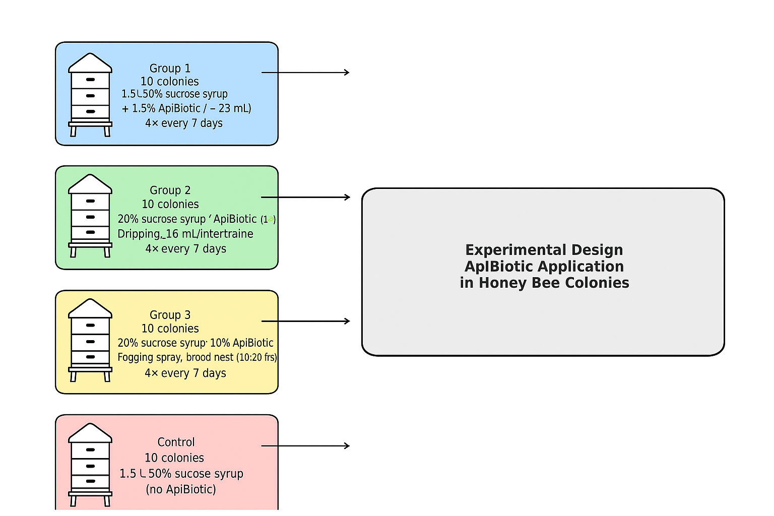
In 2022, using molecular biology techniques for the preliminary assessment of honey bee colony microbiomes, the research team transitioned in 2023 to classical microbiology approaches, employing culture-based methods with confirmation by MALDI-TOF/MS. The microbiological diagnostics focused on detecting Paenibacillus larvae in brood and honey samples. Species-level confirmation was performed using the Bruker MALDI Biotyper database. The study included honey bee colonies of equal strength and similar developmental stage, the bee colonies occupied 3.5, and less frequently 4, standard Wielkopolski hive bodies. The experiment was conducted during June–August 2023. At that time, results indicated that the microbiome of each honey bee colony within this apiary contained approximately 104 CFU/g of vegetative, live Paenibacillus larvae bacteria, primarily present in the microbiota of open brood at the larval stage. Having these data and recognizing the significant microbiological load in the honey bee colonies and the pressure exerted by P. larvae on the native microbiome, a decision was made, as a preventive measure, to administer the ApiBiotic preparation in order to assess its potential effect on the abundance of P. larvae in each tested hive. The study aimed to determine whether the supplement, in the form of a feed mixture, is capable of halting the further progression and proliferation of P. larvae bacteria and preventing the development of full-scale infection and bacterial overgrowth. The apiary was divided into three experimental groups. The first group (marked 1), consisting of 10 honey bee colonies, was fed four times at 7-day intervals with 1.5 L of 50% sucrose syrup supplemented with 1.5% v/v of the ApiBiotic preparation (approximately 23 mL). In the second group (marked 2), also comprising 10 colonies, a mixture was administered four times at 7-day intervals, containing 30% sucrose syrup mixed 1:1 by volume with the ApiBiotic preparation and applied by dripping 18 mL along the bee-occupied interframe spaces. The third experimental group (marked 3), likewise consisting of 10 colonies, received four applications at 7-day intervals in the form of a fogging spray covering all combs in the brood nest (usually 10–20 frames) on both sides (About 20 ml per comb), using a mixture of 20% sucrose syrup with 10% v/v ApiBiotic. The control group (marked 4) consisted of 10 honey bee colonies, which received 1.5 L of 50% sucrose syrup without any ApiBiotic supplementation, 4 times every 7 days (Figure 1). On the days of the preparation’s application, beekeeper inspectors conducted general inspections of the bee colonies and their hives, assessing potential disease symptoms as well as the overall condition of the colonies. After the experiment was completed, brood samples were collected twice from all experimental and control groups to perform microbiological diagnostics using a culture panel targeting P. larvae, with species confirmation by MALDI-TOF/MS proteomic analysis. Additionally, a third and final sampling was conducted 14 days after the conclusion of the research program to assess the ability of P. larvae to regenerate in the larval microbiome. The results, including the abundance of P. larvae, were compared between the period before the preventive application and immediately after the completion of the prophylaxis ApiBiotic application.
2.3. Working Methodology: Brood Samples Microbiological Analysis
Before the application of ApiBiotic and after the completion of the ApiBiotic feeding program, 1–3-day-old larvae were collected from open brood in each hive using sterile tweezers and sterile urine containers. Samples were taken from approximately 8–10 frames in the brood nest, with around 30–40 larvae per hive. The samples were cooled (4 ° C) and transported to the laboratory. No clinical signs of American foulbrood were observed in the hives; the brood remained healthy throughout the research program. Qualified beekeepers, who also served as sample collectors, strictly adhered to established sanitary rules to avoid contaminating the collected samples or transferring microorganisms from one hive to another. They wore special veterinary sanitary suits, sterile gloves, and footwear. Beekeeping tools (frame hooks and hive tongs) were disinfected with bleach (5% sodium hypochlorite) after inspecting each hive before beginning the examination of the next hive. Sterile, single-use tweezers and urine containers were used for sample collection, and gloves were changed regularly. Larvae samples from each hive were placed in sterile Schott bottles (250 cm³), weighed, and supplemented with sterile grinding beads and a small volume of sterile phosphate buffer (approximately 40 cm³; Argenta, Poznań, Poland). The mixtures were gently homogenized by shaking for 5 minutes to obtain a uniform, milky suspension. The suspension was then serially diluted using sterile physiological saline to reduce the number of accompanying bacteria. Microbiological inoculations were performed by surface plating 100 µL of the diluted suspension and spreading it with a microbiological spreader onto selective MYPGP agar supplemented with nalidixic acid (BTL, Łódź, Poland) to suppress unwanted microflora. Incubation was carried out under microaerophilic conditions using CampyGen atmosphere generators (Argenta, Poznań, Poland) at 35°C for 48–72 hours (Figure 2). Colonies that developed, appearing off-white and semi-matte, were randomly picked and subjected to MALDI TOF-MS analysis, performed through an external service. Identification was generally unambiguous, with colonies confirmed as Paenibacillus larvae, genotype Eric I. The number of colonies was expressed as CFU/g on a logarithmic scale. Additionally, samples were collected and plated to determine the number of lactic acid bacteria using MRS agar (Argenta, Poznań, Poland). The plates were incubated for 48–72 hours under anaerobic conditions at 38 °C. Sampling, culturing, and result interpretation were conducted 7 days prior to the start of ApiBiotic product testing and 3 days after the administration of the final, fourth dose (in 31 day). Additionally, a third and final sampling was conducted 14 days after the conclusion of the research program to assess the ability of P. larvae to regenerate in the larval microbiome (in 45 days). The detection limit was set at a log10 level of 1.7, which corresponded to a complete absence of grown colonies.
2.4. Data Analysis
ANOVA analysis and parametric tests were applied. In the experiment, four independent groups were formed (three experimental and one control). Measurements were repeated before and after application, and the numerical data were time-related. No preliminary logarithmic data transformation was performed. Therefore, a repeated measures ANOVA was applied using GraphPad Prism 10.5.0 (GraphPad Inc., San Diego, CA, USA) software. The dependent variable was log(CFU), while the factors were: experimental group (4 levels) and measurement time (before/after). This allowed us to determine whether the groups differ overall, whether CFU changed after application, and whether different groups responded differently to the preparation. Additionally, the delta of achieved cycle thresholds was calculated, and the analysis of differences concerned the state before and after the application of the preparation. The data were visualized using boxplots of log(CFU) for each group before and after treatment, as well as spaghetti plots in the form of lines connecting the before-and-after measurements for each bee colony and group individually. The purpose of the boxplot visualization was to show the distribution of the logarithmic number of P. larvae log(CFU) in each experimental and control group before and after the intervention with the preparation. The purpose of the spaghetti plot was to show changes in the logarithmic number log(CFU) of bacteria over time in each bee colony, where each line represents one colony, the y-axis represents log(CFU), and the x-axis represents time (before and after application). This illustrates the individual response of colonies to the preventive treatment, showing both the effect of the preparation (boxplot) and the individual differences between bee colonies (spaghetti plot). In the case of a small population, non-parametric tests would probably have been applied, but this was not necessary. Additionally, bar charts were used to present the increases in lactic acid bacteria numbers after the completed biosecurity treatments using ApiBiotic. Additionally, a parameter for the condition of the bee colony was introduced. In practice, this refers to the number of brood frames: very good (more than 10 Wielkopolski frames), good (7–10 Wielkopolski frames), and poor (fewer than 7 Wielkopolski frames).
3. Results
After four weeks of administering various formulations of the ApiBiotic preparation in different configurations and combinations, results demonstrated a strong suppressive effect of the tested pharmacobiotic on Paenibacillus larvae (Table 1, Figure 3). In most hives of the treatment group, P. larvae was undetectable in the native larval microbiome following the ApiBiotic-supplemented feeding program. Minor quantitative differences were observed depending on the route of administration. Across all experimental setups, a significant 2–3 log₁₀ reduction in bacterial load was recorded compared to the control group and baseline levels before application (Figure 4, Figure 5, Figure 6, Figure 7). It was observed that the number of lactic acid bacteria increased by 6–7 logarithmic cycles (Figure 8, Figure 9, Figure 10., Figure 11). These findings indicate a potent preventive effect, inhibiting the proliferation of P. larvae within the bee colony microbiome and thereby preventing the onset of clinical symptoms. At the end of the 4-week feeding period, no negative effects of the preparation on the bee colonies were observed in the context of colony management or bees’ behaviour. Throughout the study period and after its conclusion, no symptoms of American foulbrood (AFB) were observed, including in the control group. The detection limit was set at a log10 level of 1.7, which corresponded to a complete absence of grown colonies.
3.1. Number of Bee Colonies Released from Microbiological Risk and P. larvae Pressure After the Application of ApiBiotic
Bee colonies were identified in which the risk of P. larvae proliferation was successfully reduced, thereby preventing the potential development of infection. No differences were observed in the mechanism or efficacy of the preventive effect between test groups 1–3 or the methods of ApiBiotic application. In all test groups, it was established that from the beginning to the end of the experiment, the abundance of initially detected P. larvae cells was reduced by 2–3 orders of magnitude. Following an additional culture performed 14 days after the conclusion of the testing program, no bee colonies were identified in which P. larvae abundance had significantly recovered. The data were not included in tables or figures, as they did not contribute relevant information to the study. Throughout the study period, no deterioration in the health, overall condition, behavior, or occurrence of AFB symptoms was observed in the test group colonies. Exceptions were two cases in the control group, involving queen loss and increased defensiveness. Bee colonies from group 4 (control), in which P. larvae abundance remained constant or increased, were subjected to further observation. The apiary owner was instructed that in the event of clinical symptoms in the brood, they should contact the district veterinary officer to conduct official testing under the administrative procedure (Table 1). The results were interpreted following the analysis of the raw data provided in Section 3.2 (3.2; Figure 3).
3.2. Changes in P. larvae Abundance in Groups 1-4 Without Subsequent Suspicion of American Foulbrood Infection
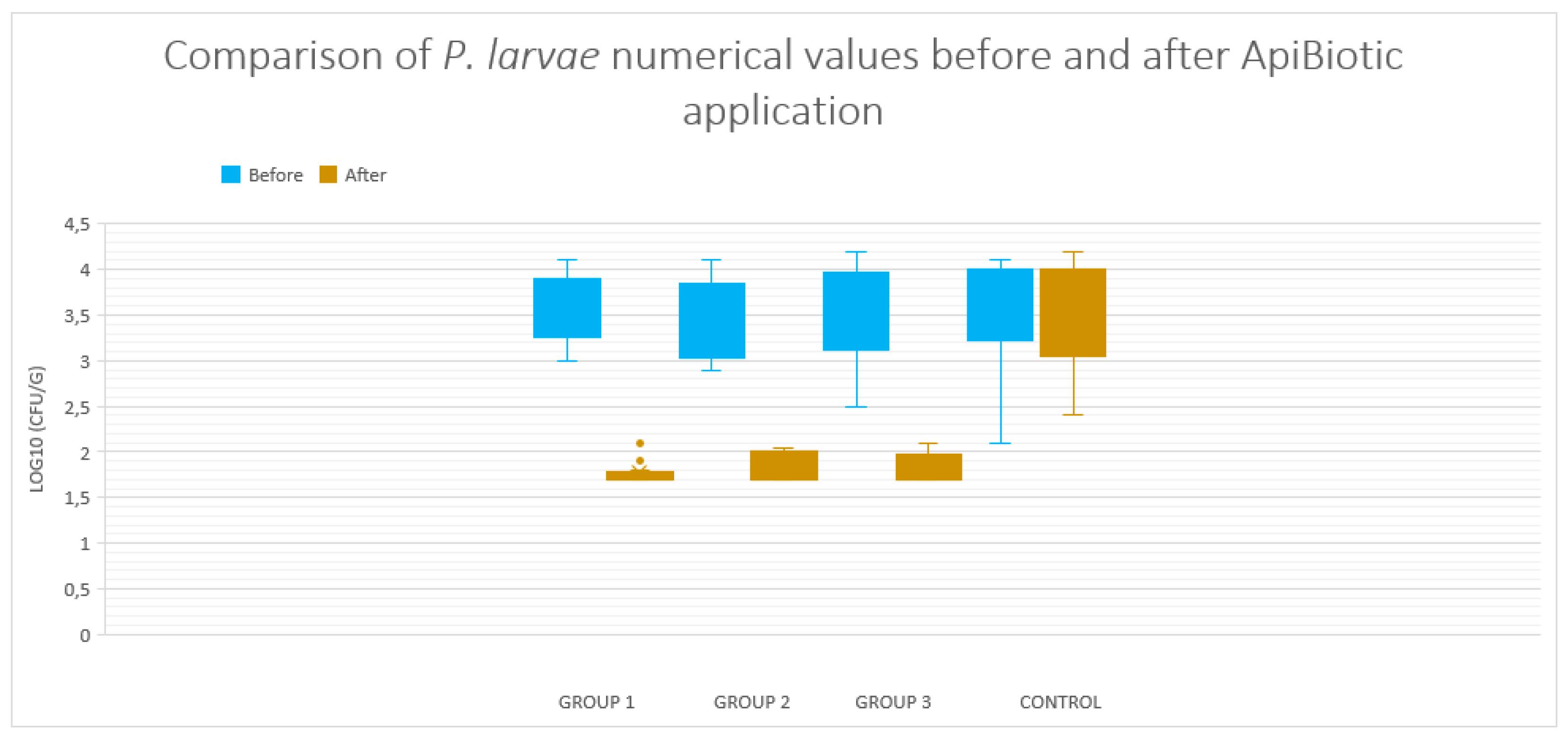
The detection limit was set at a log10 level of 1.7, which corresponded to a complete absence of grown colonies. After the completion of the 4-week application of the metabolic preparation ApiBiotic, the logarithmic growth of P. larvae cells was successfully halted, and the initial abundance was reduced by up to 3 logarithmic cycles. This effect persists even after the preventive treatments have ended. This provides evidence that ApiBiotic is highly suitable for biosecurity purposes and can serve as a tool for the preventive management of the microbiome in bee colonies. No significant differences were observed between the routes of administration: via feed (group 1), by dripping into the spaces between frames in the hive (group 2), or by spraying brood combs (group 3). The efficacy of the preparation contrasts with the control group, in which the P. larvae population remained unchanged, at the same level as before the application of the preparation (Figure 3).
Figure 3.
Comparison of P. larvae numerical values before and after ApiBiotic application between experimental groups using a boxplot.
Figure 3.
Comparison of P. larvae numerical values before and after ApiBiotic application between experimental groups using a boxplot.
3.3. Individual Responses of Microbiomes of Brood with Changes in log10(CFU) of P. larvae in Honey Bee Colonies Before and After ApiBiotic Application in 1-4 Groups
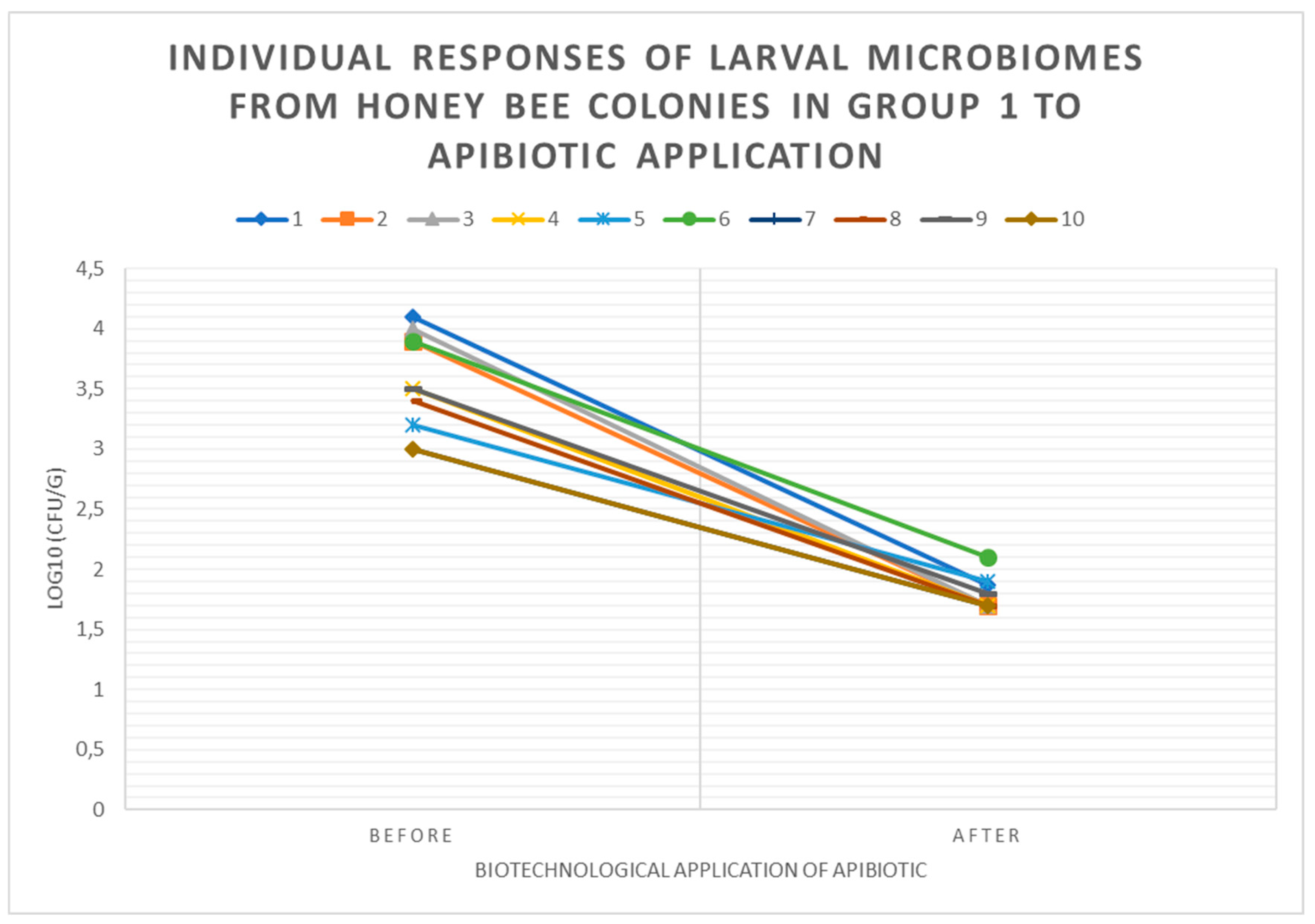
In group 1, at the level of individual bee colonies, a drastic decrease in the P. larvae population was observed after the application of the metabolic preparation. The exact timing of the logarithmic reduction (after which day or week) is unknown, as the methodology included microbiological diagnostics at only two time points. It is certain that after 4 weeks of repeated feeding treatments of the bee colonies with 1.5 L of 50% sucrose syrup supplemented with 1.5% v/v of the ApiBiotic preparation (approximately 23 mL), further growth of P. larvae was successfully halted, preventing bacteremia and infection (Figure 4). Following an additional culture performed 14 days after the conclusion of the testing program, no bee colonies were identified in which P. larvae abundance had significantly recovered. The data were not included in tables or figures, as they did not contribute relevant information to the study.
Figure 4.
Individual responses of larval microbiomes from honey bee colonies in group 1 to ApiBiotic application using spaghetti plot.
Figure 4.
Individual responses of larval microbiomes from honey bee colonies in group 1 to ApiBiotic application using spaghetti plot.
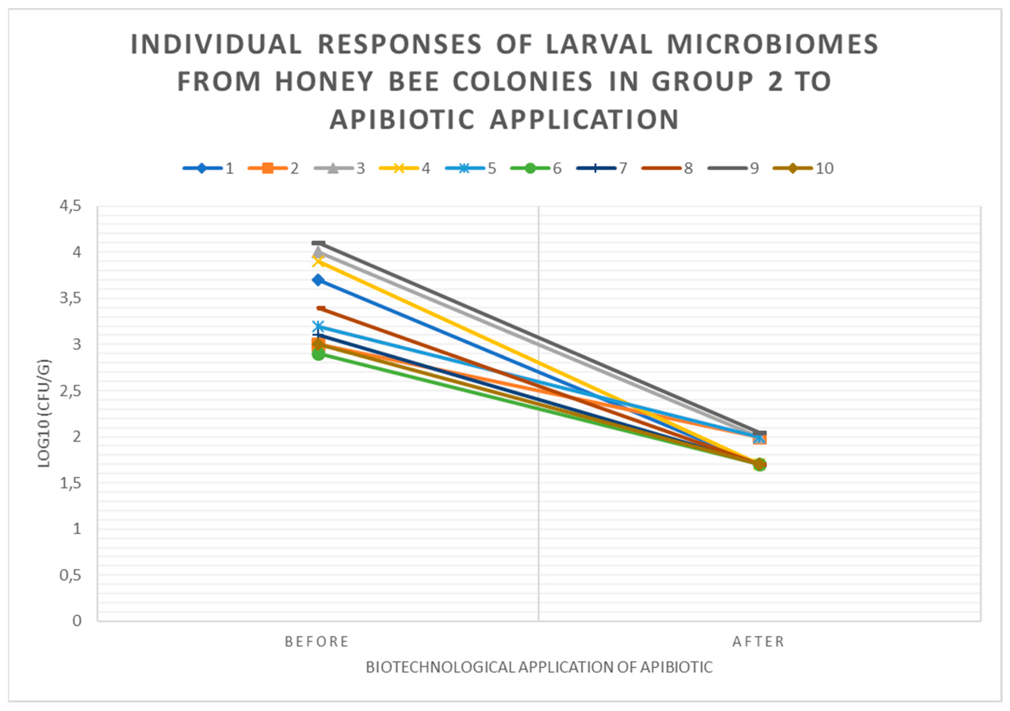
In group 2, at the level of individual bee colonies, a drastic decrease in the P. larvae population was observed after the application of the metabolic preparation. The exact timing of the logarithmic reduction by 1–3 log cycles is unknown, as the methodology included microbiological diagnostics at only two time points. It is certain that after 4 weeks of repeated treatment with a mixture containing 30% sucrose syrup mixed 1:1 by volume with the ApiBiotic preparation and applied by dripping 18 mL along the bee-occupied interframe spaces, further growth of P. larvae was successfully halted, preventing bacteremia and infection (Figure 5). Following an additional culture performed 14 days after the conclusion of the testing program, no bee colonies were identified in which P. larvae abundance had significantly recovered. The data were not included in tables or figures, as they did not contribute relevant information to the study.
Figure 5.
Individual responses of larval microbiomes from honey bee colonies in group 2 to ApiBiotic application using spaghetti plot.
Figure 5.
Individual responses of larval microbiomes from honey bee colonies in group 2 to ApiBiotic application using spaghetti plot.
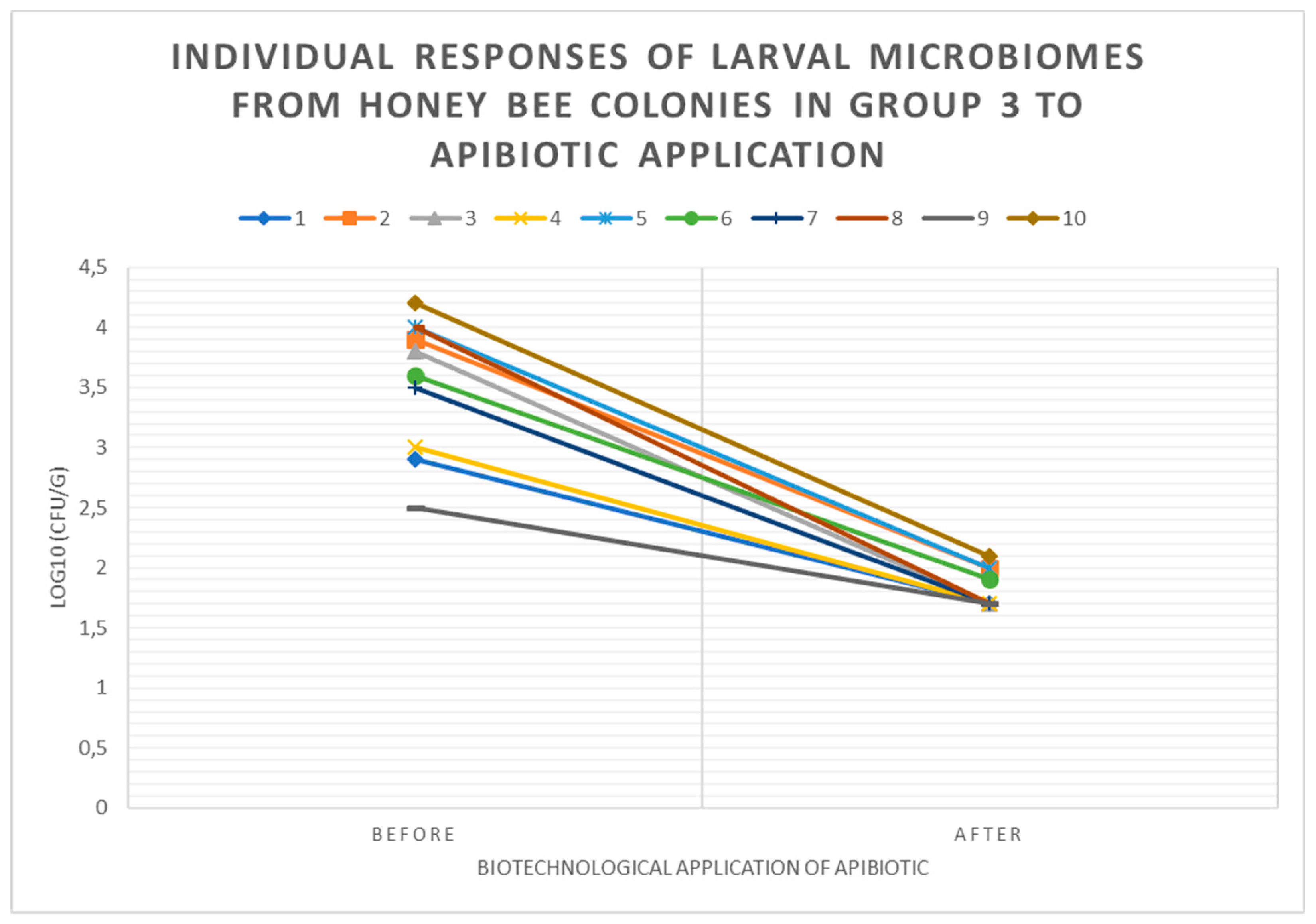
In group 3, at the level of individual bee colonies, a drastic decrease in the P. larvae population was observed after the application of the metabolic preparation. Similar to groups 1 and 2, a rapid declining trend was maintained. The exact timing of the logarithmic reduction by 1–3 log cycles is unknown, as the methodology included microbiological diagnostics at only two time points. It is certain that after 4 weeks of repeated treatment in the form of a fogging spray covering all combs in the brood nest (usually 10–20 frames) on both sides (about 20 mL per comb), using a mixture of 20% sucrose syrup with 10% v/v ApiBiotic, further growth of P. larvae was successfully halted, preventing bacteremia and infection (Figure 6). Following an additional culture performed 14 days after the conclusion of the testing program, no bee colonies were identified in which P. larvae abundance had significantly recovered. The data were not included in tables or figures, as they did not provide relevant information to the study.
Figure 6.
Individual responses of larval microbiomes from honey bee colonies in group 3 to ApiBiotic application using spaghetti plot.
Figure 6.
Individual responses of larval microbiomes from honey bee colonies in group 3 to ApiBiotic application using spaghetti plot.
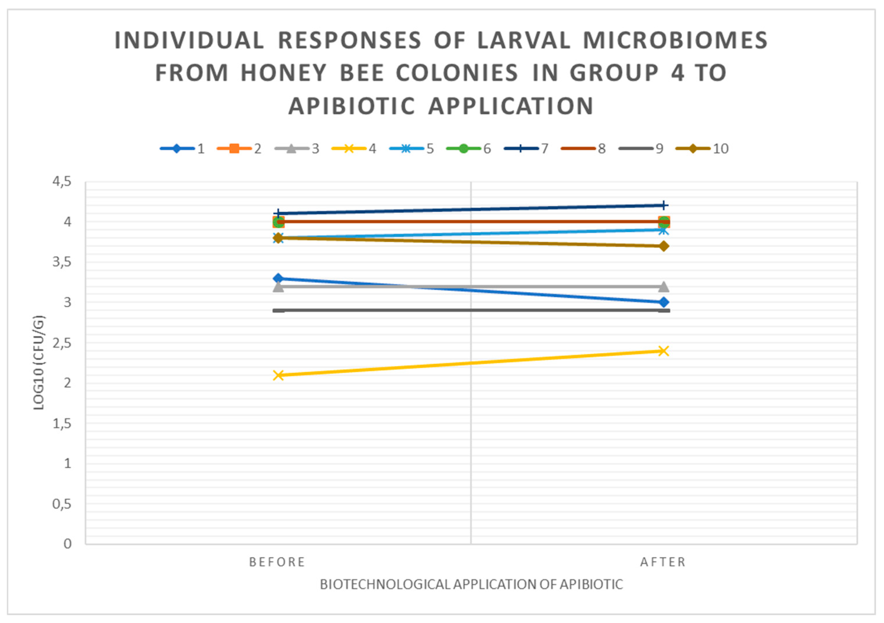
In the control group 4, which did not receive any application of the preparation, no significant changes in P. larvae abundance were observed. In two individual bee colonies, a slight, minor increase in P. larvae numbers was noted, which may indicate a progressing process toward bacterial overgrowth (Figure 7).
Figure 7.
Individual responses of larval microbiomes from honey bee colonies in group 4 without ApiBiotic application using spaghetti plot.
Figure 7.
Individual responses of larval microbiomes from honey bee colonies in group 4 without ApiBiotic application using spaghetti plot.
3.4. Individual Responses of Microbiomes Brood: Changes in log10(CFU) of LAB in Honey Bee Colonies Before and After ApiBiotic Application
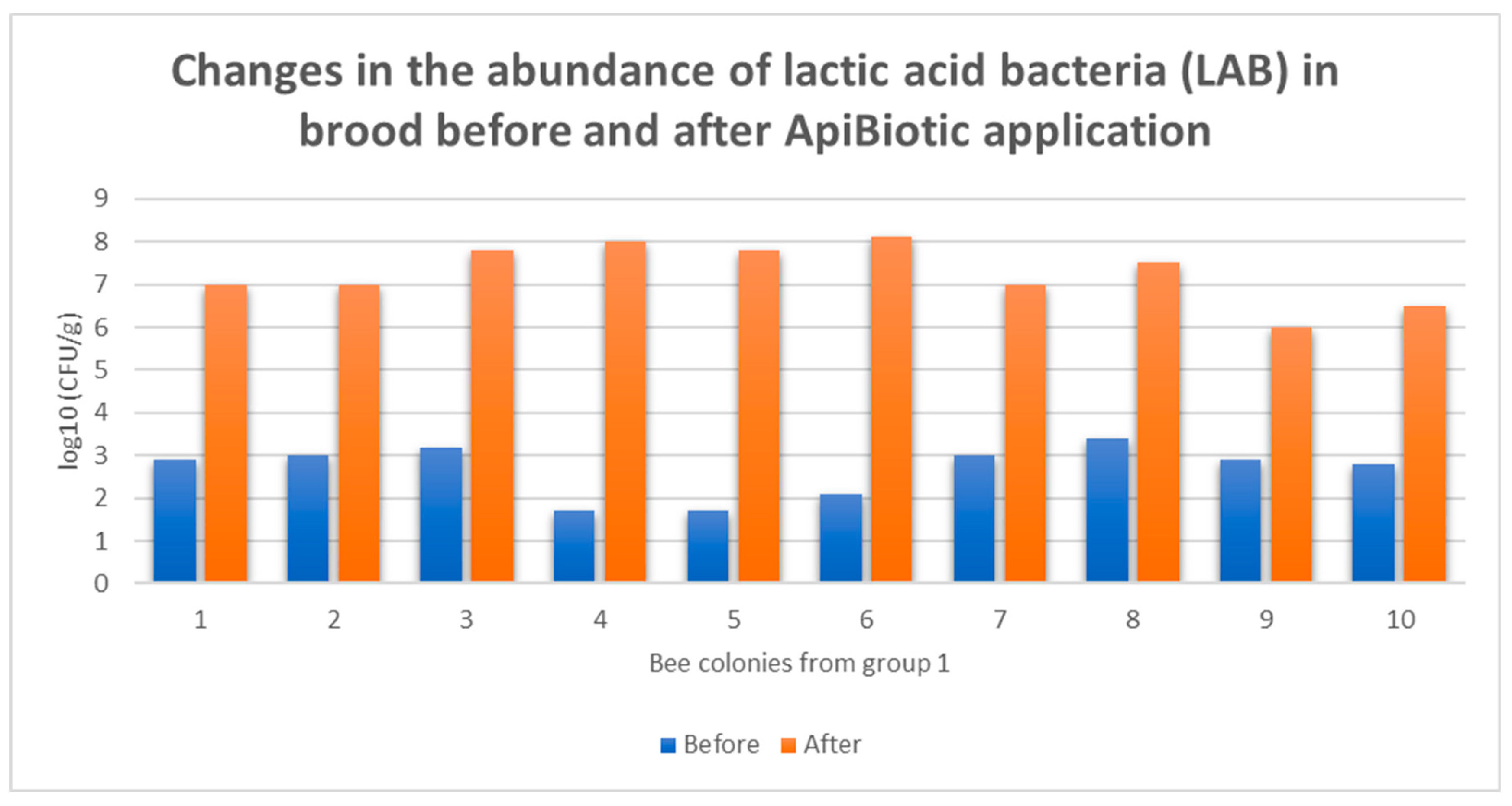
In group 1, after the completion of four rounds of feeding the bee colonies with 1.5 L of 50% sucrose syrup supplemented with 1.5% v/v of the ApiBiotic preparation (approximately 23 mL), a statistically significant increase in lactic acid bacteria was observed, reaching even 4–6 logarithmic cycles. This demonstrates the extent to which the bee microbiome had been depleted. Beneficial microorganisms, specifically lactic acid bacteria from the preparation, successfully colonized the hive environments in the studied colonies, leading to the displacement and decolonization of P. larvae from this microbiota (Figure 8).
Figure 8.
Changes in the abudance of lactic acid bacteria (LAB) in brood before and after ApiBiotic application in group 1.
Figure 8.
Changes in the abudance of lactic acid bacteria (LAB) in brood before and after ApiBiotic application in group 1.
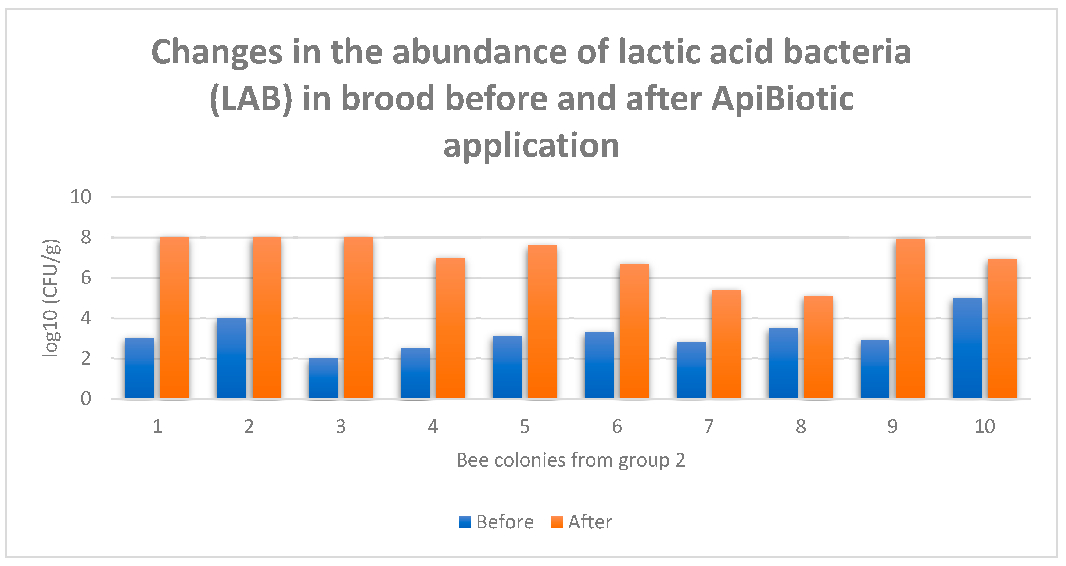
In group 2, after the completion of four rounds of treatment with a mixture containing 30% sucrose syrup mixed 1:1 by volume with the ApiBiotic preparation and applied by dripping 18 mL along the bee-occupied interframe spaces, a statistically significant increase in lactic acid bacteria was observed, reaching up to 6 logarithmic cycles (in bee colony 3), which demonstrates the extent to which the bee microbiome had been depleted. The smallest increase in lactic acid bacteria occurred in colony 8, where an increase of 1.5 logarithmic cycles was recorded. Beneficial microorganisms, specifically lactic acid bacteria from the preparation, successfully colonized the hive environments in the studied colonies, leading to the displacement and decolonization of P. larvae from this microbiota (Figure 9).
Figure 9.
Changes in the abundance of lactic acid bacteria (LAB) in brood before and after ApiBiotic application in group 2.
Figure 9.
Changes in the abundance of lactic acid bacteria (LAB) in brood before and after ApiBiotic application in group 2.
In group 3, after the completion of four rounds of treatment in the form of a fogging spray covering all combs in the brood nest (usually 10–20 frames) on both sides (about 20 mL per comb), using a mixture of 20% sucrose syrup with 10% v/v ApiBiotic, a statistically significant increase in lactic acid bacteria was observed, reaching over 6 logarithmic cycles (in bee colonies 5 and 6), demonstrating the extent to which the bee microbiome had been depleted and unoccupied by beneficial microorganisms. The smallest increase in lactic acid bacteria occurred in colony 10, where an increase of just under 2 logarithmic cycles was recorded. Beneficial microorganisms, specifically lactic acid bacteria from the preparation, successfully colonized the hive environments in the studied colonies, leading to the displacement and decolonization of P. larvae from this microbiota (Figure 10). Similar to the bee colonies in groups 1 and 2, it was possible to restructure the microbiota in the treated hives, inducing antagonistic interactions and stimulating competition between beneficial and pathogenic microorganisms (Figure 8, Figure 9., Figure 10).
Figure 10.
Changes in the abudance of lactic acid bacteria (LAB) in brood before and after ApiBiotic application in group 3.
Figure 10.
Changes in the abudance of lactic acid bacteria (LAB) in brood before and after ApiBiotic application in group 3.

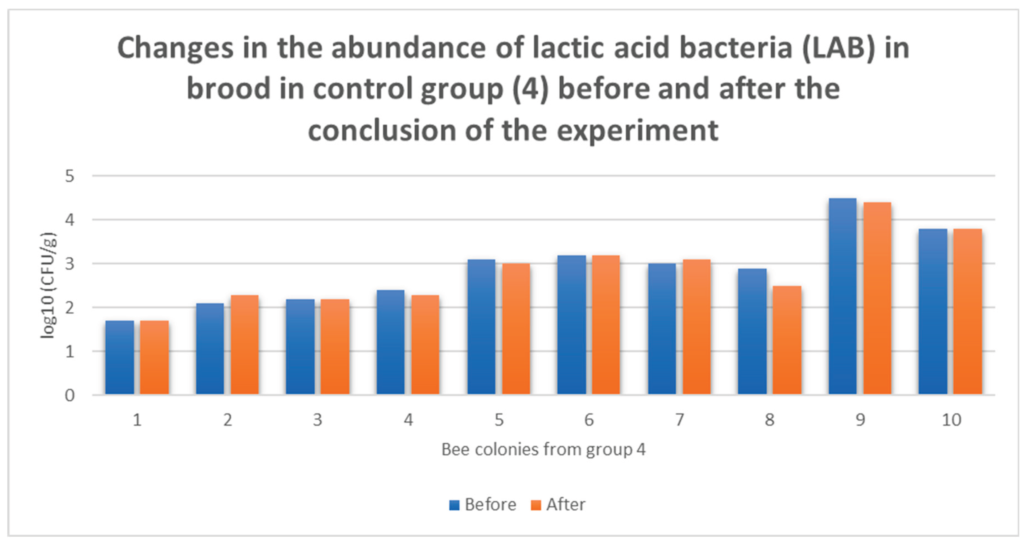
In the control group, no significant changes in lactic acid bacteria abundance were observed. Minor increases or decreases were noted, but these were not relevant to the conducted assay protocol (Figure 11).
Figure 11.
Changes in the abudance of lactic acid bacteria (LAB) in brood without ApiBiotic application in group 4.
Figure 11.
Changes in the abudance of lactic acid bacteria (LAB) in brood without ApiBiotic application in group 4.
4. Discussion
Studies have shown that ApiBiotic, as proposed in the composition described in this work (Section 2.1), is an excellent feed mixture with metabiotic properties, suitable for preventive use in bee colonies in three forms of administration, supporting biological processes that prevent excessive proliferation of P. larvae cells. The product, with an ecological pharmacobiotic profile, exhibits both bacteriostatic and bactericidal activity under simulated conditions approximating real operational in vivo environments (Technology Readiness Levels TRL VI–VIII). ApiBiotic, which leverages the synergistic action of probiotic bacteria and plant-herbal bioferments, can also be classified as a synbiotic. It can be administered to hives as a supplement to feed in the form of a concentrated 50% or 60% sucrose solution, by dripping into interframe spaces combined with a 30% sucrose solution, or by spraying/fogging brood combs with an open brood using a mixture containing 20% sucrose solution supplemented with 10% v/v ApiBiotic. All three described routes of administration produce a preventive effect and reduce the pressure of the P. larvae pathogen on the physiological, native hive microbiome, thereby blocking further bacterial proliferation (Figure 3 – Figure 10, Table 1). The preparation can also be used as an additive to winter feed, protecting the microbiome of bee colonies against potential disturbances of its homeostasis. Such preventive treatments, which support natural biological processes, align well with the principles of Evidence-Based Medicine (EBM) and Evidence-Based Practices (EBP). Evidence-based medicine (EBM) is medicine, including veterinary medicine, that is based on facts or scientific evidence. It is a clinical practice approach that relies on the best available, reliable, and up-to-date scientific evidence, integrating it with clinical knowledge as well as patient preferences and values to make the best diagnostic and therapeutic decisions. This approach assumes the use of research results that demonstrate the efficacy and safety of therapies, as well as reliable data analyses [28]. Evidence-based practice (EBP) is a set of practices based on evidence or scientific data. The term refers to an approach that integrates the best available scientific evidence with clinical knowledge and patient preferences to make informed and effective decisions across various fields, such as medicine, psychology, or physiotherapy. The foundation of this practice is the use of reliable and current research findings to guide decision-making [29]. It is also worth mentioning a third approach that has potential in beekeeping and apicultural practices, namely Evidence-Based Management (EBMgt). This is management based on evidence, an approach in organizational or strategic management that relies on making decisions based on the best available evidence, analogous to evidence-based medicine (EBM). The main feature of EBMgt is the use of the best scientific evidence so that managerial decisions are based on current, reliable research and data analyses, such as reports, scientific articles, and meta-analyses. Another important feature is the integration of practical experience, meaning that the experience of managers and expert knowledge should also be taken into account when making decisions [30,31]. ApiBiotic perfectly aligns with the concept of biosafety, as it acts preventively by limiting the growth of pathogenic P. larvae bacteria in the hive while simultaneously supporting the natural microbiome of bees. The preparation utilizes the synergistic action of probiotic bacteria and plant-herbal bioferments, enabling effective colonization of the hive environment by beneficial microorganisms, leading to the decolonization of pathogens. As a result, it reduces the risk of infection, maintains the microbiological balance in the hive, and strengthens the natural defense mechanisms of bee colonies, fully adhering to the principles of biosafety based on prevention and pathogen control [32,33,34]. The metaphylaxis discussed in this paper should go hand in hand with biosafety measures, and in the authors’ view, this combination should form the core preventive strategy for controlling infections caused by Paenibacillus larvae.
Author Contributions
Conceptualization, P.N. and P.M.; methodology, P.N., P.M. and P. C.; software, P.N.; validation, P.N., D.K..; formal analysis, P.N., P.M.; investigation, P.N., D.K. and P.C.; resources, P.M.; data curation, P.N., D.K.; writing—original draft preparation, P.N.; writing—review and editing, P.N., P.M.; visualization, P.N.; supervision, P.C.; project administration, P.N, P.C.; funding acquisition, P.C. All authors have read and agreed to the published version of the manuscript.
Funding
This research was funded by Polish Ministry of Science and Higher Education, grant number N0CBR000.7117.UWD.6/CBR/2021
Institutional Review Board Statement
Not applicable
Informed Consent Statement
Not applicable
Data Availability Statement
The original contributions presented in this study are included in the article. Further inquiries can be directed to the corresponding author(s).
Acknowledgments
We thank the laboratory specialists at the Institute of Microbiological Technologies in Turek for excellent technical assistance.
Conflicts of Interest
The authors declare no conflicts of interest
Abbreviations
The following abbreviations are used in this manuscript:
| AFB | American Foulbrood |
| OTC | Oxytetracycline hydrochloride |
| UK | United Kingdom |
| MRL | Maximum Residue Limit |
| EU | European Union |
| UV | Ultraviolet |
| CFU | Colony Forming Unit |
| CLSI | Clinical and Laboratory Standards Institute |
| EUCAST | European Committee on Antimicrobial Susceptibility Testing |
| MIC | Minimum Inhibitory Concentration |
| MBC | Minimum Bactericidal Concentration |
| TRL | Technology Readiness Level |
| LAB | Lactic Acid Bacteria |
| CT-46 | Line of bees belonging to Apis mellifera carnica |
| EBM | Evidence-Based Medicine |
| EBP | Evidence-Based Practice |
| EMBgt | Evidence-Based Management |
References
- Rosa Fuselli, S.; Gimenez Martinez, P.; Fuentes, G.; María Alonso-Salces, R.; Maggi, M. Prevention and Control of American Foulbrood in South America with Essential Oils: Review. In Beekeeping - New Challenges; Eduardo Rebolledo Ranz, R., Ed.; IntechOpen, 2020; ISBN 9781838804671 9781838804763.
- Mosca, M.; Gyorffy, A.; Milito, M.; Di Ruggiero, C.; De Carolis, A.; Pietropaoli, M.; Giannetti, L.; Necci, F.; Marini, F.; Smedile, D.; et al. Antibiotic Use in Beekeeping: Implications for Health and Environment from a One-Health Perspective. Antibiotics 2025, 14, 359. [Google Scholar] [CrossRef]
- Joshi, N.K.; Ngugi, H.K.; Biddinger, D.J. Bee Vectoring: Development of the Japanese Orchard Bee as a Targeted Delivery System of Biological Control Agents for Fire Blight Management. Pathogens 2020, 9, 41. [Google Scholar] [CrossRef] [PubMed]
- Bolognesi, M.; Zuniga, A.I.; Cordova, L.G.; Mallinger, R.; Peres, N.A. Bee Vectoring Technology Using Clonostachys Rosea as a Biological Control for Botrytis Fruit Rot of Strawberry in Florida. Plant Health Progress 2024, 25, 179–184. [Google Scholar] [CrossRef]
- Griescom, D.C. Bee Vectoring Technologies: A Revolutionary Approach to Sustainable Crop Protection. Advances in Crop Science and Technology 2025, 13, 1–2. [Google Scholar] [CrossRef]
- Mejias, E. American Foulbrood and the Risk in the Use of Antibiotics as a Treatment. In Modern Beekeeping - Bases for Sustainable Production; Eduardo Rebolledo Ranz, R., Ed.; IntechOpen, 2020. ISBN 9781838801557 9781838801564.
- Bulson, L.; Becher, M.A.; McKinley, T.J.; Wilfert, L. Long-term Effects of Antibiotic Treatments on Honeybee Colony Fitness: A Modelling Approach. J Appl Ecol 2021, 58, 70–79. [Google Scholar] [CrossRef]
- Maggi, M.; Antúnez, K.; Invernizzi, C.; Aldea, P.; Vargas, M.; Negri, P.; Brasesco, C.; De Jong, D.; Message, D.; Teixeira, E.W.; et al. Honeybee Health in South America. Apidologie 2016, 47, 835–854. [Google Scholar] [CrossRef]
- Masood, F.; Thebeau, J.M.; Cloet, A.; Kozii, I.V.; Zabrodski, M.W.; Biganski, S.; Liang, J.; Marta Guarna, M.; Simko, E.; Ruzzini, A.; et al. Evaluating Approved and Alternative Treatments against an Oxytetracycline-Resistant Bacterium Responsible for European Foulbrood Disease in Honey Bees. Sci Rep 2022, 12, 5906. [Google Scholar] [CrossRef]
- European Food Safety Authority Report for 2019 on the Results from the Monitoring of Veterinary Medicinal Product Residues and Other Substances in Live Animals and Animal Products. EFS3 2021, 18. [CrossRef]
- Kędzierska-Matysek, M.; Teter, A.; Skałecki, P.; Topyła, B.; Domaradzki, P.; Poleszak, E.; Florek, M. Residues of Pesticides and Heavy Metals in Polish Varietal Honey. Foods 2022, 11, 2362. [Google Scholar] [CrossRef] [PubMed]
- Eissa, F.; Taha, E.-K.A. Contaminants in Honey: An Analysis of EU RASFF Notifications from 2002 to 2022. J Consum Prot Food Saf 2023, 18, 393–402. [Google Scholar] [CrossRef]
- Bee Research Institute Dol. Hygiene in the Apiary: A Manual for Hygienic Beekeeping; BeeShop Project, 6th Framework Programme of the EU: Dol, Czech Republic, 2006. Bee Research Institute Dol. Hygiene in the Apiary: A Manual for Hygienic Beekeeping; BeeShop Project, 6th Framework Programme of the EU: Dol, Czech Republic, 2006.
- Vandegrift, R.; Bateman, A.C.; Siemens, K.N.; Nguyen, M.; Wilson, H.E.; Green, J.L.; Van Den Wymelenberg, K.G.; Hickey, R.J. Cleanliness in Context: Reconciling Hygiene with a Modern Microbial Perspective. Microbiome 2017, 5, 76. [Google Scholar] [CrossRef]
- Dai, Z.; Sevillano-Rivera, M.C.; Calus, S.T.; Bautista-de los Santos, Q.M.; Eren, A.M.; van der Wielen, P.W.J.J.; Ijaz, U.Z.; Pinto, A.J. Disinfection Exhibits Systematic Impacts on the Drinking Water Microbiome. Microbiome 2020, 8, 42. [Google Scholar] [CrossRef]
- Mäklin, T.; Thorpe, H.A.; Pöntinen, A.K.; Gladstone, R.A.; Shao, Y.; Pesonen, M.; McNally, A.; Johnsen, P.J.; Samuelsen, Ø.; Lawley, T.D.; et al. Strong Pathogen Competition in Neonatal Gut Colonisation. Nat Commun 2022, 13, 7417. [Google Scholar] [CrossRef]
- Woelfel, S.; Silva, M.S.; Stecher, B. Intestinal Colonization Resistance in the Context of Environmental, Host, and Microbial Determinants. Cell Host & Microbe 2024, 32, 820–836. [Google Scholar] [CrossRef]
- Xu, W.-B.; Wang, Y.-F.; Meng, S.-Y.; Zhang, X.-T.; Wang, Y.-R.; Liu, Z.-Y. Effects of Antibiotic and Disinfectant Exposure on the Mouse Gut Microbiome and Immune Function. Microbiol Spectr 2024, 12, e00611–24. [Google Scholar] [CrossRef]
- Mayer, T.; Teutloff, E.; Unger, K.; Lehenberger, P.; Agler, M.T. Deterministic Colonization Arises Early during the Transition of Soil Bacteria to the Phyllosphere and Is Shaped by Plant–Microbe Interactions. Microbiome 2025, 13, 102. [Google Scholar] [CrossRef] [PubMed]
- Grießhammer, A.; de la Cuesta-Zuluaga, J.; Müller, P.; Gekeler, C.; Homolak, J.; Chang, H.; Schmitt, K.; Planker, C.; Schmidtchen, V.; Gallage, S.; et al. Non-Antibiotics Disrupt Colonization Resistance against Enteropathogens. Nature 2025, 644, 497–505. [Google Scholar] [CrossRef] [PubMed]
- ISSUU Controlling American Foulbrood without Antibiotics. Available online: https://issuu.com/beesfd/docs/91_bfdj_jun2009/s/14114781 (accessed on 4 September 2025).
- Hristov, Y.V.; Allsopp, M.H.; Wossler, T.C. Investigating Hygienic Behaviour and AFB Resistance of Apis Mellifera Capensis Colonies: Are Cape Honey Bees Hygienic and How Well Do They Cope with the Disease? Apidologie 2025, 56, 73. [Google Scholar] [CrossRef]
- Dickel, F.; Bos, N.M.P.; Hughes, H.; Martín-Hernández, R.; Higes, M.; Kleiser, A.; Freitak, D. The Oral Vaccination with Paenibacillus Larvae Bacterin Can Decrease Susceptibility to American Foulbrood Infection in Honey Bees—A Safety and Efficacy Study. Front. Vet. Sci. 2022, 9. [Google Scholar] [CrossRef] [PubMed]
- Callens, B.; Persoons, D.; Maes, D.; Laanen, M.; Postma, M.; Boyen, F.; Haesebrouck, F.; Butaye, P.; Catry, B.; Dewulf, J. Prophylactic and Metaphylactic Antimicrobial Use in Belgian Fattening Pig Herds. Preventive Veterinary Medicine 2012, 106, 53–62. [Google Scholar] [CrossRef]
- Maples, W.E.; Brorsen, B.W.; Peel, D.; Hicks, B. Observational Study of the Effect of Metaphylaxis Treatment on Feedlot Cattle Productivity and Health. Front. Vet. Sci. 2022, 9, 947585. [Google Scholar] [CrossRef]
- Brown, A.; Rodriguez, V.; Pfister, J.; Perreten, V.; Neumann, P.; Retschnig, G. The Dose Makes the Poison: Feeding of Antibiotic-Treated Winter Honey Bees, Apis Mellifera, with Probiotics and b-Vitamins. Apidologie 2022, 53, 19. [Google Scholar] [CrossRef]
- Credille, B.; Berghaus, R.D.; Jane Miller, E.; Credille, A.; Schrag, N.F.D.; Naikare, H. Antimicrobial Metaphylaxis and Its Impact on Health, Performance, Antimicrobial Resistance, and Contextual Antimicrobial Use in High-Risk Beef Stocker Calves. Journal of Animal Science 2024, 102, skad417. [Google Scholar] [CrossRef] [PubMed]
- Siwek, J. Evidence-Based Medicine: Common Misconceptions, Barriers, and Practical Solutions. afp 2018, 98, 343–344. [Google Scholar]
- Gudeta, T.G.; Terefe, A.B.; Mengistu, G.T.; Sori, S.A. A Systematic Review and Meta-Analysis of Evidence-Based Practice and Its Associated Factors among Health Professionals in Ethiopia. BMC Health Serv Res 2024, 24, 1518. [Google Scholar] [CrossRef]
- Barends, E., Rousseau, D. M., & Briner, R. B. Evidence-Based Management: The Basic Principles; 2014. ISBN 9789462285057.
- Sohrabi, Z.; Zarghi, N. Evidence-Based Management: An Overview. Creative Education 2015, 6, 1776–1781. [Google Scholar] [CrossRef]
- Gillespie, J.R. The Underlying Interrelated Issues of Biosecurity. javma 2000, 216, 662–664. [Google Scholar] [CrossRef]
- WOAH Terrestrial Manual 2018 Biosafety and biosecurity: standard for managing biological risk in the veterinary laboratory and animal facilities. in; 2018.
- Beeckman, D.S.A.; Rüdelsheim, P. Biosafety and Biosecurity in Containment: A Regulatory Overview. Front. Bioeng. Biotechnol. 2020, 8. [Google Scholar] [CrossRef]
Figure 1.
Methodology of ApiBiotic administration.
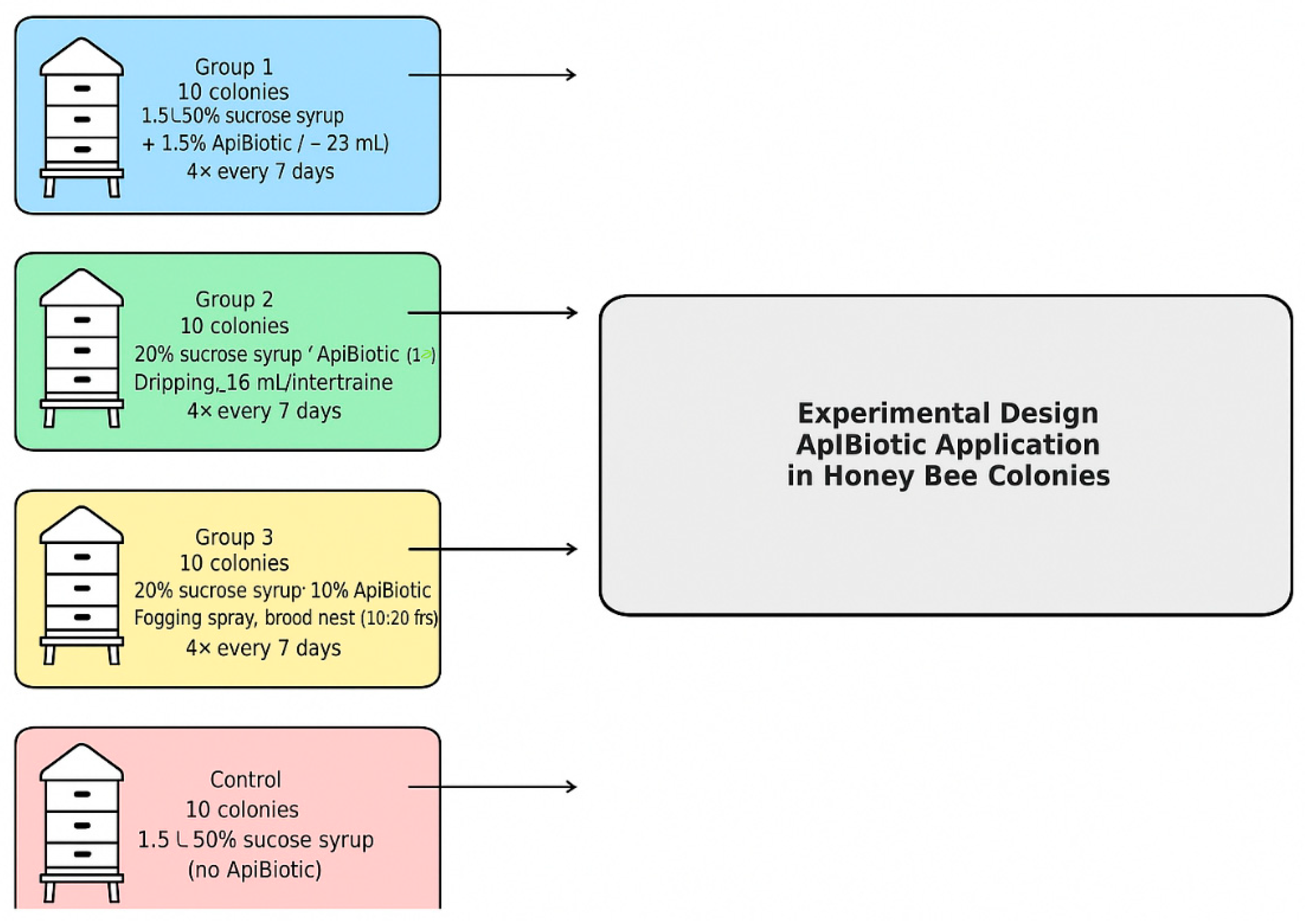
Figure 2.
Stages of performing a microbiological culture.
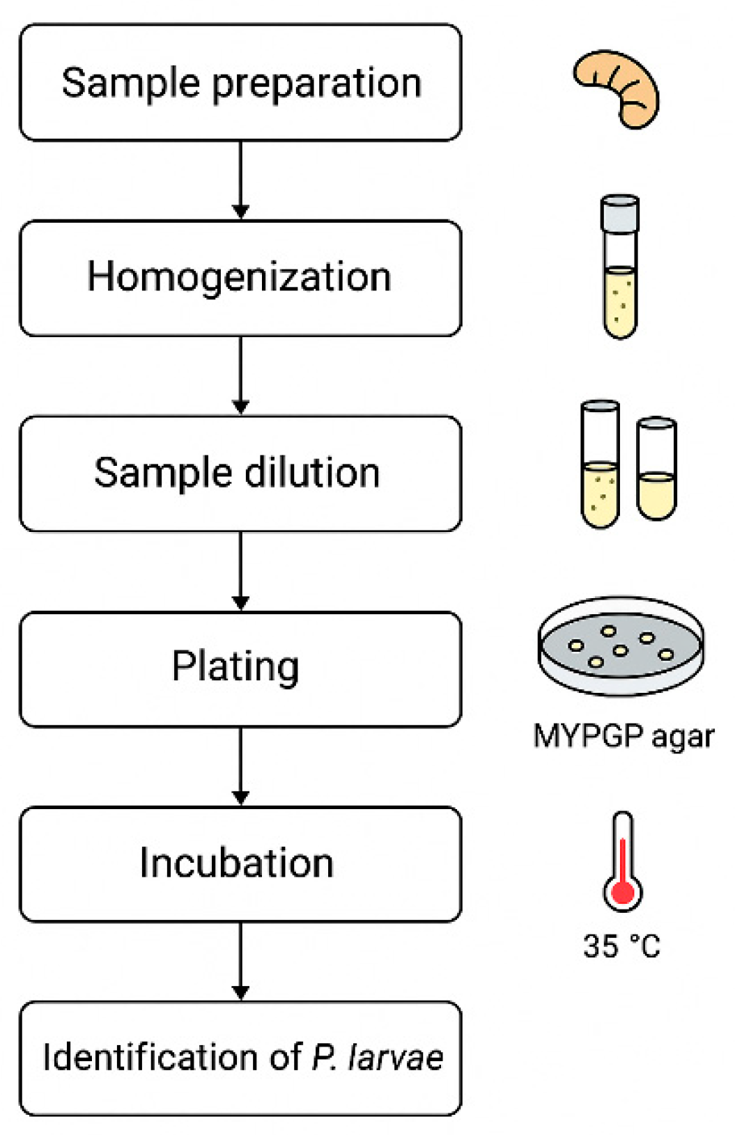

Table 1.
Number of bee colonies with varying levels of microbiological risk from P. larvae.
| Group | Overall condition of the bee colony | Total number of colonies | Number of bee colonies at microbiological risk | Number of bee colonies with deterioration of health, condition, or behaviour, and the appearance of clinical symptoms during the course of the experiment | Number of bee colonies with deterioration of health, condition, or behaviour, and the appearance of clinical symptoms after the experiment | Number of bee colonies rescued from microbiological risk (reduction and suppression of P. larvae in the microbiome |
The number of bee colonies in which microbiological risk was re-identified 14 days after the conclusion of the research program. |
|---|---|---|---|---|---|---|---|
| 1 | Very good | 9 | 9 | 0 | 0 | 9 | 0 |
| Good | 1 | 1 | 0 | 0 | 1 | 0 | |
| Poor | 0 | 0 | 0 | 0 | 0 | 0 | |
| 2 | Very good | 10 | 10 | 0 | 0 | 10 | 0 |
| Good | 0 | 0 | 0 | 0 | 0 | 0 | |
| Poor | 0 | 0 | 0 | 0 | 0 | 1 | |
| 3 | Very good | 8 | 8 | 0 | 0 | 8 | 0 |
| Good | 2 | 2 | 0 | 0 | 2 | 0 | |
| Poor | 0 | 0 | 0 | 0 | 0 | 0 | |
| 4 | Very good | 7 | 7 | 1* | 0 | 0 | 7 |
| Good | 2 | 2 | 1** | 0 | 0 | 2 | |
| Poor | 1 | 1 | 0 | 0 | 0 | 1 |
*Increased aggression (defensive instinct) of bees observed on days 14 and 21 during inspections in the bee colony marked 4VG6. **Queenlessness was observed on day 21, and the colony was provided with a new queen in the bee colony marked 4G1.
Disclaimer/Publisher’s Note: The statements, opinions and data contained in all publications are solely those of the individual author(s) and contributor(s) and not of MDPI and/or the editor(s). MDPI and/or the editor(s) disclaim responsibility for any injury to people or property resulting from any ideas, methods, instructions or products referred to in the content. |
© 2025 by the authors. Licensee MDPI, Basel, Switzerland. This article is an open access article distributed under the terms and conditions of the Creative Commons Attribution (CC BY) license (https://creativecommons.org/licenses/by/4.0/).
Copyright: This open access article is published under a Creative Commons CC BY 4.0 license, which permit the free download, distribution, and reuse, provided that the author and preprint are cited in any reuse.


