Preprint
Communication

This version is not peer-reviewed.

Multi-Target Anti-Inflammatory Potential of Amentoflavone and Hypericin: Molecular Docking with TNF-α, iNOS, JAK3, and Prostaglandin Enzymes

Submitted:

07 September 2025

Posted:

09 September 2025

You are already at the latest version

Abstract

Multiple signaling pathways, including cytokine regulation, nitric oxide production, and prostaglandin biosynthesis, drive chronic inflammation. Natural polyphenolic compounds are promising therapeutic candidates due to their multitarget potential and favorable safety profiles. Here, we performed molecular docking of amentoflavone and hypericin against key inflammatory targets: TNF-α (PDB: 2AZ5), inducible nitric oxide synthase – iNOS (PDB: 3E7G), tyrosine-protein kinase JAK3 (PDB: 6HZV), prostaglandin E synthase (PDB: 4AL1), and prostaglandin reductase 3 (PDB: 7ZEJ). Both compounds exhibited strong binding affinities, in several cases surpassing native ligands. Amentoflavone showed the highest affinity for iNOS and prostaglandin reductase, while hypericin strongly targeted JAK3 and TNF-α. Overall, both natural products demonstrate the ability to bind with higher affinity and potentially greater stability than the native ligands, making them promising candidates for further experimental validation. Our findings suggest that Amentoflavone and Hypericin are potential multi-target anti-inflammatory agents that could serve as natural therapeutic options for chronic inflammatory diseases, although further in vitro and in vivo studies are required.

Keywords: 
;  ;  ;  

1. Introduction

Inflammation underlies numerous pathological conditions, ranging from autoimmune diseases to cancer and cardiovascular disorders[1,2,3,4]. Key mediators include tumor necrosis factor-α (TNF-α), a cytokine central to systemic inflammation[5]; inducible nitric oxide synthase (iNOS), responsible for excessive nitric oxide production[6]; and Janus kinase 3 (JAK3), a pivotal kinase in cytokine signaling [7]. Furthermore, prostaglandin E synthase [8] and prostaglandin reductase 3 regulate prostaglandin metabolism, contributing to pain, fever, and inflammatory responses [9].
Conventional anti-inflammatory drugs (NSAIDs, corticosteroids, JAK inhibitors) often face limitations, including side effects and target selectivity [10]. Thus, natural bioactive compounds capable of interacting with multiple inflammatory targets are gaining attention.
Amentoflavone, a biflavonoid [11], and hypericin, a naphthodianthrone, are well-documented for antioxidant, antiviral, and anti-inflammatory properties [12]. However, their molecular interactions with key inflammatory proteins remain incompletely characterized.
This study investigates the binding potential of Amentoflavone and Hypericin against major inflammation-related proteins through molecular docking, aiming to identify their role as multi-target anti-inflammatory agents.

2. Computational Methods

Protein Targets

Protein structures:
  • TNF-α (PDB: 2AZ5)
  • iNOS (PDB: 3E7G)
  • JAK3 (PDB: 6HZV)
  • Prostaglandin E synthase (PDB: 4AL1)
  • Prostaglandin reductase 3 (PDB: 7ZEJ)
Proteins were prepared by removing water molecules, adding hydrogens, and assigning Gasteiger charges.

Docking Protocol

Docking studies were performed using the AutoDock Vina software [14]. The exhaustiveness parameter was set to 8, and the grid boxes were centered on the active or binding sites of each protein. Binding energies (kcal/mol) were calculated, and the top-ranked poses were analyzed to identify hydrogen bonds, π-π stacking, and hydrophobic interactions [13,14,15]. Prior to docking, all ligands underwent energy minimization using the MMFF94 force field. For validation, native ligands from crystallographic complexes were included as controls to allow comparative assessment of binding affinities.

Ligand Preparation

Hypericin and Amentoflavone were retrieved from the PubChem database (https://pubchem.ncbi.nlm.nih.gov/) in 3D SDF format. Each compound was prepared using PyRx [15], where geometry optimization was performed with the MMFF94 force field and a gradient-based optimization algorithm. Following energy minimization, hydrogen atoms and Gasteiger charges were assigned using AutoDock Tools. The ligands were then converted to the PDBQT format, making them ready for docking simulations with AutoDock Vina [13,14,15]. Figure 1 shows a comparative representation of the chemical structures of Amentoflavone and Hypericin

Center Grid Box Settings for AutoDock Vina Using PyRx

PDB 2AZ5–
  • Center Coordinates: X = -18.7515057672, Y = 74.8086603805, Z = 34.290280357
  • Grid Box Size: X = 18.920029768, Y = 17.3964637205, Z = 20.1995711195
  • Exhaustiveness: 8
PDB 3E7G- Nitric oxide synthase, inducible
  • Center Coordinates: X = 55.4997858867, Y = 21.4642422109, Z = 78.9058415436
  • Grid Box Size: X = 19.4004780461, Y = 19.4004780461, Z = 19.4004780461
  • Exhaustiveness: 8
PDB 6HZV- Tyrosine-protein kinase JAK3
  • Center Coordinates: X = 10.2267815987, Y = 25.1838265236, Z = 3.38777306213
  • Grid Box Size: X = 18.8755088683, Y = 20.0250877888, Z = 16.9945448487
  • Exhaustiveness: 8
PDB 3E7G- Nitric oxide synthase, inducible
  • Center Coordinates: X = 55.4997858867, Y = 21.4642422109, Z = 78.9058415436
  • Grid Box Size: X = 19.4004780461, Y = 19.4004780461, Z = 19.4004780461
  • Exhaustiveness: 8
PDB 4AL1- PROSTAGLANDIN E SYNTHASE
Center Coordinates:X = 8.94077929828, Y = -10.514637373, Z = -4.4598946305236
  • Grid Box Size: X = 15.6470506825, Y = 15.6470506825, Z = 15.6470506825
  • Exhaustiveness: 8
PDB 7ZEJ- Prostaglandin reductase 3
Center Coordinates:X = -25.1770670891, Y = 3.425788618, Z = 5.38307014308
  • Grid Box Size: X = 17.7910383025, Y = 17.8825350772, Z = 17.8825350772
  • Exhaustiveness: 8

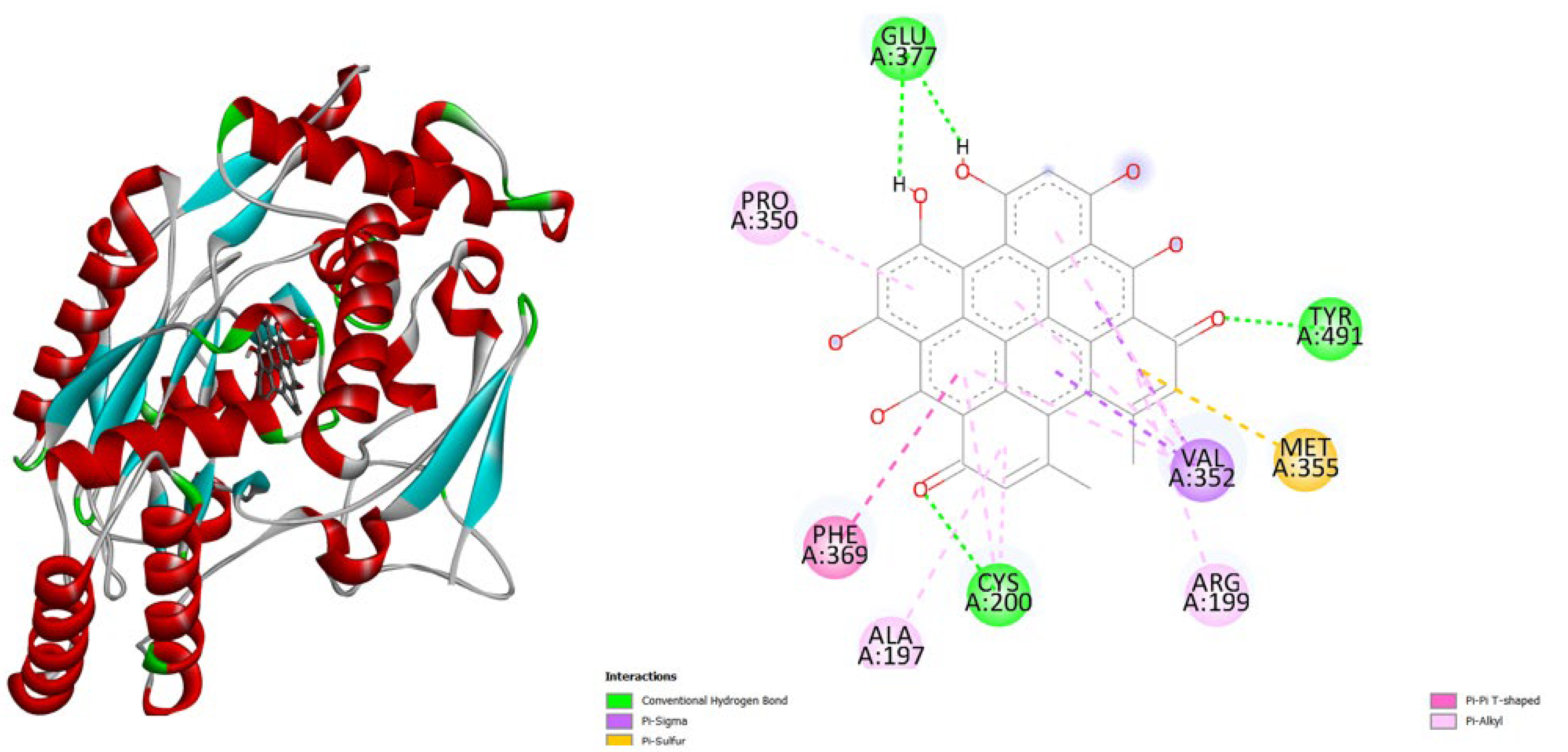
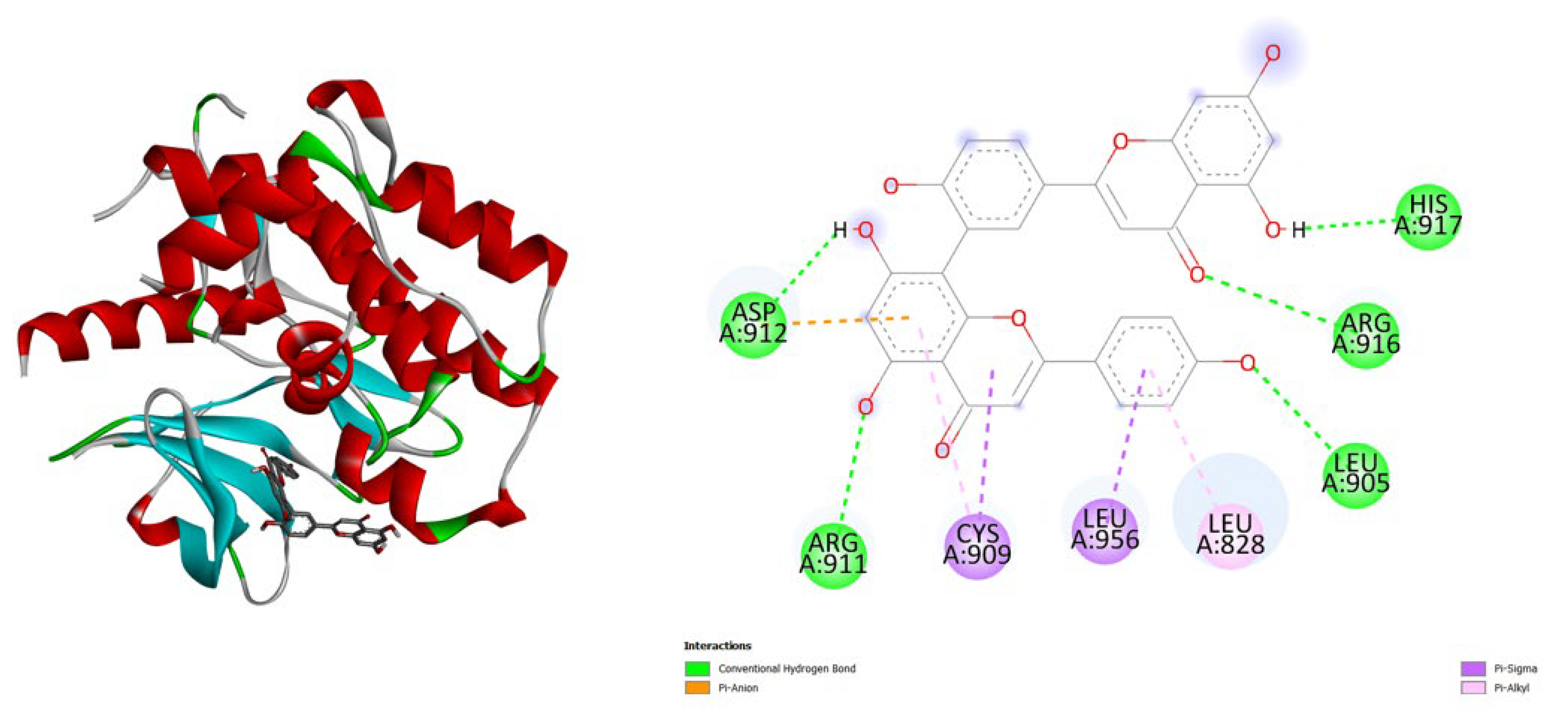
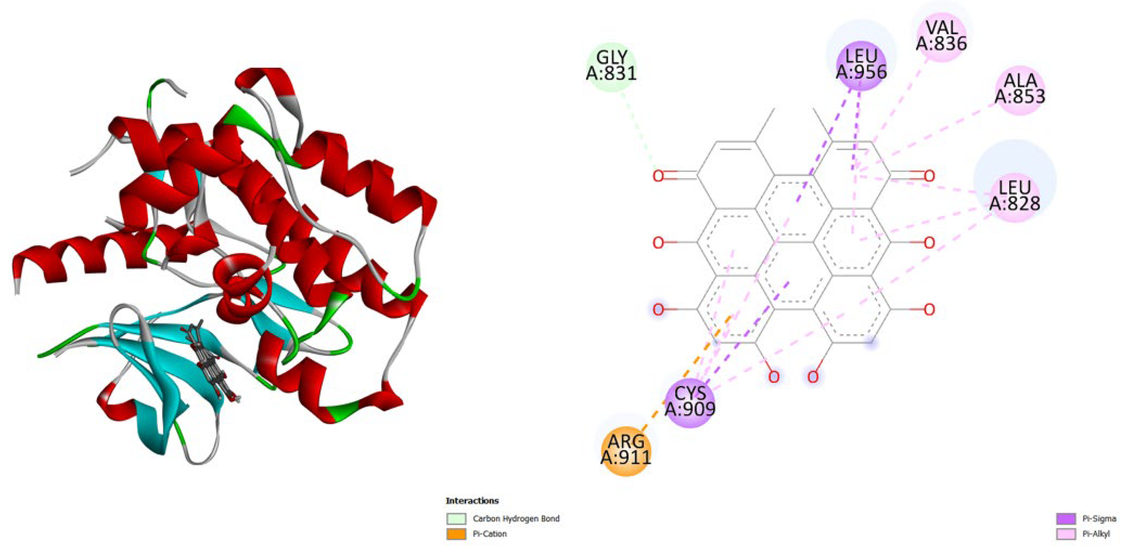
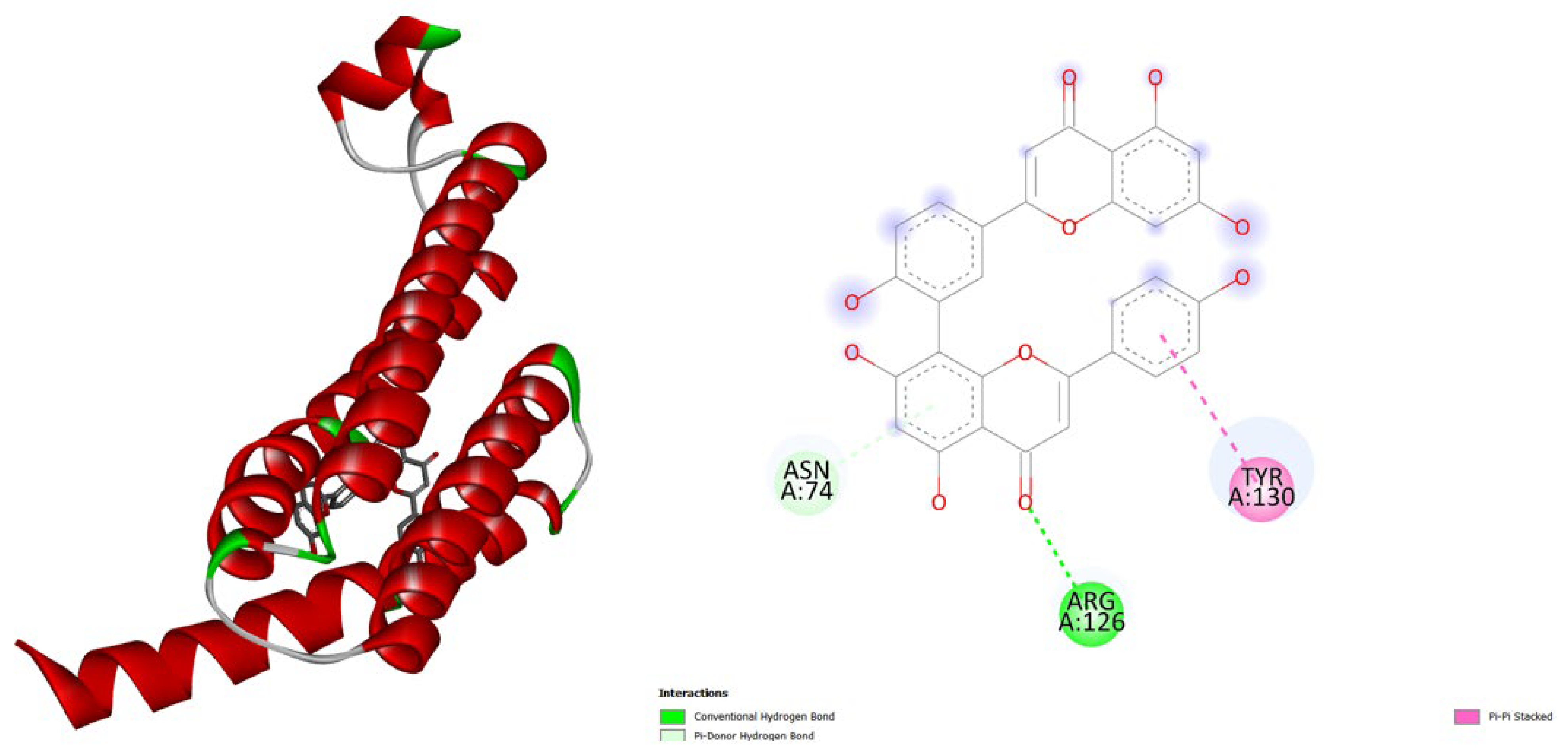
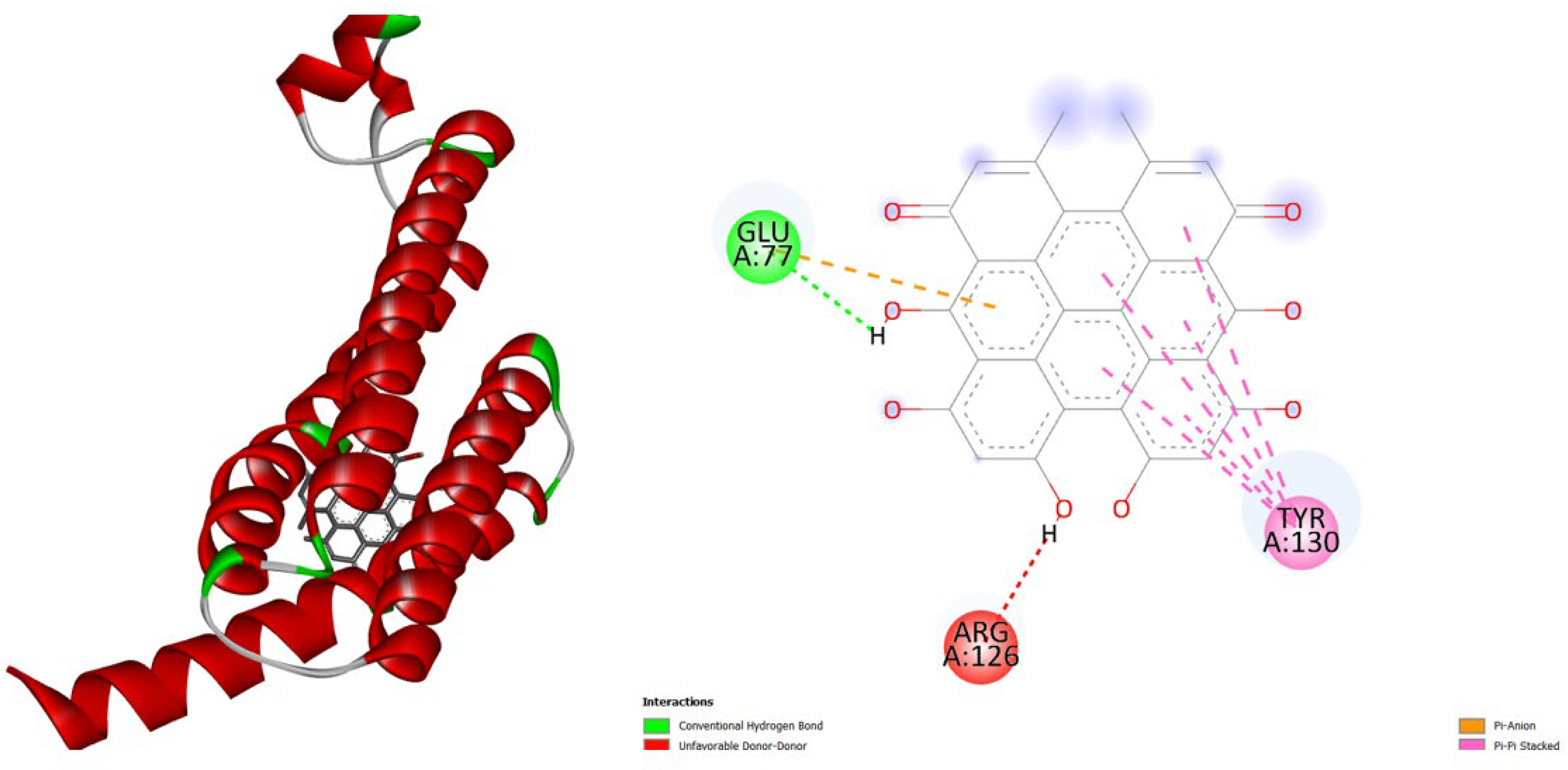
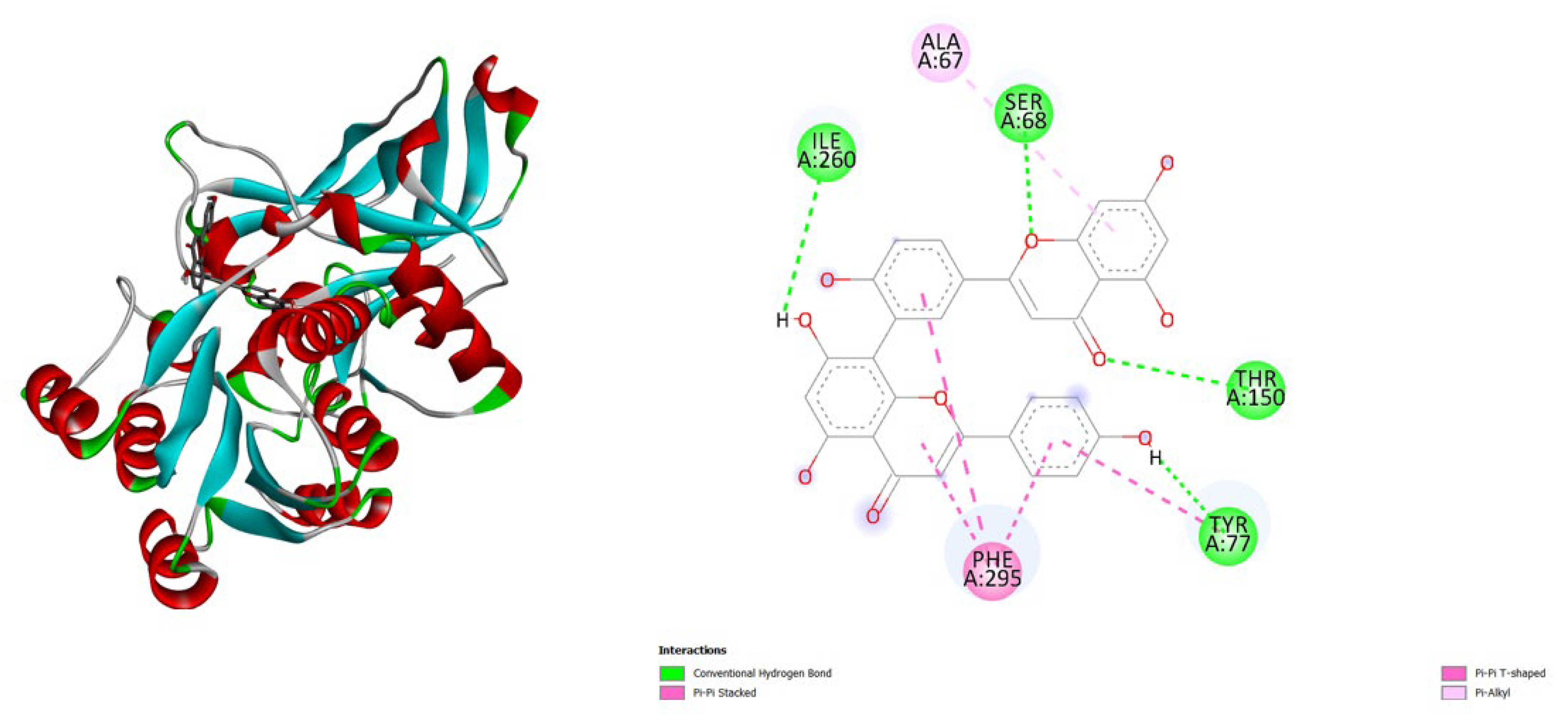
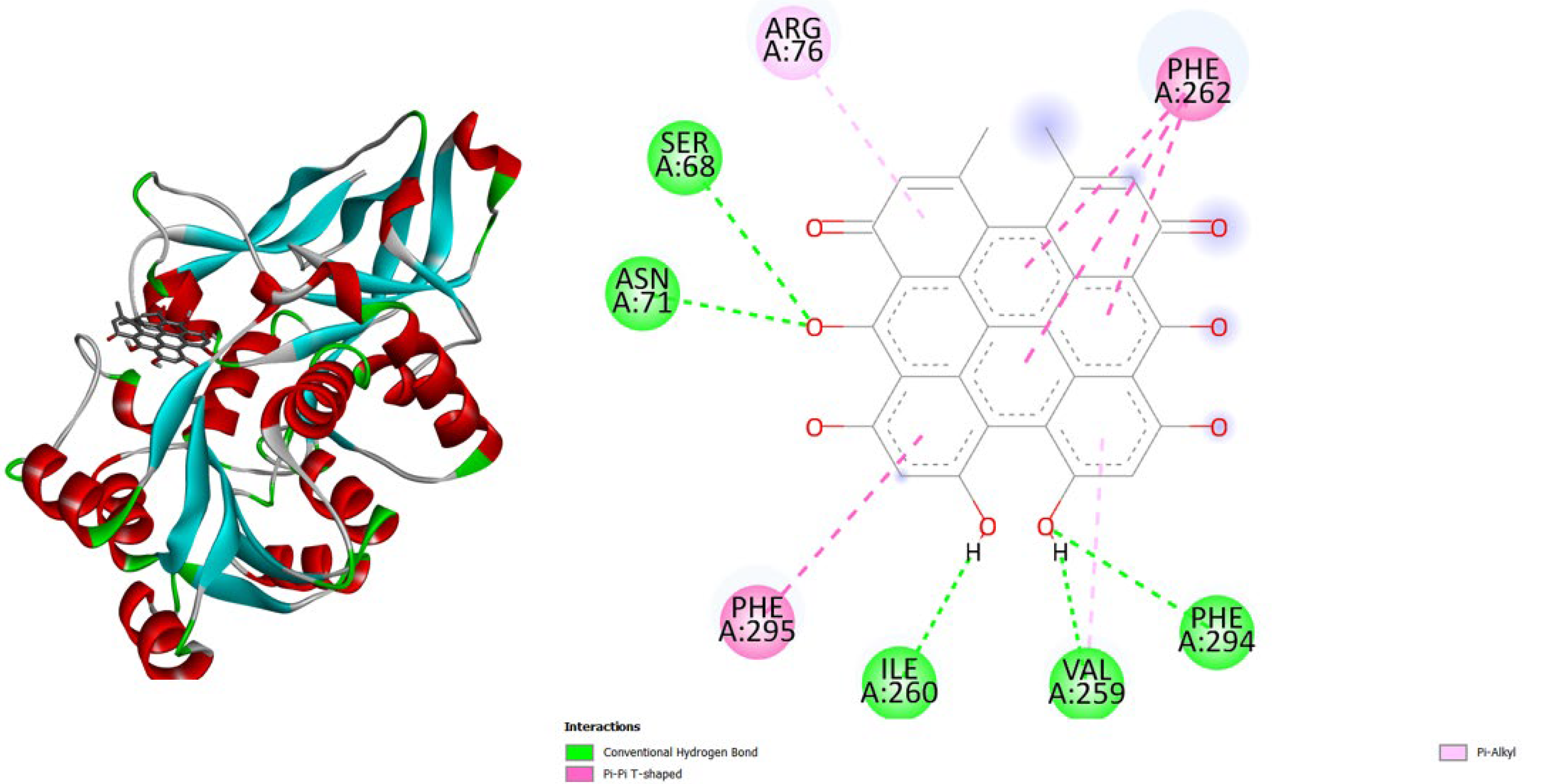
3. Results and Discussion

Inflammation plays a central role in a wide range of pathological conditions, including autoimmune disorders, cancer, and cardiovascular diseases [1,2,3,4]. Key mediators of these processes include tumor necrosis factor-α (TNF-α), a cytokine essential for systemic inflammatory responses [5]; inducible nitric oxide synthase (iNOS), which drives excessive nitric oxide production [6]; and Janus kinase 3 (JAK3), a critical enzyme in cytokine signaling pathways [7]. In addition, prostaglandin E synthase [8] and prostaglandin reductase 3 modulate prostaglandin metabolism, contributing to pain, fever, and the broader inflammatory response [9].
While conventional anti-inflammatory therapies—such as NSAIDs, corticosteroids, and JAK inhibitors—have proven effective, they are often limited by side effects and restricted target specificity [10]. Consequently, there is growing interest in natural bioactive compounds capable of simultaneously modulating multiple inflammatory targets, offering a potentially safer and more versatile therapeutic approach. This work aims to elucidate the binding interactions, identify critical residues involved, and assess the potential of these natural compounds to modulate multiple inflammatory pathways, providing a foundation for future in vitro and in vivo validation.
Molecular docking is a computational approach extensively employed to predict and analyze interactions between small molecules and target proteins. By evaluating the binding affinity and orientation of ligands within protein active sites, docking offers critical insights into the molecular mechanisms of drug action and supports the rational design of therapeutics.
Through docking analysis, researchers can assess the structural complementarity between ligands and their targets, pinpoint crucial interacting residues, and evaluate the stability of protein–ligand complexes [13,14,15]. Compared to experimental techniques, which are often time-consuming and resource-intensive, in silico docking provides a rapid and cost-effective means to screen multiple compounds and predict their potential efficacy before laboratory validation. The primary goal of this study is to investigate the potential of Amentoflavone and Hypericin as multi-target anti-inflammatory agents by performing molecular docking analysis against key inflammatory proteins, including TNF-α, iNOS, JAK3, and prostaglandin-producing enzymes.
This computational study was performed by AutoDock Vina 1.2 within the PyRx platform (https://pyrx.sourceforge.io) [15], the binding potential of natural compounds against Clostridium botulinum.
From the table:
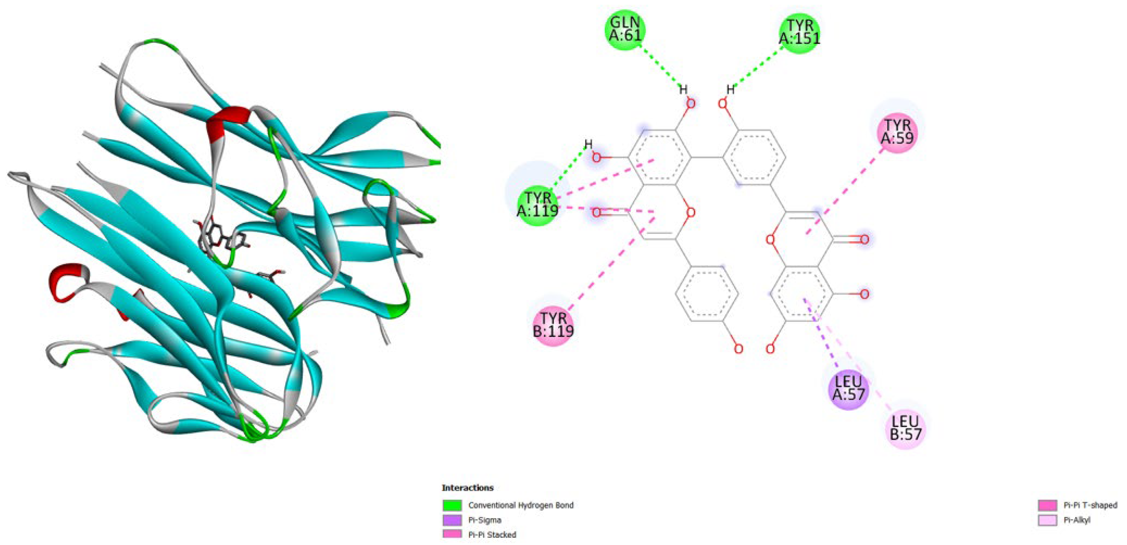
  • TNF-α (2AZ5): Hypericin binds slightly stronger (-9.9) than Amentoflavone (-9.5) and both outperform the native ligand (-8.8).
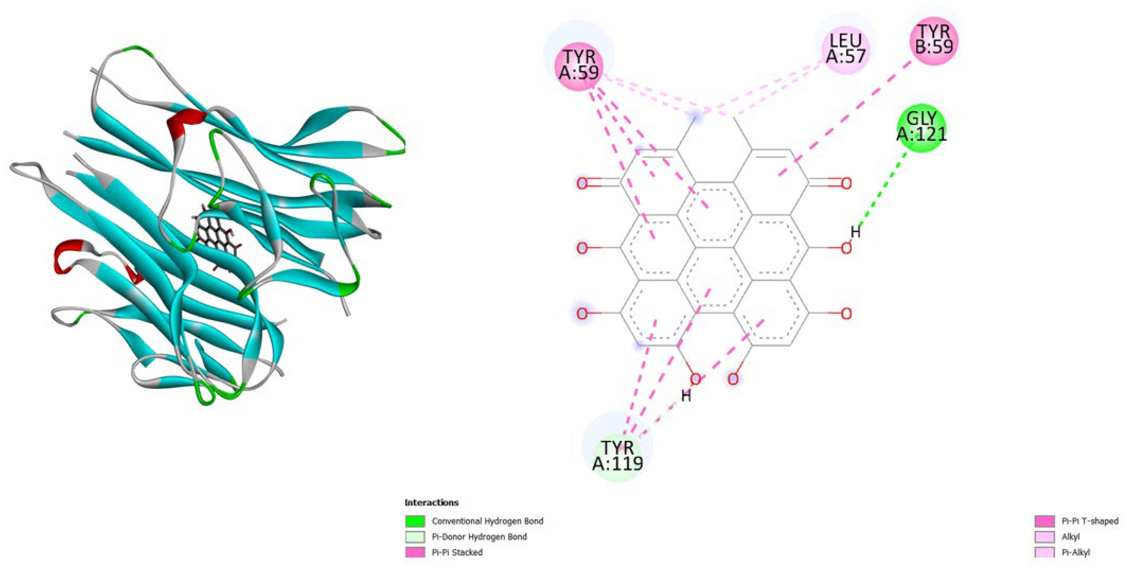
  • iNOS (3E7G): Amentoflavone shows the strongest binding (-11.3), followed by Hypericin (-10.4), both much better than the native ligand (-6.9).
  • JAK3 (6HZV): Hypericin has the strongest predicted affinity (-11.4), surpassing Amentoflavone (-9.5) and the native ligand (-9.2).
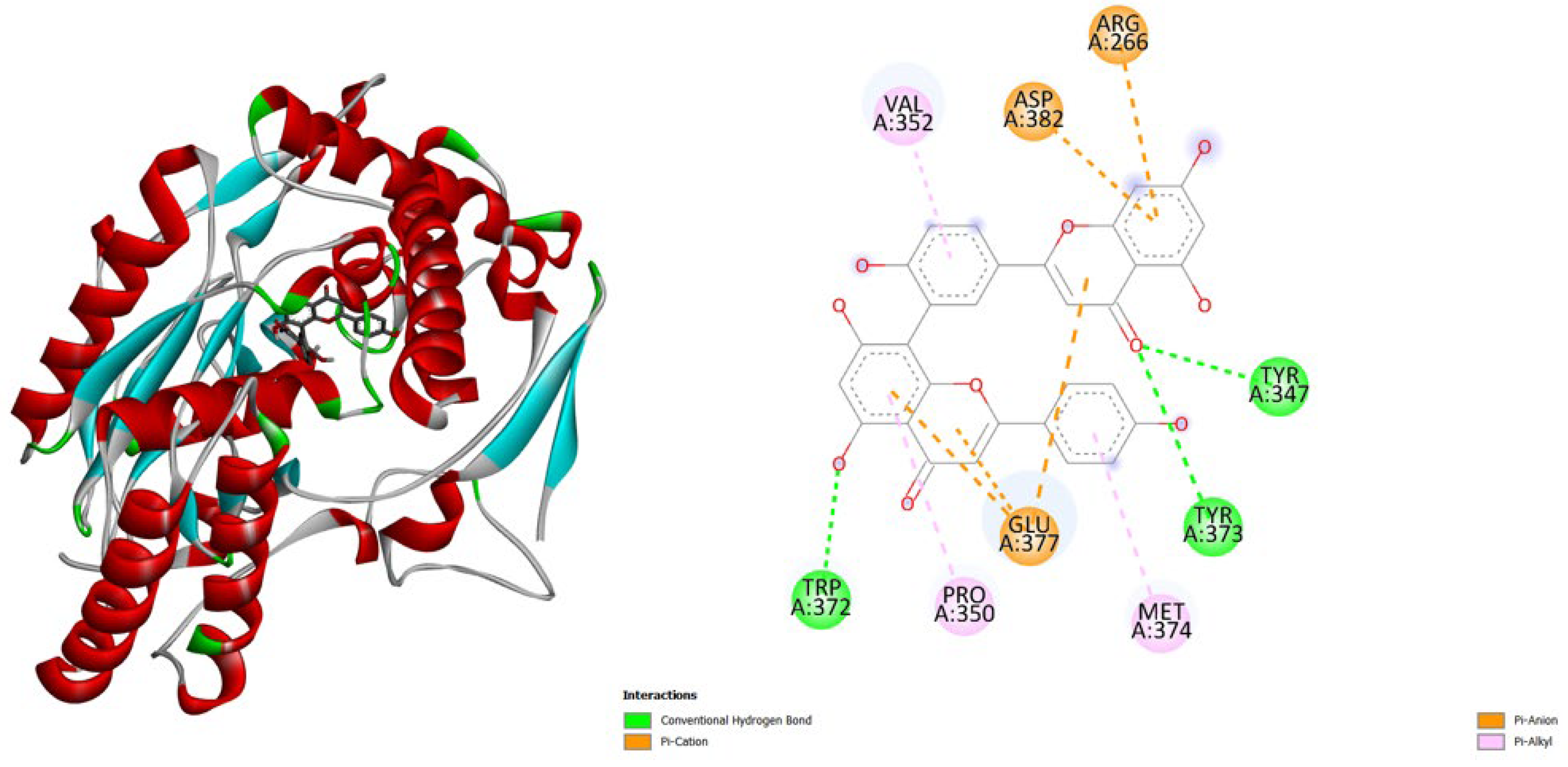
  • PGE synthase (4AL1): Amentoflavone (-7.7) binds slightly better than Hypericin (-7.1), both stronger than the native ligand (-5.9).
  • PG reductase 3 (7ZEJ): Amentoflavone (-11.0) has the strongest binding, followed by Hypericin (-9.6) and the native ligand (-8.7).
From Table 1, both Amentoflavone and Hypericin exhibit stronger predicted binding affinities than the native ligands across all five protein targets. Hypericin demonstrates the highest affinity for JAK3 and TNF-α, whereas Amentoflavone binds most strongly to iNOS and PG reductase 3.
Hypericin tends to bind better to TNF-α and JAK3, suggesting a potential preference for these targets.
Amentoflavone shows stronger binding to iNOS and PG reductase 3, indicating a potential selective advantage for these proteins.
In PGE synthase, both compounds show moderate improvement over the native ligand, but differences are small.
Overall, both natural products demonstrate the ability to bind with higher affinity and potentially greater stability than the native ligands, making them promising candidates for further experimental validation.
These findings suggest that both compounds are promising candidates for modulating the activity of these proteins.
From Figure 2, Figure 3, Figure 4, Figure 5, Figure 6, Figure 7, Figure 8, Figure 9, Figure 10 and Figure 11, we present the 3D and 2D representations of docked Amentoflavone and docked Hypericin into their respective protein targets. These visualizations were generated using Discovery Studio Biovia to identify the amino acid residues involved in the interactions [16].
All docking analyses were performed in the ligand-binding site pocket where the crystal ligand was originally located in the protein. The objective was to evaluate whether hypericin and amentoflavone exhibit lower binding energies, which would indicate stronger interactions and potentially greater stability within the protein-ligand complex.
Docking analyses indicate that both Hypericin and Amentoflavone bind more strongly than the native ligands in most target proteins, as evidenced by their more negative docking energies. This suggests enhanced stability of these natural compounds within the ligand-binding site pockets, although the relative binding strength varies by protein. Hypericin generally shows stronger binding for 2AZ5 and 6HZV, whereas Amentoflavone is preferred for 3E7G and 7ZEJ. ( See Table 1).
General speaking, across all five proteins, both Amentoflavone and Hypericin show more negative docking energies than the native ligands in almost all cases. This suggests that these natural compounds may form stronger and more stable interactions with the protein binding pockets compared to the native ligands. More negative docking energies generally correlate with higher binding affinity and potential biological activity. ( See Table 1).

4. Conclusions

Amentoflavone and Hypericin demonstrate potent, multi-target binding affinities against central mediators of inflammation, including cytokines, kinases, and prostaglandin-related enzymes. These findings support further in vitro and in vivo validation and suggest that these natural compounds could be developed as broad-spectrum anti-inflammatory agents with fewer side effects than current synthetic drugs.

References

  1. Schmid-Schönbein, G.W. Analysis of inflammation. Annu. Rev. Biomed. Eng. 2006, 8, 93–151. [Google Scholar] [CrossRef] [PubMed]
  2. Serhan, C.N.; Ward, P.A.; Gilroy, D.W. (Eds.). Fundamentals of inflammation. Cambridge University Press: 2010.
  3. Nathan, C. Points of control in inflammation. Nature 2002, 420, 846–852. [Google Scholar] [CrossRef] [PubMed]
  4. Medzhitov, R. Origin and physiological roles of inflammation. Nature 2008, 454, 428–435. [Google Scholar] [CrossRef] [PubMed]
  5. Popa, C.; Netea, M.G.; Van Riel, P.L.; Van Der Meer, J.W.; Stalenhoef, A.F. The role of TNF-α in chronic inflammatory conditions, intermediary metabolism, and cardiovascular risk. Journal of lipid research 2007, 48, 751–762. [Google Scholar] [CrossRef] [PubMed]
  6. Nussler, A.K.; Billiar, T.R. Inflammation, immunoregulation, and inducible nitric oxide synthase. Journal of leukocyte biology 1993, 54, 171–178. [Google Scholar] [CrossRef] [PubMed]
  7. Malaviya, R.; Laskin, D.L.; Malaviya, R. Janus kinase-3 dependent inflammatory responses in allergic asthma. International immunopharmacology 2010, 10, 829–836. [Google Scholar] [CrossRef] [PubMed]
  8. Williams, T.J. Prostaglandin E2, prostaglandin I2 and the vascular changes of inflammation. British Journal of Pharmacology 1979, 65, 517. [Google Scholar] [CrossRef] [PubMed]
  9. Ricciotti, E.; FitzGerald, G.A. Prostaglandins and inflammation. Arteriosclerosis, thrombosis, and vascular biology 2011, 31, 986–1000. [Google Scholar] [CrossRef] [PubMed]
  10. Ali, K.A.; Maity, A.; Roy, S.D.; Pramanik, S.D.; Das, P.P.; Shaharyar, M.A. (2023). Insight into the mechanism of steroidal and non-steroidal anti-inflammatory drugs. In How synthetic drugs work (pp. 61–94). Academic Press.
  11. Xiong, X. , Tang, N., Lai, X., Zhang, J., Wen, W., Li, X.,... & Liu, Z. Insights into amentoflavone: A natural multifunctional biflavonoid. Frontiers in Pharmacology 2021, 12, 768708. [Google Scholar] [PubMed]
  12. Hammer, K.D.; Birt, D.F. Evidence for contributions of interactions of constituents to the anti-inflammatory activity of Hypericum perforatum. Critical reviews in food science and nutrition 2014, 54, 781–789. [Google Scholar] [CrossRef] [PubMed]
  13. Jakhar, R.; Dangi, M.; Khichi, A.; Chhillar, A.K. Relevance of molecular docking studies in drug designing. Current Bioinformatics 2020, 15, 270–278. [Google Scholar] [CrossRef]
  14. Trott, O.; Olson, A.J. AutoDock Vina: improving the speed and accuracy of docking with a new scoring function, efficient optimization, and multithreading. Journal of computational chemistry 2010, 31, 455–461. [Google Scholar] [CrossRef] [PubMed]
  15. Dallakyan, S.; Olson, A.J. (2014). Small-molecule library screening by docking with PyRx. In Chemical biology: methods and protocols (pp. 243–250). New York, NY: Springer New York.
  16. Baroroh, U.; Biotek, M.; Muscifa, Z.S.; Destiarani, W.; Rohmatullah, F.G.; Yusuf, M. Molecular interaction analysis and visualization of protein-ligand docking using Biovia Discovery Studio Visualizer. Indonesian Journal of Computational Biology (IJCB) 2023, 2, 22–30. [Google Scholar] [CrossRef]
Figure 1. Chemical structures of Amentoflavone and Hypericin. The figure provides a side-by-side comparison of the two ligands, highlighting their structural features relevant for molecular docking studies.
Preprints 175729 g001
Figure 2. 3D representation and 2D interaction diagrams of docked Amentoflavone -9.5 kcal/mol with the crystal structure of Tumor Necrosis Factor (TNF-α). PDB code 2AZ5 . The figure was generated using Discovery Studio Biovia Visualizer.
Preprints 175729 g002
Figure 3. 3D representation and 2D interaction diagrams of docked Hypericin -9.9 kcal/mol with the crystal structure of Tumor Necrosis Factor (TNF-α). PDB code 2AZ5 . The figure was generated using Discovery Studio Biovia Visualizer.
Preprints 175729 g003
Figure 4. 3D representation and 2D interaction diagrams of docked Amentoflavone -11.3 kcal/mol with the crystal structure of Nitric oxide synthase, inducible. PDB code 3E7G. The figure was generated using Discovery Studio Biovia Visualizer.
Preprints 175729 g004
Figure 5. 3D representation and 2D interaction diagrams of docked Hypericin -10.4 kcal/mol with the crystal structure of Nitric oxide synthase, inducible. PDB code 3E7G. The figure was generated using Discovery Studio Biovia Visualizer.
Preprints 175729 g005
Figure 6. 3D representation and 2D interaction diagrams of docked Amentoflavone -9.5 kcal/mol with the crystal structure of Tyrosine-protein kinase JAK3. PDB Code 6HZV. The figure was generated using Discovery Studio Biovia Visualizer.
Preprints 175729 g006
Figure 7. 3D representation and 2D interaction diagrams of docked Hypericin -11.4 kcal/mol with the crystal structure of Tyrosine-protein kinase JAK3. PDB Code 6HZV. The figure was generated using Discovery Studio Biovia Visualizer.
Preprints 175729 g007
Figure 8. 3D representation and 2D interaction diagrams of docked Amentoflavone -7.7 kcal/mol with the crystal structure of PROSTAGLANDIN E SYNTHASE. PDB Code 4AL1 . The figure was generated using Discovery Studio Biovia Visualizer.
Preprints 175729 g008
Figure 9. 3D representation and 2D interaction diagrams of docked Hypericin -7.1 kcal/mol with the crystal structure of PROSTAGLANDIN E SYNTHASE. PDB Code 4AL1 . The figure was generated using Discovery Studio Biovia Visualizer.
Preprints 175729 g009
Figure 10. 3D representation and 2D interaction diagrams of docked Amentoflavone -11.0 kcal/mol with the crystal structure of Prostaglandin reductase 3. PDB Code 7ZEJ . The figure was generated using Discovery Studio Biovia Visualizer.
Preprints 175729 g010
Figure 11. 3D representation and 2D interaction diagrams of docked Hypericin -9.6 kcal/mol with the crystal structure of Prostaglandin reductase 3. PDB Code 7ZEJ . The figure was generated using Discovery Studio Biovia Visualizer.
Preprints 175729 g011
Table 1. shows the binding affinities (in kcal/mol) of two natural compounds, Amentoflavone and Hypericin, compared with the native ligand for five different protein targets. Lower (more negative) values indicate stronger predicted binding.
Protein (PDB) Amentoflavone Hypericin Native ligand
TNF-α (2AZ5) -9.5 -9.9 -8.8
iNOS (3E7G) -11.3 -10.4 -6.9
JAK3 (6HZV) -9.5 -11.4 -9.2
PGE synthase (4AL1) -7.7 -7.1 -5.9
PG reductase 3 (7ZEJ) -11.0 -9.6 -8.7
Disclaimer/Publisher’s Note: The statements, opinions and data contained in all publications are solely those of the individual author(s) and contributor(s) and not of MDPI and/or the editor(s). MDPI and/or the editor(s) disclaim responsibility for any injury to people or property resulting from any ideas, methods, instructions or products referred to in the content.
Copyright: This open access article is published under a Creative Commons CC BY 4.0 license, which permit the free download, distribution, and reuse, provided that the author and preprint are cited in any reuse.
Prerpints.org logo

Preprints.org is a free preprint server supported by MDPI in Basel, Switzerland.

Subscribe

Disclaimer

Terms of Use

Privacy Policy

Privacy Settings

© 2026 MDPI (Basel, Switzerland) unless otherwise stated