Preprint
Review

This version is not peer-reviewed.

Secretan’s Syndrome of the Hand: Literature Review and Surgical Case Report of a Rarely Documented Condition

A peer-reviewed article of this preprint also exists.

Submitted:

29 August 2025

Posted:

01 September 2025

You are already at the latest version

Abstract

Background: Secretan’s syndrome is a rare and under-recognized condition characterized by chronic, indurated, non-pitting edema of the dorsal hand with thumb sparing. Fewer than a few dozen cases have been reported worldwide, mostly as isolated case reports, and its pathogenesis remains debated between traumatic, inflammatory, and factitious mechanisms. This article presents a surgically managed hyperplastic case and a review of the literature, emphasizing how principles of precision medicine can guide diagnosis and treatment. Materials and Methods: A 36-year-old healthcare worker developed progressive dorsal swelling of the left hand following minor trauma, with marked restriction of metacarpophalangeal flexion. Laboratory tests and radiographs were normal. MRI demonstrated peritendinous fibrosis encasing the extensor tendons. Psychiatric evaluation excluded factitious behavior. Given the functional limitation and MRI evidence of fibrosis, selective dorsal fasciotomies and extensor tendon tenolysis were performed. A systematic literature review was conducted to summarize epidemiology, clinical and imaging features, histopathology, and management options. Results: Histology revealed fibro-adipose tissue with chronic inflammatory changes and CD68+ histiocytic aggregates; microbiological cultures were negative. Postoperative rehabilitation enabled significant functional recovery. The literature review confirmed the scarcity of published cases and the absence of standardized guidelines. MRI consistently proved to be the most informative imaging tool, while surgical treatment was described only in hyperplastic, refractory forms. Conclusions: This case and review illustrate how a precision medicine approach can optimize management of rare disorders. Early MRI-based diagnosis, multidisciplinary assessment, and phenotype-driven surgical intervention allowed tailored treatment and favorable outcome. Personalized care that integrates clinical features, imaging findings, and patient-specific factors may improve results in hyperplastic Secretan’s syndrome despite the limited evidence base.

Keywords: 
;  ;  ;  ;  ;  

1. Introduction

Secretan’s syndrome, first described by Henri-François Secretan in 1901, is a rare and often under-recognized condition defined by persistent, non-pitting, indurated edema of the dorsal hand after minor trauma [1]. Classically, the swelling predominates over the metacarpal region with sparing of the thumb and may cause finger flexion limitation and prolonged functional impairment [7,9]. Early reports in Swiss workers highlighted the chronicity of the edema and raised the possibility of secondary gain in selected contexts [8,9]. Histology typically shows fibrotic induration and peritendinous fibrosis, albeit non-specific findings [1,3]. Clinically, the course ranges from spontaneous resolution to a hyperplastic, fibrotic form that may require surgical management [7,10]. Imaging contributes to characterization: radiographs are often unremarkable, whereas MRI depicts peritendinous edema and fibrotic involvement of the extensor compartments [4]. Pathogenesis remains debated. Proposed mechanisms include post-traumatic/inflammatory pathways akin to lymphedema or reflex sympathetic dystrophy, and a factitious component associated with self-inflicted injury or tight bandaging; psychiatric comorbidity has been described in atypical presentations [2,11,12]. Although the hand is the typical site, rare cases involve the foot and ankle [3]. Recent reviews call for greater awareness and more standardized diagnostic criteria to guide conservative care and to identify candidates for surgery in hyperplastic forms [5,6].
We report a surgically confirmed case of Secretan’s syndrome of the hand and critically appraise the literature to outline presentation, imaging and histology, differential diagnosis, and management.

2. Materials and Methods of Literature Review

A structured literature search was conducted in PubMed, Embase, and Scopus up to March 2025, in accordance with the Preferred Reporting Items for Systematic Reviews and Meta-Analyses (PRISMA) guidelines. The search terms included “Secretan’s syndrome”, “factitious lymphedema”, “chronic dorsal hand edema”, and “peritendinous fibrosis”. We included case reports, case series, and reviews that described clinical presentation, imaging, histology, treatment, or outcome of patients with Secretan’s syndrome. Publications in English, French, and Italian were considered. Articles lacking sufficient clinical detail or reporting unrelated conditions were excluded. The reference lists of selected papers were screened to identify additional relevant reports.

3. Review of the Literature

Secretan’s syndrome was first described in 1901 and remains one of the rarest entities in hand surgery. Fewer than a few dozen cases have been reported, predominantly from Europe. Most publications consist of isolated case reports, with only a handful of small series available.

3.1. Epidemiology

The condition predominantly affects middle-aged adults, with no clear sex predilection. Occupational or repetitive microtrauma has been reported as a common trigger, though factitious etiology has been documented in several cases

3.2. Clinical Presentation

The hallmark is chronic, indurated, non-pitting edema on the dorsum of the hand, typically sparing the thumb. Limitation of MCP joint motion, especially flexion, is frequent. Pain and functional impairment are variably reported.

3.3. Imaging

Radiographs are usually normal. MRI is consistently identified as the most useful diagnostic tool, demonstrating peritendinous edema, fibrotic bands, and fascial thickening. These findings help distinguish Secretan’s syndrome from cellulitis, venous thrombosis, and CRPS.

3.4. Histology

Reported histology is non-specific, with fibrous tissue, inflammatory infiltrates, and peritendinous fibrosis. No pathognomonic pattern has been defined.

.3.5 Management

Treatment strategies vary widely. Many cases resolve with conservative measures such as compression, rest, and physiotherapy. Psychiatric evaluation is strongly recommended when factitious etiology is suspected. Surgical intervention is rarely described and generally reserved for hyperplastic, refractory cases with functional compromise. Outcomes after surgery are variable but often favorable when fibrosis and adhesions are clearly documented.
A summary of the reported cases in the literature is presented in Table 1.

4. Detailed Case Description

4.1. Patient Information

A 36-year-old female healthcare worker (employed in a nursing home) presented with progressive dorsal swelling of the left hand following minor trauma. She had no relevant medical history, no psychiatric disorders, and no chronic medication use.

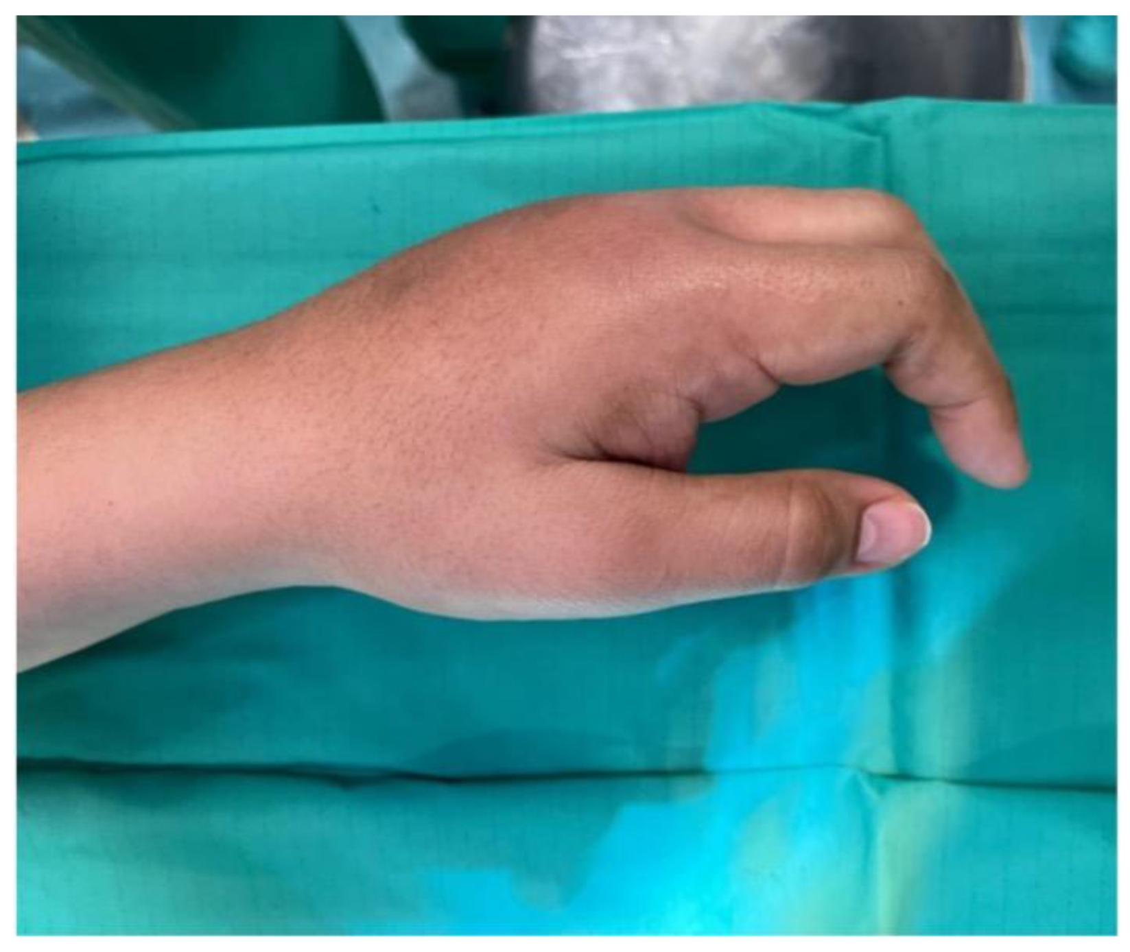
4.2. Clinical Findings

Examination revealed a firm, indurated, non-pitting edema over the dorsum of the hand with sparing of the thumb (Figure 1). Metacarpophalangeal (MCP) flexion of digits II–V was restricted to <20°, while extension was complete. The patient reported pain radiating into the forearm and significant limitations in both professional duties and daily activities. Grip strength was reduced on qualitative testing.

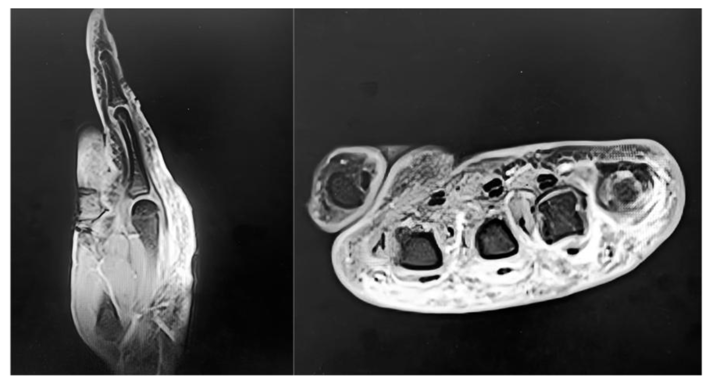
4.3. Diagnostic Assessment

Routine blood tests, erythrocyte sedimentation rate, and C-reactive protein were normal. The patient was afebrile. Radiographs showed no fractures, osteolysis, or arthropathy. Magnetic resonance imaging (MRI) of the left hand (03/02/2025) demonstrated peritendinous edema and a hypointense fibrotic band encasing the extensor tendons within compartments II–V (Figure 2). The differential diagnosis included complex regional pain syndrome (CRPS), infectious or non-infectious extensor tenosynovitis, lymphedema, superficial venous thrombosis, cellulitis/fasciitis, and factitious injury. The combination of dorsal indurated non-pitting edema, thumb sparing, negative laboratory tests, and MRI findings was consistent with Secretan’s syndrome.
Given the literature describing possible factitious components, a psychiatric consultation was obtained both preoperatively and postoperatively with diagnosis of episodic mood disorder (ICD-9-CM code 296.90). No evidence of self-harm or active factitious behavior was identified, though psycho-educational support and follow-up were recommended to reinforce adherence to care.

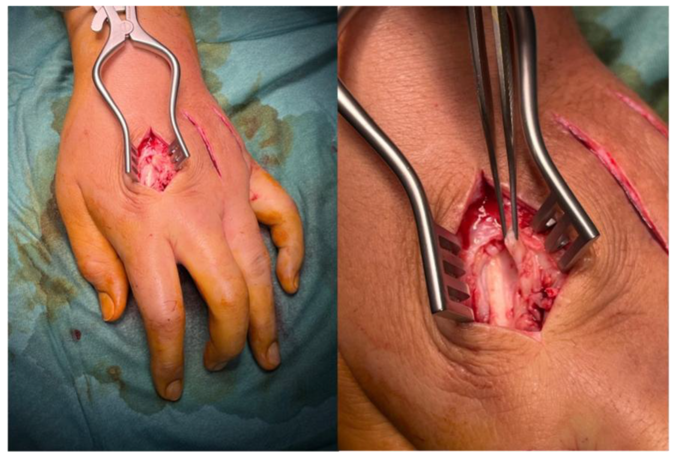
4.4. Therapeutic Intervention

On 12/04/2025, surgery was performed under brachial plexus block in a day-surgery setting. A longitudinal dorsal incision was made across three intermetacarpal spaces (II–IV). Intraoperatively, extensive peritendinous fibrosis and adhesions of the extensor tendons were observed, without abscesses or purulent collections (Figure 3). Selective fasciotomies and extensor tenolysis were carried out. Three tissue samples were collected for culture and histological examination. The wound was closed in layers, with a sterile dressing and functional bandage. Standard analgesia and early mobilization were prescribed.

4.5. Microbiological and Histological Findings

Cultures were negative. Histology (26/04/2025) showed fibro-adipose tissue with edema, chronic inflammation, amorphous material deposition, and aggregates of CD68+ histiocytic cells. These findings were non-specific but consistent with hyperplastic peritendinous fibrosis.

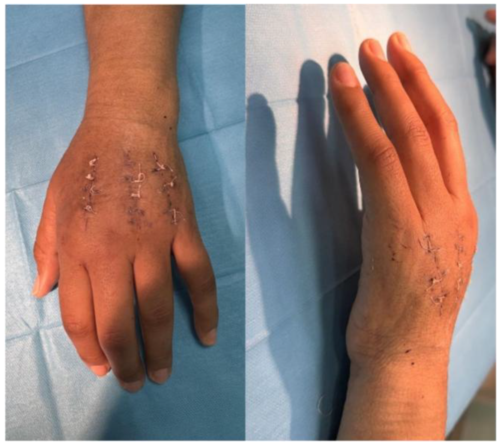
4.6. Follow-Up and Outcomes

At 18 days postoperatively, MCP flexion was still limited to ~20° (Figure 4). At 4 weeks, motion improved to ~45° following initiation of physiotherapy, electrostimulation, and a home exercise program. At 3 months, extension was complete and flexion stabilized around 45°, with reduced pain and no recurrence of swelling. The patient resumed driving by 4 months. At 6 months, after structured rehabilitation and good compliance, MCP flexion reached ~70°, with marked reduction in pain and edema. No surgical or infectious complications occurred.
Functional outcomes at baseline, postoperative, and follow-up are summarized in Table 2.

5. Discussion

The present case demonstrates a classic clinical-radiological phenotype of Secretan’s syndrome: dorsal, indurated, non-pitting edema with thumb sparing, unremarkable laboratory workup, and MRI evidence of peritendinous fibrosis. Histology confirmed hyperplastic fibrotic tissue with CD68+ histiocytic aggregates, consistent with but not diagnostic for the syndrome.
Two aspects were crucial in this case: the imaging-based early diagnosis prevented unnecessary empiric antibiotic therapies or prolonged diagnostic delays; and the multidisciplinary management, including psychiatric evaluation, was essential to exclude factitious behavior and to support adherence to treatment. It is also important to underline that the clinical presentation may easily mimic other conditions such as complex regional pain syndrome (CRPS) or lymphedema, which reinforces the need for careful differential diagnosis supported by imaging.
Surgical management was indicated due to extensive fibrosis and persistent functional limitation, aligning with the few previously reported cases requiring intervention. Functional recovery was substantial, with MCP flexion improving from <20° preoperatively to ~70° at 6 months.
When compared with the literature, our case aligns with Redfern et al. [9], who reported functional improvement after surgical release of peritendinous fibrosis. A similar role for surgery in refractory disease was noted by Abnousi and Chou [3], who described resolution following excision of fibrotic tissue in the foot. Conversely, most reports, such as those by Angelini et al. [1] and Collet et al. [8], describe conservative management, indicating that surgery should be considered only in selected cases. MRI has repeatedly been identified as the most informative diagnostic tool. In our patient it confirmed peritendinous fibrosis and guided surgical planning, consistent with the findings of Whitney and Jones [4]. Histological results, although non-specific, resembled those reported by Lemmens et al. [5], who described fibrotic changes without pathognomonic features.
Overall, this case highlights the value of a personalized approach: while conservative treatment remains first-line, surgery may be justified in hyperplastic forms with functional impairment, provided that imaging supports the diagnosis and psychiatric assessment excludes factitious causes.

6. Conclusions

Secretan’s syndrome remains a diagnostic challenge due to its rarity and heterogeneous presentation. This case illustrates how precision medicine principles can be applied: combining clinical phenotype, imaging findings, psychiatric evaluation, and tailored surgical management. In hyperplastic forms with refractory fibrosis, selective fasciotomies and tenolysis, followed by structured rehabilitation, can restore function and improve quality of life.
Further studies and standardized diagnostic criteria are needed to better define the role of surgery versus conservative treatment in this rare condition.

Author Contributions

Conceptualization, A.C. and E.G.; methodology, S.P.; formal analysis, A.C.; investigation, E.D.D.and G.V.; data curation, A.C.; writing—original draft preparation, A.C. and G.V.; writing—review and editing, A.C., S.P. and E.G.; supervision, L.R. All authors have read and agreed to the published version of the manuscript.

Funding

This research received no external funding.

Institutional Review Board Statement

Not applicable.

Informed Consent Statement

Written informed consent for publication of this case report and accompanying images was obtained from the patient.

Acknowledgments

We thank the patient for the collaboration and consenting to publication.

Conflicts of Interest

The authors declare no conflict of interest.

Abbreviations

The following abbreviations are used in this manuscript:
CRPS Complex Regional Pain Syndrome
ICD-9-CM International Classification of Diseases, Ninth Revision, Clinical Modification
MCP Metacarpophalangeal
MRI Magnetic Resonance Imaging
PRISMA Preferred Reporting Items for Systematic Reviews and Meta-Analyses
VAS Visual Analog Scale

References

  1. Angelini, G.; Meneghini, C.L.; Vena, G.A. Secretan’s syndrome: An artefact oedema of the hand. Contact Dermatitis 1982, 8, 345–346. [Google Scholar] [CrossRef] [PubMed]
  2. Demircioğlu, D.; Öztürk Durmaz, E.; Sezer, E.; Şahin, S. Secretan syndrome: A fluctuating case of factitious lymphedema. Cutis 2021, 108, E23–E24. [Google Scholar] [CrossRef] [PubMed]
  3. Abnousi, F.; Chou, L.B. Secretan’s disease of the foot: A case report and review. Foot Ankle Int. 2008, 29, 248–250. [Google Scholar] [CrossRef] [PubMed]
  4. Whitney, T.M.; Jones, N.F. Magnetic resonance imaging findings in Secretan’s disease. J. Hand Surg. Am. 1995, 20, 464–466. [Google Scholar] [CrossRef] [PubMed]
  5. Lemmens, L.; van Doninck, J.; de Smet, L. Secretan’s syndrome: Case report. Acta Chir. Belg. 2019, 119, 123–124. [Google Scholar] [CrossRef] [PubMed]
  6. Tebbaa El Hassali, A.; Barrached, M.; Lachkar, A.; Abdeljaouad, N.; Yacoubi, H. Secretan’s syndrome: It is time to talk about it. Cureus 2024, 16, e62580. [Google Scholar] [CrossRef] [PubMed]
  7. Fleming, J.P. Secretan’s disease. Plast. Reconstr. Surg. 1977, 60, 617–618. [Google Scholar] [CrossRef] [PubMed]
  8. Collet, S.; Forli, A.; Carpentier, P.H.; Laviolette, F.; Imbert, B.; Blaise, S. Secretan’s syndrome: Myth or pathomimia? J. Mal. Vasc. 2014, 39, 67–72. [Google Scholar] [CrossRef] [PubMed]
  9. Redfern, A.B.; Curtis, R.M.; Wilgis, E.F.S. Experience with peritendinous fibrosis of the dorsum of the hand. J. Hand Surg. Am. 1982, 7, 380–383. [Google Scholar] [CrossRef] [PubMed]
  10. Grobmyer, A.J., 3rd; Bruner, J.M.; Dragstedt, L.R., 2nd. Closed lymphangioplasty in Secretan’s disease. Arch. Surg. 1968, 97, 81–83. [Google Scholar] [CrossRef] [PubMed]
  11. Winkelmann, R.K.; Barker, S.M. Factitious lymphoedema, Secretan’s syndrome. Dermatol. Clin. 1990, 8, 205. [Google Scholar] [CrossRef]
  12. Birman, M.V.; Lee, D.H. Factitious disorders of the upper extremity. J. Am. Acad. Orthop. Surg. 2012, 20, 78–85. [Google Scholar] [CrossRef] [PubMed]
Figure 1. Preoperative documentation. Dorsal clinical view of the left hand showing firm, non-pitting edema with thumb sparing.
Preprints 174399 g001
Figure 2. Preoperative MRI of the left hand showing peritendinous edema and a hypointense fibrotic band encasing the extensor tendons (compartments II–V).
Preprints 174399 g002
Figure 3. Intraoperative findings through dorsal approaches to intermetacarpal spaces II–IV, showing extensive peritendinous fibrosis and adhesions of the extensor tendons; selective fasciotomies and tenolysis were performed.
Preprints 174399 g003
Figure 4. Early postoperative views. Anteroposterior and lateral aspect of the hand showing surgical incisions in healing phase, residual edema, and limited MCP flexion.
Preprints 174399 g004
Table 1. Reported cases of Secretan’s syndrome in the literature.
Author, Year No. of Cases Site Diagnostic Findings Management Outcome
Secretan, 1901 Original description Hand Indurated dorsal edema, thumb sparing Conservative Chronic, variable resolution
Angelini et al., 1982 [1] 1 Hand Non-pitting edema, histology: fibrosis Conservative Partial resolution
Redfern et al., 1982 [9] 4 Hand Peritendinous fibrosis Surgical release Functional improvement
Grobmyer et al., 1968 [10] 1 Hand Attempted closed lymphangioplasty Surgical Persistent edema
Fleming, 1977 [7] 1 Hand Indurated dorsal edema, fibrotic tissue Conservative Chronic course
Whitney & Jones, 1995 [4] 1 Hand MRI: peritendinous fibrosis Conservative Persistent limitation
Winkelmann & Barker, 1990 [11] 2 Hand Factitious lymphedema suspected Psychiatric + conservative Resolution with follow-up
Abnousi & Chou, 2008 [3] 1 Foot Dorsal edema, fibrotic band Surgical excision Symptom resolution
Collet et al., 2014 [8] 3 Hand Chronic edema, possible factitious cases Conservative, psychiatric Variable
Lemmens et al., 2019 [5] 1 Hand Classic phenotype, biopsy fibrosis Conservative Persistent symptoms
Demircioğlu et al., 2021 [2] 1 Hand Fluctuating edema, factitious suspicion Psychiatric + conservative Symptom control
Tebbaa El Hassali et al., 2024 [6] 1 Hand Chronic indurated edema Conservative Stable with follow-up
Birman & Lee, 2012 [12] Review Upper limb Factitious disorders of the extremity Psychiatric/varied Highlights diagnostic overlap
Table 2. Functional outcomes of the present case.
Timepoint VAS (0–10) QuickDASH (0–100) Grip Strength
(Jamar, % vs contralateral)
Preoperative
Early post-op (1 month)
3-Month Follow-up
8 65 40%
5 50 50%
4 35 65%
6-Month Follow-up 2 20 75%
* VAS: Visual Analog Scale; DASH: Disabilities of the Arm, Shoulder and Hand questionnaire.
Disclaimer/Publisher’s Note: The statements, opinions and data contained in all publications are solely those of the individual author(s) and contributor(s) and not of MDPI and/or the editor(s). MDPI and/or the editor(s) disclaim responsibility for any injury to people or property resulting from any ideas, methods, instructions or products referred to in the content.
Copyright: This open access article is published under a Creative Commons CC BY 4.0 license, which permit the free download, distribution, and reuse, provided that the author and preprint are cited in any reuse.
Prerpints.org logo

Preprints.org is a free preprint server supported by MDPI in Basel, Switzerland.

Subscribe

Disclaimer

Terms of Use

Privacy Policy

Privacy Settings

© 2026 MDPI (Basel, Switzerland) unless otherwise stated