
Submitted:
21 August 2026
Posted:
25 August 2026
You are already at the latest version
Abstract
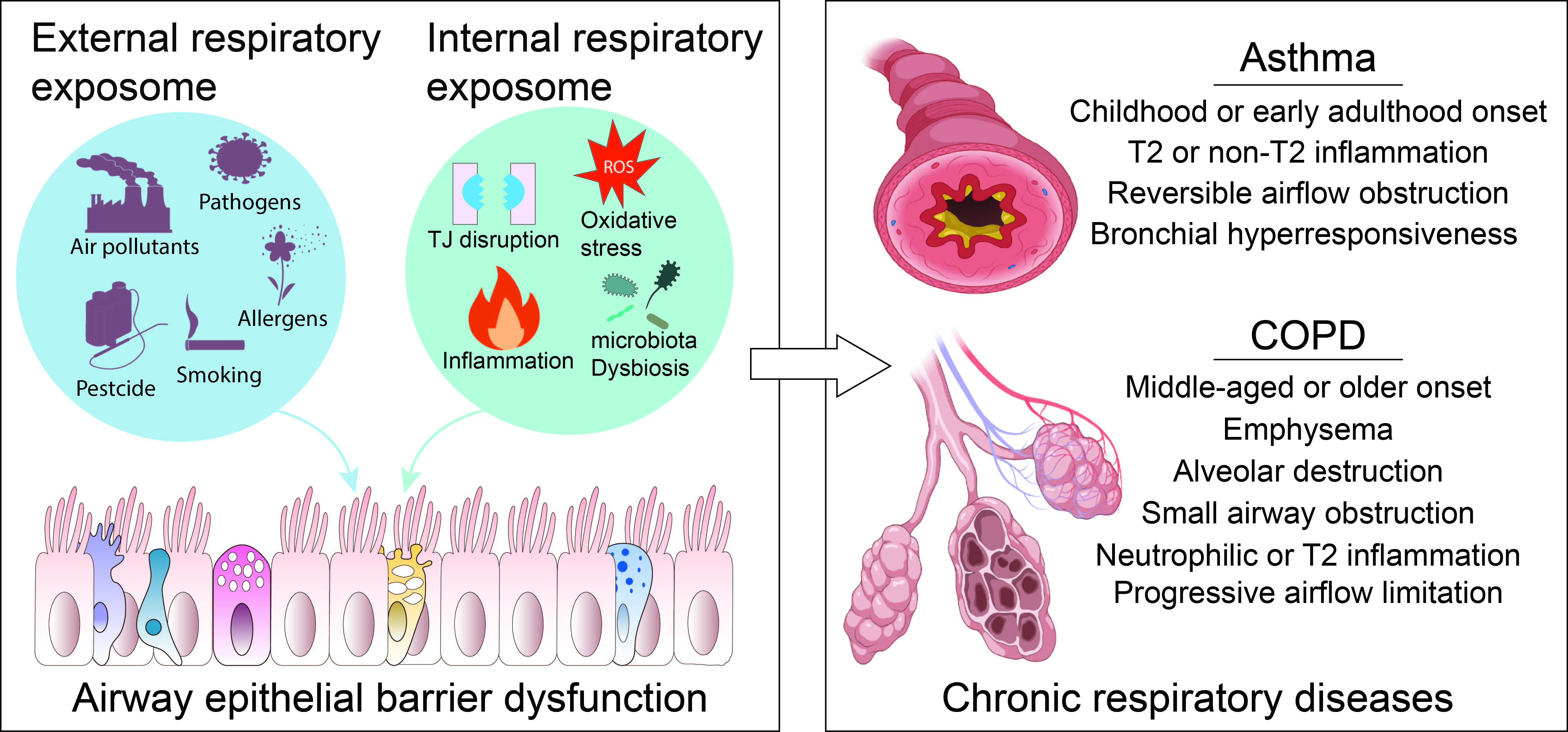
The airway epithelial barrier (AEB) is a critical transducer that maintains respiratory homeostasis. This review proposes a novel dual-domain respiratory exposome framework that conceptualizes how the external exposome (cumulative inhaled exposures) and the internal exposome (endogenous immune, metabolic, and endocrine networks) converge to modulate epithelial resilience dynamically. Excessive cumulative pressure disrupts this homeostatic balance, compromising junctional integrity, triggering oxidative stress, and provoking inflammation. These perturbations contribute to maladaptive airway remodeling in chronic respiratory diseases (CRDs), such as asthma and chronic obstructive pulmonary disease (COPD). Furthermore, emerging biologics that target epithelial alarmins and barrier-disruptive mediators are summarized. Finally, a next-generation precision medicine paradigm that leverages computational modeling to integrate individual expotypes (external exposure profiles) with endotypes (internal molecular signatures) is outlined. This framework offers a unified strategy for restoring AEB function and optimizing CRD management.
Keywords:
airway epithelial barrier
; respiratory exposome
; chronic respiratory diseases
; precision medicine
1. Introduction
Commonly diagnosed chronic respiratory diseases (CRDs), including conditions like chronic obstructive pulmonary disease (COPD), asthma, idiopathic pulmonary fibrosis (IPF), and cystic fibrosis (CF), represent significant and growing global health challenges.[1,2] With a frequency of 454.6 million cases and almost 4 million fatalities annually, CRDs were the third-ranking cause of death globally in 2019. [3] Among CRDs, asthma is the most common non-communicable disease in children, whereas COPD is the leading cause of death.[1,4]
The respiratory epithelium is a vital interface between the internal biological milieu and the external environment.[5] Extending from the trachea to the alveoli, this continuous cellular lining forms the Airway Epithelial Barrier (AEB), serving as the primary defense against inhaled pathogens, allergens, and environmental insults. The structural integrity and immunological functionality of the AEB are critical for maintaining respiratory homeostasis; conversely, barrier dysfunction is recognized as a hallmark of CRD pathogenesis.[6,7]
Industrialization and urbanization expose the respiratory system to increasingly complex mixtures of harmful substances. To scrutinize these influences, the concept of the "respiratory exposome" is adopted. While the general exposome is defined as the cumulative measure of environmental influences,[8] we specifically define the "respiratory exposome" as the sum of agents interacting with the airway: the external respiratory exposome (e.g., pollutants and microbes entering from the airway lumen) and the internal respiratory exposome (e.g., systemic inflammation, oxidative stress, and metabolic byproducts acting from the basolateral side).
Precision medicine regimens targeting inflammation-associated “endotypes” have proven effective for both asthma and COPD.[9] However, despite advances in biologic therapies targeting downstream cytokines (e.g., IL-5, IL-4, and TSLP), a significant proportion of patients—reported as 30-40% in severe asthma—remain uncontrolled,[10] highlighting a critical gap in our understanding of disease initiation. The AEB is not merely a passive shield but an active transducer, where the "external exposome" is biologically converted into the "endotype.”
In this Review, a unified framework that integrates high-resolution exposomics with epithelial barrier biology is proposed. The review first delineates the molecular architecture of the AEB and its disruption by environmental insults, and then discusses evidence on how these barrier-exposome interactions shape distinct disease endotypes in asthma and COPD. Finally, it is argued that the next generation of precision medicine must move beyond 'reactive' biologics to 'predictive' strategies that combine individual expotypes (external profiles) with barrier-derived endotypes, leveraging the computational fusion of sensor data and multi-omics.
2. AEB Structure and Functions
2.1. Airway Epithelial Cells (AECs) Landscape
Numerous epithelial cell types, such as ciliated, club, tuft, goblet, ionocytes, neuroendocrine (NE), mucous, serous, myoepithelial, basal, and type 1 and 2 alveolar epithelial cells, are found in the airways of both humans and mice.[11] Ciliated, goblet, and club cells were discovered as a result of the early identification and classification of AECs, which mostly depended on conventional histological procedures, namely staining protocols and various types of microscopy. However, the identification of uncommon and functionally distinct cell types, such as tuft cells, ionocytes, and neuroendocrine (NE) cells, has been revolutionized by the development of single-cell RNA sequencing (scRNA-seq).[12] scRNA-seq has facilitated the identification of the molecular signatures of various human respiratory epithelial cell types (Table 1).
2.2. Intercellular Junction Complexes
Cellular junctions in the epithelium are generally classified into four main groups: TJs (tight junctions), AJs (adherens junctions), desmosomes, and GJs (gap junctions). They are involved in the complex network of epithelial cells in the lungs, which perform essential tasks in gas exchange and preserve lung integrity.[18]
TJs are the primary intercellular junction complexes responsible for regulating intercellular permeability and barrier function of the airway epithelium.[19] In freeze-fracture electron microscopy (FFEM) micrographs, AJs appear as a circular network of TJ strands or fibrils, which align with the focal points where the outer plasma membrane leaflets closely meet in transmission electron microscopy (TEM) images. TJ strands are now defined as being formed by claudin family proteins. The claudin family in mammals comprises 27 proteins that share the structural topology of tetraspan membrane segments and two extracellular loops.[20] The TJ complex comprises other components, including the TJ-associated MARVEL protein (TAMP) family members (occludin, tricellulin, and marvelD3)[21] and the junctional adhesion molecule (JAM) family members (JAM-A).[22]
Beneath the TJ membrane is a plaque composed of scaffold proteins, such as the zonula occludens family (ZO-1, ZO-2, and ZO-3), as well as others, including MUPP1 and MAGI-1.[23] These proteins contain specialized domains, including PDZ (postsynaptic density 95, PSD-85; Discs large, Dlg; Zonula occludens-1, ZO-1), SH3 (Src homology 3), and GK (guanylate kinase) domains, which facilitate protein-protein interactions and signal transduction. ZO-1 features a region that binds to actin, linking the transmembrane proteins of TJs to a network of branched actin filaments.[24]
TJs and AJs are closely linked in both location and function, and together, they are known as apical junctional complexes (AJCs). Both TJs and AJs form continuous bands that wrap around the top areas of epithelial cells, collaborating to unify individual cells into a single tissue and create a barrier. The AJ is positioned immediately below the TJ and provides the structural integrity necessary for cell-cell adhesion. AJs are coupled to E-cadherin via a heterodimeric complex of α-catenin and β-catenin, which is connected to actin filaments (F-actins).[25] Nectins are crucial AJ components. They are transmembrane proteins that, along with cadherins, form adhesive domains in cells. Their ectodomains facilitate adhesion, whereas their cytosolic regions connect the adhesive contact to the F-actin cytoskeleton via the adaptors Afadin and PLEKHA7.[26]
2.3. Barrier Functions of Airway Epithelium
The AEB is a sophisticated biological interface that serves as the primary defense against environmental challenges while maintaining essential respiratory functions. As a physical barrier, the airway epithelium produces mucus primarily through goblet cells and submucosal glands (SMG), creating a protective layer that traps inhaled pathogens, particles, and allergens. This mucus contains mucins and high-molecular-weight glycoproteins, such as MUC5AC and MUC5B, which form a viscoelastic barrier essential for airway protection. In addition, TJCs at the apicolateral border of epithelial cells form a selective physical barrier that controls paracellular permeability. Furthermore, the recycling powerstroke movements of the cilia of the ciliated cells lining the airway epithelium propel the mucus, along with trapped particles and microbes, away from the lungs toward the throat for expulsion.[7,27]
As a chemical barrier, the pulmonary epithelium actively secretes a variety of substances that contribute to the chemical defense of the lungs. These secretions form a complex milieu that neutralizes harmful agents and maintains a healthy airway environment. AECs produce various antimicrobial proteins and peptides (AMPPs). These AMPPs exhibit antimicrobial activity against viral, fungal, bacterial, and protozoan pathogens and play a pivotal role in host defense.[28]
The AEB is also an immunological barrier. It is equipped with an array of pattern recognition receptors (PRRs), including Toll-like receptors (TLRs) and Nod-like receptors (NLRs). These receptors act as sentinels, constantly surveilling the airways for the presence of pathogen-associated molecular patterns (PAMPs) from invading microorganisms and damage-associated molecular patterns (DAMPs) released from injured or stressed cells. Upon detection of these danger signals, epithelial cells initiate a cascade of intracellular signaling pathways, leading to the production and release of various inflammatory mediators, including cytokines and chemokines. These mediators recruit and activate immune cells, orchestrating a coordinated immune response that eliminates the threat and restores tissue homeostasis. This early response is crucial for modulating subsequent immune activity.[29]
3. The Respiratory Exposome: A Dual-Domain Interface
Adopting the foundational definition of the exposome as the cumulative measure of environmental influences and associated biological responses throughout the lifespan, this paradigm fundamentally shifts the focus from isolated, single-exposure models to the totality of lifelong exposures.[8,30] Within this context, it is posited that the respiratory system represents a unique dual-domain interface. The AEB functions not only as a physical shield but also as a dynamic transducer, simultaneously processing signals from the external environment via the apical surface and the host’s systemic milieu via the basolateral surface. Consequently, the respiratory exposome is conceptualized as two distinct yet interacting functional domains: the external and internal respiratory exposomes (Figure 1).
3.1. The External Respiratory Exposome
The External Respiratory Exposome comprises a continuous, simultaneous barrage of exogenous agents that directly interact with the apical side of the airway epithelium via inhalation. Rather than acting independently, these agents are typically encountered as highly complex and synergistic mixtures. These factors are broadly stratified into abiotic and biotic factors. The abiotic category encompasses airborne pollutants, such as particulate matter (PM2.5 and PM10) and gaseous pollutants, such as ozone and nitrogen dioxide, alongside complex anthropogenic mixtures, including cigarette smoke (CS) and vaping aerosols. Mechanistically, these agents frequently induce direct cytotoxicity and oxidative damage to the apical membranes. Furthermore, emerging physical factors, including microplastics and substantial variations in inhaled air temperature and humidity, have been identified as direct physical disruptors of the mucosal barrier.[31] Conversely, biotic stressors include aerobiological agents such as respiratory viruses (e.g., Influenza, Rhinovirus, and RSV), bacteria, fungal spores, and allergens such as house dust mites and pollen. These biological entities often compromise the barrier by physically breaching the epithelium, inducing cell death, or enzymatically degrading junctional complexes.[32]
3.2. The Internal Respiratory Exposome
In contrast to the external respiratory exposome, the internal respiratory exposome comprises endogenous chemical and biological factors originating from the host's systemic physiology that impose stress on the AEB from the basolateral side. These "internal exposures" influence the resilience of epithelial cells to external insults. The primary components of this domain are systemic and local inflammation. While innate inflammatory cytokines (e.g., IL-1β, IL-6, TNF-α) and alarmins initiate the early inflammatory cascade, Type 1 (IFN-γ), Type 2 (IL-4, IL-5, IL-13), and Type 3 (IL-17A) inflammatory mediators function as pivotal orchestrators that coordinate specific downstream pathways. The ultimate physical disruption of the AEB is mediated by recruited terminal immune effectors. For instance, type 3 inflammation promotes the accumulation of neutrophils that release destructive elastase and extracellular traps (NETs); type 2 responses unleash eosinophil granule proteins (MBP/ECP) and mast cell tryptase; and type 1 signaling hyperactivates macrophages and promotes epithelial apoptosis. Collectively, these downstream effector molecules directly cleave junctional proteins, inducing severe barrier dysfunction. In contrast, other cytokines such as IL-10, IL-22, IL-35, and TGF-β are protective and play roles in barrier repair.[33]
Additionally, the airway epithelium is chemically "exposed" to metabolic signals from distant organs, particularly through the gut-lung axis of communication. Gut microbiota-derived short-chain fatty acids, tryptophan metabolites, and polyamines are involved in airway immune homeostasis and AEB stability;[34] p-cresol sulfate (PCS), derived from gut microbes, circulates systemically to modulate EGFR/TLR4 signals in airway epithelial cells and ameliorates allergic inflammation, as demonstrated in murine models and in vitro systems.[35] Finally, the internal biochemical background shaped by factors such as insulin, glucocorticoids, vitamin D, and sex steroids modulates AEB stability.[36] Ultimately, the convergence of apical (external) and basolateral (internal) exposures determines the dynamic resilience of the AEB. When the allostatic load—resulting from the totality of these simultaneous environmental and systemic pressures—exceeds the barrier’s compensatory capacity, homeostatic balance collapses. This threshold breach precipitates the specific molecular pathologies and disease endotypes discussed in the subsequent sections.
Importantly, these internal mediators do not act in isolation; rather, they constitute an integrated network in which external exposures (e.g., air pollution, diet, smoking) shape the composition of the gut microbiome, which in turn modulates systemic metabolite profiles (SCFAs, PCS, tryptophan metabolites) that modulate AEB integrity. Conversely, systemic hormonal and metabolic states—insulin resistance, vitamin D deficiency, glucocorticoid levels—can pre-determine epithelial resilience before any external insult occurs. The allostatic load model thus represents the cumulative integration of both apical and basolateral pressures.
3.3. Bidirectional Interplay Between the External and Internal Respiratory Exposome
A critical conceptual question is whether the external exposome initiates airway pathology through direct epithelial injury, which then generates an internal inflammatory milieu, or whether a pre-existing systemic dysregulation—such as metabolic syndrome, hormonal imbalance, or chronic low-grade inflammation—primes the AEB to be more susceptible to external insults. a bidirectional model in which both directions coexist and interact is proposed (Figure 1). In the classical forward direction, inhaled pollutants or pathogens directly disrupt TJ integrity, triggering alarmin release and downstream immune activation, thereby generating an internal inflammatory exposome. [37]However, the reverse direction is equally important: systemic conditions (e.g., obesity-associated low-grade inflammation, gut dysbiosis, or hormonal dysregulation) can pre-weaken the AEB, lowering its threshold for external triggers.[38] COPD provides a compelling illustration. Tobacco smoke (external exposome) is the primary initiating insult, yet COPD develops in only ~20% of smokers [39], and airway inflammation persists years after smoking cessation. [40] This persistence suggests that once established, the internal exposome—including tissue-resident memory T cells, epigenetic reprogramming of epithelial cells, and sustained oxidative stress—can maintain disease activity autonomously, even when the primary external driver is removed. [41,42,43] This bidirectional framework has direct therapeutic implications: interventions may need to target both the external exposure source and the self-sustaining internal inflammatory network to achieve disease modification.
4. The AEB Damage Involved in the Development of CRDs
4.1. Asthma
Asthma is a chronic respiratory condition defined by reversible airflow limitation, persistent airway inflammation, heightened bronchial hyperresponsiveness (BHR), and airway remodeling, which is associated with a molecular process known as Epithelial-Mesenchymal Transition (EMT). [33] AEB dysfunction is a significant contributor to asthma development. The environmental triggers of asthma primarily include inhaled dust mites, molds, pollen, smoke, and other airborne particulates. Detrimental exposures, especially during the vulnerable early life stage, cause structural compromise in the AEB, including disruption of the AJC and detachment of ciliated cells, which in turn increases mucosal permeability, allowing more allergens and pathogens to traverse the epithelium into the submucosa, where they encounter immune cells.[44] In most patients with asthma, the epithelial barrier is dysfunctional, a state strongly associated with a significant downregulation of the key junctional proteins E-cadherin and claudin-18, which are core components of the apical junctional complex (AJC).[45]
Epithelial barrier disruption in asthma is associated with elevated oxidative stress due to various environmental factors. The overproduction of ROS during oxidative stress serves as a critical signal that activates key pathways, including NF-κB and Src-family kinases. This activation subsequently triggers downregulation and structural disorganization of junction proteins like ZO-1, occludins, claudins, and E-cadherin, and thus directly impairs the airway epithelial barrier by damaging AJC proteins via decreasing transepithelial electrical resistance and increasing permeability. Cytokines induced by oxidative stress, such as TNF-α and IL-1β, further promote junctional disruption and inflammation.[46]
Epithelial cells also respond to damaging environmental factors by releasing key alarmin cytokines, notably thymic stromal lymphopoietin (TSLP), IL-25, and IL-33, which initiate immune alerts. A newly discovered alarmin is TNF-like cytokine 1A (TL1A), a member of the TNF cytokine superfamily that is expressed by epithelial cells and binds to the trimeric receptor, DR3 (death receptor 3).[47] Alarmins target dendritic cells (DCs), group 2 innate lymphoid cells (ILC2s), and T-cells. This stimulation promotes the recruitment and activation of Th2 cells and other immune effectors, which in turn secrete classic type 2 (T2) cytokines, including IL-4, IL-5, and IL-13. The release of these cytokines initiates a cascade that amplifies the key pathological features of asthma: eosinophilic inflammation, airway hyperresponsiveness, excessive mucus production, and structural airway remodeling. Specifically, IL-5 orchestrates the recruitment of eosinophils to the airways. As critical terminal effectors, infiltrating eosinophils release toxic granule proteins and elaborate novel mediators. Recent evidence has demonstrated that eosinophil-derived interleukin-24 (IL-24) directly disrupts epithelial tight junction integrity and promotes epithelial-mesenchymal transition (EMT)-like changes, thereby aggravating profibrotic airway remodeling in murine models of allergic asthma.[48] Notably, ILC2s are especially prevalent in asthmatic airway tissues and serve as a major source of IL-5 and IL-13 upon alarmin exposure. This positions them as critical players in both the initiation and sustenance of T2 immune responses in the lungs. Consequently, the orchestration of T2 inflammation is primarily governed by the functions of Th2 cells and ILC2s.[49] Under the influence of Th2 cytokines, B cells undergo class switching to produce allergen-specific immunoglobulin E (IgE). This antibody binds with high affinity to FcεRI receptors on mast cells and basophils. Subsequent exposure to the allergen, which crosslinks receptor-bound IgE, triggers cellular degranulation. This releases preformed and newly synthesized mediators, including histamine, leukotrienes, prostaglandins, and cytokines, which mediate the acute symptoms of bronchoconstriction and mucus secretion.[50] Mast cells located in the airway smooth muscles or mucosa contribute to direct and indirect bronchial hypersensitivity. Mast cells in connective tissues are also associated with bronchial hypersensitivity, and intraepithelial mast cells play a role in epithelial barrier dysfunction and airway remodeling.[51]
AEB disruption also alters the airway microbiome and causes dysbiosis. In a healthy lung microbiome, the dominant phyla are Firmicutes and Bacteroidetes, with the genera Prevotella, Veillonella, and Streptococcus being the most prevalent genera. However, in individuals with asthma, there is an increase in Proteobacteria, along with a rise in non-Proteobacteria groups (including Sphingomonadaceae), such as Porphyromonas and Fusobacterium. Additionally, variations in the mycobiome are evident across asthma endotypes, with Fusarium, Cladosporium, and Aspergillus particularly abundant in T2-high asthma.[52]
4.2. COPD
Chronic obstructive pulmonary disease (COPD) is one of the leading causes of death worldwide. This progressive illness, characterized by chronic bronchitis, small airway obstruction, and emphysema, significantly contributes to worldwide mortality rates. The primary risk factors for COPD are tobacco smoking, and smoke from biomass fuel used for cooking and heating are also risk factors. An umbrella review identified several exposome risk factors for COPD, including smoking, ambient air pollution such as nitrogen dioxide, low BMI, indoor biomass burning, childhood asthma, and occupational dust exposure.[53]
The COPD bronchial epithelium showed a lower ciliary beat frequency (CBF) and higher dyskinesia index (DI) than healthy controls. It also exhibited a significant loss of ciliated cells, accompanied by shorter cilia and ultrastructural defects in the axoneme. Studies have consistently confirmed the presence of these defects across airway regions, from the nasal epithelium to the bronchioles, in patients with COPD and in smokers. Additionally, bronchial epithelial cells from patients with COPD exhibit an impaired ability to differentiate into ciliated cells, resulting in a sustained deficiency of functional cilia in the airway epithelium.[54,55] These combined changes compromise mucociliary clearance, contributing to mucus retention, chronic infections, and airway inflammation, which are characteristic features of COPD.[56]
In COPD, chronic inhalation of harmful pollutants, such as cigarette smoke (CS) and biomass smoke, generates ROS and promotes oxidative stress in AECs. This oxidative burden leads to inflammation, secretion of proinflammatory cytokines, altered epigenetic modifications, and premature epithelial cell senescence, resulting in the loss of epithelial integrity and promoting barrier dysfunction.[57]
CS extract downregulates multiple TJ and AJ proteins and causes airway epithelial barrier damage. [58] Woodsmoke contains numerous toxic substances similar to those found in CS, such as carbon monoxide, polycyclic aromatic hydrocarbons, and ROS. These elements may lead to the deterioration of alveolar structure and function via a p44/42 MAPK-dependent pathway.[59] Moreover, when macrophages are exposed to PM2.5, they decrease E-cadherin levels in alveolar epithelial cells, causing dysfunction in the alveolar epithelial barrier and leading to excessive degradation of the extracellular matrix (ECM), ultimately contributing to COPD progression.[60]
Abnormal epithelial remodeling in smokers and patients with COPD involves EMT, a process characterized by the loss of defining epithelial traits, such as cellular polarity, intercellular adhesion, and anchorage to the basement membrane. This transition is characterized by the downregulation of epithelial markers (e.g., E-cadherin and ZO-1) and the upregulation of various mesenchymal components. These include cytoskeletal and adhesion proteins (vimentin, N-cadherin, and fibronectin) and key regulatory transcription factors (Snail, Slug, and Twist). These molecular and morphological changes contribute to airway remodeling, fibrosis, and obstruction, which are commonly observed in patients with COPD.[61] EMT in COPD promotes pathological tissue remodeling by promoting extracellular matrix deposition and worsening epithelial barrier dysfunction, leading to persistent inflammation and impaired repair mechanisms. Overall, EMT is a critical pathological mechanism linking chronic cigarette smoke exposure to airway fibrosis, remodeling, and an increased risk of lung cancer in COPD.[62]
The observation that only a subset of smokers develops COPD highlights the critical role of the internal exposome in determining individual susceptibility to external insults. Genetic predisposition, epigenetic modifications, pre-existing low-grade systemic inflammation, and gut-lung axis dysbiosis may collectively prime the airway epithelium, lowering its threshold for cigarette smoke-induced damage. [63,64] Even after smoking cessation, the persistence of airway inflammation and the progressive nature of COPD indicate that the internal exposome—driven by sustained oxidative stress, activated tissue-resident immune cells, and aberrant repair mechanisms—can perpetuate disease activity independently. [65,66]
While traditional exacerbation studies have primarily attributed acute events to single triggers—such as respiratory viruses (rhinovirus, influenza, RSV), bacterial infections, or aeroallergens—emerging evidence suggests that co-exposures and sequential exposures may act synergistically to lower the threshold for exacerbation. For instance, prior exposure to PM2.5 or ozone enhances airway epithelial susceptibility to viral infection by suppressing innate antiviral responses and increasing viral receptor expression. [67] The exposome framework explicitly recognizes that cumulative and interactive effects of multiple concurrent exposures—rather than single-agent causality—may better reflect the real-world complexity of exacerbation triggers. However, direct evidence for multi-exposure causality in human exacerbation events remains limited. [68]
5. CRD Precision Medicine by Targeting Airway Epithelial Barrier
The goal of precision medicine is to personalize therapeutic strategies by accounting for a patient's unique genetic profile, biomarkers, and psychosocial environment. In the context of CRDs, this approach has evolved from a purely phenotypic focus, which describes visible clinical manifestations and symptoms, to an endotype-based model. While phenotypes help define the clinical presentation,[69] endotypes reveal the underlying cellular and molecular pathways that underlie the disease.[70] Ultimately, successful precision medicine relies on integrating these clinical observations with mechanistic insights to target specific “treatable traits.”
Broadly, asthma endotypes fall into two categories: T2 high and non-T2. T2 high asthma is driven by T2 cytokines, notably IL-4, IL-5, and IL-13. The three key biomarkers of T2 asthma are blood eosinophils, FeNO, and IgE. A cardinal mark of T2 high asthma is the large number of eosinophils recruited to the inflammatory site, further amplifying T2 inflammation and driving goblet cell metaplasia or basement membrane thickening, which leads to tissue remodeling.[71]
The absence of T2 inflammation markers characterizes the non-T2 asthma endotype, which typically involves neutrophilic or paucigranulocytic airway inflammation. It does not exhibit elevated eosinophil levels or the classical T2-high cytokine profile commonly seen in allergic asthma and often shows poor responsiveness to corticosteroid therapy. This endotype includes subtypes defined by increased neutrophil presence (neutrophilic asthma) or normal levels of neutrophils and eosinophils (paucigranulocytic asthma). The underlying molecular mechanisms involve pathways such as the activation of Th1 and Th17 cells, as well as inflammasomes like NLRP3, which contribute to corticosteroid resistance and persistent airway inflammation. Biomarkers for non-T2 asthma are less well-defined but may include sputum neutrophil counts and markers such as IL-6 and MMP-9. This endotype generally represents a heterogeneous group with variable clinical courses and limited targeted treatment options currently available.[72]
In approximately 10–40% of adults with COPD, there is evidence of airway inflammation driven by T2 mechanisms. This is typically assessed by measuring eosinophil levels in the airway or bloodstream. High eosinophil counts identify the most thoroughly researched COPD endotype and are linked to an increased risk of exacerbations.[73] Since T2-inflammation is present in asthma (50-70% of asthma patients), COPD, and certain forms of interstitial lung diseases, targeting this endotype can enhance clinical outcomes in responsive individuals while reducing unnecessary side effects in those less likely to benefit from other specific therapies.[74]
Because many patients with T2-high asthma do not respond to approved IgE- or T2 cytokine-targeting therapies, the trend in precision medicine has shifted toward targeting AEB.[75] In both asthma and COPD, the epithelial barrier exhibits functional and structural abnormalities, including increased release of epithelial alarmins, such as TSLP and IL-33, in response to environmental triggers, such as viruses, allergens, pollutants, and cigarette smoke. This heightened alarmin release is associated with the initiation and exacerbation of airway inflammation and disease progression, as well as the potential use of alarmin singletons as a precision therapy target.[76]
Several biologics targeting the IL-33/ST2 (IL-33 receptor) axis have been shown to be effective in clinical trials. In Phase II asthma trials, itepekimab, an anti-33 monoclonal antibody, significantly reduced the loss of asthma control events and improved lung function compared to placebo, although its efficacy was moderate.[77] For COPD, phase 2a and 3 clinical studies showed that itepekimab reduced exacerbation rates and improved lung function, particularly in former smokers with moderate-to-severe disease. Astegolimab, an anti-ST2 antibody, significantly reduced the annualized asthma exacerbation rate (AER) in a diverse cohort of individuals with severe asthma, including eosinophil-high (T2) and eosinophil-low (non-T2) subgroups. In clinical trials, at the highest tested dose of 490 mg, astegolimab reduced AER by 43% overall and by 54% specifically in patients with eosinophil-low disease, indicating a strong effect in the non-T2 subgroup.[78] Tozorakimab is a novel human anti-IL-33 monoclonal antibody with a dual mechanism of action. It potently inhibits both the reduced oxidized (IL-33ox) and (IL-33red) isoforms of IL-33, consequently blocking signaling through the IL-33 receptor ST2 and the RAGE/EGFR-mediated pathways. A Phase 2a COPD trial of tozorakimab demonstrated positive efficacy signals in a subgroup of COPD patients, although full results have yet to be reported.[79]
Similarly, clinical trials have demonstrated the efficacy of anti-TSLP antibodies. Tezepelumab, a monoclonal antibody targeting TSLP, has shown strong efficacy in asthma by minimizing exacerbations and enhancing respiratory function in individuals with severe and uncontrolled asthma. Furthermore, tezepelumab effectively reduces exacerbations across both T2 high (>150/µL blood eosinophils) and T2 low phenotypes of asthma.[80] In COPD, tezepelumab is recognized for its potential to modulate upstream inflammatory pathways and is being evaluated in clinical trials.[81]
6. Future Directions: Monitoring the Respiratory Exposome for Precision Respiratory Medicine
Advancements in exposome methodology are opening new avenues for systematically measuring exposure characteristics in both external and internal environments at individual and population levels. For example, satellites equipped with optical, infrared, microwave, synthetic aperture radar (SAR), and hyperspectral imagers can capture data across different wavelengths and detect pollution in the air, water, and soil. Geographical Information Technologies (GIT), such as the Global Positioning System (GPS), can be used to monitor individuals’ geographic locations and potential pollution in the surrounding area. Personal, portable sensors implemented in various wearables have been developed to detect a wide range of environmental factors (air pollution, noise, temperature, and green space) and a suite of corresponding health metrics, including cardiovascular (blood pressure and heart rate), respiratory (lung function), and behavioral/emotional parameters.[82] Smartphone-based geospatial tools and sensors provide real-time, high-resolution data on individual exposure to multiple environmental factors.[83]
To determine the internal exposome, omics technologies, such as transcriptomics, proteomics, metabolomics, epigenomics, and microbiomics analyses, provide unprecedented breadth and high-throughput capabilities for comprehensive profiling of molecular changes. However, molecular techniques that provide in-depth analysis of a few targeted individual biological molecules, such as specific proteins, genes, and metabolites, and their interactions, remain essential for precisely elucidating molecular pathways and mechanisms.[9] For the respiratory exposome, biological samples derived from the airway epithelium are particularly valuable for omics analysis and elucidating molecular mechanisms. These samples can be obtained from primary nasal, tracheal, and bronchial epithelial cells, which are collected using cytology brushes or bronchoscopes, or from in vitro culture systems, such as air-liquid interface, airway organoids, 3D-printed airway models, or airway-on-chip, or other novel alternative methods.[84]
In addition to the aforementioned technologies, high-resolution mass spectrometry (HRMS) offers approaches to concurrently assess a large array of external and internal compounds, providing a resolution of both external and internal chemical exposomes.[85] The external exposome profile defines the expotype, whereas the internal exposome defines the patient's endotype. As a proposed roadmap for future research, the integration of the expotype and endotype (Figure 2) represents a conceptual framework for advancing precision respiratory medicine. In this paradigm, the external exposome profile defines the expotype—a specific subset of exposures accumulated over time and space.[86] — while the internal exposome informs the patient's endotype. The convergence of these two domains through computational modeling could, in principle, enable personalized risk stratification and targeted intervention. However, substantial practical barriers remain before this vision can be realized, including the lack of standardized exposome measurement protocols, the computational challenge of integrating high-dimensional multi-omics data with spatiotemporal exposure data, the need for prospective validation in independent cohorts, and the absence of regulatory frameworks for exposome-guided clinical decision-making. Future research should focus on developing and validating computational models that can demonstrate improved clinical outcomes over current endotype-only approaches.
7. Limitations
This review proposes that the AEB plays a central role in maintaining respiratory homeostasis and defending against environmental insults, but is also associated with CRDs. The impact of the respiratory exposome on the AEB is a key driver of the onset and progression of CRDs, as it promotes barrier disruption, inflammation, structural remodeling, and susceptibility to infection. Precision medicine approaches that incorporate respiratory exposome knowledge with molecular and clinical data offer a promising avenue for personalizing prevention and treatment strategies focused on restoring epithelial barrier function and modulating immune responses, ultimately improving outcomes for patients with CRDs.
However, several limitations of the proposed dual-domain respiratory exposome framework should be acknowledged. First, this framework is conceptual and has not been prospectively validated in clinical cohorts. The integration of external and internal exposome data into actionable expotypes and endotypes remains a theoretical roadmap rather than an operational clinical tool. Second, much of the mechanistic evidence supporting the relationship between specific exposures and AEB disruption derives from animal models (particularly murine) and in vitro air-liquid interface cultures. While these systems provide valuable mechanistic insights, their translational relevance to human disease requires further confirmation. Third, the gut-lung axis and hormonal regulation data discussed in Section 3.2 are largely based on associative or preclinical studies; causal evidence in human populations is currently limited. Fourth, the fundamental question of whether AEB dysfunction is a cause or a consequence of CRDs remains unresolved (Section 3.3). A bidirectional relationship likely exists, but longitudinal studies tracking epithelial barrier function before disease onset are needed to establish temporal causality. Fifth, practical measurement of the internal respiratory exposome at the airway tissue level remains challenging. Current omics technologies applied to bronchial biopsies, nasal epithelial brushes, and exhaled breath condensates provide only partial snapshots, and standardized protocols for comprehensive airway exposome profiling have yet to be established. Finally, the computational integration of multi-dimensional exposome data (Figure 2) faces substantial technical barriers, including data heterogeneity, missing measurements, and the lack of validated integrative modeling frameworks. Future research should prioritize prospective cohort studies that simultaneously measure external 
List of Abbreviations
AEB: Airway Epithelial Barrier
AECs: Airway Epithelial Cells
AER: Asthma Exacerbation Rate
AJCs: Apical Junctional Complexes
AJs: Adherens Junctions
AMPPs: Antimicrobial Proteins and Peptides
AT1/AT2: Alveolar Type 1/2 Cells
BHR: Bronchial Hyperresponsiveness
CBF: Ciliary Beat Frequency
CF: Cystic Fibrosis
COPD: Chronic Obstructive Pulmonary Disease
CRDs: Chronic Respiratory Diseases
CS: Cigarette Smoke
DAMPs: Damage-Associated Molecular Patterns
DCs: Dendritic Cells
DI: Dyskinesia Index
DR3: Death Receptor 3
ECM: Extracellular Matrix
EMT: Epithelial-Mesenchymal Transition
FFEM: Freeze-Fracture Electron Microscopy
GIT: Geographical Information Technologies
GJs: Gap Junctions
GK: Guanylate Kinase
GPS: Global Positioning System
HRMS: High-Resolution Mass Spectrometry
IgE: Immunoglobulin E
IL-24: Interleukin-24
ILC2s: Group 2 Innate Lymphoid Cells
IPF: Idiopathic Pulmonary Fibrosis
JAM: Junctional Adhesion Molecule
NE: Neuroendocrine
NETs: Extracellular Traps
NLRs: Nod-Like Receptors
PAMPs: Pathogen-Associated Molecular Patterns
PCS: p-cresol Sulfate
PDZ: Postsynaptic Density 95 (PSD-85), Discs Large (Dlg), Zonula Occludens-1 (ZO-1)
PNEC: Pulmonary Neuroendocrine Cell
PRRs: Pattern Recognition Receptors
SAR: Synthetic Aperture Radar
scRNA-seq: Single-Cell RNA Sequencing
SH3: Src Homology 3
SMG: Submucosal Gland
T2: Type 2
TAMP: TJ-Associated MARVEL Protein
TEM: Transmission Electron Microscopy
TJs: Tight Junctions
TL1A: TNF-Like Cytokine 1A
TLRs: Toll-Like Receptors
TSLP: Thymic Stromal Lymphopoietin
Funding
The author received no specific funding for this work.
Ethical approval
Not applicable.
Consent to participate
Not applicable.
Consent to publish
Not applicable.
Authors’ contribution
Y.S. conceptualized the framework, conducted the literature review, and wrote the manuscript.
Availability of data and materials
Not applicable.
Competing interests
The author declares no competing interests.
Authorship
The author has read the journal policies and is submitting the manuscript in accordance with those policies.
Availability of data and materials
Not applicable.
Competing interests
The author declares no competing interests.
References
- Feng, H.; Li, Z.; Zheng, R. The global burden of chronic respiratory diseases attributable to tobacco from 1990 to 2021: a global burden of disease study 2021. BMC Public Health 2025, 25, 456. [Google Scholar] [CrossRef]
- GBD Chronic Respiratory Disease Collaborators. Prevalence and attributable health burden of chronic respiratory diseases, 1990-2017: a systematic analysis for the Global Burden of Disease Study 2017. Lancet Respir. Med. 2020, 8, 585–96. [Google Scholar] [CrossRef]
- GBD Chronic Respiratory Disease Collaborators. Global burden of chronic respiratory diseases and risk factors, 1990-2019: an update from the Global Burden of Disease Study 2019. EClinicalMedicine 2023, 59, 101936. [Google Scholar] [CrossRef]
- García-Marcos, L.; Chiang, C.-Y.; Asher, M.I.; Marks, G.B.; El Sony, A.; Masekela, R.; et al. Asthma management and control in children, adolescents, and adults in 25 countries: a Global Asthma Network Phase I cross-sectional study. Lancet Glob. Health 2023, 11, e218–28. [Google Scholar] [CrossRef]
- Lee, P.H.; Park, S.; Lee, Y.G.; Choi, S.M.; An, M.H.; Jang, A.S. The Impact of Environmental Pollutants on Barrier Dysfunction in Respiratory Disease. Allergy Asthma Immunol. Res. 2021, 13, 850–62. [Google Scholar] [CrossRef]
- Ferraro, V.A.; Zanconato, S.; Carraro, S. The Epithelial Barrier Hypothesis in Food Allergies: The State of the Art. Nutrients 2025, 17, 1014. [Google Scholar] [CrossRef]
- Akdis, C.A. Does the epithelial barrier hypothesis explain the increase in allergy, autoimmunity and other chronic conditions? Nat. Rev. Immunol. 2021, 21, 739–51. [Google Scholar] [CrossRef]
- Wild, C.P. Complementing the Genome with an “Exposome”: The Outstanding Challenge of Environmental Exposure Measurement in Molecular Epidemiology. Cancer Epidemiol. Biomark. Prev. 2005, 14, 1847–50. [Google Scholar] [CrossRef]
- Hu, X. The lung exposome: Accelerating precision respiratory health. In The Lung. Third Edition.; Academic Press: New York City, 2025; pp. 629–45. [Google Scholar] [CrossRef]
- Gyawali, B.; Georas, S.N.; Khurana, S. Biologics in severe asthma: a state-of-the-art review. Eur. Respir. Rev. 2025, 34, 240088. [Google Scholar] [CrossRef] [PubMed]
- Hogan, B.; Tata, P.R. Cellular organization and biology of the respiratory system. Nat. Cell Biol. 2019. [Google Scholar] [CrossRef]
- Travaglini, K.J.; Nabhan, A.N.; Penland, L.; Sinha, R.; Gillich, A.; Sit, R.V.; et al. A molecular cell atlas of the human lung from single-cell RNA sequencing. Nature 2020, 587, 619–25. [Google Scholar] [CrossRef]
- Lee, W.; Lee, S.; Yoon, J.-K.; Lee, D.; Kim, Y.; Han, Y.B.; et al. A single-cell atlas of in vitro multiculture systems uncovers the in vivo lineage trajectory and cell state in the human lung. Exp. Mol. Med. 2023, 55, 1831–42. [Google Scholar] [CrossRef]
- Martins, L.R.; Sieverling, L.; Michelhans, M.; Schiller, C.; Erkut, C.; Grünewald, T.G.P.; et al. Single-cell division tracing and transcriptomics reveal cell types and differentiation paths in the regenerating lung. Nat. Commun. 2024, 15, 2246. [Google Scholar] [CrossRef]
- Shah, V.S.; Waghray, A.; Lin, B.; Bhagwat, A.; Monga, I.; Slyper, M.; et al. Single cell profiling of human airway identifies tuft-ionocyte progenitor cells displaying cytokine-dependent differentiation bias in vitro. Nat. Commun. 2025, 16, 5180. [Google Scholar] [CrossRef]
- Deprez, M.; Zaragosi, L.-E.; Truchi, M.; Becavin, C.; Ruiz García, S.; Arguel, M.-J.; et al. A Single-Cell Atlas of the Human Healthy Airways. Am. J. Respir. Crit. Care Med. 2020, 202, 1636–45. [Google Scholar] [CrossRef]
- Yu, W.; Moninger, T.O.; Thurman, A.L.; Xie, Y.; Jain, A.; Zarei, K.; et al. Cellular and molecular architecture of submucosal glands in wild-type and cystic fibrosis pigs. Proc. Natl. Acad. Sci. 2022, 119, e2119759119. [Google Scholar] [CrossRef]
- Kageyama, T.; Ito, T.; Tanaka, S.; Nakajima, H. Physiological and immunological barriers in the lung. Semin Immunopathol. 2024, 45, 533–47. [Google Scholar] [CrossRef]
- Otani, T.; Furuse, M. Tight Junction Structure and Function Revisited. Trends Cell Biol. 2020, 30, 805–17. [Google Scholar] [CrossRef]
- Gunzel, D.; Yu, A.S. Claudins and the modulation of tight junction permeability. Physiol. Rev. 2013, 93, 525–69. [Google Scholar] [CrossRef]
- Saito, A.C.; Endo, C.; Fukazawa, Y.; Higashi, T.; Chiba, H. Effects of TAMP family on the tight junction strand network and barrier function in epithelial cells. Ann. N Acad. Sci. 2022, 1517, 234–50. [Google Scholar] [CrossRef]
- Otani, T.; Nguyen, T.P.; Tokuda, S.; Sugihara, K.; Sugawara, T.; Furuse, K.; et al. Claudins and JAM-A coordinately regulate tight junction formation and epithelial polarity. J. Cell Biol. 2019, 218, 3372–96. [Google Scholar] [CrossRef]
- Gonzalez-Mariscal, L.; Betanzos, A.; Nava, P.; Jaramillo, B.E. Tight junction proteins. Prog. Biophys. Mol. Biol. 2003, 81, 1–44. [Google Scholar] [CrossRef]
- Godbole, N.M.; Chowdhury, A.A.; Chataut, N.; Awasthi, S. Tight Junctions, the Epithelial Barrier, and Toll-like Receptor-4 During Lung Injury. Inflammation 2022, 45, 2142–62. [Google Scholar] [CrossRef]
- Balda, M.S.; Matter, K. Tight junctions. Curr. Biol. 2023, 33, R1135–40. [Google Scholar] [CrossRef]
- Troyanovsky, S.M. Adherens junction: the ensemble of specialized cadherin clusters. Trends Cell Biol. 2023, 33, 374–87. [Google Scholar] [CrossRef]
- Leung, M.R.; Sun, C.; Zeng, J.; Anderson, J.R.; Niu, Q.; Huang, W.; et al. Structural diversity of axonemes across mammalian motile cilia. Nature 2025, 637, 1170–7. [Google Scholar] [CrossRef]
- Di, Y.P.; Kuhn, J.M.; Mangoni, M.L. Lung antimicrobial proteins and peptides: from host defense to therapeutic strategies. Physiol. Rev. 2024, 104, 1643–77. [Google Scholar] [CrossRef]
- Burgoyne, R.A.; Fisher, A.J.; Borthwick, L.A. The Role of Epithelial Damage in the Pulmonary Immune Response. Cells 2021, 10. [Google Scholar] [CrossRef] [PubMed]
- Wild, C.P. The exposome: from concept to utility. Int. J. Epidemiol. 2012, 41, 24–32. [Google Scholar] [CrossRef] [PubMed]
- Celebi Sozener, Z.; Ozdel Ozturk, B.; Cerci, P.; Turk, M.; Gorgulu Akin, B.; Akdis, M.; et al. Epithelial barrier hypothesis: Effect of the external exposome on the microbiome and epithelial barriers in allergic disease. Allergy 2022, 77, 1418–49. [Google Scholar] [CrossRef]
- Gao, N.; Rezaee, F. Airway Epithelial Cell Junctions as Targets for Pathogens and Antimicrobial Therapy. Pharmaceutics 2022, 14, 2619. [Google Scholar] [CrossRef]
- Russell, R.J.; Boulet, L.-P.; Brightling, C.E.; Pavord, I.D.; Porsbjerg, C.; Dorscheid, D.; et al. The airway epithelium: an orchestrator of inflammation, a key structural barrier and a therapeutic target in severe asthma. Eur. Respir. J. 2024, 63, 2301397. [Google Scholar] [CrossRef]
- Lejeune, S.; Kaushik, A.; Parsons, E.S.; Chinthrajah, S.; Snyder, M.; Desai, M.; et al. Untargeted metabolomic profiling in children identifies novel pathways in asthma and atopy. J. Allergy Clin. Immunol. 2024, 153, 418–34. [Google Scholar] [CrossRef]
- Wypych, T.P.; Pattaroni, C.; Perdijk, O.; Yap, C.; Trompette, A.; Anderson, D.; et al. Microbial metabolism of l-tyrosine protects against allergic airway inflammation. Nat. Immunol. 2021, 22, 279–86. [Google Scholar] [CrossRef]
- Pelizzo, G.; Calcaterra, V.; Baldassarre, P.; Marinaro, M.; Taranto, S.; Ceresola, M.; et al. The impact of hormones on lung development and function: an overlooked aspect to consider from early childhood. Front Endocrinol. 2024, 15, 1425149. [Google Scholar] [CrossRef]
- Zhang, Y.; Han, Y.; Wang, Y.; Chen, W.; Wang, T.; Xu, Y.; et al. Personal PM2.5 -Bound Metal Exposure Profiles and Their Associations with Inflammatory Biomarkers among Older Adults in Beijing. Env. Sci. Technol. 2026, acs.est.6c01853. [Google Scholar] [CrossRef]
- Habibovic, A.; Hristova, M.; Morris, C.R.; Lin, M.-C.J.; Cruz, L.C.; Ather, J.L.; et al. Diet-induced obesity worsens allergen-induced type 2/type 17 inflammation in airways by enhancing DUOX1 activation. Am. J. Physiol.-Lung Cell Mol. Physiol. 2023, 324, L228–42. [Google Scholar] [CrossRef]
- Stolz, D. Chronic obstructive pulmonary disease risk: does genetics hold the answer? Lancet Respir. Med. 2020, 8, 653–4. [Google Scholar] [CrossRef]
- Negasi, Z.H.; Nommi, N.; Liu, C.; Tesfaigzi, Y. Persistence of emphysema following cessation of cigarette smoke exposure requires a susceptibility factor. Am. J. Physiol.-Lung Cell Mol. Physiol. 2024, 326, L431–9. [Google Scholar] [CrossRef]
- Cheon, I.S.; Son, Y.M.; Sun, J. Tissue-resident memory T cells and lung immunopathology. Immunol. Rev. 2023, 316, 63–83. [Google Scholar] [CrossRef]
- Ragusa, R.; Bufano, P.; Tognetti, A.; Laurino, M.; Caselli, C. Recent Evidences of Epigenetic Alterations in Chronic Obstructive Pulmonary Disease (COPD): A Systematic Review. Int. J. Mol. Sci. 2025, 26, 2571. [Google Scholar] [CrossRef]
- Yeung-Luk, B.H.; Wally, A.; Swaby, C.; Jauregui, S.; Lee, E.; Zhang, R.; et al. Epigenetic Reprogramming Drives Epithelial Disruption in Chronic Obstructive Pulmonary Disease. Am. J. Respir. Cell Mol. Biol. 2024, 70, 165–77. [Google Scholar] [CrossRef]
- Heijink, I.H.; Kuchibhotla, V.N.S.; Roffel, M.P.; Maes, T.; Knight, D.A.; Sayers, I.; et al. Epithelial cell dysfunction, a major driver of asthma development. In Allergy; Wiley, 2020; Volume 75, pp. 1902–17. [Google Scholar] [CrossRef] [PubMed]
- Sweerus, K.; Lachowicz-Scroggins, M.; Gordon, E.; LaFemina, M.; Huang, X.; Parikh, M.; et al. Claudin-18 deficiency is associated with airway epithelial barrier dysfunction and asthma. J. Allergy Clin. Immunol. 2017, 139, 72–81 e1. [Google Scholar] [CrossRef]
- Zhang, B.; Feng, X.; Tian, L.; Xiao, B.; Hou, L.; Mo, B.; et al. Epithelial-mesenchymal transition in asthma: its role and underlying regulatory mechanisms. Front Immunol. 2025, 16, 1519998. [Google Scholar] [CrossRef]
- Varricchi, G.; Poto, R.; Criscuolo, G.; Strisciuglio, C.; Nair, P.; Marone, G. TL1A, a novel alarmin in airway, intestinal, and autoimmune disorders. In J Allergy Clin Immunol; Elsevier BV, 2025; Volume 155, pp. 1420–34. [Google Scholar] [CrossRef]
- Wu, Y.-R.; Hsing, C.-H.; Chiu, C.-J.; Chiang, B.-L.; Hsu, Y.-H. Eosinophil-derived interleukin-24 compromises epithelial integrity and aggravates airway remodeling in mouse models of allergic asthma. Nat. Commun. 2025, 16, 9248. [Google Scholar] [CrossRef]
- Kato, A.; Kita, H. The immunology of asthma and chronic rhinosinusitis. Nat. Rev. Immunol. 2025. [Google Scholar] [CrossRef]
- Owen, C.E. Immunoglobulin E: role in asthma and allergic disease: lessons from the clinic. Pharmacol. Ther. 2007, 113, 121–33. [Google Scholar] [CrossRef]
- Lambrecht, B.N.; Ahmed, E.; Hammad, H. The immunology of asthma. Nat. Immunol. 2025, 26, 1233–45. [Google Scholar] [CrossRef]
- Yi, X.; Gao, J.; Wang, Z. The human lung microbiome-A hidden link between microbes and human health and diseases. Imeta 2022, 1, e33. [Google Scholar] [CrossRef]
- Holtjer, J.C.S.; Bloemsma, L.D.; Beijers, R.; Cornelissen, M.E.B.; Hilvering, B.; Houweling, L.; et al. Identifying risk factors for COPD and adult-onset asthma: an umbrella review. Eur. Respir. Rev. 2023, 32. [Google Scholar] [CrossRef]
- Hedstrom, U.; Oberg, L.; Vaarala, O.; Dellgren, G.; Silverborn, M.; Bjermer, L.; et al. Impaired Differentiation of Chronic Obstructive Pulmonary Disease Bronchial Epithelial Cells Grown on Bronchial Scaffolds. Am. J. Respir. Cell Mol. Biol. 2021, 65, 201–13. [Google Scholar] [CrossRef]
- Hessel, J.; Heldrich, J.; Fuller, J.; Staudt, M.R.; Radisch, S.; Hollmann, C.; et al. Intraflagellar transport gene expression associated with short cilia in smoking and COPD. PLoS ONE 2014, 9, e85453. [Google Scholar] [CrossRef]
- Thomas, B.; Koh, M.S.; O’Callaghan, C.; Allen, J.C.; Rutman, A.; Hirst, R.A.; et al. Dysfunctional Bronchial Cilia Are a Feature of Chronic Obstructive Pulmonary Disease (COPD). COPD 2021, 18, 657–63. [Google Scholar] [CrossRef]
- Ortiz-Quintero, B.; Martinez-Espinosa, I.; Perez-Padilla, R. Mechanisms of Lung Damage and Development of COPD Due to Household Biomass-Smoke Exposure: Inflammation, Oxidative Stress, MicroRNAs, and Gene Polymorphisms. Cells 2022, 12. [Google Scholar] [CrossRef]
- Aghapour, M.; Raee, P.; Moghaddam, S.J.; Hiemstra, P.S.; Heijink, I.H. Airway Epithelial Barrier Dysfunction in Chronic Obstructive Pulmonary Disease: Role of Cigarette Smoke Exposure. Am. J. Respir. Cell Mol. Biol. 2018, 58, 157–69. [Google Scholar] [CrossRef]
- Zeglinski, M.R.; Turner, C.T.; Santacruz, S.; Pawluk, M.A.; Zhao, H.; Carlsten, C.; et al. Wood Smoke Disrupts Alveolar Epithelial Barrier Function through a p44/42 MAPK Signaling Pathway. FASEB J. [Internet] 2019, 33. [Google Scholar] [CrossRef]
- Guo, X.; Yang, S.; Zhu, H.; Liu, F.; Li, K.; Li, G.; et al. Involvement of M2 macrophages polarization in PM2.5-induced COPD by upregulating MMP12 via IL4/STAT6 pathway. Ecotoxicol. Env. Saf. 2024, 283, 116793. [Google Scholar] [CrossRef]
- Su, X.; Wu, W.; Zhu, Z.; Lin, X.; Zeng, Y. The effects of epithelial-mesenchymal transitions in COPD induced by cigarette smoke: an update. Respir. Res. 2022, 23, 225. [Google Scholar] [CrossRef]
- Yang, J.; Shaykhiev, R. Reply: Epithelial-Mesenchymal Transition: A Necessary New Therapeutic Target in Chronic Obstructive Pulmonary Disease? Am. J. Respir. Crit. Care Med. 2017, 196, 394–5. [Google Scholar] [CrossRef]
- Wang, Y.; Li, X.; Gao, F. Chronic obstructive pulmonary disease: in-depth analysis of microbiota association and innovative prevention and treatment approaches from the gut-lung axis perspective. Front Immunol. 2025, 16, 1549865. [Google Scholar] [CrossRef]
- Pang, X.; Huang, P.; Huang, S.; Liu, X. The gut–lung axis: a new perspective on the impact of atmospheric particulate matter exposure on chronic obstructive pulmonary disease. Front Immunol. 2025, 16, 1657675. [Google Scholar] [CrossRef]
- Dong, L.-L.; Liu, Z.-Y.; Chen, K.-J.; Li, Z.-Y.; Zhou, J.-S.; Shen, H.-H.; et al. The persistent inflammation in COPD: is autoimmunity the core mechanism? Eur. Respir. Rev. 2024, 33, 230137. [Google Scholar] [CrossRef]
- Qi, Y.; Yan, Y.; Tang, D.; Han, J.; Zhu, X.; Cui, M.; et al. Inflammatory and Immune Mechanisms in COPD: Current Status and Therapeutic Prospects. J. Inflamm. Res. 2024, Volume 17, 6603–18. [Google Scholar] [CrossRef]
- Bourdrel, T.; Annesi-Maesano, I.; Alahmad, B.; Maesano, C.N.; Bind, M.-A. The impact of outdoor air pollution on COVID-19: a review of evidence from in vitro, animal, and human studies. Eur. Respir. Rev. 2021, 30, 200242. [Google Scholar] [CrossRef]
- Eguiluz-Gracia, I.; Mathioudakis, A.G.; Bartel, S.; Vijverberg, S.J.H.; Fuertes, E.; Comberiati, P.; et al. The need for clean air: The way air pollution and climate change affect allergic rhinitis and asthma. Allergy 2020, 75, 2170–84. [Google Scholar] [CrossRef]
- Chung, K.F.; Adcock, I.M. How variability in clinical phenotypes should guide research into disease mechanisms in asthma. Ann. Am. Thorac. Soc. 2013, 10 Suppl, S109–17. [Google Scholar] [CrossRef]
- Kuruvilla, M.E.; Lee, F.E.; Lee, G.B. Understanding Asthma Phenotypes, Endotypes, and Mechanisms of Disease. Clin. Rev. Allergy Immunol. 2019, 56, 219–33. [Google Scholar] [CrossRef] [PubMed]
- Lu, Y.; Huang, Y.; Li, J.; Huang, J.; Zhang, L.; Feng, J.; et al. Eosinophil extracellular traps drive asthma progression through neuro-immune signals. Nat. Cell Biol. 2021, 23, 1060–72. [Google Scholar] [CrossRef]
- Adrish, M.; Akuthota, P. Approach to non-type 2 asthma. Respir. Med. 2023, 216, 107327. [Google Scholar] [CrossRef]
- Bhatt, S.P.; Agusti, A.; Bafadhel, M.; Christenson, S.A.; Bon, J.; Donaldson, G.C.; et al. Phenotypes, Etiotypes, and Endotypes of Exacerbations of Chronic Obstructive Pulmonary Disease. Am. J. Respir. Crit. Care Med. 2023, 208, 1026–41. [Google Scholar] [CrossRef]
- Beech, A.; Higham, A.; Booth, S.; Tejwani, V.; Trinkmann, F.; Singh, D. Type 2 inflammation in COPD: is it just asthma? Breathe Sheff. 2024, 20, 230229. [Google Scholar] [CrossRef]
- Akenroye, A.; Boyce, J.A.; Kita, H. Targeting alarmins in asthma: From bench to clinic. J. Allergy Clin. Immunol. 2025, 155, 1133–48. [Google Scholar] [CrossRef]
- Calderon, A.A.; Dimond, C.; Choy, D.F.; Pappu, R.; Grimbaldeston, M.A.; Mohan, D.; et al. Targeting interleukin-33 and thymic stromal lymphopoietin pathways for novel pulmonary therapeutics in asthma and COPD. Eur. Respir. Rev. 2023, 32. [Google Scholar] [CrossRef]
- Wechsler, M.E.; Ruddy, M.K.; Pavord, I.D.; Israel, E.; Rabe, K.F.; Ford, L.B.; et al. Efficacy and Safety of Itepekimab in Patients with Moderate-to-Severe Asthma. N Engl. J. Med. 2021, 385, 1656–68. [Google Scholar] [CrossRef]
- Kelsen, S.G.; Agache, I.O.; Soong, W.; Israel, E.; Chupp, G.L.; Cheung, D.S.; et al. Astegolimab (anti-ST2) efficacy and safety in adults with severe asthma: A randomized clinical trial. J. Allergy Clin. Immunol. 2021, 148, 790–8. [Google Scholar] [CrossRef]
- Singh, D.; Guller, P.; Reid, F.; Doffman, S.; Seppala, U.; Psallidas, I.; et al. A phase 2a trial of the IL-33 mAb tozorakimab in patients with COPD: FRONTIER-4. Eur. Respir. J. 2025. [Google Scholar] [CrossRef]
- Agache, I.; Adcock, I.M.; Baraldi, F.; Chung, K.F.; Eguiluz-Gracia, I.; Johnston, S.L.; et al. Personalised therapeutic approaches for asthma. J. Allergy Clin. Immunol. 2025. [Google Scholar] [CrossRef]
- Bagnasco, D.; De Ferrari, L.; Bondi, B.; Candeliere, M.G.; Mincarini, M.; Riccio, A.M.; et al. Thymic Stromal Lymphopoietin and Tezepelumab in Airway Diseases: From Physiological Role to Target Therapy. Int. J. Mol. Sci. 2024, 25, 5972. [Google Scholar] [CrossRef]
- Turner, M.C.; Nieuwenhuijsen, M.; Anderson, K.; Balshaw, D.; Cui, Y.; Dunton, G.; et al. Assessing the Exposome with External Measures: Commentary on the State of the Science and Research Recommendations. Annu Rev. Public Health 2017, 38, 215–39. [Google Scholar] [CrossRef]
- Wan, M.; Simonin, E.M.; Johnson, M.M.; Zhang, X.; Lin, X.; Gao, P.; et al. Exposomics: a review of methodologies, applications, and future directions in molecular medicine. EMBO Mol. Med. 2025, 17, 599–608. [Google Scholar] [CrossRef]
- Lagowala, D.A.; Kwon, S.; Sidhaye, V.K.; Kim, D.-H. Human microphysiological models of airway and alveolar epithelia. Am. J. Physiol.-Lung Cell Mol. Physiol. 2021, 321, L1072–88. [Google Scholar] [CrossRef]
- Liu, X.; Zhao, X.; Li, X.; Lv, S.; Ma, R.; Qi, Y.; et al. PM(2.5) triggered apoptosis in lung epithelial cells through the mitochondrial apoptotic way mediated by a ROS-DRP1-mitochondrial fission axis. J. Hazard Mater. 2020, 397, 122608. [Google Scholar] [CrossRef]
- Martin-Sanchez, F.; Atienza-Maderuelo, M.; Lopez-Campos, G.; Collado, P. Use of informatics to characterise the exposome of COVID-19. BMJ Health Care Inf. 2021, 28. [Google Scholar] [CrossRef]
Figure 1.
The dual-domain respiratory exposome and its dynamic modulation of the airway epithelial barrier (AEB). The respiratory exposome is conceptualized as two interacting functional domains exerting pressure on the AEB. The external exposome (top) comprises inhaled abiotic insults (e.g., airborne pollutants, cigarette smoke, detergents, microplastics) and biotic stressors (e.g., viruses, bacteria, fungi, allergens). These exogenous agents compromise the apical surface through tight junction (TJ) degradation, oxidative damage, direct cytotoxicity, and physical breach. The AEB (middle) functions as a dynamic physical and immunological transducer, composed of heterogeneous epithelial lineages (including basal, tuft, club, goblet, PNEC, ionocytes, and alveolar cells) integrated with resident immune cells and neural networks. The internal exposome (bottom) influences the basolateral resilience of the epithelium through three primary systemic axes: (1) Systemic and local immune mediators, representing a critical equilibrium between barrier-disruptive mediators (stratified into innate, Type 1, Type 2, and Type 3 inflammatory drivers) and barrier-protective/restorative mediators (e.g., IL-10, IL-22, TGF-β); (2) Gut-lung axis metabolites, such as short-chain fatty acids and p-cresol sulfate, which deliver distant microbiome-derived regulatory signals; and (3) Hormones, including insulin, glucocorticoids, vitamin D, and sex steroids, which shape the foundational biochemical environment. Abbreviations: AT1/AT2, alveolar type 1/2 cells; C. albicans, Candida albicans; M. tuberculosis, Mycobacterium tuberculosis; PNEC, pulmonary neuroendocrine cell; S. pneumoniae, Streptococcus pneumoniae; SMG, submucosal gland; TJ, tight junction.
Figure 1.
The dual-domain respiratory exposome and its dynamic modulation of the airway epithelial barrier (AEB). The respiratory exposome is conceptualized as two interacting functional domains exerting pressure on the AEB. The external exposome (top) comprises inhaled abiotic insults (e.g., airborne pollutants, cigarette smoke, detergents, microplastics) and biotic stressors (e.g., viruses, bacteria, fungi, allergens). These exogenous agents compromise the apical surface through tight junction (TJ) degradation, oxidative damage, direct cytotoxicity, and physical breach. The AEB (middle) functions as a dynamic physical and immunological transducer, composed of heterogeneous epithelial lineages (including basal, tuft, club, goblet, PNEC, ionocytes, and alveolar cells) integrated with resident immune cells and neural networks. The internal exposome (bottom) influences the basolateral resilience of the epithelium through three primary systemic axes: (1) Systemic and local immune mediators, representing a critical equilibrium between barrier-disruptive mediators (stratified into innate, Type 1, Type 2, and Type 3 inflammatory drivers) and barrier-protective/restorative mediators (e.g., IL-10, IL-22, TGF-β); (2) Gut-lung axis metabolites, such as short-chain fatty acids and p-cresol sulfate, which deliver distant microbiome-derived regulatory signals; and (3) Hormones, including insulin, glucocorticoids, vitamin D, and sex steroids, which shape the foundational biochemical environment. Abbreviations: AT1/AT2, alveolar type 1/2 cells; C. albicans, Candida albicans; M. tuberculosis, Mycobacterium tuberculosis; PNEC, pulmonary neuroendocrine cell; S. pneumoniae, Streptococcus pneumoniae; SMG, submucosal gland; TJ, tight junction.
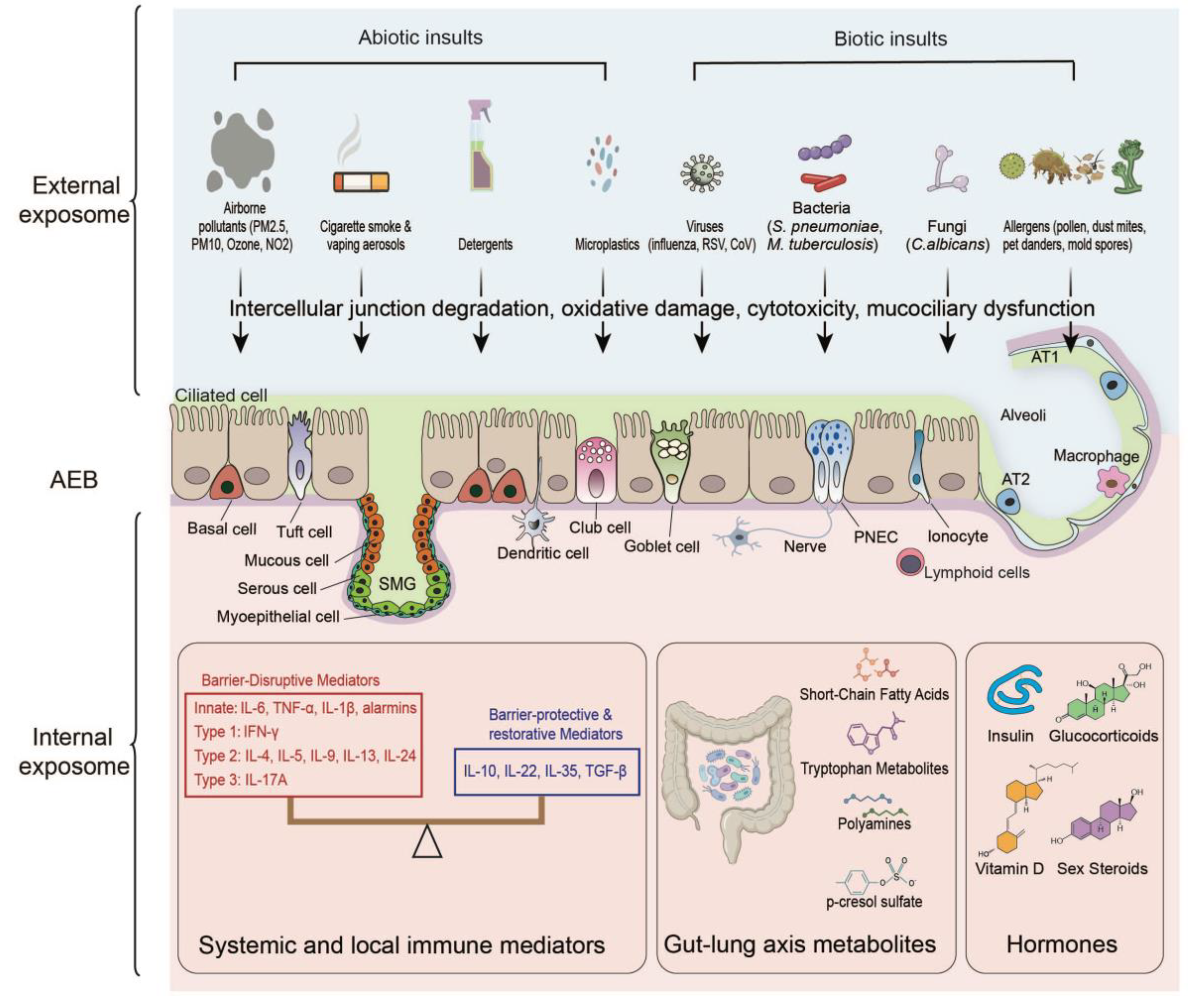
Figure 2.
Proposed conceptual framework for integrating external and internal exposome data using computational models to derive expotypes and endotypes for precision respiratory medicine. This framework represents a roadmap for future research; validation in prospective clinical cohorts is required before clinical translation.
Figure 2.
Proposed conceptual framework for integrating external and internal exposome data using computational models to derive expotypes and endotypes for precision respiratory medicine. This framework represents a roadmap for future research; validation in prospective clinical cohorts is required before clinical translation.
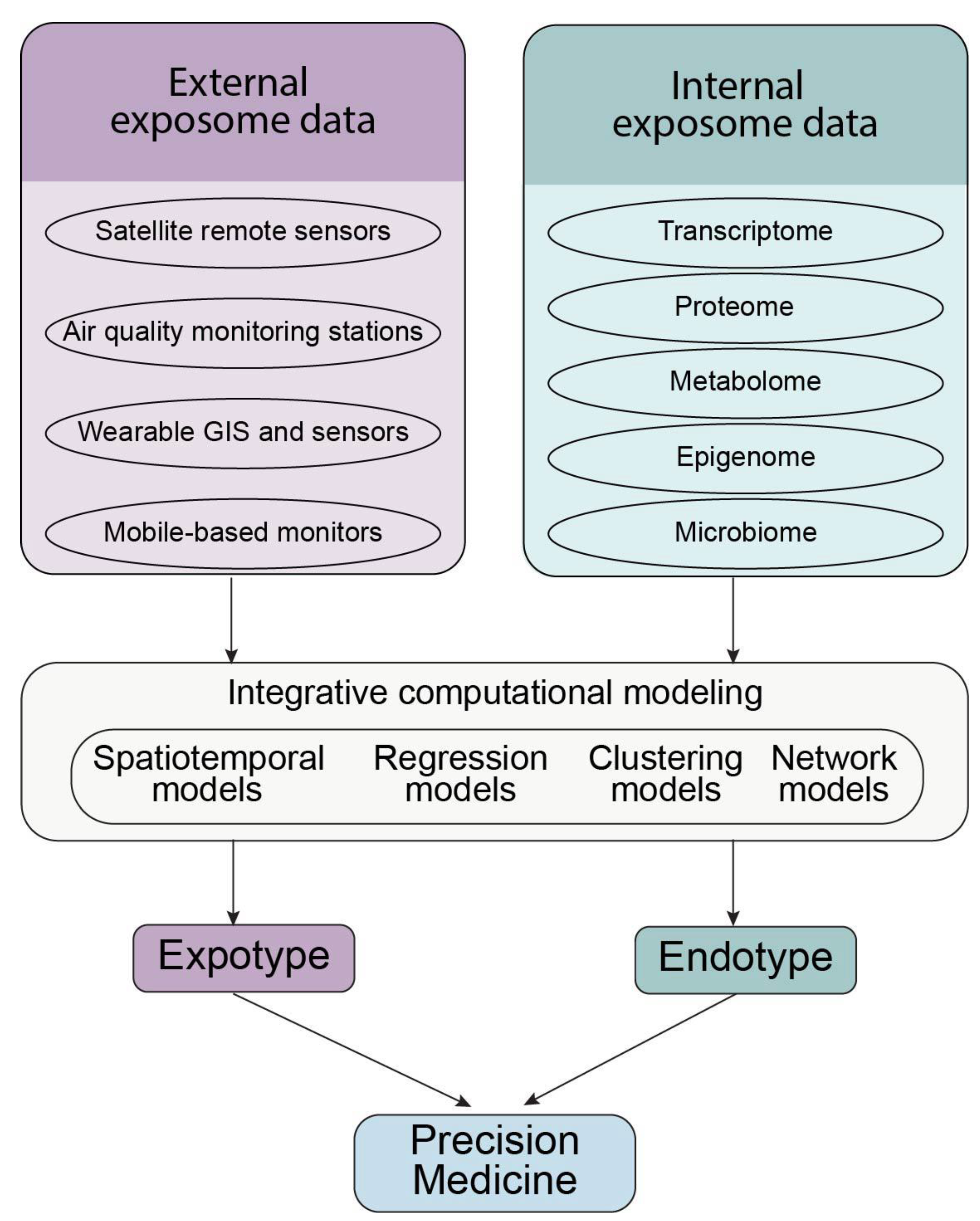

Table 1.
Molecular signatures for the identification of human respiratory epithelial cells.
| Cell Type | Key Molecular Markers | Reference |
| Ciliated cells | FOXJ1, PIFO, TPPP3, SNTN | [13] |
| Goblet cells | MUC5AC, MUC5B, MSMB | [13] |
| Club cells | Muc5b, Bpifb1, Scgb3a1, Scgb3a2, and Bpifa1 | [14] |
| Tuft cells | TRPM5, GNG13, RGS13, TAS1R1, GNAT3, IL25 | [15] |
| Ionocytes | BSND, CFTR, ATP6V0D2, ATP6V1B1, CLCNKA, CLCNKB | [15] |
| Neuroendocrine (NE) cells | SCL1, CALCA, CHGA, CPE | [15] |
| Mucous cells | MUC5B (high), MUC5AC (low/absent) | [16] |
| Serous cells | LTF, LYZ, PIP | [16] |
| Myoepithelial cells | ACTA2, KRT5, PCP4 | [17] |
| Basal cells | TP63, KRT5, KRT15 | [13] |
Disclaimer/Publisher’s Note: The statements, opinions and data contained in all publications are solely those of the individual author(s) and contributor(s) and not of MDPI and/or the editor(s). MDPI and/or the editor(s) disclaim responsibility for any injury to people or property resulting from any ideas, methods, instructions or products referred to in the content. |
© 2026 by the authors. Licensee MDPI, Basel, Switzerland. This article is an open access article distributed under the terms and conditions of the Creative Commons Attribution (CC BY) license (http://creativecommons.org/licenses/by/4.0/).
Copyright: This open access article is published under a Creative Commons CC BY 4.0 license, which permit the free download, distribution, and reuse, provided that the author and preprint are cited in any reuse.