Submitted:
18 August 2025
Posted:
19 August 2025
You are already at the latest version
Abstract
Cell-free tumor DNA (cftDNA) and cell-free tumor RNA (cftRNA) are emerging as powerful biomarkers for cancer detection, monitoring, and prognosis. These nucleic acids, released into the bloodstream by tumor cells, carry cancer-specific genetic and epigenetic alterations and can be detected non-invasively. Detection before clinical diagnosis offers a unique opportunity for earlier intervention yet requires longitudinal cohort studies to establish pre-diagnostic biomarker profiles. Current technologies enable sensitive quantification of cftDNA and cftRNA, with spike-in controls allowing absolute quanti-fication of single nucleosome-bound cftDNA, addressing a key limitation in liquid biopsy assays. Advances such as DNA-PAINT now permit single-molecule resolution detection of point mutations and methylation patterns characteristic of cancer, while new proteomics methods can identify the tissue of origin of exosome-derived nucleic acids. This review discusses state-of-the-art detection strategies for cftDNA and cftRNA, highlights gaps in longitudinal sampling, and outlines future research directions toward integrating mul-ti-omic liquid biopsy approaches for improved early diagnosis, monitoring, and relapse detection.
Keywords:
cell-free tumor DNA
; cell-free tumor RNA
; DNA-PAINT
; nucleosome-bound DNA
; liquid biopsy
; cancer biomarkers
; exosomes
; proteomics
1. Introduction
Cancer remains one of the leading causes of morbidity and mortality worldwide, with outcomes heavily influenced by the stage at diagnosis [1]. Traditional diagnostic approaches—including tissue biopsies, imaging modalities, and serum protein markers—often fall short in detecting early-stage disease or predicting recurrence [2]. Liquid biopsies, which analyze components of tumor-derived material circulating in the bloodstream, offer a promising, minimally invasive alternative for cancer detection, prognosis, and therapeutic monitoring [3,4].
Cell-free tumor DNA (cftDNA), found mostly bound to nucleosomes [5,6], and cell-free tumor RNA (cftRNA), found mostly in extracellular vesicles [7], have emerged as promising non-invasive biomarkers for cancer detection, monitoring, and prognosis. These nucleic acids are released into the bloodstream from tumor cells through apoptosis, necrosis, or active secretion. Because they carry tumor-specific genetic and epigenetic alterations, cftDNA and cftRNA offer a unique molecular snapshot of the tumor's current state, potentially enabling earlier diagnosis and better prediction of disease progression than traditional imaging or tissue biopsy approaches [8].
In cancer management, the ability to detect and quantify cftDNA and cftRNA before diagnosis offers the possibility of identifying malignant disease at a stage when intervention is most effective [9]. This requires large-scale longitudinal cohort studies in which samples from healthy individuals are collected and monitored over time to identify those who later develop cancer [9]. Such studies could reveal early molecular events in tumorigenesis and define baseline biomarker profiles for different populations [9].
Beyond diagnosis, cftDNA and cftRNA can provide valuable information for monitoring therapeutic response and detecting relapse before clinical symptoms or imaging findings appear [10]. In this context, sensitive and specific detection methods are essential, with technological advances continuing to push the boundaries of what is possible in liquid biopsy assays [10]. Spike-in Controls [11], for example, now allow for the absolute quantification of single nucleosome-bound cftDNA in serum or plasma, addressing a key analytical challenge for liquid biopsy research [12].
While this review focuses on cell-free nucleic acids, it is important to acknowledge that circulating tumor cells (CTCs) represent another class of liquid biopsy biomarker [13]. CTCs are intact tumor cells shed from primary or metastatic sites into the bloodstream, and they can provide complementary information about tumor biology, including cellular morphology, protein expression, and functional assays [13]. However, the biology, isolation techniques, and clinical applications of CTCs differ significantly from cftDNA and cftRNA[13]. Given these distinctions, the detailed discussion of CTC-based methods is beyond the scope of this review, though future integrated approaches may combine both CTC and cell-free nucleic acid analyses for comprehensive cancer profiling.
The following sections will examine the current state of detection technologies for cftDNA and cftRNA, including their use in pre-diagnostic and relapse monitoring contexts, with an emphasis on advanced methods for quantifying single nucleosome-bound cftDNA and detecting RNA species from extracellular vesicles.
2. Materials and Methods
This review is based on a comprehensive literature search of peer-reviewed publications from PubMed, Scopus, and Google Scholar, using search terms including “cftDNA detection,” “cftRNA,” “liquid biopsy,” “DNA methylation,” “epigenetic kits,” “Epicypher,” and “longitudinal cancer biomarkers.” Studies included spanned preclinical and clinical applications across a variety of cancer types, with emphasis on methodologies that demonstrate high analytical performance and potential for translational use.
For technology-specific sections, we focused on the most used and emerging commercial platforms. For cftDNA, these included CUT&Tag [14], CUT&RUN [15], and Spike-in Control assays [11], bisulfite conversion kits (e.g., Zymo Research, Qiagen), droplet digital PCR (ddPCR) [16], BEAMing (Beads, Emulsion, Amplification, and Magnetics), and hybrid-capture next generation sequencing (NGS) methods [17]. For cftRNA, we evaluated protocols employing exosomal RNA extraction (e.g., ExoRNeasy, Qiagen, Inc.), RNA-seq with UMIs (Unique Molecular Identifiers) [18], and transcript-specific reverse transcription with multiplex amplification (reviewed in [19]).
We also included recent data from longitudinal studies such as the Circulating Cell-free Genome Atlas (CCGA), sponsored by GRAIL, LLC (Menlo Park, CA)[20], and the DETECT-A trial[21], which have provided key insights into the timing and predictive power of cell-free nucleic acids. An early version of the Graphic Abstract was made by ChatGPT5, but was heavily modified by the author. ChatGPT5 was also used to improve the quality and readability of the text.
3. Results
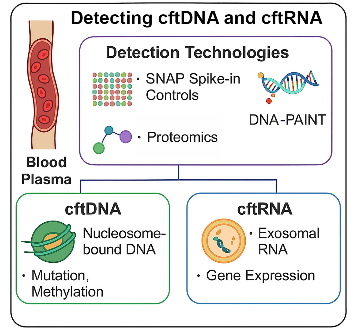
3.1. Detection Technologies for cftDNA
This section reviews current and emerging methods for identifying and characterizing circulating tumor DNA in patient blood samples. It highlights approaches ranging from advanced sequencing and digital PCR to novel single-molecule and epigenetic profiling technologies that improve sensitivity, specificity, and clinical utility in cancer diagnostics (Table 1).
3.1.1. Quantifying Nucleosome-Bound cftDNA Using Epicypher SNAP Spike-in Controls
Cell-free tumor DNA (cftDNA) is often fragmented to mono- and di-nucleosomal lengths, making it essential to precisely quantify and characterize nucleosome-bound DNA in plasma [12]. Epicypher’s SNAP (Semi-synthetic Nucleosome Spike-in) controls represent a powerful advancement in this area[11]. These spike-ins consist of synthetic mononucleosomes with defined DNA and histone modifications that mimic endogenous chromatin particles [11]. When added to plasma-derived cftDNA workflows[35], SNAP controls serve as internal standards to normalize assay variability and calibrate recovery efficiency, enabling absolute quantification of single nucleosome-bound DNA fragments (https://www.epicypher.com/products/spike-in-controls/) [11].
Unlike CUT&Tag [14] and CUT&RUN [15]—which are more suitable for mapping histone modifications on chromatin within intact cells—SNAP spike-ins are directly applicable to low-input biofluids like plasma [11]. They allow researchers to assess not only total cftDNA yield but also infer nucleosome positioning and histone post-translational modification (PTM) patterns in circulating tumor-derived chromatin [11]. This is critical for detecting epigenetic changes linked to cancer type, stage, or progression.
3.1.2. DNA Methylation Detection
DNA methylation profiling is one of the most robust and specific biomarkers for cancer detection. Cell-free methylated DNA immunoprecipitation sequencing (cfMeDIP-seq) [29,36], enzymatic methyl-seq (EM-seq) [37], and bisulfite conversion-based platforms (e.g., Zymo EZ DNA Methylation, Qiagen EpiTect) are widely used [38]. These methods can identify tissue-specific methylation patterns that are highly enriched in cancer, such as those in SEPT9 (colorectal cancer) [39], SHOX2 (lung cancer) [40,41], and GSTP1 (prostate cancer) [42,43]. Commercial kits are now capable of single-base resolution methylation detection with ultra-low input, making them suitable for liquid biopsies.
3.1.3. Fragmentomics and Size-Selective Enrichment
The size and structure of cftDNA fragments offer an additional diagnostic layer. Tumor-derived cftDNA often exhibits distinct fragmentation patterns (~147–166 bp, corresponding to mononucleosomes), jagged ends, and preferred cleavage motifs [44]. Size-selection and fragmentomics approaches, including automated microfluidic electrophoresis (e.g., Agilent TapeStation) or targeted sequencing of preferred size bands, can enrich tumor-specific cftDNA and improve mutation or epigenetic signal detection [44].
3.1.4. Mutation Detection: Digital PCR and NGS Panels
Mutation detection remains a cornerstone of cftDNA diagnostics. Droplet digital PCR (ddPCR) and BEAMing offer allele-specific sensitivity for known hotspot mutations, down to 0.01% variant allele frequency[16]. Broader mutation profiling is enabled by hybrid-capture and amplicon-based next-generation sequencing (NGS) platforms such as Guardant360 [45,46] and FoundationOne Liquid CDx [32,47]. Incorporating unique molecular identifiers (UMIs) into NGS libraries has significantly reduced error rates, making it feasible to confidently detect ultra-rare variants in plasma DNA [48,49].
3.1.5. DNA-PAINT in Cancer Detection
DNA-PAINT (DNA-based Point Accumulation for Imaging in Nanoscale Topography) is an emerging super-resolution microscopy method that offers unprecedented sensitivity and resolution in detecting cancer-associated DNA alterations [50]. By using short, fluorescently labeled DNA “imager” strands that transiently bind to complementary “docking” strands, DNA-PAINT achieves nanoscale resolution imaging without requiring complex optics [51].
-
High Sensitivity and Multiplexing:DNA-PAINT can detect DNA sequences at extremely low concentrations (femtomolar range), making it ideal for identifying rare cancer-associated mutations in liquid biopsies. It also supports multiplexing, allowing simultaneous detection of multiple oncogenic mutations, methylation sites, or genomic rearrangements in the same sample [51].
-
Point Mutation and Methylation Detection:Recent developments enable DNA-PAINT to identify single-nucleotide variants and methylation patterns characteristic of cancer DNA [50]. Methylated DNA often has a higher melting temperature than unmethylated DNA, and pattern recognition approaches like those used in the GRAIL platform can be applied to DNA-PAINT datasets for classification of cancer type and stage [50].
-
Single-Molecule Resolution for Amplified DNA:DNA-PAINT can visualize amplified DNA sequences, chromosomal rearrangements, and telomeric repeats with high spatial precision, providing both quantitative and structural insights into tumor DNA architecture [51].
-
Exosome and Tissue-of-Origin Profiling:When applied to exosome-derived nucleic acids, DNA-PAINT can be combined with advanced proteomics and proximity-barcoding methods that profile the protein cargo of exosomes to identify their tissue of origin [52]. This integration allows determination of where cftDNA originated, adding an important layer of diagnostic specificity [52].
-
Advantages Over Other Methods:Compared to fluorescence in situ hybridization (FISH) or PCR-based assays, DNA-PAINT offers superior spatial resolution, lower background noise, and reduced need for amplification, minimizing the risk of introducing errors [51]. The method’s flexibility enables adaptation for DNA, RNA, or protein targets, making it a versatile addition to the cancer biomarker detection toolkit [51].
Together, these technologies provide a multifaceted toolkit for analyzing cftDNA—each contributing to mutation, epigenetic, or structural biomarker development. SNAP spike-in controls enhance quantitative rigor, particularly in early detection or treatment monitoring contexts where changes in nucleosome-bound DNA abundance may reflect subtle tumor dynamics [11].
3.2. Detection Technologies for cftRNA
Detecting and quantifying cell-free tumor RNA (cftRNA) remains technically challenging due to its inherent instability and low abundance in plasma. Nevertheless, several methods have emerged to improve RNA preservation, enrichment, and analysis—making cftRNA a promising biomarker for cancer detection, especially for gene expression and fusion transcript profiling (Table 2).
3.2.1. RNA Stabilization and Isolation
Because cftRNA is highly susceptible to RNase degradation, immediate stabilization after blood draw is critical. Specialized blood collection tubes (e.g., PAXgene Blood ccfRNA Tubes, Streck RNA Complete BCT) stabilize RNA for up to several days at room temperature [64,65]. Following plasma separation, RNA is extracted using kits optimized for low-abundance RNA species (e.g., Qiagen miRNeasy Serum/Plasma Kit, Norgen cfRNA/cfDNA Purification Kit), which co-purify small RNAs, mRNAs, and lncRNAs [64,65].
3.2.2. Exosomal RNA Enrichment
A significant portion of circulating RNA is packaged within extracellular vesicles such as exosomes. These lipid-bound vesicles protect RNA from degradation and often carry tumor-specific transcripts. Kits such as ExoRNeasy (Qiagen) [66]and ExoQuick (System Biosciences) [67,68] isolate exosomal RNA, which can then be profiled using RNA-Seq, RT-qPCR, or digital PCR. Exosomal RNA has been shown to carry oncogenic transcripts like EGFRvIII in glioblastoma [69,70] and fusion RNAs such as EML4-ALK in lung cancer [71].
3.2.3. Reverse Transcription and Amplification Technologies
Low-input RNA samples require highly efficient reverse transcription and cDNA amplification. The use of random primers, target-specific primers, or UMIs helps preserve transcript complexity and allows for quantitative tracking of individual RNA molecules. Platforms such as Takara SMARTer kits and Qiagen's QIAseq miRNA library kits are optimized for ultra-low input and small RNA profiling.
3.2.4. RNA-Seq and Digital PCR Applications
RNA-Seq is increasingly used to analyze cftRNA, particularly when targeting known cancer-associated gene panels. However, full transcriptome sequencing is often impractical due to low RNA yield [72]. As an alternative, targeted panels (e.g., AmpliSeq Transcriptome, Oncomine Focus RNA) enable deep coverage of oncogenic transcripts and fusions with high sensitivity [73]. Digital droplet PCR (ddPCR) offers a lower-cost and highly quantitative alternative, especially for recurrent fusion transcripts (e.g., BCR-ABL [74], TMPRSS2-ERG [75]) or splice variants [76].
3.2.5. Circular and Non-Coding RNAs
Emerging research indicates that circular RNAs (circRNAs) and long non-coding RNAs (lncRNAs) are stable and informative components of cftRNA [77]. These species are often overexpressed in cancer and resistant to exonuclease degradation (reviewed in [78]). Detection platforms are being developed to capture and sequence circRNAs and lncRNAs from plasma with high specificity [77].
Together, these RNA detection strategies enable increasingly sensitive, specific, and reproducible profiling of tumor-derived transcripts from peripheral blood. While still limited by stability and technical complexity compared to cftDNA, cftRNA offers dynamic expression-based biomarkers that may provide additional dimensions for cancer detection and prognosis.
3.3. cftDNA and cftRNA from Non-Blood Body Fluids
While blood plasma remains the most widely used and validated source for cftDNA and cftRNA analysis, emerging research highlights the potential of other body fluids as complementary or alternative sources for cancer detection, prognosis, and monitoring. These “non-blood liquid biopsies” can, in some cases, offer higher sensitivity for certain malignancies, particularly when tumors are anatomically close to the sampled fluid or when blood-based cftDNA and cftRNA are scarce (Figure 1) [79].
3.3.1. cftDNA and cftRNA from Cerebrospinal fluid (CSF) for CNS Tumor Detection and Monitoring
Cerebrospinal fluid (CSF) is increasingly recognized as a uniquely informative biofluid for the analysis of cell-free tumor DNA (cftDNA) in patients with primary and metastatic tumors of the central nervous system (CNS) [80]. The blood–brain barrier severely limits the release of tumor-derived nucleic acids into peripheral circulation, meaning that plasma-based cftDNA assays often yield false negatives or very low variant allele fractions in CNS malignancies [80]. In contrast, CSF—obtained via lumbar puncture or, less commonly, ventricular access—lies in direct contact with CNS tumors, including gliomas, medulloblastomas, and brain metastases from systemic cancers such as lung, breast, and melanoma [80]. This anatomical proximity results in higher cftDNA concentrations, reduced dilution by non-tumor cfDNA, and lower interference from hematopoietic background DNA, thereby enabling more accurate mutation profiling [80].
Molecular analysis of CSF-derived cftDNA has demonstrated clinical utility in multiple domains: (1) improving diagnostic sensitivity when surgical biopsy is not feasible [81], (2) enabling molecular subtyping and detection of actionable driver alterations such as IDH1/2 [82], TERT promoter [83], or H3K27M mutations in gliomas [84], (3) guiding targeted therapy selection in metastatic CNS disease (e.g., EGFR T790M in lung adenocarcinoma brain metastases) [85], (4) tracking minimal residual disease (MRD) and early recurrence before radiographic progression [84], and (5) monitoring the emergence of treatment resistance [86]. Beyond DNA point mutations, copy number changes, structural rearrangements, and methylation signatures in CSF, cftDNA and cftRNA offers additional diagnostic and prognostic value.
Despite its advantages, CSF sampling for any disease carries limitations: lumbar puncture is moderately invasive and unsuitable for some patients [87]; cftDNA yields can vary with tumor location, size, and degree of leptomeningeal involvement [88]; and standardized pre-analytical protocols (e.g., cell-free DNA stabilization, storage, and extraction methods) remain lacking for any bodily fluid [89]. Furthermore, while targeted NGS and digital PCR have shown high analytical sensitivity, ultra-low input requirements, assay harmonization, and cross-center reproducibility are still ongoing challenges [90]. Morearge, prospective, multi-institutional trials are needed to fully establish the clinical validity and utility of CSF-derived cftDNA as a frontline or complementary liquid biopsy modality for CNS malignancies [91].
3.3.2. cftDNA and cftRNA from Urine for Bladder, Kidney, and Prostate Cancer
Urinary cftDNA offers a compelling, entirely non-invasive source for tumor-derived genetic material, particularly for cancers of the urinary tract such as bladder [92], kidney [93], and prostate cancer [94], but also for distant malignancies that shed DNA into the circulation and subsequently into urine via glomerular filtration [95]. The key advantage of urine sampling is the ease and frequency with which it can be obtained, facilitating serial monitoring without the logistical or psychological barriers associated with phlebotomy. Tumor-derived DNA in urine exists both as cell-free fragments from plasma ultrafiltration (transrenal DNA) [95] and as locally shed DNA from exfoliated cancer cells within the urinary tract [92].
Analytical sensitivity can be challenged by lower DNA concentrations compared to plasma and the presence of high-molecular-weight genomic DNA from normal urothelial cells, which can dilute tumor-specific signals [96]. Pre-analytical variables such as diurnal variation, sample volume, and the need for immediate stabilization of nucleic acids are critical to preserve fragment integrity and methylation patterns [96]. Advanced enrichment strategies, including size selection to capture the shorter, tumor-derived fragments and targeted methylation profiling, have improved detection rates for early disease and minimal residual disease (MRD) [96]. With standardized workflows, urinary cftDNA could complement plasma-based assays or serve as a frontline tool for cancers with direct urinary tract involvement.
3.3.3. cftDNA and cftRNA from Saliva for Cancers of the Aerodigestive Tract
Saliva represents an underutilized but highly accessible medium for cftDNA analysis, particularly valuable for head and neck squamous cell carcinoma (HNSCC), oral cavity cancers, and other tumors within the aerodigestive tract that shed DNA locally into oral fluids [97]. Saliva-based cftDNA sampling is non-invasive, painless, and suitable for frequent home-based collection, offering a unique opportunity for population-level cancer screening and monitoring [98]. The tumor-derived DNA within saliva may be derived from local tumor shedding, gingival crevicular fluid, or transudation from plasma, creating a heterogeneous mixture of DNA fragment sources [98].
Challenges include enzymatic degradation by oral nucleases, contamination from oral microbiota, and variability in saliva production rates influenced by hydration, circadian rhythms, or glandular pathology [97]. Optimized collection devices with nucleic acid stabilizers and rapid downstream processing are crucial to preserving fragment size profiles and epigenetic signatures [97]. Molecular assays such as targeted NGS, droplet digital PCR, and methylation-specific PCR have shown high sensitivity for detecting tumor mutations and HPV DNA in HNSCC patients [99]. Saliva-based cftDNA, especially when paired with plasma analysis, could expand the reach of precision oncology into community and remote settings [100].
3.3.4. cftDNA and cftRNA from Pleural Fluid for Malignant Cancers
Pleural effusions are often rich in tumor-derived DNA when caused by malignant processes such as metastatic lung, breast, or ovarian cancers, or malignant mesothelioma [101]. Because pleural fluid directly bathes tumor deposits in the pleural space, cftDNA from this compartment may display higher variant allele fractions (VAFs) and greater tumor heterogeneity than concurrent plasma samples [102]. This makes pleural fluid a valuable source for genomic profiling when tissue biopsy is not feasible or yields insufficient material [102]. In addition to somatic mutation detection, pleural fluid cftDNA preserves fragmentomic and methylomic signatures that may reflect the tumor’s local microenvironment [103].
However, the utility of pleural fluid is limited by its invasive collection method (thoracentesis), potential sampling bias due to loculated effusions [104], and variability in cftDNA yield depending on fluid cellularity and turnover rates [101]. Standardization of cell-free supernatant processing—separating cftDNA from cellular gDNA—is essential to prevent signal dilution [101]. Given its high tumor content, pleural fluid cftDNA has particular promise in guiding targeted therapy selection, resistance mutation tracking, and real-time disease monitoring for pleural malignancies [79].
3.3.5. Advantages of Non-Blood Fluids
Non-blood fluids offer several advantages:
- Higher local cftDNA concentration when sampling near the tumor site (e.g., CSF for CNS tumors, urine for bladder cancer) [79].
- Utility for inaccessible tumors, where tissue biopsy is high-risk or impractical [79].
- Enhanced tumor heterogeneity profiling, as sampling from multiple fluids may capture distinct subclonal populations [101].
- Facilitation of serial monitoring, particularly with easily collectible fluids such as urine or saliva [97].
However, these approaches face technical and clinical challenges, including variability in cfDNA yield, the influence of local inflammation or infection on background DNA levels, and the need for standardized pre-analytical and analytical workflows [105,106]. While still in the translational phase for many cancer types, multi-fluid cfDNA and cfRNA analysis holds promise for expanding the reach and resolution of liquid biopsy–based oncology [105,106].
4. Discussion
The field of circulating nucleic acid biomarkers has evolved rapidly over the past decade, driven by technological advances that increase sensitivity, specificity, and the amount of information retrievable from small plasma volumes. Among the most promising advances for cftDNA detection are SNAP Spike-in Controls [77], which enable quantitative measurement of single nucleosome-bound DNA in plasma. Unlike conventional ChIP-seq-based methods, these controls are optimized for fragmented DNA typical of plasma samples, providing a rigorous reference for methylation and histone modification studies [77]. Their use addresses a persistent challenge in plasma cfDNA research: distinguishing true biological variation from technical noise.
DNA-PAINT offers an entirely different advantage—single-molecule resolution combined with multiplexing capability [51]. Its potential in liquid biopsy is twofold: direct detection of point mutations and methylation profiling from individual cftDNA molecules, and the possibility of identifying DNA fragment patterns indicative of tissue-of-origin [50]. The latter is particularly promising when integrated with emerging proteomic approaches to analyze exosomal protein cargo [52], which can reveal the tissue type from which the DNA or RNA originated. Such integrative analysis could transform both early detection and relapse monitoring.
Despite these advances, significant challenges remain, as detailed in the next section (Figure 2). Biological variability, tumor heterogeneity, and technical inconsistencies can complicate interpretation. The requirement for ultra-sensitive detection also increases susceptibility to contamination and background signals. Furthermore, while current methods excel in retrospective analyses, their real-world performance in prospective, longitudinal cohorts is less well-established.
5. Limitations and Future Directions
Future research on cftDNA and cftRNA biomarkers should prioritize multi-omic integration. Combining single-molecule DNA-PAINT for genetic and epigenetic profiling with proteomic tissue-of-origin mapping can create comprehensive diagnostic signatures [50]. Longitudinal cohort studies should be designed to collect pre-diagnostic and post-diagnostic plasma samples to identify early molecular changes that precede clinical symptoms.
However, several limitations remain before these approaches can be fully implemented in clinical practice (Figure 2). One major challenge is the low concentration of cftDNA and cftRNA in the bloodstream during early cancer stages or after effective treatment, necessitating highly sensitive detection platforms. Additionally, there is currently a lack of standardization in sample collection, storage, assay procedures, and data analysis workflows, making cross-study comparisons difficult and slowing regulatory approval.
Cost and accessibility also remain significant barriers; many advanced detection methods require specialized equipment and expertise, limiting their adoption in routine diagnostics. Furthermore, the risk of false positives and false negatives persists due to the difficulty of distinguishing tumor-derived nucleic acids from background material shed by healthy cells or arising from benign physiological conditions.
Addressing these limitations will require coordinated efforts to establish standardized operating procedures and quality control measures. Automation of workflows, including SNAP Spike-in–based nucleosome quantification, could enhance scalability and reproducibility. For cftRNA specifically, the development of more robust RNA preservation and isolation methods will be essential to maintain integrity and maximize diagnostic yield.
Another priority is expanding bioinformatic frameworks capable of integrating large, multi-modal datasets from DNA, RNA, and proteomics. Machine learning could be leveraged to detect subtle, clinically relevant molecular patterns predictive of disease progression or relapse. Finally, regulatory frameworks must evolve alongside technological advances to ensure that new assays are validated rigorously but adopted rapidly enough to benefit patient care.
6. Conclusions
The integration of circulating tumor DNA and RNA analysis across both blood and non-blood biofluids holds significant promise for advancing early cancer detection, monitoring therapeutic response, and guiding personalized treatment strategies. While current challenges—such as assay standardization, sensitivity in low-tumor-burden settings, and validation in large, diverse cohorts—must be addressed, ongoing technological innovations and multiomics approaches are rapidly enhancing the clinical utility of these biomarkers. Ultimately, the convergence of improved detection platforms, robust bioinformatics pipelines, and expanded biofluid sampling will enable liquid biopsy technologies to transition from research tools to routine components of precision oncology.
Funding
This research was funded by the National Institutes of Health, grant numbers 5UG3OD023285, 5P42ES030991, and 1P30ES036084.
Acknowledgments
The author has reviewed and edited the output and takes full responsibility for the content of this publication.
Conflicts of Interest
The author declares no conflicts of interest.
Abbreviations
The following abbreviations are used in this manuscript:
| cftDNA | Cell free tumor DNA |
| cftRNA | Cell free tumor RNA |
| cfDNA | Cell free DNA (not necessarily from tumor) |
| cfRNA | Cell free RNA (not necessarily from tumor) |
| UMI | Unique molecular identifier |
| ddPCR | Droplet digital polymerase chain reaction |
| WGS | Whole genome sequencing |
| PTM | Post translational modifications |
| DNA-PAINT | DNA points accumulation for imaging in nanoscale topography |
| FISH | Fluorescent in situ hybridization |
| EV | Extracellular vesicle |
| RT-qPCR | Reverse transcriptase quantitative polymerase chain reaction |
| circRNA | Circular RNA |
| lncRNA | Long noncoding RNA |
| FRET | Fluorescence resonance energy transfer |
| MRD | Minimal residual disease |
| CSF | Cerebral spinal fluid |
| HNSCC | Head and neck small cell carcinoma |
| VAF | Variable allele frequency |
References
- Crosby, D.; Bhatia, S.; Brindle, K.M.; Coussens, L.M.; Dive, C.; Emberton, M.; Esener, S.; Fitzgerald, R.C.; Gambhir, S.S.; Kuhn, P.; et al. Early detection of cancer. Science 2022, 375, eaay9040. [Google Scholar] [CrossRef]
- Milner, D.A., Jr.; Lennerz, J.K. Technology and Future of Multi-Cancer Early Detection. Life (Basel) 2024, 14. [Google Scholar] [CrossRef]
- Xu, Y.; Zhu, S.; Xia, C.; Yu, H.; Shi, S.; Chen, K.; He, Y.; Deng, C.; Jin, H.; Liu, J.; et al. Liquid biopsy-based multi-cancer early detection: an exploration road from evidence to implementation. Sci Bull (Beijing) 2025. [Google Scholar] [CrossRef]
- Madar, S.; Amor, R.E.; Furman-Assaf, S.; Friedman, E. Innovative Approaches to Early Detection of Cancer-Transforming Screening for Breast, Lung, and Hard-to-Screen Cancers. Cancers (Basel) 2025, 17. [Google Scholar] [CrossRef]
- Martins, I.; Ribeiro, I.P.; Jorge, J.; Goncalves, A.C.; Sarmento-Ribeiro, A.B.; Melo, J.B.; Carreira, I.M. Liquid Biopsies: Applications for Cancer Diagnosis and Monitoring. Genes (Basel) 2021, 12. [Google Scholar] [CrossRef]
- Nikanjam, M.; Kato, S.; Kurzrock, R. Liquid biopsy: current technology and clinical applications. J Hematol Oncol 2022, 15, 131. [Google Scholar] [CrossRef] [PubMed]
- Loy, C.; Ahmann, L.; De Vlaminck, I.; Gu, W. Liquid Biopsy Based on Cell-Free DNA and RNA. Annu Rev Biomed Eng 2024, 26, 169–195. [Google Scholar] [CrossRef] [PubMed]
- Hassan, S.; Shehzad, A.; Khan, S.A.; Miran, W.; Khan, S.; Lee, Y.S. Diagnostic and Therapeutic Potential of Circulating-Free DNA and Cell-Free RNA in Cancer Management. Biomedicines 2022, 10. [Google Scholar] [CrossRef] [PubMed]
- Ranganathan, K.; Kurian, N.S.; Goswami, H.M.; Rishi, K.D.; Veldore, V.H. Exploring the clinical utility of liquid biopsy with cfDNA in cancer: A systematic review. J Liq Biopsy 2024, 5, 100150. [Google Scholar] [CrossRef]
- Keller, L.; Belloum, Y.; Wikman, H.; Pantel, K. Clinical relevance of blood-based ctDNA analysis: mutation detection and beyond. Br J Cancer 2021, 124, 345–358. [Google Scholar] [CrossRef]
- Grzybowski, A.T.; Shah, R.N.; Richter, W.F.; Ruthenburg, A.J. Native internally calibrated chromatin immunoprecipitation for quantitative studies of histone post-translational modifications. Nat Protoc 2019, 14, 3275–3302. [Google Scholar] [CrossRef]
- Bronkhorst, A.J.; Ungerer, V.; Oberhofer, A.; Gabriel, S.; Polatoglou, E.; Randeu, H.; Uhlig, C.; Pfister, H.; Mayer, Z.; Holdenrieder, S. New Perspectives on the Importance of Cell-Free DNA Biology. Diagnostics (Basel) 2022, 12. [Google Scholar] [CrossRef]
- Saha, S.; Araf, Y.; Promon, S.K. Circulating tumor DNA in cancer diagnosis, monitoring, and prognosis. J Egypt Natl Canc Inst 2022, 34, 8. [Google Scholar] [CrossRef] [PubMed]
- Kaya-Okur, H.S.; Wu, S.J.; Codomo, C.A.; Pledger, E.S.; Bryson, T.D.; Henikoff, J.G.; Ahmad, K.; Henikoff, S. CUT&Tag for efficient epigenomic profiling of small samples and single cells. Nat Commun 2019, 10, 1930. [Google Scholar] [CrossRef]
- Skene, P.J.; Henikoff, S. An efficient targeted nuclease strategy for high-resolution mapping of DNA binding sites. Elife 2017, 6. [Google Scholar] [CrossRef] [PubMed]
- Clausen, F.B.; Jorgensen, K.; Wardil, L.W.; Nielsen, L.K.; Krog, G.R. Droplet digital PCR-based testing for donor-derived cell-free DNA in transplanted patients as noninvasive marker of allograft health: Methodological aspects. PLoS One 2023, 18, e0282332. [Google Scholar] [CrossRef] [PubMed]
- Clark, T.A.; Chung, J.H.; Kennedy, M.; Hughes, J.D.; Chennagiri, N.; Lieber, D.S.; Fendler, B.; Young, L.; Zhao, M.; Coyne, M.; et al. Analytical Validation of a Hybrid Capture-Based Next-Generation Sequencing Clinical Assay for Genomic Profiling of Cell-Free Circulating Tumor DNA. J Mol Diagn 2018, 20, 686–702. [Google Scholar] [CrossRef]
- Cabus, L.; Lagarde, J.; Curado, J.; Lizano, E.; Perez-Boza, J. Current challenges and best practices for cell-free long RNA biomarker discovery. Biomark Res 2022, 10, 62. [Google Scholar] [CrossRef]
- Tang, Y.T.; Huang, Y.Y.; Zheng, L.; Qin, S.H.; Xu, X.P.; An, T.X.; Xu, Y.; Wu, Y.S.; Hu, X.M.; Ping, B.H.; et al. Comparison of isolation methods of exosomes and exosomal RNA from cell culture medium and serum. Int J Mol Med 2017, 40, 834–844. [Google Scholar] [CrossRef]
- Bryce, A.H.; Thiel, D.D.; Seiden, M.V.; Richards, D.; Luan, Y.; Coignet, M.; Zhang, Q.; Zhang, N.; Hubbell, E.; Kurtzman, K.N.; et al. Performance of a Cell-Free DNA-Based Multi-cancer Detection Test in Individuals Presenting With Symptoms Suspicious for Cancers. JCO Precis Oncol 2023, 7, e2200679. [Google Scholar] [CrossRef]
- Lennon, A.M.; Buchanan, A.H.; Kinde, I.; Warren, A.; Honushefsky, A.; Cohain, A.T.; Ledbetter, D.H.; Sanfilippo, F.; Sheridan, K.; Rosica, D.; et al. Feasibility of blood testing combined with PET-CT to screen for cancer and guide intervention. Science 2020, 369. [Google Scholar] [CrossRef]
- Newman, A.M.; Bratman, S.V.; To, J.; Wynne, J.F.; Eclov, N.C.; Modlin, L.A.; Liu, C.L.; Neal, J.W.; Wakelee, H.A.; Merritt, R.E.; et al. An ultrasensitive method for quantitating circulating tumor DNA with broad patient coverage. Nat Med 2014, 20, 548–554. [Google Scholar] [CrossRef] [PubMed]
- Newman, A.M.; Lovejoy, A.F.; Klass, D.M.; Kurtz, D.M.; Chabon, J.J.; Scherer, F.; Stehr, H.; Liu, C.L.; Bratman, S.V.; Say, C.; et al. Integrated digital error suppression for improved detection of circulating tumor DNA. Nat Biotechnol 2016, 34, 547–555. [Google Scholar] [CrossRef] [PubMed]
- Diehl, F.; Schmidt, K.; Choti, M.A.; Romans, K.; Goodman, S.; Li, M.; Thornton, K.; Agrawal, N.; Sokoll, L.; Szabo, S.A.; et al. Circulating mutant DNA to assess tumor dynamics. Nat Med 2008, 14, 985–990. [Google Scholar] [CrossRef]
- Pinheiro, L.B.; Coleman, V.A.; Hindson, C.M.; Herrmann, J.; Hindson, B.J.; Bhat, S.; Emslie, K.R. Evaluation of a droplet digital polymerase chain reaction format for DNA copy number quantification. Anal Chem 2012, 84, 1003–1011. [Google Scholar] [CrossRef] [PubMed]
- Quan, P.L.; Sauzade, M.; Brouzes, E. dPCR: A Technology Review. Sensors (Basel) 2018, 18. [Google Scholar] [CrossRef]
- Cristiano, S.; Leal, A.; Phallen, J.; Fiksel, J.; Adleff, V.; Bruhm, D.C.; Jensen, S.O.; Medina, J.E.; Hruban, C.; White, J.R.; et al. Genome-wide cell-free DNA fragmentation in patients with cancer. Nature 2019, 570, 385–389. [Google Scholar] [CrossRef]
- Shen, S.Y.; Singhania, R.; Fehringer, G.; Chakravarthy, A.; Roehrl, M.H.A.; Chadwick, D.; Zuzarte, P.C.; Borgida, A.; Wang, T.T.; Li, T.; et al. Sensitive tumour detection and classification using plasma cell-free DNA methylomes. Nature 2018, 563, 579–583. [Google Scholar] [CrossRef]
- Shen, S.Y.; Burgener, J.M.; Bratman, S.V.; De Carvalho, D.D. Preparation of cfMeDIP-seq libraries for methylome profiling of plasma cell-free DNA. Nat Protoc 2019, 14, 2749–2780. [Google Scholar] [CrossRef]
- Wilson, S.L.; Shen, S.Y.; Harmon, L.; Burgener, J.M.; Triche, T., Jr.; Bratman, S.V.; De Carvalho, D.D.; Hoffman, M.M. Sensitive and reproducible cell-free methylome quantification with synthetic spike-in controls. Cell Rep Methods 2022, 2, 100294. [Google Scholar] [CrossRef]
- Bauml, J.M.; Li, B.T.; Velcheti, V.; Govindan, R.; Curioni-Fontecedro, A.; Dooms, C.; Takahashi, T.; Duda, A.W.; Odegaard, J.I.; Cruz-Guilloty, F.; et al. Clinical validation of Guardant360 CDx as a blood-based companion diagnostic for sotorasib. Lung Cancer 2022, 166, 270–278. [Google Scholar] [CrossRef] [PubMed]
- Woodhouse, R.; Li, M.; Hughes, J.; Delfosse, D.; Skoletsky, J.; Ma, P.; Meng, W.; Dewal, N.; Milbury, C.; Clark, T.; et al. Clinical and analytical validation of FoundationOne Liquid CDx, a novel 324-Gene cfDNA-based comprehensive genomic profiling assay for cancers of solid tumor origin. PLoS One 2020, 15, e0237802. [Google Scholar] [CrossRef]
- Jungmann, R.; Avendano, M.S.; Woehrstein, J.B.; Dai, M.; Shih, W.M.; Yin, P. Multiplexed 3D cellular super-resolution imaging with DNA-PAINT and Exchange-PAINT. Nat Methods 2014, 11, 313–318. [Google Scholar] [CrossRef]
- van Wee, R.; Filius, M.; Joo, C. Completing the canvas: advances and challenges for DNA-PAINT super-resolution imaging. Trends Biochem Sci 2021, 46, 918–930. [Google Scholar] [CrossRef]
- Diaz, I.M.; Nocon, A.; Held, S.A.E.; Kobilay, M.; Skowasch, D.; Bronkhorst, A.J.; Ungerer, V.; Fredebohm, J.; Diehl, F.; Holdenrieder, S.; et al. Pre-Analytical Evaluation of Streck Cell-Free DNA Blood Collection Tubes for Liquid Profiling in Oncology. Diagnostics (Basel) 2023, 13. [Google Scholar] [CrossRef]
- Feng, B.; Liu, T.; Feng, Y.; Huang, J.; Qin, J. From sensitivity to public health: integrating cfMeDIP-seq into early breast cancer detection strategies. J Transl Med 2024, 22, 1083. [Google Scholar] [CrossRef]
- Tanaka, Y.; Mizuguchi, R.; Koseki, N.; Suzuki, H.; Suzuki, T. Quality assessment of enzymatic methyl-seq library constructed using crude cell lysate. Biochem Biophys Res Commun 2024, 696, 149488. [Google Scholar] [CrossRef]
- Huang, J.; Wang, L. Cell-Free DNA Methylation Profiling Analysis-Technologies and Bioinformatics. Cancers (Basel) 2019, 11. [Google Scholar] [CrossRef] [PubMed]
- Zhang, J.; Li, C.; An, Y.; Wang, B.; Liang, G. Comparative analysis of SDC2 and SEPT9 methylation tests in the early detection of colorectal cancer: a systematic review and meta-analysis. Front Med (Lausanne) 2024, 11, 1460233. [Google Scholar] [CrossRef]
- Guo, Y.; Wu, P.; Liao, Q.; Huang, Z. Association of DNA methylation of RASSF1A and SHOX2 with lung cancer risk: A systematic review and meta-analysis. Medicine (Baltimore) 2024, 103, e40042. [Google Scholar] [CrossRef] [PubMed]
- Xie, M.; Zhao, Y.; Hou, X.; Li, N.; Liu, H.; Zhang, X.; Xu, X. SHOX2 and RASSF1A methylation in diagnosing malignant pleural effusion induced by lung cancer. Clin Chim Acta 2025, 572, 120273. [Google Scholar] [CrossRef] [PubMed]
- Ji, R.; Zhang, L.; Xing, Y.; Zhu, Y.; Mao, K.; Wang, M.; Xuan, K.; Yang, Y.; Lo, R.; Luo, B.; et al. Clinical significance of urine-based automated detection of GSTP1 methylation for the diagnosis of suspected prostate cancer patients. Transl Androl Urol 2025, 14, 1204–1213. [Google Scholar] [CrossRef]
- Mharrach, I.; Aqerrout, M.; Tadlaoui, K.A.; Laraqui, A.; Ghazzaly, A.E.; Ennaji, M.M. Methylation status of GSTP1 and APC promoter genes as potential biomarkers for early detection and tumor aggressiveness in prostate cancer: insights from a Moroccan cohort. Mol Biol Rep 2025, 52, 711. [Google Scholar] [CrossRef]
- Qi, T.; Pan, M.; Shi, H.; Wang, L.; Bai, Y.; Ge, Q. Cell-Free DNA Fragmentomics: The Novel Promising Biomarker. Int J Mol Sci 2023, 24. [Google Scholar] [CrossRef]
- Gupta, R.; Othman, T.; Chen, C.; Sandhu, J.; Ouyang, C.; Fakih, M. Guardant360 Circulating Tumor DNA Assay Is Concordant with FoundationOne Next-Generation Sequencing in Detecting Actionable Driver Mutations in Anti-EGFR Naive Metastatic Colorectal Cancer. Oncologist 2020, 25, 235–243. [Google Scholar] [CrossRef]
- Qi, Z.; Tokuhiro, S.; Odegaard, J.I.; Wienke, S.; Karnoub, M.; Feng, W.; Shiga, R.; Smit, E.F.; Goto, Y.; De Langen, A.J.; et al. Analytical and Clinical Validation of the Plasma-Based Guardant360 CDx Test for Assessing HER2 (ERBB2) Mutation Status in Patients with Non-Small-Cell Lung Cancer for Treatment with Trastuzumab Deruxtecan in DESTINY-Lung01/02. J Mol Diagn 2025, 27, 119–129. [Google Scholar] [CrossRef]
- Isla, D.; Alvarez, R.; Arnal, M.; Arriola, E.; Azkarate, A.; Azkona, E.; Garcia-Campelo, R.; Garrido, P.; Nadal, E.; Ortega, A.L.; et al. Detection of genomic alterations in liquid biopsies from patients with non-small cell lung cancer using FoundationOne Liquid CDx: a cost-effectiveness analysis. J Med Econ 2024, 27, 1379–1387. [Google Scholar] [CrossRef]
- Dunwell, T.L.; Dailey, S.C.; Ottestad, A.L.; Yu, J.; Becker, P.W.; Scaife, S.; Richman, S.D.; Wood, H.M.; Slaney, H.; Bottomley, D.; et al. Adaptor Template Oligo-Mediated Sequencing (ATOM-Seq) is a new ultra-sensitive UMI-based NGS library preparation technology for use with cfDNA and cfRNA. Sci Rep 2021, 11, 3138. [Google Scholar] [CrossRef] [PubMed]
- Streubel, A.; Stenzinger, A.; Stephan-Falkenau, S.; Kollmeier, J.; Misch, D.; Blum, T.G.; Bauer, T.; Landt, O.; Am Ende, A.; Schirmacher, P.; et al. Comparison of different semi-automated cfDNA extraction methods in combination with UMI-based targeted sequencing. Oncotarget 2019, 10, 5690–5702. [Google Scholar] [CrossRef] [PubMed]
- Bauer, J.; Reichl, A.; Tinnefeld, P. Kinetic Referencing Allows Identification of Epigenetic Cytosine Modifications by Single-Molecule Hybridization Kinetics and Superresolution DNA-PAINT Microscopy. ACS Nano 2024, 18, 1496–1503. [Google Scholar] [CrossRef]
- Piantanida, L.; Li, I.T.S.; Hughes, W.L. Advancements in DNA-PAINT: applications and challenges in biological imaging and nanoscale metrology. Nanoscale 2025, 17, 14016–14034. [Google Scholar] [CrossRef]
- Wu, D.; Yan, J.; Shen, X.; Sun, Y.; Thulin, M.; Cai, Y.; Wik, L.; Shen, Q.; Oelrich, J.; Qian, X.; et al. Profiling surface proteins on individual exosomes using a proximity barcoding assay. Nat Commun 2019, 10, 3854. [Google Scholar] [CrossRef]
- Lakshmi, S.; Hughes, T.A.; Priya, S. Exosomes and exosomal RNAs in breast cancer: A status update. Eur J Cancer 2021, 144, 252–268. [Google Scholar] [CrossRef]
- Jenjaroenpun, P.; Kremenska, Y.; Nair, V.M.; Kremenskoy, M.; Joseph, B.; Kurochkin, I.V. Characterization of RNA in exosomes secreted by human breast cancer cell lines using next-generation sequencing. PeerJ 2013, 1, e201. [Google Scholar] [CrossRef] [PubMed]
- Kugeratski, F.G.; Hodge, K.; Lilla, S.; McAndrews, K.M.; Zhou, X.; Hwang, R.F.; Zanivan, S.; Kalluri, R. Quantitative proteomics identifies the core proteome of exosomes with syntenin-1 as the highest abundant protein and a putative universal biomarker. Nat Cell Biol 2021, 23, 631–641. [Google Scholar] [CrossRef]
- Garcia-Martin, R.; Brandao, B.B.; Thomou, T.; Altindis, E.; Kahn, C.R. Tissue differences in the exosomal/small extracellular vesicle proteome and their potential as indicators of altered tissue metabolism. Cell Rep 2022, 38, 110277. [Google Scholar] [CrossRef]
- Roshani, M.; Baniebrahimi, G.; Mousavi, M.; Zare, N.; Sadeghi, R.; Salarinia, R.; Sheida, A.; Molavizadeh, D.; Sadeghi, S.; Moammer, F.; et al. Exosomal long non-coding RNAs: novel molecules in gastrointestinal cancers' progression and diagnosis. Front Oncol 2022, 12, 1014949. [Google Scholar] [CrossRef]
- Kocabey, S.; Chiarelli, G.; Acuna, G.P.; Ruegg, C. Ultrasensitive and multiplexed miRNA detection system with DNA-PAINT. Biosens Bioelectron 2023, 224, 115053. [Google Scholar] [CrossRef] [PubMed]
- Capone, I.; Bozzi, F.; Dagrada, G.P.; Verderio, P.; Conca, E.; Busico, A.; Testi, M.A.; Monti, V.; Duca, M.; Proto, C.; et al. Targeted RNA-sequencing analysis for fusion transcripts detection in tumor diagnostics: assessment of bioinformatic tools reliability in FFPE samples. Explor Target Antitumor Ther 2022, 3, 582–597. [Google Scholar] [CrossRef]
- Haas, B.J.; Dobin, A.; Li, B.; Stransky, N.; Pochet, N.; Regev, A. Accuracy assessment of fusion transcript detection via read-mapping and de novo fusion transcript assembly-based methods. Genome Biol 2019, 20, 213. [Google Scholar] [CrossRef]
- Kim, J.; Kang, C.; Shin, S.; Hohng, S. Rapid quantification of miRNAs using dynamic FRET-FISH. Commun Biol 2022, 5, 1072. [Google Scholar] [CrossRef]
- Urbanek, M.O.; Nawrocka, A.U.; Krzyzosiak, W.J. Small RNA Detection by in Situ Hybridization Methods. Int J Mol Sci 2015, 16, 13259–13286. [Google Scholar] [CrossRef]
- Shaba, E.; Vantaggiato, L.; Governini, L.; Haxhiu, A.; Sebastiani, G.; Fignani, D.; Grieco, G.E.; Bergantini, L.; Bini, L.; Landi, C. Multi-Omics Integrative Approach of Extracellular Vesicles: A Future Challenging Milestone. Proteomes 2022, 10. [Google Scholar] [CrossRef]
- Ma, Y.; Wang, Y.; He, L.; Du, J.; Li, L.; Bie, Z.; Li, Y.; Xu, X.; Zhou, W.; Wu, X.; et al. Preservation of cfRNA in cytological supernatants for cfDNA & cfRNA double detection in non-small cell lung cancer patients. Cancer Med 2024, 13, e70197. [Google Scholar] [CrossRef] [PubMed]
- Zhong, P.; Bai, L.; Hong, M.; Ouyang, J.; Wang, R.; Zhang, X.; Chen, P. A Comprehensive Review on Circulating cfRNA in Plasma: Implications for Disease Diagnosis and Beyond. Diagnostics (Basel) 2024, 14. [Google Scholar] [CrossRef] [PubMed]
- Su, B.; Jeyhani, M.; Thillainadesan, G.; Sheng, M.; Wunsche, R.; Dayarathna, T.; Cimolai, K.; Weng, H.; Jerzak, K.J.; Liu, S.K.; et al. Next Generation Aqueous Two-Phase System for Gentle, Effective, and Timely Extracellular Vesicle Isolation and Transcriptomic Analysis. J Extracell Vesicles 2025, 14, e70058. [Google Scholar] [CrossRef]
- Du, K.; Sun, X.; Tang, X.; Xu, H.; Duan, P.; Xu, Q.; Sang, M. [Effects of storage temperature and time on quality of plasma exosomes extracted by ExoQuick(TM) and Umibio kits]. Xi Bao Yu Fen Zi Mian Yi Xue Za Zhi 2020, 36, 330–336. [Google Scholar]
- Gemoll, T.; Rozanova, S.; Roder, C.; Hartwig, S.; Kalthoff, H.; Lehr, S.; ElSharawy, A.; Habermann, J.K. Protein Profiling of Serum Extracellular Vesicles Reveals Qualitative and Quantitative Differences After Differential Ultracentrifugation and ExoQuick(TM) Isolation. J Clin Med 2020, 9. [Google Scholar] [CrossRef]
- Ghazi, B.; Harmak, Z.; Rghioui, M.; Kone, A.S.; El Ghanmi, A.; Badou, A. Decoding the secret of extracellular vesicles in the immune tumor microenvironment of the glioblastoma: on the border of kingdoms. Front Immunol 2024, 15, 1423232. [Google Scholar] [CrossRef] [PubMed]
- Yekula, A.; Yekula, A.; Muralidharan, K.; Kang, K.; Carter, B.S.; Balaj, L. Extracellular Vesicles in Glioblastoma Tumor Microenvironment. Front Immunol 2019, 10, 3137. [Google Scholar] [CrossRef]
- Reclusa, P.; Laes, J.F.; Malapelle, U.; Valentino, A.; Rocco, D.; Gil-Bazo, I.; Rolfo, C. EML4-ALK translocation identification in RNA exosomal cargo (ExoALK) in NSCLC patients: a novel role for liquid biopsy. Transl Cancer Res 2019, 8, S76–S78. [Google Scholar] [CrossRef] [PubMed]
- Lin, Y.; Rasmussen, M.H.; Christensen, M.H.; Frydendahl, A.; Maretty, L.; Andersen, C.L.; Besenbacher, S. Evaluating Bioinformatics Processing of Somatic Variant Detection in cfDNA Using Targeted Sequencing with UMIs. Int J Mol Sci 2024, 25. [Google Scholar] [CrossRef] [PubMed]
- Pei, X.M.; Yeung, M.H.Y.; Wong, A.N.N.; Tsang, H.F.; Yu, A.C.S.; Yim, A.K.Y.; Wong, S.C.C. Targeted Sequencing Approach and Its Clinical Applications for the Molecular Diagnosis of Human Diseases. Cells 2023, 12. [Google Scholar] [CrossRef] [PubMed]
- Bochicchio, M.T.; Petiti, J.; Berchialla, P.; Izzo, B.; Giugliano, E.; Ottaviani, E.; Errichiello, S.; Rege-Cambrin, G.; Venturi, C.; Luciano, L.; et al. Droplet Digital PCR for BCR-ABL1 Monitoring in Diagnostic Routine: Ready to Start? Cancers (Basel) 2021, 13. [Google Scholar] [CrossRef]
- Check, M.H.; Ernst, S.E.; Sfanos, K.S. Use of Droplet Digital PCR for Consistent Detection of TMPRSS2:ERG Gene Fusion Transcripts Initiated In Vitro. Prostate 2025. [Google Scholar] [CrossRef]
- Grudda, T.; Jiang, C.; Lo, C.M.; Skinner, N.E.; Sulkowski, M.S.; Balagopal, A.; Thio, C.L. High-dimensional droplet digital PCR of multiple hepatitis B splice variants in serum, liver, and tissue culture. J Virol Methods 2025, 336, 115155. [Google Scholar] [CrossRef]
- Feng, X.Y.; Zhu, S.X.; Pu, K.J.; Huang, H.J.; Chen, Y.Q.; Wang, W.T. New insight into circRNAs: characterization, strategies, and biomedical applications. Exp Hematol Oncol 2023, 12, 91. [Google Scholar] [CrossRef]
- Pisignano, G.; Michael, D.C.; Visal, T.H.; Pirlog, R.; Ladomery, M.; Calin, G.A. Going circular: history, present, and future of circRNAs in cancer. Oncogene 2023, 42, 2783–2800. [Google Scholar] [CrossRef]
- Werner, B.; Warton, K.; Ford, C.E. Transcending Blood-Opportunities for Alternate Liquid Biopsies in Oncology. Cancers (Basel) 2022, 14. [Google Scholar] [CrossRef]
- Xiao, F.; Lv, S.; Zong, Z.; Wu, L.; Tang, X.; Kuang, W.; Zhang, P.; Li, X.; Fu, J.; Xiao, M.; et al. Cerebrospinal fluid biomarkers for brain tumor detection: clinical roles and current progress. Am J Transl Res 2020, 12, 1379–1396. [Google Scholar]
- Otsuji, R.; Fujioka, Y.; Hata, N.; Kuga, D.; Hatae, R.; Sangatsuda, Y.; Nakamizo, A.; Mizoguchi, M.; Yoshimoto, K. Liquid Biopsy for Glioma Using Cell-Free DNA in Cerebrospinal Fluid. Cancers (Basel) 2024, 16. [Google Scholar] [CrossRef]
- Crucitta, S.; Pasqualetti, F.; Gonnelli, A.; Ruglioni, M.; Luculli, G.I.; Cantarella, M.; Ortenzi, V.; Scatena, C.; Paiar, F.; Naccarato, A.G.; et al. IDH1 mutation is detectable in plasma cell-free DNA and is associated with survival outcome in glioma patients. BMC Cancer 2024, 24, 31. [Google Scholar] [CrossRef]
- Fontanilles, M.; Marguet, F.; Beaussire, L.; Magne, N.; Pepin, L.F.; Alexandru, C.; Tennevet, I.; Hanzen, C.; Langlois, O.; Jardin, F.; et al. Cell-free DNA and circulating TERT promoter mutation for disease monitoring in newly-diagnosed glioblastoma. Acta Neuropathol Commun 2020, 8, 179. [Google Scholar] [CrossRef] [PubMed]
- Cantor, E.; Wierzbicki, K.; Tarapore, R.S.; Ravi, K.; Thomas, C.; Cartaxo, R.; Nand Yadav, V.; Ravindran, R.; Bruzek, A.K.; Wadden, J.; et al. Serial H3K27M cell-free tumor DNA (cf-tDNA) tracking predicts ONC201 treatment response and progression in diffuse midline glioma. Neuro Oncol 2022, 24, 1366–1374. [Google Scholar] [CrossRef]
- Shi, L.; Tang, J.; Tao, H.; Guo, L.; Wu, W.; Wu, H.; Liu, Z.; Tong, L.; Wu, W.; Li, H.; et al. Detection of EGFR Mutations in Cerebrospinal Fluid of EGFR-Mutant Lung Adenocarcinoma With Brain Metastases. Front Oncol 2021, 11, 622142. [Google Scholar] [CrossRef]
- Riviere-Cazaux, C.; Dong, X.; Mo, W.; Kumar, R.; Dai, C.; Carlstrom, L.P.; Munoz-Casabella, A.; Ghadimi, K.; Nesvick, C.L.; Andersen, K.M.; et al. Longitudinal Glioma Monitoring via Cerebrospinal Fluid Cell-Free DNA. Clin Cancer Res 2025, 31, 881–889. [Google Scholar] [CrossRef]
- Shaw, L.M.; Arias, J.; Blennow, K.; Galasko, D.; Molinuevo, J.L.; Salloway, S.; Schindler, S.; Carrillo, M.C.; Hendrix, J.A.; Ross, A.; et al. Appropriate use criteria for lumbar puncture and cerebrospinal fluid testing in the diagnosis of Alzheimer's disease. Alzheimers Dement 2018, 14, 1505–1521. [Google Scholar] [CrossRef]
- Fitzpatrick, A.; Iravani, M.; Mills, A.; Childs, L.; Alaguthurai, T.; Clifford, A.; Garcia-Murillas, I.; Van Laere, S.; Dirix, L.; Harries, M.; et al. Assessing CSF ctDNA to Improve Diagnostic Accuracy and Therapeutic Monitoring in Breast Cancer Leptomeningeal Metastasis. Clin Cancer Res 2022, 28, 1180–1191. [Google Scholar] [CrossRef]
- Peng, H.; Pan, M.; Zhou, Z.; Chen, C.; Xing, X.; Cheng, S.; Zhang, S.; Zheng, H.; Qian, K. The impact of preanalytical variables on the analysis of cell-free DNA from blood and urine samples. Front Cell Dev Biol 2024, 12, 1385041. [Google Scholar] [CrossRef] [PubMed]
- Lih, C.J.; Harrington, R.D.; Sims, D.J.; Harper, K.N.; Bouk, C.H.; Datta, V.; Yau, J.; Singh, R.R.; Routbort, M.J.; Luthra, R.; et al. Analytical Validation of the Next-Generation Sequencing Assay for a Nationwide Signal-Finding Clinical Trial: Molecular Analysis for Therapy Choice Clinical Trial. J Mol Diagn 2017, 19, 313–327. [Google Scholar] [CrossRef] [PubMed]
- Liu, A.P.Y.; Smith, K.S.; Kumar, R.; Paul, L.; Bihannic, L.; Lin, T.; Maass, K.K.; Pajtler, K.W.; Chintagumpala, M.; Su, J.M.; et al. Serial assessment of measurable residual disease in medulloblastoma liquid biopsies. Cancer Cell 2021, 39, 1519–1530. [Google Scholar] [CrossRef]
- Tse, R.T.; Zhao, H.; Wong, C.Y.; Cheng, C.K.; Kong, A.W.; Peng, Q.; Chiu, P.K.; Ng, C.F.; Teoh, J.Y. Urinary Cell-Free DNA in Bladder Cancer Detection. Diagnostics (Basel) 2021, 11. [Google Scholar] [CrossRef] [PubMed]
- Nuzzo, P.V.; Berchuck, J.E.; Korthauer, K.; Spisak, S.; Nassar, A.H.; Abou Alaiwi, S.; Chakravarthy, A.; Shen, S.Y.; Bakouny, Z.; Boccardo, F.; et al. Detection of renal cell carcinoma using plasma and urine cell-free DNA methylomes. Nat Med 2020, 26, 1041–1043. [Google Scholar] [CrossRef] [PubMed]
- He, W.; Xiao, Y.; Yan, S.; Zhu, Y.; Ren, S. Cell-free DNA in the management of prostate cancer: Current status and future prospective. Asian J Urol 2023, 10, 298–316. [Google Scholar] [CrossRef] [PubMed]
- Dermody, S.M.; Bhambhani, C.; Swiecicki, P.L.; Brenner, J.C.; Tewari, M. Trans-Renal Cell-Free Tumor DNA for Urine-Based Liquid Biopsy of Cancer. Front Genet 2022, 13, 879108. [Google Scholar] [CrossRef]
- Huang, F.F.; Di, X.F.; Bai, M.H. Analysis of urine cell-free DNA in bladder cancer diagnosis by emerging bioactive technologies and materials. Front Bioeng Biotechnol 2024, 12, 1458362. [Google Scholar] [CrossRef]
- Kumar, P.; Gupta, S.; Das, B.C. Saliva as a potential non-invasive liquid biopsy for early and easy diagnosis/prognosis of head and neck cancer. Transl Oncol 2024, 40, 101827. [Google Scholar] [CrossRef]
- Rigotti, P.; Polizzi, A.; Quinzi, V.; Blasi, A.; Lombardi, T.; Lo Muzio, E.; Isola, G. Cell-Free DNA as a Prognostic Biomarker in Oral Carcinogenesis and Oral Squamous Cell Carcinoma: A Translational Perspective. Cancers (Basel) 2025, 17. [Google Scholar] [CrossRef]
- Mattox, A.K.; D'Souza, G.; Khan, Z.; Allen, H.; Henson, S.; Seiwert, T.Y.; Koch, W.; Pardoll, D.M.; Fakhry, C. Comparison of next generation sequencing, droplet digital PCR, and quantitative real-time PCR for the earlier detection and quantification of HPV in HPV-positive oropharyngeal cancer. Oral Oncol 2022, 128, 105805. [Google Scholar] [CrossRef]
- Swarup, N.; Cheng, J.; Choi, I.; Heo, Y.J.; Kordi, M.; Aziz, M.; Arora, A.; Li, F.; Chia, D.; Wei, F.; et al. Multi-faceted attributes of salivary cell-free DNA as liquid biopsy biomarkers for gastric cancer detection. Biomark Res 2023, 11, 90. [Google Scholar] [CrossRef]
- Chang, S.C.; Wei, Y.F.; Chen, C.Y.; Lai, Y.C.; Hu, P.W.; Hung, J.C.; Chang, C.Y. Profiling Cell-Free DNA from Malignant Pleural Effusion for Oncogenic Driver Mutations in Patients with Treatment-Naive Stage IV Adenocarcinoma: A Multicenter Prospective Study. Mol Diagn Ther 2024, 28, 803–810. [Google Scholar] [CrossRef]
- Guo, Z.; Xie, Z.; Shi, H.; Du, W.; Peng, L.; Han, W.; Duan, F.; Zhang, X.; Chen, M.; Duan, J.; et al. Malignant pleural effusion supernatant is an alternative liquid biopsy specimen for comprehensive mutational profiling. Thorac Cancer 2019, 10, 823–831. [Google Scholar] [CrossRef]
- Zhang, N.; Li, Y.; Zhang, H.; Dong, Y.; Zhang, C.; Du, W.; Long, C.; Xing, X.; Li, K.; Liu, Z.; et al. Performance of SHOX2 and RASSF1A methylation assay in supernatants and matched cell pellets for the diagnosis of malignant pleural effusion. Clin Chim Acta 2024, 553, 117699. [Google Scholar] [CrossRef]
- Na, M.J. Diagnostic tools of pleural effusion. Tuberc Respir Dis (Seoul) 2014, 76, 199–210. [Google Scholar] [CrossRef] [PubMed]
- Connal, S.; Cameron, J.M.; Sala, A.; Brennan, P.M.; Palmer, D.S.; Palmer, J.D.; Perlow, H.; Baker, M.J. Liquid biopsies: the future of cancer early detection. J Transl Med 2023, 21, 118. [Google Scholar] [CrossRef] [PubMed]
- Qureshi, Z.; Altaf, F.; Khanzada, M.; Safi, A.; Asghar, Z.; Warraich, D.; Shah, S. Liquid biopsies for early detection and monitoring of cancer: advances, challenges, and future directions. Ann Med Surg (Lond) 2025, 87, 3244–3253. [Google Scholar] [CrossRef] [PubMed]
Figure 1.
Detection of cftDNA and cftRNA from blood and non-blood biofluids. (a) Isolation of cftDNA and cftRNA from blood and serum, such as for multiple cancer detection methods. (b) Isolation from cerebral spinal fluid (CSF), such as from brain cancer patients. (c) Isolation from urine, such as from bladder cancer patients. (d) Isolation from saliva, such as from throat cancer patients. (e) Isolation from pleural fluid, such as from lung cancer patients.
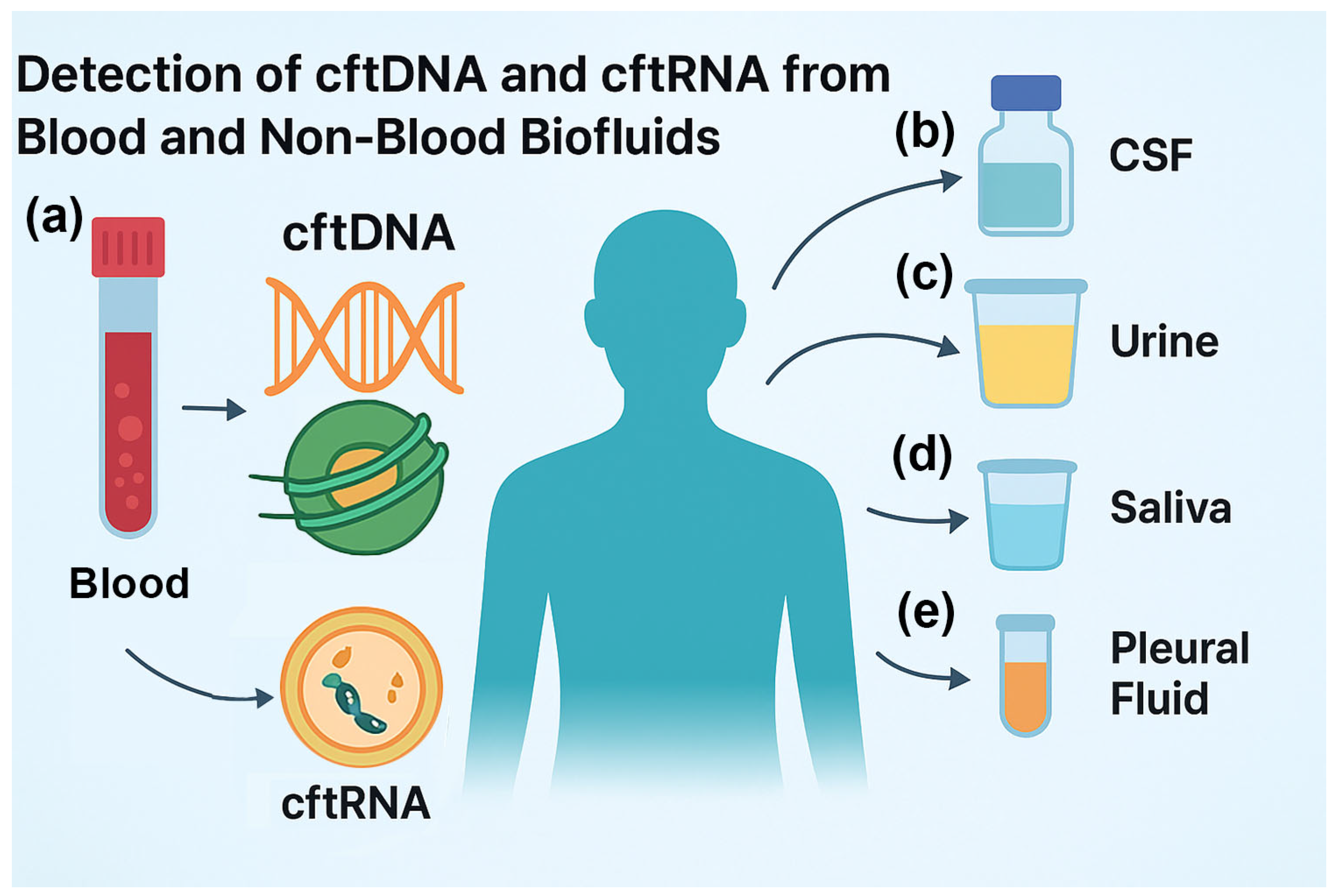
Figure 2.
Limitations of cftDNA and cftRNA in the early detection of cancer. (a) Low concentrations in early stages. In the early stages, when the tumor is small, there will be low to undetectable levels of cftDNA and cftRNA in the biofluids. (b) Lack of standardization. Biofluids, cfDNA, EVs, cfRNA, and detection methods vary according to the laboratory. (c) Cost and accessibility. The kits and NGS assays are typically expensive and usually not covered by insurance. (d) Potential for false positives and negatives. The assays have a high rate of false positives and negatives, making it difficult for doctors to prescribe treatments based on the results.
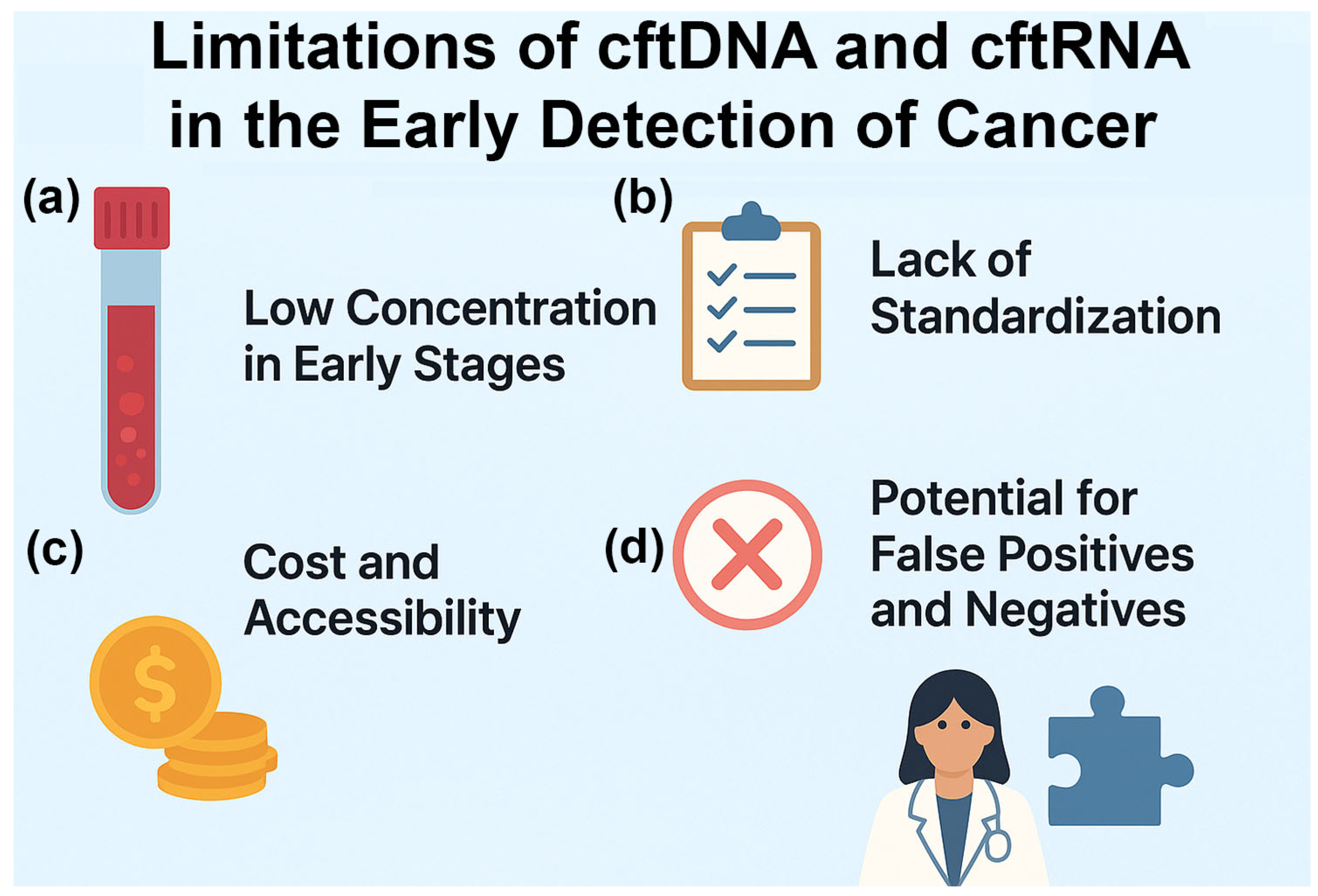

Table 1.
cftDNA detection methods, short description, and references.
| Method | Short description | References |
| CAPP-Seq | Cancer Personalized Profiling by deep Sequencing; Targeted hybrid-capture NGS selector approach designed for ultrasensitive, broad patient-coverage quantitation of ctDNA (mutation, indel, rearrangement, CNV). | [22] |
| iDES | Integrated digital error suppression — molecular barcodes (UMIs) + in-silico background filtering to reduce NGS errors and lower limit of detection. | [23] |
| BEAMing | Beads, Emulsion, Amplification, Magnetics; Emulsion PCR + bead capture and flow cytometry readout for ultra-sensitive detection of known hotspot mutations (digital PCR class). | [24] |
| ddPCR | Droplet digital PCR; Partitioned PCR (droplets) for absolute quantitation of known single nucleotide variants (SNVs) and small insertion-deletions (indels); highly sensitive for low variant allele frequencies (VAFs) and orthogonal validation. | [25,26] |
| sWGS | Shallow whole-genome sequencing; Low-coverage WGS to detect genome-wide copy number alterations and tumor fraction estimation from cfDNA. | [27] |
| Fragmentomics (DELFI) | Genome-wide cfDNA fragmentation profiling (fragment size, end-motifs, nucleosome signals) used with machine learning (ML) to detect and localize cancer. | [27] |
| cfMeDIP-seq | DNA methylome profiling; Immunoprecipitation-based enrichment for methylated cfDNA enabling low-input methylome profiling and tissue-specific cancer signals (bisulfite-free options). | [28,29] |
| cfMeDIP-spike | Synthetic spike-in DNA controls (variable length/GC/CpG) for normalization and absolute quantification in cfMeDIP workflows. | [30] |
| FSE-EME | Fragment size selection & end-motif enrichment; Size-selection/enrichment for mono-nucleosomal-sized fragments or defined end motifs to enrich tumor-derived cfDNA prior to library prep. | [27] |
| cfDNA NGS panels | Commercial comprehensive cfDNA NGS panels (Guardant360, FoundationOne® Liquid CDx); Clinically validated, large targeted panels for therapy selection, monitoring and companion diagnostics; include hybrid-capture, UMIs, and bioinformatic QC. | [31,32] |
| SM-AFD | Single-molecule / amplification-free detection (emerging); Experimental approaches that minimize amplification bias (single-molecule sequencing, nanopore/PacBio with specialized prep) for direct detection of fragmented cfDNA. | [27,28] |
| DNA-PAINT | Super-resolution, single-molecule imaging that uses transient DNA hybridization to detect point mutations, methylation patterns, and amplified sequences at femtomolar sensitivity. | [33,34] |
Table 2.
cftRNA / exosomal RNA detection methods, short description, and representative references (PMID).
Table 2.
cftRNA / exosomal RNA detection methods, short description, and representative references (PMID).
| Method | Short description | References |
| EV-RNA-seq | Exosome / extracellular vesicle (EV) isolation + exo-RNA sequencing; Enrichment of EVs (ultracentrifugation, size-exclusion, precipitation, commercial kits) followed by RNA extraction and sequencing. | [53] |
| Targeted RT-qPCR | ddPCR for miRNAs and fusion transcripts; Sensitive quantification of defined transcripts using RT-qPCR or ddPCR on exoRNA or total plasma RNA. | [53,54] |
| UMI-RNA-seq | RNA-Seq with UMIs and low-input library preps; Low-input RNA-Seq protocols optimized for extracellular RNA to avoid amplification bias. | [54] |
| EV-proteomics | Exosomal proteomics for tissue-of-origin determination; Mass-spectrometry proteomic profiling of exosome cargo proteins to infer tissue/cell-type origin. | [55,56] |
| circRNA / lncRNA profiling | Circular RNAs and lncRNAs in exosomes are resistant to degradation and can yield cancer-specific biomarkers. | [57] |
| DNA-PAINT-miRNA | DNA-PAINT and DNA-origami nanoarrays for amplification-free detection of miRNAs in fluids/exosomes. | [58] |
| Targeted Fus-RNA-seq | Fusion transcript detection (targeted RNA panels); Targeted RNA assays for fusion transcript detection in plasma/exosomes. | [59,60] |
| Ago-FISH, FRET-FISH | Ago-FISH, FRET-FISH and other single-molecule RNA detection; Single-molecule RNA detection methods for multiplexed, amplification-free detection of short RNAs. | [61,62] |
| exo-RNA + proteomics | Integration: multi-omic exo-RNA + proteomics pipelines; Combined workflows analyzing exosomal RNA, DNA and protein cargo for integrated biomarker signatures. | [63] |
Disclaimer/Publisher’s Note: The statements, opinions and data contained in all publications are solely those of the individual author(s) and contributor(s) and not of MDPI and/or the editor(s). MDPI and/or the editor(s) disclaim responsibility for any injury to people or property resulting from any ideas, methods, instructions or products referred to in the content. |
© 2025 by the authors. Licensee MDPI, Basel, Switzerland. This article is an open access article distributed under the terms and conditions of the Creative Commons Attribution (CC BY) license (http://creativecommons.org/licenses/by/4.0/).
Copyright: This open access article is published under a Creative Commons CC BY 4.0 license, which permit the free download, distribution, and reuse, provided that the author and preprint are cited in any reuse.


