Submitted:
13 August 2025
Posted:
14 August 2025
You are already at the latest version
Abstract
In previous work we have used the computer program AmyloGram to assess the amyloidogenic potential of proteins observed using mass spectrometry-based proteomics in thrombi extracted from individuals who had suffered an ischaemic stroke. As anticipated from our experimental observation of substantial amounts of amyloid in such clots, the AmyloGram scores were very high and entirely consistent with the amyloid nature of such thrombi. We here apply a similar strategy to assess the amyloidogenic nature of proteins in thrombi removed from venous thromboembolisms including pulmonary embolisms, similarly finding very high AmyloGram scores. The same is true for atherosclerotic plaques as determined from multiple studies in which the data were readily available. Amyloidogenesis is a specific activity or subset of a class of proteins known to adopt very different macrostates, in which amyloidogenesis to create insoluble fibrils is more or less irreversible. Another subset of multi-state proteins, whose conformational interchanges are much more reversible, involves what are referred to as ‘fold-switching’ or ‘metamorphic’ proteins’. We here use AmyloGram to analyse the amyloidogenic potential of these too, finding that while some are highly amyloidogenic their amyloidogenic potential is considerably more heterogeneous and little different from that of the overall proteome within Uniprot.
Keywords:
amyloid
; fibrinaloid
; proteomics
; inflammation
; amyloidogenic sequences
; embolism
; atherosclerosis
Introduction
Following our discovery that blood can clot into an anomalous amyloid form [1, 2] to produce what we and others refer to as fibrinaloid microclots [3-17], we have more recently discovered experimentally that the macroclots retrieved from ischaemic stroke are also amyloid in nature [10, 18].
Among the conventional pathogenic amyloidoses [19-22], it is well known that amyloidogenic cross-seeding can be occurring [23-28]. This has led (i) to the recognition that this is also occurring in the microclots (as reflected in the complex proteomes so observed [15, 29-31]) and (ii) to predictions that this would also be true in macroclots [16].
A variety of computational programs exist for predicting the amyloidogenicity of a protein (summarised in [32] and in Table 1 of [15]), and we recently used one of these, AmyloGram [33, 34], to predict the amyloidogenicity of proteins observed in a variety of ischaemic stroke thrombi [35]. In that work [35], we ‘calibrated’ the system with proteins annotated by humans at Uniprot, where (with a generous margin) every one of these had an AmyloGram score exceeding 0.7 (as did 79% of all human polypeptides). Specifically, of the 83,567 proteins that were analysed, 66,190 (79.2%) had AmyloGram scores exceeding 0.7, while 45,169 (54.1%) exceeded 0.8 and 7409 (8.9%) were above 0.9. The implication was that any thrombus with a higher percentage than 0.7 (or a much higher median AmyloGram score) could or would effectively be enriched in amyloidogenic proteins, and this turned out very much to be the case [35], consistent with the experimental observations [10, 18].
As well as thrombi retrieved from ischaemic stroke, we also predicted that a variety of other thrombi, for which experimental proteomic data existed, including those from venous thromboembolisms including pulmonary embolism and deep vein thrombosis, and various cardiac issues, would also be amyloid in nature, though we did not test this with AmyloGram [16]. The purpose of the present work is, where the data are available in a suitable format, precisely to perform those analyses. We conclude that in all cases where proteomic data are available the thrombi are, like those from ischaemic stroke, expected to be amyloid in nature.
Materials and Methods
Just as with our previous endeavour [35] this work uses the online http://biongram.biotech.uni.wroc.pl/AmyloGram/ or R-based versions of AmyloGram (see https://github.com/michbur/AmyloGram or https://cran.r-project.org/web/packages/AmyloGram/index.html) to determine the AmyloGram scores; protein identification data are given in the original publications cited, or in Tables here.
Results
Venous Thromboembolism
In the case of thrombi removed from individuals following a venous thromboembolism, we identified the 18 proteins given in the data from Stachowicz and colleagues (their Table 1, that included concentrations) [36], and ran the list on the AmyloGram server, with the results shown in Figure 1. With one exception, each scores above 0.7, with a median score exceeding 0.86, strongly implying that these clots are amyloid in nature.
Pulmonary Embolism
The data we used for these studies came from a study of Bryk and colleagues [37], whose Table (their Supplementary Information Table S1) contained 198 polypeptides that were both in thrombi retrieved following a pulmonary embolism and were also differentially expressed relative to controls (normal clots more-or-less reflect the standard plasma proteome [15]). Six proteins (SERPINA1, IGKC;IGKV1-8, IGHM, IGLL5, IGHG3, SRRM1) would not run with the R code for some reason and were added via the Web server at http://biongram.biotech.uni.wroc.pl/AmyloGram/. The data are illustrated in Figure 2, where it may be observed that 190/198 have AmyloGram scores exceeding 0.7, 148/198 exceeding 0.8, 108 exceeding 0.85, and 22 (labelled in Fig 2) exceeding 0.9. The highest AmyloGram score is held by the human telomerase reverse transcriptase TERT (Uniprot O14746) with a value of 0.956. Again it is very clear that the amyloidogenicity of proteins enriched in the clots taken following a pulmonary embolism are very much greater than the average, a finding consistent with recent studies from other thrombi [10, 15, 16, 18, 35] and – since insoluble amyloids are well known to be much more refractory to proteolysis than are soluble proteins – one that plausibly underpins their resistance to fibrinolysis.
Atherosclerosis and Acute Myocardial Infarction
Lipoprotein proteomes have been reviewed by [38]. However, our interest here is in atherosclerotic plaques, whose proteomes have been studied by several groups [38-51], with some papers providing more easily accessible/analysable data than others.
Langley and colleagues [43] sought biomarkers of high-risk atherosclerotic plaques, and identified a 4-biomarker signature (matrix metalloproteinase 9, S100A8/S100A9 (calprotectin), cathepsin D, and galectin-3-binding protein). Their AmyloGram scores are respectively 0.9148, 0.7768 (for S100A8), 0.7941, and 0.8661. Clearly, each is highly amyloidogenic, and we previously highlighted galectin-3-binding protein (LG3BP, Uniprot Q08380) [16] as being a major player in essentially every kind of persistent thrombus.
Rocchiccioli and colleagues [52] found 31 proteins that were differentially secreted from atherosclerotic plaques. ELISA assays of plasma samples confirmed a significantly higher concentration of thrombospondin-1 and vitamin D binding protein in atherosclerotic subjects; as with LG3BP above, we had previously highlighted thrombospondin-1 as being a major player in essentially every kind of persistent thrombus [16], not least as it has long been known [53-55] that it is actually incorporated into fibrin during thrombus formation.
Theofilatos and colleagues [46] also determined a 4-protein atherosclerotic plaque signature for high-risk cardiovascular mortality, consisting of calponin-1 (Uniprot P51911), Vitamin K-dependent protein C (Uniprot P04070), serpin H1 (Uniprot P50454), and versican (Uniprot P13611). Their AmyloGram scores are, respectively, 0.6934, 0.9148, 0.8789, and 0.8627.
Alonso-Orgaz and colleagues [42] assessed the proteome of the human coronary thrombus in patients with ST-segment elevation acute myocardial infarction, using means of three different approaches involving a separation step followed by mass spectrometry. 46 proteins could be identified using all three methods [42], and these are illustrated in Figure 3. All 46 have an AmyloGram score exceeding 0.7, 35 have scores that exceed 0.8, while 29 (labelled in the Figure) exceed more than 0.85, and three exceed 0.9. The median value (between proteins scoring 0.856 and 0.863) is 0.86. Interestingly, talin-1, with a score of 0.851 is among them and is a known amyloidogen [56]. Obviously these are again very high scores, serving to underline the fact that such thrombi are likely to be amyloid in nature, and this recognition adds a major means of explaining why they are so resistant to proteolysis. Amyloid deposition in the thrombus associated with cardiac amyloidosis is of course known [57], and (although seemingly not widely recognised) amyloid deposition is in fact an established feature of atherosclerotic plaques [58-63], so in one sense the high AmyloGram scores here are unsurprising.
Wang and colleagues [51] identified 11 proteins associated with coronary atherosclerosis (Their Table 1), and their AmyloGram scores are given in Figure 4. Every single entry has an AmyloGram score exceeding 0.75.
Hansmeier et al. [64] identified 20 proteins that were raised in atherosclerotic plaques (their Supplementary Table S3), and these are tabulated, along with their AmyloGram scores and lengths, in Figure 5. All proteins again have AmyloGram scores exceeding 0.7, many being considerably greater.
The data from the 2025 study of Lorentzen and colleagues [49] (their Supplementary Information Table S2, provided there as an Excel sheet, with 128 peptides and 83 unique proteins) analysed proteins in atherosclerotic plaque from the perspective of their ease of degradation/ plaque stability or otherwise, though our interest here is simply which proteins were present and their AmyloGram scores. The data are given in Figure 6. All but 5 proteins (some represented by multiple peptides) have AmyloGram scores exceeding 0.7.
Aragonès and colleagues [40] compared the proteome of carotid atherosclerotic plaque and non-diseased mammary artery; 25 proteins showed statistically significant differences. Their median AmyloGram score is 0.845, and they are listed, along with their AmyloGram scores, in Figure 7.
Overall, there is a high enrichment of amyloidogenic proteins in each of the eight studies of atheromatous plaques as reviewed here.
Fold-Switching Proteins
Amyloidogenic proteins, including prions, clearly represent a set of proteins that can exist in multiple stable macrostates under a given set of conditions. The usual form or conformational ensemble, as synthesised at the ribosome, commonly has abundant α-helices whereas the amyloid forms are much richer in β-sheets, specifically the crossed-β structure [65-70], that is the characteristic of amyloid and the one to which fluorogenic stains such as thioflavin T bind [71-74]. While the amyloid form is significantly more stable thermodynamically (amyloidogenesis, involving accretion of multiple molecules leading to insoluble fibril formation, is essentially irreversible), the two forms are separated via a substantial energy barrier of some 36 kJ.mol-1 [75-80].
It has long been known that the same peptide sequences can adopt quite different conformations in different proteins [81-84](and, for that matter, that similar conformations can have very different dynamics [85, 86]). In particular, this ability to adopt multiple stable conformations or macrostates, usually referred to as polymorphs [4, 78, 87-113], is very much true of amyloids. Equally, there is a more general class of proteins that can adopt multiple macrostates, in this case often often – but not always – reversibly. These macrostates or conformations probably have different evolutionarily selected functions, and proteins exhibiting this are known as fold-switching [114-128] or metamorphic [117, 129-141] proteins. Some 6% are known to be fibril-forming [120], and the question thus arose as to whether these might also have a tendency to be more amyloidogenic.
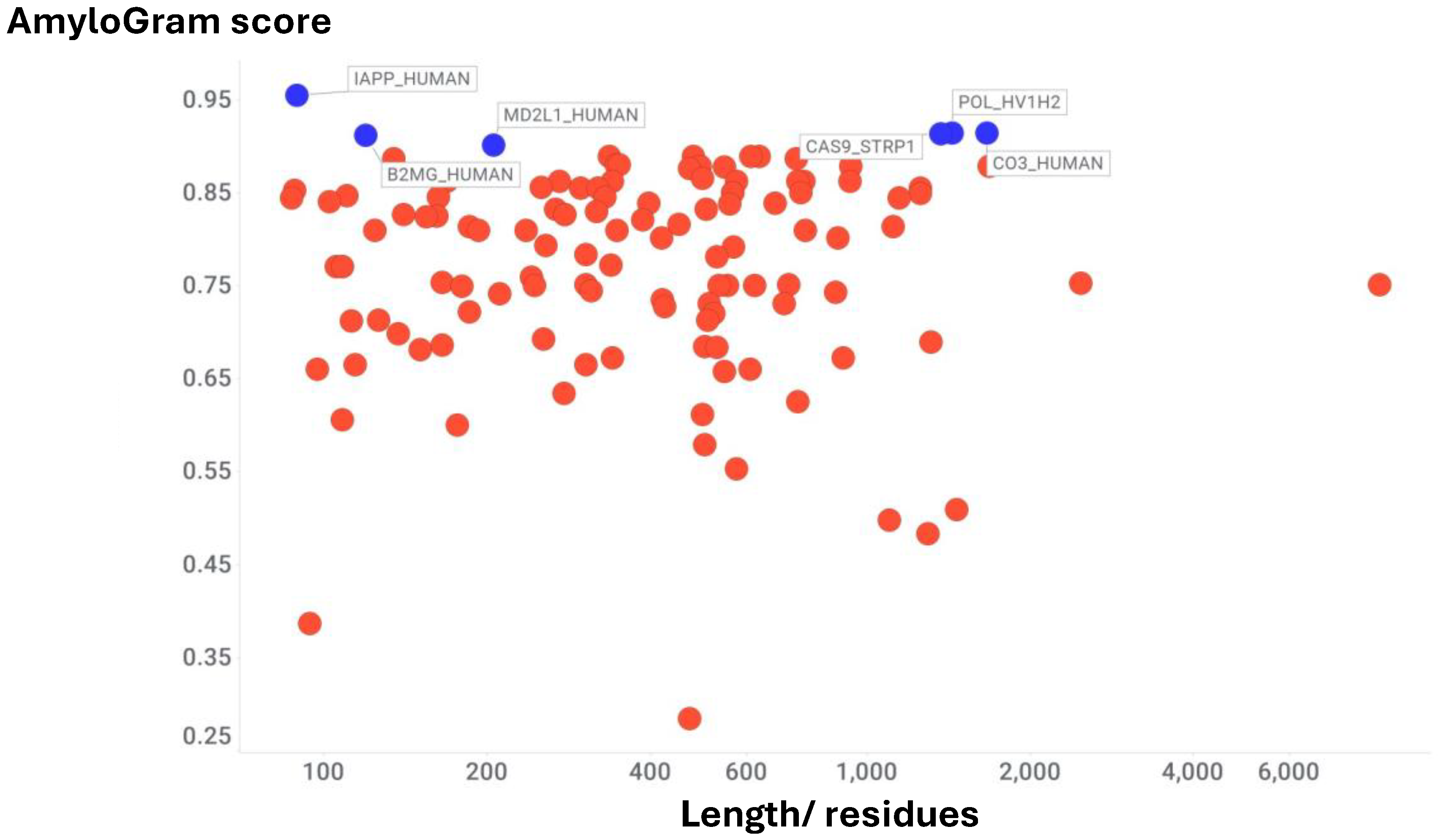
A list of fold-switching proteins (see [122]) was kindly provided by Dr Lauren Porter. It is based on PDB references, some of which are different 3D structures of the same protein, and we have cut this down to provide one representative of each as per Table S1 of the Supplementary Information. This leaves 121 examples, and their AmyloGram scores and lengths are plotted in Figure 8.
For context, the median AmyloGram score of the 204 proteins labelled at Uniprot as amyloid (following human analysis) was 0.88, while the median score for all human proteins was 0.81 [35]. The median score for fold-switching proteins was 0.81, meaning that in general they did not tend overall to be unusually amyloidogenic or otherwise. This is reasonable, as fold-switching is based on a relatively short subsequence (‘fold-switching regions’) of the protein of interest [118, 142]. This said, the distribution of overall AmyloGram scores is significantly heterogeneous, since some of them, with AmyloGram scores in excess of 0.9, such as the well-known amyloidogenic proteins amylin (islet amyloid polypeptide) [143-145] and β2microglobulin [146-148], certainly are amyloid in nature, while complement C3 seems to be involved in cross-seeding and thus guilty by association [149-151]. HIV reverse transcriptase is also of interest, as it has been implicated in amyloidogenesis as part of Alzheimer’s dementia [152]. The numbers (out of 121) of polypeptides with AmyloGram scores exceeding 0.7, 0.75, 0.8, 0.85 and 0.9 are, respectively, 95, 83, 64, 35 and 6.
Discussion
The amyloidogenic clotting of blood to make fibrinaloid microclots (commonly with an equivalent diameter of 2-200 μm) is now well established, and has been described in dozens of papers from multiple laboratories (e.g. [1-17, 31, 153-156]). More recently, it was established that the macroclots (of over 1 mm diameter) that can be thrombectomised following an ischaemic stroke are also amyloid in character [10, 18], and there is also evidence that amyloid is a feature of atherosclerotic plaques [60-63]. Cross seeding, in which an amyloid protein induces amyloid formation in other amyloidogenic proteins that can then become part of the same fibril, is also commonplace [15, 23-25, 27, 28, 109, 157-182], as are amyloid-nucleic acid interactions [183-188].
Consequently, one can predict (correctly) that insoluble amyloid structures will tend to accumulate preferentially those proteins that are themselves more amyloidogenic than normal [15, 16]. In a recent study [35] we used the amyloid prediction program AmyloGram [33, 34] to assess this for the proteome of macroclots extracted following an ischaemic stroke, finding that the AmyloGram scores for the proteins in the stroke thrombus proteome (as measured by a number of groups) were indeed noticeably greater than the average for proteins [35].
The purpose of the present study was to assess this kind of phenomenon for macroclots taken from other diseases, such as venous thromboembolism and pulmonary embolism, and also for the many examples in which the proteome of insoluble atherosclerotic plaques had been analysed. The conclusion from the analyses above was again that in all cases the proteomes displayed a very strong tendency towards amyloidogenicity, consistent with self-seeding and providing a ready explanation both for why they are insoluble and – since amyloids are notoriously resistant to proteolysis (e.g. [70, 189-193]) – for why the thrombi are rather resistant to the normal mechanisms of fibrinolysis.
There are no necessary changes in the primary sequence of proteins following their amyloid formation; notwithstanding, amyloids can form multiple, stable variants known as polymorphs, and the insolubilisation of amyloids when they form fibrils is thermodynamically more-or-less irreversible. However, another class of proteins that can switch conformation dramatically but reversibly, including from α-helices to β-sheets, are referred to as fold-switching or metamorphic proteins. It was thus of interest to assess whether or not these too tended to be unusually amyloidogenic. The answer is that while some examples such as amylin and β2microglobulin are indeed highly amyloidogenic, the median amyloidogenicity as reflected in their overall AmyloGram score was more or less identical to that of the proteins in Uniprot. Since it is recognised that relatively short subsequences of amino acids are actually responsible for the fold switching, this is possibly not surprising, but it was worth assessing.
Conclusion
Having established the fact that the AmyloGram scores of proteins embedded in the thrombi extracted following an ischaemic stroke are sufficient to predict that they are highly amyloidogenic [35], as they are experimentally [10, 18], it was of interest to assess the amyloidogenic potential in thrombi from other thrombi such as those involving venous thromboembolisms [16]. In every case it was found that the proteomes of these thrombi involved highly amyloidogenic proteins. The same was true in a series of studies of atherosclerotic plaques. Given that the simple presence of these plaques and thrombi indicates clearly (by definition) that they are resistant to the normal methods of fibrinolysis, it is clear that novel means will be required to effect their removal. This provides exciting opportunities.
Author Contributions
All authors contributed to the conceptualization of the study, data analysis, manuscript drafting, and final editing.
Funding
D.B.K. acknowledges support from the Balvi Foundation (grant 18) and the Novo Nordisk Foundation (grant NNF20CC0035580). E.P. received funding from the National Research Foundation of South Africa (grant 142142), the South African Medical Research Council (Self-Initiated Research grant), and the Balvi Foundation. KD acknowledges support from Research Ireland (Grant Number 13/RC/2073_P2). The content and findings presented here are the sole responsibility of the authors and do not necessarily reflect the views of the funding agencies.
Data Availability
All data supporting the findings of this study are available within the article. Stroke thrombus proteomic datasets were obtained from previously published studies and are cited accordingly.
Acknowledgments
We thank Dr Lauren Porter (NIH/National Library of Medicine) for furnishing data on fold-switching proteins and for useful discussions.
Conflicts of Interest
E.P. is a named inventor on a patent application related to the use of fluorescence-based methods for microclot detection in Long COVID. The funding bodies had no involvement in the design of the study, data collection or analysis, manuscript preparation, or the decision to submit for publication. The other authors have no disclosures.
References
- Pretorius, E. , Mbotwe, S., Bester, J., Robinson, C. J. and Kell, D. B. (2016) Acute induction of anomalous and amyloidogenic blood clotting by molecular amplification of highly substoichiometric levels of bacterial lipopolysaccharide. J R Soc Interface 2016, 123, 20160539. [Google Scholar] [CrossRef]
- Pretorius, E. , Page, M. J., Hendricks, L., Nkosi, N. B., Benson, S. R. and Kell, D. B. (2018) Both lipopolysaccharide and lipoteichoic acids potently induce anomalous fibrin amyloid formation: assessment with novel Amytracker™ stains. J R Soc Interface 2018, 15, 20170941. [Google Scholar] [CrossRef]
- Kell, D. B. , Laubscher, G. J. and Pretorius, E. (2022) A central role for amyloid fibrin microclots in long COVID/PASC: origins and therapeutic implications. Biochem J 2022, 479, 537–559. [Google Scholar] [CrossRef] [PubMed]
- Kell, D. B. and Pretorius, E. (2023) Are fibrinaloid microclots a cause of autoimmunity in Long Covid and other post-infection diseases? Biochem J 2023, 480, 1217–1240. [Google Scholar] [CrossRef]
- Nunes, J. M. , Kruger, A., Proal, A., Kell, D. B. and Pretorius, E. (2022) The occurrence of hyperactivated platelets and fibrinaloid microclots in Myalgic Encephalomyelitis/Chronic Fatigue Syndrome (ME/CFS). Research Square 2022. [Google Scholar] [CrossRef]
- Nunes, J. M. , Kruger, A., Proal, A., Kell, D. B. and Pretorius, E. (2022) The occurrence of hyperactivated platelets and fibrinaloid microclots in Myalgic Encephalomyelitis/Chronic Fatigue Syndrome (ME/CFS). Pharmaceuticals (Basel) 2022, 15, 931. [Google Scholar] [CrossRef] [PubMed]
- Turner, S. , Naidoo, C., Usher, T., Kruger, A., Venter, C., Laubscher, G. J., Khan, M. A., Kell, D. B. and Pretorius, E. (2022) Increased levels of inflammatory molecules in blood of Long COVID patients point to thrombotic endotheliitis. medRxiv 2022, 2022.2010.2013.22281055. [Google Scholar] [CrossRef]
- Turner, S. , Khan, M. A., Putrino, D., Woodcock, A., Kell, D. B. and Pretorius, E. (2023) Long COVID: pathophysiological factors and abnormal coagulation. Trends Endocrinol Metab 2023, 34, 321–344. [Google Scholar] [CrossRef]
- Dalton, C. F. , de Oliveira, M. I. R., Stafford, P., Peake, N., Kane, B., Higham, A., Singh, D., Jackson, N., Davies, H., Price, D., Duncan, R., Tattersall, N., Barnes, A. and Smith, D. P. (2024) Increased fibrinaloid microclot counts in platelet-poor plasma are associated with Long COVID. medRxiv 2024, 2024.2004.2004.24305318. [Google Scholar] [CrossRef]
- Grixti, J. M. , Chandran, A., Pretorius, J. H., Walker, M., Sekhar, A., Pretorius, E. and Kell, D. B. (2024) The clots removed from ischaemic stroke patients by mechanical thrombectomy are amyloid in nature. medRxiv 2024, 10.1101/2024.1111.1101.24316555v24316551. [Google Scholar] [CrossRef]
- Grixti, J. M. , Theron, C. W., Salcedo-Sora, J. E., Pretorius, E. and Kell, D. B. (2024) Automated microscopic measurement of fibrinaloid microclots and their degradation by nattokinase, the main natto protease. J Exp Clin Appl Chin Med 2024, 5, 30–55. [Google Scholar] [CrossRef]
- Kell, D. B. , Khan, M. A., Kane, B., Lip, G. Y. H. and Pretorius, E. (2024) Possible role of fibrinaloid microclots in Postural Orthostatic Tachycardia Syndrome (POTS): focus on Long COVID. J Personalised Medicine 2024, 14, 170. [Google Scholar] [CrossRef] [PubMed]
- Kell, D. B. and Pretorius, E. (2024) Potential roles of fibrinaloid microclots in fibromyalgia syndrome. OSF preprint. [CrossRef]
- Kell, D. B. , Lip, G. Y. H. and Pretorius, E. (2024) Fibrinaloid Microclots and Atrial Fibrillation. Biomedicines 2024, 12, 891. [Google Scholar] [CrossRef]
- Kell, D. B. and Pretorius, E. (2024) Proteomic evidence for amyloidogenic cross-seeding in fibrinaloid microclots. Int J Mol Sci 2024, 25, 10809. [Google Scholar] [CrossRef]
- Kell, D. B. and Pretorius, E. (2025) The proteome content of blood clots observed under different conditions: successful role in predicting clot amyloid(ogenicity). Molecules 2025, 30, 668. [Google Scholar] [CrossRef]
- Kell, D. B. , Pretorius, E. and Zhao, H. (2025) A direct relationship between ‘blood stasis’ and fibrinaloid microclots in chronic, inflammatory and vascular diseases, and some traditional natural products approaches to treatment. Pharmaceuticals 2025, 18, 712. [Google Scholar] [CrossRef]
- Grixti, J. M. , Chandran, A., Pretorius, J. H., Walker, M., Sekhar, A., Pretorius, E. and Kell, D. B. (2025) Amyloid presence in acute ischemic stroke thrombi: observational evidence for fibrinolytic resistance. Stroke 2025, 56, e165–e167. [Google Scholar] [CrossRef] [PubMed]
- Blancas-Mejía, L. M. and Ramirez-Alvarado, M. (2013) Systemic amyloidoses. Annu Rev Biochem 2013, 82, 745–774. [Google Scholar] [CrossRef]
- Lavatelli, F. , di Fonzo, A., Palladini, G. and Merlini, G. (2016) Systemic amyloidoses and proteomics: The state of the art. EuPA Open Proteom 2016, 11, 4–10. [Google Scholar] [CrossRef] [PubMed]
- Nevone, A. , Merlini, G. and Nuvolone, M. (2020) Treating protein misfolding diseases: therapeutic successes against systemic amyloidoses. Front Pharmacol 2020, 11, 1024. [Google Scholar] [CrossRef]
- Palladini, G. and Merlini, G. (2013) Systemic amyloidoses: what an internist should know. Eur J Intern Med 2013, 24, 729–739. [Google Scholar] [CrossRef] [PubMed]
- Chaudhuri, P. , Prajapati, K. P., Anand, B. G., Dubey, K. and Kar, K. (2019) Amyloid cross-seeding raises new dimensions to understanding of amyloidogenesis mechanism. Ageing Res Rev 2019, 56, 100937. [Google Scholar] [CrossRef]
- Ge, W. Y. , Deng, X., Shi, W. P., Lin, W. J., Chen, L. L., Liang, H., Wang, X. T., Zhang, T. D., Zhao, F. Z., Guo, W. H. and Yin, D. C. (2023) Amyloid protein cross-seeding provides a new perspective on multiple diseases in vivo. Biomacromolecules 2023, 24, 1–18. [Google Scholar] [CrossRef]
- Ivanova, M. I. , Lin, Y., Lee, Y. H., Zheng, J. and Ramamoorthy, A. (2021) Biophysical processes underlying cross-seeding in amyloid aggregation and implications in amyloid pathology. Biophys Chem 2021, 269, 106507. [Google Scholar] [CrossRef]
- Larsson, J. , Hellstrand, E., Hammarström, P. and Nyström, S. (2023) SARS-CoV-2 Spike amyloid fibrils specifically and selectively accelerates amyloid fibril formation of human prion protein and the amyloid β peptide. bioRxiv 2023, 2023.2009.2001.555834. [Google Scholar] [CrossRef]
- Ren, B. , Zhang, Y., Zhang, M., Liu, Y., Zhang, D., Gong, X., Feng, Z., Tang, J., Chang, Y. and Zheng, J. (2019) Fundamentals of cross-seeding of amyloid proteins: an introduction. J Mater Chem B 2019, 7, 7267–7282. [Google Scholar] [CrossRef] [PubMed]
- Westermark, G. T. , Fandrich, M., Lundmark, K. and Westermark, P. (2018) Noncerebral Amyloidoses: Aspects on Seeding, Cross-Seeding, and Transmission. Cold Spring Harb Perspect Med 2018, 8, a024323. [Google Scholar] [CrossRef] [PubMed]
- Kruger, A. , Vlok, M., Turner, S., Venter, C., Laubscher, G. J., Kell, D. B. and Pretorius, E. (2022) Proteomics of fibrin amyloid microclots in Long COVID/ Post-Acute Sequelae of COVID-19 (PASC) shows many entrapped pro-inflammatory molecules that may also contribute to a failed fibrinolytic system. Cardiovasc Diabetol 2022, 21, 190. [Google Scholar] [CrossRef]
- Pretorius, E. , Vlok, M., Venter, C., Bezuidenhout, J. A., Laubscher, G. J., Steenkamp, J. and Kell, D. B. (2021) Persistent clotting protein pathology in Long COVID/ Post-Acute Sequelae of COVID-19 (PASC) is accompanied by increased levels of antiplasmin. Cardiovasc Diabetol 2021, 20, 172. [Google Scholar] [CrossRef]
- Schofield, J. , Abrams, S. T., Jenkins, R., Lane, S., Wang, G. and Toh, C. H. (2024) Microclots, as defined by amyloid-fibrinogen aggregates, predict risks of disseminated intravascular coagulation and mortality. Blood Adv 2024, 8, 2499–2508. [Google Scholar] [CrossRef]
- Iglesias, V. , Chilimoniuk, J., Pintado-Grima, C., Barcenas, O., Ventura, S. and Burdukiewicz, M. (2024) Aggregating amyloid resources: A comprehensive review of databases on amyloid-like aggregation. Comput Struct Biotechnol J 2024, 23, 4011–4018. [Google Scholar] [CrossRef]
- Burdukiewicz, M. , Sobczyk, P., Rödiger, S., Duda-Madej, A., Mackiewicz, P. and Kotulska, M. (2017) Amyloidogenic motifs revealed by n-gram analysis. Sci Rep 2017, 7, 12961. [Google Scholar] [CrossRef] [PubMed]
- Szulc, N. , Burdukiewicz, M., Gąsior-Głogowska, M., Wojciechowski, J. W., Chilimoniuk, J., Mackiewicz, P., Šneideris, T., Smirnovas, V. and Kotulska, M. (2021) Bioinformatics methods for identification of amyloidogenic peptides show robustness to misannotated training data. Sci Rep 2021, 11, 8934. [Google Scholar] [CrossRef] [PubMed]
- Kell, D. B. , Doyle, K. M., Salcedo-Sora, E., Sekhar, A., Walker, M. and Pretorius, E. (2025) AmyloGram reveals amyloidogenic potential in stroke thrombus proteomes. bioRxiv 2025, 2025.2007.2007.663482. [Google Scholar] [CrossRef]
- Stachowicz, A. , Siudut, J., Suski, M., Olszanecki, R., Korbut, R., Undas, A. and Wisniewski, J. R. (2017) Optimization of quantitative proteomic analysis of clots generated from plasma of patients with venous thromboembolism. Clin Proteomics 2017, 14, 38. [Google Scholar] [CrossRef]
- Bryk, A. H. , Natorska, J., Ząbczyk, M., Zettl, K., Wisniewski, J. R. and Undas, A. (2020) Plasma fibrin clot proteomics in patients with acute pulmonary embolism: Association with clot properties. J Proteomics 2020, 229, 103946. [Google Scholar] [CrossRef] [PubMed]
- Nieddu, G. , Formato, M. and Lepedda, A. J. (2023) Searching for Atherosclerosis Biomarkers by Proteomics: A Focus on Lesion Pathogenesis and Vulnerability. Int J Mol Sci 2023, 24, 15175. [Google Scholar] [CrossRef]
- Hao, P. , Ren, Y., Pasterkamp, G., Moll, F. L., de Kleijn, D. P. V. and Sze, S. K. (2014) Deep proteomic profiling of human carotid atherosclerotic plaques using multidimensional LC-MS/MS. Proteomics Clin Appl 2014, 8, 631–635. [Google Scholar] [CrossRef]
- Aragonès, G. , Auguet, T., Guiu-Jurado, E., Berlanga, A., Curriu, M., Martinez, S., Alibalic, A., Aguilar, C., Hernandez, E., Camara, M. L., Canela, N., Herrero, P., Ruyra, X., Martin-Paredero, V. and Richart, C. (2016) Proteomic profile of unstable atheroma plaque: increased neutrophil defensin 1, clusterin, and apolipoprotein E levels in carotid secretome. J Proteome Res 2016, 15, 933–944. [Google Scholar] [CrossRef]
- Bagnato, C. , Thumar, J., Mayya, V., Hwang, S. I., Zebroski, H., Claffey, K. P., Haudenschild, C., Eng, J. K., Lundgren, D. H. and Han, D. K. (2007) Proteomics analysis of human coronary atherosclerotic plaque: a feasibility study of direct tissue proteomics by liquid chromatography and tandem mass spectrometry. Mol Cell Proteomics 2007, 6, 1088–1102. [Google Scholar] [CrossRef] [PubMed]
- Alonso-Orgaz, S. , Moreno-Luna, R., López, J. A., Gil-Dones, F., Padial, L. R., Moreu, J., de la Cuesta, F. and Barderas, M. G. (2014) Proteomic characterization of human coronary thrombus in patients with ST-segment elevation acute myocardial infarction. J Proteomics 2014, 109, 368–381. [Google Scholar] [CrossRef]
- Langley, S. R. , Willeit, K., Didangelos, A., Matic, L. P., Skroblin, P., Barallobre-Barreiro, J., Lengquist, M., Rungger, G., Kapustin, A., Kedenko, L., Molenaar, C., Lu, R., Barwari, T., Suna, G., Yin, X., Iglseder, B., Paulweber, B., Willeit, P., Shalhoub, J., Pasterkamp, G., Davies, A. H., Monaco, C., Hedin, U., Shanahan, C. M., Willeit, J., Kiechl, S. and Mayr, M. (2017) Extracellular matrix proteomics identifies molecular signature of symptomatic carotid plaques. J Clin Invest 2017, 127, 1546–1560. [Google Scholar] [CrossRef]
- Lorentzen, L. G. , Hansen, G. M., Iversen, K. K., Bundgaard, H. and Davies, M. J. (2022) Proteomic Characterization of Atherosclerotic Lesions In Situ Using Percutaneous Coronary Intervention Angioplasty Balloons-Brief Report. Arterioscler Thromb Vasc Biol 2022, 42, 857–864. [Google Scholar] [CrossRef]
- Chandran, M. , S, S., Abhirami, Chandran, A., Jaleel, A. and Plakkal Ayyappan, J. (2023) Defining atherosclerotic plaque biology by mass spectrometry-based omics approaches. Mol Omics 2023, 19, 6–26. [Google Scholar] [CrossRef]
- Theofilatos, K. , Stojkovic, S., Hasman, M., van der Laan, S. W., Baig, F., Barallobre-Barreiro, J., Schmidt, L. E., Yin, S., Yin, X., Burnap, S., Singh, B., Popham, J., Harkot, O., Kampf, S., Nackenhorst, M. C., Strassl, A., Loewe, C., Demyanets, S., Neumayer, C., Bilban, M., Hengstenberg, C., Huber, K., Pasterkamp, G., Wojta, J. and Mayr, M. (2023) Proteomic Atlas of Atherosclerosis: The Contribution of Proteoglycans to Sex Differences, Plaque Phenotypes, and Outcomes. Circ Res 2023, 133, 542–558. [Google Scholar] [CrossRef]
- Cruz-González, G. , Meschia, J. F., Madden, B. J., Prudencio, M., Polania-Sandoval, C. A., Hartwell, J., Oyefeso, E., Benchaaboune, R., Brigham, T., Sandhu, S. J. S., Charlesworth, C., Pujari, G. P., Petrucelli, L., Pandey, A. and Erben, Y. (2024) Recent advances in proteomic analysis to study carotid artery plaques. JVS Vasc Sci 2024, 5, 100215. [Google Scholar] [CrossRef]
- Lorentzen, L. G. , Yeung, K., Eldrup, N., Eiberg, J. P., Sillesen, H. H. and Davies, M. J. (2024) Proteomic analysis of the extracellular matrix of human atherosclerotic plaques shows marked changes between plaque types. Matrix Biol Plus 2024, 21, 100141. [Google Scholar] [CrossRef] [PubMed]
- Lorentzen, L. G. , Yeung, K., Zitkeviciute, A., Yang-Jensen, K. C., Eldrup, N., Eiberg, J. P. and Davies, M. J. (2025) N-Terminal Proteomics Reveals Distinct Protein Degradation Patterns in Different Types of Human Atherosclerotic Plaques. J Proteome Res 2025, 24, 144–157. [Google Scholar] [CrossRef]
- Wang, C. , Feng, Y., Liu, X., Sun, H., Guo, Z., Shao, J., Li, K., Chen, J., Shu, K., Kong, D., Wang, J., Li, Y., Lei, X., Li, C., Liu, B., Sun, W. and Lai, Z. (2025) Molecular landscape of atherosclerotic plaque progression: insights from proteomics, single-cell transcriptomics and genomics. BMC Med 2025, 23, 254. [Google Scholar] [CrossRef] [PubMed]
- Wang, H. , Xie, F., Wang, M., Ji, J., Song, Y., Dai, Y., Wang, L., Kang, Z. and Cao, L. (2025) Identification of potential therapeutic targets for coronary atherosclerosis from an inflammatory perspective through integrated proteomics and single-cell omics. Int J Mol Sci 2025, 26, 6201. [Google Scholar] [CrossRef]
- Rocchiccioli, S. , Pelosi, G., Rosini, S., Marconi, M., Viglione, F., Citti, L., Ferrari, M., Trivella, M. G. and Cecchettini, A. (2013) Secreted proteins from carotid endarterectomy: an untargeted approach to disclose molecular clues of plaque progression. J Transl Med 2013, 11, 260. [Google Scholar] [CrossRef] [PubMed]
- Bale, M. D. , Westrick, L. G. and Mosher, D. F. (1985) Incorporation of thrombospondin into fibrin clots. J Biol Chem 1985, 260, 7502–7508. [Google Scholar] [CrossRef]
- Bale, M. D. and Mosher, D. F. (1986) Effects of thrombospondin on fibrin polymerization and structure. J Biol Chem 1986, 261, 862–868. [Google Scholar] [CrossRef] [PubMed]
- Bale, M. D. (1987) Noncovalent and covalent interactions of thrombospondin with polymerizing fibrin. Semin Thromb Hemost 1987, 13, 326–334. [Google Scholar] [CrossRef]
- Ellis, C. , Ward, N. L., Rice, M., Ball, N. J., Walle, P., Najdek, C., Kilinc, D., Lambert, J. C., Chapuis, J. and Goult, B. T. (2024) The structure of an amyloid precursor protein/talin complex indicates a mechanical basis of Alzheimer's disease. Open Biol 2024, 14, 240185. [Google Scholar] [CrossRef]
- Otake, M. and Abe, D. (2023) Coronary Artery Thrombus in Cardiac Amyloidosis. JACC Cardiovasc Interv 2023, 16, 2454–2455. [Google Scholar] [CrossRef]
- Westermark, P. , Mucchiano, G., Marthin, T., Johnson, K. H. and Sletten, K. (1995) Apolipoprotein A1-derived amyloid in human aortic atherosclerotic plaques. Am J Pathol 1995, 147, 1186–1192. [Google Scholar]
- Mucchiano, G. I. , Jonasson, L., Haggqvist, B., Einarsson, E. and Westermark, P. (2001) Apolipoprotein A-I-derived amyloid in atherosclerosis. Its association with plasma levels of apolipoprotein A-I and cholesterol. Am J Clin Pathol 2001, 115, 298–303. [Google Scholar] [CrossRef] [PubMed]
- Howlett, G. J. and Moore, K. J. (2006) Untangling the role of amyloid in atherosclerosis. Curr Opin Lipidol 2006, 17, 541–547. [Google Scholar] [CrossRef]
- Teoh, C. L. , Griffin, M. D. and Howlett, G. J. (2011) Apolipoproteins and amyloid fibril formation in atherosclerosis. Protein Cell 2011, 2, 116–127. [Google Scholar] [CrossRef]
- Howlett, G. J. , Ryan, T. M. and Griffin, M. D. W. (2019) Lipid-apolipoprotein interactions in amyloid fibril formation and relevance to atherosclerosis. Biochim Biophys Acta Proteins Proteom 2019, 1867, 502–507. [Google Scholar] [CrossRef]
- Khowdiary, M. M. , Al-Kuraishy, H. M., Al-Gareeb, A. I., Albuhadily, A. K., Elhenawy, A. A., Rashwan, E. K., Alexiou, A., Papadakis, M., Fetoh, M. E. A. and Batiha, G. E. (2025) The Peripheral Amyloid-beta Nexus: Connecting Alzheimer's Disease with Atherosclerosis through Shared Pathophysiological Mechanisms. Neuromolecular Med 2025, 27, 20. [Google Scholar] [CrossRef]
- Hansmeier, N. , Buttigieg, J., Kumar, P., Pelle, S., Choi, K. Y., Kopriva, D. and Chao, T. C. (2018) Identification of mature atherosclerotic plaque proteome signatures using aata-independent acquisition mass spectrometry. J Proteome Res 2018, 17, 164–176. [Google Scholar] [CrossRef] [PubMed]
- Riek, R. and Eisenberg, D. S. (2016) The activities of amyloids from a structural perspective. Nature 2016, 539, 227–235. [Google Scholar] [CrossRef] [PubMed]
- Biancalana, M. , Makabe, K. and Koide, S. (2010) Minimalist design of water-soluble cross-beta architecture. Proc Natl Acad Sci U S A 2010, 107, 3469–3474. [Google Scholar] [CrossRef] [PubMed]
- Jahn, T. R. , Makin, O. S., Morris, K. L., Marshall, K. E., Tian, P., Sikorski, P. and Serpell, L. C. (2010) The common architecture of cross-beta amyloid. J Mol Biol 2010, 395, 717–727. [Google Scholar] [CrossRef]
- Nelson, R. , Sawaya, M. R., Balbirnie, M., Madsen, A. O., Riekel, C., Grothe, R. and Eisenberg, D. (2005) Structure of the cross-beta spine of amyloid-like fibrils. Nature 2005, 435, 773–778. [Google Scholar] [CrossRef]
- Sabaté, R. and Ventura, S. (2013) Cross-beta-sheet supersecondary structure in amyloid folds: techniques for detection and characterization. Methods Mol Biol 2013, 932, 237–257. [Google Scholar] [CrossRef]
- Sawaya, M. R. , Hughes, M. P., Rodriguez, J. A., Riek, R. and Eisenberg, D. S. (2021) The expanding amyloid family: Structure, stability, function, and pathogenesis. Cell 2021, 184, 4857–4873. [Google Scholar] [CrossRef]
- Biancalana, M. , Makabe, K., Koide, A. and Koide, S. (2009) Molecular mechanism of thioflavin-T binding to the surface of beta-rich peptide self-assemblies. J Mol Biol 2009, 385, 1052–1063. [Google Scholar] [CrossRef]
- Biancalana, M. and Koide, S. (2010) Molecular mechanism of Thioflavin-T binding to amyloid fibrils. Biochim Biophys Acta 2010, 1804, 1405–1412. [Google Scholar] [CrossRef]
- Gade Malmos, K. , Blancas-Mejia, L. M., Weber, B., Buchner, J., Ramirez-Alvarado, M., Naiki, H. and Otzen, D. (2017) ThT 101: a primer on the use of thioflavin T to investigate amyloid formation. Amyloid 2017, 24, 1–16. [Google Scholar] [CrossRef]
- Xue, C. , Lin, T. Y., Chang, D. and Guo, Z. (2017) Thioflavin T as an amyloid dye: fibril quantification, optimal concentration and effect on aggregation. R Soc Open Sci 2017, 4, 160696. [Google Scholar] [CrossRef]
- Chiti, F. and Dobson, C. M. (2009) Amyloid formation by globular proteins under native conditions. Nat Chem Biol 2009, 5, 15–22. [Google Scholar] [CrossRef]
- Cohen, F. E. and Prusiner, S. B. (1998) Pathologic conformations of prion proteins. Annu Rev Biochem 1998, 67, 793–819. [Google Scholar] [CrossRef] [PubMed]
- Arden, B. G. , Borotto, N. B., Burant, B., Warren, W., Akiki, C. and Vachet, R. W. (2020) Measuring the Energy Barrier of the Structural Change That Initiates Amyloid Formation. Anal Chem 2020, 92, 4731–4735. [Google Scholar] [CrossRef] [PubMed]
- Kell, D. B. and Pretorius, E. (2017) Proteins behaving badly. Substoichiometric molecular control and amplification of the initiation and nature of amyloid fibril formation: lessons from and for blood clotting. Progr Biophys Mol Biol 2017, 123, 16–41. [Google Scholar] [CrossRef]
- Noji, M. , Samejima, T., Yamaguchi, K., So, M., Yuzu, K., Chatani, E., Akazawa-Ogawa, Y., Hagihara, Y., Kawata, Y., Ikenaka, K., Mochizuki, H., Kardos, J., Otzen, D. E., Bellotti, V., Buchner, J. and Goto, Y. (2021) Breakdown of supersaturation barrier links protein folding to amyloid formation. Commun Biol 2021, 4, 120. [Google Scholar] [CrossRef]
- Arutyunyan, A. , Seuma, M., Faure, A. J., Bolognesi, B. and Lehner, B. (2025) Massively parallel genetic perturbation suggests the energetic structure of an amyloid-beta transition state. Sci Adv 2025, 11, eadv1422. [Google Scholar] [CrossRef] [PubMed]
- Frauenfelder, H. , Sligar, S. G. and Wolynes, P. G. (1991) The energy landscapes and motions of proteins. Science 1991, 254, 1598–1603. [Google Scholar] [CrossRef] [PubMed]
- Wilson, I. A. , Haft, D. H., Getzoff, E. D., Tainer, J. A., Lerner, R. A. and Brenner, S. (1985) Identical short peptide sequences in unrelated proteins can have different conformations: a testing ground for theories of immune recognition. Proc Natl Acad Sci U S A 1985, 82, 5255–5259. [Google Scholar] [CrossRef] [PubMed]
- Abrudan, A. , Pujalte Ojeda, S., Joshi, C. K., Greenig, M., Engelberger, F., Khmelinskaia, A., Meiler, J., Vendruscolo, M. and Knowles, T. P. J. (2025) Multi-state Protein Design with DynamicMPNN. ed.)^eds.). arXiv 2025, arXiv:2507.21938. [Google Scholar]
- Bonomi, M. and Vendruscolo, M. (2019) Determination of protein structural ensembles using cryo-electron microscopy. Curr Opin Struct Biol 2019, 56, 37–45. [Google Scholar] [CrossRef]
- Jiménez-Osés, G. , Osuna, S., Gao, X., Sawaya, M. R., Gilson, L., Collier, S. J., Huisman, G. W., Yeates, T. O., Tang, Y. and Houk, K. N. (2014) The role of distant mutations and allosteric regulation on LovD active site dynamics. Nat Chem Biol 2014, 10, 431–436. [Google Scholar] [CrossRef]
- Osuna, S. , Jiménez-Osés, G., Noey, E. L. and Houk, K. N. (2015) Molecular dynamics explorations of active site structure in designed and evolved enzymes. Acc Chem Res 2015, 48, 1080–1089. [Google Scholar] [CrossRef]
- Tycko, R. (2015) Amyloid polymorphism: structural basis and neurobiological relevance. Neuron 2015, 86, 632–645. [Google Scholar] [CrossRef]
- Aubrey, L. D. , Blakeman, B. J. F., Lutter, L., Serpell, C. J., Tuite, M. F., Serpell, L. C. and Xue, W. F. (2020) Quantification of amyloid fibril polymorphism by nano-morphometry reveals the individuality of filament assembly. Commun Chem 2020, 3, 125. [Google Scholar] [CrossRef]
- Banerjee, S. , Baghel, D., Edmonds, H. O. and Ghosh, A. (2024) Heterotypic Seeding Generates Mixed Amyloid Polymorphs. Small Sci 2024, 4, 2400109. [Google Scholar] [CrossRef] [PubMed]
- Cao, Q. , Boyer, D. R., Sawaya, M. R., Ge, P. and Eisenberg, D. S. (2019) Cryo-EM structures of four polymorphic TDP-43 amyloid cores. Nat Struct Mol Biol 2019, 26, 619–627. [Google Scholar] [CrossRef]
- De Luca, G. , Fennema Galparsoro, D., Sancataldo, G., Leone, M., Fodera, V. and Vetri, V. (2020) Probing ensemble polymorphism and single aggregate structural heterogeneity in insulin amyloid self-assembly. J Colloid Interface Sci 2020, 574, 229–240. [Google Scholar] [CrossRef]
- Errico, S. , Fani, G., Ventura, S., Schymkowitz, J., Rousseau, F., Trovato, A., Vendruscolo, M., Bemporad, F. and Chiti, F. (2025) Structural commonalities determined by physicochemical principles in the complex polymorphism of the amyloid state of proteins. Biochem J 2025, 482, 87–101. [Google Scholar] [CrossRef]
- Farzadfard, A. , Kunka, A., Mason, T. O., Larsen, J. A., Norrild, R. K., Dominguez, E. T., Ray, S. and Buell, A. K. (2024) Thermodynamic characterization of amyloid polymorphism by microfluidic transient incomplete separation. Chem Sci 2024, 15, 2528–2544. [Google Scholar] [CrossRef]
- Guenther, E. L. , Ge, P., Trinh, H., Sawaya, M. R., Cascio, D., Boyer, D. R., Gonen, T., Zhou, Z. H. and Eisenberg, D. S. (2018) Atomic-level evidence for packing and positional amyloid polymorphism by segment from TDP-43 RRM2. Nat Struct Mol Biol 2018, 25, 311–319. [Google Scholar] [CrossRef] [PubMed]
- Heerde, T. , Schütz, D., Lin, Y. J., Münch, J., Schmidt, M. and Fändrich, M. (2023) Cryo-EM structure and polymorphic maturation of a viral transduction enhancing amyloid fibril. Nat Commun 2023, 14, 4293. [Google Scholar] [CrossRef]
- Kakinen, A. , Adamcik, J., Wang, B., Ge, X., Mezzenga, R., Davis, T. P., Ding, F. and Ke, P. C. (2018) Nanoscale inhibition of polymorphic and ambidextrous IAPP amyloid aggregation with small molecules. Nano Res 2018, 11, 3636–3647. [Google Scholar] [CrossRef]
- Kollmer, M. , Close, W., Funk, L., Rasmussen, J., Bsoul, A., Schierhorn, A., Schmidt, M., Sigurdson, C. J., Jucker, M. and Fändrich, M. (2019) Cryo-EM structure and polymorphism of Abeta amyloid fibrils purified from Alzheimer's brain tissue. Nat Commun 2019, 10, 4760. [Google Scholar] [CrossRef] [PubMed]
- Li, D. and Liu, C. (2023) Molecular rules governing the structural polymorphism of amyloid fibrils in neurodegenerative diseases. Structure 2023, 31, 1335–1347. [Google Scholar] [CrossRef]
- Louros, N. , van der Kant, R., Schymkowitz, J. and Rousseau, F. (2022) StAmP-DB: a platform for structures of polymorphic amyloid fibril cores. Bioinformatics 2022, 38, 2636–2638. [Google Scholar] [CrossRef]
- Lövestam, S. , Li, D., Wagstaff, J. L., Kotecha, A., Kimanius, D., McLaughlin, S. H., Murzin, A. G., Freund, S. M. V., Goedert, M. and Scheres, S. H. W. (2024) Disease-specific tau filaments assemble via polymorphic intermediates. Nature 2024, 625, 119–125. [Google Scholar] [CrossRef] [PubMed]
- Matiiv, A. B. , Trubitsina, N. P., Matveenko, A. G., Barbitoff, Y. A., Zhouravleva, G. A. and Bondarev, S. A. (2022) Structure and Polymorphism of Amyloid and Amyloid-Like Aggregates. Biochemistry (Mosc) 2022, 87, 450–463. [Google Scholar] [CrossRef]
- Nguyen, B. A. , Singh, V., Afrin, S., Yakubovska, A., Wang, L., Ahmed, Y., Pedretti, R., Fernandez-Ramirez, M. D. C., Singh, P., Pekala, M., Cabrera Hernandez, L. O., Kumar, S., Lemoff, A., Gonzalez-Prieto, R., Sawaya, M. R., Eisenberg, D. S., Benson, M. D. and Saelices, L. (2024) Structural polymorphism of amyloid fibrils in ATTR amyloidosis revealed by cryo-electron microscopy. Nat Commun 2024, 15, 581. [Google Scholar] [CrossRef]
- Ostermeier, L. , de Oliveira, G. A. P., Dzwolak, W., Silva, J. L. and Winter, R. (2021) Exploring the polymorphism, conformational dynamics and function of amyloidogenic peptides and proteins by temperature and pressure modulation. Biophys Chem 2021, 268, 106506. [Google Scholar] [CrossRef]
- Pansieri, J. , Halim, M. A., Vendrely, C., Dumoulin, M., Legrand, F., Sallanon, M. M., Chierici, S., Denti, S., Dagany, X., Dugourd, P., Marquette, C., Antoine, R. and Forge, V. (2018) Mass and charge distributions of amyloid fibers involved in neurodegenerative diseases: mapping heterogeneity and polymorphism. Chem Sci 2018, 9, 2791–2796. [Google Scholar] [CrossRef]
- Sharma, K. , Stockert, F., Shenoy, J., Berbon, M., Abdul-Shukkoor, M. B., Habenstein, B., Loquet, A., Schmidt, M. and Fändrich, M. (2024) Cryo-EM observation of the amyloid key structure of polymorphic TDP-43 amyloid fibrils. Nat Commun 2024, 15, 486. [Google Scholar] [CrossRef] [PubMed]
- Shenoy, J. , Lends, A., Berbon, M., Bilal, M., El Mammeri, N., Bertoni, M., Saad, A., Morvan, E., Grelard, A., Lecomte, S., Theillet, F. X., Buell, A. K., Kauffmann, B., Habenstein, B. and Loquet, A. (2023) Structural polymorphism of the low-complexity C-terminal domain of TDP-43 amyloid aggregates revealed by solid-state NMR. Front Mol Biosci 2023, 10, 1148302. [Google Scholar] [CrossRef] [PubMed]
- van der Kant, R. , Louros, N., Schymkowitz, J. and Rousseau, F. (2022) Thermodynamic analysis of amyloid fibril structures reveals a common framework for stability in amyloid polymorphs. Structure 2022, 30, 1178–1189 e1173. [Google Scholar] [CrossRef]
- Wilkinson, M. , Xu, Y., Thacker, D., Taylor, A. I. P., Fisher, D. G., Gallardo, R. U., Radford, S. E. and Ranson, N. A. (2023) Structural evolution of fibril polymorphs during amyloid assembly. Cell 2023, 186, 5798–5811 e5726. [Google Scholar] [CrossRef] [PubMed]
- Zhang, M. , Hu, R., Chen, H., Gong, X., Zhou, F., Zhang, L. and Zheng, J. (2015) Polymorphic Associations and Structures of the Cross-Seeding of Abeta1-42 and hIAPP1-37 Polypeptides. J Chem Inf Model 2015, 55, 1628–1639. [Google Scholar] [CrossRef]
- Close, W. , Neumann, M., Schmidt, A., Hora, M., Annamalai, K., Schmidt, M., Reif, B., Schmidt, V., Grigorieff, N. and Fändrich, M. (2018) Physical basis of amyloid fibril polymorphism. Nat Commun 2018, 9, 699. [Google Scholar] [CrossRef]
- Fändrich, M. , Nyström, S., Nilsson, K. P. R., Bockmann, A., LeVine, H., 3rd and Hammarström, P. (2018) Amyloid fibril polymorphism: a challenge for molecular imaging and therapy. J Intern Med 2018, 283, 218–237. [Google Scholar] [CrossRef]
- Cendrowska, U. , Silva, P. J., Ait-Bouziad, N., Muller, M., Guven, Z. P., Vieweg, S., Chiki, A., Radamaker, L., Kumar, S. T., Fändrich, M., Tavanti, F., Menziani, M. C., Alexander-Katz, A., Stellacci, F. and Lashuel, H. A. (2020) Unraveling the complexity of amyloid polymorphism using gold nanoparticles and cryo-EM. Proc Natl Acad Sci U S A 2020, 117, 6866–6874. [Google Scholar] [CrossRef]
- Aubrey, L. D. and Radford, S. E. (2025) How is the Amyloid Fold Built? Polymorphism and the Microscopic Mechanisms of Fibril Assembly. J Mol Biol 2025, 169008. [Google Scholar] [CrossRef]
- Bryan, P. N. and Orban, J. (2010) Proteins that switch folds. Curr Opin Struct Biol 2010, 20, 482–488. [Google Scholar] [CrossRef]
- Dishman, A. F. , Tyler, R. C., Fox, J. C., Kleist, A. B., Prehoda, K. E., Babu, M. M., Peterson, F. C. and Volkman, B. F. (2021) Evolution of fold switching in a metamorphic protein. Science 2021, 371, 86–90. [Google Scholar] [CrossRef]
- Porter, L. L. (2021) Predictable fold switching by the SARS-CoV-2 protein ORF9b. Protein Sci 2021, 30, 1723–1729. [Google Scholar] [CrossRef]
- Artsimovitch, I. and Ramirez-Sarmiento, C. A. (2022) Metamorphic proteins under a computational microscope: Lessons from a fold-switching RfaH protein. Comput Struct Biotechnol J 2022, 20, 5824–5837. [Google Scholar] [CrossRef] [PubMed]
- Chakravarty, D. , Schafer, J. W. and Porter, L. L. (2023) Distinguishing features of fold-switching proteins. Protein Sci 2023, 32, e4596. [Google Scholar] [CrossRef] [PubMed]
- Chakravarty, D. and Porter, L. L. (2025) Fold-switching Proteins. ed.)^eds.). p. arXiv:2507. arXiv 2025, arXiv:2507.10839. [Google Scholar]
- Kim, A. K. and Porter, L. L. (2021) Functional and Regulatory Roles of Fold-Switching Proteins. Structure 2021, 29, 6–14. [Google Scholar] [CrossRef] [PubMed]
- LiWang, A. , Porter, L. L. and Wang, L. P. (2021) Fold-switching proteins. Biopolymers 2021, 112, e23478. [Google Scholar] [CrossRef]
- Porter, L. L. and Looger, L. L. (2018) Extant fold-switching proteins are widespread. Proc Natl Acad Sci U S A 2018, 115, 5968–5973. [Google Scholar] [CrossRef]
- Retamal-Farfán, I. , González-Higueras, J., Galaz-Davison, P., Rivera, M. and Ramírez-Sarmiento, C. A. (2023) Exploring the structural acrobatics of fold-switching proteins using simplified structure-based models. Biophys Rev 2023, 15, 787–799. [Google Scholar] [CrossRef]
- Zuber, P. K. , Daviter, T., Heissmann, R., Persau, U., Schweimer, K. and Knauer, S. H. (2022) Structural and thermodynamic analyses of the beta-to-alpha transformation in RfaH reveal principles of fold-switching proteins. Elife 2022, 11, e76630. [Google Scholar] [CrossRef]
- Cai, M. , Ying, J., Lopez, J. M., Huang, Y. and Clore, G. M. (2025) Unraveling structural transitions and kinetics along the fold-switching pathway of the RfaH C-terminal domain using exchange-based NMR. Proc Natl Acad Sci U S A 2025, 122, e2506441122. [Google Scholar] [CrossRef] [PubMed]
- Zhang, N. , Sood, D., Guo, S. C., Chen, N., Antoszewski, A., Marianchuk, T., Dey, S., Xiao, Y., Hong, L., Peng, X., Baxa, M., Partch, C., Wang, L. P., Sosnick, T. R., Dinner, A. R. and LiWang, A. (2024) Temperature-dependent fold-switching mechanism of the circadian clock protein KaiB. Proc Natl Acad Sci U S A 2024, 121, e2412327121. [Google Scholar] [CrossRef] [PubMed]
- Seifi, B. and Wallin, S. (2025) Impact of N-Terminal Domain Conformation and Domain Interactions on RfaH Fold Switching. Proteins 2025, 93, 608–619. [Google Scholar] [CrossRef] [PubMed]
- Schafer, J. W. and Porter, L. L. (2023) Evolutionary selection of proteins with two folds. Nat Commun 2023, 14, 5478. [Google Scholar] [CrossRef]
- Lella, M. and Mahalakshmi, R. (2017) Metamorphic proteins: emergence of dual protein folds from one primary sequence. Biochemistry 2017, 56, 2971–2984. [Google Scholar] [CrossRef]
- Goodchild, S. C. , Curmi, P. M. G. and Brown, L. J. (2011) Structural gymnastics of multifunctional metamorphic proteins. Biophys Rev 2011, 3, 143. [Google Scholar] [CrossRef]
- Yadid, I. , Kirshenbaum, N., Sharon, M., Dym, O. and Tawfik, D. S. (2010) Metamorphic proteins mediate evolutionary transitions of structure. Proc Natl Acad Sci U S A 2010, 107, 7287–7292. [Google Scholar] [CrossRef]
- Appadurai, R. , Nagesh, J. and Srivastava, A. (2021) High resolution ensemble description of metamorphic and intrinsically disordered proteins using an efficient hybrid parallel tempering scheme. Nat Commun 2021, 12, 958. [Google Scholar] [CrossRef]
- Madhurima, K. , Nandi, B. and Sekhar, A. (2021) Metamorphic proteins: the Janus proteins of structural biology. Open Biol 2021, 11, 210012. [Google Scholar] [CrossRef]
- Chen, N. , Das, M., LiWang, A. and Wang, L. P. (2020) Sequence-Based Prediction of Metamorphic Behavior in Proteins. Biophys J 2020, 119, 1380–1390. [Google Scholar] [CrossRef] [PubMed]
- Das, M. , Chen, N., LiWang, A. and Wang, L. P. (2021) Identification and characterization of metamorphic proteins: Current and future perspectives. Biopolymers 2021, 112, e23473. [Google Scholar] [CrossRef]
- Porter, L. L. , Artsimovitch, I. and Ramírez-Sarmiento, C. A. (2024) Metamorphic proteins and how to find them. Curr Opin Struct Biol 2024, 86, 102807. [Google Scholar] [CrossRef]
- Meerbott, K. B. , Monhemi, H., Travaglini, L., Sawicki, A., Ramamurthy, S., Slocik, J. M., Dennis, P. B., Glover, D. J., Walsh, T. R. and Knecht, M. R. (2025) Metamorphic Proteins to Achieve Conformationally Selective Material Surface Binding. Small 2025, 21, e2408141. [Google Scholar] [CrossRef]
- Toso, J. , Pennacchietti, V., Di Felice, M., Ventura, E. S., Toto, A. and Gianni, S. (2025) Topological determinants in protein folding dynamics: a comparative analysis of metamorphic proteins. Biol Direct 2025, 20, 44. [Google Scholar] [CrossRef]
- LiWang, A. and Orban, J. (2025) Unveiling the cold reality of metamorphic proteins. Proc Natl Acad Sci U S A 2025, 122, e2422725122. [Google Scholar] [CrossRef] [PubMed]
- Dishman, A. F. and Volkman, B. F. (2022) Design and discovery of metamorphic proteins. Curr Opin Struct Biol 2022, 74, 102380. [Google Scholar] [CrossRef] [PubMed]
- Solomon, T. L. , He, Y., Sari, N., Chen, Y., Gallagher, D. T., Bryan, P. N. and Orban, J. (2023) Reversible switching between two common protein folds in a designed system using only temperature. Proc Natl Acad Sci U S A 2023, 120, e2215418120. [Google Scholar] [CrossRef]
- Mishra, S. , Looger, L. L. and Porter, L. L. (2019) Inaccurate secondary structure predictions often indicate protein fold switching. Protein Sci 2019, 28, 1487–1493. [Google Scholar] [CrossRef]
- Cooper, G. J. S. , Day, A. J., Willis, A. C., Roberts, A. N., Reid, K. B. M. and Leighton, B. (1989) Amylin and the amylin gene: structure, function and relationship to islet amyloid and to diabetes mellitus. Biochim Biophys Acta 1989, 1014, 247–258. [Google Scholar] [CrossRef]
- Höppener, J. W. M. , Oosterwijk, C., van Hulst, K. L., Verbeek, J. S., Capel, P. J. A., de Koning, E. J. P., Clark, A., Jansz, H. S. and Lips, C. J. M. (1994) Molecular physiology of the islet amyloid polypeptide (IAPP)/amylin gene in man, rat, and transgenic mice. J Cell Biochem 1994, 55, 39–53. [Google Scholar] [CrossRef] [PubMed]
- Watanabe-Nakayama, T. , Sahoo, B. R., Ramamoorthy, A. and Ono, K. (2020) High-Speed Atomic Force Microscopy Reveals the Structural Dynamics of the Amyloid-beta and Amylin Aggregation Pathways. Int J Mol Sci 2020, 21. [Google Scholar] [CrossRef]
- Eichner, T. and Radford, S. E. (2011) Understanding the complex mechanisms of beta2-microglobulin amyloid assembly. FEBS J 2011, 278, 3868–3883. [Google Scholar] [CrossRef]
- Heegaard, N. H. H. (2009) beta2-microglobulin: from physiology to amyloidosis. Amyloid 2009, 16, 151–173. [Google Scholar] [CrossRef]
- Radford, S. E. , Gosal, W. S. and Platt, G. W. (2005) Towards an understanding of the structural molecular mechanism of beta(2)-microglobulin amyloid formation in vitro. Biochim Biophys Acta 2005, 1753, 51–63. [Google Scholar] [CrossRef] [PubMed]
- Shim, K. H. , Kim, D., JuKang, M., Pyun, J. M., Park, Y. H., Youn, Y. C., Park, K., Suk, K., Lee, H. W., Gomes, B. F., Zetterberg, H., An, S. S. A., Kim, S. and Alzheimers Dis All Markers (ADAM) Research Group. (2024) Subsequent correlated changes in complement component 3 and amyloid beta oligomers in the blood of patients with Alzheimer's disease. Alzheimers & Dementia 2024, 20, 2731–2741. [Google Scholar] [CrossRef]
- Wu, T. , Dejanovic, B., Gandham, V. D., Gogineni, A., Edmonds, R., Schauer, S., Srinivasan, K., Huntley, M. A., Wang, Y. Y., Wang, T. M., Hedehus, M., Barck, K. H., Stark, M., Ngu, H., Foreman, O., Meilandt, W. J., Elstrott, J., Chang, M. C., Hansen, D. V., Carano, R. A. D., Sheng, M. and Hanson, J. E. (2019) Complement C3 Is Activated in Human AD Brain and Is Required for Neurodegeneration in Mouse Models of Amyloidosis and Tauopathy. Cell Reports 2019, 28, 2111. [Google Scholar] [CrossRef]
- Saito, S. , Yamashiro, T., Yamauchi, M., Yamamoto, Y., Noguchi, M., Tomita, T., Kawakami, D., Shikata, M., Tanaka, T. and Ihara, M. (2022) Complement 3 Is a Potential Biomarker for Cerebral Amyloid Angiopathy. Journal of Alzheimers Disease 2022, 89, 381–387. [Google Scholar] [CrossRef]
- Ellis, R. J. , Pal, S., Achim, C. L., Sundermann, E., Moore, D. J., Soontornniyomkij, V. and Feldman, H. (2024) Alzheimer-type cerebral amyloidosis in the context of HIV infection: implications for a proposed new treatment approach. J Neuroimmune Pharmacol 2024, 19, 27. [Google Scholar] [CrossRef]
- Bergaglio, T. , Synhaivska, O. and Nirmalraj, P. N. (2023) Digital holo-tomographic 3D maps of COVID-19 microclots in blood to assess disease severity. bioRxiv 2009, 2023.2009.2012.557318. [Google Scholar] [CrossRef]
- Okuducu, Y. K. , Boribong, B., Ellett, F., Hajizadeh, S., VanElzakker, M., Haas, W., Pillai, S., Fasano, A., Irimia, D. and Yonker, L. (2024) Evidence Circulating Microclots and Activated Platelets Contribute to Hyperinflammation Within Pediatric Post Acute Sequala of COVID. Am J Respir Crit Care Med 2024, 209, A2247. [Google Scholar]
- Appelman, B. , Charlton, B. T., Goulding, R. P., Kerkhoff, T. J., Breedveld, E. A., Noort, W., Offringa, C., Bloemers, F. W., van Weeghel, M., Schomakers, B. V., Coelho, P., Posthuma, J. J., Aronica, E., Joost Wiersinga, W., van Vugt, M. and Wüst, R. C. I. (2024) Muscle abnormalities worsen after post-exertional malaise in long COVID. Nat Commun 2024, 15, 17. [Google Scholar] [CrossRef]
- Rasouli, R. , Hartl, B. and Konecky, S. D. (2025) Low-intensity ultrasound lysis of amyloid microclots in a lab-on-chip model. Front Bioeng Biotechnol 2025, 13, 1604447. [Google Scholar] [CrossRef] [PubMed]
- Alexandrov, A. I. , Serpionov, G. V., Kushnirov, V. V. and Ter-Avanesyan, M. D. (2016) Wild type huntingtin toxicity in yeast: Implications for the role of amyloid cross-seeding in polyQ diseases. Prion 2016, 10, 221–227. [Google Scholar] [CrossRef]
- Anand, B. G. , Prajapati, K. P. and Kar, K. (2018) Abeta(1-40) mediated aggregation of proteins and metabolites unveils the relevance of amyloid cross-seeding in amyloidogenesis. Biochem Biophys Res Commun 2018, 501, 158–164. [Google Scholar] [CrossRef]
- Bardin, T. , Daskalov, A., Barrouilhet, S., Granger-Farbos, A., Salin, B., Blancard, C., Kauffmann, B., Saupe, S. J. and Coustou, V. (2021) Partial Prion Cross-Seeding between Fungal and Mammalian Amyloid Signaling Motifs. mBio 2021, 12, e02782-02720. [Google Scholar] [CrossRef] [PubMed]
- Daskalov, A. , Martinez, D., Coustou, V., El Mammeri, N., Berbon, M., Andreas, L. B., Bardiaux, B., Stanek, J., Noubhani, A., Kauffmann, B., Wall, J. S., Pintacuda, G., Saupe, S. J., Habenstein, B. and Loquet, A. (2021) Structural and molecular basis of cross-seeding barriers in amyloids. Proc Natl Acad Sci U S A 2021, 118, e2014085118. [Google Scholar] [CrossRef]
- Fan, X. , Zhang, X., Yan, J., Xu, H., Zhao, W., Ding, F., Huang, F. and Sun, Y. (2024) Computational Investigation of Coaggregation and Cross-Seeding between Abeta and hIAPP Underpinning the Cross-Talk in Alzheimer's Disease and Type 2 Diabetes. J Chem Inf Model 2024, 64, 5303–5316. [Google Scholar] [CrossRef]
- Hartman, K. , Brender, J. R., Monde, K., Ono, A., Evans, M. L., Popovych, N., Chapman, M. R. and Ramamoorthy, A. (2013) Bacterial curli protein promotes the conversion of PAP248-286 into the amyloid SEVI: cross-seeding of dissimilar amyloid sequences. PeerJ 2013, 1, e5. [Google Scholar] [CrossRef]
- Hashimoto, M. , Ho, G., Takamatsu, Y., Wada, R., Sugama, S., Takenouchi, T., Waragai, M. and Masliah, E. (2019) Possible Role of Amyloid Cross-Seeding in Evolvability and Neurodegenerative Disease. J Parkinsons Dis 2019, 9, 793–802. [Google Scholar] [CrossRef]
- Hu, R. , Ren, B., Zhang, M., Chen, H., Liu, Y., Liu, L., Gong, X., Jiang, B., Ma, J. and Zheng, J. (2017) Seed-Induced Heterogeneous Cross-Seeding Self-Assembly of Human and Rat Islet Polypeptides. ACS Omega 2017, 2, 784–792. [Google Scholar] [CrossRef]
- Koloteva-Levine, N. , Aubrey, L. D., Marchante, R., Purton, T. J., Hiscock, J. R., Tuite, M. F. and Xue, W. F. (2021) Amyloid particles facilitate surface-catalyzed cross-seeding by acting as promiscuous nanoparticles. Proc Natl Acad Sci U S A 2021, 118, e2104148118. [Google Scholar] [CrossRef]
- Larsson, A. , Malmström, S. and Westermark, P. (2011) Signs of cross-seeding: aortic medin amyloid as a trigger for protein AA deposition. Amyloid 2011, 18, 229–234. [Google Scholar] [CrossRef] [PubMed]
- Nirwal, S. , Bharathi, V. and Patel, B. K. (2021) Amyloid-like aggregation of bovine serum albumin at physiological temperature induced by cross-seeding effect of HEWL amyloid aggregates. Biophys Chem 2021, 278, 106678. [Google Scholar] [CrossRef]
- Peim, A. , Hortschansky, P., Christopeit, T., Schroeckh, V., Richter, W. and Fandrich, M. (2006) Mutagenic exploration of the cross-seeding and fibrillation propensity of Alzheimer's beta-amyloid peptide variants. Protein Sci 2006, 15, 1801–1805. [Google Scholar] [CrossRef] [PubMed]
- Qi, R. , Luo, Y., Wei, G., Nussinov, R. and Ma, B. (2015) Aβ “Stretching-and-Packing” Cross-Seeding Mechanism Can TriggerTau Protein Aggregation. J Phys Chem Lett 2015, 6, 3276–3282. [Google Scholar] [CrossRef]
- Ren, B. , Hu, R., Zhang, M., Liu, Y., Xu, L., Jiang, B., Ma, J., Ma, B., Nussinov, R. and Zheng, J. (2018) Experimental and Computational Protocols for Studies of Cross-Seeding Amyloid Assemblies. Methods Mol Biol 2018, 1777, 429–447. [Google Scholar] [CrossRef]
- Tang, Y. , Zhang, D., Zhang, Y., Liu, Y., Miller, Y., Gong, K. and Zheng, J. (2022) Cross-seeding between Abeta and SEVI indicates a pathogenic link and gender difference between alzheimer diseases and AIDS. Commun Biol 2022, 5, 417. [Google Scholar] [CrossRef]
- Tang, Y. , Zhang, D., Liu, Y., Zhang, Y., Zhou, Y., Chang, Y., Zheng, B., Xu, A. and Zheng, J. (2022) A new strategy to reconcile amyloid cross-seeding and amyloid prevention in a binary system of alpha-synuclein fragmental peptide and hIAPP. Protein Sci 2022, 31, 485–497. [Google Scholar] [CrossRef]
- Tang, Y. , Zhang, D. and Zheng, J. (2023) Repurposing Antimicrobial Protegrin-1 as a Dual-Function Amyloid Inhibitor via Cross-seeding. ACS Chem Neurosci 2023, 14, 3143–3155. [Google Scholar] [CrossRef]
- Tang, Y. , Zhang, D., Chang, Y. and Zheng, J. (2023) Atrial Natriuretic Peptide Associated with Cardiovascular Diseases Inhibits Amyloid-beta Aggregation via Cross-Seeding. ACS Chem Neurosci 2023, 14, 312–322. [Google Scholar] [CrossRef]
- Vaneyck, J. , Segers-Nolten, I., Broersen, K. and Claessens, M. M. A. E. (2021) Cross-seeding of alpha-synuclein aggregation by amyloid fibrils of food proteins. J Biol Chem 2021, 296, 100358. [Google Scholar] [CrossRef]
- Yamamoto, N. , Matsuzaki, K. and Yanagisawa, K. (2005) Cross-seeding of wild-type and hereditary variant-type amyloid beta-proteins in the presence of gangliosides. J Neurochem 2005, 95, 1167–1176. [Google Scholar] [CrossRef]
- Yuzu, K. , Yamamoto, N., Noji, M., So, M., Goto, Y., Iwasaki, T., Tsubaki, M. and Chatani, E. (2021) Multistep Changes in Amyloid Structure Induced by Cross-Seeding on a Rugged Energy Landscape. Biophys J 2021, 120, 284–295. [Google Scholar] [CrossRef]
- Zhang, M. , Hu, R., Liang, G., Chang, Y., Sun, Y., Peng, Z. and Zheng, J. (2014) Structural and energetic insight into the cross-seeding amyloid assemblies of human IAPP and rat IAPP. J Phys Chem B 2014, 118, 7026–7036. [Google Scholar] [CrossRef] [PubMed]
- Zhang, M. , Hu, R., Ren, B., Chen, H., Jiang, B., Ma, J. and Zheng, J. (2017) Molecular Understanding of Abeta-hIAPP Cross-Seeding Assemblies on Lipid Membranes. ACS Chem Neurosci 2017, 8, 524–537. [Google Scholar] [CrossRef] [PubMed]
- Zhang, Y. , Zhang, M., Liu, Y., Zhang, D., Tang, Y., Ren, B. and Zheng, J. (2021) Dual amyloid cross-seeding reveals steric zipper-facilitated fibrillization and pathological links between protein misfolding diseases. J Mater Chem B 2021, 9, 3300–3316. [Google Scholar] [CrossRef] [PubMed]
- Zhou, Y. , Smith, D., Leong, B. J., Brannstrom, K., Almqvist, F. and Chapman, M. R. (2012) Promiscuous cross-seeding between bacterial amyloids promotes interspecies biofilms. J Biol Chem 2012, 287, 35092–35103. [Google Scholar] [CrossRef] [PubMed]
- Burdukiewicz, M. , Rafacz, D., Barbach, A., Hubicka, K., Bąkaaronla, L., Lassota, A., Stecko, J., Szymańska, N., Wojciechowski, J. W., Kozakiewicz, D., Szulc, N., Chilimoniuk, J., Jęśkowiak, I., Gąsior-Glogowska, M. and Kotulska, M. (2023) AmyloGraph: a comprehensive database of amyloid-amyloid interactions. Nucleic Acids Res 2023, 51, D352–D357. [Google Scholar] [CrossRef] [PubMed]
- Braun, S. , Humphreys, C., Fraser, E., Brancale, A., Bochtler, M. and Dale, T. C. (2011) Amyloid-associated nucleic acid hybridisation. PLoS One 2011, 6, e19125. [Google Scholar] [CrossRef]
- Di Domizio, J. , Zhang, R., Stagg, L. J., Gagea, M., Zhuo, M., Ladbury, J. E. and Cao, W. (2012) Binding with nucleic acids or glycosaminoglycans converts soluble protein oligomers to amyloid. J Biol Chem 2012, 287, 736–747. [Google Scholar] [CrossRef] [PubMed]
- Di Domizio, J. , Dorta-Estremera, S., Gagea, M., Ganguly, D., Meller, S., Li, P., Zhao, B., Tan, F. K., Bi, L., Gilliet, M. and Cao, W. (2012) Nucleic acid-containing amyloid fibrils potently induce type I interferon and stimulate systemic autoimmunity. Proc Natl Acad Sci U S A 2012, 109, 14550–14555. [Google Scholar] [CrossRef]
- Rha, A. K. , Das, D., Taran, O., Ke, Y., Mehta, A. K. and Lynn, D. G. (2020) Electrostatic Complementarity Drives Amyloid/Nucleic Acid Co-Assembly. Angew Chem Int Ed Engl 2020, 59, 358–363. [Google Scholar] [CrossRef]
- Pretorius, E. , Thierry, A., Sanchez, C., Ha, T., Pastor, B., Mirandola, A., Pisareva, E., Prevostel, C., Laubscher, G., Usher, T., Venter, C., Turner, S., Waters, M. and Kell, D. B. (2024) Circulating microclots are structurally associated with Neutrophil Extracellular Traps and their amounts are strongly elevated in long COVID patients. Res Square 4666. [Google Scholar] [CrossRef]
- Kukreja, R. and Latham, M. P. (2025) Molecular recognition and structural plasticity in amyloid-nucleic acid complexes. J Struct Biol 2025, 217, 108233. [Google Scholar] [CrossRef]
- Aguzzi, A. and Calella, A. M. (2009) Prions: protein aggregation and infectious diseases. Physiol Rev 2009, 89, 1105–1152. [Google Scholar] [CrossRef]
- Bansal, A. , Schmidt, M., Rennegarbe, M., Haupt, C., Liberta, F., Stecher, S., Puscalau-Girtu, I., Biedermann, A. and Fändrich, M. (2021) AA amyloid fibrils from diseased tissue are structurally different from in vitro formed SAA fibrils. Nat Commun 2021, 12, 1013. [Google Scholar] [CrossRef]
- Lambeth, T. R. and Julian, R. R. (2021) Proteolysis of amyloid β by lysosomal enzymes as a function of fibril morphology. ACS Omega 2021, 6, 31520–31527. [Google Scholar] [CrossRef] [PubMed]
- Schönfelder, J. , Pfeiffer, P. B., Pradhan, T., Bijzet, J., Hazenberg, B. P. C., Schönland, S. O., Hegenbart, U., Reif, B., Haupt, C. and Fändrich, M. (2021) Protease resistance of ex vivo amyloid fibrils implies the proteolytic selection of disease-associated fibril morphologies. Amyloid 2021, 28, 243–251. [Google Scholar] [CrossRef] [PubMed]
- Stepanenko, O. V. , Sulatsky, M. I., Mikhailova, E. V., Stepanenko, O. V., Kuznetsova, I. M., Turoverov, K. K. and Sulatskaya, A. I. (2021) Trypsin induced degradation of amyloid fibrils. Int J Mol Sci 2021, 22, 4828. [Google Scholar] [CrossRef] [PubMed]
Figure 1.
AmyloGram scores of 18 proteins given in the data from Stachowicz and colleagues [36] (their Table 1) that included concentrations in the thrombi. The colour scale reflects the AmyloGram score.
Figure 1.
AmyloGram scores of 18 proteins given in the data from Stachowicz and colleagues [36] (their Table 1) that included concentrations in the thrombi. The colour scale reflects the AmyloGram score.
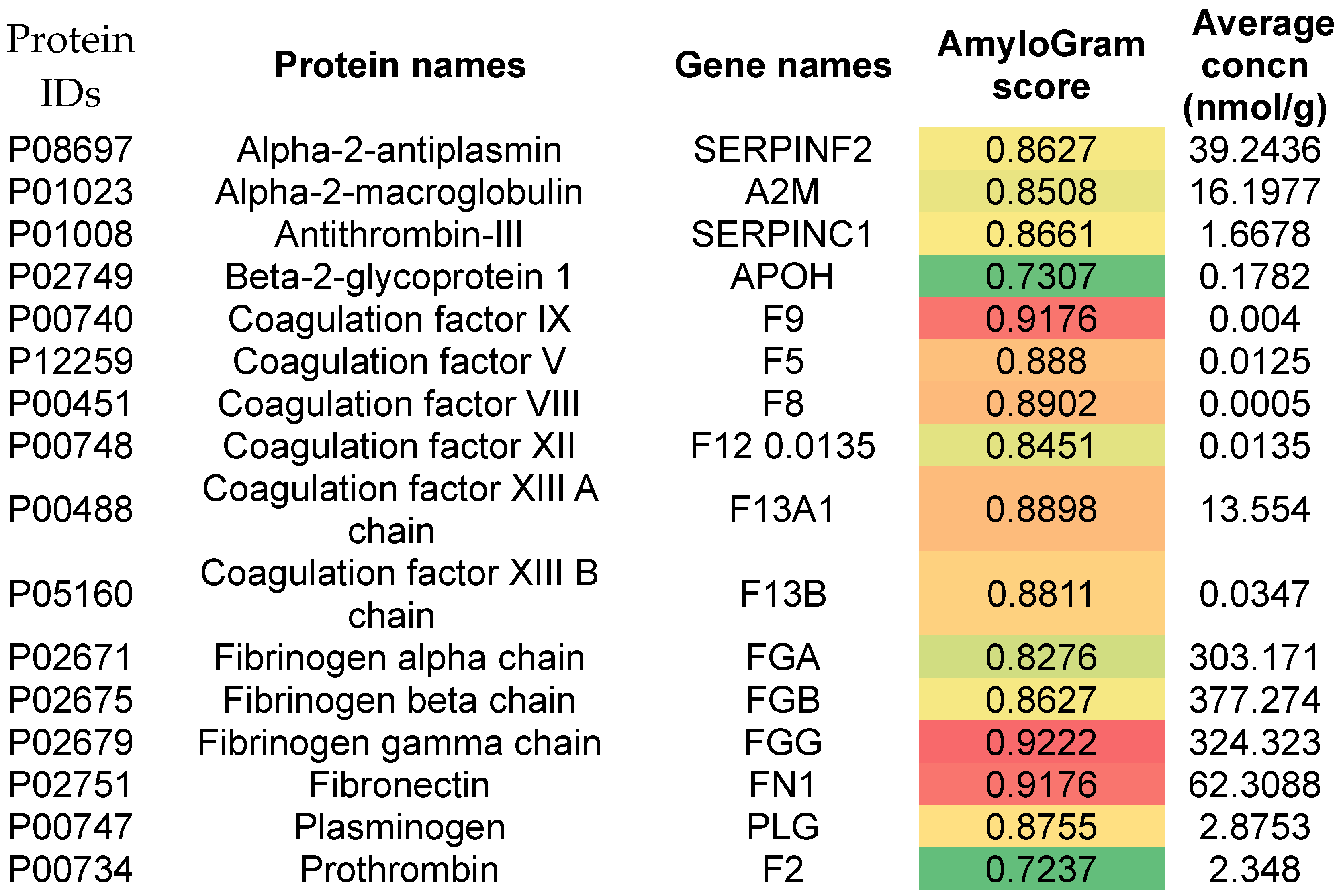

Figure 2.
AmyloGram scores for 198 polypeptides that were both found in thrombi retrieved following a pulmonary embolism and were also differentially expressed relative to those of control clots. Those 22 with an AmyloGram score exceeding 0.9 are labelled.
Figure 2.
AmyloGram scores for 198 polypeptides that were both found in thrombi retrieved following a pulmonary embolism and were also differentially expressed relative to those of control clots. Those 22 with an AmyloGram score exceeding 0.9 are labelled.
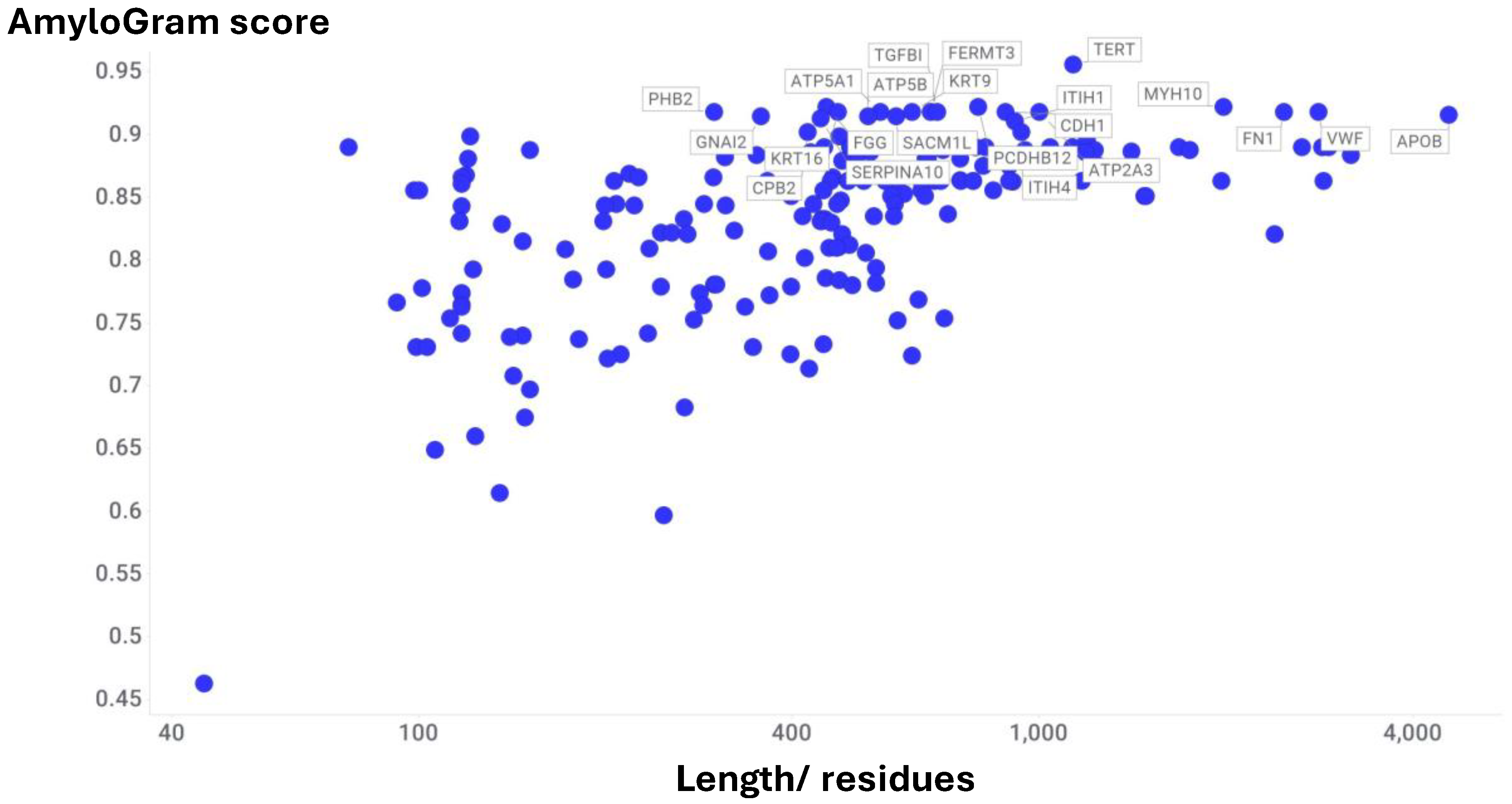
Figure 3.
AmyloGram scores of proteins in the proteome of the human coronary thrombus in patients with ST-segment elevation acute myocardial infarction. 46 proteins were found present in each case when assessed with three different separation/mass spectrometric methods. Data taken from the Supplementary information provided with reference [42].
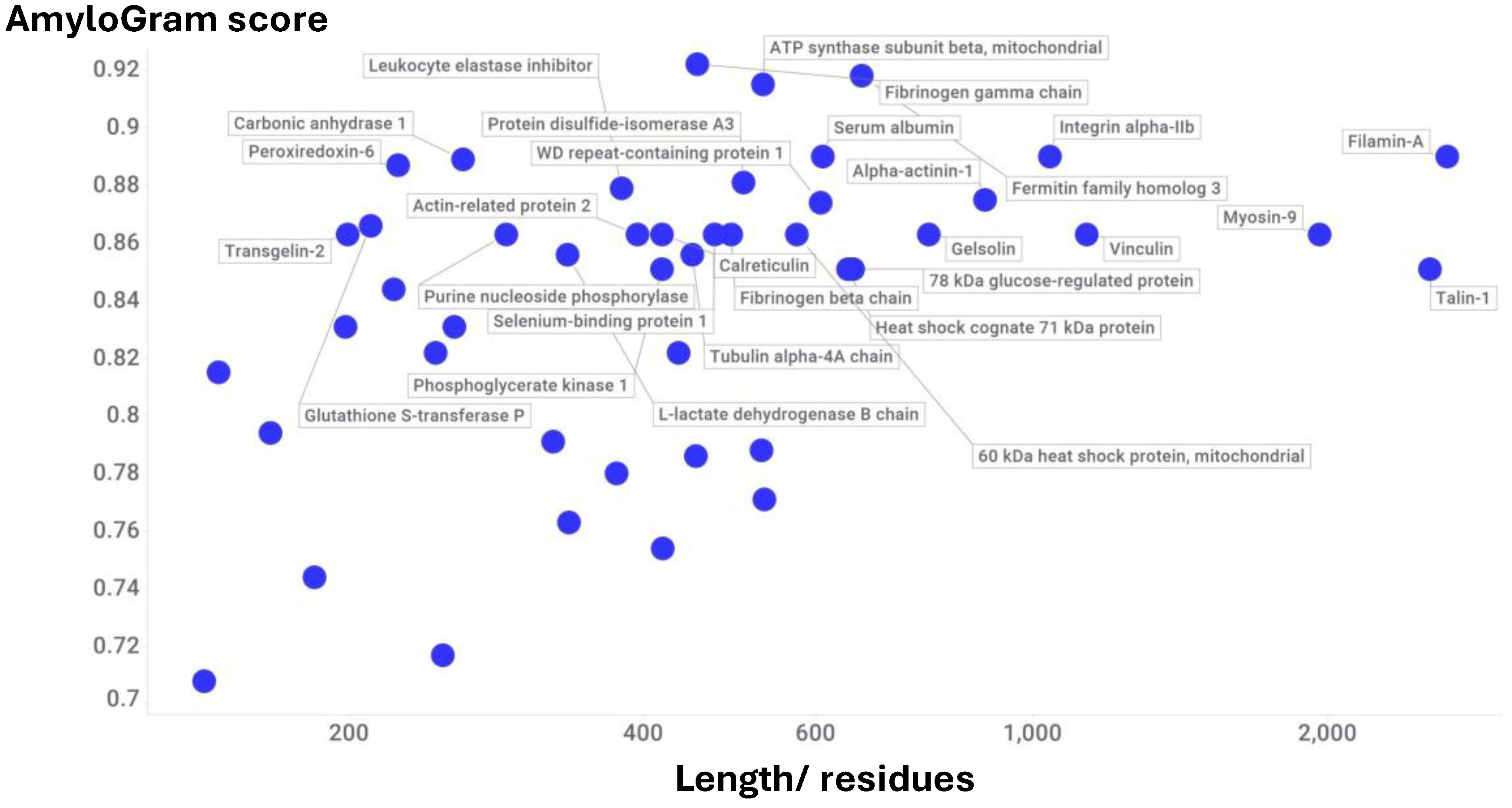
Figure 4.
11 proteins identified by Wang and colleagues (Table 1 of [51]) as being associated with coronary atherosclerosis. The colouring reflects the AmyloGram score.
Figure 4.
11 proteins identified by Wang and colleagues (Table 1 of [51]) as being associated with coronary atherosclerosis. The colouring reflects the AmyloGram score.
Figure 5.
20 proteins that were raised in atherosclerotic plaques (Supplementary Table S3 of [64]) and their AmyloGram scores.
Figure 5.
20 proteins that were raised in atherosclerotic plaques (Supplementary Table S3 of [64]) and their AmyloGram scores.
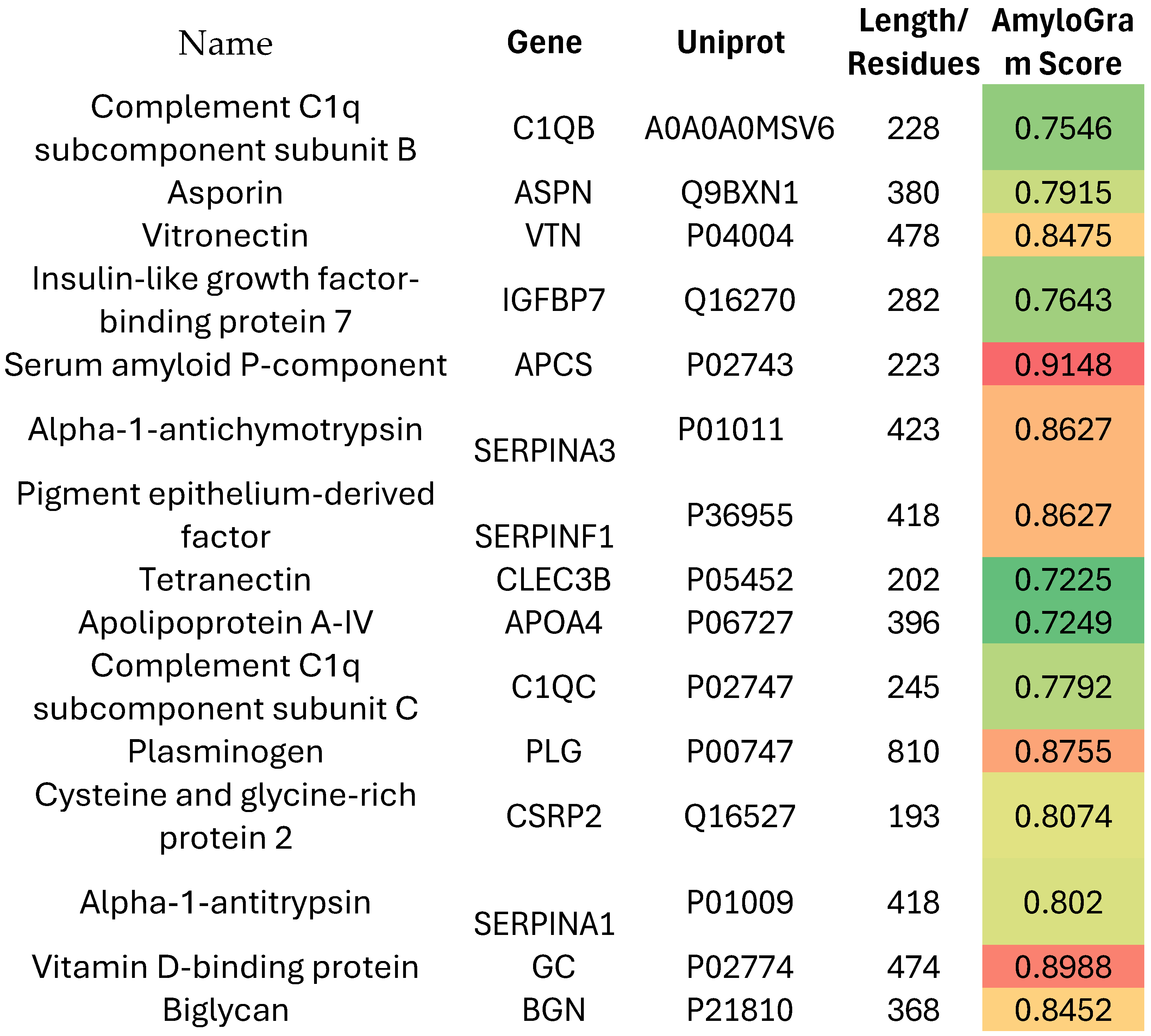
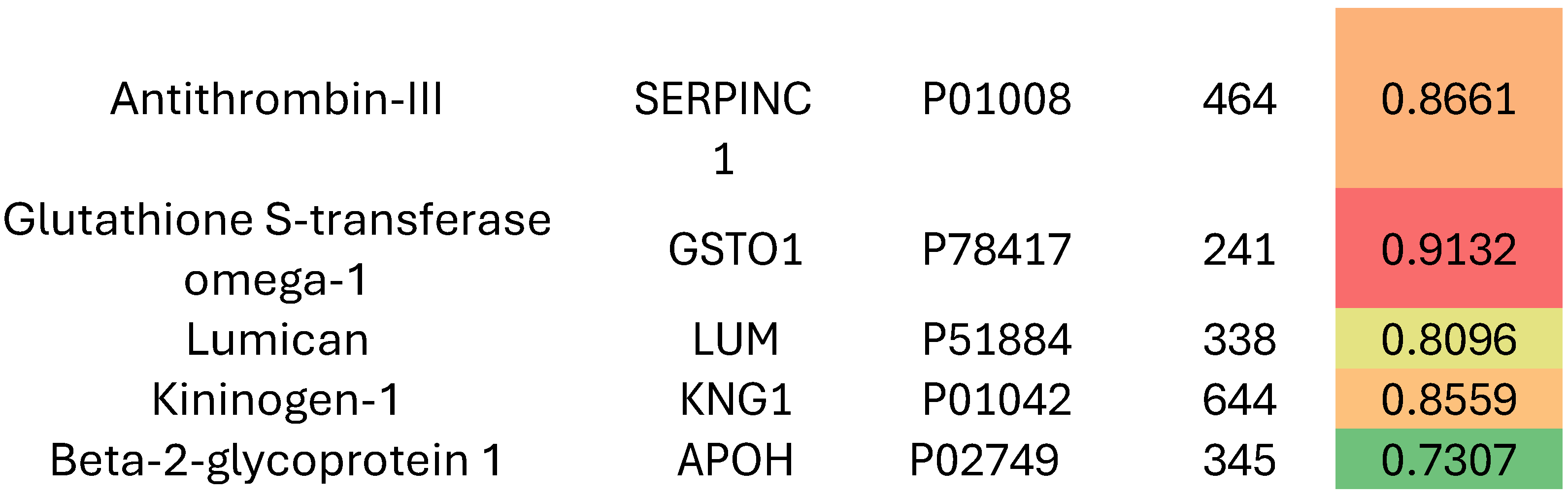
Figure 6.
83 proteins enriched differentially in atherosclerotic plaque from the perspective of their ease of degradation/ plaque stability. Protein identification data are obtained from Supplementary Table S2 of a study of Lorentzen and colleagues [49]. Again almost all have an AmyloGram score in excess of 0.7.
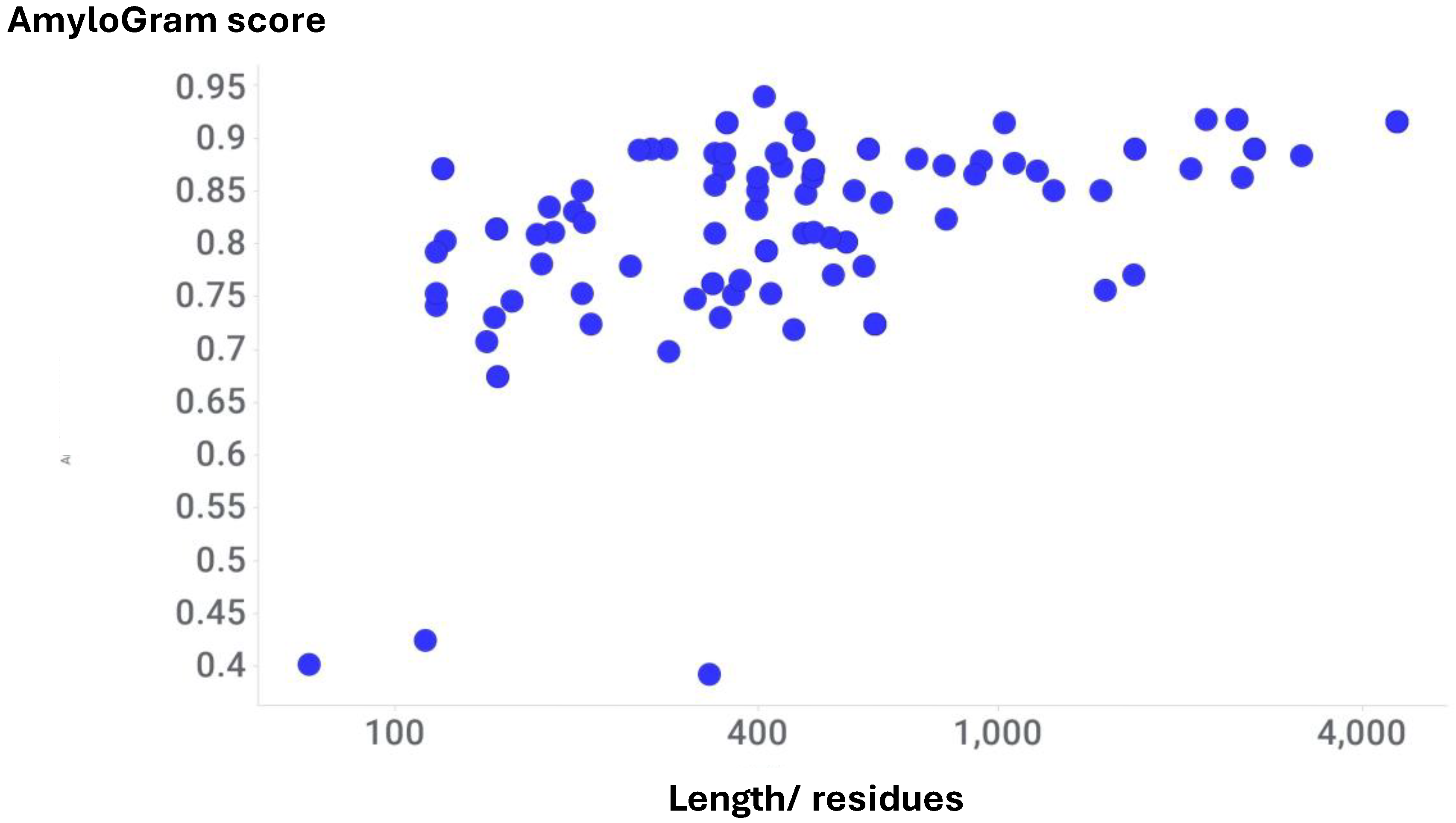
Figure 7.
25 proteins that were raised in the proteome of carotid atherosclerotic plaque relative to non-diseased mammary artery [40], along with their Amyogram scores (that are also encoded in colour).
Figure 7.
25 proteins that were raised in the proteome of carotid atherosclerotic plaque relative to non-diseased mammary artery [40], along with their Amyogram scores (that are also encoded in colour).
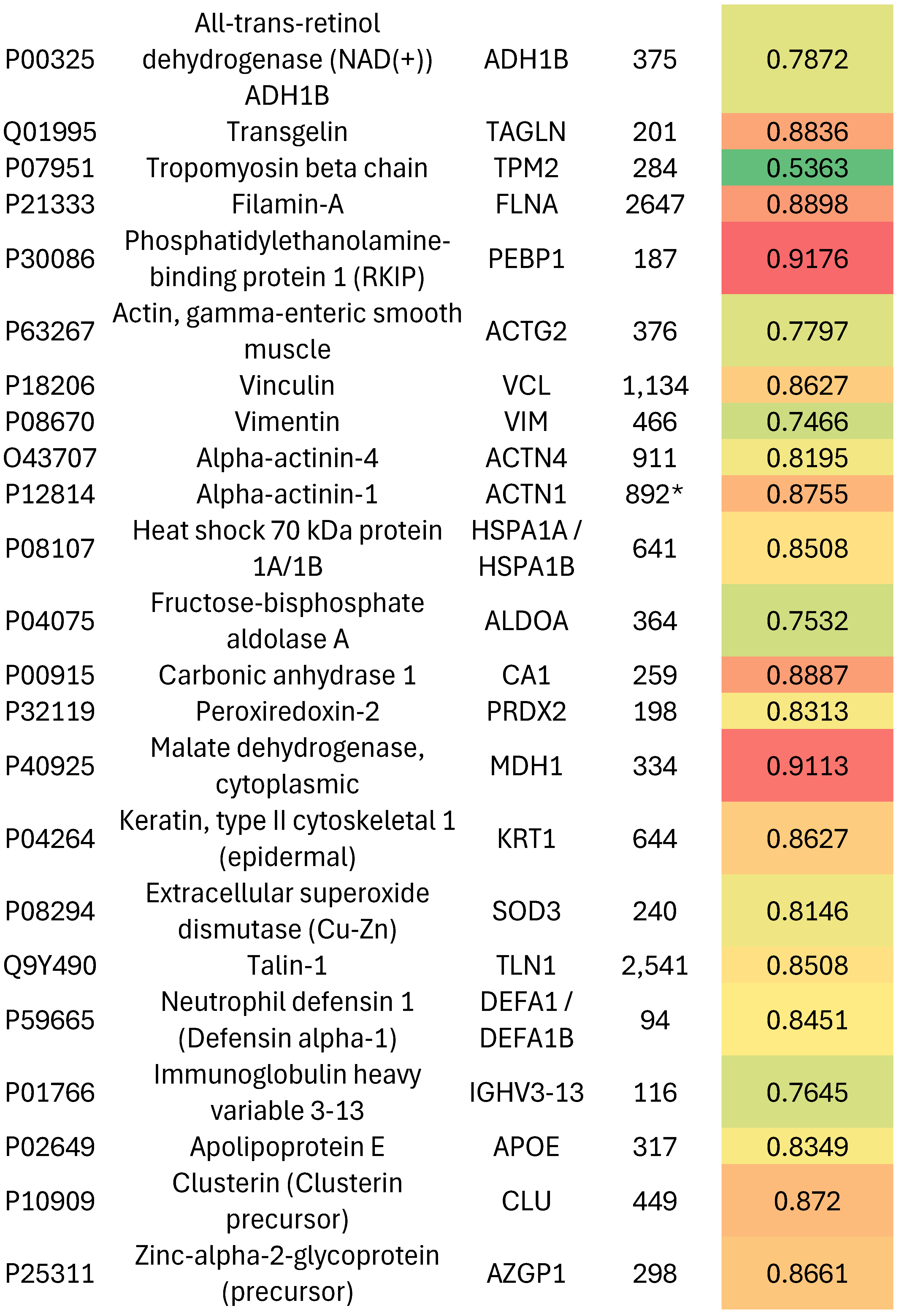
Figure 8.
AmyloGram scores of 121 fold-switching proteins. Those six with an AmyloGram score exceeding 0.9 are marked in blue. 95 of the 121 have an AmyloGram score exceeding 0.7.
Figure 8.
AmyloGram scores of 121 fold-switching proteins. Those six with an AmyloGram score exceeding 0.9 are marked in blue. 95 of the 121 have an AmyloGram score exceeding 0.7.
Disclaimer/Publisher’s Note: The statements, opinions and data contained in all publications are solely those of the individual author(s) and contributor(s) and not of MDPI and/or the editor(s). MDPI and/or the editor(s) disclaim responsibility for any injury to people or property resulting from any ideas, methods, instructions or products referred to in the content. |
© 2025 by the authors. Licensee MDPI, Basel, Switzerland. This article is an open access article distributed under the terms and conditions of the Creative Commons Attribution (CC BY) license (http://creativecommons.org/licenses/by/4.0/).
Copyright: This open access article is published under a Creative Commons CC BY 4.0 license, which permit the free download, distribution, and reuse, provided that the author and preprint are cited in any reuse.


