Preprint
Article

This version is not peer-reviewed.

IgM and IgG Epitope Mapping of the Porin Outer Membrane Protein-2a from Brucela abortus: Potential Biomarkers for Detecting Exposure to Brucellosis

Submitted:

06 August 2025

Posted:

11 August 2025

You are already at the latest version

Abstract

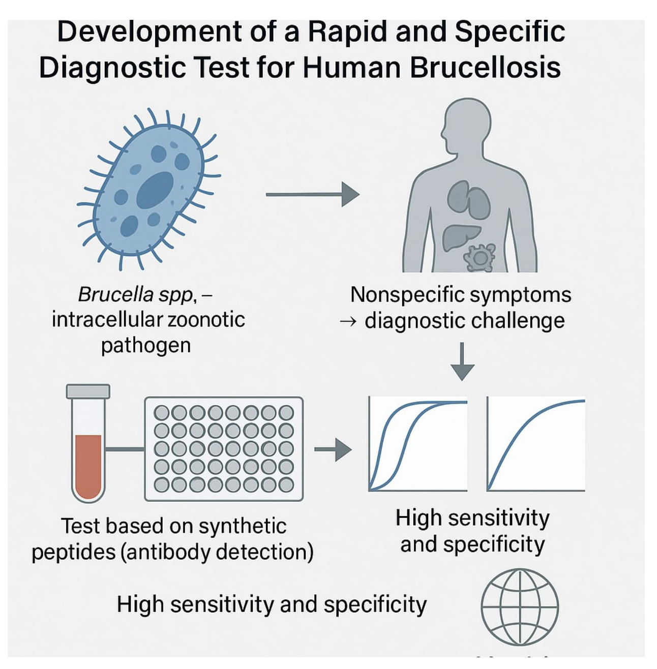
Brucellosis is one of the most serious zoonotic diseases worldwide, affecting both human and animal health. In humans, the disease often presents with diverse and nonspecific symptoms, making laboratory confirmation essential for accurate diagnosis and effective treatment. However, traditional diagnostic methods, including serological tests, suffer from limitations such as low sensitivity and high false-positive rates, underscoring the need for improved diagnostic strategies. This underscores the need for improved assays that enable rapid and reliable laboratory detection. In this study, we performed comprehensive IgM and IgG epitope mapping of the Omp-2a protein using sera from Brucella-infected patients. We identified epitopes, created chimeric peptides, and tested their diagnostic value with ELISA. Materials and Methods: The IgM and IgG epitopes of the Omp-2a protein were identified through SPOT synthesis on cellulose membranes, utilizing sera obtained from seropositive individuals. Potential cross-reactive epitopes were screened using peptide database searches and ELISA. Long, chimeric, multi-epitope peptides were synthesized and tested using ELISA on sera from 40 patients to evaluate their diagnostic performance. Results: Three major IgM and seven IgG linear B-cell epitopes were identified. None of the selected epitopes showed cross-reactivity with proteins from other significant human pathogenic organisms, as determined by analysis of peptide databases. Six peptide epitopes were confirmed using peptide-ELISA. Two chimeric peptides (50 and 60 amino acids) containing Brucella-specific IgM and IgG epitopes demonstrated high diagnostic performance, with a sensitivity of 99.96% and a specificity of 100%. Conclusion: Our study identified and validated IgM and IgG epitopes of the transmembrane Omp-2a protein from Brucella abortus. The use of these specific epitopes as biomarkers presents a promising strategy for developing accurate serological assays, potentially enhancing Brucellosis diagnosis and facilitating better monitoring of infection-induced immunity.

Keywords: 
;  ;  ;  ;  ;  ;  

1. Introduction

Brucellosis is one of the most common and endemic zoonotic diseases affecting both animals and humans in many countries; however, this disease remains neglected in human medicine, and nationwide control activities and surveillance programs have never been adequately structured [1,2,3]. Among twelve known Brucella spp., only B. melitensis, B. abortus, B. canis, and B. suis (except biovar 2) are associated with human infection and are facultative intracellular pathogens [4,5].
Brucellosis is an occupational disease affecting mostly people in contact with animals or animal products, particularly in agricultural countries where most of the population is involved in livestock farming and land cultivation.
The clinical symptoms of human brucellosis are nonspecific and can be mistaken for those of other diseases that cause fever [6,7]. Infection in humans is mainly associated with undulant fever, severe back pain, headache, joint pain, fatigue, myalgia, weight loss, loss of appetite, depression, and night sweats. Arthritis, orchitis, endocarditis, hepatomegaly, and splenomegaly have been seen in patients who suffered from brucellosis [8,9,10].
The disease is transmitted from unhealthy animals to humans via infected fluids and discharges such as blood, vaginal secretions, placental fluids, and aborted fetuses [11]. Bacterial transmission from person to person is rare, but sexual contact and breastfeeding have been proven to transmit Brucella [12]. Common routes of infection include direct inoculation through cuts, inoculation through the conjunctival sac of the eyes, abrasions of the skin, and inhaling infectious aerosols [13,14]. Occupational exposure to animals and direct contact with sick animals, as well as consuming contaminated milk and milk products, are the most common risk factors for human brucellosis [15,16]. The prevalence of brucellosis in livestock, particularly in low- and middle-income countries, has significant socioeconomic consequences [17,18] and poses substantial public health challenges [19,20]. In some parts of the world, the incidence of brucellosis is increasing daily due to a lack of awareness [21].
Early diagnosis of infected animals and humans is a crucial step in reducing the incidence of brucellosis. Thus, laboratory assays such as culturing, serology, and molecular analysis can frequently be used to diagnose brucellosis [22,23]. Although serological markers and PCR-based techniques are extremely sensitive, and extensive experience has been gained with these techniques in the laboratory diagnosis of brucellosis [24], a culture is still considered to be the "gold standard" due to the importance of this aspect of public health and clinical care.
In endemic regions, however, serological tests remain the primary method of diagnosis due to their low cost, user-friendliness, and strong ability to provide a negative prediction, so they are commonly used. A nucleic acid amplification assay, which is highly sensitive, specific, and safe, enables rapid disease diagnosis [25,26,27]. Patients who have reportedly fully healed may continue to have positive molecular test results for an extended period [28]. Therefore, cultures and serological methods will continue to be the primary tools for diagnosing and monitoring human brucellosis if no commercial tests or studies demonstrate adequate interlaboratory reproducibility.
Over the past few years, several studies have been conducted to develop Brucella vaccines [29,30,31,32,33,34], but the problem of controlling brucellosis in both humans and animals remains challenging [28]. Therefore, rapid and specific diagnosis is still necessary for both the treatment and prevention of infectious diseases, especially in humans, since all animal species that test positive for the disease must be slaughtered [35,36].
Classical serological tests, such as the tube agglutination test [37,38], rose Bengal plate test [39,40], and complement fixation test [41], as well as commercial enzyme-linked immunosorbent assay kits [42], are based on the detection of antibodies to the cell wall polysaccharide antigens of Brucella spp smooth strains. As a result, they do not exclude cross-reactions with related bacteria [43,44,45] and fail to differentiate between infected and vaccinated animals. Over the past decades, many attempts have been made to identify immunoreactive and pathogen-specific protein antigens. To date, several studies have investigated Brucella spp full recombinant proteins and chimeric cell wall proteins, as the best antigens for diagnosing brucellosis in humans [46,47,48,49,50] and animals [51,52,53,54,55,56]. However, the available results on the specificity and sensitivity of serological tests based on cell wall proteins are ambiguous and sometimes contradictory [28,57].
Therefore, this study aimed to identify the linear B-epitopes of the B. abortus Omp-2a protein, specifically those recognized by IgM and IgG, and to develop multiepitope polypeptides to explore their utility in the development of a rapid IgM and/or IgG diagnostic assay. The Omp-2a belongs to the alphaproteobacterial porin family, a multi-pass β-barrel outer membrane protein that induces a large passive diffusion pore at the center of each barrel, allowing small, hydrophilic molecular materials to cross the outer membrane [58,59].

2. Materials and Methods

2.1. Human Sera

In this study, blood samples from 40 patients diagnosed with brucellosis, confirmed by a commercial ELISA, were analyzed. Sera were obtained between 2012 and 2022 by the Central Public Health Laboratory of Rondônia (LACEN-Ro) and the Infectiology Service at the Oswaldo Cruz Policlinic in Rondônia, Brazil.
The other twenty-one sera samples used were from healthy individuals (21) obtained from the blood bank donor HEMORIO-RJ. For this study, patients' identities were kept anonymous.

2.2. Spot Synthesis

Two libraries of 163 peptides, each covering the entire sequence of Omp-2a (UniProt Q44620) from B. abortus (biovar 1, strain 9-941), were synthesized. These peptides were 15 residues in length, with a 10-residue overlap, and were obtained using a synthesizer (Auto-Spot ASP222, Intavis Bioanalytical Instruments AG, Köln, Germany) and the F-moc (9-fluorenylmethoxy carbonyl) strategy, as previously described [60].
As positive controls, the following peptides were used: GYPKDGNAFNNLDRI (Clostridium tetani; G20), KEVPALTAVETGATN (Poliovirus; G21), YPYDVPDYAGYP YDV (Hemagglutinin, Influenza; G22), GDFIDYEELREQLGG (Influenza A virus H3N2; G23), and YPGEFADYEELREQL (Influenza A virus Jakarta H1N1; G24).

2.3. Screening and Measurement of Spot Signal Intensities

Cellulose membranes (amino-PEG500-UC540; Intavis Bioanalytical Instruments, Köln, Germany) were equilibrated with TBS (50 mM Tris-buffered saline, pH 7.0) and blocked overnight at 4°C with TBS containing 3% casein and 0.1% Tween 20 (TBS-CT). After extensive washing with TBS-T (Tris-buffered saline, 0.1% Tween 20, pH 7.0), the membranes containing the peptide libraries were incubated for 2 hours with a sera pool from twenty different patients (1:250) in TBS-CT and then washed again with TBS-T.
Next, the membranes were incubated with alkaline phosphatase (AP)-labeled goat anti-human IgM (huIgM, 1:5000 in TBS-T; KPL, Gaithersburg, MD, USA; Lot #070466) or goat anti-human IgG (huIgG, 1:5000 in TBS-CT; Thermo, Lot #JUA1121836) for 1 hour, washed with TBS-T, and given a final wash in CBS (50 mM citrate-buffered saline, pH 7.0). Chemiluminescent CDP-Star® Substrate (0.25 mM) with Nitro-Block-II™ Enhancer (Applied Biosystems, Waltham, MA, USA) was then added to complete the reaction.
As described previously [61], chemiluminescent signals were detected using an Odyssey FC (LI-COR Bioscience, Lincoln, NE, USA). Briefly, a digital image file was generated at a resolution of 5 MP, and the signal intensities were quantified using the TotalLab TL100 software (v. 2009, Nonlinear Dynamics, Newcastle, Tyne, UK). The signal intensity (SI) used as a background was set by negative controls spotted on each membrane. Finally, a comparative analysis of the reactivity index of the spots, normalized on a dimensional hierarchical level, was conducted using the approach previously described [62].

2.4. Preparation of the Chimeric Peptides

Eight multi-antigen peptides (MAP4) epitope [PP225, PP226, PP227, PP228, PP229, PP231, PP232, PP233 (15 residues)] were synthesized using the F-moc protocol and the tetrameric TentaGel-S-NH2 resin (Intavis Bioanalytical Instruments AG, Köln, Germany) (Table S1) [63]. The synthesis of the two chimeric peptides, PP230-IgG (60 residues) and PP234 (50 residues), was conducted using XXX resin. Constructs were synthesized on a MultiPep-1 automated peptide synthesizer (CEM Corp, Charlotte, NC, USA), employing tetrafunctional Fmoc-amino acids with TFA-labile side chain protection where necessary. Residues of the monovalent (tail') segment, starting with bis-Fmoc Lys, were coupled using single-step protocols. After sequence assembly, F-moc groups were removed, and the peptide-resin was cleaved and deprotected with TFA/H₂O/EDT/TIS (94:2.5:2.5:1.0 v/v) for 90 min. The peptides were precipitated with cold diethyl ether, centrifuged three times for 10 minutes at 4°C, then dissolved in 10% aqueous Acetic Acid, dried, and stored as a lyophilized powder. MAP4 was dissolved in water, centrifuged at 10,000 g for 60 min at 15°C, and the supernatant filtered with a Centricon 10 filter. The single peptides were utilized without prior purification, and their identity was verified by MS (MALDI-TOF or electrospray).

2.5. Enzyme-Linked Immunosorbent Assay (ELISA)

The in-house ELISA was performed as previously described [64]. Briefly, 96-well plates (Immulon 2HB; Thermo Fisher, Waltham, MA, USA), were incubated (12 h at 4 °C) with 50 µl synthetic peptide solution (100 µg/mL in Na2CO3-NaHCO3 buffer, 0.1 M, pH 9.6). After three washes with PBS pH 7.2, the free sites on the plate were blocked for 90 min with 200 μL of a 2% skim milk solution in PBS containing 0.1% Tween 20, pH 7.2. After three further washes in PBS, 50 µl of patient serum (1:50) was added to the wells and the plate incubated for 90 min at 37 °C. After further washes with PBS, 50 µl of goat anti-human IgG (H+L) conjugated to alkaline phosphatase (AP) (1: 5000) (Thermo Fisher, Waltham, MA, USA) was added to the wells and the plate incubated for 90 min at 37 °C. After a series of three washes in PBS pH 7.2, the pNPP substrate was added, and the plates were incubated for 15 min. Next, 50 µl of 3N NaOH were added, and the reading was done after 30-120 min at 405 nm on a FlexStation3 (Molecular Devices, Sunnyvale, Ca, USA).
For comparison, a commercial ELISA assay (classic kit, Serion, Curitiba, Brazil) was used. Briefly, precoated micro test plates were incubated with patients' sera, and after removal of unbound material, anti-human Ig-AP conjugate was allowed to react with the immune complex. After washing away excess conjugation, pNPP was added, and specific antibody binding was measured photometrically.

2.6. Bioinformatics and In-Silico Analysis Model

The complete sequence of Omp-2a (Q44620) of B. abortus was retrieved from the National Center for Biotechnology Information, USA (http://www.ncbi. nlm.nih.gov). Epitope locations within the three-dimensional molecular structure of Omp-2ae were identified by generating in silico protein models using the I-TASSER server (http://zhanglab.ccmb, accessed April 10, 2024). Models were selected based on their C-score and TM-score, and the 3D models were validated using the AlphaFold v3 database [65].
Multiple sequence alignments were performed using the programs ClustalW (http:// www.ebi.ac.uk/clustalw) and BioEdit (http://www.mbio.ncsu.edu/BioEdit/bioedit.html).

2.7. Statistical Analysis

ELISA tests were statistically analyzed using Med Calc software version 20.218 [66]. A statistical difference was considered significant if the p-value was ≤ 0.05. Initially, the outcomes for each peptide were reported as a reactivity index (RI), determined by the optical density (OD) ratio of a particular sample to the cut-off OD values for each test. All RI values were classified as positive (>1.00) or negative (< 0.005); those with a value of 0.005 were statistically significant.

2.8. Ethical Standards

This work was approved by the FIOCRUZ Ethics in Research Committee (CAAE: 23784114.8.0000.5248, 15 May 2014) and Federal Fluminense University (23/04/21 UFF)

3. Results

3.1. Epitope Mapping of the Omp-2a

IgM and IgG linear B epitopes of the full Omp-2a (321 aa) protein recognized by the patient’s sera were identified using a library of 163 synthetic peptides covalently linked to a cellulose membrane (Figure 1A and Figure 2A) and developed as described before. Panel B shows the chemiluminescence intensity measurement using the patient’s serum, and Panel C shows the hierarchical distribution of the peptides according to their reaction intensity. For IgM, the most reactive peptide was C1, followed by C2, D5, and others. For IgG, the most reactive peptides were C1, C2, D7, and B18.
The intensities were normalized using the reactivity of the positive control as 100%. The ratio of the synthesized peptides and their respective positions in the membranes is shown in Table S2.
The overlapping sequences of the positive peptides defined 10 epitopes for Omp-2a, comprising 3 IgM and 7 IgG epitopes recognized by the patient’s sera pool (Table 1).

3.2. Shared Epitopes and Selection of Specific Brucella sp Epitopes

Three common structural overlapping epitopes recognized by both IgM and IgG were identified. These include the sequence NNSRHDGQYG, shared by epitopes Omp-2a/1M and Omp-2a/4G; NGFSAVIALE, found in Omp-2a/2M and Omp-2a/5G; and KFGGE, present in Omp-2a/3M and Omp-2a/10G (Figure 3). The remaining four IgG epitopes (6G, 7G, 8G, and 9G) were IgG isotype-specific and did not overlap with IgM epitopes.
Regarding specificity based on sequence database analysis, only the 3M and 10G epitope was exclusively found in the Brucella genus, indicating genus specificity, ie. are present in Omp-2a of the structure of Brucella spp. but absent in other bacteria such as E. coli, Shigella boydii, and Yersinia spp.. The other eight epitopes exhibited sequence homology with proteins from various non-pathogenic genera, including Rhodotorula, Bartonella, Ochrobactrum, and Falsochrobactrum (Table 1). However, none of these genera are known to include human pathogens.
Figure 4 presents the alignment results of the B. bovis Omp-2a sequence with autotransporter proteins from various genera, demonstrating a general identity of 70% or more. In the figure, the colored rectangles demarcate only the unique linear B-epitopes (either IgM or IgG) that have been identified. No correlation was identified with Yersinia sp proteins.

3.3. Structural Studies

The secondary structure elements of Omp-2a (Q44620) were identified using the TMpred tool, as described in the previous section. The protein has 11 alpha-helical transmembrane (TM) and two conserved domains (signal peptide and autotransporter) and tandem repeats, indicating that Omp-2a is an authentic OmpA (Figure 5) but shares no sequence homology with any other porin and a higher number of negatively changed residues in the exposed loops than Omp-2b [45].
The Omp-2a 3D model was constructed by homology modeling using as a template the available data toxin (ABC protein complex) Yersinia entomophaga, which presented the highest "score". The models generated by the I-Tasser server presented reliability parameters with C-score values of 0.48 and RMSD = 8.6 (± 4.5 Ǻ). The 3D model, illustrating the spatial distribution of epitopes, is shown in Figure 5. The amino acids that define the 10 (IgM and IgG) epitopes were located and marked on the model of secondary structure. From this analysis, we can conclude that all epitopes are facing the external surface of the protein and are thus accessible to the immune system. All are "linear" structures with "coils" characteristics of linear B epitopes.
The protein presents 16-beta-barrel sheets, with large surface-exposed loops, that form a TM pore at the center of each barrel. The pore is partially occluded by a peptide loop that folds into the pore lumen. The larger pore formed by Omp-2a may be advantageous for intracellular growth when the bacterium competes with the host cell for nutrients whose concentration is particularly low within the phagosome.

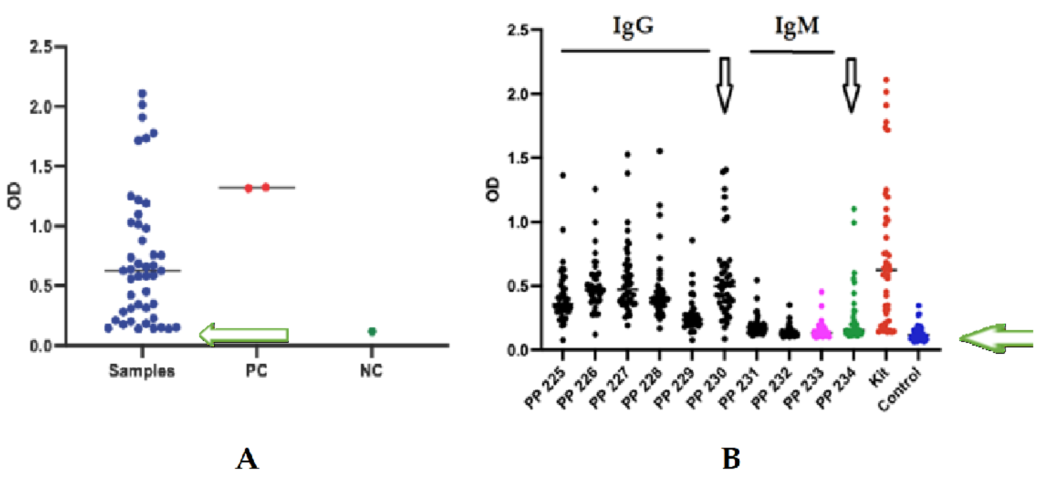
3.4. Enzyme Immunoassay with Human Serum

From the sera of forty brucellosis suspect patients, nineteen were identified as positive with a higher optical density (OD) using a commercial kit and were included in this study. The results are depicted in Figure 6A. Among the nineteen analysed sera, only 1-3 did not exceed the cutoff values for all eight peptides analysed (PP225-PP229 and PP231-PP233), whereas sera from twenty-one apparently healthy individuals consistently fell 3-5 units above the cutoff point (Figure 6B) .
Cutoffs for each peptide (PP225-PP227 and PP231-233) were established using statistical analysis with ROC curve (Figure 7), as detailed in Table S3.
The ROC curves illustrate the diagnostic test’s discriminatory ability across different cutoff values, highlighting the trade-offs between optical sensitivity and specificity. The points closest to the top left corner of the ROC curve indicate the most effective diagnostic thresholds,
Sensitivity, defined as the proportion of true positive results (TP) relative to all affected patients (TP + false negative [FN], ranged from 0.88 to 0.95, indicating a strong statistical correlation between sera of patients with brucellosis and healthy individual sera results.
Specificity, representing the proportion of true negatives (TN) among controls (TN + false positive results [FPR]), ranged from 90% to 95%. The control group analyzed in the ROC curve analysis consisted of healthy individuals, demonstrating that the in-house ELISA test peptide exhibits 90-95% specificity for the diagnosis of Brucellosis in humans, a similar value obtained by the commercial kit.
ROC curves provide quantitative assessments of diagnostic test accuracy, with the area under the curve indicating overall efficiency. Peptide PP225-PP228 and P230 demonstrated higher immunogenicity (90% specificity and 94% sensitivity) (Table S2).

4. Discussion

Brucellosis diagnosis requires further refinement in terms of sensitivity and specificity, particularly during the acute phase [67]. Research indicates that diagnosing brucellosis is challenging due to difficulties with serological examinations. Previous studies have reported variable accuracy in ELISA-based detection of Brucella-specific IgM and IgG antibodies, with some suggesting a combined specificity of up to 100% [68,69]. However, the persistence of antibodies in cases without active infection complicates clinical interpretation.
Therefore, identifying epitope-specific immune responses may enhance diagnostic accuracy by targeting epitopes associated with active and recent infection. This study focused on the Omp-2a protein for epitope mapping due to its antigenic properties, membrane localization [70], and critical role in immune responses. Therefore, it was used in association with other proteins in the process of vaccination and diagnosis as a recombinant protein [71].
Our results identified ten immunoreactive epitopes on the B. abortus Omp-2a protein that reacted with anti-IgM and anti-IgG antibodies (Table 1). As no cross-reactivity was found with other human pathogens, the finding supports the molecular identification of Omp-2a from B. abortus as a suitable target for diagnostic use. Numerous studies have highlighted the physicochemical properties of B cell epitopes, such as the surface accessibility of membrane-bound or free antibodies [72,73]. This characteristic enables antibodies to effectively bind to and neutralize their biological targets [74]. Additionally, understanding the dynamics of humoral immune responses during Brucella infection is crucial for enhancing serodiagnostic tools and developing effective vaccine candidates. In human brucellosis, IgM antibodies typically appear within the first week of infection and peak around two months later, whereas IgG antibodies emerge after the second week and reach their maximal levels between six- and eight-weeks post-infection. Although IgG responses align more closely with disease progression, the detection of IgM in the absence of IgG, or vice versa, can lead to diagnostic ambiguity [75]. This highlights the need for refined diagnostic markers that can distinguish between the early and late phases of infection with high specificity and sensitivity.
This pattern reflects both the immunological stage of infection and the antigenic architecture of the Omp-2a protein, a 16-layer β-barrel secondary structure with large surface-exposed loops, which can naturally make accessible unique linear B cell epitopes. [76].
The relative scarcity of IgM epitopes identified in this work may be attributed to isotype switching during infection, as most patients included in our study were in a later phase of disease or under treatment. The process of isotype switching is a hallmark of adaptive immunity, involving recombination of constant regions in immunoglobulin genes to generate antibody classes with different effector functions and half-lives. This transition, while essential for developing high-affinity IgG antibodies, can obscure the presence of short-lived IgM responses, which are typically useful markers of recent or acute infection.
Interestingly, three of the IgM epitopes (Omp-2a/1M, 2M, and 3M) shared sequences with corresponding IgG epitopes (Omp-2a/4G, 5G, and 10G), indicating a sustained immune response against these antigenic determinants across different stages of infection. In contrast, four IgG epitopes (Omp-2a/6G to 9G) were uniquely recognized, possibly reflecting epitopes that induce IgG responses with no preceding or concurrent IgM reactivity, suggesting either rapid class switching or late-stage immune memory development. These findings support the notion that certain epitopes may be more relevant for the early diagnosis of brucellosis, while others could serve as markers of chronic or convalescent phases.
The validation of these immunodominant epitopes/peptides via ELISA confirmed their reactivity with sera from Brucella-infected patients. To assess their diagnostic potential and specificity, four epitopes with no homology to proteins from unrelated microorganisms were further evaluated. Notably, peptides PP225–PP229, mapped to the loops, were recognized by both IgM and IgG, indicating their capacity to elicit long-lasting immune responses. However, cross-reactivity observed within the epitopes 2M/5G and 7G and proteins from Bartonella tamiae, a Gram-negative bacterium isolated from the blood of Tailândia patients, suggests that these epitopes must be avoided when preparing a specific and universal diagnostic test for Brucellosis.
On the other hand, Omp-2a and Omp-2b are homologous antigens [77], but do not share the same linear epitopes.
In summary, this study advances our understanding of human humoral responses to Brucella Omp-2a by identifying epitope-specific IgM and IgG reactivity patterns [78]. The identification of three epitopes (1M/4G, 2M/5G, and 3M/10G) recognized by both IgM and IgG isotypes and five recognized only by IgG (exhibiting high specificity highlights their potential utility in the development of improved serological IgM/IgG or only IgG assays for brucellosis. These findings underscore the importance of epitope-level analysis in distinguishing between infection stages and improving diagnostic precision, thereby providing valuable insights into both clinical management and future vaccine design.

5. Conclusions

This study demonstrates the potential of synthetic peptides as effective substitutes for native proteins in immunodiagnostic assays for Brucellosis. Using peptide arrays, we established a high-throughput and cost-efficient platform for the identification of disease-specific linear B-cell epitopes. A total of three IgM and seven IgG epitopes were identified from the Omp-2a protein. Subsequently, eight peptides were selected for further validation through ELISA-based assays. These peptides demonstrated strong diagnostic performance, with ROC curve analyses confirming high sensitivity, specificity, and overall accuracy in distinguishing individuals infected with Brucellosis from healthy controls. Statistical analyses further reinforced the significant immunoreactivity of these peptides. Together, these findings highlight the diagnostic value of peptide-based assays and support their application in the development of reliable, specific, and scalable serological tests for Brucellosis.

Supplementary Materials

The following supporting information can be downloaded at the website of this paper posted on Preprints.org, Table S1: List of the synthesized epitope/peptides. Glycine (G) residues in larger letters were added to standardize the length of all synthetic single peptides. For chimeric peptide #234, glycines were also used to separate the epitopes and ensure consistent peptide size. Table S2: List of Synthesized Peptides Covering the Entire Sequence of Omp-2 Protein (Uniprot Database) from Brucella abortus. This table details the synthesized peptides covering the entire Omp-2 protein sequence, including positive controls (F3, F4, F5, F11, F12, F13) and negative controls (F9, F17). The overlapping of positive peptides is highlighted in red. Table S3: Values of the statistical analysis of ELISA and ROC (Receiver Operating Characteristic) data. The area under the curve (AUC) serves as a measure of the test's overall accuracy.

Author Contributions

Conceptualization, S.G.D-S. and F.R.S.; methodology, A.F.N., G.C.L.; P.N-P.; J.P.R.S. and L.R.G.; data curation, F.R.S.; writing—original draft preparation, S.G.D-S.; writing—review and editing, S.G.D-S.; supervision, F.R.S. and S.G.D-S.. All authors have read and agreed to the published version of the manuscript. All authors have read and agreed to the published version of the manuscript.

Funding

This research was funded by Carlos Chagas Filho Foundation of Research Support of the State of Rio de Janeiro (FAPERJ #010.101.029/2018) and the Brazilian Council for Scientific Research (CNPq #301744/2019-0).

Institutional Review Board Statement

The study was conducted in accordance with the Declaration of Helsinki and approved by the Ethics Research Committee of Faculty of Medicine/Federal Fluminense University (protocol code CAAE:46304621.8.0000.5243) and Oswaldo Cruz Institute/ /FIOCRUZ (protocol code CAAE:23784114.8.0000.5248).

Informed Consent Statement

The consent of the patients was sought, and informed consent was obtained from all subjects involved in the study to publish this paper.

Data Availability Statement

All data required to evaluate the conclusions of this study are presented in the paper and/or Supplementary Materials. Additional data related to this study can be obtained from the corresponding author upon reasonable request

Acknowledgments

P.N-P. and G.C.L. are postdoctoral fellows from the INOVA-FIOCRUZ Program. A.F.N. and J.P.R.S.C. are DSc fellows from the Post-Graduation Program on Science and Biotechnology, Federal Fluminense University.

Conflicts of Interest

The authors declare no conflict of interest.

References

  1. WHO 2019. Neglected tropical diseases. World Health Organiztion. Available online: http://www.who.int/neglecteddiseases/ zoonoses/infections_more/en/ (accessed on 24 May 2025).
  2. WHO, 2001. WHO recommended strategies for the prevention and control of communicable Diseases. Available online: https://www.who.int/news-room/fact-sheets/detail/brucellosis (accessed on 24 May 2025).
  3. Qureshi, K.A.; Parvez, A.; Fahmy, N.A.; Abdel Hady, B.H.; Kumar, S.; Ganguly, A.; Atiya, A.; Elhassan, G.O.; Alfadly, S.O.; Parkkila, S.; Aspatwar, A. Brucellosis: epidemiology, pathogenesis, diagnosis and treatment-a comprehensive review. Ann Med 2023, 55, 2295398. [Google Scholar] [CrossRef]
  4. Swai, E.S.; Schoonman, L. Human brucellosis: Seroprevalence and risk factors related to high risk occupational groups in Tanga Municipality, Tanzania. Zoonoses Public Health 2009, 56, 183–187. [Google Scholar] [CrossRef]
  5. Laine CG, Johnson VE, Scott HM, Arenas-Gamboa AM. Global estimate of human brucellosis incidence. Emerg Infect Dis 2023, 29, 1789–1797. [CrossRef]
  6. Franco, M.P.; Mulder, M.; Gilman, R.H.; Smits, H.L. Human brucellosis. Lancet Infect Dis 2007, 7, 775–786. [Google Scholar] [CrossRef]
  7. Saddique, A.; Ali, S.; Akhter, S.; Khan, I.; Neubauer, H.; Melzer, F.; Khan, A.U.; Azam, A.; El-Adawy, H. Acute febrile illness caused by Brucella abortus Infection in humans in Pakistan. Int J Environ Res Public Health 2019, 16, 4071. [Google Scholar] [CrossRef]
  8. Hassan, K.S.; Schuster, H.; Al-Rawahi, A.; Balkhair, A. Clinical presentations of brucellosis over a four-year period at Sultan Qaboos University Hospital and armed forces hospital, Muscat, Oman. Sultan Qaboos Univ Med J 2021, 21, e282–e288. [Google Scholar] [CrossRef]
  9. Zheng, R.; Xie, S.; Lu, X.; Sun, L.; Zhou, Y.; Zhang, Y.; et al. . A systematic review and meta-analysis of epidemiology and clinical manifestations of human brucellosis in China. BioMed Res Int 2018, 2018, 5712920. [Google Scholar] [CrossRef]
  10. Kiiza, D.; Denagamage, T.; Serra, R.; Maunsell, F.; Kiker, G.; Benavides, B.; Hernandez, J.A. A systematic review of economic assessments for brucellosis control interventions in livestock populations. Prev Vet Med 2023, 213, 105878. [Google Scholar] [CrossRef] [PubMed]
  11. Ducrotoy, M.J.; Bertu, W.J.; Ocholi, R.A.; Gusi, A.M.; Bryssinckx, W.; Welburn, S.; Moriyon, I. Brucellosis as an emerging threat in developing economies: Lessons from Nigeria. PLoS Negl Trop Dis 2014, 8, 1–18. [Google Scholar] [CrossRef]
  12. Agasthya, A.S.; Isloor, S.; Krishnamsetty, P. Seroprevalence study of human brucellosis by conventional tests and indigenous indirect enzyme-linked immunosorbent assay. Sci. World J 2012, 20, 104–239. [Google Scholar] [CrossRef]
  13. Herrick, J.A.; Lederman, R.J.; Sullivan, B.; Powers, J.H.; Palmore, T.N. Brucella arteritis: Clinical manifestations, treatment, and prognosis. Lancet Infect Dis 2014, 14, 520–526. [Google Scholar] [CrossRef]
  14. Blacksell, S.D.; Dhawan, S.; Kusumoto, M.; Le, K.K.; Summermatter, K.; O'Keefe, J.; Kozlovac, J.; Almuhairi, S.S.; Sendow, I.; Scheel, C.M.; et al. The biosafety research road map: The search for evidence to support practices in the laboratory-Bacillus anthracis and Brucella melitensis. Appl Biosaf 2023, 28, 72–86. [Google Scholar] [CrossRef] [PubMed]
  15. Dadar, M.; Tabibi, R.; Alamian, S.; Caraballo-Arias, Y.; Mrema, E.J.; Mlimbila, J.; Chandrasekar, S.; Dzhusupov, K.; Sulaimanova, C.; Alekesheva, L.Z.; et al. Safety concerns and potential hazards of occupational brucellosis in developing countries: A review. J Public Health (Berl) 2023, 31, 1681–1690. [Google Scholar] [CrossRef]
  16. Memish, Z.A.; Balkhy, H.H. Brucellosis and international travel. J. Travel. Med. 2004, 11, 49–55. 10. Rahman, A.K.; Dirk, B.; Fretin, D.; Saegerman, C.; Ahmed, M.U.; Muhammad, N.; Hossain, A.; Abatih, E. Seroprevalence and risk factors for brucellosis in a high-risk group of individuals in Bangladesh. Foodborne Pathog Dis 2012, 9, 190–197. [Google Scholar] [CrossRef]
  17. Lokamar, P.N.; Kutwah, M.A.; Atieli, H.; Gumo, S.; Ouma, C. Socio-economic impacts of brucellosis on livestock production and reproduction performance in Koibatek and Marigat regions, Baringo County, Kenya. BMC Veter Res 2020, 16, 61. [Google Scholar] [CrossRef]
  18. Lai, S.; Chen, Q.; Li, Z. Human brucellosis: An ongoing global health challenge. China CDC Wkly 2021, 3, 120. [Google Scholar] [CrossRef]
  19. Vrioni, G.; Gartzonika, C.; Kostoula, A.; Boboyianni, C.; Papadopoulou, C.; Levidiotou, S. Application of a polymerase chain reaction enzyme immunoassay in peripheral whole blood and serum specimens for diagnosis of acute human brucellosis. Eur J Clin Microbiol Infect Dis 2004, 23, 194–199. [Google Scholar] [CrossRef]
  20. Nikokart 202 Freire, M.L.; Machado de Assis, T.S.; Silva, S.N.; Cota, G. Diagnosis of human brucellosis: Systematic review and meta-analysis. PLoS Negl Trop Dis 2024, 18, e0012030. [Google Scholar] [CrossRef]
  21. Arif, S.; Thomson, P.C.; Hernandez-Jover, M.; McGill, D.M.; Warriach, H.M.; Heller, J. Knowledge, attitudes and practices (KAP) relating to brucellosis in small holder dairy farmers in two provinces in Pakistan. PLoS ONE 2017, 12, e0173365. [Google Scholar] [CrossRef]
  22. Dal, T.; Kara, S.S.; Cikman, A.; Balkan, C.E.; Acıkgoz, Z.C.; Zeybek, H.; Uslu, H.; Durmaz, R. Comparison of multiplex real-time polymerase chain reaction with serological tests and culture for diagnosing human brucellosis. J Infect Public Health 2019, 12, 337–342. [Google Scholar] [CrossRef]
  23. Di Bonaventura, G.; Angeletti, S.; Ianni, A.; Petitti, T.; Gherardi, G. Microbiological laboratory diagnosis of human brucellosis: An overview. Pathogens 2021, 10, 1623. [Google Scholar] [CrossRef]
  24. Gusi, A.M.; Bertu, W.J.; Jesús de Miguel, M.; Dieste-Pérez, L.; Smits, H.L.; Ocholi, R.A.; Blasco, J.M.; Moriyón, I.; Muñoz, P.M. Comparative performance of lateral flow immunochromatography, iELISA and rose bengal tests for the diagnosis of cattle, sheep, goat and swine brucellosis. PLoS NeglTrop Dis 2019, 13, e0007509. [Google Scholar] [CrossRef]
  25. Hinić, V.; Brodard, I.; Thomann, A.; Cvetnić, Z.; Makaya, P.V.; Frey, J.; Abril, C. Novel identification and differentiation of Brucella melitensis, B. abortus, B. suis, B. ovis, B. canis, and B. neotomae suitable for both conventional and real-time PCR systems. J Microbiol Methods. 2008, 75, 375–378. [Google Scholar] [CrossRef]
  26. Zeybek, H.; Acikgoz, Z.C.; Dal, T.; Durmaz, R. Optimization and validation of a real-time polymerase chain reaction protocol for the diagnosis of human brucellosis. Folia Microbiol (Praha) 2020, 65, 353–361. [Google Scholar] [CrossRef] [PubMed]
  27. Freire, M.L.; Machado de Assis, T.S.; Silva, S.N.; Cota, G. Diagnosis of human brucellosis: Systematic review and meta-analysis. PLoS Negl Trop Dis 2024, 18, e0012030. [Google Scholar] [CrossRef]
  28. Elbehiry, A.; Aldubaib, M.; Marzouk, E.; Abalkhail, A.; Almuzaini, A.M.; Rawway, M.; Alghamdi, A.; Alqarni, A.; Aldawsari, M.; Draz, A. The development of diagnostic and vaccine strategies for early detection and control of human brucellosis, particularly in endemic areas. Vaccines (Basel) 2023, 11, 654. [Google Scholar] [CrossRef]
  29. Masjedian Jezi, F.; Razavi, S.; Mirnejad, R.; Zamani, K. Immunogenic and protective antigens of Brucella as vaccine candidates. Comp Immunol Microbiol Infect Dis 2019, 65, 29–36. [Google Scholar] [CrossRef]
  30. Rezaei, M.; Rabbani-Khorasgani, M.; Zarkesh-Esfahani, S.H.; Emamzadeh, R.; Abtahi, H. Prediction of the Omp16 epitopes for the development of an epitope-based vaccine against Brucellosis. Infect Disord Drug Targets 2019, 19, 36–45. [Google Scholar] [CrossRef]
  31. Sha, T.; Li, Z.; Zhang, C.; Zhao, X.; Chen, Z.; Zhang, F.; Ding, J. Bioinformatics analysis of candidate proteins Omp2b, P39 and BLS for Brucella multivalent epitope vaccines. Microb Pathog 2020, 147, 104318. [Google Scholar] [CrossRef] [PubMed]
  32. Chen, Z.; Zhu, Y.; Sha, T.; Li, Z.; Li, Y.; Zhang, F.; Ding, J. Design of a new multi-epitope vaccine against Brucella based on T and B cell epitopes using bioinformatics methods. Epidemiol Infect 2021, 149, e136. [Google Scholar] [CrossRef] [PubMed]
  33. Oliveira KC, Brancaglion GA, Santos NCM, Araújo LP, Novaes E, Santos RL, Oliveira SC, Corsetti PP, de Almeida LA. Epitope-based vaccine of a Brucella abortus putative small RNA target induces protection and less tissue damage in mice. Front Immunol 2021, 12, 778475. [CrossRef]
  34. Tarrahimofrad, H.; Zamani, J.; Hamblin, M.R.; Darvish, M.; Mirzaei, H. A designed peptide-based vaccine to combat Brucella melitensis, B. suis and B. abortus: Harnessing an epitope mapping and immunoinformatics approach. Biomed Pharmacother 2022, 155, 113557. [Google Scholar] [CrossRef]
  35. Casalinuovo, F.; Ciambrone, L.; Cacia, A.; Rippa, P. Contamination of Bovine, Sheep and Goat Meat with Brucella Spp. Ital J Food Saf 2016, 5, 5913. Ital J Food Saf 2016, 5, 5913. [Google Scholar] [CrossRef]
  36. Wareth, G.; Dadar, M.; Ali, H.; Hamdy, M.E.R.; Al-Talhy, A.M.; Elkharsawi, A.R.; El Tawab, A.A.A.; Neubauer, H. The perspective of antibiotic therapeutic challenges of brucellosis in the Middle East and North African countries: Current situation and therapeutic management. Transbound Emerg Dis 2022, 69, e1253–e1268. [Google Scholar] [CrossRef]
  37. Binnicker, M.J.; Theel, E.S.; Larsen, S.M.; Patel, R. A high percentage of serum samples that test reactive by enzyme immunoassay for anti-Brucella antibodies are not confirmed by the standard tube agglutination test. Clin Vaccine Immunol 2012, 19, 1332–1334. [Google Scholar] [CrossRef]
  38. Mikailov, M.M.; Gunashev, S.A.; Yanikova, E.A.; Halikov, A.A.; Bulashev, A.K. Indirect hemagglutination assay for diagnosing brucellosis: Past, present, and future. Vet World 2024, 17, 811–819. [Google Scholar] [CrossRef]
  39. Chothe, S.K.; Saxena, H.M. Innovative modifications to Rose Bengal plate test enhance its specificity, sensitivity and predictive value in the diagnosis of brucellosis. J Microbiol Methods 2014, 97, 25–28. [Google Scholar] [CrossRef] [PubMed]
  40. Gemcioglu, E.; Erden, A.; Karabuga, B.; Davutoglu, M.; Ates, I.; Kücüksahin, O.; Güner, R. False positivity of Rose Bengal test in patients with COVID-19: case series, uncontrolled longitudinal study. Sao Paulo Med J 2020, 138, 561–562. [Google Scholar] [CrossRef] [PubMed]
  41. Mantur, B.G.; Amarnath, S.K.; Shinde, R.S. Review of clinical and laboratory features of human brucellosis. Indian J Med Microbiol 2007, 25, 25,188–202. [Google Scholar] [CrossRef]
  42. Prince, H.E.; Lopez, J.; Yeh, C.; Tablante, J.; Morgan, J.; Kaneko, B.; Duffey, P. Performance characteristics of the Euroimmun enzyme-linked immunosorbent assay kits for Brucella IgG and IgM. Diagn Microbiol Infect Dis 2009, 65, 99–102. [Google Scholar] [CrossRef] [PubMed]
  43. Chart, H.; Okubadejo, O.; Rowe, B. The serological relationship between Escherichia coli O157 and Yersinia enterocolitica O9 using sera from patients with brucellosis. Epidemiol. Infect. 1992, 108, 77–85. [Google Scholar] [CrossRef]
  44. Erdenebaatar, J.; Bayarsaikhan, B.; Watarai, M.; Makino, S.; Shirahata, T. Enzyme-linked immunosorbent assay to differentiate the antibody responses of animals infected with Brucella species from those of animals infected with Yersinia enterocolitica O9. Clin. Diagn. Lab. Immunol. 2003, 10, 710–714. [Google Scholar] [CrossRef] [PubMed]
  45. Rawlins, ML; Gerstner, C.; Hill, H.R.; Litwin, C.M. Evaluation of a western blot method for the detection of Yersinia antibodies: evidence of serological cross-reactivity between Yersinia outer membrane proteins and Borrelia burgdorferi. Clin Diagn Lab Immunol. 2005, 12, 1269–1274. [CrossRef]
  46. Yin, D.; Li, L.; Song, X.; Li, H.; Wang, J.; Ju, W.; Qu, X.; Song, D.; Liu, Y.; Meng, X.; et al. A novel multi-epitope recombined protein for diagnosis of human brucellosis. BMC Infect Dis 2016, 16, 219. [Google Scholar] [CrossRef] [PubMed]
  47. Koyuncu, I.; Kocyigit, A.; Ozer, A.; Selek, S.; Kirmit, A.; Karsen, H. Diagnostic potential of Brucella melitensis Rev1 native Omp28 precursor in human brucellosis. Cent Eur J Immunol 2018, 43, 81–89. [Google Scholar] [CrossRef]
  48. Yin, D.; Bai, Q.; Zhang, J.; Xu, K.; Li, J. A novel recombinant multiepitope protein candidate for the diagnosis of brucellosis: A pilot study. J Microbiol Methods 2020, 174, 105964. [Google Scholar] [CrossRef]
  49. Yin, D.; Bai, Q.; Wu, X.; Li, H.; Shao, J.; Sun, M.; Jiang, H.; Zhang, J. Paper-based ELISA diagnosis technology for human brucellosis based on a multiepitope fusion protein. PLoS Negl Trop Dis. 2021, 15, e0009695. [Google Scholar] [CrossRef]
  50. Gonçalves, A.A.M.; Ribeiro, A.J.; Resende, C.A.A.; Couto, C.A.P.; Gandra, I.B.; Dos Santos Barcelos, I.C.; da Silva, J.O.; Machado, J.M.; Silva, K.A.; Silva, L.S.; et al. Recombinant multiepitope proteins expressed in Escherichia coli cells and their potential for immunodiagnosis. Microb Cell 2024, 23, 145. [Google Scholar] [CrossRef]
  51. Bulashev, A.; Akibekov, O.; Syzdykova, A.; Suranshiyev, Z.; Ingirbay, B. Use of recombinant Brucella outer membrane proteins 19, 25, and 31 for serodiagnosis of bovine brucellosis. Vet World 2020, 13, 1439–1447. [Google Scholar] [CrossRef]
  52. Bulashev, A.K.; Ingirbay, B.K.; Mukantayev, K.N.; Syzdykova, A.S. Evaluation of chimeric proteins for serological diagnosis of brucellosis in cattle. Vet World. 2021, 14, 2187–2196. [Google Scholar] [CrossRef]
  53. Yin, D.; Bai, Q.; Zhang, J.; Xu, K.; Li, J. A novel recombinant multiepitope protein candidate for the diagnosis of brucellosis: A pilot study. J Microbiol Methods 2020, 2020. 174, 105964. [Google Scholar] [CrossRef]
  54. Yin, D.; Bai, Q.; Wu, X.; Li, H.; Shao, J.; Sun, M.; Zhang, J. A multi-epitope fusion protein-based p-ELISA method for diagnosing bovine and goat Brucellosis. Front Vet Sci 2021, 8, 708008. [Google Scholar] [CrossRef]
  55. Golchin, M.; Mollayi, S.; Mohammadi, E.; Eskandarzade, N. Development of a diagnostic indirect ELISA test for detection of Brucella antibody using recombinant outer membrane protein 16 kDa (rOMP16). Vet Res Forum. 2022, 13, 387–391. [Google Scholar] [CrossRef]
  56. Binnicker, M.J.; Theel, E.S.; Larsen, S.M.; Patel, R. A high percentage of serum samples that test reactive by enzyme immunoassay for anti-Brucella antibodies are not confirmed by the standard tube agglutination test. Clin Vaccine Immunol. 2012, 19, 1332–4. [Google Scholar] [CrossRef]
  57. Paquet, J.Y.; Vinals, C.; Wouters, J.; Letesson, J.J.; Depiereux, E. Topology prediction of Brucella abortus Omp2b and Omp2a porins after critical assessment of transmembrane beta strands prediction by several secondary structure prediction methods. J Biomol Struct Dyn 2000, 17, 747–757. [Google Scholar] [CrossRef] [PubMed]
  58. Bulashev, A.; Eskendirova, S. Brucellosis detection and the role of Brucella spp. cell wall proteins. Vet World. 2023, 16, 1390–1399. [Google Scholar] [CrossRef] [PubMed]
  59. Roussel, G.; Matagne, A.; De Bolle, X.; Perpète, E.A.; Michaux, C. Purification, refolding and characterization of the trimeric Omp2a outer membrane porin from Brucella melitensis. Protein Expr Purif 2012, 83, 198–204. [Google Scholar] [CrossRef] [PubMed]
  60. De-Simone, S.G.; Napoleão-Pêgo, P.; Lechuga, G.C.; Carvalho, J.P.R.S.; Gomes, L.R.; Cardoso, S.V.; Morel, C.M.; Provance-Jr, D.W.; Silva, F.R.S. High-throughput IgG epitope mapping of tetanus neurotoxin: Implications for immunotherapy and vaccine design. Toxins 2023, 15, 239. [Google Scholar] [CrossRef]
  61. De-Simone, S.G.; Souza, A.L.A.; Melgarejo, A.R.; Aguiar, A.S.; Provance, D.W., Jr. Development of elisa assay to detect specific human IgE anti-therapeutic horse sera. Toxicon 2017, 138, 37–42. [Google Scholar] [CrossRef]
  62. Moutsinas, G.; Shuaib, C.; Guo, W.; Jarvis, S. Graph hierarchy: A novel framework to analyze hierarchical structures in complex networks. Sci Rep 2021, 11, 13943. [Google Scholar] [CrossRef]
  63. Silva, F.R.; Napoleão-Pego, P.; De-Simone, S.G. Identification of linear B epitopes of pertactin of Bordetella pertussis induced by immunization with whole and acellular vaccine. Vaccine 2014, 32, 6251–6258. [Google Scholar] [CrossRef]
  64. De-Simone, S.G.; Gomes, L.R.; Napoleão-Pêgo, P.; Lechuga, G.C.; de Pina, J.S.; da Silva, F.R. Epitope mapping of the diphtheria toxin and development of an ELISA-specific diagnostic assay. Vaccines (Basel) 2021, 9, 313. [Google Scholar] [CrossRef]
  65. Jumper, J.; Evansm, R.; Pritzel, A.; Green, T.; Figurnov, M.; Ronneberger, O.; Tunyasuvunakool, K.; Bates, R.; Žídek, A.; Potapenko, A.; et al. Highly accurate protein structure prediction with AlphaFold. Nature 2021, 596, 583–589. [Google Scholar] [CrossRef]
  66. MedCalc® Statistical Software; Version 20.218. Available on https://www.filehorse.com/download-medcalc/download/. (accessed on 22 January 2025).
  67. Liu, C.M.; Suo, B.; Zhang, Y. Analysis of clinical manifestations of acute and chronic brucellosis in patients admitted to a public general hospital in Northern China. Int J Gen Med 2021, 14, 8311–8316. [Google Scholar] [CrossRef]
  68. Özdemir, M.; Feyzioğlu, B.; Kurtoğlu, M.G.; Doğan, M.; Dağı, H.T.; Yüksekkaya, Ş.; Keşli, R.; Baysal, B. A comparison of immuncapture agglutination and ELISA methods in serological diagnosis of brucellosis. Int J Med Sci 2011, 8, 428–432. [Google Scholar] [CrossRef]
  69. Asaad, A.M.; Alqahtani, J.M. Serological and molecular diagnosis of human brucellosis in Najran, Southwestern Saudi Arabia. J Infect Public Health 2012, 5, 189–194. [Google Scholar] [CrossRef]
  70. Mobasheri, H.; Ficht, T.A.; Marquis, H.; Lea, E.J.; Lakey, J.H. Brucella Omp2a and Omp2b porins: single channel measurements and topology prediction. FEMS Microbiol Lett 1997, 155, 23–30. [Google Scholar] [CrossRef] [PubMed]
  71. Pathak, P.; Kumar, A.; Thavaselvam, D. Evaluation of recombinant porin (rOmp2a) protein as a potential antigen candidate for serodiagnosis of human Brucellosis. BMC Infect Dis 2017, 17, 485. [Google Scholar] [CrossRef] [PubMed]
  72. Yan, T.; Jia, H.; Xie, H.; Wu, Q.; Chen, W.; Wu, L.; Lian, D. Enhanced diagnostic accuracy of combined serological and bacteriological tests for brucella infection. Am J Transl Res. 2024, 16, 3915–3921. [Google Scholar] [CrossRef]
  73. Yao, M.; Guo, X.; Wu, X.; Bai, Q.; Sun, M.; Yin, D. Evaluation of the combined use of major outer membrane proteins in the serodiagnosis of brucellosis. Infect Drug Resist 2022, 15, 4093–4100. [Google Scholar] [CrossRef] [PubMed]
  74. Kringelum, J.V.; Nielsen, M.; Padkjær, S.B.; Lund, O. Structural analysis of B-cell epitopes in antibody:protein complexes. Mol Immunol. 2013, 53, 24–34. [Google Scholar] [CrossRef] [PubMed]
  75. Solís García Del Pozo, J.; Lorente Ortuño, S.; Navarro, E.; Solera, J. Detection of IgM antibrucella antibody in the absence of IgGs: a challenge for the clinical interpretation of brucella serology. PLoS Negl Trop Dis 2014, 8, e3390. [Google Scholar] [CrossRef]
  76. Paquet, J.Y.; Diaz, M.A.; Genevrois, S.; Grayon, M.; Verger, J.M.; de Bolle, X.; Lakey, J.H.; Letesson, J.J.; Cloeckaert, A. Molecular, antigenic, and functional analyses of Omp2b porin size variants of Brucella spp. J Bacteriol 2001, 183, 4839–4847. [Google Scholar] [CrossRef]
  77. Marquis, H.; Ficht, T.A. The Omp2 gene locus of Brucella abortus encodes two homologous outer membrane proteins with properties characteristic of bacterial porins. Infect Immun 1993, 61, 3785–3790. [Google Scholar] [CrossRef] [PubMed]
  78. Aranís, J.C.; Oporto, C.J.; Espinoza, M.; Riedel, K.I.; Pérez, C.C.; García, C.P. Usefulness of the determination of IgG and IgM antibodies by ELISA and immunocapture in a clinical series of human brucellosis. Rev Chilena Infectol 2008, 25, 116–121. [Google Scholar]
Figure 1. Mapping of linear IgM B-cell epitopes of the porin protein Omp-2a (Q44620; 321 aa) of B. abortus (biovar 1; strain 9-941). A library of 163 peptides, each 15 amino acid residues long with a 10-residue overlap, was tested for IgG reactivity against a pool of sera from patients diagnosed with brucellosis (n=15). (A) Immunoreactivity spot-membrane and (B) intensity of the chemiluminescent signal. (C) Analysis of the hierarchical epitope recognition. The amino acid sequence of the peptides synthesized and their position on the membrane are shown in Table S2.
Preprints 171465 g001
Figure 2. Mapping of linear IgG B-cell epitopes of porin protein Omp-2a (Q44620; 321 aa) of B. abortus (biovar 1; strain 9-941). A library of 163 peptides, each 15 amino acid residues long with a 10-residue overlap, was tested for IgG reactivity against a pool of sera from patients diagnosed with brucellosis (n = 15). (A) Immunoreactivity spot-membrane and (B) intensity of the chemiluminescent signal. (C) Analysis of the hierarchical epitope recognition. The amino acid sequence of the peptides synthesized and their position on the membrane are shown in Table S2.
Preprints 171465 g002
Figure 3. Venn diagram of the IgM and IgG common epitopes.
Preprints 171465 g003
Figure 4. Sequence alignment of outer membrane proteins-2a. The alignment was carried out using ClustalW and included the following proteins: (1) Q44620 (Brucella abortus), (2) A5VPH9 (B. ovis), (3) Q45325 (B. neotomae), (4) A9MA14 (B. canis), (6) P0DI93 (B. suis), (6) Q7CNU3 (B. melitensis), (7) A6X2A5 (B. anthropi), (8) (Ochrobactrum anthropi), (9) A0A316JA34_9 (Falsochrobactrum shanghaiense), (10) Q45324 (B. neotomae), (11) YY3 (Bartonella tamiae). The regions marked in yellow rectangles indicate 10 epitopes present in Brucella sp but absent in E. coli and S. boydii. The alignment follows the specified numbering order.
Preprints 171465 g004
Figure 5. 3D structure of Omp-2a from Brucella abortus showing the locations of immunodominant IgM (A) and IgG (B) epitopes. The structural model of Omp-2a (UniProt ID: Q44620) was generated using the I-TASSER server, with the toxin ABC protein complex from Yersinia entomophaga serving as the template.
Preprints 171465 g005
Figure 6. The cohort of sera from patients with Brucellosis was meticulously evaluated using a commercial ELISA (A) and detection of IgG antibodies. The specific responses were then validated against the synthetic peptides corresponding to the immunodominant IgG and IgM peptide/epitopes (B). Arrows indicate the cut-off point, a testament to the precision of our methodology. Graph B shows the OD of every MAP4 single peptide (PP225-PP229, fifteen residues) reactive with IgG and IgM (PP231-PP233, fifteen residues) antibodies from the patient's serum and control (1:50 dilution). Peptides PP230 (IgG, sixty residues) and PP234 (IgM, fifty residues) represent chimeric peptides. Serum from healthy individuals was used as a control, a crucial element that ensures the validity and reliability of our findings.
Preprints 171465 g006
Figure 7. ROC curves illustrate the sensitivity and specificity of the evaluated peptides. Panels A to J correspond to peptides PP225 (A), PP226 (B), PP227 (C), PP228 (D), PP229 (E), PP230 (F), PP231 (G), PP232 (H), PP233 (I), and PP234 (J), respectively. Panel K represents the commercial Serion ELISA classic IgG kit used as a reference.
Preprints 171465 g007
Table 1. List of the immunodominnt IgM and IgG epitopes identified in Omp-2a of B. abortus through SPOT synthesis and their predicted secondary structure (C, coil; H, helix; S, strand) based on I-TASSER * prediction (https://zhanglab. ccmb.med.umich.edu/ I-TASSER; accessed on 28 December 2023).
Code Sequence Peptide start Peptide end 2nd Structure Ig type Specificity
Omp-2a/1M NNSRHDGQYGDFSDD 116 130 S+C IgM Brucella sp., Rhodotorula sp.
Omp-2a/2M NGFSAVIALE 151 165 S+C IgM Brucella sp., Bartonella tamiae, Ochrobactrum sp.
Omp-2a/3M FTITPEVSYTKFGGE 285 300 S+C IgM Brucella sp.,
Omp-2a/4G FNYTSNNSRHDGQYG 111 125 S+C IgG Brucella sp., Rhodotorula sp.
Omp-2a/5G TFTGGNGFSAVIALE 146 160 S+C IgG Brucella sp., Bartonella tamiae, Ochrobactrum sp.
Omp-2a/6G VAYDSVIEEWATKVRGDVNI 196 215 S+C IgG Brucella sp., Pseudochrobactrum saccharolyticum
Omp-2a/7G NYGQWGGDWA 236 245 C+S IgG Brucella sp., Falsochrobactrum ovis, Bartonella tamiae
Omp-2a/8G VWGGAKFIAPEKATF 246 260 S IgG Brucella sp., Falsochrobactrum ovis, Ochrobactrum anthropi
Omp-2a/9G HDDWGKTAVTANVAY 266 280 C+S IgG Brucella sp., Falsochrobactrum ovis
Omp-2a/10G KFGGEWKDTVAEDNA 296 310 C IgG Brucella sp.
Disclaimer/Publisher’s Note: The statements, opinions and data contained in all publications are solely those of the individual author(s) and contributor(s) and not of MDPI and/or the editor(s). MDPI and/or the editor(s) disclaim responsibility for any injury to people or property resulting from any ideas, methods, instructions or products referred to in the content.
Copyright: This open access article is published under a Creative Commons CC BY 4.0 license, which permit the free download, distribution, and reuse, provided that the author and preprint are cited in any reuse.
Prerpints.org logo

Preprints.org is a free preprint server supported by MDPI in Basel, Switzerland.

Subscribe

Disclaimer

Terms of Use

Privacy Policy

Privacy Settings

© 2026 MDPI (Basel, Switzerland) unless otherwise stated