

Submitted:
31 July 2025
Posted:
01 August 2025
You are already at the latest version
Abstract
Alzheimer's disease and type 2 diabetes mellitus are two of the most important global public health issues. While historically regarded in the context of distinct clinical and pathological entities, the latest evidence promises an enormous pathophysiological overlap between these conditions—based on the dysfunction of neuroglial cells. Microglia, astrocytes, and oligodendrocytes, all of which play crucial functions in immune surveillance, metabolic regulation, and synaptic homeostasis of the central nervous system, undergo revolutionary alterations in both Alzheimer's Disease (AD) and Type 2 Diabetes (T2D). These alterations include chronic low-grade inflammation, insulin resistance, mitochondrial damage, oxidative stress, and maladaptive intercellular communication—fostering an environment favourable to neurodegeneration and cognitive impairment.In this review we shed light on the interconnected and convergent functions of glial cells in AD and T2D pathophysiology, emphasizing the shared cellular mechanisms that bind these conditions. We address how glial metabolic reorganization, inflammasome activation, and disrupted neuroglial crosstalk drive disease progression. We also talk about therapeutic strategies that address glial dysfunction, such as anti-inflammatory medications, metabolic modulators, and precision medicine strategies that aim at glial subtype-selective vulnerabilities.By re-prioritizing glial cells as central players in the pathologies of both AD and T2D, this article fosters a more holistic understanding and treatment of these diseases. Restoration of glial homeostasis offers a promising avenue for early intervention, neuroprotection, and disease modification in the age in which metabolic and cognitive well-being are inextricably intertwined.
Keywords:
Alzheimer's disease
; type 2 diabetes
; neuroglia
; neurodegeneration
1. Introduction
Alzheimer’s disease (AD) and type 2 diabetes mellitus (T2D) are conventionally considered separate conditions —one predominantly impacting the central nervous system, while the other based in the systemic metabolic dysfunction. However, increasing molecular and epidemiological evidence confirms a significant overlap in their pathophysiology [1,2]. In addition to having comparable risk factors and comorbidities, they share common biochemical pathways and cellular mechanisms that contribute to disease progression [3]. The neuroglial population in the brain is at the leading edge of this intersection. Glial cells, which were once thought of as passive structural support for neurons, have since been proven to be vital and active modulators of synapse modulation, immunological responses, and neuronal homeostasis. Chronic neuroinflammation, impaired metabolic signalling, synaptic impairment, and eventually cognitive loss are all signs of acute neuroglial cell dysfunction in both AD and T2D [4]. This growing evidence of scientific literature suggests that neuroglial dysregulation is a key factor in relating the systemic abnormalities seen in T2D to the neuropathological characteristics of AD.
Alzheimer’s disease (AD) is traditionally characterized by the deposition of extracellular amyloid-beta (Aβ) plaques, formation of intracellular neurofibrillary tangles composed of hyperphosphorylated tau, and progressive neuronal loss. Nonetheless, these neuropathological hallmarks alone do not fully account for the heterogeneous clinical manifestations of AD, particularly in view of the limited success of amyloid-centric therapeutic strategies. Increasing evidence suggests chronic glial activation and sustained neuroinflammation as central, primary drivers of disease progression rather than secondary responses to amyloid and tau pathology. Glial cells—including microglia, astrocytes, and oligodendrocytes—exhibit persistent reactivity to Aβ, tau aggregates, and various metabolic stressors, leading to the release of proinflammatory cytokines, reactive oxygen species, and metabolic disturbances that compromise neuronal function and survival [5]. The concept of glial priming has emerged as a critical framework in AD research, suggesting that early-life environmental or metabolic insults induce lasting alterations in glial reactivity, thereby predisposing the brain to heightened and maladaptive neuroinflammatory responses in later life. This paradigm underscores the pivotal role of neuroglial cells not only in initiating but also in perpetuating the neurodegenerative cascade observed in AD.
Neuroinflammation is increasingly recognized as highly relevant to diabetes mellitus (DM) as well. The central nervous system is now understood to be vulnerable to the systemic metabolic disturbances inherent in diabetes, and thus, the brain cannot be considered isolated from the consequences of chronic hyperglycemia, insulin resistance, and dyslipidemia. The above abnormalities lead to diabetic encephalopathy, a condition associated with mild cognitive impairment, synaptic plasticity, and detectable brain changes. In the absence of overt dementia, people with diabetes mellitus have been observed to have slower processing speed, reduced executive performance, and lower memory scores. In addition, neuropathological changes found in the diabetic brain show striking similarities with those of early Alzheimer’s disease (AD), including chronic glial activation, increased oxidative stress, insulin resistance, and mitochondrial dysfunction [6]. In this environment, microglia enter a hyper-reactive, proinflammatory state; astrocytes display a glycolytic shift in metabolism, reduced glutamate uptake, and enhanced role in excitotoxicity; and oligodendrocytes (as well as their progenitors) show delayed maturation and increased vulnerability, culminating in white matter loss and disconnection. These glial changes are strikingly like each other and strongly implicate that AD and DM may not be independent comorbid conditions but rather part of a continuum with metabolic and inflammatory dysfunction that culminates in neurodegeneration [7].
The integrity of metabolic health, especially brain insulin signalling, has been implicated as a crucial intersection between the pathophysiology of AD and type 2 diabetes (T2D). The notion of AD as “type 3 diabetes” is more than a catchy label or metaphor and has become a means to highlight the important role of central insulin resistance in neurodegeneration, as both AD and T2D are associated with defects at the cellular and molecular levels [3]. In particular, the loss of central insulin signalling leads to impaired synaptic plasticity, altered energy metabolism, and glial dysregulation.
Neuroglial cells are particularly insulin-sensitive and need intact insulin signalling to function at their best. Astrocytes need insulin for best glucose uptake and glycogen synthesis; microglia use insulin signalling pathways to control their inflammatory reactions; and oligodendrocytes utilize insulin for metabolic support, survival, and myelin maintenance. In AD and T2D, disrupted insulin signalling in these cells triggers a cascade of pathogenic consequences—ranging from metabolic malfunction, chronic neuroinflammation, and disrupted structural integrity of neural circuits. In addition, central nervous system insulin resistance is most often linked to mitochondrial dysfunction, elevated oxidative stress, and perturbation in lipid metabolism. All these other factors also raise glial pathology, all together compromising neural resilience and accelerating cognitive decline in different pathological conditions.
These convergent mechanisms were found, and this has radically changed our comprehension of Alzheimer's disease (AD) and diabetes mellitus (DM). Rather than considering them distinct clinical syndromes, there is a strong case to be made for both as disorders that arise from dysregulated neuroglial communication and pathological glial reactions to a wide range of metabolic and pathological stressors. Changing focus has given rise to the development of therapeutic strategies targeted at the promotion of glial cell function—such as treatments to restore metabolic plasticity of astrocytes, to inhibit microglial-induced neuroinflammation, to enhance oligodendrocyte resilience, and to restore insulin sensitivity in the central nervous system. Importantly, drugs such as GLP-1 receptor agonists, initially developed for type 2 diabetes, are now being clinically tested for their neuroprotective actions in AD, a reflection of the importance of the convergent glial and metabolic pathways [8].
In this review, we discuss the central role of glial cells in mediating the pathophysiological convergence between AD and DM. We outline how glial-induced neuroinflammation, metabolic reorganization, and intercellular crosstalk disruption synergize to amplify neurodegeneration in both conditions. By illuminating these converging neuroglial mechanisms, we hope to facilitate new therapeutic directions with potential to cure patients with either—or both—of these increasingly common and interconnected diseases.
2. The Functional Roles of Glial Cells in Brain Homeostasis
The central nervous system is an integrated system of neurons and glial cells that work in concert with one another to maintain cerebral functions and plasticity. Although neurons have historically been considered the main players in information processing, glial cells—microglia, astrocytes, and oligodendrocytes—have been more understood to be not only passive facilitators but also active drivers of the modulation of neural dynamics, homeostasis, and response to injury. The coordinated activities of these cellular constituents are essential to maintain metabolic balance, immune surveillance, modulation of synaptic functions, and structural stability throughout the CNS. An appreciation of their physiological roles is necessary before one can consider the pathological changes in Alzheimer's disease and diabetes mellitus.
2.1. Microglia: Immune Surveillance and Synaptic Maturation
Microglia are the brain-resident immune cells contributing 10-15% of human brain that arise early in embryonic life from yolk sac progenitors and enter the CNS well in advance of the appearance of peripheral macrophages [9]. Out of the two main phenotypes of microglia, M1 are proinflammatory and releases harmful cytokines and chemokines which can damage neurons whereas M2 microglia are anti-inflammatory and neuroprotective promoting tissue repair and regeneration [10]. They perform wide range of functions like neuronal network preservation, synaptic pruning for trophic support, debris clearance and, angiogenesis [11]. This ability of microglia is facilitated by their capacity to consistently survey the environment and swiftly respond to the alterations by expressing a variety of markers, different morphologies in addition to their phagocytic activity [12]. Under normal conditions, microglia are present in a ramified morphology and enter a "surveying" state, constantly extending and retracting processes to chart the microenvironment. Chronic M1 activation is implicated in both AD and DM contributing neuroinflammation and cognitive decline [13]. Microglia are essential for the detection of homeostatic changes, the clearance of cellular debris, and the promotion of neuroimmune communication.
In addition to immune defence, microglia also play a key role in neurodevelopmental functions, particularly synaptic pruning. By identifying less active or redundant synapses, microglia refine neural circuits during critical periods of brain maturation, a process regulated by neuronal activity and complement signalling (e.g., C1q, C3) [14]. In adult life, they remain to coordinate synaptic plasticity and memory consolidation, in part through secretion of trophic factors and cytokines upon physiological stimuli. Their activity is tightly regulated, and under healthy conditions, microglia are in a delicate balance between responsiveness and restraint—willing to act, when necessary, but silent sentinels when all is calm [15].
2.2. Astrocytes: The Metabolic Integrators and Neurovascular Coordinators
Astrocytes, the most abundant type of glial cell in the central nervous system, play a staggering array of functions necessary for both neuronal survival, operational efficacy and, brain health. Their star-shaped processes cover synapses, blood vessels, and nodes of Ranvier, positioning them at the centre of cerebral signalling and metabolic pathways [16]. At the synaptic level, astrocytes control the modulation of neurotransmitter removal, specifically glutamate, by excitatory amino acid transporters (EAAT1/2) thereby preventing damage from excitotoxicity and enabling rapid synaptic reset. Astrocytes also clear extracellular potassium to maintain the ionic microenvironment stability following neuronal firing [17].
Astrocytes are metabolic partners to neurons. Glial cells produce glucose and deliver lactate to neurons through the astrocyte-neuron lactate shuttle, a system through which neurons are provided with the substrate of choice for energy, especially during periods of increased synaptic activity [18]. They are also the brain's major glycogen depot and can deliver the energy stores to meet transient energy demands. The end-feet of astrocytes provide the structural basis of the blood-brain barrier (BBB), which works in concert with endothelial cells and pericytes to regulate delivery of nutrients and protect the central nervous system from peripheral insults [19].
In addition, astrocytes contribute to neurovascular coupling, the process by which local neural activity can control cerebral blood flow. By releasing vasoactive molecules upon stimulation by glutamate, astrocytes enable the matching of blood flow to metabolic demand—a fundamental process for the preservation of cognitive function. Astrocytes also release a variety of growth factors, cytokines, and extracellular matrix proteins that contribute to neuronal growth, synaptic integrity, and tissue repair [20]
2.3. Oligodendrocytes: Offering Axonal Insulation and Metabolic Support
Oligodendrocytes are most famous for their function in the construction of the myelin sheath enveloping axons and allowing electrical impulses to be propagated rapidly by saltatory conduction. One oligodendrocyte may myelinate several segments of axons, allowing the compact packing of the brain without compromising signal speed [21]. Myelin is not merely a structural element but a metabolic interface. Myelin saves neuronal energy by decreasing the cost of action potential conduction and decreasing ion exchange over long axon lengths.
Interestingly, oligodendrocytes do not simply passively insulate the axons. They engage actively in metabolic processes, delivering energy substrates like lactate to the axons via monocarboxylate transporters [22]. This function becomes particularly crucial with enhanced neuronal activity or in the case of an energy availability limitation. Oligodendrocyte precursor cells (OPCs), which are a population of proliferative glial precursors, exist in adult life and maintain the capacity to mature into functional oligodendrocytes, thus playing a role in myelin plasticity and repair.
Aside from their conventional roles, oligodendrocytes also interact with astrocytes and microglia. This interaction is necessary in development and following injury, where glial cooperation is necessary for remyelination and repair of the nervous system. Maintenance of white matter integrity, which depends on oligodendrocyte integrity, is necessary for the best brain communication and cognition. Damage to these cells may compromise connectivity and worsen the cognitive dysfunction in most neurological disorders [23].
2.4. Glial Intercommunication: A Comprehensive Network of Neural Support
Although each glial type has specific functions, their activities depend heavily on one another and are strongly coordinated. Microglia, astrocytes, and oligodendrocytes interact in real time by direct contact, paracrine signalling, and extracellular vesicle transfer. Glial crosstalk allows for fine-tuning of immune responses, metabolic support, synaptic activity, and structural repair [24]. For instance, microglia-derived signals control astrocyte reactivity, whereas astrocytes release factors that control the activation thresholds of microglia and oligodendrocyte maturation. They collectively constitute a neuroglial network that dynamically adapts to the brains' needs during development, activity states, and stress conditions.
Under homeostatic conditions, this network functions highly efficiently. But under perturbed conditions—by chronic metabolic disease, neurodegenerative processes, or aging—this glial network can become maladaptive. Microglia can become chronically inflammatory, astrocytes can become metabolically inflexible, and oligodendrocytes can lose the ability to uphold white matter integrity [25]. Such maladaptive glial states form the core of Alzheimer's disease and diabetes mellitus pathophysiology, as described in the following sections. Figure 1. illustrates the synergistic roles of glial subtypes in maintaining neuronal and vascular homeostasis in the CNS.
3. Glial Cells-Mediated Neuroinflammation in Alzheimer's Disease
Alzheimer's disease (AD) is not only being recognized, increasingly, as a neurodegenerative disorder resulting from the deposition of amyloid-β plaques and hyperphosphorylated tau tangles, but also as a multifaceted disorder with chronic, self-perpetuating neuroinflammation at its core [26]. Central to this inflammatory process are glial cells—microglia, to be sure, but also astrocytes and oligodendrocytes—once relegated to the status of mere brain support cells but now recognized as key facilitators of both beneficial and pathological processes. Glial cells are essential under normal physiological conditions for synaptic modulation, immune surveillance, and metabolic support. But in AD, the same cells respond to chronic retinopathies and metabolic stressors in a manner that ultimately threatens the survival of neurons, thus playing a central role in the pathogenesis of the disease [27].
3.1. Microglial Activation and Immune Imbalance
Microglia are the brain's innate immune cells and the first line of response to injury or pathologic stimuli. During the initial stages of Alzheimer's disease (AD), microglia play a role in surveillance and containment functions by trying to clear accumulating amyloid-β via phagocytosis. These activities are aided by receptors like TREM2, CD33, and other Toll-like receptors that recognize misfolded proteins and induce clearance processes [28]. With growing numbers and chronicity of amyloid-β aggregates, however, microglia undergo phenotypic modifications that drive them toward a pro-inflammatory, neurodegenerative phenotype known as disease-associated microglia (DAM). The DAM phenotypes are characterized by a suppression of homeostatic gene expression in parallel with an induction of inflammatory markers like IL-1β, TNF-α, IL-6, and complement components like C1q [29]. The activation of the NLRP3 inflammasome pathway in these microglia also amplifies the inflammatory response, leading to pyroptosis and release of reactive oxygen species [30].
With age, this chronic inflammatory condition is pathological. Instead of promoting Aβ clearance, chronically activated microglia contribute to synaptic pruning, neuronal connectivity loss, and astrocyte recruitment to the inflammatory microenvironment (Figure 2). TREM2 genetic variants—essential in regulating microglial metabolism and phagocytic function—have been significantly associated with Alzheimer's disease risk, especially by disrupting microglial energy metabolism and functional pliability [31]. As the microglia develop their metabolic dysfunction, switching to aerobic glycolysis from oxidative phosphorylation, their amyloid clearance is reduced, and their inflammatory outputs are increased. As a result, microglia, once protectors of the central nervous system, increasingly assume a role that potentiates pathology through chronic immune activation and metabolic disruption.
3.2. Astrocyte Activation and Metabolic Disruption
Astrocytes, the most abundant glial cells in the human brain, have a central role in neurotransmitter balance, ionic gradients, synaptic plasticity, and metabolic interaction with neurons. But in AD, astrocytes also change dramatically. Exposure to Aβ, hyperphosphorylated tau, and inflammatory cytokines secreted by microglia triggers a reactive phenotype in astrocytes more commonly referred to as the A1 phenotype [32]. Reactive astrocytes are distinguished by hypertrophic morphology, increased GFAP expression, and secretion of neurotoxic factors such as C3 and pro-inflammatory cytokines. A1 astrocytes lose their neuroprotective functions, such as their essential function in glutamate clearance through EAAT1 and EAAT2 transporters, and this triggers excitotoxicity and additional neuronal damage [33].
In addition, astrocytes of the AD brain display unique metabolic dysfunctions. In healthy conditions, astrocytes support neuronal activity by the astrocyte–neuron lactate shuttle, fermenting glucose to lactate, which is then used by neurons for oxidative metabolism. In AD and type 2 diabetes, this mechanism is disrupted [34]. Reactive astrocytes exhibit defective mitochondrial function, defective uptake of glucose, and transition to aerobic glycolysis. This metabolic rewiring restricts their capacity to deliver adequate metabolic substrates to neurons, impairing cognitive processing and synaptic function. Parallel to this, insulin signalling pathways in astrocytes become dampened in the AD and diabetic brains, further reducing their metabolic responsiveness. The shared astrocytic insulin resistance in the two conditions illustrates a common mechanistic vulnerability to metabolic disorders and neurodegeneration [35].
It should be emphasized that the chronic activation of astrocytes is not an isolated phenomenon. Microglial signals such as IL-1α, TNF-α, and C1q have been shown to induce the neurotoxic A1 phenotype, which reflects the intricate interaction between glial subpopulations [36]. Thus, A1 astrocytes can amplify microglial inflammation, thus establishing an auto reinforcing mechanism that perpetuates glial pathology and neuroinflammation. This interaction destabilizes the glia-neuron interface as well as reorganizes the extracellular environment in manners that compromise synaptic plasticity and neuronal survival [37].
3.3. Oligodendrocyte Vulnerability and White Matter Damage
Although oligodendrocytes have until now been underemphasized in the context of Alzheimer's disease, their function in ensuring the integrity of white matter is increasingly realized, particularly in the context of cognitive deficits related to deranged neuronal connectivity. They are crucial in ensuring the myelination of axons and offer metabolic support to neurons. In the Alzheimer's disease-affected brain, oligodendrocytes demonstrate evidence of dysfunction, such as decreased expression of myelin-associated genes, disrupted energy metabolism, and enhanced vulnerability to inflammatory cell injury. Pro-inflammatory cytokines such as IL-6 and interferon-γ suppress the maturation of oligodendrocyte precursor cells, leading to decreased numbers of myelinated axons and degeneration of the integrity of neural networks [38].
As myelin is degenerated, not only is the speed of the transmission of signals compromised, but so too is the structural stability of axons. This white matter degeneration, combined with disruption of metabolic regulation between glial cells and neurons, can contribute to the occurrence of early disconnection syndrome in Alzheimer's disease. Moreover, like astrocytes, oligodendrocytes are cells that exhibit metabolic responsiveness. Their functional effectiveness relies upon optimal mitochondrial operation and lipid synthesis, both of which are adversely affected in the inflammatory and insulin-resistant cerebral environment of Alzheimer's disease and diabetes [39]. Oligodendrocytes, thus, seem to be another glial component of the cell network vulnerable to chronic inflammation and metabolic derangement.
3.4. Converging Pathways and Glial Communication in Neurodegenerative Processes
Their interaction between microglia, astrocytes, and oligodendrocytes creates a tightly interwoven glial network that becomes grossly dysregulated in Alzheimer's disease. The pathologic changes seen in one glial population inevitably cascade through others, impairing not only glial homeostasis but also their support of neurons. Microglia-astrocyte interactions are an example of this bidirectional communication, where the inflammatory output of one cell type programs the dysfunction of another [40]. In addition, astrocyte-oligodendrocyte interactions are essential for myelin maintenance, and disruptions in these contribute to white matter pathology.
The convergence of glial dysfunction with the metabolic pathologies of type 2 diabetes underlines the systemic nature of neurodegeneration. Insulin resistance, mitochondrial dysfunction, and deranged glucose metabolism—characteristics of diabetes—have a profound effect on glial cells, reconstituting their normal functions into disease-sustaining pathologies. Neuroinflammation characteristic of Alzheimer's disease, therefore, cannot be viewed in isolation but needs to be understood as a systemic, multidimensional process, wherein glial dysfunction is both a mediator and an outcome of disease progression [41].
Therapeutically, this new understanding of glial cell biology makes possible the creation of treatments aimed at blocking individual glial pathways. Modulation of microglial activation, restoration of astrocytic metabolic plasticity, promotion of oligodendrocyte maturation, and interference with maladaptive glial crosstalk are some of the new approaches being considered [24]. Furthermore, identification of the metabolic cause of glial dysfunction offers an unprecedented potential of repurposing antidiabetic drugs, such as GLP-1 receptor agonists, to treat glial-mediated neurodegeneration in Alzheimer's disease.
4. Glial Cell-Mediated Neuroinflammation in Diabetes Mellitus
Despite diabetes mellitus (DM) and type 2 diabetes (T2D) having traditionally been viewed as a peripheral metabolopathy, the accumulating evidence now recognizes the brain as a vulnerable target of diabetic pathology. "Diabetic encephalopathy" has been coined to characterize the broad spectrum of cognitive and neuropsychiatric dysfunction seen in diabetic individuals, including memory impairment, slowing of processing speed, and predisposition to neurodegenerative disease [43]. Central to this cerebral impairment is a chronic, low-grade neuroinflammatory state, mediated largely by glial cells. Microglial, astroglial, and oligodendroglial function, key modulators of central nervous system homeostasis, are radically disrupted by chronic hyperglycemia, insulin resistance, and oxidative stress. These glial functional changes not only mirror the inflammatory signatures of Alzheimer's disease but also actively participate in its pathogenesis in diabetic individuals [44].
4.1. Microglial Priming and Chronic Inflammatory Phenotype
Microglia in diabetes have a state of chronic activation called "priming," where such cells are hyper-sensitive to further inflammatory stimuli. Hyper-glycemia, advanced glycation end-products (AGEs), and disturbed lipid metabolism are all potent microglial Toll-like receptor (TLR4) activators that drive them towards a pro-inflammatory phenotype [45]. Activated microglia are defined by increased release of TNF-α, IL-1β, IL-6, and nitric oxide, which further contribute to oxidative stress and blood-brain barrier (BBB) disruption. Of note, evidence has been presented that even before overt cognitive symptoms are evident, diabetic model microglia show morphological changes—such as retracted processes and enlarged cell bodies—consistent with chronic activation [46].
The role of insulin signalling in microglial biology is an increasing area of interest. Microglia have been reported to express insulin receptors, and activation of the receptors has an immunomodulatory role in anti-inflammatory signalling cascades. In systemic insulin resistance, microglia become refractory to the regulatory effects of insulin, leading to a predisposition for chronic inflammation [47]. Additionally, hyperinsulinemia can disrupt autophagic flux and mitochondrial function in these cells, impairing their clearance of neuronal waste and toxic aggregates. These changes in function suggest that microglial dysfunction in diabetes is not a reactive process but can be an underlying cause of neuronal injury, particularly in susceptible regions like the hippocampus and prefrontal cortex.
4.2. Astrocytic Dysfunction: Metabolic and Inflammatory Disruption
Astrocytes are crucial metabolic partners to neurons, and their pathophysiology in diabetes has far-reaching implications for cerebral energetics, neurotransmission, and redox balance. In diabetes, astrocytes experience profound metabolic changes. Chronic hyperglycemia allows intracellular glucose and lactate accumulation, thus altering their redox status and mitochondrial function. This change elevates the generation of reactive oxygen species (ROS), NF-κB activation, and pro-inflammatory cytokine secretion [48]. At the same time, astrocytes are characterized by downregulation of glutamate transporter expression (GLT-1/EAAT2), which suppresses synaptic glutamate clearance, thus promoting excitotoxicity [49].
Notably, insulin signalling pathways in astrocytes are impaired in diabetes. Astrocytes have insulin receptors that otherwise regulate glucose uptake and allow for glycogen synthesis. In insulin resistance, this pathway is absent, leading to reduced glucose utilization and impaired support of the lactate shuttle to neurons. Neurons are thus presented with an energy deficit in the presence of high levels of glucose in systemic circulation—a paradox that speaks to the complexity of diabetic brain metabolic processes. In addition, impairment of astrocytic control over extracellular potassium and neurotransmitter levels adds to the neuronal milieu, predisposing to hyperexcitability and cognitive dysfunction [40].
Reactive astrogliosis with hypertrophy and increased GFAP expression is a frequent finding in diabetic brains. Unlike the trophic astrocytic responses in acute injury, the diabetic reactive phenotype is maladaptive. These astrocytes secrete high levels of proinflammatory mediators, such as IL-6 and CCL2, stimulating microglial activation and BBB disruption. Conversely, BBB permeability permits peripheral immune mediators and metabolic toxins to enter the CNS, stimulating the glial inflammatory loop [51]. This glial crosstalk—primarily through cytokine signalling—is a chronic amplifier of neuroinflammation and synaptic damage.
4.3. Oligodendrocyte Damage and White Matter Damage
Oligodendrocytes, which are involved in myelination of axons and in metabolic support, are highly influenced by the diabetic condition. Experiments with human patients and diabetic animal models have shown decreased expression of myelin-associated proteins, elevated markers of oligodendrocyte stress, and defective maturation of oligodendrocyte precursor cells (OPCs) [52]. The interaction among hyperglycemia, oxidative stress, and pro-inflammatory cytokines results in oligodendrocyte lineage development impairment, thus giving rise to demyelination and atrophy of white matter. Table 1. summarizes the key glial pathophysiological changes in AD and DM, highlighting both disease-specific and converging features.
Taken together, these findings highlight that glial dysfunction in diabetes mellitus is not isolated to metabolic dysregulation but extends deeply into neuroinflammatory and degenerative processes reminiscent of Alzheimer’s pathology. The phenotypic overlap—characterized by insulin resistance, mitochondrial stress, and proinflammatory glial activation—suggests that AD and DM may share a convergent pathobiological axis rooted in neuroglial maladaptation. In the following section, we explore this intersection in detail, emphasizing the molecular and cellular mechanisms through which glial cells orchestrate shared pathways of neurodegeneration across both disorders.
5. Common Mechanistic Pathways: Neuroglial Communication in Alzheimer's Disease and Diabetes Mellitus
The overlapping pathology of Alzheimer's disease (AD) and diabetes mellitus (DM) has transitioned from epidemiological correlation to a rich tapestry of overlapping cellular mechanisms, which are predominantly driven by glial cells. Microglia, astrocytes, and oligodendrocytes—the major non-neuronal cells in the central nervous system—do not merely react in analogous fashion to chronic metabolic and inflammatory stressors in the two conditions but also become involved in maladaptive crosstalk that exerts an amplifying effect on neuronal susceptibility [53]. In this holistic perspective, AD and DM are not merely comorbid disorders but self-strengthening diseases linked by glial-mediated inflammation, metabolic reprogramming, and aberrant intercellular communication.
5.1. Glial Insulin Resistance and Metabolic Rewiring
One of the most powerful connections between DM and AD is defective insulin signalling, not just in neurons but in glial cells as well (Figure 3.). Astrocytes, with a crucial role in brain energy metabolism, contain insulin receptors that regulate glucose uptake and glycogen synthesis. In both conditions, astrocyte insulin resistance ablates the astrocyte-neuron lactate shuttle, denying neurons essential metabolic substrates despite peripheral hyperglycemia. This disparity—of blood glucose plenty but functional starvation of the brain—has been referred to as "brain insulin resistance" and is seen as reduced ATP production, oxidative stress, and mitochondrial impairments [34].
Microglia and oligodendrocytes are affected as well. In a normally functioning brain, insulin signalling in microglia regulates inflammatory tone, favouring resolution and repair. In insulin resistance, however, microglia take on a chronically activated phenotype, releasing excess cytokines and reactive oxygen species. This condition of continuous activation—termed "primed microglia"—is shared by both AD and diabetic models and is driven by common metabolic derangements like deranged mitochondrial dynamics and lipid loading [54]. Oligodendrocytes, which require insulin and IGF-1 signalling for maturation and survival, are demyelinated and have compromised energy exchange in both conditions. The ensuing white matter damage underlies cognitive impairment and decreased network efficiency.
5.2. Inflammasome Activation and Cytokine Loops
Both AD and DM are characterized by ongoing, low-grade inflammation, frequently initiated by hyperactive glial responses. One critical element of this neuroinflammatory state is the NLRP3 inflammasome, a multi-protein complex highly expressed by microglia. Upon activation by cues such as amyloid-β, advanced glycation end-products (AGEs), saturated fatty acids, or hyperglycemia, the inflammasome acts to initiate cleavage of pro-IL-1β into its activated form. This activity not only fosters additional glial activation but also disrupts the blood-brain barrier and sensitizes neurons to apoptosis. Astrocytes, in turn, react to microglial-derived IL-1β, TNF-α, and C1q by assuming a reactive phenotype—usually referred to as the neurotoxic A1 state [55]. These astrocytes diminish the expression of protective genes and, instead, secrete inflammatory mediators that enhance microglial activation, creating a pathogenic feedback cycle. This astrocyte-microglia axis has been implicated in synaptic loss, excitotoxicity of glutamate, and disrupted neurovascular coupling in both AD and DM. Additionally, the same inflammatory cytokines also inhibit oligodendrocyte precursor cell differentiation, inhibiting remyelination and disrupting connectivity within neural networks.
5.3. Oxidative Stress and Mitochondrial Dysfunction
Oxidative stress constitutes a second unifying characteristic of glial pathology in AD and DM. In both pathologies, increased levels of ROS (reactive oxygen species) derive from mitochondrial dysfunction, NADPH oxidase activation, and long-term exposure to cytokines. Astrocytes, classically considered antioxidative buffers, have impaired glutathione synthesis and redox disturbances in the diabetic and Alzheimer's brain. Microglia, subjected to pro-inflammatory stress, become glycolytic (the Warburg effect), contributing to low-quality energy production and additional ROS production [56].
Mitochondrial fragmentation and dysfunctional biogenesis are ubiquitous findings in all three glial cell types in each disease. This energy deficit not only impacts glial function but also restricts their ability to sustain neurons. Oligodendrocytes, with their energetically high requirements for myelin production, are particularly vulnerable. With damage, their inability to provide axonal insulation is a causal component of the "disconnection syndrome" in the early cognitive impairment in AD and T2D.
5.4. Glial Crosstalk and the Disruption of Neuroglial Harmony
The disruption of glial-glial communication is probably the most important commonality. Under health, microglia, astrocytes, and oligodendrocytes work together to maintain synaptic homeostasis, immune balance, and structural support. Under disease, this synergism is disrupted. For instance, reactive microglia trigger A1 astrocytes, which in turn perpetuate microglial inflammation. Likewise, astrocytic dysfunction disrupts trophic support to oligodendrocytes, interrupting myelin maintenance. In AD and DM, this network of glia becomes trapped in a maladaptive cycle of inflammation, metabolic stress, and inadequate repair [57].
These alterations are not additive but synergistic, frequently potentiating injury beyond that of their collective components. The consequence is a pathological milieu in which neurons, previously susceptible to metabolic and excitotoxic stress, have lost the buffering and protective functions of glia. Cognitive symptoms such as memory loss, attentional deficits, and disrupted executive function represent the clinical demonstration of this cellular chaos.
5.5. Converging Evidence and Clinical Implications
The increasing appreciation of these common glial mechanisms is redefining our knowledge of both Alzheimer's disease and diabetes mellitus. It undermines long-standing silos that have isolated neurology from endocrinology and Favors the proposition that glia, including astrocytes and microglia, could be key points of intersection between systemic metabolic derangement and central neurodegeneration.
This shift in concept has significant therapeutic implications. Drugs that reverse insulin sensitivity in glia, like GLP-1 receptor agonists (e.g., liraglutide), or those affecting glial inflammation (e.g., NLRP3 inhibitors, metformin, or flavonoids), would provide dual utility against both conditions [58]. Addressing the metabolic-inflammation interface in glial cells could be the silver bullet to decelerate or forestall cognitive impairment in people with diabetes and alleviate the burden of Alzheimer's disease in aging populations. Table 2. presents a synthesis of shared molecular pathways between AD and DM, focusing on neuroinflammation, metabolic dysfunction, protein aggregation, and synaptic loss.
6. Therapeutic Horizons Directed Towards Glial Dysfunction in Alzheimer's Disease and Diabetes Mellitus
Increased awareness of glial cells as active participants—and not passive bystanders—of both Alzheimer's disease (AD) and diabetes mellitus (DM) pathology has created new therapeutic possibilities. Conventional therapies have been predominantly focused on neurons, with amyloid plaques in AD and glucose levels in DM serving as the common target. Yet, new evidence points to the fact that restoration of glial homeostasis—through modulation of inflammation, metabolic derangement, and intercellular communication—would prove more efficacious in breaking the common pathologic cascade that interlinks these two disorders. This shift in thinking is particularly crucial since glial malfunction tends to precede overt neuronal injury, providing a broader therapeutic window for early intervention.
6.1. Glial Modulation of Inflammation
One of the most viable glial-focused therapy targets is modulating neuroinflammation. Microglia and astrocytes in AD and DM are chronically activated, with resulting persistent release of cytokines such as IL-1β, TNF-α, and IL-6, all of which mediate synaptic loss and cognitive deterioration. Therapies targeting the NLRP3 inflammasome, including MCC950 and other small-molecule inhibitors, have been promising in preclinical models by diminishing microglial-mediated inflammation and improving cognitive function [59].
Non-steroidal anti-inflammatory drugs (NSAIDs) and minocycline, a tetracycline derivative, have also been explored for their potential to suppress glial activation. Results, though, have been inconsistent in clinical trials, in part due to timing and blood-brain barrier constraints. Increasingly, it is believed that these compounds can be beneficial only if administered at the outset of the disease process—prior to loss of neurons irreversibly. Selective cytokine inhibitors, including IL-1 receptor antagonists or TNF blockers, are currently being repurposed from the fields of rheumatology and immunology for potential use in neurodegenerative and metabolic brain disorders [60].
6.2. Restoring Metabolic Flexibility in Glia
Astrocytes, microglia, and oligodendrocytes are glucose and lipid metabolism-dependent cells that are dependent on tightly controlled glucose and lipid pathways to sustain their functions. In both AD and DM, glial cells adopt glycolytic metabolism (the Warburg effect), become oxidatively stressed, and lose the efficiency of mitochondria. Therapeutic interventions aimed at re-metabolizing glial functions have thus accelerated.
Metformin, a first-line medication for type 2 diabetes, has also been demonstrated to promote mitochondrial biogenesis and lower reactive oxygen species in glial cells. While its neuroprotective action in AD is still under investigation, initial observations promise cognitive improvement in diabetic patients [61]. Resveratrol, nicotinamide riboside, and other NAD+ enhancers have been found to reestablish redox balance and enhance glial bioenergetics, particularly in astrocytes.
Presumably most promising are GLP-1 receptor agonists like liraglutide and semaglutide that have displayed dual efficacy in both peripheral glucose control and central neuroprotection. These drugs penetrate the brain across the blood-brain barrier and have direct effects on glial cells, attenuating microglial activation and enhancing astrocytic insulin sensitivity. Clinical trials such as the ELAD (Evaluating Liraglutide in Alzheimer's Disease) study are ongoing to assess these drugs for cognitive outcomes in AD patients with especially comorbid metabolic risk factors [58].
6.3. Glial Resilience and Repair
In addition to reducing pathology, therapeutic interventions are being explored to enhance glial regeneration and functional reprogramming. In oligodendrocytes, remyelinating agents—e.g., the histamine antagonist clemastine—are yielding initial promise. These drugs operate by facilitating the maturation of oligodendrocyte precursor cells (OPCs), which in diabetic and Alzheimer's brains are frequently arrested by inflammatory signalling.
Astrocytes can be reprogrammed from a reactive A1 state to a neuroprotective A2 phenotype with the use of epigenetic modulators or small molecules targeting STAT3, HDACs, or microRNAs. In parallel, PPAR-γ agonists (e.g., pioglitazone) not only enhance systemic insulin sensitivity but also regulate glial gene expression, suppress neuroinflammation, and restore metabolic function. While some of these trials have had setbacks, notably from poor CNS penetration or off-target effects, next-generation molecules are being optimized for delivery to the brain [62].
In addition, cell-based therapies are under investigation, such as transplantation of gene-engineered glial progenitors or mesenchymal stem cells possessing glia-supportive functions. Though still at an early stage of development, such strategies represent an evolution from damage limitation to active restitution of glial homeostasis.
6.4. Systems Approaches and Precision Glial Medicine
As our appreciation for glial heterogeneity increases—with the emergence of single-cell RNA sequencing, spatial transcriptomics, and multi-omics platforms—the prospect of precision medicine targeting specific glial phenotypes becomes ever more real. Disease-associated microglia (DAM), A1 astrocytes, and metabolically stressed oligodendrocytes each might necessitate a unique therapy. Precision glial medicine seeks to find the correct target, in the correct glial subtype, at the correct time.
Integration of glial activity biomarkers—e.g., CSF GFAP, NfL, or PET imaging of TSPO (glial activation marker) along with systemic metabolic markers can facilitate early diagnosis and personalized treatment strategies for cognitively at-risk patients at risk for cognitive decline from AD or DM [63].
Taken together, these findings highlight that glial dysfunction in diabetes mellitus is not isolated to metabolic dysregulation but extends deeply into neuroinflammatory and degenerative processes reminiscent of Alzheimer’s pathology. The phenotypic overlap—characterized by insulin resistance, mitochondrial stress, and proinflammatory glial activation—suggests that AD and DM may share a convergent pathobiological axis rooted in neuroglial maladaptation. In the following section, we explore this intersection in detail, emphasizing the molecular and cellular mechanisms through which glial cells orchestrate shared pathways of neurodegeneration across both disorders.
7. Future Perspectives and Challenges
Although significant advances have been made in deciphering the common neuroglial mechanisms of Alzheimer's disease (AD) and type 2 diabetes mellitus (T2D), much remains to be uncovered on how to translate these discoveries into successful clinical interventions. The dynamic, intricate nature of glial biology is an opportunity as well as a challenge for the next generation of diagnostic and therapeutic avenues.
The most promising area is in the creation of glia-specific biomarkers that may serve as pre-clinical markers of disease onset or progression. Improved single-cell transcriptomics, proteomics, and non-invasive imaging technologies (e.g., TSPO-PET) hold the promise to identify unique glial phenotypes—such as A1 astrocytes and disease-associated microglia (DAM)—at pre-clinical stages. These technologies need validation across a wide range of populations and standardization across platforms to allow for reproducibility and diagnostic use.
A second key direction includes precision glial medicine, in which treatment is matched to glial subtype, stage of disease, and concomitant metabolic status. This will require more profound insights into glial heterogeneity, which occurs not only by cell type but also regionally, by age, sex, and systemic environment. Translating this complexity into clinically useful frameworks will be enabled only by integrative modelling techniques, such as systems biology, machine learning, and network-based models of therapeutic prediction.
From a therapeutic perspective, although various repurposed metabolic drugs (e.g., metformin, GLP-1 receptor agonists) and anti-inflammatory drugs have proven promising in preclinical models, their clinical efficacy in modulating glial pathology in humans is not proven. The blood–brain barrier (BBB) remains a daunting barrier, frequently restricting CNS bioavailability of hopeful systemic drugs. Thus, future research endeavours should focus on CNS-penetrant formulations, targeted delivery systems, and nanotechnology-based platforms to enhance therapeutic specificity.
Further, clinical trials need to start including glial-focused endpoints—e.g., imaging biomarkers of neuroinflammation, levels of glial proteins in cerebrospinal fluid (CSF), or shifts in cognitive–metabolic axes—rather than merely counting on neuronal or behavioural endpoints. Longitudinal cohort studies on individuals with diabetes at risk for cognitive impairment might also inform the temporal progression of glial alterations and identify best therapeutic windows.
Lastly, interdisciplinary dialogue between clinicians, data scientists, bioengineers, endocrinologists, and neuroscientists will be required to untangle the glia-dependent associations among brain and body health. Policies of open data sharing, glial marker panel standardization, and clinical research cohort diversity will be imperative to drive the field forwards equitably and powerfully.
8. Conclusions
The complex interplay between Alzheimer’s disease (AD) and type 2 diabetes mellitus (T2D) is increasingly being understood through the lens of neuroglial dysfunction. Far from being secondary responders, glial cells—particularly microglia, astrocytes, and oligodendrocytes—serve as early instigators and amplifiers of the inflammatory and metabolic disturbances that define both diseases. This review synthesizes growing evidence that chronic glial activation, metabolic reprogramming, and intercellular signalling disruptions create a shared pathological landscape between AD and T2D, contributing to synaptic loss, cognitive impairment, and neurodegeneration.
Importantly, these shared glial pathologies offer a promising window for translational intervene [64] . Importantly, these glial-driven alterations are not merely reactive outcomes but active contributors to disease amplification through feedback loops of inflammation, metabolic distress, and glia–glia miscommunication.
The convergence of pathological features, such as impaired insulin signalling, chronic neuroinflammation, and oxidative stress, indicates that therapeutic strategies targeting glial metabolism and immune modulation hold promise for dual efficacy. Pharmacological agents including GLP-1 receptor agonists, NLRP3 inhibitors, and metabolic modulators like metformin have shown preclinical efficacy in restoring glial function and improving cognitive outcomes [65]. Moreover, the emergence of precision glial medicine, powered by single-cell omics and spatial profiling, offers a path forward to develop subtype-specific interventions tailored to glial heterogeneity across disease stages and patient profiles [66].
Future research must prioritize the early identification of glial dysfunction as a predictive biomarker and therapeutic window, especially in individuals with metabolic syndrome or preclinical cognitive symptoms. Multimodal approaches that integrate glial-targeted therapy with systemic metabolic control may offer the most effective route to modify disease trajectories. Ultimately, preserving neuroglial homeostasis represents a pivotal axis not only in preventing or delaying neurodegeneration in diabetic populations but in redefining treatment paradigms for complex brain–body disorders.
Author Contributions
A.B., as the first author, was responsible for data collection and drafting the manuscript. T.K., contributed to data collection and manuscript drafting. A.B. is the corresponding author, reflecting her leadership in supervision, conceptualization and manuscript finalization. All authors have read and agreed to the published version of the manuscript.
Funding
This research received no external funding.
Data Availability Statement
This narrative review synthesizes information from previously published studies, which are appropriately cited within the manuscript. No new data were generated or analysed in this study. Therefore, data sharing is not applicable.
Conflicts of Interest
The authors declare no conflicts of interest.
Abbreviations
The following abbreviations are used in this manuscript:
| Abbreviation | Full Form |
| AD | Alzheimer’s Disease |
| DM | Diabetes Mellitus |
| T2D | Type 2 Diabetes Mellitus |
| CNS | Central Nervous System |
| Aβ | Amyloid-beta |
| IAPP | Islet Amyloid Polypeptide |
| ROS | Reactive Oxygen Species |
| IL-1β | Interleukin-1 beta |
| TNF-α | Tumor Necrosis Factor-alpha |
| IL-6 | Interleukin-6 |
| NLRP3 | NOD-, LRR- and pyrin domain-containing protein 3 (inflammasome) |
| BBB | Blood–Brain Barrier |
| GFAP | Glial Fibrillary Acidic Protein |
| GLP-1 | Glucagon-like Peptide-1 |
| OPCs | Oligodendrocyte Precursor Cells |
| NAD+ | Nicotinamide Adenine Dinucleotide (oxidized form) |
| HDACs | Histone Deacetylases |
| PPAR-γ | Peroxis |
References
- Ayoub, S.; Arabi, M.; Al-Najjar, Y.; Laswi, I.; Outeiro, T.F.; Chaari, A. Glycation in Alzheimer’s Disease and Type 2 Diabetes: The Prospect of Dual Drug Approaches for Therapeutic Interventions. Molecular Neurobiology 2025, 2025, 1–24. [Google Scholar] [CrossRef] [PubMed]
- Rojas, M.; Chávez-Castillo, M.; Bautista, J.; Ortega, Á.; Nava, M.; Salazar, J.; et al. Alzheimer’s disease and type 2 diabetes mellitus: Pathophysiologic and pharmacotherapeutics links. World J Diabetes 2021, 12, 745. [Google Scholar] [CrossRef] [PubMed]
- Kciuk, M.; Kruczkowska, W.; Gałęziewska, J.; Wanke, K.; Kałuzińska-Kołat, Ż.; Aleksandrowicz, M.; et al. Alzheimer’s Disease as Type 3 Diabetes: Understanding the Link and Implications. International Journal of Molecular Sciences 2024, 25, 11955. [Google Scholar] [CrossRef] [PubMed]
- Chauhan, A.; Dubey, S.; Jain, S. Association Between Type 2 Diabetes Mellitus and Alzheimer’s Disease: Common Molecular Mechanism and Therapeutic Targets. Cell Biochem Funct 2024, 42, e4111. [Google Scholar] [CrossRef]
- Nasb, M.; Tao, W.; Chen, N. Alzheimer’s Disease Puzzle: Delving into Pathogenesis Hypotheses. Aging Dis 2024, 15, 43. [Google Scholar] [CrossRef]
- Berlanga-Acosta, J.; Guillén-Nieto, G.; Rodríguez-Rodríguez, N.; Bringas-Vega, M.L.; García-del-Barco-Herrera, D.; Berlanga-Saez, J.O.; et al. Insulin Resistance at the Crossroad of Alzheimer Disease Pathology: A Review. Front Endocrinol (Lausanne) 2020, 11, 560375. [Google Scholar] [CrossRef]
- Han, J.; Zhang, Z.; Zhang, P.; Yu, Q.; Cheng, Q.; Lu, Z.; et al. The roles of microglia and astrocytes in neuroinflammation of Alzheimer’s disease. Front Neurosci 2025, 19, 1575453. [Google Scholar] [CrossRef]
- Nauck, M.A.; Quast, D.R.; Wefers, J.; Meier, J.J. GLP-1 receptor agonists in the treatment of type 2 diabetes – state-of-the-art. Mol Metab 2020, 46, 101102. [Google Scholar] [CrossRef]
- Vidal-Itriago, A.; Radford, R.A.W.; Aramideh, J.A.; Maurel, C.; Scherer, N.M.; Don, E.K.; et al. Microglia morphophysiological diversity and its implications for the CNS. Front Immunol 2022, 13. [Google Scholar] [CrossRef]
- Guo, S.; Wang, H.; Yin, Y. Microglia Polarization From M1 to M2 in Neurodegenerative Diseases. Front Aging Neurosci 2022, 14, 815347. [Google Scholar] [CrossRef]
- Guedes, J.R.; Ferreira, P.A.; Costa, J.M.; Cardoso, A.L.; Peça, J. Microglia-dependent remodeling of neuronal circuits. J Neurochem 2022, 163, 74. [Google Scholar] [CrossRef] [PubMed]
- Paolicelli, R.C.; Sierra, A.; Stevens, B.; Tremblay, M.E.; Aguzzi, A.; Ajami, B.; et al. Microglia states and nomenclature: A field at its crossroads. Neuron 2022, 110, 3458–3483. [Google Scholar] [CrossRef] [PubMed]
- Li, T.; Lu, L.; Pember, E.; Li, X.; Zhang, B.; Zhu, Z. New Insights into Neuroinflammation Involved in Pathogenic Mechanism of Alzheimer’s Disease and Its Potential for Therapeutic Intervention. Cells 2022, 11, 1925. [Google Scholar] [CrossRef] [PubMed]
- Cowan, M.; Petri, W.A. Microglia: Immune Regulators of Neurodevelopment. Front Immunol 2018, 9, 2576. [Google Scholar] [CrossRef]
- Müller, L.; Di Benedetto, S.; Müller, V. From Homeostasis to Neuroinflammation: Insights into Cellular and Molecular Interactions and Network Dynamics. Cells 2025, 14, 54. [Google Scholar] [CrossRef]
- Liu, Z.; Chopp, M. Astrocytes, therapeutic targets for neuroprotection and neurorestoration in ischemic stroke. Prog Neurobiol 2016, 144, 103–120. [Google Scholar] [CrossRef]
- Daniels, M.; Brown, D.R. Astrocytes Regulate N-Methyl-d-aspartate Receptor Subunit Composition Increasing Neuronal Sensitivity to Excitotoxicity. Journal of Biological Chemistry 2001, 276, 22446–22452. [Google Scholar] [CrossRef]
- Magistretti P, neuroscience IA-N reviews, 2018 undefined. Lactate in the brain: from metabolic end-product to signalling molecule. NatureComPJ Magistretti, I AllamanNature Reviews Neuroscience, 2018•natureCom 2018, 19, 235–249. [CrossRef]
- Paisley, C.E.; Kay, J.N. Seeing stars: Development and function of retinal astrocytes. Dev Biol 2021, 478, 144–154. [Google Scholar] [CrossRef]
- Petzold, G.C.; Murthy, V.N. Role of Astrocytes in Neurovascular Coupling. Neuron 2011, 71, 782–797. [Google Scholar] [CrossRef]
- Simons, M.; Nave, K.A. Oligodendrocytes: Myelination and Axonal Support. Cold Spring Harb Perspect Biol 2016, 8, a020479. [Google Scholar] [CrossRef]
- Philips, T.; Rothstein, J.D. Oligodendroglia: metabolic supporters of neurons. J Clin Invest 2017, 127, 3271. [Google Scholar] [CrossRef]
- Liu, L.R.; Liu, J.C.; Bao, J.S.; Bai, Q.Q.; Wang, G.Q. Interaction of Microglia and Astrocytes in the Neurovascular Unit. Front Immunol 2020, 11, 514779. [Google Scholar] [CrossRef]
- Domingues, H.S.; Portugal, C.C.; Socodato, R.; Relvas, J.B. Oligodendrocyte, Astrocyte, and Microglia Crosstalk in Myelin Development, Damage, and Repair. Front Cell Dev Biol 2016, 4, 71. [Google Scholar] [CrossRef]
- Afridi, R.; Suk, K. Targeting glial metabolism in neurodegenerative diseases: detail matters. Neural Regen Res 2023, 18, 2393. [Google Scholar] [CrossRef]
- Zhang, H.; Wei, W.; Zhao, M.; Ma, L.; Jiang, X.; Pei, H.; et al. Interaction between Aβ and Tau in the Pathogenesis of Alzheimer’s Disease. Int J Biol Sci 2021, 17, 2181. [Google Scholar] [CrossRef] [PubMed]
- Yu, Y.; Chen, R.; Mao, K.; Deng, M.; Li, Z. The Role of Glial Cells in Synaptic Dysfunction: Insights into Alzheimer’s Disease Mechanisms. Aging Dis 2024, 15, 459. [Google Scholar] [CrossRef] [PubMed]
- Miao, J.; Ma, H.; Yang, Y.; Liao, Y.; Lin, C.; Zheng, J.; et al. Microglia in Alzheimer’s disease: pathogenesis, mechanisms, and therapeutic potentials. Front Aging Neurosci 2023, 15, 1201982. [Google Scholar] [CrossRef] [PubMed]
- Gao, C.; Jiang, J.; Tan, Y.; Chen, S. Microglia in neurodegenerative diseases: mechanism and potential therapeutic targets. Signal Transduct Target Ther 2023, 8, 359. [Google Scholar] [CrossRef]
- Harijith, A.; Ebenezer, D.L.; Natarajan, V. Reactive oxygen species at the crossroads of inflammasome and inflammation. Front Physiol 2014, 5, 352. [Google Scholar] [CrossRef]
- Valiukas, Z.; Tangalakis, K.; Apostolopoulos, V.; Feehan, J. Microglial activation states and their implications for Alzheimer’s Disease. J Prev Alzheimers Dis 2025, 12, 100013. [Google Scholar] [CrossRef] [PubMed]
- Chen, Z.; Yuan, Z.; Yang, S.; Zhu, Y.; Xue, M.; Zhang, J.; et al. Brain Energy Metabolism: Astrocytes in Neurodegenerative Diseases. CNS Neurosci Ther 2022, 29, 24. [Google Scholar] [CrossRef] [PubMed]
- Yang, K.; Liu, Y.; Zhang, M. The Diverse Roles of Reactive Astrocytes in the Pathogenesis of Amyotrophic Lateral Sclerosis. Brain Sci 2024, 14, 158. [Google Scholar] [CrossRef] [PubMed]
- Beard, E.; Lengacher, S.; Dias, S.; Magistretti, P.J.; Finsterwald, C. Astrocytes as Key Regulators of Brain Energy Metabolism: New Therapeutic Perspectives. Front Physiol 2022, 12, 825816. [Google Scholar] [CrossRef]
- Zong, Y.; Li, H.; Liao, P.; Chen, L.; Pan, Y.; Zheng, Y.; et al. Mitochondrial dysfunction: mechanisms and advances in therapy. Signal Transduction and Targeted Therapy 2024, 2024, 1–29. [Google Scholar] [CrossRef]
- Garland, E.F.; Hartnell, I.J.; Boche, D. Microglia and Astrocyte Function and Communication: What Do We Know in Humans? Front Neurosci 2022, 16, 824888. [Google Scholar] [CrossRef]
- Hinkle, J.T.; Dawson, V.L.; Dawson, T.M. The A1 astrocyte paradigm: New avenues for pharmacologic intervention in neurodegeneration. Mov Disord 2019, 34, 959. [Google Scholar] [CrossRef]
- Zou, P.; Wu, C.; Liu, T.C.Y.; Duan, R.; Yang, L. Oligodendrocyte progenitor cells in Alzheimer’s disease: from physiology to pathology. Transl Neurodegener 2023, 12, 52. [Google Scholar] [CrossRef]
- Monte SMde la Grammas, P. Insulin Resistance and Oligodendrocyte/Microvascular Endothelial Cell Dysfunction as Mediators of White Matter Degeneration in Alzheimer’s Disease. Alzheimer’s Disease. [CrossRef]
- Lee, H.G.; Wheeler, M.A.; Quintana, F.J. Function and therapeutic value of astrocytes in neurological diseases. Nat Rev Drug Discov 2022, 21, 339. [Google Scholar] [CrossRef]
- Zhang, F.; Jiang, L. Neuroinflammation in Alzheimer’s disease. Neuropsychiatr Dis Treat 2015, 11, 243. [Google Scholar] [CrossRef]
- Shpakov, A.O.; Derkach, K.V.; Berstein, L.M. Brain signaling systems in the Type 2 diabetes and metabolic syndrome: promising target to treat and prevent these diseases. Future Sci OA. [CrossRef]
- Cai, Y.; Liu, J.; Wang, B.; Sun, M.; Yang, H. Microglia in the Neuroinflammatory Pathogenesis of Alzheimer’s Disease and Related Therapeutic Targets. Front Immunol 2022, 13, 856376. [Google Scholar] [CrossRef]
- Menini, S.; Iacobini, C.; Vitale, M.; Pugliese, G. The Inflammasome in Chronic Complications of Diabetes and Related Metabolic Disorders. Cells, 1812. [Google Scholar] [CrossRef]
- Qin, J.; Ma, Z.; Chen, X.; Shu, S. Microglia activation in central nervous system disorders: A review of recent mechanistic investigations and development efforts. Front Neurol 2023, 14, 1103416. [Google Scholar] [CrossRef]
- Darwish, R.; Alcibahy, Y.; Bucheeri, S.; Albishtawi, A.; Tama, M.; Shetty, J.; et al. The Role of Hypothalamic Microglia in the Onset of Insulin Resistance and Type 2 Diabetes: A Neuro-Immune Perspective. Int J Mol Sci 2024, 25, 13169. [Google Scholar] [CrossRef] [PubMed]
- González, P.; Lozano, P.; Ros, G.; Solano, F. Hyperglycemia and Oxidative Stress: An Integral, Updated and Critical Overview of Their Metabolic Interconnections. Int J Mol Sci 2023, 24, 9352. [Google Scholar] [CrossRef] [PubMed]
- Pajarillo, E.; Digman, A.; Nyarko-Danquah, I.; Son, D.S.; Soliman, K.F.A.; Aschner, M.; et al. Astrocytic transcription factor REST upregulates glutamate transporter EAAT2, protecting dopaminergic neurons from manganese-induced excitotoxicity. J Biol Chem 2021, 297, 101372. [Google Scholar] [CrossRef] [PubMed]
- Rothhammer, V.; Quintana, F.J. Control of autoimmune CNS inflammation by astrocytes. Semin Immunopathol 2015, 37, 625. [Google Scholar] [CrossRef]
- Narine, M.; Colognato, H. Current Insights Into Oligodendrocyte Metabolism and Its Power to Sculpt the Myelin Landscape. Front Cell Neurosci 2022, 16, 892968. [Google Scholar] [CrossRef]
- Mariotti, R.; Szablewski, L. Associations Between Diabetes Mellitus and Neurodegenerative Diseases. International Journal of Molecular Sciences 2025, 26, 542. [Google Scholar] [CrossRef]
- Alexaki, V.I. The Impact of Obesity on Microglial Function: Immune, Metabolic and Endocrine Perspectives. Cells 2021, 10, 1584. [Google Scholar] [CrossRef]
- Gąssowska-Dobrowolska, M.; Chlubek, M.; Kolasa, A.; Tomasiak, P.; Korbecki, J.; Skowrońska, K.; et al. Microglia and Astroglia—The Potential Role in Neuroinflammation Induced by Pre- and Neonatal Exposure to Lead (Pb). Int J Mol Sci 2023, 24, 9903. [Google Scholar] [CrossRef]
- Deng, Q.; Wu, C.; Parker, E.; Liu, T.C.Y.; Duan, R.; Yang, L. Microglia and Astrocytes in Alzheimer’s Disease: Significance and Summary of Recent Advances. Aging Dis 2024, 15, 1537. [Google Scholar] [CrossRef] [PubMed]
- Ziar, R.; Tesar, P.J.; Clayton, B.L.L. Astrocyte and oligodendrocyte pathology in Alzheimer’s disease. Neurotherapeutics 2025, 22, e00540. [Google Scholar] [CrossRef] [PubMed]
- Collins, L.; Costello, R.A. Glucagon-Like Peptide-1 Receptor Agonists. StatPearls 2024.
- Kamila, P.; Kar, K.; Chowdhury, S.; Chakraborty, P.; Dutta, R.; SS; et al. Effect of neuroinflammation on the progression of Alzheimer’s disease and its significant ramifications for novel anti-inflammatory treatments. IBRO Neurosci Rep 2025, 18, 771–782. [Google Scholar] [CrossRef]
- Mallick, R.; Basak, S.; Chowdhury, P.; Bhowmik, P.; Das, R.K.; Banerjee, A.; et al. Targeting Cytokine-Mediated Inflammation in Brain Disorders: Developing New Treatment Strategies. Pharmaceuticals 2025, 18, 104. [Google Scholar] [CrossRef]
- Kruczkowska, W.; Gałęziewska, J.; Buczek, P.; Płuciennik, E.; Kciuk, M.; Śliwińska, A. Overview of Metformin and Neurodegeneration: A Comprehensive Review. Pharmaceuticals 2025, 18, 486. [Google Scholar] [CrossRef]
- Lu, W.; Wen, J. Metabolic reprogramming and astrocytes polarization following ischemic stroke. Free Radic Biol Med 2025, 228, 197–206. [Google Scholar] [CrossRef]
- Noh, M.Y.; Kwon, H.S.; Kwon, M.S.; Nahm, M.; Jin, H.K.; Bae, J.S.; et al. Biomarkers and therapeutic strategies targeting microglia in neurodegenerative diseases: current status and future directions. Mol Neurodegener 2025, 20, 82. [Google Scholar] [CrossRef]
- Verkhratsky, A.; Augusto-Oliveira, M.; Pivoriūnas, A.; Popov, A.; Brazhe, A.; Semyanov, A. Astroglial asthenia and loss of function, rather than reactivity, contribute to the ageing of the brain. Pflugers Arch 2021, 473, 753–774. [Google Scholar] [CrossRef]
- Hölscher, C. Brain insulin resistance: role in neurodegenerative disease and potential for targeting. Expert Opin Investig Drugs 2020, 29, 333–348. [Google Scholar] [CrossRef] [PubMed]
- Masuda, T.; Sankowski, R.; Staszewski, O.; Böttcher, C.; Amann, L.; Sagar et, a.l. Spatial and temporal heterogeneity of mouse and human microglia at single-cell resolution. Nature 2019, 566, 388–392. [Google Scholar] [CrossRef]
- Kellar, D.; Craft, S. Brain insulin resistance in Alzheimer’s disease and related disorders: mechanisms and therapeutic approaches. Lancet Neurol 2020, 19, 758–766. [Google Scholar] [CrossRef]
- Ly, N.; Zheng, Y.; Griffiths, J.M.; van der Merwe, R.; Agoram, B.; Parnes, J.R.; et al. Pharmacokinetic and Pharmacodynamic Modeling of Tezepelumab to Guide Phase 3 Dose Selection for Patients With Severe Asthma. J Clin Pharmacol 2021, 61, 901–912. [Google Scholar] [CrossRef] [PubMed]
- Sasaki-Hamada, S.; Ikeda, M.; Oka, J.I. Glucagon-like peptide-2 rescues memory impairments and neuropathological changes in a mouse model of dementia induced by the intracerebroventricular administration of streptozotocin. Sci Rep. [CrossRef]
- Tian, Y.; Jing, G.; Ma, M.; Yin, R.; Zhang, M. Microglial activation and polarization in type 2 diabetes-related cognitive impairment: A focused review of pathogenesis. Neurosci Biobehav Rev 2024, 165, 105848. [Google Scholar] [CrossRef]
- Toledano, A.; Rodríguez-Casado, A.; Älvarez, M.I.; Toledano-Díaz, A. Alzheimer’s Disease, Obesity, and Type 2 Diabetes: Focus on Common Neuroglial Dysfunctions (Critical Review and New Data on Human Brain and Models). Brain Sciences 2024, 14, 1101. [Google Scholar] [CrossRef]
- Henn, R.E.; Noureldein, M.H.; Elzinga, S.E.; Kim, B.; Savelieff, M.G.; Feldman, E.L. Glial-neuron crosstalk in health and disease: A focus on metabolism, obesity, and cognitive impairment. Neurobiol Dis 2022, 170, 105766. [Google Scholar] [CrossRef]
Figure 1.
Homeostatic Roles of Microglia, Astrocytes, and Oligodendrocytes in the Healthy Brain.
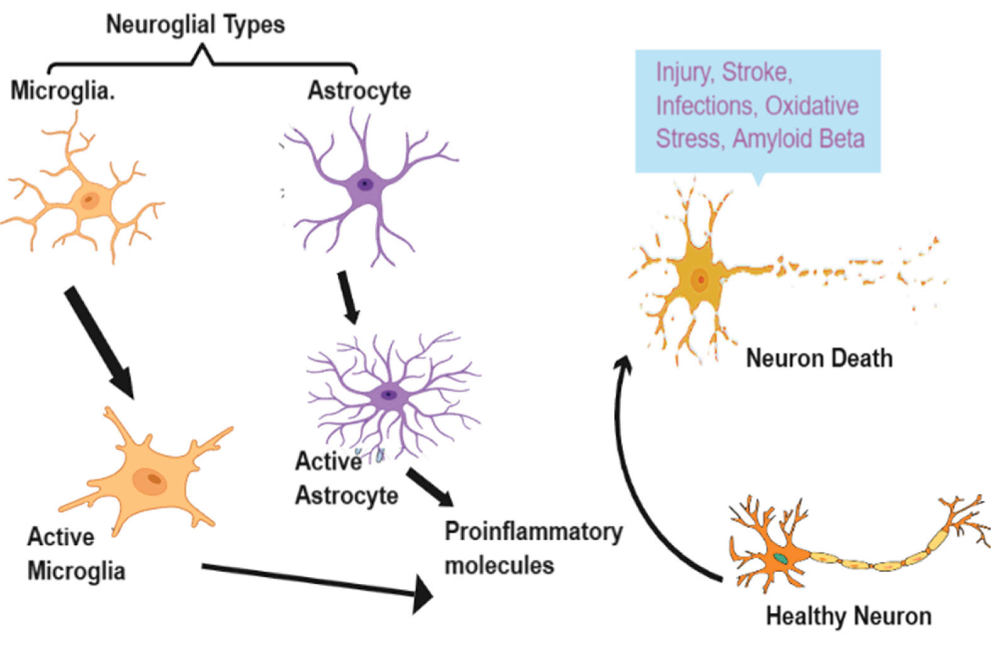
Figure 2.
Reactive astrocytes' function in Aβ production.
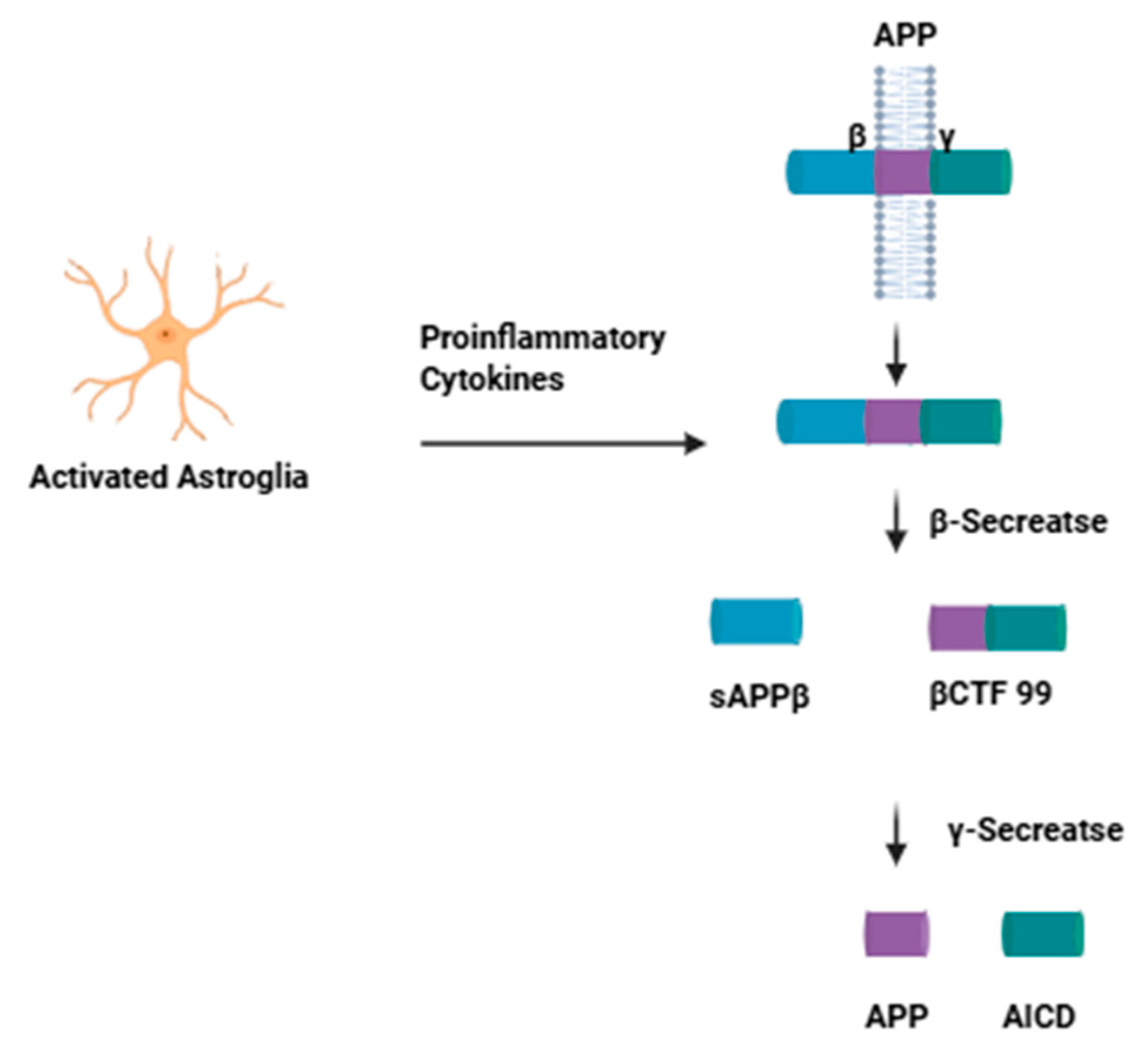
Figure 3.
Summary of common glial dysfunctional pathways in Alzheimer's Disease and Diabetes Mellitus.
Figure 3.
Summary of common glial dysfunctional pathways in Alzheimer's Disease and Diabetes Mellitus.
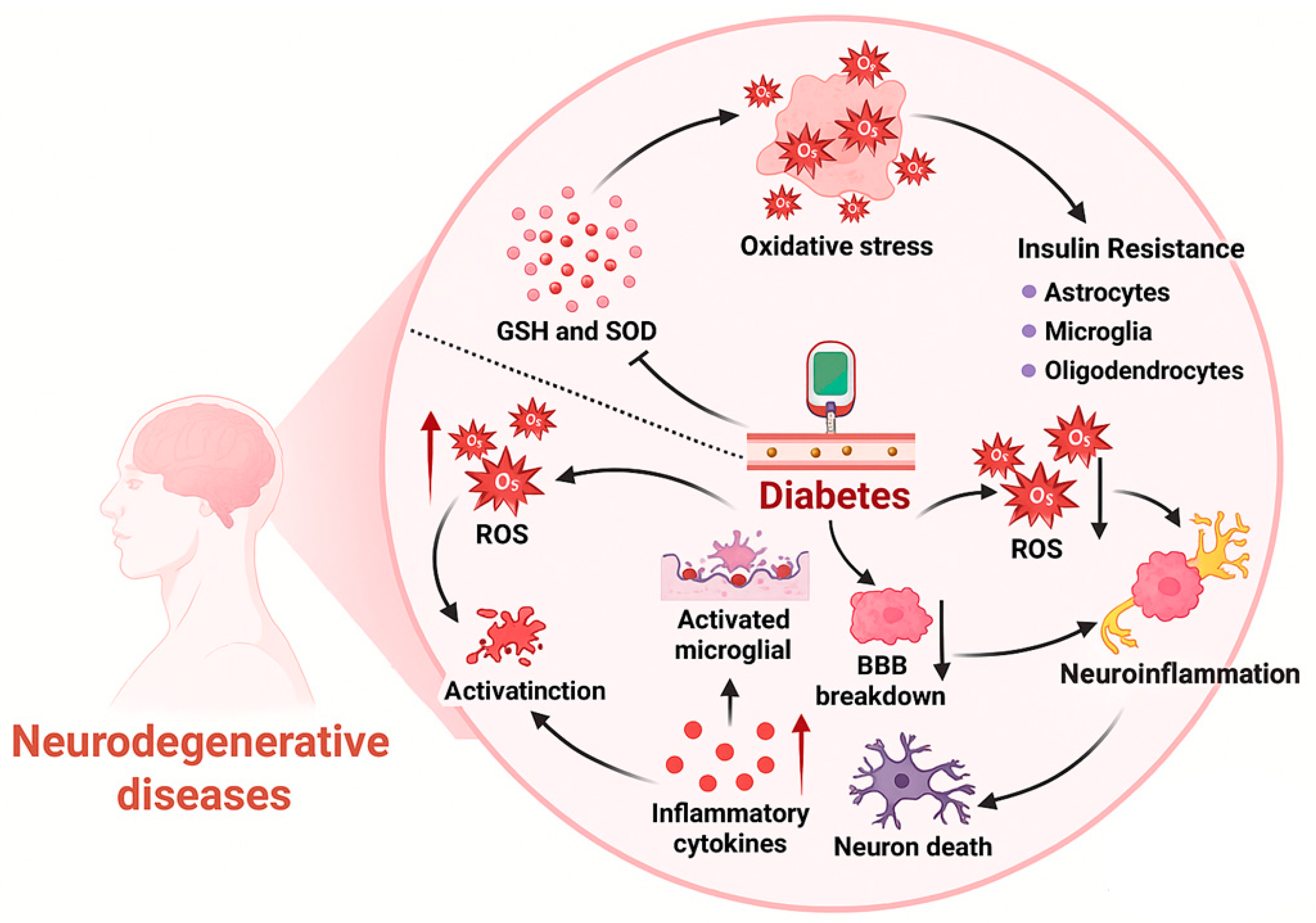

Table 1.
Shared pathophysiological Pathways in Alzheimer’s Disease and Diabetes Mellitus.
| Pathway | Key Findings | Scientific References |
|---|---|---|
| Neuroinflammation | Microglial activation leads to pro-inflammatory cytokine release and chronic inflammation in both AD and DM brains. Increased microglial activity correlates with neuronal injury and cognitive impairment. | [67] |
| Metabolic Dysfunction | Impaired glucose metabolism and insulin resistance present in AD and DM. These changes drive oxidative stress, energy deficits, and promote neurodegeneration. | [68] |
| Protein Aggregation | Accumulation of amyloid-β and tau in AD parallels islet amyloid polypeptide (IAPP) deposition in DM, with evidence for cross-seeding between these peptides. | [69] |
| Synaptic Dysfunction | Both diseases exhibit synaptic loss and deficits in neurotransmission (e.g., glutamate, GABA, acetylcholine), directly linked to cognitive decline. | [70] |
Table 2.
Mechanisms of neurological crosstalk disruption in AD and DM.
| Mechanism | Neuronal-Glial Interaction Disruption | Scientific References |
| Microglial Activation | Persistent M1 (pro-inflammatory) microglial state promotes neurodegeneration and impairs neuroprotection. | [67] |
| Astrocyte Dysfunction | Astrocytes fail to maintain metabolic support, promote oxidative stress, and amplify neuroinflammation. | [71] |
| Oligodendrocyte Impairment | Metabolic syndrome and hyperglycemia disrupt oligodendrocyte energy supply, impacting myelination and axonal health. | [72] |
| Cytokine Network Alteration | Excessive TNF-α, IL-1β, and other cytokines from glia contribute to sustained inflammation and tissue injury. | [67] |
Disclaimer/Publisher’s Note: The statements, opinions and data contained in all publications are solely those of the individual author(s) and contributor(s) and not of MDPI and/or the editor(s). MDPI and/or the editor(s) disclaim responsibility for any injury to people or property resulting from any ideas, methods, instructions or products referred to in the content. |
© 2025 by the authors. Licensee MDPI, Basel, Switzerland. This article is an open access article distributed under the terms and conditions of the Creative Commons Attribution (CC BY) license (http://creativecommons.org/licenses/by/4.0/).
Copyright: This open access article is published under a Creative Commons CC BY 4.0 license, which permit the free download, distribution, and reuse, provided that the author and preprint are cited in any reuse.


