Submitted:
25 July 2025
Posted:
28 July 2025
You are already at the latest version
Abstract
Shechita is the method of slaughter prescribed by Jewish law. It involves a single swift continuous uninterrupted cut through both carotid arteries and jugular veins, the trachea, esophagus and other structures. Shechita in cattle causes a rapid, massive drop in blood volume and pressure that quickly eliminates cortical function causing near-instantaneous loss of consciousness (LOC) and loss of pain perception. Shechita has long been subjected to criticism, with recent concerns focusing on cattle claiming that animals remain conscious for a prolonged period after the cut. Critics argue that the vertebral arteries, which are not severed during Shechita, may continue supplying blood to the brain, thereby delaying LOC. However, a critical review of the scientific literature reveals that this theory is based on misinformation and fundamental misconceptions about the cerebral blood supply in cattle, misconceptions that have been repeatedly propagated within the current literature.
Keywords:
shechita
; carotid arteries
; vertebral arteries
; blood flow
; brain
; cortex
; consciousness
1. Introduction
Slaughter with prior stunning is generally mandated on animal welfare grounds, as it is intended to induce rapid unconsciousness before bleeding. It is widely regarded as the standard against which Shechita and other forms of non-stun slaughter (e.g., Halal) are compared. In the Jewish religion, Shechita is the traditional method of slaughter of designated (‘kosher’) mammals and birds for dietary consumption. A trained worker (shochet) uses a surgically sharp knife (chalaf) that is twice as long as the width of the animal’s neck. The blade must be perfectly smooth and free of any nicks [1,2]. The shochet cuts the carotid arteries, jugular veins, trachea, esophagus, the vagus and sympathetic nerves in a single, rapid, continuous movement [2,3,4].
The bovine cerebral cortex receives blood supply from both the carotid and vertebral arteries; this differs from sheep and goats, where the cortex relies solely on the carotid arteries [5,6]. The internal maxillary artery, a direct continuation of the external carotid artery, is the main source of blood supply to the brain for both cattle and small ruminants. In cattle, following the experimental occlusion or clamping of carotid arteries, flow alterations are such that the vertebral arteries can compensate and supply blood to the brain [7]. This is primarily due to the fact that the bovine cardiovascular system contains an important anastomosis (vertebral-occipital) which connects the anterior and posterior circulations of the brain [5,6,8,9]. In the intact circulatory system, the vertebral-occipital anastomosis serves as a conduit for blood flow from the carotid arteries to the vertebral arteries [7]. (Figure 1)
In cattle, the vertebral arteries reach the rete mirabile but also connect back to carotid arteries via the vertebral-occipital anastomoses. In the context of Shechita, pressure alterations from cut carotids causes blood flow pattern reversal where blood from the vertebral arteries, by way of the anastomoses, travels into the carotids towards the rostral end of the cut. This phenomenon obeys the law of hemodynamics which describe blood flow to the path of least resistance. This results in a complete stoppage of blood flow to the cattle brain [6,7,10] (Figure 2).
After the Shechita, retrograde blood flow from the rostral end of the severed carotid arteries can be observed. This flow originates from the vertebral arteries but never reaches the rete or brain due to the vertebral-occipital anastomosis, as explained above.
Flow redirection dramatically reduces blood in the cerebrovascular system such that total cerebral blood flow is rapidly reduced to 5% or less of normal blood flow [3,6,7,11,12,13].
In experiments in which calves had the vertebral-occipital anastomoses ligated followed by cutting of the carotids, the blood pressure in vertebral arteries declined slowly, from 190 mm/Hg to 50 mm/Hg in 13.8 seconds [7]. This again confirms the role of vertebral-occipital anastomoses in conveying the blood flow between vertebral and carotid arteries and in the case of cut carotids, from vertebral arteries to the rostral end of cut carotids [6,7,10].
During severe hemorrhagic hypotension, cerebral vascular resistance tends to increase rather than decrease, perhaps due to the passive collapse of the vascular channels and because of sympathetically mediated cerebral autoregulatory mechanisms [14]. This further contributes to blood flowing along the path of least resistance, meaning that increased vascular resistance in the cortex diverts blood away from it. Furthermore, in the context of Shechita, in order to preserve vital functions, blood flow is redistributed away from the cortical grey matter towards the brainstem [11,14]. Additionally, the brainstem and spinal cord tissues are at baseline more resilient to changes in the blood supply and tissue oxygenation [15]. The brainstem, thalamus and hypothalamus are more resistant to ischemia than the hippocampus and cortical structures [16]. These phenomena are part of the reason why the brainstem and spinal cord remain functional longer than the cerebral cortex after Shechita. This explains why many of the brainstem reflexes such as the corneal reflex or spinal reflexes [6] and kicking, paddling movements of the legs often persist for several seconds or minutes after Shechita. These however are mere reflexes and in no way indicative of conscious activities and are similarly present after stunning [6,17,18,19]. These movements neither indicate nor cause animal suffering, as the cortex is no longer functional and therefore pain perception is impossible. In fact, some of the reflexes observed occur precisely because the cortex is no longer able to suppress them, serving as a sign of the cessation of cortical activity and, therefore, of unconsciousness. The cortex is extremely intolerant of ischemia. According to Dukes [7] interruption of flow to the brain results in unconsciousness within two seconds and irreversible tissue damage can occur within a few minutes [7]. The cerebral cortex is fundamental to the experience of pain, as it is where conscious perception occurs.
Arguments against slaughtering cattle by severing both carotid arteries without prior stunning focus on the presence of vertebral arteries that supply the rete mirabile. It is suggested that these arteries could maintain enough blood flow to prevent sufficient cerebral ischemia, potentially preserving cortical function for some time and delaying LOC. It is also argued that occlusions may develop in the severed carotid arteries, reducing blood loss and allowing the vertebral arteries to maintain sufficient cerebral blood flow to preserve cortical function, thereby delaying LOC and pain perception [20].
2. Materials and Methods
A literature review was conducted on the arterial blood supply to the brain in bovines following Shechita, or external carotid severance, with particular focus on the role of the vertebral arteries. PubMed database was searched with key words “calf AND vertebral artery AND blood flow OR pressure AND slaughter”; “vertebral artery AND bovine AND blood pressure AND Shechita OR halal”; accessed June 2025. Only articles which included evaluations or measurements of blood flow and/or blood pressure in the vertebral arteries were included. Furthermore, references were considered from above mentioned literature to other works which again included blood flow or blood pressure studies in vertebral arteries. Generative artificial intelligence (GenAI) has not been used in this paper.
3. Results
The literature search resulted in the inclusion of 14 manuscripts. Studies dating from 1920-2025 were identified that examined the changes in blood supply through carotid, maxillary and vertebral arteries after Shechita in bovines.
Vertebral Artery Pressure Measurements
Lieben et al. (1920) used a manometer to measure blood pressure in the vertebral arteries and the aorta of bovines before and after Shechita. They observed a significant, >50%, drop in vertebral artery pressure within 2–3 seconds after Shechita, a drop not consistent with sustained consciousness [12].
Dukes et al. [7] measured blood pressure in the vertebral arteries of bovines during Shechita and found that pressures dropped, on average, by 50.2 mmHg/second from 135-160 mmHg during first 3 seconds; and by more than 60% within 2 seconds; a level that is considered below that needed to sustain consciousness in most animals [7]. An observational report noted that the first movements seen after Shechita, typically leg movements, occurred on average at 20.5 seconds. Sporri et. al reported uncoordinated movements starting on average 48 sec (15 to 120 sec) after Shechita [6].
Sporri (1965) measured the blood pressure in the internal maxillary artery (head); ulnar artery and femoral artery (periphery) in cattle before and after cutting of the carotids. Following the cut, measured blood pressures dropped at varying rates. In the peripheral ulnar and femoral arteries, the decline was relatively slow, falling from 180 mmHg systolic to 80 mmHg over 25 seconds. In contrast, pressure in the internal maxillary artery dropped much more rapidly, decreasing by 80 mmHg within one second, representing 25 percent of the initial value, and reaching zero within five seconds [6]. (Figure 3)
Sahlstedt et al. (1928) measured blood flow through the vertebral artery before and after Shechita. The study demonstrated that while the vertebral arteries supply approximately 16% to 20% of bovine cerebral flow under normal conditions, this flow drops to about 1/30th to 1/40th (2.5%-3%) of the normal amount within 20 seconds after Shechita [21].
Data are summarized in Table 1 below.
Investigating the efficacy of the vertebral arteries in maintaining functional blood pressure and cerebral blood flow yields differing results when analyzing blood pressure versus blood flow dynamics.
Baldwin and Bell [5] observed that the vertebral arteries alone can maintain a blood flow sufficient to sustain sensibility in calves only if the circulatory system is intact (e.g. not cut) and the common carotid arteries are clamped; this is because the clamping of carotids, in an intact circulatory system, induces a compensatory blood flow increase in the vertebral arteries [7], even if only for a limited duration [23].
Dukes [7] measured the average vertebral blood flow of 8 anesthetized calves, with functional (non-ligated) carotids (20 measurements total). The results showed a flow of 27.7 ml/min in the vertebral arteries, while carotids blood flow was 87 to 120 ml/min (312% to 433% more). The clamping of carotids in calves induced a (non-constant) modest increase of blood pressure of only 5-10 mm/Hg in vertebral arteries, while blood flow greatly increased by 345%. Therefore, blood flow does not increase proportionally with respect to blood pressure.
This increase of blood flow in the vertebral arteries does not happen if both the carotid arteries and the vertebral-occipital anastomoses are clamped. Thus, indicating that the blood is flowing to the vertebral arteries from above the clamping of carotid arteries, through the occipital-vertebral anastomoses [7]. The above-mentioned data indicate that all or almost all of the blood that the vertebral arteries can supply to the brain in emergency situations (blockage of the common carotid arteries) must take its route via the anastomosis between the vertebral artery and the carotid artery [7,10].
Shaw, Bager and Devine (1990) evaluated the role of vertebral artery blood supply to the brain, considering its impact on cortical activity (measured using EEG) under the following conditions: 1. Carotid arteries are severed, and their rostral ends are ligated to stop the retrograde blood flow from the vertebral arteries through the occipital-vertebral anastomoses; assuming the vertebral arteries would maintain sufficient blood flow to the brain to prolong cortical activity. 2. Carotids are severed and vertebral arteries are ligated; assuming this
ligation would affect the blood supply to the brain and then accelerate the loss of cortical activity. Results were that the ligation of the vertebral arteries or the carotid clamping did not induce significant differences in the time to loss of cortical activity on EEG, indicating that even when all the available vertebral blood is preserved, this flow cannot maintain cortical functions [24].
Anil [25] measured blood flow and blood pressure in eight calves after a neck stick. The neck stick involved a lateral stab in the neck, followed by a transverse incision in a retrograde way to sever the soft tissues and the skin in the neck (a cut that is distinctly different to the Shechita cut). Some of the calves were shackled and hoisted after the cut, which could have affected the levels of both pressure and flow at the level of the vertebral arteries. Some of the animals developed occlusion of the carotids, which again, similar to carotid clamping, may have increased blood flow in the vertebral arteries. Anil [25] reported a prompt reduction in vertebral artery blood flow, which was maintained at approximately 30% of its initial level for approximately 3 min, even in calves which developed carotid occlusion. Considering that the physiological blood flow in vertebral arteries in the intact circulatory system is 25 to 30 ml/min [7], contributing only 1/4th of the blood supply to the brain [13], and that after the cut of carotid arteries the flow in vertebral arteries decrease to 2.5% to 3% within 20 seconds [21], even with the observed 30% blood flow (equivalent to 8 to 10 ml/min) a claim relative to efficacy in maintenance of a functional cortex cannot be supported.
Bager, Devine & Gilbert [10] evaluated the average blood flow to the brain of calves after severing both carotid arteries, by measuring the blood flow from the rostral end of severed jugular veins. The assumption was that this blood should represent the residual blood directed to brain after the cut of both the carotid arteries. The average jugular blood flow was 4.8 ml/min/100g of brain tissue, similar to the 5.2 ml/min/100g, as calculated by Rabkin [13]. Bager, Devine, and Gilbert [10] attributed the markedly reduced cerebral blood flow to the rerouting of vertebral artery circulation via the vertebral-occipital anastomoses toward the rostral ends of the severed carotid arteries. This rapid arterial flow from the rostral carotid stumps is illustrated schematically in Figure 2 [10]. In this study, calves were electrically, head-only, stunned which is theoretically reversible but they did not regain consciousness. Two calves developed carotid occlusions following the cut. In these animals, an increase in jugular blood flow was observed, representing residual blood from the vertebral arteries that perfused the brain and subsequently drained via the jugular veins. The calculated blood flows were 29.1 ml/min/100g brain and 16.3 ml/min/100g brain, respectively. These values are markedly lower than those reported by Terlouw and Kishimoto [26,27], or by Bager, Devine, and Gilbert [10], who cited previous research indicating flows of 74 ml/min/100g brain, considered necessary to sustain consciousness. Indeed, in these two calves, consciousness was not regained.
Bongert et al. [17] experimentally ligated the vertebral arteries of cattle prior to Shechita and observed that the reflexive kicking behavior following the cut was identical to that seen in animals with intact vertebral circulation. He concluded that these findings support the interpretation that the post-cut kicking movements are non-conscious motor responses, mediated by brainstem and spinal reflexes, and occur independently of vertebral blood flow.
Rabkin (2025) conducted the first systematic review of the cardiovascular effects of Shechita and calculated cerebral blood flow on the basis of published changes in systemic and vertebral artery blood pressure in cattle. He determined that, following the cut of carotids, within single digit seconds the vertebral circulation cannot maintain cerebral blood flow at a level sufficient to sustain brain function [13].
Flow Detection Based on Colorant Injections
Vertebral artery blood flow has also been studied through the injection of various contrast materials, including ink, dye, and charcoal.
Lieben (1930) demonstrated that a charcoal emulsion injected into a vertebral artery branch of an anesthetized bull calf reached the brain only if administered before Shechita. Injections initiated as early as 5 seconds after Shechita failed to reach the brain, providing direct evidence of the rapid cessation of cerebral blood flow following the cut [22].
Levinger (1976) injected India ink into the left ventricle of anesthetized animals immediately prior to severing the carotid arteries. After a prolonged period, large amounts of ink were found in the spleen, liver, kidneys, and lungs. In contrast, only small amounts were detected on the inferior surface of the brain, and no ink was observed in the cerebral cortex [28].
Baldwin and Bell (1963) injected fluorescent dyes into the carotid and vertebral arteries to study their distribution following alternate clamping of each artery. They concluded that in cattle, the entire brain is perfused by carotid blood mixed with vertebral blood, while the cervical spinal
cord is supplied exclusively by the vertebral arteries [5].
Blackman et al. (1986) injected methylene blue via an intracardiac catheter in two calves and observed that sequential dye boluses continued to diffuse through the vertebral arteries for over 100 seconds after severing both carotid arteries and jugular veins. The vessel pressure was not measured. Rather only the presence or absence of blood flow was determined by the diffusion of the methylene blue solution [29].
Carotid Artery Occlusions
Carotid artery occlusion has been reported to occur after slaughter when the severed artery retracts into its surrounding connective tissue sheath, effectively sealing the vessel [20]. The reported incidence of such occlusions following slaughter without stunning varies widely in the literature, ranging from 1% to 30%. Bager et al. also observed carotid occlusion occurring during the neck cut after electrical stunning [10]. It has been proposed that carotid occlusions may delay LOC by preserving sufficient blood flow through the vertebral arteries to maintain cerebral perfusion. While the reasons for carotid occlusion are not discussed in detail in this paper, further information is available in a recent review by Rabkin [13].
Baldwin and Bell [5] showed that in course of unilateral common carotid clamping (similar to occlusion) an immediate sustained increase in blood flow, which averaged 72%, occurred in the contralateral common carotid artery. Simultaneously, the same authors noted that the flow in the ipsilateral vertebral artery showed an increase of 107 % [5]. The same study showed that clamping the carotid artery resulted in a small (13 %), immediate increase in flow in the contralateral common carotid, but had no effect on vertebral artery blood flow [5].
4. Discussion
Criticisms of Shechita in cattle often center on the hypothesis that, because the vertebral arteries remain intact, cutting only the external carotid arteries may not sufficiently reduce cerebral blood flow to induce a rapid and humanely acceptable LOC. Additionally, there is concern that occlusion of the severed carotid arteries may help maintain sufficient blood flow through the vertebral arteries to the brain, potentially contributing to a delay in LOC. These concerns are raised repeatedly, yet they are supported by little scientific evidence. The underlying assumptions are primarily if not entirely based on anatomical speculation rather than on direct measurements or empirical validation of the hypotheses. A simple anatomical description of the arterial supply to the brain does not necessarily imply that these vessels continue to function fully when the circulatory system is disrupted or severed as it is in Shechita.
The current literature review found little to no evidence supporting the hypothesis that the vertebral artery provides sufficient blood flow to maintain consciousness in bovines slaughtered by Shechita. For reasons that remain unclear, several of the above-mentioned studies, many of which are in the German language, have been largely forgotten or excluded from the modern literature. Instead, in these published physiological studies, the authors describe in detail the lack of a significant role for the vertebral arteries in supplying blood to the cattle brain after the simultaneous severing of both carotid arteries. They emphasize that this cut leads to an almost immediate cessation of cerebral blood flow and, consequently, a rapid LOC as similarly reported by Hascalovici et al. in a recent systematic review article [30].
Although experimental occlusion of the carotid arteries alone does not result in immediate LOC [7], and may even lead to increased flow and pressure in the vertebral arteries [10], the severance of the carotids, as occurs in Shechita, creates an open circulatory system with rapid loss of blood volume and pressure. This physiological collapse precludes the maintenance of consciousness, even when the vertebral arteries remain anatomically intact [7,30]. Severing the carotid arteries causes an immediate loss of both cerebral blood flow and perfusion pressure due to the sudden drop in mean arterial pressure when the vascular system is breached. This loss of cerebral perfusion pressure is further exacerbated by rapid and continuous exsanguination [30]. As stated, Dukes observed a decrease in vertebral arterial pressure of more than 60% within two seconds of the carotid arteries being severed [7]. Similarly, Sporri (1965) reported a drop in internal maxillary artery pressure to 25% of its original value within one second of carotid transection [6]. These drops in blood pressure are consistent with those recorded by Lieben et al. in the vertebral arteries [12]. Lieben (1920) also noted that the onset of unconsciousness occurred simultaneously with the sudden drop in blood pressure [12]. Such pronounced decreases in blood pressure are considered inadequate to sustain consciousness in approximately 80% of humans [31]. Cerebral perfusion pressures below 50 mmHg are associated with hypoperfusion and the risk of ischemia [32].
A rapid drop in systolic blood pressure (SBP) of 50-60 mmHg in cattle is a significant physiological event, in the context of slaughter; the severing of the carotid arteries drastically reduces blood flow to the brain, and the limited compensatory mechanisms of vertebral arteries are not sufficient to maintain adequate cerebral blood flow; the brain rapidly becomes ischemic and unconscious [13]. Bager observed that vertebral blood, reaching the carotid rete and the Circle of Willis after carotids cut, should have a pressure as high as 30-40 mm Hg to be able to perfuse brain tissue [10]. This value is lost within 2 seconds after carotids cut [7].
Vertebral Artery Flow
Alterations in blood flow can lead to at least two relevant outcomes that impact cerebral perfusion and LOC during Shechita. First, in situ flow is reduced, leading to decreased effective perfusion of the cerebral cortex. Second, flow is redistributed along paths of least resistance within the now open circulatory system. The net result is a profound and rapid decline in cerebral blood flow following Shechita, consistent with Sahlstedt’s observation that vertebral artery flow drops to just 2 to 3 percent of normal levels within 20 seconds of carotid severance [21].
During experimental clamping of both the carotid arteries and the vertebral-occipital anastomosis, the residual blood flow through the vertebral arteries is insufficient to maintain consciousness, resulting in loss of posture and LOC in cattle within an average of 10.5 to 11.5 seconds [7]. The recovery of consciousness and posture following the release of the vertebral-occipital anastomosis clamp further demonstrates that vertebral blood flow alone cannot fully support cortical function [7]. In a typical hydraulic stanchion squeeze chute, commonly used in commercial feed yards, cattle may collapse due to pressure exerted on the carotid arteries when the chute is poorly managed. If the animal remains collapsed and the operator does not release it promptly, death can occur within 30 seconds [23]. This further demonstrates that vertebral artery blood flow, which is not affected in a standard hydraulic stanchion squeeze chute, is insufficient to maintain brain function even in the absence of profuse hemorrhage.
The carotid artery system supplies approximately 74% of cerebral blood flow, while the vertebral artery system accounts for the remaining 26% [13]. In humans, cerebral blood flow ranges is approximately 35 ml per 100 grams of brain tissue per minute. When blood flow falls below 35 ml/100 g/min, the level of consciousness begins to decline [27]. Similarities in physiological blood flow to the cortex between humans and calves [26,27] suggest that the oxygen requirements of the cattle brain are comparable. Given that a cattle brain weighs between 400 and 650 grams [33], it would require a minimum blood flow of 35 ml per 100 grams of tissue per minute or a total of 150 to 227 ml/min. This level of blood flow can only be achieved by the combined contributions of the carotid and vertebral arteries, but not by the vertebral arteries alone, as measured by Duke [7] and confirmed through field observations by Grandin [23].
The Shechita cut also severs the vagosympathetic trunk, thereby eliminating sympathetic vasoconstrictor activity in the extracerebral vessels rostral to the incision. This vasodilation similarly facilitates retrograde arterial blood flow toward the rostral ends of the severed carotids [10].
Although delayed flow through the vertebral arteries has been observed in dye indicator studies [29] blood flow alone is insufficient to ensure adequate tissue oxygenation. Both blood pressure and flow are essential, and the mere presence of blood flow offers no indication of cortical function or the state of consciousness. Anil [34] reported that 94% of the total potential blood loss in cattle occurs within 90 seconds after carotid severance, regardless of stunning. Consequently, the continued diffusion of dye along the vertebral arteries for up to 100 seconds, as observed by Blackman [29], must be explained by mechanisms other than mechanical drag, given the dramatically reduced, and nearly halted, blood flow. Instead, this phenomenon is more plausibly explained by passive diffusion along a concentration gradient, consistent with Fick’s law, which governs the movement of substances from regions of higher to lower concentration in the absence of active flow [35].
Lieben [12] and Dukes [7] demonstrated a dramatic drop in vertebral artery blood pressure following carotid artery severance. When cerebral arterial pressure falls to 20–30 mmHg, cerebrovascular resistance rises to a level that effectively halts all blood flow [6,14]. The causes of flow alterations vary depending on the model used. Baldwin's 1963 clamping study [36], a highly relevant scientific experiment, should be interpreted strictly in the context of cerebral blood supply and arterial distribution within a physiologically intact circulatory system. Its conclusions regarding cortical perfusion should not be directly extrapolated to the entirely different physiological state that occurs when the main blood vessels to the head are severed.
Impact of Occlusions in Maintaining Vertebral Flow
A recurring concern in the current literature is the potential impact of arterial occlusions, particularly at the severed ends of the carotid arteries, on perpetuating vertebral flow and delaying the onset of LOC. As previously explained, the dynamics of blood flow through the vertebral-occipital anastomoses toward the path of least resistance remain relevant. If a severed carotid becomes occluded, it may indeed contribute to increased vertebral flow. However, based on available evidence [7], a significant rise in blood pressure is unlikely. Blood flow will significantly increase (>70%) in the contralateral severed common carotid artery [5].
Given the open nature of the circulatory system following Shechita, any increased vertebral flow is expected to follow the path of least resistance via the anastomoses and exit through the rostral cut end of the external carotid artery [10], thereby continuing to facilitate volume loss.
In his manuscript, Rabkin [13] argues that the term “false aneurysm” is a misnomer and that the existing data do not convincingly link it to significant blood loss. He proposes that the formation of extravascular clots, composed of fibrin and blood, at the severed ends of the carotid arteries is a consequence of slow blood flow in the later stages of slaughter rather than its cause. At this late stage, blood pressure and flow are well below the levels necessary to sustain consciousness. Moreover, he suggests that the hematoma surrounding the severed carotid artery does not effectively impede ongoing blood flow.
Conclusions
Concerns regarding continued flow or meaningful cortical perfusion through patent vertebral arteries persist. However, a substantial body of physiological evidence, much of it developed in the last century, demonstrates through direct measurements that vertebral blood flow is dramatically, rapidly, and irreversibly affected by severing the external carotid arteries. Any residual flow is insufficient to sustain cortical function, maintain consciousness, or support pain perception.
In any context, it is indefensible and unacceptable for animals to be subjected to unnecessary pain, including at time of slaughter. This must therefore be addressed by minimization of pain and time to irreversible LOC [30]. Shechita in cattle consists of an instantaneous large surface ventral neck cut, which include the bilateral cut of carotid arteries, resulting in an abrupt loss of cortical blood flow, thus causing near immediate LOC. This paper addresses objections to Shechita that are based on assumptions regarding blood pressure and cerebral blood flow following the severing of both external carotid arteries in cattle, as well as concerns related to post-cut arterial occlusions. Historical clinical experiments remain highly relevant, providing empirical data on the physiological changes in blood pressure and flow after carotid severance. These findings offer a concrete basis for evaluating the role of the vertebral arteries, in contrast to the more speculative interpretations found in some later literature. These early studies consistently indicate a quick drop of blood supply to the brain following Shechita, which occurs within single digit seconds. They demonstrate that the Shechita cut affects blood pressure and blood flow to the brain in a quick, catastrophic, irreversible manner. Blood pressure and flow drop almost immediately, severely compromising the supply to the cerebral cortex at the pressure required to maintain its function. While some residual blood supply may preserve limited activity in the more primitive regions of the brain, such as the brainstem, these functions should not be mistaken for pain perception. The true intent of Shechita is to prevent unnecessary suffering in animals [37]. From this perspective, Shechita should be regarded as a humane method of slaughter.
Author Contributions
Conceptualization, J.H. and P.P.; methodology, J.H.; software, J.H.; validation, K.Y., G.SJ.; formal analysis, J.H.; investigation, P.P.; resources, -- ; data curation, J.H., K.Y.; writing—original draft preparation, P.P.; writing—review and editing, J.H.; visualization, K.Y.; supervision, G.SJ.; project administration, --; funding acquisition, -- All authors have read and agreed to the published version of the manuscript.
Funding
None
Institutional Review Board Statement
Not applicable
Informed Consent Statement
Not applicable
Data Availability Statement
Results from references indicated can be found at links indicated; results from references without link can be accessed at: Praxis Institute collection; https://praxisi.org/blog/category/scientific-studies/1
Acknowledgments
Conflicts of Interest
The authors declare no conflicts of interest
References
- Grandin, T.; Regenstein, J. Religious slaughter and animal welfare: A discussion for meat scientists. Meat Focus International 1994, 3, 115-123.
- Bager, F., Devine, C.E., Gilbert, K.V., Jugular blood flow in calves after head-only electrical stunning and throat-cutting, Meat Sci. (1988);22(3):237-43. [CrossRef]
- Levinger, I.M. Shechita in the Light of the Year 2000: Critical review of the scientific aspects of methods of slaughter and shechita; Machon Maskil L'David: 1995.
- Nangeroni, L.; Kennett, P. An electroencephalographic study of the effect of shechita slaughter on cortical function in ruminants. Report prepared for the Research Institute of Religious Jewry 1963, 44.
- Baldwin, B.; Bell, F. Blood flow in the carotid and vertebral arteries of the sheep and calf. The Journal of Physiology 1963, 167, 448. [CrossRef]
- Spörri, H. Schaechten und Tierschutz. University of Zurich, Zurich, Switzerland, 1965.
- Dukes, H. A study of blood pressure and blood flow in the vertebral arteries of ruminants; U.S Agriculture Research Service: Meat Inspection Division: 1958.
- King, A.S. Physiological and clinical anatomy of the domestic mammals. Volume 1. Central nervous system; Oxford University Press: 1987.
- Skerritt, G. King's applied anatomy of the central nervous system of domestic mammals; John Wiley & Sons: 2018.
- Bager, F.; Devine, C.; Gilbert, K. Jugular blood flow in calves after head-only electrical stunning and throat-cutting. Meat Science 1988, 22, 237-243. [CrossRef]
- Bager, F.; Braggins, T.; Devine, C.; Graafhuis, A.; Mellor, D.; Tavener, A.; Upsdell, M. Onset of insensibility at slaughter in calves: Effects of electroplectic seizure and exsanguination on spontaneous electrocortical activity and indices of cerebral metabolism. Research in Veterinary Science 1992, 52, 162-173. [CrossRef]
- Lieben, S. Flow Conditions in the Vertebral Artery Following Shechita Cut; Institute of Physiology at the German University in Prague.: 1920.
- Rabkin, S. The cardiovascular effects of shechita in cattle: a systematic review shows the fallacies that vertebral artery will preserve cerebral blood flow and false aneurysms occur in severed carotid arteries. Journal of the American Veterinary Medical Association 2025, 1, 1-10. [CrossRef]
- Marcus, M.L.; Heistad, D.D. Effects of sympathetic nerves on cerebral blood flow in awake dogs. American Journal of Physiology-Heart and Circulatory Physiology 1979, 236, H549-H553. [CrossRef]
- Brisson, C.D.; Hsieh, Y.-T.; Kim, D.; Jin, A.Y.; Andrew, R.D. Brainstem neurons survive the identical ischemic stress that kills higher neurons: insight to the persistent vegetative state. PloS one 2014, 9, e96585. [CrossRef]
- Terlouw, C.; Bourguet, C.; Deiss, V. Consciousness, unconsciousness and death in the context of slaughter. Part I. Neurobiological mechanisms underlying stunning and killing. Meat Science 2016, 118, 133-146. [CrossRef]
- Bongert, J. Food science, including meat inspection and slaughtering. On the question of slaughter - Does the Jewish ritual slaughter of animals constitute cruelty to animals? Veterinary Weekly Journal 1927, 43.
- Rosen, S.D. Physiological insights into shechita. Veterinary Record 2004, 154, 759-765. [CrossRef]
- Terlouw, C.; Bourguet, C.; Deiss, V.; Mallet, C. Origins of movements following stunning and during bleeding in cattle. Meat science 2015, 110, 135-144. [CrossRef]
- Gregory, N.G.; von Wenzlawowicz, M.; Alam, R.M.; Anil, H.M.; Yeşildere, T.; Silva-Fletcher, A. False aneurysms in carotid arteries of cattle and water buffalo during shechita and halal slaughter. Meat Science 2008, 79, 285-288. [CrossRef]
- Sahlstedt, A.V. Nagra forsok aft pa filsiologiskt experimentell Vag fa en objektiv grundval for bedomande av judiska slakmetoden ur djurskydssynpunkt. Veterinarmoto 1928, 640.
- Lieben, S. Die Strömungsverhältnisse in der A. vertebralis nach dem Schächtschnitt (Flow conditions in the vertebral artery following shechita cut). Prager Arch Tiermid (np, 1930), pt. A 1930, 10.
- Grandin, T. Restraint of Livestock. Available online: https://www.grandin.com/references/abdlps.html (accessed on 06/2025).
- Shaw, F.; Bager, F.; Devine, C. The role of the vertebral arteries in maintaining spontaneous electrocortical activity after electrical stunning and slaughter in calves. New Zealand Veterinary Journal 1990, 38, 14-16. [CrossRef]
- Anil, M.H.; McKinstry, J.L.; Gregory, N.G.; Wotton, S.B.; Symonds, H. Welfare of calves - 2. Increase in vertebral artery blood flow following exsanguination by neck sticking and evaluation of chest sticking as an alternative slaughter method. Meat Science 1995, 41, 113-123. [CrossRef]
- Kishimoto, M.; Kushida, K.; Yamada, K. Perfusion computed tomographic measurements of cerebral blood flow variables in live Holstein calves. American Journal of Veterinary Research 2018, 79, 177-180. [CrossRef]
- Terlouw, C.; Bourguet, C.; Deiss, V. Consciousness, unconsciousness and death in the context of slaughter. Part II. Evaluation methods. Meat Science 2016, 118, 147-156. [CrossRef]
- Levinger, I.M. Shechita. Religious, historical and scientific aspects; Gur Arye Institute: New York, USA, 1976.
- Blackman, N.; Cheetham, K.; Blackmore, D. Differences in blood supply to the cerebral cortex between sheep and calves during slaughter. Research in veterinary science 1986, 40, 252-254. [CrossRef]
- Hascalovici, J.R.; Schipper, H.M.; Regenstein, J.M.; Rosen, S.D.; Zivotofsky, A.; St-Jean, G.; Freilich, S.; Morrison, T.J.; Rabkin, S.W.; Haut, S. Rapid loss of consciousness in cattle following nonstun slaughter: evidence from a systematic review. American Journal of Veterinary Research 2025, 1, 1-10. [CrossRef]
- Njemanze, P.C. Critical limits of pressure-flow relation in the human brain. Stroke 1992, 23, 1743-1747. [CrossRef]
- Hopkins, A. Head trauma. Veterinary Clinics: Small Animal Practice 1996, 26, 875-891. [CrossRef]
- Gökyar, A.; Cokluk, C. Using of fresh cadaveric cow brain in the microsurgical training model for sulcal-cisternal and fissural dissection. Journal of Neurosciences in Rural Practice 2018, 9, 26. [CrossRef]
- Anil, M.H.; Yesildere, T.; Aksu, H.; Matur, E.; McKinstry, J.; Weaver, H.; Erdogan, O.; Hughes, S.; Mason, C. Comparison of Halal slaughter with captive bolt stunning and neck cutting in cattle: exsanguination and quality parameters. Animal Welfare 2006, 15, 325-330. [CrossRef]
- Fick, A. On liquid diffusion. Journal of membrane science 1995, 100, 33-38. [CrossRef]
- Baldwin, B., Bell, F.; The anatomy of the cerebral circulation of the sheep and ox. The dynamic distribution of the blood supplied by the carotid and vertebral arteries to cranial regions. 1963, J. Ant. Lond. (97)2, 203-215.
- Gurtman, R. Shehitah: Jewish Ritual Slaughter. 2005, http://nrs.harvard.edu/urn-3:HUL.InstRepos:8852091.
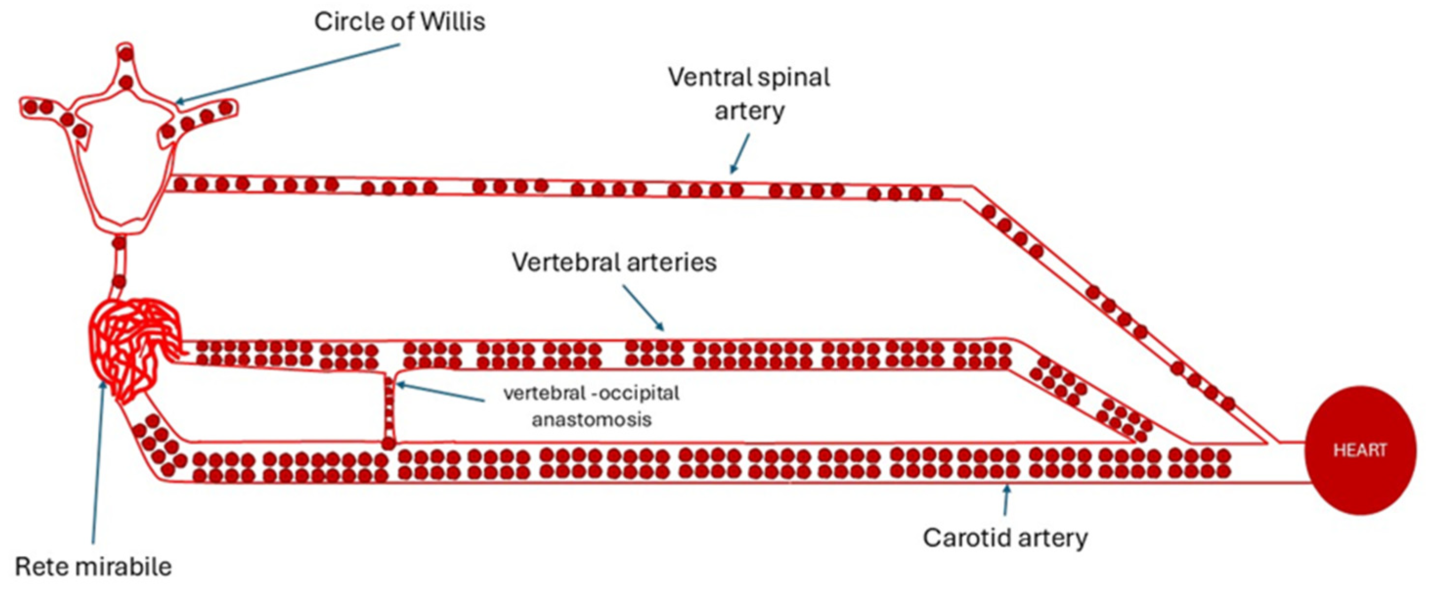
Figure 2.
Changes in blood flow to the brain, after cutting the carotids. Original diagram from [10]. The large arrow indicates the position where the carotid was cut in the experiment. The small arrows indicate the proposed route of flow of the major part of vertebral artery blood.
Figure 2.
Changes in blood flow to the brain, after cutting the carotids. Original diagram from [10]. The large arrow indicates the position where the carotid was cut in the experiment. The small arrows indicate the proposed route of flow of the major part of vertebral artery blood.
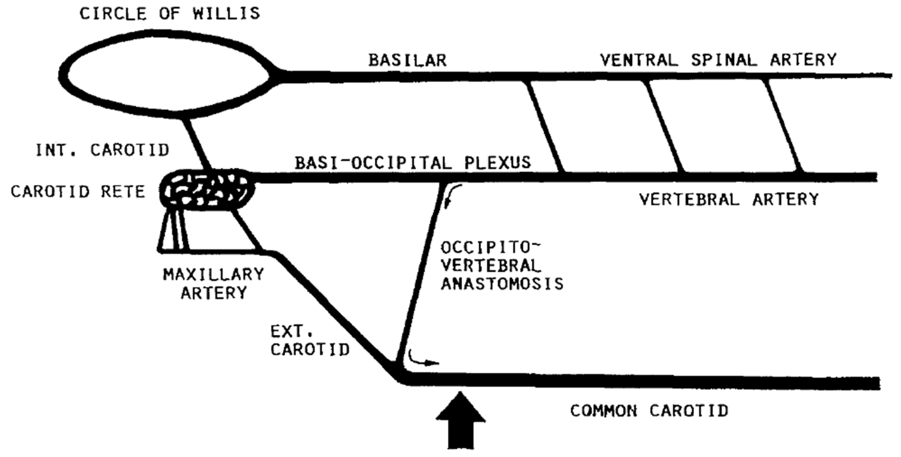
Figure 3.
Drop in blood pressure in vertebral, internal maxillary and other arteries, following the cut of carotids in cattle, as in original works [6,12].
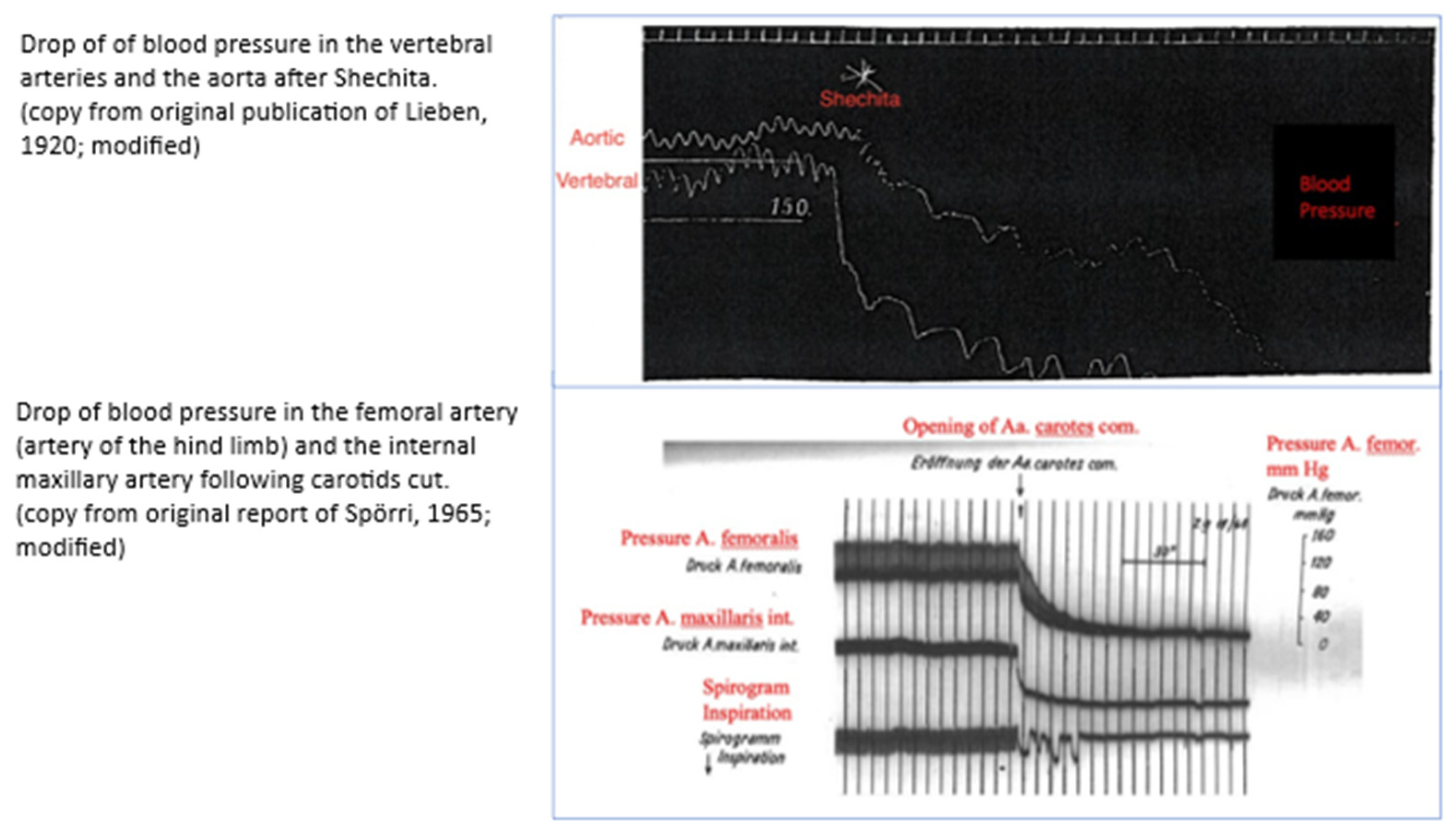

Table 1.
Changes in blood pressure and blood supply, in arteries supplying the brain, after carotids cut.
Table 1.
Changes in blood pressure and blood supply, in arteries supplying the brain, after carotids cut.
| Vessel | |||
| Blood pressure: mm/Hg | |||
| Vessel | Before cut | After cut | Reference |
| Vertebral artery | Drop >50% in 2-3 seconds | [12] | |
| Vertebral artery | 135-160 | Drop of 50.2 mmHg/second | [7] |
| Internal Maxillary artery | 130-135 | Drop by 80 mmHg within 1 second 0 mmHg at 5 seconds |
[6] |
| Blood flow: ml/min; or contribution % | |||
| Vertebral artery | 20% | Down to 2.5% - 3% after cut | [21] |
| Vertebral artery | Not evidenced at 5 seconds after cut | [22] | |
| Jugular veins | 3.6 ml/min/100g brain ±1.4 (4.8% of normal flow) |
[10] | |
Disclaimer/Publisher’s Note: The statements, opinions and data contained in all publications are solely those of the individual author(s) and contributor(s) and not of MDPI and/or the editor(s). MDPI and/or the editor(s) disclaim responsibility for any injury to people or property resulting from any ideas, methods, instructions or products referred to in the content. |
© 2025 by the authors. Licensee MDPI, Basel, Switzerland. This article is an open access article distributed under the terms and conditions of the Creative Commons Attribution (CC BY) license (http://creativecommons.org/licenses/by/4.0/).
Copyright: This open access article is published under a Creative Commons CC BY 4.0 license, which permit the free download, distribution, and reuse, provided that the author and preprint are cited in any reuse.


