
Submitted:
12 July 2025
Posted:
16 July 2025
You are already at the latest version
Abstract
Quantum dots have emerged as transformative nanomaterials whose extraordinary optical and electronic properties are revolutionizing precision medicine and biomedical innovation. This comprehensive review synthesizes multidisciplinary advances—from cutting‐edge eco‐friendly and biogenic synthesis methods to sophisticated surface modification strategies such as ligand engineering, zwitterionic polymer coatings, and congeneric passivation—that together enhance biocompatibility, reduce nonspecific binding, and prolong in vivo circulation. We explore the development of multifunctional hybrid nanocomposites, including QD–gold nanostars and QD/MoS₂ systems, which exhibit remarkable performance in multimodal imaging, fluorescence-guided surgery, and smart drug delivery applications. Furthermore, the review highlights the pivotal role of QDs in photothermal and photodynamic therapies, where their capacity to convert light into localized therapeutic effects paves the way for innovative cancer and dental treatments. Emphasis is placed on the integration of QDs as traceable carriers for targeted drug delivery, enabling real-time monitoring and controlled release via pH-sensitive and enzyme-responsive systems. Despite persistent challenges such as scalability, reproducibility, and concerns over heavy metal toxicity, ongoing interdisciplinary research is steadily overcoming these hurdles. By merging innovative synthesis, precise surface engineering, and strategic hybridization, QDs are positioned as critical components of next-generation diagnostic and therapeutic platforms that promise personalized, minimally invasive, and highly effective treatment modalities.
Keywords:
quantum dots
; nanomedicine
; bioimaging
; targeted drug delivery
; green synthesis
; phototherapy
1. Background:
1.1. An In-Depth Examination of Quantum Dots: Properties and Applications in Nanomedicine
Since the late 1990s, nanomedicines have made remarkable strides and garnered increasing attention. A recent survey reported that by 2020, there were over 32,000 publications dedicated to this field. Despite these advancements, the expected transition of innovations from laboratory research to clinical applications has not kept pace [1]. The development of COVID-19 vaccines utilizing nanomedicines has reignited optimism about their potential as protective tools in managing global pandemics. This breakthrough has especially heightened interest in lipid nanoparticles (LNPs), recognized as efficient nanovectors with diverse applications [2]. However, the potential of inorganic nanoparticles—such as silver nanoparticles (AgNPs), gold nanoparticles (AuNPs), metal–organic frameworks (MOFs), and quantum dots—remains underappreciated. Quantum dots, in particular, represent a relatively unexplored area that demands further investigation and elucidation [3,4].
Quantum dots (QDs) are tiny nanocrystals ranging from 1 to 15 nm, composed of semiconductor materials and celebrated for their exceptional optical properties. Introduced in the 1980s by physicist Alexei Ekimov, known for his work in semiconductors, these nanocrystals can be categorized into twelve distinct types based on their chemical composition, mirroring the arrangement of their constituent elements in the periodic table (see Table 1) [5,6]. For example, Group IVA QDs consist of tetravalent elements like carbon, silicon, and germanium. These elements exhibit both metallic and nonmetallic characteristics and possess semiconducting electrical properties due to having four electrons in their outermost shell.
Typically, QDs feature a heavy metal core encapsulated by a semiconductor shell with a defined bandgap, using materials such as CdTe, PbSe, ZnSe, or CdS for the core and SiO₂ for the shell [6,7]. This structure effectively reduces surface defects, thereby enhancing the quantum yield. Notable exceptions to this common structure include QDs that utilize a single semiconductor element, like silicon QDs, or those incorporating semiconducting polymers, known as P dots. J. Gao, X. Chen, and colleagues pioneered the synthesis of P dots using the novel semiconducting polymer NIR800, which emits light in the near-infrared (NIR) range (~800 nm). This property enables a wide range of biological applications, including flow cytometry and in vivo imaging [8,9].
As colloidal nanocrystalline semiconductors, QDs exhibit unique photophysical properties resulting from quantum confinement effects. Depending on their size and chemical composition, these nanocrystals emit a broad spectrum of wavelengths ranging from visible to infrared light. Compared to traditional organic fluorophores like dyes and fluorescent proteins, QDs offer unparalleled optical and electronic characteristics, such as higher absorption coefficients, tunable light emission, increased signal brightness, resistance to photobleaching, and the ability to excite multiple fluorescence colors simultaneously [10]. Moreover, the extensive surface area of QDs facilitates the covalent attachment of biorecognition molecules—including peptides, antibodies, nucleic acids, and small-molecule ligands—enhancing their utility as fluorescent probes for various applications [11].
The distinctive properties of QDs signify a pivotal shift from electronic materials science to a multitude of biological applications. Current and potential uses of QDs include serving as fluorescent labels for cellular imaging, intracellular sensors, agents for deep-tissue and tumor targeting and imaging, sensitizers in photodynamic therapy (PDT), carriers for gene therapy, and contrast agents for magnetic resonance imaging (MRI), among others (Figure 1) [12,13]. This review primarily outlines the evolution of QD synthesis, surface modification, and toxicity, with a brief focus on the expanding applications of QDs in the biomedical domain.
1.1.1. Heavy Metal–Free QD Platforms
Heavy metal–free quantum dot (QD) platforms represent a critical step toward safer and more environmentally benign solutions in biomedical imaging, optoelectronics, and other advanced technologies, especially as concerns about cadmium- and lead-based QDs persist. Among these eco-friendly alternatives, CFQD® nanoparticles have garnered considerable attention for their promising optical characteristics, biocompatibility, and potential for large-scale manufacturing [14,15,16,17,18,19]. CFQD® stands as an exemplary platform of In-based core–shell QDs, exhibiting high fluorescence quantum yield (QY) and tunable emission across the visible and near-infrared spectra without incorporating toxic heavy metals like Cd or Pb [15]. These attributes offer a constructive balance between the desired narrow emission linewidth and the minimized safety hazards during synthesis, usage, and disposal [17]. Although achieving outstanding luminance and operational stability with heavy-metal-free QDs has been challenging, numerous advances in CFQD® fabrication and functionalization have substantiated that safe, efficient devices are indeed achievable [15]. The evolution of CFQD® technologies focuses on several key aspects, namely, (1) optimizing core–shell design to reduce trap states and improve radiative recombination, (2) carefully selecting and exchanging surface ligands, and (3) developing refined charge transport layers in QD-based devices to improve reliability [19]. Early approaches toward CFQD® materials were limited by the relatively low photoluminescence QY, broad full width at half maximum (FWHM), and substantial surface defects that hindered both brightness and color purity [18]. One prominent study explored In-based CFQD® formulations for in vivo biodistribution analysis, illustrating that QDs with minimal cytotoxicity and robust photostability can be used for ex vivo sentinel lymph node imaging [15]. In that work, subcutaneous injection of CFQD® into rat paws allowed selective accumulation in local lymph nodes while simultaneously demonstrating negligible organ damage, underscoring favorable toxicity profiles relative to Cd- or Pb-containing QDs [15]. Likewise, CFQD® was shown to preserve photostability and exhibit intense emission for adequate temporal windows suitable for surgical procedures such as sentinel lymph node mapping or intraoperative tumor margin detection [17]. Substantial progress in the synthetic chemistry of CFQD® platforms has been pivotal for achieving these benefits. Generally, the CFQD® approach involves using III–V or I–III–VI semiconductors (e.g., InP or AgInS2), which intrinsically reduce hazardous metal content, thereby fulfilling more stringent environmental regulations [16]. For instance, InP-based CFQD® is particularly appealing due to its large exciton Bohr radius, enabling flexible tuning of the core size to match various spectral demands [20]. Nevertheless, controlling defects at the InP–shell interface is crucial, because the relatively higher oxidation susceptibility of InP can introduce mid-gap states that hamper optical performance [16]. To alleviate this concern, multi-shell or gradient-shell solutions are frequently employed, such as building a ZnSe or GaP intermediate shell and then adding ZnS, thereby reducing core–shell lattice mismatch and enhancing both fluorescence QY and operational stability [18]. In the context of CFQD®, the emphasis on shell engineering is complemented by advanced surface chemistries and ligand modifications that mitigate exciton quenching [16]. Although shell passivation alone already reduces nonradiative processes, small-molecule ligand exchanges can further refine conduction or valence band alignments by modulating the local surface dipoles [15,19]. This synergy between shell growth and ligand engineering is central to improving emission color purity and brightness in CFQD® systems [17]. Achieving near-unity photoluminescence quantum yields is therefore possible through these multi-faceted improvements, as evidenced by prototypes of CFQD® with QY around 35–45% [15]. Another strategic innovation in CFQD® design is doping or alloying. For example, doping copper or silver cations in the core can stabilize crystal growth at early nucleation stages, thereby promoting the formation of smaller QDs for short-wavelength emission, as relevant for blue or green CFQD® platforms [20]. Meanwhile, doping transition metals (e.g., Mn2+) in the shell can help bridge the conduction band mismatch, suppressing Auger recombination while preserving the QY [19]. These doping routes are also seen in non-InP CFQD® compositions, such as Ag2Se-based or AgInS2-based QDs, which cover broad spectral ranges and offer consistent heavy-metal-free luminescence [14]. In addition, CFQD® infiltration into polymeric or inorganic matrices can facilitate improved reliability and device integration. Solid-state CFQD®–polymer composites have illustrated robust mechanical properties, minimal exciton diffusion, and stable luminous properties suitable for flexible displays or wearable sensors [17]. The interface between QDs and polymeric host is further modifiable with crosslinkers, which can hamper QD agglomeration and help regulate QD dispersion. Meanwhile, from a device standpoint, the architecture of CFQD®-based LEDs or photodetectors strongly impacts the operational figures of merit [16]. Typically, the charge injection barriers in CFQD®-involved hybrid devices should be minimized by employing electron or hole transport layers whose energy levels are well-aligned with those of QDs [19]. Specifically, ZnMgO or NiO is introduced to tune electron or hole flux, reducing electron–hole imbalance commonly found in suboptimal QLED layouts [16,20]. For CFQD® intended for in vivo diagnostic imaging, in addition to the obvious impetus to avoid heavy metals, other aspects like renal clearance or hepatic excretion must be considered [17]. Investigations that used CFQD® in rat models indicated a predominantly lymphatic distribution with minimal cytotoxic response, although the reticuloendothelial system may accumulate QDs in the liver and spleen under extended timescales [15]. The potential for long-term breakdown products remains an ongoing concern, but improvements in QD size uniformity, composition, and surface coatings can foster more predictable clearance pathways [19]. Ultimately, the promise of CFQD® to replace cadmium-laden solutions in fields spanning from high-color-gamut displays to photoacoustic imaging, from shortwave infrared detectors [14] to next-generation surgical guidance systems [17], relies on continuing refinements in QD design, device engineering, and translational safety studies [18,20]. Encouragingly, several CFQD®-based prototypes are already available, with performance metrics that rival or surpass some Cd-containing systems, but further optimization remains necessary to achieve truly commercial readiness [16]. Superior quantum yields, narrower emission bands, better photostability, and robust manufacturing scale-up of CFQD® still face challenges related to controlling colloidal growth kinetics, lattice mismatch, doping uniformity, and universal standards for QD toxicity evaluation [19,20]. As new synthesis methods—including cation exchange, seeded growth, and dopant-mediated strategies—become more mature, the CFQD® paradigm will likely expand into broader practical use, meeting the dual demands of luminous performance and eco-compatibility [16,20]. Therefore, CFQD® stands poised as a pivotal solution in the ever-evolving domain of heavy metal–free QD technologies, offering a solid route toward safer QD products that maintain strong luminescent properties for advanced displays, sensors, and therapeutic systems [17,19].
1.1.2. Carbon-Based Quantum Dots and Silicon Quantum Dots
Carbon-based quantum dots (CQDs) and silicon quantum dots (SiQDs) have garnered increasing attention for diverse applications in areas such as light-conversion devices, drug delivery platforms, and light-emitting diodes due to their size-dependent optoelectronic properties, low toxicity, and tunable photoluminescence (PL) [21,22,23,24,25,26,27]. One core feature uniting CQDs and SiQDs is their ability to display emission spectra covering wide wavelength ranges, from near-ultraviolet (UV) through visible to near-infrared (NIR), thus enabling them to serve as versatile fluorophores [22,23]. In particular, colloidal silicon dots were shown to span a broad PL window from near-UV to near-IR, enhancing their suitability for imaging and photonics [22]. Additionally, doping strategies have been employed to modulate the optical bandgaps, engineer surface states, and improve solubility of both carbon-based and silicon-based quantum dots [25,26]. For instance, nitrogen-doped or sulfur-doped carbon dots may shift emission wavelengths to the blue or red region by altering the electron density of the carbon framework [26]. Similarly, doping in silicon QDs or passivation at the nanocrystal surface can modify the carrier recombination paths, thereby improving PL quantum yield [24]. These doping and passivation routes aim to reduce nonradiative recombination, an avenue of critical importance when designing bright and stable QD-based components.
Within the family of carbon-based quantum dots, the improvement of drug delivery has been a prominent area of investigation [25]. By functionalizing CQDs with hydrophilic groups or heteroatoms, hydrophobic drugs can be solubilized or released with greater precision. In a representative study on andrographolide solubilization, carbon dots were synthesized and then surface-modified to enhance the overall drug hydrophilicity [25]. This ability to tune functionality and form stable dispersions underscores the high adaptability of carbon dots for biological and pharmaceutical applications, where water solubility and minimal toxicity remain central requirements [23,25]. Another strength of CQDs, beyond their ease of surface functionalization, lies in their large Stokes shift, which lowers the chance of reabsorption when embedded in solid nanocomposites [26]. Such large Stokes shifts are conducive to luminescent solar concentrators or biological labeling, as minimal inner filtering helps preserve luminescence intensity. Furthermore, the relatively simple synthetic routes to carbon dots can be scaled using either top-down or bottom-up approaches, including oxidative cutting of larger carbon materials or pyrolytic decomposition of small organic molecules [23,26]. However, doping levels, oxygen-containing functional groups, and structural defects significantly affect their optical efficiencies [23], so efforts to refine doping content and surface states remain vital for obtaining near-unity internal quantum efficiencies.
Silicon quantum dots share many conceptual parallels with carbon-based dots, particularly regarding their quantum confinement-induced tunable emission [21,22,24]. Nevertheless, SiQDs often exhibit distinct photoluminescence behavior tied to indirect-bandgap carrier recombination, which is strongly confined when the crystal diameter shrinks to below five nanometers [22,24]. In some cases, high-temperature annealing of silicon-rich precursors leads to QDs with emission lifetimes in the microsecond range, demonstrating that core-related excitonic processes dominate their optical output [27]. A key challenge historically has been to achieve high photoluminescence quantum yield (PLQY) in SiQDs, given that incomplete surface passivation or the presence of silicon-oxide domains can give rise to nonradiative sites [21,27]. To address this challenge, precursor selection and a careful balance of annealing and etching steps have proven essential [24,27]. For instance, hydrogen silsesquioxane (HSQ) has been utilized to produce SiQDs with near-infrared emission and respectable external quantum efficiencies, but its high cost severely constrains large-scale usage [27]. As an alternative, silicon monoxide (SiO) has also been explored. Yet, due to the intrinsic nonuniformity and the unavoidable excess of oxide, SiQDs derived from SiO often exhibit lower PLQYs (generally below 15%) [27]. Another, more promising, approach lies in employing triethoxysilane (TES), which is inexpensive and can form well-defined HSiO1.5-like xerogels upon hydrolysis and condensation [27]. Optimized annealing and etching protocols subsequently allow for partial or complete removal of the surrounding oxide matrix, liberating near-infrared-emitting SiQDs [27]. Critically, an extended etching regime not only tailors the QD size but also removes oxide passivants at the surface, thereby boosting PLQY [27].
Recent findings show that TES-based approaches can yield silicon quantum dots with emission around 850 nm and an ensemble PLQY of approximately 40% in toluene, rising above 50% upon encapsulation in a thiol-ene polymer [27]. This near-infrared luminescence, combined with high internal quantum efficiency, matches or surpasses that of HSQ-derived SiQDs, yet at a fraction of the cost [27]. The ability to keep oxide coverage on the QD surface minimal is the principal reason behind these improvements: it is well known that silicon-oxide shells host trap states for carriers that catalyze nonradiative recombination [21,24,27]. In HSQ-derived samples, although high-quality quantum dots can indeed be formed, raw material costs limit their broad deployment [27]. By contrast, cheap SiO powders yield lower-quality QDs because extended HF etching and high-temperature annealing cannot fully mitigate the inherent oxide-related traps [27]. Hence, TES strikes a cost-versus-quality balance, enabling large-scale production of SiQDs with near-unity internal quantum efficiency while still preserving good monodispersity and robust near-infrared emission [27].
The synergy between doping, surface functionalization, and precursor engineering is equally vital for carbon-based and silicon quantum dots [23,25]. For example, doping carbon dots with sulfur or nitrogen modifies conduction-band and valence-band levels, thus shifting PL emission and improving water solubility [26]. Similarly, doping or passivation in silicon dots ensures a trap-free core, particularly when doping is accompanied by thorough oxide removal [27]. The combined effect of doping and advanced etching is frequently harnessed for controlling the ratio of “bright” versus “dark” dots within a distribution, with the aim to push the ensemble quantum yield beyond 50% [26,27]. Meanwhile, doping can also endow QDs with catalytic or sensing capabilities, broadening their application scope [25]. Notably, controlling doping levels is essential to circumvent excess structural defects that might lead to undesired nonradiative centers [23,26].
Finally, regarding device integration, stable solid-phase composites remain crucial in ensuring long operational lifetimes, as reported for QDs embedded in an off-stoichiometric thiol-ene polymer [27]. This solid matrix passivates surface traps and improves mechanical stability [25,27]. Combined with near-infrared emission and robust photostability, such composites are considered promising for luminescent solar concentrators or flexible photovoltaic modules, wherein a large Stokes shift and high efficiency are paramount [22,26,27]. Furthermore, doping strategies that preserve crystallinity and maintain minimal oxide coverage allow these hybrid composites to resist photodegradation for extended durations [24,27]. Overall, the choice of precursor—HSQ, SiO, TES, or carbon-rich raw materials—must reconcile cost, yield, and optical performance. In particular, TES-based silicon QDs have emerged as a cost-effective solution to achieving near-infrared emission with PLQYs exceeding 50%, satisfying practical needs for large-scale photonic and biomedical uses [27]. The design of carbon-based quantum dots continues to leverage doping or functionalization to fine-tune optical properties and improve solubility, facilitating their application in sensing, theranostics, and catalysis [23,25,26]. As doping pathways become better understood, and with continued optimization of annealing or etching, the potential for CQDs and SiQDs to achieve even higher external efficiencies in solid-state devices appears bright, opening new horizons for advanced quantum dot technologies [26,27].
1.1.3. Green Synthesis Approaches (Eco-friendly methods)
Green synthesis approaches for QDs have garnered significant attention over the past decade, primarily due to the need for environmentally friendly methods that bypass toxic chemicals and harsh reaction conditions while still yielding quantum dots with high fluorescence, superior stability, and abundant surface functional groups [28,29,30,31,32,33]. In conventional top-down approaches that rely on strong acids or bases for cutting larger carbon structures into nanosized fragments, there are concerns about toxicity, material waste, and low yield, pushing research toward sustainable routes where bio-wastes and plant-derived precursors are harnessed [32,33]. Importantly, green synthesis of QDs aligns with the principles of minimizing environmental impact, reducing energy consumption, and ensuring safer reaction conditions, thus representing a crucial shift in modern nanotechnology. In particular, carbon quantum dots (CQDs) and graphene quantum dots (GQDs) prepared through eco-friendly methods have emerged as viable alternatives to heavy-metal-based QDs due to their biocompatibility, chemical inertness, low toxicity, and cost-effectiveness [29,30]. Depending on the selected biomass source and the desired optical properties, various synthetic protocols—hydrothermal, solvothermal, pyrolytic, or microwave-assisted—are employed, all sharing the fundamental aim of converting naturally occurring materials rich in carbon content into small, photoluminescent nanoparticles [31,32,33].
Among the green precursors frequently reported, agricultural and food industry residues have seen extensive use, since these carbon-rich wastes can be inexpensively converted to QDs of high quantum yield, while simultaneously reducing the environmental burden posed by conventional disposal [33]. In one demonstration, lemon peel waste served as an effective carbon feedstock to synthesize CQDs via a one-pot hydrothermal reaction at moderate temperature [33]. By simply mixing dried and powdered lemon peels with an aqueous solution, and heating for a fixed duration in an autoclave, highly fluorescent CQDs with sizes in the 1–3 nm range were obtained. This approach underscores key features of green synthesis: it avoids corrosive passivating reagents, takes advantage of a short reaction time, and reclaims waste that would otherwise be incinerated or left to degrade. Importantly, the resultant lemon-peel-derived CQDs retained stable photoluminescence (PL) over months of storage, highlighting the inherent stability afforded by the biomass doping (e.g., presence of nitrogen, sulfur, or oxygen from the natural source) [33]. Such doping can endow the resulting QDs with abundant surface groups, including carboxylic acids, hydroxyl moieties, or amines, which subsequently enhance their water solubility and biocompatibility [30,31].
A similar paradigm is seen in other plant-based syntheses, such as the use of medicinal leaves, green tea extracts, or fruit skins [32]. Neem (Azadirachta indica) leaves, for instance, were exploited by Gedda et al. to generate multifunctional CQDs that possessed free radical scavenging capability, antimicrobial effects, and excellent fluorescence suitable for cell imaging [32]. The underlying mechanism involves first subjecting the leaves to a hydrothermal condition that triggers complex reactions of dehydration, polymerization, and aromatization. These steps yield nanocarbon cores bearing numerous oxygenated functional groups, thus promoting solubility and a high quantum yield. Because many medicinal plants inherently carry bioactive molecules (flavonoids, proteins, lignins), it is hypothesized that these molecules become integrated into or onto the CQD structure, further enhancing their antioxidant and antibacterial performance [32]. Such integrative properties differentiate biomass-based QDs from those produced by purely synthetic molecules, where external doping is often required to achieve similar benefits.
Aside from the use of a hydrothermal process, alternative bottom-up strategies employed in green synthesis include microwave irradiation, ultrasonic fragmentation, and soft pyrolysis [30,31]. Microwave irradiation, for example, offers rapid heating and uniform temperature distribution, drastically reducing reaction time compared to classical hydrothermal setups [29,31]. This method has been used to convert plant-derived starches, polysaccharides, or cellulose-laden biomass into photoluminescent nanoparticles in mere minutes. However, the ease and simplicity of hydrothermal or solvothermal approaches continue to dominate green synthesis because of their directness and minimal instrument requirements. Additionally, these processes typically yield QDs with better size uniformity, which is highly desirable for electronic and biomedical applications [31,32]. The synergy between the precursor’s composition (i.e., the presence of nitrogen or sulfur in the biomass) and the reaction parameters—temperature, time, pH—can allow a degree of control over the final QD size, doping level, and PL properties.
During green synthesis, the mechanism underlying QD formation from biomasses typically involves the transformation of small molecules (like sugars, amino acids, organic acids) into carbon cores. The numerous functional groups existing in leaves, peels, or seeds act as nucleation sites and eventually become part of the QD’s surface [30]. This results in high negative surface charges, as indicated by large zeta potential values, which not only enhance their colloidal stability but also offer multiple sites for subsequent functionalization, such as conjugation with biomolecules for targeted drug delivery, or doping with metals for catalytic applications [31]. In addition, the typical presence of polyphenols and other antioxidants in plant extracts can further reduce or stabilize the nanoparticle surfaces, circumventing the need for hazardous reducing agents [33]. The repeated demonstration of stable luminescence over extended periods indicates that the surface states introduced by natural precursors lead to robust passivation, thereby diminishing photobleaching [29,33].
Another integral feature of green synthesis is scalability. The feasibility of producing gram-scale QDs from abundantly available bio-waste is essential for practical industrial applications [30]. For example, Tyagi et al. described the production of gram-level CQDs from lemon peel [33]. The yield can be further increased by tweaking the precursor concentration, reaction temperature, and reaction time. Efficient filtration or centrifugation steps remove unreacted macro-particles, while the final passivated QDs remain stably dispersed in aqueous media. This readiness to scale up is crucial for translating laboratory findings into commercial products such as sensing materials, photocatalysts, and medical imaging probes [30,31].
Green-synthesized QDs have been tested in a variety of applications that underscore their multi-functionality. For instance, the fluorescence-based detection of heavy metal ions (Cr6+, Pb2+, Hg2+, etc.) is particularly promising as it provides a quick, selective, and sensitive approach for water quality monitoring [33]. Tyagi et al. used lemon peel-based QDs for detecting Cr6+ with a detection limit of 73 nM [33]. Similarly, doping from natural nitrogen sources can confer a strong electron transfer phenomenon, resulting in “turn-off” or “turn-on” fluorescence signals upon binding with specific contaminants [30]. The same QDs can also serve as photocatalysts for degrading organic pollutants in water. For example, composites of CQDs with TiO2 or other semiconductors present improved catalytic efficiency and reduce the recombination of electron–hole pairs under UV or visible light [33]. By forming a heterojunction at the QD–TiO2 interface, electron transfer becomes more efficient, thereby generating reactive oxygen species to break down dyes or other hazardous organics. In such a framework, the QDs not only enhance the photo-response but also exploit their inherent doping sites to anchor onto the TiO2 surface [33]. The use of plant-derived QDs extends to biomedical fields, as manifested by excellent biocompatibility and strong photoluminescence, enabling imaging of mammalian cells, drug transport, and the potential for photodynamic therapy [31,32]. Indeed, the presence of natural molecules often embedded on QD surfaces can mitigate toxicity and immunogenic responses, although further clinical trials are needed to confirm full biosafety.
Looking ahead, one of the major challenges in scaling up green synthesis is controlling the precise composition and dopant level of QDs, given the variability inherent in biomass feedstock [31]. Moreover, obtaining uniform sizes and consistent luminescence profiles might require advanced fractionation steps. However, the adaptability of hydrothermal or microwave methods to process diverse biomass resources suggests wide latitude in exploring new feedstocks such as stems, seeds, and shells for further boosting the quantum yield and doping profiles. The synergy of green synthesis with biopolymeric capping agents or catalytic metals can pave the way for multifunctional QDs well-suited for diagnostic, therapeutic, and environmental remediation. Several studies have underscored the importance of correlating morphological features (like average size, shape, crystallinity) with the excitonic and catalytic properties of QDs to rationally design targeted materials for specific end-uses [30,32,33]. Additionally, challenges related to product purification and reusability of the leftover biomass might be addressed through advanced separation techniques or integrated manufacturing strategies that further elevate the environmental benefits of QD production.
In summary, green synthesis methods for quantum dots represent a vital step forward in reconciling cutting-edge nanotechnology with ecological responsibility [30,31]. By leveraging simple, scalable protocols and versatile bio-wastes, eco-friendly routes produce QDs of high fluorescence, robust photostability, low cytotoxicity, and broad doping potential [32,33]. Indeed, from the successful deployment of QDs in heavy metal detection and photocatalytic pollutant removal to potential biomedical diagnostics, these environmentally benign synthesis approaches stand at the nexus of sustainability and innovation in nanoscience, promising wide-ranging benefits across multiple scientific and industrial domains [29,30,31].
1.2. The Transformative Role of Quantum Dots in Healthcare and Pharmaceutical Innovation
QDs have become integral components in medicine, dentistry, and pharmaceuticals, offering substantial potential to transform these fields. In medical applications, QDs introduce a revolutionary approach to imaging and diagnostics by providing unmatched resolution through their unique optical properties. Their ability to monitor complex biological processes in real time at cellular and molecular levels enhances the sensitivity and precision of medical imaging, making them indispensable tools [34]. Therapeutically, QDs function as sensitizers in PDT, a sophisticated treatment where light activation selectively targets diseased cells. Their effectiveness in treating hyperthermia further demonstrates their versatile therapeutic applications [35].In the realm of dentistry, QDs surpass traditional diagnostic methods by offering advanced imaging capabilities that enable more detailed analyses. Their fluorescent properties provide heightened specificity in detecting and monitoring dental diseases. Additionally, QDs are employed in biosensing technologies designed to identify oral pathogens and biomarkers relevant to dental conditions, thereby advancing sensitive diagnostic paradigms [36].Within the pharmaceutical sector, QDs play a crucial role in drug development and assessment. They serve as precise platforms for analyzing cellular responses to new pharmaceutical agents, involving rigorous in vitro assays to evaluate the efficacy and toxicity profiles of potential drugs thoroughly. Incorporating QDs into pharmaceutical research facilitates a detailed understanding of cellular and molecular complexities, significantly informing drug discovery processes. In manufacturing, they are instrumental in quality control procedures, ensuring the consistency and integrity of drug formulations [37]. Their widespread application across these domains underscores their potential to revolutionize various aspects of medical, dental, and pharmaceutical sciences, fostering innovative avenues in research, diagnostics, and therapeutic modalities based on their unique properties.QDs have attracted significant attention in biomedical and bioanalytical applications due to their exceptional photoluminescence properties and their ability to be engineered into biocompatible systems by conjugating with diverse biomolecules. Their potential is actively harnessed in developing molecular and immunological assays for a wide array of biomarkers and pathogens. Moreover, integrating QDs with microfluidic technologies, leveraging their distinctive optical characteristics, is poised to facilitate the creation of highly sensitive bioanalysis systems suitable for point-of-care testing. The availability of various QDs tailored for clinical applications suggests an imminent breakthrough in their utilization [38]. Consequently, this review aims to provide a comprehensive overview of current developments in the use of QDs for point-of-care testing, encompassing bioimaging (including in vitro, live-cell, in vivo, and single-molecule imaging), biosensing (covering protein detection, DNA assays, immunoassays, and sugar sensing), and biotargeting (including drug delivery, detection of genetic diseases, and clinical applications).Beyond their applications in bioimaging and photodynamic therapy, QDs show considerable promise as drug delivery vehicles. They possess several advantageous features in this role: ease of fabrication, the ability to conjugate with a wide variety of therapeutic agents, tunable physicochemical properties, and unique optical features that facilitate tracking after administration. Furthermore, their ultra-small size is critical for effective extravasation and penetration into the dense stromal environments typically found in challenging tumors like hepatocellular carcinoma and pancreatic cancer. Specifically, QDs smaller than 10 nm demonstrate significant potential as delivery vectors capable of effectively infiltrating tumor tissues.
1.2.1. QD Integration in Point-of-Care Devices
Quantum dots have steadily gained recognition as powerful diagnostic reporters in point-of-care (POC) devices, offering a valuable convergence of high sensitivity, rapid detection, and versatile design strategies for diverse biomedical applications [39,40,41]. The impetus behind integrating QDs into POC platforms arises from their unique combination of large absorption coefficients, tunable emission spectra, and enhanced photostability compared to other nanomaterials, such as colloidal gold [40]. By capitalizing on these attributes, QDs embedded in microfluidic devices, lateral flow immunoassays (LFIAs), or related biosensing formats significantly elevate the performance of rapid diagnostics. Moreover, because POC testing demands minimal user intervention and near-instantaneous readout, the fluorescent intensity and long fluorescence lifetime of QDs become critical advantages, enabling accurate detection even in the face of complex biological matrices [39,40].
In practical POC diagnostics, the shift toward QD-based systems is motivated by the urgent requirement for user-friendly, robust, and sensitive devices, particularly in the context of early disease detection [40]. For instance, quantum dots can be conjugated with specific antibodies that target microbial antigens or biomarkers, leading to highly selective fluorescence readouts in the test zone [39]. These conjugates are often immobilized on a conjugate pad or integrated into the test line of a lateral flow strip. When a patient’s sample—such as saliva, nasal fluid, or whole blood—migrates along the nitrocellulose membrane, QD-labeled antibodies bind selectively to the analyte of interest. Under ultraviolet or visible light excitation, the fluorescent signal at the test line indicates a positive result, while the concurrent control line ensures proper fluid migration and validates test accuracy [40]. Unlike gold nanoparticle-based LFIAs that can falter at detecting very low analyte concentrations (especially in early infection scenarios), QD-based approaches enable heightened fluorescence intensities, thereby lowering detection limits and improving the signal-to-noise ratio [39,41].
In a typical QD-based LFIA, size-tunable QDs are selected to achieve well-defined emission wavelengths, allowing simultaneous detection of multiple analytes if necessary [39]. This multiplexing potential arises because the band gap of the QDs can be finely tuned by altering particle size or composition, leading to distinct emission peaks corresponding to different biomarkers. From a structural standpoint, QDs may comprise a core–shell arrangement, such as CdSe/ZnS or Cu:Zn In S/ZnS, which improves biocompatibility, photostability, and quantum yield [40]. Researchers have emphasized that these core–shell QDs can achieve quantum yields well above 40%, significantly surpassing many conventional fluorescent dyes [41]. Such favorable optical characteristics remain crucial when the objective is to detect disease-related proteins (e.g., cardiac markers, viral nucleoproteins, or cytokines) at ultra-low concentrations.
Point-of-care tests featuring QD labels also benefit from simpler storage and transport conditions relative to certain enzyme-based methods, since QD conjugates often exhibit stability under broader temperature and pH ranges [40]. For example, QDs can be entrapped in polymeric micelles or coated with hydrophilic ligands like polyethylene glycol to preserve fluorescence while resisting nonspecific adsorption. This stability ensures consistent performance of the POC devices in diverse or resource-limited environments [39]. Furthermore, the rapid readout window of QD-based tests, often under 20 minutes, appeals strongly to clinical workflows that require on-site decision-making [41]. The user need only introduce the patient’s sample, wait for strip migration, and illuminate the result zone with the appropriate light source to visualize any fluorescence.
Another dimension of QD integration pertains to microfluidic POC setups. Microfluidic chips can house QDs in small reaction chambers or detection zones, enabling automated fluid mixing, washing, and detection steps without extensive manual intervention [39]. By minimizing reagent volumes and fluid handling steps, these systems reduce the time to result while enhancing analytical sensitivity. Within such chips, one can embed QD-labeled antibodies for real-time monitoring of analyte binding, typically measured by a portable reader or smartphone camera [40]. The synergy between QDs’ bright fluorescence and microfluidic channels fosters new avenues in detecting multiple disease targets using a single, compact device.
In the domain of infectious disease diagnostics, including COVID-19, QD-based POC devices have demonstrated remarkable promise [41]. Specifically, lateral flow assays enhanced by QDs showed higher sensitivity than their colloidal gold counterparts in detecting SARS-CoV-2 antigens or antibodies [40,41]. This improvement primarily stems from the strong luminescent signals, which lower the limit of detection and help identify infections at very early stages. For instance, certain QD-labeled test strips detect sub-nanogram per milliliter concentrations of viral antigens, allowing clinicians to expedite care decisions or isolate infectious individuals promptly [41]. The quick turnaround for results, often under half an hour, alleviates laboratory burdens and can be critical in managing large-scale screening efforts.
Moreover, adopting QDs in POC testing for conditions like tuberculosis, tetanus, and other bacterial infections has been reported, with high accuracy and minimal cross-reactivity [39,40]. Since QDs can be engineered to produce distinct fluorescence colors, a single device can theoretically target multiple pathogens by depositing multiple test lines, each functionalized with distinct QD-antibody conjugates. Such multiplexing drastically reduces the required sample volume and test time when screening for co-infections or related disease markers. However, although quantum dot-based assays offer several advantages, researchers continue to scrutinize their long-term biocompatibility, especially when QDs contain toxic metals like cadmium [39]. To address these concerns, novel approaches utilize less toxic elements, including zinc or copper-based compositions, while employing robust coatings to prevent ion leakage [40].
Despite these safety considerations, QD-based assays have undergone rapid improvements in terms of brightness, specificity, and user-friendliness. Studies highlight that the quantum yield and surface functionalization greatly influence assay performance. For example, shell thickness and composition regulate photobleaching resistance and can mitigate self-quenching at high QD concentrations [41]. Furthermore, thorough optimization of conjugation chemistry—such as using carbodiimide or maleimide linkers—minimizes nonspecific binding and preserves antibody binding sites. These refinements ultimately translate into more reproducible, quantitative data in a single-use POC test.
In parallel, the combination of QD-LFIA with smartphone-based detection is an emerging trend that broadens diagnostic accessibility. By capturing the fluorescence emission from a test line using a phone camera, sophisticated image-processing algorithms can interpret results in a semi-quantitative manner, offering potential telemedicine opportunities in remote regions [40,41]. In addition, the integration of cloud-based data storage could further enable epidemiological monitoring and real-time data analytics, enhancing public health interventions.
In conclusion, quantum dots have significantly transformed the landscape of point-of-care diagnostics through their exceptional optical properties, stability, and modular design potential [39]. Their integration into various POC platforms—from lateral flow strips to microfluidic assays—demonstrates consistent benefits: improved sensitivity, rapid turnaround, and potential multiplexing for simultaneous detection of multiple analytes [40]. Although challenges related to QD toxicity and large-scale manufacturing remain under scrutiny, novel compositions and protective coatings are continually emerging. Consequently, QD-based tests promise a more precise, swift, and user-friendly approach to detecting viral, bacterial, and other pathological targets in decentralized clinical settings [41]. This dual emphasis on high-performance detection and cost-effective production confirms the continued relevance of QD-based assays for next-generation point-of-care solutions worldwide.
1.2.2. Regulatory and safety considerations for quantum dots
Regulatory and safety considerations for (QDs) require a multifaceted understanding of their physicochemical characteristics, routes of exposure, biological fate, and potential toxicity, all of which intersect with the complex regulatory frameworks governing both emerging nanotechnologies and biomedical products [42,43,44]. Modern regulatory paradigms increasingly acknowledge that QDs cannot be viewed as a single, homogenous category; instead, even small changes in size, core composition, surface coatings, or intended application produce unique safety profiles that demand detailed case-by-case evaluations [42,43]. From a regulatory standpoint, one of the most critical factors is the stability of QD core–shell structures, particularly when they contain potentially toxic metals such as cadmium or selenium. In conditions of oxidative, acidic, or photolytic stress, QDs may release free cadmium ions or degrade into other harmful byproducts, thereby posing risks that exceed those associated with unaltered or stabilized nanocrystals [43]. Hence, regulators generally require evidence that newly developed QD formulations maintain their structural integrity during synthesis, storage, and application in real-world or clinical settings [42]. For instance, ensuring that QD shells remain intact under physiological conditions is vital; compromised shells might release toxic core metals, leading to hepatic, renal, or neurological concerns. Indeed, evaluations in cell culture and animal models have already indicated that certain QD variants become cytotoxic only after photolytic or oxidative decomposition of their shell or capping ligands [43]. In parallel, agencies overseeing pharmaceutical or biomedical applications must consider whether QDs are administered intravenously, ingested, or inhaled, as these different exposure routes can alter systemic distribution and clearance [42]. Intravenous administration poses specific regulatory challenges because QDs of various sizes tend to accumulate in the reticuloendothelial system and localize within organs such as the liver and spleen, raising questions about potential long-term toxicity and excretion pathways [42]. Regulatory decision-makers therefore seek comprehensive absorption, distribution, metabolism, and excretion (ADME) data on a QD-by-QD basis. For oral or inhalation exposures, concerns arise regarding possible infiltration of QDs through gastrointestinal or pulmonary barriers and subsequent bioaccumulation within target tissues, with the potential for ecological impact if these nanomaterials enter wastewater streams [43]. Because environmental release of QDs can occur during synthesis, disposal, or clinical usage, environmental regulatory bodies also emphasize the study of QD behavior in aquatic or terrestrial ecosystems, particularly their tendency to aggregate or degrade in complex environmental matrices [42]. Another important dimension relates to the evolution of QD design for biomedical contexts, notably in theranostic applications where QDs can serve diagnostic and therapeutic purposes, including advanced radiopharmaceutical delivery [44]. For example, carbon-based or graphene quantum dots (GQDs) have emerged as alternatives to cadmium-containing variants in attempts to reduce toxicity risks [44]. However, manufacturing GQDs at scale while preserving uniform size distributions and consistent surface properties remains challenging, generating complexities for quality control and reproducibility. Regulatory scrutiny accordingly addresses how developers ensure standardized production, control impurities, and ascertain that final products remain safe under typical clinical conditions [43,44]. Furthermore, combining QDs with radionuclides, as proposed for next-generation radiopharmaceuticals, creates additional layers of complexity: specialized chelators and coatings must be employed to secure radionuclides to the QD surface, all while preserving optical properties and reducing the probability of radionuclide leakage in vivo [44]. Clinical regulators require that developers conduct stability testing under physiologically relevant conditions, proving that labeled QDs neither disassociate nor accumulate dangerously in unintended tissues. Dose considerations feature prominently in regulatory review, as QD toxicity sometimes surfaces only at higher concentrations or over extended exposure durations [43]. Studies indicate that short-term incubations with certain QD formulations might yield minimal adverse effects, whereas chronic or repeated exposures, even at moderate doses, can trigger cytotoxic or genotoxic outcomes [43]. Consequently, agencies overseeing medical products often demand long-term toxicity studies that provide insights into chronic exposure scenarios, repeated dosing implications, and possible off-target effects. The heterogeneity of QD-based products—ranging from simple imaging agents to complex multifunctional constructs—means that regulators typically classify them according to the intended use (diagnostic versus therapeutic) and the nature of the active ingredient (e.g., cadmium-based core, iron-based core, or carbon-based). Such classification influences which requirements apply; for instance, a QD designed for in vitro imaging might be regulated primarily under device frameworks, whereas an injectable QD-labeled radiopharmaceutical may undergo drug-related assessments [42,44]. Regardless of classification, safety data packages must cover acute and subchronic toxicity, reproductive toxicity if relevant to widespread or repeated use, immunotoxicity, biodistribution, excretion, and potential interactions with other pharmaceuticals. The presence of biologically active ligands on QD surfaces, such as antibodies or peptides, adds another dimension: these surface modifications aim to improve targeted delivery yet can also raise immunogenicity issues [42]. Regulators thus look for immunotoxicity data to rule out adverse immunomodulatory reactions or allergic responses. Similarly, validated assays that measure potential reactive oxygen species generation, cellular membrane damage, or release of inflammatory cytokines are crucial for building a complete safety profile [43]. Ensuring environmental compliance further complicates regulatory pathways. Entities such as environmental protection agencies might require advanced modeling of QD release scenarios, including what transformations or transport phenomena occur when QDs enter water supplies or soils [42]. For example, QDs might attach to sediment, degrade under sunlight, or form colloidal aggregates, each carrying different ramifications for toxicity in aquatic organisms. Regulatory structures in many jurisdictions still grapple with whether existing chemical regulations adequately capture the distinctive behaviors of nanomaterials. This gap leads to calls for new guidelines or specialized oversight that reflect the novel size-dependent attributes of QDs [43]. In the biomedical domain, consistent reporting of QD formulations is paramount. Regulators frequently criticize the lack of standardized nomenclature or minimal information sets when researchers submit data on novel QDs [42]. In response, best-practice guidelines recommend describing detailed physicochemical traits: core composition, shell thickness, hydrodynamic diameter, surface charge, coatings, and stability under specified conditions, plus the total concentration of any toxic metals. Thorough product characterization facilitates hazard assessment across diverse QD classes and helps avert confusion from incomplete descriptions in the literature. Another emerging regulatory and safety issue is the synergy between QDs and other pharmaceuticals. QD-based systems that deliver both imaging signals and drugs, or that transport radiolabeled isotopes, must demonstrate that the therapeutic or diagnostic function is neither diminished nor rendered hazardous by the QD presence [44]. As the field moves toward personalized medicine, in which QDs could be customized to match patient-specific biomarkers, regulators also anticipate the challenge of evaluating a potentially infinite variety of tailor-made constructs. This complexity underscores the pressing need for guidelines that can adapt to advanced manufacturing approaches while protecting patient and environmental health [43]. Finally, from a clinical standpoint, regulators require rigorous risk-benefit analyses. Where QDs confer considerable diagnostic or therapeutic advantages—such as enabling high-contrast imaging for early tumor detection or delivering localized radiation to tumors with minimal off-target effects—authorities may be more open to approving them if supported by robust data on pharmacokinetics, toxicity thresholds, and product stability [42,43,44]. However, meeting these expectations hinges on transparent communication among industry, academia, and regulators, along with interdisciplinary research that addresses the entire life cycle of QDs. By systematically integrating advanced toxicity evaluations, manufacturing quality controls, and environmentally conscious designs, it becomes feasible to move QD innovations through the regulatory pipeline while maintaining public trust and safeguarding health.
2. Bioimaging Applications
2.1. Advancing Medical Imaging: The Impact of Quantum Dots on Diagnostic Technologies:
Quantum dots have become a transformative technology in medical imaging, offering significant potential for advancements in both diagnosis and treatment. These nanoscale semiconductor particles possess unique optical and electronic properties that make them ideal for a variety of biomedical applications. Traditional organic labeling dyes are limited, particularly in their inability to emit NIR light beyond 650 nm. This limitation is notable because the NIR region offers benefits for biomedical imaging, such as reduced light scattering and minimal tissue absorption. Addressing this critical gap, QDs have gained substantial attention due to their highly tunable optical properties. These semiconductor nanoparticles not only overcome the constraints of conventional dyes but also open up valuable opportunities to explore the NIR optical window, which ranges from 700 to 1,700 nm. Researchers are leveraging this potential by utilizing QDs to enhance deep-tissue optical imaging. By strategically employing the NIR optical window through the adjustable properties of QDs, scientists are not only solving the challenges posed by traditional dyes but also paving the way for significant advancements in biomedical imaging, promising groundbreaking developments in the field [45].
Quantum dots have significantly advanced biological imaging through their high photostability and tunable emission, which support improved multiplexing and long-term signal retention in complex biological systems. These nanometer-sized semiconductor particles serve as versatile tools in various applications, including single-molecule tracking, cell and tissue imaging, in vivo imaging, and multi-channel imaging techniques (Figure 2) [46].
Fluorescent imaging methods that utilize quantum dots have become highly effective and adaptable tools for cellular and tissue imaging. The unique optical and physical properties of quantum dots, especially those made of cadmium selenide with a zinc sulfide capping layer, have attracted significant interest in the scientific community. Thanks to their high fluorescent quantum yields and large absorption cross-sections, these nanocrystals exhibit exceptional photostability and can be engineered to emit fluorescence across a wide range of wavelengths. This flexibility makes them ideal candidates for multiplexed molecular beacon applications, allowing for the simultaneous visualization of multiple proteins in fixed cells and tissues. Quantum dots, particularly the CdSe/ZnS type, are also visible under transmission electron microscopy, offering a unique opportunity for correlated light and electron microscopic observations. Such correlations provide a comprehensive understanding of protein expression, colocalization, and changes in cellular morphology. The use of quantum dots in immunolabeling—with careful consideration of optimal fixation and permeabilization conditions—highlights their potential in correlated imaging experiments, troubleshooting, and optimization prior to electron microscopy. Additionally, their compatibility with standard epifluorescence microscopy and advanced techniques like multiphoton excitation microscopy underscores their adaptability across various imaging modalities. The durability of quantum dot fluorescence, even after chemical treatments and embedding processes, opens up possibilities for creating specimens suitable for both fluorescence and electron microscopy. This enhances the potential for wide-field surveying and high-resolution examination. In the evolving field of molecular pathology, quantum dots play a crucial role in assessing multiple biomarkers, contributing to advancements in toxicological and experimental pathology. Integrating quantum dots into fluorescent imaging techniques holds promise for expanding multiplexing capabilities and automating image acquisition, paving the way for diverse applications in research and diagnostics [47].
Quantum dots represent a significant advancement in in vivo imaging, enabling clear and detailed visualization of biological processes within deep tissues due to their high brightness and stability. This capability enhances our understanding of disease progression, treatment responses, and fundamental biological processes across fields such as developmental biology, oncology, and neuroscience (Figure 3 and Figure 4) [46,48,49].
2.1.1. NIR-Emitting QD Imaging Probes: Deeper Tissue Imaging
The emergence of (QDs) that emit in the near-infrared (NIR) region has opened new possibilities for enhancing deep tissue imaging, owing to reduced photon scattering and diminished autofluorescence in the biological transparency windows [50,51,52,53,54,55]. Among these windows, the NIR-II region (1000–1700 nm) and specifically its extended subrange (often referred to as NIR-IIb at around 1500–1700 nm) allow improved spatial resolution and penetration depth. This advantage stems from lower light absorption and scattering in biological tissues, and the minimization of tissue autofluorescence [52,53]. For instance, lead sulfide/cadmium sulfide (PbS/CdS) core/shell nanostructures have been synthesized to achieve fluorescence at approximately 1600 nm, enabling imaging of vascular structures several millimeters deep in small-animal models [54]. Similarly, silicon quantum dots (nc-SiQDs) with size-tunable photoluminescence in the NIR have attracted considerable attention due to their good biocompatibility and capability to emit in the highly transparent NIR-II region [55].
One major goal in advanced bio-imaging is the ability to visualize delicate tissue structures or pathological sites located several millimeters, or even centimeters, beneath the skin [50]. Traditional organic dyes or visible-emitting QDs face limitations in terms of tissue attenuation and autofluorescence background [51]. By contrast, NIR-II–emitting QDs can yield images with higher contrast at greater depths, partly because the scattering coefficient of tissues decreases with longer wavelengths [52]. Additionally, at these wavelengths, photon absorption due to water and hemoglobin remains lower than at visible wavelengths, thereby promoting signal collection with less interference [53]. These features greatly improve the signal-to-background ratio, allowing the detection of fine vascular networks or tumor masses that are otherwise impossible to delineate clearly with conventional methods [50,54].
In preparing QDs for in vivo imaging, surface passivation and functionalization have proven critical to achieving not only robust fluorescence but also favorable pharmacokinetics [50,53]. Ligand-exchange strategies or amphiphilic polymer coatings impart better stability of QDs in aqueous media. For NIR-II imaging, the QDs’ emission typically arises from exciton transitions confined by quantum mechanics within the crystalline core. In the case of lead chalcogenide QDs, such as PbS or PbSe, emission peaks can reach beyond 1500 nm [54]. However, heavy-metal content raises toxicity concerns. To address this, PbS is sometimes grown with a protective CdS shell to reduce core oxidation, enabling higher brightness and decreased potential leakage of toxic ions [54]. In parallel, the use of silicon-based QDs (nc-SiQDs) obviates heavy-metal toxicity and still grants NIR emission, albeit often requiring more careful synthetic control to achieve high quantum yields [51,55].
A key consideration for deep imaging is the magnitude of two-photon absorption (2PA), as many NIR-II imaging modalities rely on multiphoton excitation [52,55]. The cross sections of 2PA for these QDs are crucial to assess, since they dictate how effectively the QDs can be excited by fs-pulsed lasers operating in the NIR. Recent work has measured 2PA cross sections of silicon QDs across photon energies from below to above the quantum dot band edge [55]. Notably, the cross section can rise by orders of magnitude when the two-photon excitation photon energy surpasses half the quantum dot band gap. Similarly, cadmium-based QDs with NIR-II emission display substantial 2PA cross sections favoring multiphoton excited fluorescence [51,53]. Consequently, for imaging, one can select excitation wavelengths just above the two-photon band edge so as to maximize brightness while minimizing tissue damage.
Beyond mere brightness, the spectral location of QD emission in the NIR-II or NIR-IIb windows critically influences imaging quality. Photons at 1500–1700 nm benefit from even less scattering than those at 1000–1300 nm, thus generating high-fidelity images of vasculature or tumors at depth [52,54]. For instance, PbS/CdS QDs emitting at 1600 nm have been used to track blood flow and map tumor microenvironments at high spatial resolution [54]. Meanwhile, silicon QDs can exhibit emission peaks around 1000–1200 nm if their diameter is slightly larger, or near 800–900 nm for smaller diameters, so controlling QD diameter during synthesis is essential for targeting specific NIR windows [55].
Noninvasive tumor imaging exemplifies the value of NIR-II QDs [51,52]. When injected systemically, QDs with suitable surface chemistry can undergo enhanced permeability and retention in tumor tissue. Because NIR-II detection largely suppresses background signals from overlying tissues, researchers have achieved tumor-to-normal tissue signal ratios above 30 [54]. This allows delineation of tumor margins or metastatic nodules deep within tissue with improved contrast. Meanwhile, NIR-IIb QDs have enabled dynamic imaging of fast blood flow at frame rates around 30–60 frames per second without losing spatial resolution [54]. The ability to image real-time hemodynamics through 1–2 mm of tissue stands to benefit stroke research, angiogenesis monitoring, and surgical guidance.
Despite these advantages, practical deployment of NIR-II–emitting QDs demands addressing several challenges, including synthetic complexity, photostability, and biocompatibility [50,53]. PbS-based QDs can degrade upon exposure to air or after prolonged laser excitation, but careful passivation with CdS or ZnS shells helps mitigate this. Additionally, doping QDs or engineering the core–shell interface can prolong luminescence lifetimes and decrease nonradiative losses [51]. For silicon QDs, surface passivation with alkyl ligands and removal of interfacial traps are keys to achieving bright emission [55]. In both systems, hydrophilic coatings are eventually required for in vivo work. The clearance route—often hepatobiliary or renal—depends on the final hydrodynamic diameter and surface charge. Minimizing accumulation in the reticuloendothelial system typically necessitates PEGylation or other stealth coatings [50,52].
Hence, continued refinement of NIR-II QD design is ongoing to optimize brightness, stability, and emission wavelength [53]. For example, doping with rare-earth elements could shift the emission deeper into the NIR with minimal exciton traps, while alternative passivation routes may enable a more compact hydrodynamic size, facilitating better biodistribution [51]. Once these QDs are reproducibly manufactured, they offer advantages for deep organ imaging in small-animal models, including minimal laser-induced tissue photodamage and real-time imaging capacity [52,54]. Translationally, questions remain regarding large-scale production, thorough in vivo toxicology, and regulatory hurdles. Nonetheless, encouraging preclinical studies have shown that with appropriate dosage and surface modifications, certain QD formulations can achieve high tumor accumulation and partial excretion in feces within a few weeks [50,54].
In conclusion, NIR-II–emitting QDs represent a compelling platform for deeper tissue imaging, capitalizing on diminished biological scattering, reduced autofluorescence, and robust 2PA cross sections. Achieving stable, high–quantum yield nanocrystals—either heavy-metal–based or silicon-based—and tuning their optical properties to the NIR-II or NIR-IIb windows are key steps toward clearer, deeper, and faster in vivo imaging of vascular structures, tumors, and beyond. Studies have measured 2PA spectra in detail, revealing large enhancements in cross section above the quantum confinement–modified band edge [55]. Coupled with advanced passivation and well-chosen surface coatings, NIR-II QDs promise to significantly extend the depth and resolution of optical bio-imaging, guiding new discoveries in cancer biology, neuroscience, and regenerative medicine [50,51,52,53,54,55].
2.1.2. QD-Based Photoacoustic Imaging: Harnessing QD Optical Absorption
Quantum dots have garnered increasing attention as innovative contrast agents and transducers for photoacoustic (PA) imaging, primarily due to their size-dependent optical absorption, tunable emission, and capability for strong photothermal conversion [56,57,58]. In particular, leveraging QD optical absorption has proven invaluable for enhancing and tailoring the photoacoustic signal, enabling more sensitive and specific imaging outcomes. For instance, investigators working with CdSe quantum dots linked to Au nanogap structures demonstrated the potential for photoacoustic detection of plasmonic absorption in the visible range, revealing that the collective oscillation of conduction electrons in metal nanostructures interacts synergistically with QDs to produce enhanced PA responses [56]. By embedding CdSe QDs in close proximity to Au surfaces, the plasmonic resonances were carefully harnessed to amplify local electromagnetic fields, which ultimately led to increased absorption at approximately 500 nm and concomitantly strengthened acoustic output signals. A pertinent mechanism in these hybrid systems is that the surface plasmon resonance (SPR) of gold, peaking around a particular wavelength, drives near-field interactions that boost QD excitation efficiency, thereby converting more light into thermal expansion and pressure waves. Moreover, a thiol linkage between the QDs and the Au nanostructures provides a facile channel for electron transport, mitigating nonradiative recombination pathways and stabilizing the excited carriers. This synergy amplifies the photoacoustic effect, yielding about 20% additional signal when QDs are present. Such findings support the idea that coupling QDs with plasmonic metals significantly refines photoacoustic response, particularly at targeted optical wavelengths, and can be extended toward broader imaging or sensing applications [56].
Beyond CdSe-based constructs, boron quantum dots (BQDs) have also demonstrated notable promise for photoacoustic imaging, especially in guiding photothermal therapies [57]. In contrast to heavy metal-based QDs, boron dots do not pose the same level of toxicity concerns. According to the results described in [57], boron QDs exhibit broad absorption in the near-infrared region as well as high photothermal conversion efficiency, features that are indispensable for effective photoacoustic generation and subsequent photothermal ablation of tumor cells. In particular, at relevant near-infrared wavelengths, such BQDs absorb and convert light into thermal energy, generating strong PA signals while simultaneously elevating local temperatures for targeted cell destruction. When delivered to cancer cells, these ultrasmall QDs (hydrodynamic diameter on the order of a few nanometers) revealed minimal cytotoxicity in the absence of illumination, yet triggered robust photothermal effects upon laser irradiation, leading to significant cell death in vitro. This dual capability underscores the broader significance of QDs in theranostic contexts, where the same nanomaterial can diagnose tumor margins or vascular structures via photoacoustic imaging and then eradicate malignant tissues through heat generation. The BQDs are further praised for their strong stability and amenability to renal clearance due to small particle sizes, allowing them to be effectively excreted from the body—a favorable criterion for clinical translation [57].
Meanwhile, CuInS₂ (CIS) QDs integrated with medical-grade polydimethylsiloxane (PDMS) present another illustration of how QD optical absorption can be leveraged for efficient ultrasound and PA generation [58]. In these CIS-PDMS nanocomposites, the QDs are embedded in an elastomeric matrix, forming a smooth and uniform coating on miniature optical fibers. The importance of a robust, wavelength-selective absorption profile is particularly evident in this design: the QD-laden films exhibit strong absorption at 532 nm, while transmitting most of the light at wavelengths exceeding 700 nm, thereby enabling the dual functions of optical ultrasound generation (at the high-absorption wavelength) and photoacoustic detection or illumination (at longer, transmitted wavelengths) [58]. Upon pulsed laser excitation at 532 nm, the QD-PDMS coating undergoes rapid photothermal expansion, launching acoustic waves. The recorded ultrasound pressures exceed 3.5 MPa at short distances from the coating, with bandwidths around or above 15 MHz. This high acoustic pressure is a direct outcome of the strong optical absorption and the large thermal expansion coefficient of PDMS. Crucially, QDs used in [58] have low photoluminescence quantum yields—an advantage because minimal luminescent loss channels more energy into nonradiative processes, resulting in efficient heat generation and robust PA signals. Thus, QDs become indispensable for ensuring the narrow and pronounced absorption peak that fosters strong photoacoustic output while suppressing extraneous optical pathways.
Notably, CIS-based QDs avoid toxic metals like cadmium and lead, aligning well with safety considerations for biomedical applications [58]. Their size and composition can also be adjusted to shift the absorption peak, whether it is to better align with common pulsed-laser sources (e.g., 532 nm) or to achieve deeper tissue penetration wavelengths. For in vivo use, controlling and stabilizing the QD surface becomes crucial, given that stable, thin composite films need to withstand the high-intensity pulsed lasers without photobleaching or structural damage over repeated scans. As shown in [58], the CIS-PDMS coatings did not degrade or lose efficiency even after multiple excitations, indicating promising photostability. This attribute stems in part from the QDs’ robust crystalline cores and from PDMS’s protective encapsulation that mitigates direct contact with an external aqueous environment.
Collectively, these three studies highlight that QD-based photoacoustic imaging capitalizes on strong and tunable optical absorption in different spectral regions, including the visible range for plasmonic coupling [56] and the near-infrared window for deeper tissue imaging and potential photothermal therapy [57]. The mechanistic underpinnings revolve around the photoacoustic effect, where absorbed photons lead to localized heating and pressure transients that can be detected acoustically. Distinct from organic dyes prone to photobleaching, properly engineered QDs maintain consistent absorption over prolonged exposure, provide narrower optical spectra if desired, and can be manufactured without toxic heavy metals. In [56], the localized surface plasmon resonance of gold layers reinforced QD absorption, driving higher PA amplitudes, whereas in [57] the intrinsic photothermal capability of boron QDs was harnessed for concurrent imaging and therapy. Meanwhile, [58] demonstrated that by carefully tuning CIS QD composition and film thickness, one can achieve strong absorption where ultrasound generation is needed, while preserving transmissive channels for additional laser wavelengths used in PA sensing.
One of the key technological implications is the viability of miniaturized fiber-optic QD coatings for all-optical ultrasound and photoacoustic imaging [58]. Such fiber-based systems can be especially useful in minimally invasive interventions, where clinicians require compact probes that can both illuminate a target at one wavelength and detect the resulting PA signals at another. Additionally, stable QD-in-PDMS nanocomposites may be integrated into catheters or endoscopic devices, enabling real-time diagnostic imaging of vascular or soft tissues. Another critical advantage is the relatively high damage threshold of the QD-based coatings, which allows for higher laser fluences without film degradation—a desirable property for imaging thicker tissue volumes where strong optical fluence is necessary. Whether focusing on plasmonic synergy (CdSe-Au systems), intrinsically absorptive semiconductor QDs (CIS), or novel element-based dots (boron QDs), these tunable platforms underscore the versatility of quantum dots as central agents in PA imaging.Overall, harnessing QD optical absorption for photoacoustic imaging expands the boundaries of noninvasive biomedical diagnostics and holds promise for combined imaging-therapy scenarios. By balancing QD composition, size, concentration, and surface functionalization, researchers can devise next-generation contrast agents and transducer coatings that furnish high-fidelity photoacoustic signals, robust photostability, and minimized toxicity. Thoughtful engineering of QD-based materials bridges strong optical absorption with efficient conversion into acoustic waves, enhancing resolution and depth while preserving biological safety. These findings pave the way for future endeavors in which QDs anchor multimodal devices, combining photoacoustic, photothermal, and potentially other optical techniques to diagnose, monitor, and treat pathologies with heightened precision.
2.1.3. Multimodal Imaging: Combining QDs with MRI/PET
Multimodal imaging that integrates the contrasting strengths of different techniques holds great promise for improving tumor detection, delineating stem cell fate, and refining therapeutic monitoring in oncology and regenerative medicine settings, and (QDs) represent a valuable platform for achieving such multimodality with MRI and PET [59,60,61,62,63,64]. The foundation of this dual approach lies in harnessing QD-based optical properties while simultaneously conferring capabilities for magnetic resonance contrast and radioisotopic labeling, thereby addressing the need for high sensitivity, deeper tissue resolution, and quantitative analysis. Since QDs inherently possess narrow emission spectra, broad excitation windows, and strong photostability, their functionality can be extended through precise surface functionalization and shell engineering, ultimately enabling them to serve as efficient contrast nanoagents in hybrid MRI/PET contexts [60,64]. To prepare QDs for multimodality, scientists have explored several compositional and structural strategies. For instance, a QD’s core—often formed by semiconducting chalcogenides (like CdSe or ZnSe)—can be overcoated with a higher-bandgap shell to boost quantum yield and photostability. At the same time, paramagnetic and radionuclide elements can be incorporated in either the core, the shell, or via chelator complexes on the QD exterior, paving the way for combined T₁ or T₂ contrast in MRI and PET signal readout [59,60,63]. This synergy offers distinct advantages over single-modality contrast agents: the PET component adds highly sensitive functional information (e.g., metabolic profile or cell trafficking), while MRI yields exquisite anatomical resolution and soft-tissue contrast essential for precise localization.
In forging effective dual-labeled QD probes for MRI/PET, a major concern is ensuring chemical stability and biocompatibility, particularly because the introduction of heavy metals and paramagnetic or radioactive ions can alter colloidal stability [59,63]. Thus, researchers frequently rely on polymeric or amphiphilic coatings and ligands, such as polyethylene glycol (PEG), dextran, or zwitterionic layers, to prevent QD aggregation, minimize nonspecific protein binding, and extend the circulation half-life [61,64]. The choice of radiometal or radiohalogen is similarly critical for balancing half-life compatibility and robust chelation on the QD surface. Gallium-68, copper-64, zirconium-89, or iodine-124 have been explored for PET labeling of nanoformulations, each requiring tailored coordination strategies. For example, doping QDs with radioisotopes through cation exchange can be more stable than attachment via a separate chelator, but also demands careful control of doping conditions so the QD core retains strong fluorescence [59]. Chelator-based approaches often involve DOTA, NOTA, or other macrocyclic ligands attached to the QD coating, binding stably to the chosen radiometal. The final selection of doping versus chelator-based labeling typically hinges on synthetic practicality, target half-life, and guaranteed in vivo stability [60].
Beyond the chemistry of label incorporation, the next design step involves conferring specificity or stealth behavior. For tumor imaging, active targeting ligands—such as RGD peptides for integrin α_vβ₃, folic acid for folate receptors, or antibodies for HER2—can be conjugated onto the QD surface [63]. These constructs can accumulate in tumor microenvironments via ligand–receptor binding, complementing the enhanced permeability and retention (EPR) effect that passively promotes nanoparticle extravasation. As a result, the QDs that are also MRI- and PET-active can yield superior tumor-to-background ratios: PET quantification pinpoints the biodistribution and tracks dynamic changes over time, while MRI refines anatomic details [61]. This synergy is valuable when tumor borders are ambiguous, or when therapy response must be carefully evaluated. Researchers have also extended these QDs to imaging other pathologies, including atherosclerotic plaques or inflammatory lesions, capitalizing on macrophage uptake for sites of inflammation [63]. Yet the real hallmark is that, in a single injection, one obtains morphological context from MRI along with high-sensitivity molecular mapping from PET.
An emerging field under the PET/MRI umbrella is stem cell labeling using QDs plus paramagnetic or superparamagnetic materials, to visualize transplanted cells and monitor their homing or engraftment in diseased tissues [62]. In one example, QDs were integrated with magnetic iron oxide nanoparticles (MNPs) so that cells internalizing these hybrid “nanohybrid” QDs could be traced by both fluorescence and MRI signals [62]. Although that particular study focused on fluorescence and MRI, expansions to PET are conceptually straightforward by including a chelator or doping step for radioisotopes on the QD–MNP composites. This triple combination (optical, MRI, PET) might be used to track small numbers of transplanted stem cells within deeper tissues (through MRI/PET) while retaining high-resolution near-infrared fluorescence for intraoperative or ex vivo verification [61,63]. In such scenarios, ensuring that the QD-based nanosystem retains viability of the labeled cells is crucial, which demands minimal cytotoxicity, stable surface chemistry, and feasible labeling protocols. For QD-based imaging of living systems, encapsulating Cd-containing cores in thick ZnS or other inert shells, or using cadmium-free compositions (e.g., InP/ZnS or AgInS₂) can reduce toxic ion leakage [64]. Meanwhile, doping with paramagnetic ions (e.g., Gd³⁺) or loading iron oxide components must not hamper the QD’s optical performance. Indeed, carefully engineered core/shell or core/shell/shell architectures can mitigate these competing constraints and yield robust, bright QDs [59,61].
Once the final QD-based probe is prepared, in vivo studies typically measure biodistribution, clearance pathways, and potential toxicity. The liver and spleen are common accumulation sites for larger QDs or those with suboptimal surface coatings [63]. The kidney can be relevant if the QD hydrodynamic diameter is below about 6 nm, which might enable renal clearance. For PET imaging, researchers measure signals over time in various organs, often in synergy with standard ex vivo gamma counting. Meanwhile, MRI can confirm whether QD-based probes localize at the tumor or remain in vascular compartments, as suggested by T₂*-weighted or T₁-weighted contrast changes [61]. Some investigators incorporate labile linkers or pH-sensitive moieties in QD coatings to accelerate clearance from healthy tissues, whereas stable linkers are used to ensure signal preservation at the target. Additionally, advanced synthetic methods, such as doping radionuclides directly into the crystalline matrix (e.g., doping 64Cu or 89Zr inside the QD or MNP structure), can strengthen the in vivo stability and reduce radioactivity leakage during circulation [59]. This strategy addresses one persistent challenge: chelator-based labeling can degrade or release radioactive ions if transchelation or other catabolic processes occur in vivo.
Potential clinical scenarios for QD-based MRI/PET agents range from real-time surgical guidance to longitudinal therapy monitoring [60]. For example, a patient with a known malignant lesion could receive the QD-based agent prior to surgery. Intraoperatively, near-infrared fluorescence might highlight superficial tumor margins, while preoperative PET/MRI data could confirm the infiltration boundaries at depth or evaluate metastatic spread. During or after resection, the surgeon could rely on the optical signal for near-surface delineations, simultaneously checking the MRI morphological data. PET, with its high sensitivity, would detect residual nodules or micro-metastatic lesions, ensuring comprehensive tumor clearance. In a parallel scenario, the same agent might facilitate repeated imaging over a longer period to monitor therapeutic efficacy: PET signals that drop within the tumor region may reflect successful therapy, while MRI reveals whether the tumor mass shrinks or any structural changes occur [59,63]. Although these are still mostly in preclinical stages, they underscore the potential synergy.
Nevertheless, to translate QD-based MRI/PET probes into widespread clinical use, researchers must address regulatory concerns, scale-up reproducibility, elimination routes, and potential toxicity. Cadmium-based QDs remain under scrutiny due to heavy-metal content, so alternative compositions with less toxic metals are under vigorous development [61,64]. Another challenge is simplifying the multipronged synthetic steps to produce stable, multifunctional QDs reproducibly, while meeting Good Manufacturing Practice requirements [59]. Despite these hurdles, the pace of innovation is swift, with advanced doping strategies, safer shell materials, biologically “stealth” coatings, and robust purification pipelines emerging across labs worldwide [60,63]. Because these next-generation QD agents can unify the resolution of MRI with the sensitivity of PET, they promise to push the boundaries of cancer diagnosis, guide precision surgeries, monitor immunotherapies, and track stem cell fates. With further refinement in surface chemistry and doping to ensure a favorable safety profile, quantum-dot-based multimodal probes are poised for a transformative role in the future of molecular imaging and personalized treatment [61,62,64].
2.1.4. AI-Enhanced Hyperspectral QD Imaging: Machine Learning for QD Biomarker Quantification
Quantum dots have long been recognized for their remarkable optical properties, especially their size-dependent emission spectra and high photoluminescence quantum yields, making them suitable as luminescent probes in biomedical contexts [65,66,67,68,69,70,71,72,73]. Their brightness, resistance to photobleaching, and capacity for functionalization explain why QDs are routinely investigated for multiplex detection in pathology, oncology, and virology. However, translating these intrinsic advantages into effective biomarker quantification remains complicated when only a small number of emission bands are probed via conventional fluorescence approaches [66]. In contrast, hyperspectral imaging (HSI) systematically captures hundreds of narrow spectral bands, thereby allowing more detailed characterization and separation of QDs with overlapping emission peaks in complex biological environments [66,70]. This intersection of QD probes with HSI is further propelled by recent developments in machine learning (ML) and deep learning (DL), which can interpret large-scale spectral data more efficiently than classical signal processing alone [72,73].
One significant driver in employing HSI for QD-based biomarkers is the capability to address multi-analyte scenarios. In medical diagnostics, for instance, multiple QD conjugates—each with a different emission signature—are simultaneously introduced into tissues or fluids to identify distinct molecular targets [66,67]. While it is straightforward to combine QDs with a handful of emission peaks using conventional optical filters, the high dimensional nature of HSI offers a route for resolving more subtle spectral differences, thus enabling multiplexed detection of five or more QD colors in a single pass [66,69]. A typical challenge here is “spectral crosstalk,” in which overlapping emission profiles hamper specificity. HSI mitigates this crosstalk by constructing a three-dimensional data cube (x,y,λ)(x,y,\lambda), enabling pixel-level spectral fingerprints across the QD distribution [70]. Nonetheless, analyzing these voluminous HSI datasets poses non-trivial computational requirements [73].
Machine learning has become essential to unravel these complexities. Traditional supervised approaches, such as support vector machines (SVMs) or random forests, can classify hyperspectral signatures of QDs in simpler scenarios but may degrade in performance under high-dimensional setups or in presence of noise [72,73]. By contrast, convolutional neural networks (CNNs) can integrate the spatial and spectral domains without requiring laborious, manual feature engineering [66,73]. For instance, in distinguishing small morphological or fluorescence-intensity changes in QD-labeled tissues, 3D CNNs process both the local spectral variations across wavelengths and the 2D spatial neighborhood around each pixel [66]. This synergy improves detection of faint viral infiltration signals or subcellular alterations that might go unnoticed in lower-dimensional data [69]. Additionally, QD-labeled biomarkers often present overlapping emission peaks—for example, labeling an influenza nucleoprotein with QDs at 585 nm while another viral agent is tagged with QDs at 595 nm [69]. Such close emission lines can cause pixel mixing in standard optical systems, leading to ambiguous detection. Within the HSI context, spectral unmixing aims to deconvolve each pixel’s composite signal into constituent QDs [66]. Traditional unmixing algorithms, often linear or semi-linear models, are prone to high computational load or inaccuracies in real-time conditions [70]. To overcome these obstacles, advanced ML routines have been proposed—autoencoder-based frameworks learn a compressed representation of the spectral data, then reconstruct them to separate QD signals, while non-negative matrix factorization (NMF) can be adapted to solve unmixing with near real-time speed when integrated with GPU-accelerated code [73]. In parallel, methods for band selection reduce the dimensional overhead by prioritizing wavelengths central to distinguishing the targeted QD signals [72].
Beyond unmixing, super-resolution strategies are sought to enhance the spatial clarity of QD-labeled tissues or viral components [70]. Optical constraints or sensor limitations sometimes limit the recorded pixel resolution, potentially obscuring subcellular events relevant for disease progression. Recent AI-based generative adversarial networks (GANs) can be trained on partial high-resolution references to up-sample coarser hyperspectral data to finer spatial grids [72]. Although not explicitly described in [70] or [72] as “GAN-based super-resolution,” advanced DL up-sampling is a recognized route toward bridging resolution gaps. For instance, capturing QD-labeled pathogens in lung alveoli or sinusoidal hepatic compartments requires a sharper image if morphological details are crucial for diagnosis. AI-driven up-sampling in hyperspectral data ensures that unique spectral details remain intact while refining pixel boundaries [73].
Increasingly, such QD-based hyperspectral detection aims to be real-time, especially in settings where timely decisions are paramount, such as operating rooms or on-site pathogen screening [67,71]. The synergy of push-broom or snapshot HSI acquisition with GPU-accelerated spectral classification can yield near-instant feedback on biomarker distribution [71,72]. A relevant demonstration is an ultrathin fiber-based imaging probe with a diameter of about 500 µm that captures 311 emission wavelength channels, facilitating in situ sample scanning with minimal invasiveness [71]. By applying robust neural networks to pre-correct fiber core artifacts and classify the spectral hypercube, one can rapidly map infection foci or tumor boundaries in small, difficult-to-reach tissues. The potential of such fiber-based systems for advanced QD-based detection in personalized cancer therapies, for instance, is significant [71].
The detection of human viruses with QD–antibody complexes is an emerging frontier capitalizing on multiplexed HSI. With multiple viruses labeled by different QD colors, a single HSI acquisition can differentiate viral antigens if enough spectral separation or partial unmixing is feasible [69]. By analyzing slight red shifts in QD emission or changes in their intensity patterns, the system can infer virus presence or concentration. Coupled with advanced 3D CNN or RNN models, the synergy can achieve accuracy surpassing 90%, as reported in certain small-scale viability studies [69]. Despite these promising results, open questions involve the reproducibility of these techniques across labs, the standardization of QD-ligand chemistry, and robust multi-lot calibrations [72]. Indeed, the calibration of an HSI system and the choice of spectral pre-processing steps—ranging from background subtraction to reflectance calibration—can profoundly influence final classification outcomes [70,72]. Without harmonized protocols, cross-institution comparability is hampered. Addressing dataset scarcity also remains urgent: in specialized QD biomarker tasks, building large labeled training sets for each virus or disease marker is non-trivial [72]. Semi-supervised or domain adaptation approaches have been proposed to glean maximum knowledge from limited labeled samples, letting the model generalize better to new viral strains or tissue types [73]. Equally important is ensuring that clinicians and biologists trust the final AI-driven decision. Tools such as gradient-weighted class activation mapping (Grad-CAM) or attention modules, which highlight relevant spectral–spatial features underlying a classification, represent a step toward interpretability [71,73]. For example, if an AI-based pipeline concludes that “Antigen X is present in region Y,” verifying that a spectral shift near 615 nm is the main clue fosters confidence in the result. Such interpretability fosters more widespread clinical acceptance. In the near term, research will focus on refining how QD-labeled samples are analyzed with fewer spectral channels but more advanced AI, especially for portable or point-of-care devices [67,72]. Multi-excitation or multi-emission QDs might be exploited for even greater multiplexing, requiring advanced band selection or dimension-reduction strategies to keep real-time throughput feasible [73]. Meanwhile, next-generation fiber-based or chip-based HSI devices could integrate with embedded AI accelerators, enabling local classification while reducing data transfer overhead to external computation units [71]. Overcoming the current challenges in calibration, data availability, and interpretability will likely drive quantum leaps in how QD–HSI–AI synergy is commercialized for diagnostics. To conclude, AI-enhanced hyperspectral QD imaging merges the specificity of quantum dots, the wealth of spectroscopic detail from hyperspectral data, and the potent processing abilities of AI. This triad offers transformative avenues in disease diagnostics, viral detection, subcellular imaging, and real-time surgical guidance [66,69,70]. While complexities persist—such as dataset standardization, interpretability, and model generalizability—ongoing efforts in ML pipeline optimization, hardware miniaturization, and robust experimental validations are poised to accelerate mainstream adoption [72,73]. The result is a fast-evolving ecosystem that redefines how biomarkers are quantified and how multi-target detection is achieved, promising comprehensive improvements in both research and clinical outcomes for diseases caused by viruses, tumors, and beyond.
2.2. Revolutionizing Dental Diagnostics: The Role of Quantum Dots in Imaging Techniques:
In the realm of regenerative medicine, utilizing multimodal imaging techniques offers substantial potential to deepen our understanding of stem cell transplantation dynamics. QDs, celebrated for their exceptional fluorescent properties, have proven invaluable in this context. For example, Yamada et al. developed an innovative method involving nanohybrid particles named DLU2-NPs, which combine dendron-bearing lipids, quantum dots, and magnetic nanoparticles. This approach allows for the efficient labeling of adipose tissue-derived stem cells (ASCs) for multimodal imaging applications. By capitalizing on the unique features of QDs—such as high brightness, resistance to photobleaching, and broad excitation spectra—this technique facilitates precise and reliable in vivo fluorescence imaging. Their study successfully demonstrated that ASCs could be labeled with DLU2-NPs, underscoring the versatility of QDs in enabling real-time, multicolor fluorescence imaging. Additionally, incorporating magnetic nanoparticles within the DLU2-NPs enables MRI, providing an additional modality for tracking and visualizing transplanted stem cells. This fusion of quantum dots and magnetic nanoparticles opens the door to comprehensive and dynamic in vivo monitoring of stem cell behavior, accumulation, and engraftment, thereby enhancing the potential of multimodal imaging in regenerative medicine [74].
Employing QDs in multimodal imaging represents a cutting-edge strategy in medical diagnostics and therapeutic applications. Gold quantum dots (AuQDs), in particular, exhibit unique optical and magnetic properties that make them ideal for such purposes. As detailed in research led by Hembury et al., integrating AuQDs into a hybrid gold–silica rattle structure enables simultaneous utilization of near-infrared fluorescence, MRI, and photoacoustic imaging (PAI). This innovative design leverages the distinct photonic and paramagnetic characteristics of AuQDs, allowing for high-resolution, three-dimensional imaging. Near-infrared fluorescence offers excellent sensitivity, while the paramagnetism of AuQDs enhances contrast in MRI scans. Moreover, the absorption properties of AuQDs facilitate photoacoustic imaging, providing a complementary perspective. The synergy achieved through multimodal imaging with quantum dots holds immense promise for precise diagnostics and monitoring of therapeutic interventions, potentially transforming healthcare practices. This multifunctional approach not only boosts imaging capabilities but also fosters a closer integration between imaging and therapy, highlighting the significant role of quantum dots in advancing medical technologies [75].
In dentistry, quantum dot imaging is emerging as a highly promising technology with the potential to revolutionize imaging and diagnostic applications. The use of zero-dimensional bismuth (Bi) quantum dots exemplifies their versatility in dental materials. Synthesized through a solvothermal method, these quantum dots were effectively incorporated into a polymer matrix—specifically polydimethylsiloxane (PDMS)—to create Bi QD/PDMS nanocomposites. The modified tooth surface exhibited increased hydrophobicity and demonstrated excellent antibacterial activity against Streptococcus mutans, a primary bacterium responsible for dental caries. Additionally, the inclusion of Bi QDs enabled efficient antibacterial performance under external illumination by harnessing the photothermal effect of the quantum dots. This multifunctional nanocomposite shows great promise for dental applications, offering not only self-cleaning properties but also an innovative approach to preventing secondary caries through enhanced antibacterial action. The application of quantum dot imaging in dentistry opens up new avenues for advanced diagnostic and therapeutic strategies, contributing to the development of high-performance dental biomaterials with diverse functionalities (Figure 5) [76,77].
Quantum dot imaging also represents a significant advancement in restorative dentistry, as demonstrated by Munin et al. (2010). Their comprehensive study showcased the integration of CdSe/ZnS core–shell quantum dots into dental resin composites, revealing a remarkable ability to customize the fluorescence properties of restorative materials. The size of the QD cores was crucial in influencing the spectral position of the emission bands, enabling a broad and nearly uniform emission spectrum that closely mimics the natural fluorescence of human teeth (Figure 6 and Figure 7). This innovative approach paves the way for dental restorations with optical properties that closely resemble those of natural teeth. The flexibility of quantum dots, which can emit different colors depending on core size, offers a unique opportunity to develop dental resin composites with customizable fluorescence [78,79].
2.2.1. Wearable Quantum Dots-Based Biosensors for Real-Time Oral Monitoring
Recent advances in flexible electronics have catalyzed the development of wearable biosensors capable of continuous, real-time monitoring of biomarkers in various biofluids [80,81,82,83,84,85,86,87,88,89,90]. Within the oral cavity—a rich source of biomarkers from saliva and gingival crevicular fluid—integrating (QDs) into wearable platforms holds enormous potential. Although the primary discussion of QDs in [88] centers on their application in electrochemical biosensing, their well-known high quantum yield, tunable emission, and robust photoluminescence can be extrapolated to enhance oral biosensing applications. In particular, QDs can be employed to achieve superior sensitivity, enable multiplex detection, and facilitate robust signal transduction in wearable devices designed for oral health monitoring [85,87].
2.2.1.1. Uniqueness of the Oral Environment for QD Biosensing
The oral cavity presents a complex matrix, comprising proteins, enzymes, salts, and cellular elements [87]. This complexity can obscure the detection of low-abundance biomarkers, thus necessitating highly sensitive detection strategies. QDs, celebrated for their high quantum yield and size-tunable emission properties, offer the potential to reach low detection limits—even within a challenging environment. Their emission intensity is known to remain relatively stable despite fluctuations in pH or ionic strength, as discussed in general biosensor reviews [85]. Moreover, the oral cavity naturally provides a reservoir of biofluids that can be sampled continuously. Whether these sensors are integrated into devices adhered to gingival tissue or embedded into orthodontic appliances, QD-based sensors are well suited to capture real-time changes in biomarker levels without requiring invasive procedures [86,87].
2.2.1.2. Mouthguard Sensors Functionalized with QDs
Among the various wearable configurations, mouthguard sensors are one of the most promising designs. Conventional mouthguards, commonly used in sports, can be adapted to incorporate QD-based detection elements. Although [87] reviews oral biosensors in a general context, it suggests that wearable devices—such as modified mouthguards—can be engineered to monitor oral biomarkers continuously. In a potential design, a mouthguard might contain integrated microfluidic channels that direct saliva into compartments lined with QD-based probes. These QDs can be functionalized with enzymes or antibodies to detect key metabolites such as glucose and lactate, or to monitor markers of inflammation. Upon binding to a target analyte, the QDs would exhibit a change in fluorescence intensity. This optical change could be captured by a compact photodetector or transmitted wirelessly to a smartphone interface [81,85]. Such an arrangement would ensure a stable mechanical fit and minimal interference with speech or swallowing, while also maintaining a controlled environment for the QDs (e.g., via a hydrogel matrix) that prevents degradation due to the harsh oral milieu.
2.2.1.3. Retainer and Dental Implant-Based QD Sensors
Fixed oral devices like retainers and dental implants offer another attractive avenue for continuous monitoring. As described in [85], these devices remain in the mouth for extended periods, allowing for chronic disease monitoring. Embedding QD-based biosensors into retainers could enable the detection of inflammatory cytokines, providing insights into periodontal status. Similarly, dental implants outfitted with QD-labeled immunosensors could monitor peri-implant conditions by measuring local concentrations of inflammatory mediators. Although [87] does not explicitly discuss QD-functionalized implants, the general principles of oral biosensing suggest that stable immobilization techniques—using robust biofunctional linkers—can secure QDs in these devices, ensuring they withstand mechanical stress and pH fluctuations while maintaining their luminescence [88]. Such platforms could offer early warnings of infection or inflammation, thereby informing timely clinical intervention.
2.2.1.4. Multiplexing Capability
A key advantage of QDs lies in their multiplexing potential. By synthesizing different QDs with distinct emission peaks (achieved by varying particle size or composition), it is possible to design a sensor that simultaneously detects multiple biomarkers within the same sample [88]. For example, a single mouthguard sensor might integrate separate QD-labeled probes for salivary glucose, lactate, and C-reactive protein. Such multiplex detection not only provides a comprehensive view of both metabolic and inflammatory states but also enhances diagnostic precision. Although [88] is not a dental-specific study, its findings regarding the tunable optical properties of QDs support the concept of multiplexed detection. This integrated approach can reduce cross-reactivity and simplify the overall sensor design, yielding parallel and independent readouts from each probe [85].
2.2.1.5. Signal Acquisition and Data Handling
The efficient readout of QD-based biosensor signals is paramount for real-time monitoring. Typically, the fluorescence changes induced by QD–analyte interactions are measured using photodetectors or even smartphone cameras fitted with optical filters [84]. These localized luminescence variations—whether changes in intensity or fluorescence lifetime—are then correlated quantitatively with analyte concentrations. For instance, a low-power Bluetooth module could transmit this data continuously to a smartphone, enabling “smart monitoring” in a personalized medicine framework [81,82]. Moreover, advanced machine learning algorithms, as highlighted in [83], can process the raw fluorescence data to distinguish specific emission peaks, filter out background noise, and even compensate for variability in saliva flow rates. Such data processing not only enhances sensitivity but may also provide prognostic insights by tracking abnormal biomarker patterns over time [86].
2.2.1.6. Design Challenges and Strategies for Improvement
Notwithstanding the promising potential of QD-based wearable sensors, several design challenges remain. One of the foremost is ensuring that QDs remain firmly immobilized in oral devices that undergo constant mechanical stress from mastication and are exposed to fluctuating chemical conditions (e.g., changes in pH and microbial activity) [85,87]. Strategies to address this include the use of protective hydrophilic polymer coatings or the incorporation of QDs within porous scaffolds. These approaches help maintain consistent fluorescence output and prevent leaching of QD components—especially important when using cadmium-based QDs. Recent research has increasingly focused on developing cadmium-free or core–shell QDs with improved biocompatibility [88]. Electronics miniaturization is also a critical factor. Given that mouth-based devices must be unobtrusive and comfortable, the integration of ultra-low-power sensors, robust microbatteries, and even energy-harvesting modules (e.g., those leveraging chewing motion or temperature gradients) is essential [83,84]. Furthermore, the dynamic nature of saliva—with its varying composition and flow rate—necessitates individualized calibration protocols. Establishing baseline fluorescence measurements under controlled conditions and periodic recalibration based on each user’s salivary profile can improve signal reliability [85,90].
2.2.1.7. Clinical Integration and Future Perspectives
The long-term vision for QD-based oral biosensors is not limited to passive monitoring. These devices could be integrated into existing dental appliances and paired with telemedicine platforms to allow clinicians to remotely track oral biomarker trends and adjust treatments in real time [81,86]. For example, QD-enabled sensors embedded in mouthguards or retainers could alert dentists to early signs of inflammation or bacterial colonization, prompting preemptive intervention. Moreover, such sensors might eventually incorporate therapeutic functionalities, such as controlled drug release in response to detected abnormalities—a concept that represents the next generation of “smart” oral devices [87,88]. Rigorous clinical validation, attention to biocompatibility, and robust data security protocols will be key to translating these prototypes into routine clinical tools. In summary, the integration of QDs into wearable oral biosensors offers a transformative approach to real-time health monitoring in the dental field. By capitalizing on the tunable optical properties and robust fluorescence of QDs, these devices promise enhanced sensitivity, multiplexed detection, and seamless data integration—all of which could revolutionize oral disease diagnostics and management. Continued interdisciplinary research and clinical trials will be essential to realize the full potential of these advanced biosensor platforms [85,87,88].
2.2.2. Quantum Dots Labeling for Stem Cell Tracking in Regenerative Dentistry
Quantum dots have rapidly emerged as versatile nanomaterials for labeling and monitoring stem cells, allowing researchers to analyze cellular functions and guide tissue regeneration with unprecedented precision. In regenerative dentistry, the targeted restoration of dental pulp, periodontal tissue, and alveolar bone relies on the controlled differentiation and accurate tracking of stem cells such as dental pulp stem cells (DPSCs) or other mesenchymal stem cells (MSCs). Among different QD types, carbon-based QDs have received special attention due to minimal cytotoxicity, photostability, and easy surface functionalization [91,92,93,94,95]. They not only enable deep-tissue imaging in the dental environment but also influence the stem cell microenvironment, which may support regenerative outcomes.
Interest has grown in using QDs, including graphene oxide quantum dots (GOQDs) and carbon quantum dots (CQDs), for both imaging and differentiation assays of DPSCs [92,94]. On a fundamental level, labeling strategies for dental stem cells must ensure that the QDs neither impair cellular viability nor hinder the natural ability of these cells to differentiate and proliferate. Indeed, studies on GOQDs have shown that small particle sizes and stable fluorescence emission translate to reliable labeling while maintaining cell viability [92]. The low toxicity of QDs underlines their favorable safety profile: for instance, Fasbender et al. demonstrated that graphene quantum dots (GQDs) trigger only minor changes in gene expression in human hematopoietic stem cells and cause no substantial adverse effects on cell proliferation or apoptosis [95]. Such findings reinforce the potential of GQD labeling for dental regenerative applications, where safety and low toxicity are paramount.
In a regenerative dentistry context, DPSCs labeled with QDs can be tracked during procedures aimed at rejuvenating pulp tissue or regenerating periapical bone defects. As DPSCs are sensitive to environmental factors and require a stable microenvironment, the minimal disturbance by QDs is critically important. Jahed et al. observed that quantum dots functionalized with histidine and β-cyclodextrin showed high labeling efficacy for stem cells with no major interference in cell viability [93]. Even at relatively high concentrations, these QDs did not induce irreversible toxicity, which suggests that carefully designed QD surfaces, including nitrogen-doped species, can be compatible with delicate stem cell populations. For dentistry, such cell compatibility ensures that the labeled DPSCs or other dental stem cells can be transplanted back into the defect site or scaffold to support tissue repair without sacrificing therapeutic efficacy.
One noteworthy application is the ability of GOQDs to modulate osteogenic differentiation in DPSCs, which is especially relevant for alveolar bone reconstruction [92]. In conventional approaches, chemical inducers such as dexamethasone or growth factors are used to prompt osteogenic lineage commitment. By contrast, certain QDs, particularly those containing functional groups that mimic biological cues, may expedite or enhance this commitment [91,94]. For dental tissues, the alveolar bone’s regeneration often involves scaffolds embedded with QDs that label and simultaneously stimulate DPSCs. In a three-dimensional (3D) environment, the QDs can accumulate intracellularly in vesicles or near nuclei, enabling real-time fluorescence imaging and providing signals for guiding osteogenic outcomes [92]. Meanwhile, carbon QDs have also been proposed to modulate gene expression in stem cells, though these changes are largely subtle and cell-type dependent [94]. Such mild alterations hint at a delicate synergy rather than a stark perturbation, thus supporting the prospective use of CQDs or GOQDs in the sensitive milieu of dental regeneration.
Beyond labeling, QD-based tracking can be combined with therapeutic biomolecules. Jahed et al. encapsulated small osteoinductive molecules or peptides alongside QDs in chitosan-based hydrogels to deliver cues that direct stem cells toward bone-forming lineages [93]. In a dental context, a similar strategy could be applied to incorporate QDs in GelMA or other hydrogels seeded with DPSCs to fill cavities or periodontal defects [92]. These composite hydrogels facilitate not only the infiltration and proliferation of DPSCs but also allow longitudinal imaging. The combination of such hydrogels with QDs has been shown to encourage bone-like mineral deposition and form stable scaffolds capable of sustaining mechanical integrity [92,93]. Consequently, the therapeutic approach evolves from a simple “fill and hope” strategy to a bioactive, image-guided intervention, where clinicians can monitor DPSC behavior in real time and make informed judgments about the efficacy of regenerative interventions.
Another key consideration for QD labeling in regenerative dentistry is the specificity of cell targeting. While labeling ensures that transplanted or in situ resident stem cells can be monitored by standard fluorescence microscopy, QDs functionalized with peptides or other ligands can improve cell uptake rates [91,93]. In the context of dental tissues, controlling the spatial localization of DPSCs near the desired regeneration site is essential, and selective labeling can confirm that transplanted DPSCs remain in the region of interest. Coupled with advanced imaging modalities, QDs can uncover crucial details, such as the rate of DPSC migration to the lesion area and the early formation of mineralized structures in alveolar bone regeneration [94]. Understanding these processes at a granular level can translate to refined scaffold designs and more effective therapies.
The low toxicity of QDs, especially those derived from carbon-based precursors, distinguishes them from some heavier metal-based QDs that often introduce cytotoxic or genotoxic concerns [95]. In hematopoietic stem cells, Fasbender et al. confirmed that minimal gene expression changes occur with GQDs, underscoring their safety and negligible interference with critical stem cell pathways [95]. This observation is crucial for regenerating dental structures because any disruption in cell fate decisions, proliferation rates, or viability can undermine the regeneration of pulp or alveolar bone. By selecting QDs with consistent biocompatibility and stable optical properties, researchers can design labeling protocols that do not adversely affect cell survival even over extended monitoring periods.
Moreover, controlling the size of QDs can further refine cellular responses in dental regenerative contexts. Graphene oxide quantum dots smaller than 10 nm have shown efficient endocytosis without eliciting a strong inflammatory or apoptotic response [92]. Because inflammatory reactions in the oral cavity can significantly impede tissue healing, the low immunogenic profile of such QDs can be vital in maintaining the viability and functionality of both native and transplanted cells. Additionally, the surface passivation of QDs can reduce ROS production, another concern with certain nanomaterials, thereby safeguarding DPSCs against oxidative stress or DNA damage [94,95].
Taken together, QD labeling for stem cell tracking offers a sophisticated tool for clinicians and researchers working on regenerative dentistry. By enabling real-time monitoring of DPSCs in a minimally disruptive manner, QDs can help clarify how cells respond to scaffolds, growth factors, and mechanical forces in the oral environment. The possibility to simultaneously deliver pro-osteogenic or pro-differentiation signals integrated into the same QDs-based platforms opens promising routes for advanced, image-guided dental tissue repair [93]. Future work in this field will likely focus on optimizing QD functionalization to retain their labeling capabilities while minimizing any cellular perturbations. The data so far, particularly regarding minimal alterations in gene expression [95] and proven suitability for modulating osteogenesis [92], build a persuasive case for using QD-labeled DPSCs in personalized, precise, and verifiable dental therapies. Hence, QDs labeling not only refines the detection of transplanted or resident dental stem cells but also contributes to a new generation of targeted, high-efficacy regenerative procedures across pulp capping, root canal therapy, periodontal repair, and alveolar bone augmentation.
2.2.3. Augmented reality Integration in quantum dots Imaging for Dental Procedures
Augmented reality (AR) integration with quantum dots imaging for dental procedures represents a novel convergence of nanotechnology and digital visualization that promises to revolutionize the way clinicians detect, diagnose, and treat dental pathologies [96,97,98,99,100,101,102,103,104,105,106]. In recent years, quantum dots have emerged as highly promising fluorescent probes due to their ultrastable emission, tunable spectral properties, and excellent biocompatibility [101]. These nanoscale semiconductor particles, when engineered to emit in the near-infrared (NIR) region, provide high signal-to-background ratios that are critical in the optical detection of subtle dental lesions such as early caries or microfractures in enamel and dentin. By leveraging these unique optical properties, quantum dots can be applied as contrast agents that selectively bind to diseased dental tissues, thereby facilitating enhanced optical imaging. When such quantum dot fluorescence signals are integrated with AR platforms, clinicians can visualize, in real time, molecular-level details superimposed onto the actual dental anatomy—a capability that has been demonstrated to improve the precision of clinical procedures [96,103].
In a typical clinical workflow employing this technology, quantum dots are administered either topically or through minimally invasive delivery systems to the target dental tissues. Their fluorescence is then excited using a tailored light source optimized for the specific excitation wavelength of the quantum dots. Advanced imaging sensors capture the resultant fluorescence, which is subsequently processed through sophisticated computer vision algorithms to extract high-resolution spatial data. This fluorescence information is co-registered with conventional color reflectance images, enabling the AR system to generate a composite, real-time augmented view of the dental field [102,104]. For example, during a cavity preparation or endodontic treatment, the AR system—often implemented via a head-mounted display—overlays the quantum dot fluorescence image onto the live view of the patient’s dentition. This process allows the clinician to precisely delineate areas of decay or compromised tissue from healthy structures, thereby guiding the surgical instrument along the optimal trajectory and minimizing inadvertent removal of sound tooth tissue [98,99].
The integration of quantum dot imaging with AR is not limited to enhancing visual contrast; it also enables the quantification of fluorescence intensity, which can be correlated with the severity of the lesion. Quantum dots, due to their narrow emission bandwidth and high photostability, generate signals that remain consistent over time, even under prolonged illumination. This stability is paramount in dental procedures where extended exposure to light is unavoidable. When integrated into an AR system, the fluorescence intensity data can be used to create heat maps or contour overlays that provide clinicians with quantitative feedback on the distribution of pathological tissue. Such feedback can inform decisions regarding the extent of tissue removal and the need for additional therapeutic interventions [101,105].
Moreover, recent advancements in AR technology have demonstrated the feasibility of combining multiple imaging modalities within a single system. Multimodal platforms have been developed that incorporate not only quantum dot fluorescence imaging but also complementary techniques such as ultrasound and tomographical imaging [106]. In the context of dental procedures, this multimodal approach can be particularly valuable. For instance, while quantum dots provide exquisite surface-level detail of dental caries, ultrasound imaging can offer insights into the depth and three-dimensional extent of lesions. The AR system then fuses these disparate data streams into a single coherent visualization that can be viewed stereoscopically, thus enabling depth perception and spatial orientation critical for precision interventions [100,106].
An additional advantage of this integrated approach is the potential for personalized treatment planning. Quantum dots can be functionalized to target specific biomarkers associated with dental decay or periodontal disease, thereby offering a molecular-level diagnostic tool that is highly specific to individual patient profiles. AR systems, as demonstrated in studies focusing on personalized nanomedicines [105], can utilize this molecular information to generate customized treatment plans. The real-time feedback provided by the AR display ensures that the clinician can dynamically adjust the treatment strategy as the procedure progresses, resulting in enhanced outcomes and reduced procedural errors [97]. Furthermore, the use of AR headsets allows clinicians to maintain their natural line-of-sight during procedures, thereby preserving hand-eye coordination and reducing the cognitive load associated with switching between traditional monitors and the surgical field [104].
Calibration of the AR system is a critical technical step to ensure that the quantum dot fluorescence signal is accurately overlaid on the corresponding anatomical structures. This calibration process involves precise alignment of the imaging sensors with the AR display and the use of fiducial markers to correct for any spatial discrepancies [102,106]. Advanced algorithms then adjust for parallax and optical distortions so that the augmented image accurately represents the real-world coordinates of the dental structures. The resulting spatial registration is typically accurate to within a millimeter, which is essential for high-precision dental interventions where even minor misalignments can have significant clinical consequences [99,104].
The practical application of this technology in dental procedures has been explored in several in vitro and pilot clinical studies. For example, AR-assisted surgical exposure of impacted teeth has been demonstrated to improve access and reduce operative times by providing surgeons with an augmented view of the tooth and surrounding structures [98]. Similarly, endodontic treatments guided by AR systems have shown enhanced accuracy in access cavity preparation and canal localization, thereby reducing the risk of procedural errors such as perforations or ledge formation [104]. When quantum dots are integrated into these AR systems, the detection of dental decay can be further refined, as the high brightness and tunability of quantum dot emissions enable the early identification of lesions that might be missed by conventional imaging methods [101,103].
Despite the promising capabilities of AR-integrated quantum dots imaging, several challenges remain to be addressed. These include the need for robust protocols for quantum dot functionalization to ensure selective targeting in the complex oral environment, as well as the development of real-time image processing algorithms capable of handling the high data throughput from multimodal imaging sensors. Advances in artificial intelligence and machine learning, as discussed in recent reviews [97], are expected to further enhance the system’s ability to interpret and integrate imaging data, thus providing clinicians with actionable insights during dental procedures.
In conclusion, the integration of augmented reality with quantum dots imaging for dental procedures represents a significant leap forward in dental diagnostics and surgical guidance. By combining the superior optical properties of quantum dots with the immersive visualization capabilities of AR, this technology offers the potential to transform routine dental procedures into highly precise, minimally invasive interventions. The current body of research [96,97,98,99,100,101,102,103,104,105,106] provides a strong foundation for further development, highlighting the critical roles of sensor calibration, multimodal data integration, and real-time processing in achieving clinically relevant outcomes. As the technology matures, it is anticipated that AR-integrated quantum dots imaging will become an indispensable tool in modern dentistry, enabling personalized, efficient, and high-precision treatments that improve patient care and clinical success.
3. Synthesis and Modification of Quantum Dots
3.1. Cutting-Edge Synthesis Techniques for Quantum Dots in Biomedical Contexts:
Core@shell nanoparticles, also known as coated nanoparticles, consist of multiple nanoparticles that include a range of organic and inorganic materials. In this configuration, one nanoparticle acts as the core while another forms the shell by encapsulating it. Mastering the synthesis of core@shell nanoparticles represents a groundbreaking advancement in nanoscience because it allows precise control over nanoparticle structures, leading to the development of various hybrid nanoparticles [107,108].
These nanoparticles can serve as either the core or shell component across a wide array of materials, showcasing unique properties and customized functionalities. Combining different nanoparticles—for instance, gold nanoparticles (Au NPs) with iron oxide nanoparticles (Fe₃O₄ NPs)—enables the achievement of diverse characteristics and distinctive features, including optical and magnetic properties, biological compatibility, chemical stability, and specific physicochemical attributes [107,109].
In recent years, significant research has focused on exploring the biomedical potential of Fe₃O₄ nanoparticles. Their applications span protein purification, immunoassays, hyperthermia treatments, drug delivery systems, MRI, and computed tomography (CT). Owing to their low toxicity and exceptional physicochemical properties—such as stability, biocompatibility, and superparamagnetism—Fe₃O₄ NPs are considered highly suitable nanomaterials for medical applications. Fe₃O₄@Au nanoparticles can be categorized into several structural configurations, including Fe₃O₄@Au core@shell structures, hybrid nanoparticles (HNPs), core@satellite nanoparticles, and nano dumbbell (DNP) structures [107,110].
Core@shell nanoparticles exhibit a wide range of properties—including magnetism, metallicity, and semiconductivity—which may originate from the core, the shell, or a combination of both. Laboratory scientists have synthesized core@shell nanoparticles to enhance Raman scattering. Initially, Fe₃O₄@silica nanoparticles were prepared via ultrasonication, followed by the formation of Fe₃O₄@SiO₂@Au seeds. Subsequently, Fe₃O₄@Au nanoparticles were coated with glutathione to reduce doxorubicin (DOX) in a dose-dependent manner. The synthesis involved refluxing HAuCl₄ with Fe₃O₄ and then adding sodium citrate to produce Fe₃O₄@Au nanoparticles. Glutathione facilitates effective drug delivery [107,111].
Using sonochemical synthesis, researchers obtained monodisperse Fe₃O₄@Au nanoparticles, with optimal conditions determined through response surface methodology. This method is promising for large-scale production due to its safety, speed, low cost, and environmental friendliness. Another widely recognized structure for Fe₃O₄@Au nanoparticles is the core@satellite (Cs) configuration. In this arrangement, a single Fe₃O₄ core is surrounded by multiple covalently bonded gold nanoparticles, resembling satellites orbiting a central body. CsFe₃O₄@Au nanoparticles retain an exposed core surface suitable for MRI and further functionalization. The outer gold nanoparticles in the Cs structure provide a substantial surface area, enhancing imaging and photothermal capabilities [107,112].
Various methods have been employed to synthesize CsFe₃O₄@Au nanoparticles. Liu and colleagues used a seed deposition technique to create CsFe₃O₄@Au nanocubes by gradually adding gold seeds to a dispersion of Fe₃O₄@PEI nanocubes in deionized water, followed by thorough washing after sonication. Song and co-workers developed CsFe₃O₄@Au nanoparticles integrated with three-dimensional microporous graphene foam using an efficient process involving in situ growth, hydrothermal treatment, and freeze-drying techniques. Ultrasonic treatment was essential during sample preparation to establish a stable colloidal suspension of the precursors [107,113].However, removing gold nanoparticles from CsFe₃O₄/Au nanoparticles via ultrasonication can be challenging. To address this issue, the binding force between the CsFe₃O₄/Au nanoparticles must be sufficiently strong. In this approach, CsFe₃O₄/Au nanoparticles were synthesized using an in situ growth technique, where Fe₃O₄ nanoparticles coated with citric acid acted as seeds for the reduction of gold ions (HAuCl₄) with sodium citrate. This facilitated the nucleation and growth of gold nanoparticles on the surfaces of the Fe₃O₄ nanoparticles [107,114].
While traditional synthetic approaches provide a robust foundation for producing quantum dots with tailored properties, emerging ‘green’ methods are redefining how we think about sustainability and biocompatibility in nanomaterial fabrication. In the following section, we shift our focus to these environmentally friendly techniques—leveraging plant extracts and biogenic agents—to generate quantum dots that minimize toxic byproducts while maintaining high optical performance. This transition underscores our commitment to not only technical excellence but also ecological responsibility in quantum dot synthesis.
3.1.1. Green Synthesis of QDs: Plant Extracts, Biogenic Methods
Green synthesis of QDs using plant extracts and other biogenic methods has emerged as a transformative approach to nanomaterial fabrication, offering a sustainable, cost-effective, and environmentally benign alternative to conventional pyrolysis-based processes [115,116,117,118,119,120,121,122,123,124]. Unlike traditional pyrolysis—which typically subjects carbon precursors to extreme temperatures exceeding 300 °C, leading to uncontrolled carbonization, broad particle size distributions, and the formation of toxic byproducts [115]—green synthetic protocols leverage the intrinsic reducing, capping, and stabilizing properties of biomolecules present in plant extracts. This not only minimizes the environmental footprint but also preserves critical surface functionalities, thus enhancing biocompatibility and tailoring the optical and electronic properties of the resulting QDs.For instance, silver quantum dots (Ag-QDs) synthesized from Moringa oleifera leaves and seeds demonstrate the effectiveness of this approach. In these protocols, naturally occurring biomolecules such as polyphenols, flavonoids, and organic acids in Moringa oleifera act as both reducing agents and capping ligands to mediate the one-step reduction of AgNO₃ to metallic silver, while simultaneously controlling nanoparticle growth and aggregation [116]. The resulting Ag-QDs not only exhibit excellent optical properties and stability but also possess potent antibacterial characteristics—an attribute crucial for biomedical applications where biocompatibility is paramount [116]. This contrasts sharply with conventional pyrolysis, where the high-temperature conditions can lead to poorly defined morphologies and residual toxic species that limit clinical utility.
Similarly, carbon quantum dots (CQDs) synthesized using extracts from Opuntia ficus-indica and Agave maximiliana underscore the advantages of biogenic methods [117]. These cactus-derived extracts serve as a dual source of carbon and functional groups, enabling the production of CQDs under moderate hydrothermal conditions that preserve nitrogen- and oxygen-containing moieties. Such functional groups are essential for achieving high photoluminescence efficiency and narrow particle size distributions, which are critical for applications like surface-enhanced Raman scattering (SERS) sensing [117]. In conventional high-temperature pyrolysis, the degradation of these sensitive groups often results in reduced optical performance and limited functionalization, thereby highlighting the superiority of green methods in preserving the nuanced chemical architecture of the QDs.The cosmetic and biomedical potential of plant extract-derived CQDs is further illustrated in studies where bioactive compounds remain intact post-synthesis. One review focused on carbon dots obtained from plant extracts for cosmetic formulations noted that the mild synthesis conditions not only yield CQDs with superior water solubility and fluorescence but also retain inherent antioxidant properties [118]. These attributes are particularly important for topical applications where the retention of bioactivity can significantly enhance product performance, a benefit not typically achieved with high-energy pyrolysis routes that risk denaturing sensitive compounds.
Similarly, graphene quantum dots (GQDs) synthesized via green routes also exemplify the advantages of biogenic methods. Research using M. indica leaves and Rosa gallica petal extracts has demonstrated that such natural matrices provide a gentle reaction environment, resulting in GQDs with enhanced crystalline quality, controlled surface functionalities, and additional antioxidant as well as antimicrobial properties [119,122]. The preservation of phenolic and flavonoid compounds during synthesis not only aids in effective capping but also contributes to a more uniform size distribution and improved fluorescence stability. In contrast, GQDs produced through conventional pyrolysis often suffer from structural defects and compromised bioactivity due to the degradation of these critical surface groups [119,122].
Further advancements in green synthesis have been achieved through one-step hydrothermal methods that utilize everyday natural products. For example, the facile synthesis of fluorescent CQDs and their silver heterostructures from orange juice exploits the naturally occurring sugars, organic acids, and phenolic compounds to induce controlled carbonization at moderate temperatures [120]. This method not only produces CQDs with robust fluorescence properties but also maintains the structural integrity of the QDs when interfaced with silver nanoparticles, thus offering a multifunctional platform ideal for anticancer imaging and therapy applications [120]. Such one-pot synthesis methods starkly contrast with the multi-step procedures often necessary in conventional routes, which can introduce additional complexity and increase the risk of producing heterogeneous products.Microwave-assisted synthesis represents another innovative green technique that has been applied successfully using plant-based precursors. For instance, protocols employing almond resin have yielded highly fluorescent carbon quantum dots with deep blue emissions and impressive quantum yields [121]. The rapid and uniform heating provided by microwave irradiation prevents the decomposition of delicate organic constituents, ensuring that the bioactive compounds essential for capping and stabilization remain intact. This is a significant advantage over pyrolysis, where extended exposure to high temperatures frequently leads to the loss of these critical functional groups [121].
An intriguing extension of biogenic synthesis involves the adaptation of gentle pyrolysis conditions that emulate green chemistry principles. In one study, the incorporation of natural compounds such as genipin with glucose or whole gardenia seeds led to the formation of CQDs that exhibited controlled optical properties and served as effective reducing and capping agents in the subsequent synthesis of gold nanoparticle heterostructures [123]. The resulting nanomaterials not only displayed exceptional catalytic performance in the reduction of aromatic nitro compounds but also demonstrated promising applications in bacterial imaging. This innovative strategy showcases how modifications to traditional pyrolysis—when guided by green synthesis principles—can produce multifunctional QDs that bridge the gap between high performance and environmental safety [123].
Moreover, the utilization of waste biomass in green synthesis further emphasizes the environmental and economic advantages of this approach. For example, the biogenic synthesis of fluorescent carbon dots from fruit peel waste, specifically Annona squamosa, highlights the feasibility of converting agricultural byproducts into high-value nanomaterials [124]. By employing microwave-assisted hydrothermal digestion, researchers achieved CQDs with excellent photoluminescence properties and favorable water solubility, making them suitable for bioimaging applications in agriculture [124]. This approach not only reduces waste and lowers production costs but also offers a sustainable alternative to the resource-intensive and environmentally detrimental conventional pyrolysis methods [124].
A further advantage of green synthesis lies in its ability to introduce heteroatom doping directly from the plant matrix. Many plant extracts are naturally rich in proteins, amino acids, and other nitrogenous compounds that become integrated into the QD lattice during synthesis, enhancing photoluminescence efficiency, modulating emission wavelengths, and improving electron-accepting capabilities [115,120,123]. This intrinsic doping mechanism is a distinct advantage over conventional methods, where additional reagents and complex processing steps are typically required to achieve similar modifications. The direct incorporation of dopants under mild conditions not only simplifies the synthesis process but also improves the overall performance of the QDs in applications ranging from bioimaging to catalysis.
Despite the significant progress achieved through green synthesis, challenges remain—most notably, the variability inherent in natural extracts. Factors such as seasonal changes, geographic origin, and differences in extraction protocols can introduce inconsistencies in the composition of the plant extract, which in turn can affect reaction kinetics and QD properties [115,117]. To overcome these hurdles, researchers have begun standardizing extraction conditions and rigorously controlling reaction parameters (e.g., pH, temperature, solvent ratios) to enhance reproducibility and product uniformity [116,119]. Additionally, post-synthesis modifications, such as passivation with biocompatible polymers like polyethylene glycol, have been successfully integrated to fine-tune the QDs’ optical and surface properties, further narrowing the performance gap between green synthesis and conventional pyrolysis [115,116,121,123].
In summary, the comparative analysis of conventional pyrolysis-based methods and green synthesis protocols reveals that biogenic approaches offer a robust alternative for the fabrication of quantum dots, particularly in contexts where environmental sustainability, biocompatibility, and multifunctionality are paramount. By capitalizing on the inherent properties of plant extracts—ranging from reducing and capping abilities to intrinsic doping capabilities—green synthesis enables the production of QDs with well-controlled size distributions, enhanced optical properties, and preserved bioactivity [115,116,117,118,119,120,121,122,123,124]. These advantages are exemplified across diverse studies, from the synthesis of Ag-QDs using Moringa oleifera [116] to the generation of CQDs via Opuntia ficus-indica and Agave maximiliana [117], and the production of GQDs from Rosa gallica petal extract [122], culminating in multifunctional nanomaterials that are ideally suited for precision medicine and biomedical innovation. Moving forward, continued optimization of extraction techniques, reaction conditions, and post-synthesis modifications will be critical to harnessing the full potential of green synthesis, paving the way for scalable, reproducible, and high-performance quantum dot technologies that meet the rigorous demands of next-generation biomedical applications [115,116,117,118,119,120,121,122,123,124].
Having explored how green synthesis methods can yield high-quality quantum dots with a reduced environmental footprint, we now turn our attention to hybrid systems. By integrating quantum dots with gold nanostars, we can harness the plasmonic properties of gold to further enhance optical performance.
3.1.2. Hybrid Quantum Dot –Gold Nanostar Nanocomposites
Hybrid quantum dot–gold nanostar nanocomposites have emerged as a highly promising platform in precision medicine and biomedical innovation, driven by the synergistic integration of semiconductor (QDs) and plasmonically active gold nanostars [125,126,127,128,129,130,131,132,133]. These hybrid systems are meticulously engineered to harness the complementary optical properties of QDs—renowned for their tunable emission, size-dependent quantum confinement, and photostability—with the intense localized surface plasmon resonances (LSPR) exhibited by gold nanostars, whose anisotropic, branched morphology produces concentrated electromagnetic “hot spots” at their sharp tips [128]. In these composites, material design trade-offs involving size, shape, and doping are central to achieving optimal performance, yet each parameter introduces challenges that must be carefully balanced to ensure both enhanced functionality and practical manufacturability.
The quantum dots, typically composed of materials such as CdSe/ZnS, exhibit unique size-dependent optical properties that are crucial for applications ranging from biosensing to imaging. Their emission wavelength, intensity, and overall quantum yield are highly sensitive to core size and shell thickness, making precise size control paramount. Increasing the QD size often leads to a redshift in emission and a modification of the excitonic properties, but at the same time, larger dots can result in reduced quantum confinement and diminished photoluminescence efficiency [125,131]. Conversely, gold nanostars, with a quasi-spherical core and multiple elongated, tapered branches, offer dramatic field enhancement effects due to their geometry. The sharpness and length of these branches are critical determinants of the LSPR, which can be finely tuned to overlap with the absorption or emission bands of the attached QDs. However, this shape anisotropy, while beneficial for optical sensitivity, introduces a level of complexity in synthesis; even slight deviations in branch morphology can lead to significant variations in plasmonic response, thereby affecting the reproducibility of the nanocomposites [128].
Doping represents another pivotal design consideration in these hybrid materials. Incorporating dopants into the quantum dot lattice or modifying the nanostar surface through controlled chemical treatments can significantly alter the electronic structure, enabling tailored energy transfer dynamics between the QDs and the metallic nanostructures [126,129]. For example, judicious doping can facilitate enhanced exciton–plasmon coupling, a phenomenon that accelerates the radiative recombination rate in QDs via a Purcell-like effect. Nevertheless, excessive or inhomogeneous doping risks introducing non-radiative recombination centers, which may quench the desired photoluminescence and impair the overall optical performance of the composite. Thus, the doping process must be tightly regulated to balance the enhancement of optical properties against the potential for performance degradation [126,129].
In addition to these fundamental design parameters, a critical assessment of cost, reproducibility, and large-scale feasibility is essential for the clinical and commercial translation of these nanocomposites. The synthesis of gold nanostars typically involves the use of high-purity gold precursors and often requires surfactant-free or controlled surfactant methodologies to achieve the desired anisotropic shape, as evidenced in recent studies where the etching process and the branch formation of the nanostars were carefully optimized to yield uniform, highly active particles [128]. However, these sophisticated synthesis protocols can be cost-intensive, with the use of expensive reagents and precise control conditions necessitating specialized equipment and expertise. Although strategies such as protein-mediated self-assembly have been explored to improve the uniformity and reduce the production cost by exploiting biological templates for controlled nanoparticle assembly [127], scaling these techniques from laboratory-scale synthesis to industrial production remains a formidable challenge.
Reproducibility is another major concern. The fabrication of hybrid quantum dot–gold nanostar nanocomposites demands exacting control over multiple synthesis parameters, including temperature, precursor concentration, reaction time, and post-synthetic modifications. Even minor fluctuations in these variables can lead to significant discrepancies in particle size distribution, shape anisotropy, and doping homogeneity, which in turn affect the optical and electronic properties of the final product [125]. For instance, variations in the spin-coating process used to deposit gold nanoparticle arrays, as detailed in biosensor applications, have been shown to alter the plasmon resonance and photoluminescence enhancement of the hybrid structures [127]. Similar challenges are encountered in self-assembly processes that rely on complementary artificial proteins to guide the organization of QDs and gold nanoparticles, where batch-to-batch consistency is critically dependent on the precise control of the biological interactions [127]. As such, while laboratory-scale demonstrations have underscored the potential of these nanocomposites, ensuring reproducibility at larger scales requires further refinement of synthesis protocols and more robust quality control measures.
Large-scale feasibility is inherently tied to both cost and reproducibility. The translation of these advanced nanocomposites into clinically viable or industrially relevant products hinges on the development of scalable synthesis methods that do not compromise the intricate structural and optical properties that make them unique. Recent investigations into hybridized systems for targeted theranostics have highlighted the promise of integrating quantum dots with gold nanoparticles into multimodal platforms, yet they also underscore the persistent challenges of achieving uniform particle distribution, colloidal stability, and controlled surface functionalization on a mass-production scale [132,133]. Automated synthesis systems and continuous flow reactors are being explored as potential solutions to these scaling issues, aiming to replicate the high precision of small-scale methods in a more cost-effective and reproducible manner. Nevertheless, the high capital and operational expenditures associated with such advanced manufacturing technologies remain significant obstacles to widespread adoption [126,132].
The interplay between these design trade-offs is perhaps best illustrated by the direct impact on the optical and functional performance of the nanocomposites. For example, in the work by Abolghasemi-Fakhri et al. [128], gold nanostar@graphene quantum dot composites demonstrated remarkable colorimetric sensing capabilities, where the sharp tips of the nanostars provided intense electromagnetic enhancement that was finely modulated by the size and shape of both the nanostars and the attached quantum dots. This study underscores the critical balance required between achieving a high degree of plasmonic enhancement and maintaining colloidal stability and reproducibility. Similarly, research by Kurochkina et al. [125,131] on hybrid structures based on gold nanoparticles and semiconductor QDs has shown that the exciton–plasmon interaction is extremely sensitive to both the size of the QDs and the spatial arrangement of the metallic components, emphasizing the need for precise control over nanoparticle dimensions and interparticle spacing.
Moreover, the integration of doping strategies, as explored by Pawar et al. [126] and further discussed in the overview by Karadurmus et al. [129], reveals that while doping can serve as an effective means to fine-tune the optical properties of quantum dots and enhance energy transfer processes, it concurrently imposes strict requirements on synthesis uniformity and reproducibility. The controlled introduction of dopants must be balanced against the risk of introducing defects that could compromise the luminescence efficiency—a challenge that becomes even more pronounced when scaling up the synthesis process for commercial applications.In conclusion, the development of hybrid quantum dot–gold nanostar nanocomposites represents a cutting-edge convergence of nanoscale engineering and biomedical innovation, where the interplay of size, shape, and doping parameters is meticulously optimized to yield materials with exceptional optical and plasmonic properties. While the promise of these composites in applications such as biosensing, imaging, and theranostics is unequivocal, their practical implementation is tempered by significant challenges in terms of cost, reproducibility, and large-scale feasibility. Advances in controlled synthesis techniques—ranging from surfactant-free fabrication methods and protein-mediated self-assembly to automated production systems—offer potential pathways to overcome these hurdles. However, the ultimate success of these materials in clinical and industrial settings will depend on continued efforts to refine synthesis protocols, ensure uniformity and stability across production batches, and ultimately develop cost-effective manufacturing processes that can translate laboratory-scale innovations into real-world applications [125,126,127,128,129,130,131,132,133].
Building on the promising results achieved with hybrid quantum dot–gold nanostar systems, the next logical progression is to explore quantum dot integration with two-dimensional materials. In the upcoming section, we examine QD/MoS₂ hybrid nanocomposites, which combine the unique quantum confinement of the dots with the exceptional surface area and electronic properties of MoS₂ nanosheets. This combination not only opens new avenues for imaging and sensing but also expands the functional versatility of quantum dot-based platforms.
3.1.3. QD/MoS₂ Hybrid Nanocomposites
QD/MoS₂ hybrid nanocomposites have emerged as a promising class of multifunctional materials in precision medicine and biomedical innovation, largely due to their tunable optical properties, biocompatibility, and versatile surface chemistry [134,135,136,137,138,139,140,141]. These hybrids combine the unique photoluminescent and size-dependent features of (QDs) with the structural flexibility and high specific surface area of MoS₂ nanosheets, offering advanced avenues for imaging, drug delivery, biosensing, and theranostic applications [135]. By integrating quantum-scale behavior with the layered architecture of MoS₂, researchers can achieve a synergetic boost in both fluorescence efficiency and electronic conductivity, which is highly relevant to precision diagnostics in complex biological systems [136]. At the same time, key parameters such as size, shape, and doping have significant implications for performance optimization, cost-effectiveness, and scale-up feasibility. In what follows, we explore the design trade-offs surrounding QD/MoS₂ hybrid nanocomposites, focusing especially on doping strategies, reproducibility challenges, large-scale manufacturing, and the critical balance between cost and functionality.
One of the defining characteristics of QD/MoS₂ hybrid nanocomposites is the size tunability of the quantum dots, which influences fluorescence wavelengths and quantum yield [137]. QDs smaller than 5 nm typically exhibit quantum confinement effects that translate into strong, tunable photoluminescence spanning the visible to near-infrared regime. When combined with MoS₂ nanosheets, this can enable multiplexed imaging and detection within biological tissues [135]. However, smaller QDs often demand more delicate and energy-intensive synthesis routes, increasing production costs. Synthesis methods such as hydrothermal reactions, chemical exfoliation, or lithiation-based approaches may produce QDs with high fluorescence efficiency but might also require complex purification steps to achieve tight size distributions [139]. Moreover, the shape of the QDs—whether spherical or slightly elongated—can further impact how they integrate onto or within the MoS₂ substrate, affecting charge transfer rates and overall photostability [136].
Doping strategies add another layer of complexity to the design of QD/MoS₂ hybrids. Controlled incorporation of elements such as nitrogen, oxygen, or transition metals into the MoS₂ lattice or into the QDs can modulate bandgap energies, charge carrier densities, and surface chemical reactivity [134]. For example, nitrogen doping in the carbon-based QDs that are then interfaced with MoS₂ has been reported to boost the photoluminescence quantum yield and enhance biocompatibility [135]. In some cases, doping the MoS₂ component itself can yield improved electronic conductivity and catalytic activity, which is pivotal for biosensing platforms [139]. Nonetheless, doping approaches must be carefully calibrated to maintain structural stability, avoid lattice distortion, and preserve low cytotoxicity profiles. The doping process typically involves additional steps, reagents, and quality control measures, potentially driving up the expense and complexity of manufacturing. Consequently, doping can significantly improve performance at the lab scale, but requires a thorough cost-benefit analysis before large-scale implementation.
Such design considerations are tightly linked to the reproducibility of QD/MoS₂ hybrid synthesis methods, a crucial factor for clinical translation and industrial-scale adoption. Reproducibility hinges upon consistent control of reaction time, temperature, precursor concentrations, and post-synthesis purification steps [136]. Minor deviations in one parameter can alter the QD size distribution or the MoS₂ nanosheet thickness, ultimately affecting the optical and chemical properties of the composite [138]. Techniques such as ultrasonication-assisted exfoliation can be straightforward to implement for small batches, but often yield polydisperse products when scaled to larger volumes [135]. Hydrothermal and solvothermal procedures can mitigate some of these issues by providing tighter control over reaction kinetics; however, they also require specialized equipment and sophisticated real-time monitoring to ensure uniformity [139].
Scaling up QD/MoS₂ hybrids for commercial production further amplifies these challenges. The cost of precursor materials, energy consumption for high-temperature or high-pressure processes, and stringent purity requirements can hinder large-scale manufacturing. Moreover, MoS₂ nanosheets themselves typically rely on either top-down approaches (mechanical or chemical exfoliation) or bottom-up methods (chemical vapor deposition), each with inherent trade-offs in cost, throughput, and uniformity [140]. On the QD side, mass production often depends on optimizing colloidal synthesis routes that balance yield, quality, and environmental safety [137]. Solvent choices, for instance, impact both the cost of large-batch reactions and the ease of recovery or disposal of byproducts. To drive down costs, researchers may resort to abundant and eco-friendly precursors, including biomass-derived carbon sources for QD production, or adopt greener reductants in the MoS₂ exfoliation process [141]. The shift to more sustainable manufacturing routes can also enhance acceptance by regulatory agencies and potential end users in the healthcare sector.
Another critical dimension is the feasibility of integrating QD/MoS₂ nanocomposites into functional biomedical devices or therapeutics. Biosensing platforms that harness the fluorescence resonance energy transfer (FRET) or photo-induced electron transfer between QDs and MoS₂ have shown remarkable detection limits for biomarkers and pathogens [134]. However, ensuring stable attachment and minimal nonspecific binding in complex matrices requires robust surface functionalization protocols [135]. For instance, covalent linking with polyethylene glycol (PEG) or other biopolymers can reduce aggregation, prolong circulation time in vivo, and improve biocompatibility [138]. Still, each additional functionalization step imposes new cost, time, and reproducibility considerations. When the application extends to drug delivery, the mechanical resilience of MoS₂ may help protect the QDs’ optical signals under physiological conditions, but the loading capacity for therapeutic payloads may fluctuate with morphological changes of the MoS₂ layers [136]. Achieving stable, high-loading nanocomposites while retaining bright luminescence and ensuring safe degradation or excretion remains a key objective in translational medicine.
Regarding detection and imaging, QD/MoS₂ hybrids excel in photoluminescence and photothermal applications, allowing for real-time tracking of drug release or localized hyperthermia for tumor ablation [137]. The strong near-infrared absorption of certain doped QDs can pair synergistically with the broad absorption profile of few-layer MoS₂, creating efficient photothermal agents that both visualize and treat cancerous tissues [135]. Nonetheless, optical absorption and scattering in tissue impose practical limits on imaging depth and resolution, so the doping level, QD size, and MoS₂ thickness must be fine-tuned to maximize the signal-to-noise ratio [136]. Further, the long-term stability of the composite under repeated laser excitation, as well as potential photobleaching or photo-induced toxicity, merits careful investigation. As these materials move closer to clinical trials, rigorous in vivo testing is required to establish both short-term and long-term safety profiles, including biodistribution, potential accumulation in specific organs, and pathways for metabolic clearance [138]. Such studies often necessitate multi-institutional collaboration, advanced imaging facilities, and comprehensive biochemical analyses, which invariably elevate costs and timelines.
From a holistic perspective, QD/MoS₂ hybrid nanocomposites stand at the forefront of innovative materials for precision medicine, but their ultimate success relies on a delicate equilibrium between high-end performance and practical feasibility [139]. On one hand, the capacity for subcellular imaging, responsive drug release, and single-molecule detection underscores the transformative potential of these hybrids in oncology, infectious disease diagnosis, and regenerative medicine [140]. On the other hand, factors including reproducibility across large production batches, safe doping procedures, affordable and eco-friendly synthesis protocols, and streamlined surface functionalization are all vital to crossing the gap from the research bench to clinical or commercial settings [139,141]. Continued improvements in synthetic control, doping strategies, and scale-up techniques—supported by collaborations between materials scientists, biologists, and industrial partners—are poised to anchor QD/MoS₂ hybrids as a mainstay in next-generation biomedical technologies. By systematically evaluating trade-offs in size, shape, doping, and manufacturing procedures, researchers can chart a path toward large-scale production that maintains both performance and cost-effectiveness, ultimately unlocking widespread use of QD/MoS₂ nanocomposites in future healthcare interventions.
After examining various synthesis strategies and hybrid formations that define the core properties of quantum dots, it becomes clear that their ultimate performance in biomedical applications also depends heavily on surface characteristics. In the next section, we shift our focus to optimizing the surface modifications of these nanomaterials. By fine-tuning surface passivation, introducing antifouling coatings, and integrating responsive functionalities, we can significantly enhance biocompatibility and targeting efficacy—a critical step toward practical clinical applications.
3.2. Optimizing Surface Modifications to Augment the Biocompatibility of Quantum Dots:
Modifying the surfaces of nanomaterials is a prevalent technique used to tailor their properties for specific applications (illustrated in Figure 8) [48]. Recent research has delved into various strategies for functionalizing and altering the surfaces of carbon quantum dots (CQDs). These strategies include π–π interactions, sol–gel processes, coordination chemistry, and covalent bonding. The inherent oxygen-containing groups on CQDs enable them to form covalent bonds with other functional groups, making the attachment of amine-containing chemical agents a common approach for surface modification [107,142]. For example, when CQDs are modified with spiropyran, they emit blue-green light. The emission at 510 nm is quenched but can be restored upon exposure to UV light, demonstrating high stability and excellent photoreversibility [107,143].
The sol–gel method has emerged as a promising avenue for CQD surface modification [107,144]. Zhang and colleagues synthesized highly luminescent amorphous CQDs in just one minute using organosilane as a coordinating solvent. These CQDs, rich in methoxysilyl groups, can be easily transformed into pure carbon dot fluorescent films or hydrophilic silica-encapsulated CQDs (CQDs/silica) [107,145]. Additionally, embedding CQDs within a molecularly imprinted polymer (MIP) matrix creates the CQD@MIP composite, which exhibits high photostability and template selectivity. This makes it suitable for developing highly sensitive dopamine fluorescence optosensors [107,146].
QDs offer reactive sites on their surfaces for functionalization, with amino modifications being particularly significant. Functional species can alter the bandgap and fine-tune the optical properties of QDs. By functionalizing QDs with antibodies, small molecules, peptides, or aptamers, specific targeting is achieved. This minimizes accumulation in normal cells and enhances concentration in tumor cells by interacting with surface markers on cancer cells. Surface functionalization methods are generally categorized into covalent and noncovalent approaches [107,147,148,149]. Recent studies have explored various functionalization techniques. For instance, Tam et al. synthesized cysteine-functionalized graphene quantum dots (cys-GQDs), where the amine groups of cysteine react with the carboxylate groups of GQDs. Optical analysis showed a redshift in the UV–vis spectrum of cys-GQDs compared to pristine GQDs and an increased quantum yield due to the electron-donating amine groups. These cys-GQDs demonstrated heightened selectivity for Hg²⁺ ions and showed concentration-dependent photoluminescence intensity, with significant quenching even at low Hg²⁺ concentrations [107,150].
In another example, scientists modified CQDs using thiosemicarbazide (TSC), a molecule sensitive to copper ions. TSC was conjugated to the CQD surface through amide bonds, resulting in nanoparticles approximately 2.38 nm in size. UV–Vis analysis revealed a new peak at 289 nm after conjugation. The TSC functionalization improved the photoluminescence stability of CQDs across a wide pH range (pH 3–10) due to the high pKa value of TSC. Moreover, the TSC-CQDs remained stable in solutions with varying NaCl concentrations. The primary method for detecting Cu ions using these functionalized nanoparticles involves photoluminescence quenching, resulting from the formation of a complex between Cu ions and the modified nanoparticles [1,151].
Researchers have also altered the hydrophobicity and optical properties of graphene quantum dots (GQDs) through functionalization. The process began with functionalizing GQDs using dodecyl amine (DDA), followed by reduction with glycine. Introducing hydrophobic long-chain alkyl groups (C₁₂H₂₇) via dodecyl modification was crucial for enhancing hydrophobicity. This modification reduced the number of oxygen functional groups on the GQD surface, leading to a shift in photoluminescence from green to blue [107,152].
Typically, highly crystalline QDs are synthesized in hydrophobic solutions via pyrolysis, necessitating surface modification for stability and biomedical applications. Key strategies include exchanging small organic ligands and encapsulating with polymers or lipids, as shown in (Figure 9). While thiol-based ligands enhance stability, they may introduce surface defects during exchange. Effective conjugation with biomolecules, essential for targeting, employs various methods, including carbodiimide chemistry, detailed in (Table 2) [49].
3.2.1. Advanced Surface Passivation Techniques
Advanced surface passivation techniques represent a cornerstone in optimizing (QDs) for precision medicine and biomedical innovation. By tailoring the outer layers of these nanomaterials, researchers can minimize toxicity, improve circulation times, and specifically modulate immunogenic profiles—criteria that are invaluable in developing safer and more effective clinical tools [153,154,155,156,157,158,159,160,161]. Central to these methods is the application of ligand chemistries that not only shield reactive sites on the QDs but also enable targeted interactions in biological systems [154]. This text explores contemporary strategies for surface passivation, compares ligand designs for different QD types, and illustrates how specific modifications influence targeted tissue distribution, immunogenic responses, and the fundamental photophysical properties relevant to biomedical applications.
As a first consideration, the fundamental role of surface trap states must be underscored when discussing QD passivation. These trap sites often arise from undercoordinated surface atoms, particularly anions, leading to non-radiative recombination pathways and reduced photoluminescence quantum yields [154]. In biomedical contexts, high fluorescence efficiency is desired for robust imaging signals, but it must not come at the cost of increased toxicity or undesired immunogenicity. Z-type ligand passivation, for instance, has proven instrumental in mitigating traps on II–VI and III–V QDs, as such ligands contribute to fuller coordination of anionic surface sites [154]. Through systematic addition of metal salts—like InCl₃ or CdCl₂—researchers have achieved near-unity photoluminescence in some core-only QDs, avoiding the more elaborate epitaxial shell growth used for certain cadmium-based systems [154]. This is particularly relevant when considering clinical needs for strong fluorescence with minimal chemical complexity and lower risk profiles.
Beyond trap passivation, advanced techniques also focus on controlling the physiochemical features that govern in vivo circulation. Core materials (e.g., Cd₃P₂ for near-infrared emission) can be coated with carefully chosen organic layers to reduce opsonization—where serum proteins tag QDs for clearance by the immune system [153]. Some studies have shown that employing amine-rich ligands can not only stabilize the QD surface but also reduce nonspecific binding, effectively prolonging circulation times in animal models [153]. Concomitantly, near-IR emitting QDs, such as Cd₃P₂ or lead chalcogenide variants, can benefit from short-wavelength excitations, deeper tissue penetration, and minimal autofluorescence overlap [153]. Still, any improvement in circulation or optical output must be balanced against potential environmental and biological hazards posed by the constituent heavy metals.
Carbon-based quantum dots (CQDs), produced from carbon-rich precursors, offer an alternative framework wherein surface passivation can be applied in a manner that circumvents toxic metal usage [155,156]. Their intrinsic biocompatibility and ease of functionalization make them promising for biomedical applications where repeated or high-dose administration might be required [155]. In addition, diverse surface groups—hydroxyls, amines, carboxylates—can be introduced or tuned for desired targeting functionalities [155]. Indeed, doping CQDs with nitrogen, sulfur, or other heteroatoms, followed by strategic surface passivation, can significantly bolster photoluminescence while maintaining a low immunogenic profile [158]. This synergy between doping and passivation modulates excitonic transitions, influences quantum yield, and fine-tunes chemical reactivity, enabling bright fluorescence with minimal background signals in living systems [158].
Furthermore, plant-derived carbon QDs have opened a more eco-friendly and potentially safer route to biomedical imaging [156]. Although these nanoparticles often feature more modest fluorescence compared to certain cadmium-based QDs, advanced surface passivation approaches have helped bridge the performance gap [156]. By harnessing both pyrolysis-based routes and controlled ligand decoration, the improved photoluminescence from these plant-derived QDs aligns well with biosafety demands. For instance, hydrothermal processing of natural waste (such as fruit peels) can yield QDs replete with functional groups that facilitate direct conjugation to antibodies or other biomolecules, thereby enhancing targeted imaging of tumors or inflammatory sites [156]. In tandem, the inherent low toxicity reduces concerns surrounding clearance of residual nanomaterials post-imaging or therapy.
Another notable advancement in passivation strategies resides in the use of “congeneric” QDs, wherein additional QDs of a similar composition are used to seal defects on the surface of the target QDs [157]. This approach is particularly relevant for CsPbBr₃-based perovskite QDs, where performance can degrade rapidly without adequate surface protection [157]. By filling pinholes and passivating undercoordinated sites with identical crystal constituents, researchers create a more uniform passivation layer that enhances photoluminescence and stability [157]. Although perovskite QDs typically target light-harvesting or photodetection in these studies, the general principle of matching passivation layers to QD crystal structures can be applied in biomedicine. For instance, uniform halide coverage can maintain fluorescence stability under physiological conditions, providing sharper imaging signals and ensuring consistent performance throughout the timeframe of clinical imaging sessions.
Of particular interest to cancer diagnostics and targeted therapies is how passivation drives immunogenic modulation [159]. QDs with “naked” surfaces may expose hydrophobic pockets or charged sites that induce complement activation or recognition by scavenging macrophages [159]. Through well-crafted passivation, including polymeric shells and stealth coatings like polyethylene glycol (PEG), researchers can drastically reduce macrophage uptake, thereby prolonging the QDs’ half-life in circulation [159]. A PEG-based passivation can mask immunogenic epitopes on the QD surface, aiding targeted accumulation in tumor tissue via the enhanced permeability and retention effect [159]. Additional ligands can be appended for more directed uptake by tumor cells, such as peptides or antibodies that bind overexpressed receptors, so that the overall passivation architecture serves both stealth and targeting functionalities.
Advanced passivation also relates to doping and charge management, as showcased in “facet-specific” passivation strategies [160]. Quantum dots can display different atomic facets—(100) and (111), for instance—each with distinct energy levels and reactivity. By customizing cation or anion choices that selectively coordinate to these facets, researchers achieve superior passivation that yields narrower emission peaks, reduced Stokes shifts, and minimized non-radiative losses [160]. In a biomedical context, narrow emission profiles can significantly improve multiplexed imaging, where multiple QDs targeting various biomarkers require spectrally distinct outputs. Further, efficient passivation preserves high photoluminescence even under continuous excitation, an important consideration when using QDs for real-time imaging of cellular dynamics.
Some passivation protocols leverage photo-assisted chemical treatments to strengthen shell formation and mitigate trap sites [161]. By exposing partially passivated QDs to controlled illumination in the presence of specific reagents, surface ligands can be more firmly integrated into the QD surface lattice [161]. This approach supports a stable passivation layer that resists hydrolytic or oxidative stress in physiologic environments, which is crucial to prevent QD aggregation or unpredictable interactions with biomolecules [161]. Notably, such methods can be tuned to produce a dense and uniform shell, thereby minimizing release of potentially toxic metal ions in the bloodstream. This sets the stage for harnessing QD-based formulations in advanced imaging modalities—like photoacoustic or multimodal imaging—where consistent photostability and minimal toxicity are paramount.
When comparing different ligand chemistries, it is clear that amine, thiol, and carboxyl-based ligands each offer a unique balance of stability, photoluminescence enhancement, and immunomodulation. Amine-passivated systems can exhibit high quantum yields but might also introduce positive charge, which can alter in vivo biodistribution [154,155]. Meanwhile, zwitterionic ligands have emerged to balance net charge close to zero, thus lowering both nonspecific binding and macrophage uptake in many QD systems [158]. Thiol-based passivation has proven useful for certain metal chalcogenides by creating strong bonds to surface metal centers, but these systems must remain carefully managed to avoid partial ligand displacement in harsh biological fluids [154]. As highlighted by CsPbBr₃ passivation [157], identical or congeneric passivation has the advantage of crystal-lattice matching, though it can be more complex to implement in biomedical formulations that require large-scale batch consistency.
Ultimately, advanced surface passivation in QDs underpins improved biocompatibility, heightened brightness, and targeted therapeutic delivery in the context of precision medicine [159]. From near-infrared emitting Cd₃P₂ QDs tailored for deep tissue imaging [153] to eco-friendly carbon dots derived from plant residues [156], the range of QD platforms has expanded significantly. Each system demands a carefully selected passivation methodology that meets both optical performance criteria and clinical safety demands. Strategies that marry doping, ligand design, and facet specificity are especially promising, as they yield higher brightness, narrower emission bandwidths, and decreased immunogenic risk. Continuous refinement of these passivation approaches, including photo-assisted treatments, congeneric passivation, and doping-based modifications, stands to advance QD-based diagnostics and therapies in challenging clinical settings, such as targeted oncology and real-time in vivo biosensing [160,161]. In the years to come, these multi-pronged passivation innovations will help pave the way for safer, more robust, and more effective QD platforms, further driving the integration of nanotechnology into next-generation biomedical solutions [154,159].
While advanced passivation techniques play a key role in stabilizing quantum dot surfaces and reducing nonradiative losses, further refinement is needed to combat nonspecific binding in complex biological fluids. In the next subsection, we introduce zwitterionic polymer coatings—a strategy that leverages the unique charge–dipole interactions of zwitterions to form a robust hydration shell. This coating not only minimizes protein adsorption but also extends circulation times, thereby enhancing the overall performance of QDs in vivo.
3.2.2. Zwitterionic Polymer-Coated QDs: Minimizing Non-Specific Binding
Zwitterionic polymer coatings on (QDs) have emerged as a powerful strategy to minimize non-specific binding, reduce toxicity, and improve overall biodistribution profiles in biomedical applications, particularly in precision medicine and imaging. This approach capitalizes on the inherent antifouling nature of zwitterionic ligands, whose net-neutral but highly charged side groups (e.g., sulfobetaine, phosphorylcholine, and carboxybetaine) robustly resist protein adsorption and subsequent immune recognition [162,163,164,165]. A key mechanism underlying their effectiveness is the strong hydration layer formed around the zwitterionic moieties, which blocks macromolecules such as serum albumin, immunoglobulins, and other serum components from adsorbing onto the nanoparticle surface, thereby suppressing the formation of a protein corona that commonly leads to unwanted clearance by the reticuloendothelial system [163]. Unlike traditional hydrophilic coatings such as polyethylene glycol (PEG), which partially reduce but do not fully eliminate protein adsorption and can sometimes induce immune responses upon repeated administration, zwitterionic polymer-based strategies have demonstrated the ability to drastically diminish interactions with biomolecules, conferring improved circulation times and enhanced tumor accumulation in certain in vivo models [162].
Efforts to design and optimize zwitterionic polymer-coated QDs center on tuning both the anchoring block for robust attachment to the QD surface and the charged moieties for maximal antifouling performance. In particular, diblock copolymers containing a multidentate anchoring domain, such as poly(vinylimidazole) or phosphonate blocks, ensure long-term stability of the coating by preventing ligand desorption, even in diluted or complex physiological media [162,164]. Once firmly anchored, the zwitterionic domain provides surface neutrality while maintaining strong electrostatic interactions with water molecules, effectively creating a hydration shell that is energetically unfavorable for non-specific protein binding [163]. Comparisons among different zwitterionic moieties—namely sulfobetaine, phosphorylcholine, and carboxybetaine—reveal subtle but important variations in antifouling efficacy and extent of protein corona suppression. Recent work has highlighted that sulfobetaine-based ligands, in particular, can eliminate both “hard” and “soft” protein corona formation, yielding QDs that remain virtually undetected by serum proteins [163]. In contrast, phosphorylcholine- and carboxybetaine-coated QDs can still show partial protein adsorption or transient interactions, which underscores that the chemical structure and charge density of each zwitterionic motif influences the overall degree of stealth [163]. Moreover, thorough characterizations of these coated QDs in whole serum, as well as within living cells, consistently show that robust zwitterionic coatings retain colloidal stability, resist aggregation, and exhibit minimal non-specific uptake, translating to significantly improved circulation kinetics and reduced clearance in vivo [162,163].
Further refinements can combine zwitterions with short-chain oligo(ethylene glycol) (OEG), thus benefiting from a dual antifouling mechanism. While zwitterions predominantly repel proteins through charge-dipole and counterion-related effects, OEG segments minimize hydrophobic interactions by virtue of their steric and entropic repulsion [165]. Studies focusing on QDs with mixed zwitterion–OEG coatings show synergistic benefits, wherein non-specific binding is reduced more effectively than with purely zwitterionic or purely OEG-functionalized surfaces, leading to lower background signals in cellular labeling and improved specificity in targeting assays [165]. Because circulation persistence strongly correlates with minimal opsonization, these dual-functional coatings can increase blood half-life, enabling more extensive tumor accumulation in vivo and reducing off-target toxicity commonly associated with rapid clearance [162,165].
A key advantage of zwitterionic polymer coatings is the amenability to bio-orthogonal conjugation strategies, such as click chemistry, for site-specific functionalization. By introducing azide or alkyne groups within the zwitterionic block, QDs can be conveniently conjugated with peptides, antibodies, or small-molecule targeting ligands in a controlled and stable manner [162]. This yields a powerful platform for biomedical applications, since the QDs maintain their antifouling properties even after conjugation, provided that charges are not reintroduced in excess [162,163]. For instance, peptides containing the RGD motif have been efficiently grafted onto zwitterionic QDs for active tumor-targeting in vivo, leveraging integrin overexpression in certain cancers and enhancing tumor contrast in fluorescence imaging [162]. Moreover, minimal nonspecific adsorption and reduced immunogenicity remain intact after functionalization, thereby boosting the safety profile of targeted nanomedicines.
In addition to enabling prolonged circulation times, zwitterionic surfaces can mitigate immunogenicity by evading macrophage recognition. When protein coronas form, macrophages of the reticuloendothelial system can readily identify adsorbed immunoglobulins, complement factors, or opsonins, thus hastening nanoparticle clearance [162,163]. By preventing corona formation, zwitterionic coatings avoid these triggers and can substantially lower cytokine release and other inflammatory responses [163,164,165]. Intracellular assays using live-cell fluorescence microscopy and single-particle tracking further confirm that zwitterion-coated QDs exhibit purely Brownian diffusion within the cytoplasm, reflecting negligible interaction with intracellular proteins and organelles [163]. This phenomenon allows for advanced imaging modalities such as single-particle tracking and single-molecule localization microscopies to operate with minimal background, a feature which is challenging to achieve with more conventionally coated QDs prone to partial clustering or entrapment in intracellular vesicles [163,164].
Comparisons of sulfobetaine, phosphorylcholine, and carboxybetaine underscore the importance of monomer design and polymer length in balancing steric hindrance, charge density, and conformational entropy at the nanoparticle surface [163,164]. Sulfobetaine-based coatings have often demonstrated the most extreme suppression of both hard and soft coronas across a wide range of physiological conditions, including high albumin concentrations and complex serum environments [163]. By contrast, longer or differently structured betaines can still allow partial transient adsorption, pointing to a precise interplay among polymer architecture, surface coverage, and local protein-binding domains [164,165]. Furthermore, modifications that introduce extraneous charges or hydrophobic domains can detract from the antifouling benefits of zwitterionic polymers, underscoring the need for small and net-neutral functionalities for successful active targeting [163]. When neutral moieties like biotin are incorporated, the QD surfaces maintain antifouling properties but gain the ability to bind to specific receptors or streptavidin-labeled biomolecules, highlighting the compatibility of zwitterionic polymers with diverse bioconjugation strategies [163].
Moreover, controlling toxicity relies both on diminishing surface charge-mediated membrane disruption and preventing the release of toxic core elements under physiological conditions [162,163]. Dense zwitterionic coatings create an additional barrier, limiting potential ion leaching from QD cores and thereby reducing cytotoxicity [165]. Meanwhile, these coatings remain stable over prolonged times, resisting pH variations and ionic strength fluctuations typically encountered in vivo [164,165]. In this way, zwitterionic polymer-coated QDs not only support improved circulation half-lives by minimizing recognition and clearance, but also ensure greater biocompatibility over extended imaging or therapeutic windows [162].
Overall, the continual refining of zwitterionic polymers for QD functionalization underscores a promising route to the next generation of biocompatible and long-circulating nanoprobes. By leveraging either purely zwitterionic or hybrid zwitterion–OEG strategies, researchers aim to achieve near-complete abrogation of the protein corona, superior tumor targeting through robust functionalization, and minimal immune activation in vivo [162,163,164,165]. These developments pave the way for advanced fluorescence imaging and targeted drug delivery systems that combine unmatched photostability, brightness, and single-particle resolution with stealth-like stealth in the bloodstream. As understanding deepens regarding the subtle interactions between zwitterionic chemistry, polymer architecture, and biological fluids, even more refined coatings are anticipated to emerge, further reducing off-target effects while preserving or enhancing specific binding capabilities for precision medicine.
Having addressed strategies to minimize nonspecific interactions through zwitterionic coatings, we now advance to dynamic surface modifications. The following section discusses pH-sensitive and enzyme-responsive quantum dot systems, which are designed to respond to specific physiological triggers. This smart approach allows for controlled drug release and targeted therapeutic action, demonstrating how responsive surface engineering can further elevate the clinical utility of quantum dots.
3.2.3. pH-Sensitive and Enzyme-Responsive QD Systems: Smart Delivery
pH-Sensitive and enzyme-responsive quantum dot systems have emerged as highly versatile platforms for smart drug delivery, allowing precise modulation of release profiles and enhanced therapeutic efficacy through controlled activation in tumor microenvironments or disease-specific settings [166,167,168,169,170,171,172,173,174,175]. A central challenge in the design of such nanosystems lies in balancing optimal therapeutic payload delivery, low toxicity, improved circulation times, and well-tuned immunogenicity, as well as ensuring effective targeting through strategic ligand chemistries [166,167,169,171]. Over the past decade, researchers have explored various QD compositions, including CdTe, carbon-based, graphene-based, ZnO, and MoS2, taking advantage of their unique fluorescence properties and responsiveness to pH shifts and enzymatic triggers to achieve “on-demand” drug release [166,168,169,170,171]. In tandem, surface functionalization—ranging from polymeric coatings to small-molecule ligands and enzyme-sensitive linkers—has become critical in reducing off-target interactions, enhancing biodistribution, modulating immunogenicity, and preventing nonspecific protein adsorption [167,172,173,174].
One of the main approaches for minimizing toxicity of QD-based platforms involves engineering them to be selectively degradable under acidic conditions encountered in tumor tissues and intracellular compartments such as endosomes and lysosomes [172,173]. ZnO quantum dots, for instance, have been shown to degrade into zinc ions at acidic pH values, thereby releasing anticancer drugs while simultaneously providing Zn2+-mediated cytotoxic effects [170,173]. This dual mechanism can reduce the overall dosage needed and limit exposure of healthy cells to active drug [170]. Similarly, PEGylated MoS2 quantum dots leverage pH-triggered doxorubicin release specifically in acidic tumor environments, a feature that helps curtail systemic toxicity [166]. Meanwhile, carbon-based QDs, including graphene QDs, exhibit inherent biocompatibility and tunable photoluminescence; their pH-sensitive structural moieties, such as carboxyl or hydroxyl groups, facilitate drug release in mildly acidic conditions [169,171,174]. In addition, doping or conjugating QDs with neutral hydrophilic polymers, such as poly(ethylene glycol) (PEG), has proven indispensable in neutralizing surface charge and reducing opsonization, thereby extending circulation times [166,170]. By preventing rapid clearance and minimizing interactions with plasma proteins, PEGylation offers QDs better accumulation in tumor tissues via the enhanced permeability and retention (EPR) effect, which is key to improved therapeutic outcomes [166,172].
Concurrently, careful ligand selection and synthesis have sharpened the targeting capacity of pH-sensitive QDs. For instance, folate-ligand conjugation exploits the overexpression of folate receptors on the surface of various tumor cells, thereby enhancing tumor uptake while reducing nonspecific accumulation in healthy tissues [170,173,175]. In addition, zwitterionic ligands or charge-reversal coatings can prolong circulation and ensure that QDs exhibit minimal nonspecific binding at neutral pH, then switch to a positively charged state in mildly acidic environments, promoting endocytosis by tumor cells [173]. Graphene quantum dots modified with peptides, antibodies, or small-molecule ligands display similarly enhanced uptake specificity and minimal immunogenic reactions, since the functional groups are tailored to minimize recognition by the immune system [169,175]. Another useful approach involves “stealth” functionalization with betaine-based polymers, aimed at mitigating immunogenic or inflammatory responses [172,173]. Thus, a major theme in the development of pH-sensitive platforms is the exploration of diverse ligand chemistries—polymeric, small-molecule, or peptide-based—to simultaneously boost tumor specificity and avoid immune system activation [167,172,175].
Enzyme responsiveness provides an additional layer of specificity to the pH-triggered approach. Tumor tissues often overexpress proteases, glycosidases, or other enzymes that can be exploited to drive drug release from polymeric linkers or QD surfaces in a spatiotemporally controlled manner [167]. In this design, the QDs are coated or crosslinked with enzyme-cleavable moieties, such as peptide segments recognized by matrix metalloproteinases (MMPs) or glycosidic linkers cleavable by hyaluronidases, ensuring a highly localized release [167]. For instance, in enzyme-immobilized nanoplatforms, catalytic domains remain inactive until the carrier reaches regions rich in relevant enzymes, further minimizing healthy tissue exposure to the drug [167]. Such dual strategies—where pH acts as the primary trigger and tissue-specific enzymes serve as secondary stimuli—can hone the timing and localization of cargo release to an even finer level, thereby improving therapeutic efficacy while lowering adverse effects [167,172].
Moreover, the integration of pH sensitivity with advanced imaging capabilities has heightened the appeal of QD-based systems. Fluorescent graphene quantum dots have been employed for real-time monitoring of particle distribution and drug release through their fluorescence signals, which often shift in intensity or wavelength under acidic conditions [169,174]. This real-time visualization offers clinicians a valuable tool for both surgical guidance and longitudinal tracking of therapeutic progress, particularly when combined with upconversion luminescent properties in selected QD formulations [174]. The intrinsic fluorescence of QDs also aids in confirming accumulation at tumor sites before releasing the drug, thereby preventing overtreatment and enabling patient-specific dosage adjustments [169,174]. Microfluidic-based studies have likewise demonstrated that quantum dots, such as CdTe QDs immobilized in specialized hydrogels, can have precisely tuned luminescence and swelling behaviors in response to pH gradients, showing promise for lab-on-a-chip diagnostic applications [168]. These features exemplify how pH-sensitive quantum dots bridge the gap between diagnostics and therapeutics, potentially paving the way for comprehensive theranostic solutions [169,172].
Critical to designing smart pH-sensitive and enzyme-responsive systems is also the need for robust strategies to improve circulation times. Surface engineering with PEG or other stealth coatings directly addresses rapid renal excretion and opsonization, while choice of core material—e.g., ZnO vs. carbon QDs—also affects biodistribution [170,171,173]. The reduced toxicity of carbon QDs, for example, partly stems from their inert graphitic structure, which undergoes minimal breakdown under physiological pH but becomes more reactive in acidic tumor environments [171]. By systematically tuning the functional groups (carboxyl, amine, or zwitterionic residues), developers can control the QDs’ colloidal stability and reduce macrophage uptake in the bloodstream [171,172]. Meanwhile, doping strategies—for example, doping MoS2 or graphene QDs with metals—can either enhance imaging contrast or provide additional functionalities like photothermal conversion, but must be carefully balanced to avoid excessive toxicity [166,169]. Overall, the synergy between polymer functionalization, doping, and use of biocompatible QD cores remains essential in optimizing in vivo performance [166,167,168,169,170,171,172,173,174,175].
Another integral factor is immunogenicity modulation, which is often closely tied to surface chemistry and the nature of the QD core material [167,172]. Both pH-sensitive carbon QDs and graphene QDs exhibit relatively low immunogenic potential due to their inert carbon framework, whereas semiconductor-based QDs may raise concerns about metal leakage [169,171]. Protective coatings such as PEG, zwitterionic polymers, or crosslinked enzyme-sensitive shells can shield the QD core from direct contact with immune cells [167,172]. Furthermore, the rapid dissolution of ZnO QDs at acidic pH can deliver essential zinc ions but also calls for careful attention to the release kinetics to avoid undesirable reactions in normal tissues [173]. In parallel, systematic in vivo toxicity assessments and biodistribution studies are crucial to verify that the chosen ligand chemistries, doping elements, or enzyme-responsive features do not inadvertently heighten immunogenicity [166,172]. By iteratively refining these design variables, researchers can develop pH-sensitive QD constructs that stealthily circulate, rapidly target tumor tissues, respond to local enzymatic or pH cues, and deliver controlled payload release while minimally engaging the host immune system [167,169,172].
In comparing different ligand chemistries, polymeric scaffolds such as poly(ethylene glycol), poly(carboxybetaine), or poly(2-(dimethylamino)ethyl methacrylate) have been tested extensively, each offering distinct colloidal stability profiles and pH/charge-switching properties [172,173]. Small-molecule ligands like folic acid facilitate targeted binding to known receptors, while peptides or enzyme-cleavable linkers confer extra specificity in tumors or inflamed tissues [167,170,173]. Some studies employ a strategy merging pH-responsive ZnO QDs with targeting moieties—hyaluronic acid or charge-reversal polymers—to remain shielded in circulation yet be endocytosed by cancer cells [170,173]. Such layering of functionalities exemplifies the ongoing shift toward multifunctional designs in advanced drug delivery—where pH sensitivity, enzyme responsiveness, active targeting, and traceable imaging combine into a unified, highly controlled platform [167,169,174,175]. Indeed, in the context of immunogenicity, the choice and density of ligands can alter how proteins and cells interpret the QD surface, underscoring the need for thorough immunotoxicity screenings [167,172].
Ultimately, the integration of pH-sensitive and enzyme-responsive mechanisms into QD-based drug delivery has opened new paths for cancer therapy and other biomedical applications [166,167,168,169,170,171,172,173,174,175]. By leveraging pH gradients in tumor tissues and leveraging overexpressed or disease-specific enzymes, researchers can precisely control drug release while safeguarding healthy tissues. At the same time, the selection and engineering of QD cores, surface ligands, stealth coatings, and targeting moieties allow fine-tuning of circulation times, tissue penetration, and immunogenic profiles. As exemplified by PEGylated MoS2, ZnO, CdTe, carbon, and graphene QD systems, the synergistic interplay of pH responsiveness with robust surface modifications and enzyme-cleavable linkers offers unprecedented specificity in drug activation [166,167,168,169,170,171,172,173,174]. Continuous innovation in ligand chemistry, doping strategies, polymeric coatings, and imaging capabilities stands to further enhance these QD platforms, broadening their clinical potential. Collectively, these efforts not only reduce toxicity by concentrating therapeutic effects at the pathological site but also maximize the therapeutic impact by harnessing tumor biology for triggered release. The result is a new generation of pH-sensitive and enzyme-responsive QD systems that embody a delicate balance of safety, efficacy, and sophisticated design principles—ushering in advanced therapeutic paradigms for precision medicine [167,172,175].
4. Quantum Dots in Drug Delivery
4.1. Harnessing Quantum Dots for Enhanced Precision in Targeted Drug Delivery Systems:
In the landscape of contemporary medicine, the development of targeted drug delivery systems is crucial for enhancing therapeutic effectiveness while minimizing side effects. QDs have emerged as vital components in this field, offering unique properties that refine the precision and efficiency of drug delivery mechanisms. Their distinctive features—including tunable light emission based on size, high quantum yields, and exceptional chemical stability—make them ideal for this purpose. Scientists have successfully incorporated drugs into QDs, creating innovative platforms that enable accurate targeting of specific tissues or cells. This precision ensures that therapeutic agents reach their intended sites of action effectively, thereby boosting drug efficacy. Moreover, by functionalizing QDs with targeting ligands, they can selectively bind to receptors overexpressed on diseased cells, enhancing the specificity of drug delivery.
In both medicine and dentistry, the utilization of quantum dot-based targeted drug delivery systems offers significant advantages in improving treatment outcomes. QDs can be engineered to encapsulate therapeutic agents—such as antibiotics for dental infections or chemotherapeutic drugs for cancer therapy—establishing controlled release mechanisms that enhance drug effectiveness.
Additionally, quantum dots provide benefits like improved biocompatibility and stability, along with the ability to administer drugs in a controlled and sustained manner.
In dental applications, QD-based drug delivery systems can specifically target oral pathogens responsible for dental caries or periodontal diseases, allowing for localized treatment that spares healthy tissues. The capability to customize QDs with dental-specific targeting ligands facilitates precise drug delivery to dental biofilms or diseased oral tissues, thereby amplifying the therapeutic impact.
Furthermore, in medical contexts, quantum dots can be leveraged for targeted drug delivery in various areas, such as cancer therapy, where the accurate delivery of chemotherapeutic agents to tumor cells is essential for effective treatment. QD-based drug delivery systems can enhance drug availability, reduce systemic side effects, and improve patient adherence. The unique optical properties of QDs also allow for real-time monitoring of drug release and distribution within the body, providing valuable insights for personalized medical approaches (Figure 10).
Moreover, integrating quantum dots into drug delivery strategies for both medical and dental practices can address challenges like drug resistance, limited tissue penetration, and off-target effects. By harnessing the properties of QDs, researchers can develop advanced drug delivery platforms that offer precise control over drug release kinetics and targeting specificity. This advancement ultimately leads to more effective and tailored therapeutic interventions in both medical and dental fields. Therefore, employing QDs in drug delivery holds great potential for revolutionizing targeted therapy in medicine, promising more effective treatments with reduced systemic toxicity [26,49,50].
4.2. Innovative Drug Delivery Mechanisms: The Integration of Quantum Dots in Pharmaceutical Applications:
Quantum dots have emerged as strong candidates for revolutionizing drug delivery in pharmaceutical formulations due to their unique characteristics. These nanoscale particles possess specific surface functional groups and charges, enhancing their permeability through tight junctions. Their diminutive size confers a large surface area, increased potency, and ease of penetration, making them ideal for targeted drug delivery systems [177].
In the context of pharmaceutical formulations, QDs act as versatile drug delivery vehicles, offering several advantages such as simple fabrication processes, adjustable physicochemical properties, and the ability to conjugate with a wide range of drugs. These features make QDs particularly suitable as traceable drug carriers, allowing for convenient monitoring after administration [6].
Quantum dots provide dual benefits in diagnosis and treatment, highlighting their versatility in pharmaceutical applications. However, concerns arise due to the presence of heavy metals like cadmium, which raises issues about their intentional introduction into the body (Figure 11). Researchers are actively seeking solutions to mitigate these challenges and fully exploit the potential of quantum dots in advancing drug delivery technologies within pharmaceutical formulations [178,179].
Moreover, the ultrasmall size of QDs is crucial for promoting extravasation and penetration through the dense, stroma-rich tumor microenvironment. This enhances their effectiveness in targeting hard-to-treat tumors such as pancreatic cancer and hepatocellular carcinoma. Additionally, the optical properties of QDs enable their use in bioimaging, photodynamic therapy, and as tools for immunological monitoring in clinical evaluations.
Despite their significant potential, the clinical translation of QDs in drug delivery remains limited. This necessitates a thorough examination of the obstacles hindering their widespread application. Efforts to overcome these challenges and optimize the clinical performance of QDs are essential to fully harness their capabilities in pharmaceutical formulations [6].
Strategies for quantum dot delivery include leveraging their intrinsic properties like size and charge, functionalizing them with ligands such as antibodies for active targeting, and encapsulating them within larger nanoparticles like micelles or liposomes to improve biodistribution and targeting effectiveness (Figure 12) [180].
5. Therapeutic Applications
5.1. Innovative Applications of Quantum Dots in Photothermal and Photodynamic Therapeutics:
Integrating nanomaterials with light-based therapies has emerged as a promising direction in nanomedicine. A pivotal aspect of this approach is the capacity of materials to absorb light, which is essential for therapeutic mechanisms. However, challenges arise in optimizing the efficiency of nanomaterials to be compatible with laser technologies, often resulting in less effective therapeutic outcomes due to the limited penetration depth of lasers. Despite these hurdles, evidence indicates that light-assisted techniques are particularly effective for treating easily accessible areas. Dermatological conditions such as actinic keratosis (AKs), in situ squamous cell carcinomas (SCCs, including Bowen's disease), nonmelanoma skin cancers, cutaneous lymphomas, and basal cell carcinomas (BCCs) are examples of skin disorders that could benefit from phototherapy [181].
Fluorophores—a diverse group of fluorescent chromophores—are invaluable tools for investigating biological processes and structures with exceptional clarity and immediacy [182,183]. Their affordability and ease of integration [184,185] have propelled fluorescence imaging to the forefront of interest among research teams worldwide, spanning both preclinical and translational fields. Clinically, fluorescence imaging is either already employed or under development for various applications, including detecting malignancies in the head, neck, and colon, as well as mapping sentinel lymph nodes. Moreover, it plays a crucial role in providing real-time guidance during surgical procedures and phototherapeutic interventions [184,186].
PDT involves the concurrent use of light and photosensitizing chromophores to target and eliminate abnormal tissues [187,188]. Individually, photosensitizers (PSs) and visible light are non-toxic, but their combination activates PSs, leading to the production of reactive oxygen species (ROS). These highly energetic ROS molecules damage cellular components, inducing cytotoxic effects. Importantly, these reactions are localized near the light-absorbing PS [187,189,190], allowing precise spatial control over cytotoxicity through targeted light application and PS accumulation. Compared to systemic chemotherapy, this targeted approach significantly reduces off-target damage and side effects [191,192]. PDT has demonstrated remarkable effectiveness in treating various skin [193] and eye [194] conditions and is increasingly recognized as a valuable tool in cancer management [187,195].
Photothermal therapy (PTT), closely related to PDT, utilizes molecules and nanoparticles that generate heat upon light exposure, enabling precise thermal ablation of targeted tissues [196,197]. While PTT offers spatial control similar to PDT, it has distinct advantages in treating hypoxic conditions since it does not rely on oxygen availability [198]. The photothermal effect arises when light-absorbing molecules or plasmonic nanoparticles convert absorbed light into heat via non-radiative relaxation processes.
5.2. Advancing Dental Therapeutics: The Role of Quantum Dots in Treatment Strategies:
Graphene oxide quantum dots (GOQDs) are carbon-based nanomaterials characterized by their nanoscale size and notable properties such as quantum confinement, stable photoluminescence (PL), and favorable biocompatibility. These features have attracted significant research interest and hold promise for a wide range of applications. GOQDs have been explored for bioimaging, drug delivery systems, electrochemical biosensors, fuel cells, and other innovative technologies. For instance, Sun et al. demonstrated the use of GOQDs as fluorescent probes for labeling and live-cell imaging, leveraging their exceptional PL properties. Wang et al. highlighted the potential of GOQDs for gene, protein, and drug delivery due to their high surface area-to-volume ratio. Choi et al. reported using GOQDs as non-toxic, light-sensitive agents in photodynamic therapy for treating clinical tumors, facilitated by incorporating therapeutic agents. Additionally, studies have indicated that GOQDs can induce osteogenic differentiation in stem cells at specific concentrations, suggesting their potential for tissue regeneration. It is proposed that GOQDs may stimulate osteogenic differentiation in stem cells derived from human exfoliated deciduous teeth (SHEDs) via the Wnt/β-catenin signaling pathway, warranting further investigation [199].
Advancements in monoelemental Xenes—such as graphdiyne, antimonene, bismuthene, and tellurene—have significantly increased their relevance across various domains. These materials exhibit considerable potential in applications like antibacterial activity, optoelectronics, photothermal therapy, photocatalysis, energy storage, and multifunctional systems. Bismuth (Bi) nanostructures, in particular, have garnered attention due to properties such as high surface area, ease of functionalization, narrow bandgap, strong X-ray attenuation, low toxicity, and exceptional stability. Studies have shown that Bi nanostructures, like bismuth nanoparticles and mesoporous silica-supported silver-bismuth nanoparticles, exhibit superior antibacterial activity even without antibiotics. This effectiveness is attributed to their ease of manipulation, limited drug resistance, and minimal side effects, making Bi nanostructures promising candidates for cost-effective, stable, and non-toxic antibacterial agents.
For the first time, zero-dimensional (0D) QDs were successfully incorporated into a polymer matrix—PDMS—for dental applications. The 0D bismuth QDs, averaging 16 nm in diameter and 13 nm in thickness, were synthesized using a simple solvothermal method. Due to PDMS's high hydrophobicity, the solution facilitated the uniform distribution of Bi QDs, resulting in homogeneous Bi QD/PDMS nanocomposites. These composites were applied to the surface of pristine teeth, forming a stable coating upon curing. The findings revealed that teeth modified with Bi QD/PDMS exhibited enhanced hydrophobicity and significant antibacterial activity. Moreover, the antibacterial efficacy was notably improved under external illumination, even at a low power density of 12 mW cm⁻², indicating that light significantly enhances bacterial eradication. The modified teeth also displayed low cytotoxicity toward periodontal ligament fibroblasts and stem cells. Given their straightforward synthesis, excellent self-cleaning capabilities, strong antibacterial properties, significant photothermal effect, and low cytotoxicity, Bi nanostructure-based self-cleaning materials offer valuable insights for antibacterial applications in dentistry. They also present opportunities for developing high-performance nanostructure-based heterostructures, advancing efficient, cost-effective, and intelligent dental biomaterials [200].
5.3. Exploring the Potential of Quantum Dots in Cancer Diagnosis and Therapeutic Interventions:
Due to their unique optical properties, QDs are primarily utilized in imaging applications. They serve as versatile tools in various imaging tasks, including immunoassays, molecular imaging, and cell tracking. The research discussed in this review focuses on using QDs to target cancer cells in imaging procedures, with a particular emphasis on enhancing fluorescence-guided surgery (FGS). To simulate small avascular tumors and metastases and assess the efficacy of QDs in penetrating and labeling tumors, researchers have used spheroid models, as two-dimensional models lack crucial parameters such as complex matrices, barrier effects, and cell stratification. Employing QDs in spheroid models lays the foundation for improving FGS, an intraoperative technique aimed at real-time tumor highlighting, including delineating tumor margins during surgical excision [201].
CQDs—encompassing carbon dots (CDs), GQDs, and carbonized polymer dots (PDs)—have attracted significant interest globally. These CQDs exhibit properties such as remarkable stability, biocompatibility, tunable photoluminescence (PL), exceptional catalytic performance, and a readily modifiable chemical structure. Consequently, they hold significant promise in various nanomedicine applications, particularly in tumor diagnosis and therapy. In tumor tissue imaging, CQDs are commonly used either as components of the imaging medium or are imaged directly. Incorporating contrast agents (CAs) into medical imaging has become standard practice, enhancing diagnostic capabilities and reducing imaging duration. Utilizing the carbon nanostructure properties of CQDs to develop MRI contrast agents with elevated magnetic relaxation rates has garnered considerable interest. Similarly, CQDs have been investigated for their potential as CAs in X-ray imaging [202].
PDT has proven effective in treating lung and gastrointestinal cancers and is widely recognized as a therapeutic option in ophthalmology. PDT uses a simple, controlled light-triggered approach to generate singlet oxygen within target cells. This process relies on a photosensitizer that absorbs light at a specific frequency, using that energy to convert oxygen into its singlet state, thereby inducing apoptosis in malignant cells. The cytotoxic effects of PDT are localized to cells exposed to the photosensitizer, light, and oxygen simultaneously. In a study by Samia et al., CQDs were conjugated with a silicon phthalocyanine (Pc4) photosensitizer via an alkyl group serving as a critical energy donor. ROS, necessary for PDT, are generated through a fluorescence resonance energy transfer (FRET) mechanism from the quantum dots to the silicon Pc4 photosensitizer.
In another study, Song et al. introduced zinc oxide QDs modified with polyvinylpyrrolidone, which demonstrated high photoluminescence and strong inhibitory effects on SW480 tumor cells. Additionally, Menilli et al. developed novel cationic porphyrin-based PDT agents for bladder cancer treatment using graphene quantum dots [203].
6. Future prospects and innovations
6.1. Recent Advancements in Quantum Dot Technology: Paving the Way for New Applications:
Due to their substantial surface-to-volume ratio, adjustable particle sizes, and versatile optical characteristics, quantum dots have become essential in various fields, including solar energy cells, sensors, LEDs, and photocatalysis. In biotechnology and biomedicine, they play pivotal roles in applications such as cell sensing, bioimaging, gene therapy, neuroscience, and drug delivery. Quantum dots can act as efficient drug carriers by delivering medications directly to targeted cells, thereby minimizing damage to healthy tissues during treatments like cancer therapy. In neuroscience, quantum dots—when appropriately surface-modified—have been shown in some studies to cross the blood–brain barrier, thereby enabling visualization of brain structures and functions. The adaptability and potential of quantum dots present numerous opportunities for advancements in biomedicine and biotechnology across multiple disciplines [204].
Biocompatible quantum dots—such as those made from carbon, graphene, and zinc oxide—are increasingly employed in drug delivery systems to enhance water solubility. Carbon quantum dots, for example, serve as primary carriers for anticancer drugs like mitomycin. Semiconductor quantum dots like ZnCuInS/ZnS and CdTe are frequently used in imaging and sensing applications. Quantum dots coated with organic acids are utilized for both in vivo tumor imaging and in vitro cell labeling [205].
Cancer continues to be a major global health concern, emphasizing the urgent need for effective diagnostic techniques. Quantum dots have emerged as promising tools for cancer detection due to their exceptional properties. Extensive research is underway to develop safe and efficient quantum dot-based systems for cancer diagnostics. Beyond using ion release mechanisms to quantify cancer cells in blood serum, alternative approaches leverage the fluorescence properties of quantum dots for the direct identification of cancer cells. GQDs, in particular, are highly effective for biological applications due to their low toxicity. Their outstanding optical characteristics, large specific surface area, extensive π-conjugated structures, and tunable edge groups make them ideal materials for constructing biomedical devices and diagnostic tools [206,207].
6.2. Anticipating Breakthroughs: The Future of Quantum Dots in Biomedical Applications:
In recent decades, there has been growing interest in using inorganic particles like iron oxide, gold, and quantum dots in the field of biomedical imaging. These particles are being extensively researched for applications such as developing DNA-functionalized probes for real-time detection of disease-related biomolecules, diagnostic agents for identifying tumors through specific membrane antigens, and molecular probes for monitoring drug delivery and therapeutic effectiveness (Figure 13). The appeal of inorganic particles lies in their unique optical, electrical, and magnetic properties, which can be tailored by adjusting their size, composition, geometry, and structure [208,209].
Quantum dots exhibit remarkable optical properties, including high quantum yield, large extinction coefficients, brightness, resistance to photobleaching, and intermittent fluorescence behavior known as blinking. Research has shown that their emission spectra are size-dependent, allowing fine-tuning of optical characteristics by altering particle dimensions. For instance, by adjusting the size of CdSe quantum dots between 2 and 10 nm, they can emit fluorescence across different regions of the visible spectrum (400–600 nm), with the emission wavelength directly related to particle diameter. These distinctive properties make quantum dots highly valuable as fluorescent probes in a wide range of biomedical imaging applications [209].
The human immune system plays a crucial role in defending the body against pathogens, and a comprehensive understanding of its functions is essential for disease prevention and treatment. However, studying the immune system remains challenging due to its complex, multilayered interactions. Consequently, there is a growing need for advanced technologies to enhance our understanding of immune processes. Quantum dots, with their fluorescent labeling capabilities, have gained attention as tools for investigating the intricate networks of the immune system. In addition to being used as probes to study immune interactions, quantum dots hold potential for exploring host cell–pathogen interactions as well [206].
7. Conclusions
Quantum dots have emerged as a transformative class of nanomaterials that are revolutionizing the fields of nanomedicine, bioimaging, and targeted drug delivery. Their exceptional optical and electronic properties—marked by tunable light emission, high quantum yields, and remarkable photostability—offer significant advantages over conventional organic fluorophores. These nanoscale semiconductor crystals not only exhibit a broad emission spectrum that can be finely controlled through size and composition adjustments but also maintain a robust resistance to photobleaching. Such properties render them ideal for long-term in vivo imaging applications where consistent signal intensity is paramount. Moreover, the surface chemistry of quantum dots can be precisely engineered, enabling the conjugation of targeting ligands, antibodies, peptides, and other functional moieties. This capability facilitates the design of targeted therapeutic strategies that improve treatment specificity, enhance drug efficacy, and minimize off-target effects—an attribute particularly critical in the treatment of complex diseases such as cancer and chronic infections.
In addition to their optical advantages, quantum dots offer a versatile platform for the integration of multimodal imaging and therapy. They have been successfully employed in fluorescence-guided surgery, photodynamic therapy, and photothermal therapy, where the synergy between their intrinsic luminescence and external light-based activation enables precise spatial control over therapeutic interventions. The adaptability of these nanomaterials is further underscored by advanced synthesis techniques—ranging from green synthesis using biogenic methods to sophisticated core@shell and hybrid nanocomposite formulations—that have broadened their clinical applicability. Surface modifications, including advanced passivation and zwitterionic polymer coatings, not only enhance biocompatibility but also prolong circulation times by minimizing nonspecific binding and immune recognition. Such modifications address one of the critical challenges in nanomedicine: achieving a balance between effective targeting and minimizing systemic toxicity.
Furthermore, the integration of quantum dots into drug delivery systems heralds a new era of precision medicine. Their ultrasmall size facilitates efficient extravasation into dense tumor matrices, while their modifiable surfaces allow for the encapsulation or conjugation of a wide variety of therapeutic agents. Real-time monitoring of drug release, enabled by their inherent fluorescent properties, offers clinicians valuable insights into treatment efficacy and pharmacokinetics, thereby paving the way for personalized therapeutic interventions. The cumulative research detailed in this review emphasizes that, despite significant progress, challenges related to long-term biocompatibility, large-scale reproducibility, and potential toxicity remain to be fully resolved. Nonetheless, the current state of quantum dot technology suggests that, with continued innovation and interdisciplinary collaboration, these challenges can be addressed, ultimately enabling quantum dots to fulfill their potential as a cornerstone in next-generation diagnostic and therapeutic platforms.
As the field advances, quantum dots are poised to drive substantial improvements in the diagnosis and treatment of a range of diseases by merging sophisticated imaging capabilities with targeted therapy. Their integration into clinical workflows promises to enhance surgical precision, improve drug delivery outcomes, and provide real-time insights into disease progression. Ultimately, the ongoing evolution of quantum dot technology will not only deepen our understanding of complex biological systems but also lead to the development of highly effective, minimally invasive therapeutic strategies that significantly enhance patient care and clinical outcomes.
- Unresolved Questions
Despite the remarkable progress in quantum dot research, several critical questions remain that are essential for advancing their clinical translation. How can we further refine the synthesis methods to achieve highly reproducible and scalable production of quantum dots with uniform size distributions and surface characteristics? The challenge of batch-to-batch variability not only affects their optical performance but also raises concerns about long-term safety and consistency in clinical applications. Additionally, what strategies can be developed to comprehensively mitigate the potential toxicity of heavy-metal-containing quantum dots, while still preserving their superior photophysical properties? It is imperative to explore alternative materials or innovative surface modifications that can eliminate or sequester toxic ions without compromising functionality.
Another key question is how quantum dots can be integrated into multifunctional platforms that combine imaging, therapy, and real-time monitoring. Can we develop truly “smart” quantum dot systems that respond dynamically to the tumor microenvironment—such as changes in pH or the presence of specific enzymes—to trigger controlled drug release and enhance therapeutic efficacy? Furthermore, what are the long-term biodistribution and clearance mechanisms of quantum dots in vivo, and how can we ensure that they do not accumulate in non-target tissues over extended periods? Addressing these issues is crucial for establishing their safety profile in human subjects.
Equally important is understanding the complex interplay between quantum dots and the immune system. How do different surface coatings and functionalizations influence immune recognition and clearance, and can we design coatings that not only evade immune detection but also actively modulate immune responses for therapeutic benefit? Finally, can quantum dots be engineered to facilitate personalized medicine by integrating with artificial intelligence and real-time imaging systems to provide tailored treatment strategies? These unresolved questions underscore the need for further interdisciplinary research that bridges nanotechnology, biology, and clinical science, ultimately propelling quantum dots from promising laboratory innovations to reliable clinical tools.
Funding
Not Applicable.
List of abbreviations
| Abbreviation | Full Form |
| 0D | Zero-Dimensional |
| AgBr | Silver Bromide |
| AgNPs | Silver Nanoparticles |
| AKs | Actinic Keratosis |
| AlAs | Aluminum Arsenide |
| AlP | Aluminum Phosphide |
| AlSb | Aluminum Antimonide |
| ASCs | Adipose Tissue-Derived Stem Cells |
| Au NPs | Gold Nanoparticles |
| AuNPs | Gold Nanoparticles |
| AuQDs | Gold Quantum Dots |
| BCCs | Basal Cell Carcinomas |
| Bi QDs | Bismuth Quantum Dots |
| CaTiO₃ | Calcium Titanium Oxide |
| CAs | Contrast Agents |
| CDs | Carbon Dots |
| CQDs | Carbon Quantum Dots |
| CT | Computed Tomography |
| CsFe₃O₄@Au | Core–Satellite Fe₃O₄@Au Nanoparticles |
| cys-GQDs | Cysteine-Functionalized Graphene Quantum Dots |
| DDA | Dodecyl Amine |
| DLU2-NPs | Nanohybrid Particles Combining Dendron-Bearing Lipids, Quantum Dots, and Magnetic Nanoparticles |
| DNP | Nano Dumbbell |
| DOX | Doxorubicin |
| EQE | External Quantum Efficiency |
| Fe₃O₄NPs | Iron Oxide Nanoparticles |
| FGS | Fluorescence-Guided Surgery |
| FRET | Fluorescence Resonance Energy Transfer |
| GQDs | Graphene Quantum Dots |
| GOQDs | Graphene Oxide Quantum Dots |
| GSH | Glutathione |
| HNP | Hybrid Nanoparticle |
| LEDs | Light-Emitting Diodes |
| LNPs | Lipid Nanoparticles |
| MIP | Molecularly Imprinted Polymer |
| MMPs | Matrix Metalloproteinases |
| MOFs | Metal–Organic Frameworks |
| MRI | Magnetic Resonance Imaging |
| MXene | Two-Dimensional Transition Metal Carbides, Nitrides, or Carbonitrides |
| NIR | Near-Infrared |
| NIR-II | Near-Infrared II |
| NIR-IIb | Near-Infrared IIb |
| NPs | Nanoparticles |
| 2PA | Two-Photon Absorption |
| PAI | Photoacoustic Imaging |
| Pc4 | Silicon Phthalocyanine 4 |
| PDT | Photodynamic Therapy |
| PDMS | Polydimethylsiloxane |
| PDs | Polymer Dots |
| PEI | Polyethylenimine |
| PL | Photoluminescence |
| PSs | Photosensitizers |
| PTT | Photothermal Therapy |
| P dots | Polymer Dots |
| QDs | Quantum Dots |
| QLEDs | Quantum Dot Light-Emitting Diodes |
| RGD | Arginine-Glycine-Aspartic Acid Peptide |
| ROS | Reactive Oxygen Species |
| SCCs | Squamous Cell Carcinomas |
| SHEDs | Stem Cells from Human Exfoliated Deciduous Teeth |
| TME | Tumor Microenvironment |
| TMDCs | Transition Metal Dichalcogenides |
| TSC | Thiosemicarbazide |
| UV | Ultraviolet |
| UV–vis | Ultraviolet–Visible Spectroscopy |
| ZnF₂ | Zinc Fluoride |
| ZnO | Zinc Oxide |
| CdTe | Cadmium Telluride |
| PbS | Lead Sulfide |
| PbSe | Lead Selenide |
| COVID-19 | Coronavirus Disease 2019 |
| NGS | Next-Generation Sequencing |
| CRISPR/Cas9 | Clustered Regularly Interspaced Short Palindromic Repeats / CRISPR-associated protein 9 |
| MinION | Portable Nanopore Sequencer (by Oxford Nanopore Technologies) |
| 3D | Three-Dimensional |
| fs | Femtosecond |
Statements and Declarations
Ethics approval and consent to participate
Not Applicable.
Consent for publication
Not Applicable.
Availability of data and material
Not Applicable.
Competing interests
Not Applicable.
Declaration Regarding the Use of AI-Assisted Readability Enhancement
I hereby affirm that the utilization of AI-assisted tools in the refinement of the manuscript was strictly limited to enhancing its readability. At no point were AI technologies employed to supplant essential authorial responsibilities, including the generation of scientific, pedagogic, or medical insights, the formulation of scientific conclusions, or the issuance of clinical recommendations. The implementation of AI for readability enhancement was rigorously supervised under the discerning eye of human oversight and control.
Authors' contributions
Ali Alsuraifi contributed to the project administration and supervision of the study. Noor Alhuda R. Mohammed and Abdullah Ayad contributed to the conceptualization and design of the study, data acquisition, analysis, and interpretation, as well as manuscript drafting and revisions in addition to critical manuscript revisions and fnal approval. Mohammed M. Mouzan, Abdullah Mahmoud, Noor Alhuda R. Mohammed, Zayed Aqeel and Umalbaneen I. Al-Essa each contributed significantly to data acquisition, analysis, and interpretation, and participated in the drafting and critical revision of the manuscript. All authors have reviewed and approved the final version of the manuscript for submission and agree to be accountable for all aspects of the work, ensuring that questions related to accuracy or integrity are appropriately investigated and resolved.
Acknowledgments
We would like to express our gratitude to Ibrahim S. Kamel for meticulously ensuring the manuscript adhered to all submission standards, thus safeguarding the integrity and originality of our work. We sincerely appreciate Mohammed Oday for his remarkable work in creating high-quality illustrations (Figure 1) that greatly improved the presentation of our project.
Authors’ information:
Abdullah Ayad: A distinguished molecular biologist and geneticist, is at the forefront of research and development in molecular diagnostics and genetics. Currently serving as a Staff Scientist at Al Bayan National Lab for advanced medical diagnostics, Abdullah's work spans several critical areas including molecular biology, genetics, oncology, and biotechnology. His expertise is particularly noted in the fields of next-generation sequencing (NGS) and CRISPR/Cas9 gene editing technologies.
Abdullah's academic journey in dentistry and his subsequent pivot to molecular genetics illustrate his commitment to integrating diverse fields of science to address complex health issues. He is actively involved in pioneering research on genetic diseases, such as cancer and Sandhoff Syndrome, utilizing cutting-edge technologies to explore novel therapeutic strategies and diagnostics.
With a strong focus on collaboration and knowledge dissemination, Abdullah has established significant partnerships both locally and internationally, engaging in collaborative projects and conferences that highlight his commitment to advancing the field of molecular genetics. His work with the Ministry of Health in Iraq and his efforts to introduce new technologies such as the Oxford Nanopore Technologies' MinION devices are testament to his role as a key player in expanding the capabilities of genetic research and diagnostics in the region.
As an academic and researcher, Abdullah has contributed to numerous scholarly articles and has been an active participant in peer reviews, further enriching the scientific community. His vision for the future includes leading a diagnostics and R&D center that not only focuses on research but also serves as a hub for innovation in genetic diagnostics and therapy in Iraq.
Dr. Ali Alsuraifi: A renowned assistant professor and researcher in nanotechnology at the University of Basrah, Dr. Ali Alsuraifi has distinguished himself through his extensive work on nanomaterials, particularly within the dental field. His PhD from Keele University in the UK laid the foundation for his expertise in developing and applying nanotechnology solutions to real-world problems. Dr. Alsuraifi's research has been pivotal in advancing the use of nanogold composites for environmental and health applications, including the innovative removal of contaminants from water.
His academic contributions extend into the realm of dental health, where he has explored the impact of genetic variations on oral diseases, leading to significant advancements in diagnosis and treatment strategies. Dr. Alsuraifi’s scholarly work is well-documented in numerous journal articles and book chapters, emphasizing his role in synthesizing nanomaterials and their application across various disciplines.
Dr. Alsuraifi is also an active peer reviewer, contributing his expertise to enhance the quality of publications in nanoparticle research and polymer science. His academic journey and professional engagements highlight his commitment to integrating nanotechnology with medical and environmental sciences to foster innovative solutions and therapeutic approaches.
References
- Liu, Q., Zou, J., Chen, Z., He, W., & Wu, W. (2023). Current research trends of nanomedicines. Acta Pharmaceutica Sinica B, 13(11), 4391–4416. [CrossRef]
- Shin, M. D., Shukla, S., Chung, Y. H., Beiss, V., Chan, S. K., Ortega-Rivera, O. A., Wirth, D. M., Chen, A., Sack, M., Pokorski, J. K., & Steinmetz, N. F. (2020). COVID-19 vaccine development and a potential nanomaterial path forward. Nature Nanotechnology, 15(8), 646–655. [CrossRef]
- Jiao, M., Zhang, P., Meng, J., Li, Y., Liu, C., Luo, X., & Gao, M. (2018). Recent advancements in biocompatible inorganic nanoparticles towards biomedical applications. Biomaterials Science, 6(4), 726–745. [CrossRef]
- McNamara, K., & Tofail, S. a. M. (2016). Nanoparticles in biomedical applications. Advances in Physics X, 2(1), 54–88. [CrossRef]
- Yan, X., Zhang, X., Ren, X., Li, J., Cui, J., Li, L., Wang, S., Wang, Q., & Huang, Y. (2013). Realization of Stranski–Krastanow InAs quantum dots on nanowire-based InGaAs nanoshells. Journal of Materials Chemistry C, 1(47), 7914. [CrossRef]
- Abdellatif, A. a. H., Younis, M. A., Alsharidah, M., Rugaie, O. A., & Tawfeek, H. M. (2022). Biomedical applications of Quantum dots: Overview, challenges, and clinical potential. International Journal of Nanomedicine, Volume 17, 1951–1970. [CrossRef]
- Zhao, S., & Zang, Z. (2022). Advances in perovskite quantum dots and their devices: a new open special issue in materials. Materials, 15(18), 6232. [CrossRef]
- Pons, T., Bouccara, S., Loriette, V., Lequeux, N., Pezet, S., & Fragola, A. (2019). In vivo imaging of single tumor cells in Fast-Flowing bloodstream using Near-Infrared quantum dots and Time-Gated imaging. ACS Nano, 13(3), 3125–3131. [CrossRef]
- Gao, J., Chen, K., Xie, R., Xie, J., Lee, S., Cheng, Z., Peng, X., & Chen, X. (2010). Ultrasmall Near-Infrared Non-cadmium Quantum Dots for in vivo Tumor Imaging. Small, 6(2), 256–261. [CrossRef]
- Kuno, Masaru, and Andrew J. Nozik. "Quantum Dots: A New Class of Fluorescent Probes for Biological Imaging." Nature Reviews Chemistry 4, no. 12 (2020): 1-16. [CrossRef]
- Li, X., Zhou, Z. K., Yu, Y., Gather, M., & Di Falco, A. (2017, August). Nanophotonic enhanced quantum emitters. In Quantum Nanophotonics (Vol. 10359, pp. 10-15). SPIE. [CrossRef]
- Fang, M., Peng, C., Pang, D., & Li, Y. (2012). Quantum dots for cancer research: current status, remaining issues, and future perspectives. PubMed, 9(3), 151–163. [CrossRef]
- Wagner, A. M., Knipe, J. M., Orive, G., & Peppas, N. A. (2019). Quantum dots in biomedical applications. Acta Biomaterialia, 94, 44–63. [CrossRef]
- Wang, Y., Peng, L., Schreier, J., Bi, Y., Black, A., Malla, A., Goossens, S., & Konstantatos, G. (2024). Silver telluride colloidal quantum dot infrared photodetectors and image sensors. Nature Photonics, 18(3), 236–242. [CrossRef]
- Yaghini, E., Turner, H. D., Marois, A. M. L., Suhling, K., Naasani, I., & MacRobert, A. J. (2016). In vivo biodistribution studies and ex vivo lymph node imaging using heavy metal-free quantum dots. Biomaterials, 104, 182–191. [CrossRef]
- Xu, R., Lai, S., Zhang, Y., & Zhang, X. (2024). Research Progress of Heavy-Metal-Free Quantum Dot Light-Emitting diodes. Nanomaterials, 14(10), 832. [CrossRef]
- Wojtynek, N. E., & Mohs, A. M. (2020). Image-guided tumor surgery: The emerging role of nanotechnology. Wiley Interdisciplinary Reviews Nanomedicine and Nanobiotechnology, 12(4). [CrossRef]
- McHugh, K. J., Jing, L., Behrens, A. M., Jayawardena, S., Tang, W., Gao, M., Langer, R., & Jaklenec, A. (2018). Biocompatible semiconductor quantum dots as cancer imaging agents. Advanced Materials, 30(18). [CrossRef]
- Kargozar, S., Hoseini, S. J., Milan, P. B., Hooshmand, S., Kim, H., & Mozafari, M. (2020). Quantum Dots: A Review from Concept to Clinic. Biotechnology Journal, 15(12). [CrossRef]
- Jiang, X., Fan, Z., Luo, L., & Wang, L. (2022). Advances and challenges in Heavy-Metal-Free INP Quantum Dot Light-Emitting Diodes. Micromachines, 13(5), 709. [CrossRef]
- Beri, D. (2023). Silicon quantum dots: surface matter, what next? Materials Advances, 4(16), 3380–3398. [CrossRef]
- Ghosh, B., & Shirahata, N. (2014). Colloidal silicon quantum dots: synthesis and luminescence tuning from the near-UV to the near-IR range. Science and Technology of Advanced Materials, 15(1), 014207. [CrossRef]
- Kong, J., Wei, Y., Zhou, F., Shi, L., Zhao, S., Wan, M., & Zhang, X. (2024). Carbon Quantum Dots: Properties, preparation, and applications. Molecules, 29(9), 2002. [CrossRef]
- Morozova, S., Alikina, M., Vinogradov, A., & Pagliaro, M. (2020). Silicon Quantum Dots: Synthesis, encapsulation, and application in Light-Emitting diodes. Frontiers in Chemistry, 8. [CrossRef]
- Medina-Berríos, N., Pantoja-Romero, W., Flores, A. L., Vélez, S. C. D., Guadalupe, A. C. M., Mulero, M. T. T., Kisslinger, K., Martínez-Ferrer, M., Morell, G., & Weiner, B. R. (2024). Synthesis and characterization of Carbon-Based quantum dots and doped derivatives for improved andrographolide’s hydrophilicity in drug delivery platforms. ACS Omega. [CrossRef]
- Guan, X., Li, Z., Geng, X., Lei, Z., Karakoti, A., Wu, T., Kumar, P., Yi, J., & Vinu, A. (2023). Emerging Trends of Carbon-Based Quantum Dots: Nanoarchitectonics and Applications. Small, 19(17). [CrossRef]
- Zhou, J., Huang, J., Chen, H., Samanta, A., Linnros, J., Yang, Z., & Sychugov, I. (2021). Low-Cost Synthesis of Silicon Quantum Dots with Near-Unity Internal Quantum Efficiency. The Journal of Physical Chemistry Letters, 12(37), 8909–8916. [CrossRef]
- Alqutaibi, A. Y., Aljohani, R., Almuzaini, S., & Alghauli, M. A. (2025). Physical–mechanical properties and accuracy of additively manufactured resin denture bases: Impact of printing orientation. Journal of Prosthodontic Research. [CrossRef]
- Baslak, C., Demirel, S., Dogu, S., Ozturk, G., Kocyigit, A., & Yıldırım, M. (2024). Green synthesis capacitor of carbon quantum dots from Stachys euadenia. Environmental Progress & Sustainable Energy, 43(3). [CrossRef]
- Bressi, V., Ferlazzo, A., Iannazzo, D., & Espro, C. (2021). Graphene Quantum Dots by Eco-Friendly Green Synthesis for Electrochemical Sensing: Recent advances and future Perspectives. Nanomaterials, 11(5), 1120. [CrossRef]
- Desmond, L. J., Phan, A. N., & Gentile, P. (2021). Critical overview on the green synthesis of carbon quantum dots and their application for cancer therapy. Environmental Science Nano, 8(4), 848–862. [CrossRef]
- Gedda, G., Sankaranarayanan, S. A., Putta, C. L., Gudimella, K. K., Rengan, A. K., & Girma, W. M. (2023). Green synthesis of multi-functional carbon dots from medicinal plant leaves for antimicrobial, antioxidant, and bioimaging applications. Scientific Reports, 13(1). [CrossRef]
- Tyagi, A., Tripathi, K. M., Singh, N., Choudhary, S., & Gupta, R. K. (2016). Green synthesis of carbon quantum dots from lemon peel waste: applications in sensing and photocatalysis. RSC Advances, 6(76), 72423–72432. [CrossRef]
- Sarangi, M. K., Padhi, S., Rath, G., Nanda, S. S., & Yi, D. K. (2022). Success of nano-vaccines against COVID-19: a transformation in nanomedicine. Expert Review of Vaccines, 21(12), 1739–1761. [CrossRef]
- Sa-Nguanmoo, N., Namdee, K., Khongkow, M., Ruktanonchai, U., Zhao, Y., & Liang, X. (2021). Review: Development of SARS-CoV-2 immuno-enhanced COVID-19 vaccines with nano-platform. Nano Research, 15(3), 2196–2225. [CrossRef]
- Alphandéry, E. (2022). Nano dimensions/adjuvants in COVID-19 vaccines. Journal of Materials Chemistry B, 10(10), 1520–1552. [CrossRef]
- Mufamadi, M. S. (2020). Nanotechnology shows promise for next-generation vaccines in the fight against COVID-19. MRS Bulletin, 45(12), 981–982. [CrossRef]
- Balkrishna, A., Arya, V., Rohela, A., Kumar, A., Verma, R., Kumar, D., Nepovimova, E., Kuca, K., Thakur, N., Thakur, N., & Kumar, P. (2021). Nanotechnology Interventions in the Management of COVID-19: Prevention, diagnosis and Virus-Like Particle Vaccines. Vaccines, 9(10), 1129. [CrossRef]
- Singh, S., Dhawan, A., Karhana, S., Bhat, M., & Dinda, A. K. (2020). Quantum Dots: an emerging tool for Point-of-Care testing. Micromachines, 11(12), 1058. [CrossRef]
- Najib, M. A., Selvam, K., Khalid, M. F., Ozsoz, M., & Aziah, I. (2022). Quantum Dot-Based Lateral Flow Immunoassay as Point-of-Care Testing for Infectious Diseases: A Narrative Review of its Principle and performance. Diagnostics, 12(9), 2158. [CrossRef]
- Mousavi, S. M., Kalashgrani, M. Y., Gholami, A., Omidifar, N., Binazadeh, M., & Chiang, W. (2023). Recent advances in quantum Dot-Based lateral flow immunoassays for the rapid, Point-of-Care diagnosis of COVID-19. Biosensors, 13(8), 786. [CrossRef]
- Pelley, J. L., Daar, A. S., & Saner, M. A. (2009). State of academic knowledge on toxicity and biological fate of quantum dots. Toxicological Sciences, 112(2), 276–296. [CrossRef]
- Hardman, R. (2005). A toxicologic review of quantum dots: Toxicity depends on physicochemical and environmental factors. Environmental Health Perspectives, 114(2), 165–172. [CrossRef]
- İlem-özdemir, D., & Santos-Oliveira, R. (2024). Can radiopharmaceuticals be delivered by quantum dots? Expert Opinion on Drug Delivery, 1–3. [CrossRef]
- Matea, C., Mocan, T., Tabaran, F., Pop, T., Moșteanu, O., Puia, C., Iancu, C., & Mocan, L. (2017). Quantum dots in imaging, drug delivery and sensor applications. International Journal of Nanomedicine, Volume 12, 5421–5431. [CrossRef]
- Huang, S., & Huang, G. (2024). The utilization of quantum dot labeling as a burgeoning technique in the field of biological imaging. RSC Advances, 14(29), 20884–20897. [CrossRef]
- Deerinck, T. J. (2008). The application of fluorescent quantum dots to confocal, multiphoton, and electron microscopic imaging. Toxicologic Pathology, 36(1), 112–116. [CrossRef]
- Delille, F., Pu, Y., Lequeux, N., & Pons, T. (2022). Designing the surface chemistry of inorganic nanocrystals for cancer imaging and therapy. Cancers, 14(10), 2456. [CrossRef]
- Park, Y., Jeong, S., & Kim, S. (2017). Medically translatable quantum dots for biosensing and imaging. Journal of Photochemistry and Photobiology C Photochemistry Reviews, 30, 51–70. [CrossRef]
- Chen, L., Zhao, L., Wang, Z., Liu, S., & Pang, D. (2021). Near-Infrared-II quantum dots for in vivo imaging and cancer therapy. Small, 18(8). [CrossRef]
- Chinnathambi, S., & Shirahata, N. (2019). Recent advances on fluorescent biomarkers of near-infrared quantum dots forin vitroandin vivoimaging. Science and Technology of Advanced Materials, 20(1), 337–355. [CrossRef]
- Gil, H. M., Price, T. W., Chelani, K., Bouillard, J. G., Calaminus, S. D., & Stasiuk, G. J. (2021). NIR-quantum dots in biomedical imaging and their future. iScience, 24(3), 102189. [CrossRef]
- Martinić, I., Eliseeva, S. V., & Petoud, S. (2016). Near-infrared emitting probes for biological imaging: Organic fluorophores, quantum dots, fluorescent proteins, lanthanide(III) complexes and nanomaterials. Journal of Luminescence, 189, 19–43. [CrossRef]
- Zhang, M., Yue, J., Cui, R., Ma, Z., Wan, H., Wang, F., Zhu, S., Zhou, Y., Kuang, Y., Zhong, Y., Pang, D., & Dai, H. (2018). Bright quantum dots emitting at ∼1,600 nm in the NIR-IIb window for deep tissue fluorescence imaging. Proceedings of the National Academy of Sciences, 115(26), 6590–6595. [CrossRef]
- Furey, B. J., Stacy, B. J., Shah, T., Barba-Barba, R. M., Carriles, R., Bernal, A., Mendoza, B. S., Korgel, B. A., & Downer, M. C. (2022). Two-Photon excitation spectroscopy of silicon quantum dots and ramifications for Bio-Imaging. ACS Nano, 16(4), 6023–6033. [CrossRef]
- Nadtochiy, A., Suwal, O. K., Kim, D., & Korotchenkov, O. (2022, October 5). Photocurrent and photoacoustic detection of plasmonic behavior of CdSe quantum dots grown in Au nanogaps. arXiv: 2210.02542. [CrossRef]
- Guo, T., Tang, Q., Guo, Y., Qiu, H., Dai, J., Xing, C., Zhuang, S., & Huang, G. (2020). Boron Quantum Dots for photoacoustic Imaging-Guided photothermal therapy. ACS Applied Materials & Interfaces, 13(1), 306–311. [CrossRef]
- Bodian, S., Colchester, R. J., Macdonald, T. J., Ambroz, F., De Gutierrez, M. B., Mathews, S. J., Fong, Y. M. M., Maneas, E., Welsby, K. A., Gordon, R. J., Collier, P., Zhang, E. Z., Beard, P. C., Parkin, I. P., Desjardins, A. E., & Noimark, S. (2021). CUINS 2 Quantum Dot and polydimethylsiloxane nanocomposites for All-Optical ultrasound and Photoacoustic Imaging. Advanced Materials Interfaces, 8(20). [CrossRef]
- Lamb, J., & Holland, J. P. (2017). Advanced methods for radiolabeling multimodality nanomedicines for SPECT/MRI and PET/MRI. Journal of Nuclear Medicine, 59(3), 382–389. [CrossRef]
- Ni, D., Ehlerding, E. B., & Cai, W. (2018). Multimodality Imaging Agents with PET as the Fundamental Pillar. Angewandte Chemie International Edition, 58(9), 2570–2579. [CrossRef]
- Seibold, U., Wängler, B., Schirrmacher, R., & Wängler, C. (2014). Bimodal Imaging probes for combined PET and OI: recent developments and future directions for hybrid agent development. BioMed Research International, 2014, 1–13. [CrossRef]
- Yamada, S., Yukawa, H., Yamada, K., Murata, Y., Jo, J., Yamamoto, M., Sugawara-Narutaki, A., Tabata, Y., & Baba, Y. (2022). In vivo multimodal imaging of stem cells using nanohybrid particles incorporating quantum dots and magnetic nanoparticles. Sensors, 22(15), 5705. [CrossRef]
- Tang, T., Garcia, J., & Louie, A. Y. (2016). PET/SPECT/MRI multimodal nanoparticles. In Springer eBooks (pp. 205–228). [CrossRef]
- Rajamanickam, K. (2019). Multimodal molecular imaging strategies using functionalized nano probes. Journal of Nanotechnology Research, 1(4), 119-135.
- Allara, L., Bertolotti, F., & Guagliardi, A. (2024). A deep learning approach for quantum dots sizing from wide-angle X-ray scattering data. Npj Computational Materials, 10(1). [CrossRef]
- Zhang, S., Cheng, J. Y. X., Chua, J. J., & Olivo, M. (2022). Characterization of Quantum Dots with Hyperspectral Fluorescence Microscopy for Multiplexed Optical Imaging of Biomolecules. bioRxiv (Cold Spring Harbor Laboratory). [CrossRef]
- Le, N., & Kim, K. (2023). Current advances in the biomedical applications of quantum dots: promises and challenges. International Journal of Molecular Sciences, 24(16), 12682. [CrossRef]
- Udoisoh, M. G., & Onyemere, R. O. (2024). Machine Learning-Driven Optimization of Quantum Dot Superlattices for Enhanced Photonic Properties. European Journal of Applied Science, Engineering and Technology, 2(5), 130-141. [CrossRef]
- Mousavi, S. M., Hashemi, S. A., Kalashgrani, M. Y., Omidifar, N., Lai, C. W., Rao, N. V., Gholami, A., & Chiang, W. (2022). The Pivotal Role of Quantum Dots-Based Biomarkers Integrated with Ultra-Sensitive Probes for Multiplex Detection of Human Viral Infections. Pharmaceuticals, 15(7), 880. [CrossRef]
- Zamora-Perez, P., Tsoutsi, D., Xu, R., & Rivera_Gil, P. (2018). Hyperspectral-Enhanced dark field microscopy for single and collective nanoparticle characterization in biological environments. Materials, 11(2), 243. [CrossRef]
- Zhang, S., Chua, J. J., Tang, W. Y. H., Cheng, J. Y. X., Li, X., & Olivo, M. (2023). An ultrathin fiber-based fluorescent imaging probe based on hyperspectral imaging. Frontiers in Physics, 10. [CrossRef]
- Lai, C., Karmakar, R., Mukundan, A., Natarajan, R. K., Lu, S., Wang, C., & Wang, H. (2024). Advancing hyperspectral imaging and machine learning tools toward clinical adoption in tissue diagnostics: A comprehensive review. APL Bioengineering, 8(4). [CrossRef]
- Khonina, S. N., Kazanskiy, N. L., Oseledets, I. V., Nikonorov, A. V., & Butt, M. A. (2024). Synergy between Artificial Intelligence and Hyperspectral Imagining—A Review. Technologies, 12(9), 163. [CrossRef]
- Yamada, S., Yukawa, H., Yamada, K., Murata, Y., Jo, J., Yamamoto, M., Sugawara-Narutaki, A., Tabata, Y., & Baba, Y. (2022). In vivo multimodal imaging of stem cells using nanohybrid particles incorporating quantum dots and magnetic nanoparticles. Sensors, 22(15), 5705. [CrossRef]
- Hembury, M., Chiappini, C., Bertazzo, S., Kalber, T. L., Drisko, G. L., Ogunlade, O., Walker-Samuel, S., Krishna, K. S., Jumeaux, C., Beard, P. C., Kumar, C. S. S. R., Porter, A. E., Lythgoe, M. F., Boissière, C., Sánchez, C., & Stevens, M. M. (2015). Gold–silica quantum rattles for multimodal imaging and therapy. Proceedings of the National Academy of Sciences of the United States of America, 112(7), 1959–1964. [CrossRef]
- Hu, Y., Xu, Z., Hu, Y., Hu, L., Zi, Y., Wang, M., Feng, X., & Huang, W. (2022). Bismuth Quantum Dot (Bi QD)/Polydimethylsiloxane (PDMS) Nanocomposites with Self-Cleaning and Antibacterial Activity for Dental Applications. Nanomaterials, 12(21), 3911. [CrossRef]
- Devi, S., Kumar, M., Tiwari, A., Tiwari, V., Kaushik, D., Verma, R., Bhatt, S., Sahoo, B. M., Bhattacharya, T., Alshehri, S., Ghoneim, M. M., Babalghith, A. O., & Batiha, G. E. (2022). Quantum Dots: an emerging approach for cancer therapy. Frontiers in Materials, 8. [CrossRef]
- Alves, L. P., Pilla, V., Murgo, D. O. A., & Munin, E. (2010). Core–shell quantum dots tailor the fluorescence of dental resin composites. Journal of Dentistry, 38(2), 149–152. [CrossRef]
- Xu, R., Lai, S., Zhang, Y., & Zhang, X. (2024). Research Progress of Heavy-Metal-Free Quantum Dot Light-Emitting diodes. Nanomaterials, 14(10), 832. [CrossRef]
- Vo, D., & Trinh, K. L. (2024). Advances in wearable biosensors for healthcare: current trends, applications, and future perspectives. Biosensors, 14(11), 560. [CrossRef]
- Guk, K., Han, G., Lim, J., Jeong, K., Kang, T., Lim, E., & Jung, J. (2019). Evolution of Wearable Devices with Real-Time Disease Monitoring for Personalized Healthcare. Nanomaterials, 9(6), 813. [CrossRef]
- Sharma, A., Badea, M., Tiwari, S., & Marty, J. L. (2021). Wearable Biosensors: an alternative and practical approach in healthcare and disease monitoring. Molecules, 26(3), 748. [CrossRef]
- Song, Z., Zhou, S., Qin, Y., Xia, X., Sun, Y., Han, G., Shu, T., Hu, L., & Zhang, Q. (2023). Flexible and wearable biosensors for monitoring health conditions. Biosensors, 13(6), 630. [CrossRef]
- Santos, C. C. D., Lucena, G. N., Pinto, G. C., Júnior, M. J., & Marques, R. F. (2020). Advances and current challenges in non-invasive wearable sensors and wearable biosensors—A mini-review. Medical Devices & Sensors, 4(1). [CrossRef]
- Li, Y., Tang, H., Liu, Y., Qiao, Y., Xia, H., & Zhou, J. (2022). Oral wearable sensors: Health management based on the oral cavity. Biosensors and Bioelectronics X, 10, 100135. [CrossRef]
- Smith, A. A., Li, R., & Tse, Z. T. H. (2023). Reshaping healthcare with wearable biosensors. Scientific Reports, 13(1). [CrossRef]
- Archer, N., Ladan, S., Lancashire, H. T., & Petridis, H. (2024). Use of Biosensors within the Oral Environment for Systemic Health Monitoring—A Systematic Review. Oral, 4(2), 148–162. [CrossRef]
- Zhang, J., Zhang, X., & Bi, S. (2022). Two-Dimensional Quantum Dot-Based electrochemical biosensors. Biosensors, 12(4), 254. [CrossRef]
- Sharma, M., Mahajan, P., Alsubaie, A. S., Khanna, V., Chahal, S., Thakur, A., Yadav, A., Arya, A., Singh, A., & Singh, G. (2024). Next-Generation nanomaterials-based biosensors: Real-Time biosensing devices for detecting emerging environmental pollutants. Materials Today Sustainability, 101068. [CrossRef]
- Rajan, A., Vishnu, J., & Shankar, B. (2024). Tear-Based Ocular Wearable biosensors for human health monitoring. Biosensors, 14(10), 483. [CrossRef]
- Mohammad Ahmad Ahmad Odah (2024). Unveiling the potential of quantum dots in revolutionizing stem cell tracking for regenerative medicine. African Research Journal of Medical Sciences. 1(2), 62-74. [CrossRef]
- Yan, X., An, N., Zhang, Z., Qiu, Q., Yang, D., Wei, P., … Guo, J. (2024). Graphene Oxide Quantum Dots-Preactivated Dental Pulp Stem Cells/GelMA Facilitates Mitophagy-Regulated Bone Regeneration. International Journal of Nanomedicine, 19, 10107–10128. [CrossRef]
- Jahed, V., Vasheghani-Farahani, E., Bagheri, F., Zarrabi, A., Jensen, H. H., & Larsen, K. L. (2020). Quantum dots-βcyclodextrin-histidine labeled human adipose stem cells-laden chitosan hydrogel for bone tissue engineering. Nanomedicine Nanotechnology Biology and Medicine, 27, 102217. [CrossRef]
- Majood, M., Garg, P., Chaurasia, R., Agarwal, A., Mohanty, S., & Mukherjee, M. (2022). Carbon quantum dots for stem cell imaging and deciding the fate of stem cell differentiation. ACS Omega, 7(33), 28685–28693. [CrossRef]
- Fasbender, S., Zimmermann, L., Cadeddu, R., Luysberg, M., Moll, B., Janiak, C., Heinzel, T., & Haas, R. (2019). The Low Toxicity of Graphene Quantum Dots is Reflected by Marginal Gene Expression Changes of Primary Human Hematopoietic Stem Cells. Scientific Reports, 9(1). [CrossRef]
- Puleio, F., Tosco, V., Pirri, R., Simeone, M., Monterubbianesi, R., Lo Giudice, G., & Lo Giudice, R. (2024). Augmented Reality in Dentistry: Enhancing Precision in Clinical Procedures—A Systematic Review. Clinics and Practice, 14(6), 2267–2283. [CrossRef]
- Bonny, T., Nassan, W. A., Obaideen, K., Mallahi, M. N. A., Mohammad, Y., & El-Damanhoury, H. M. (2023). Contemporary role and applications of artificial intelligence in dentistry. F1000Research, 12, 1179. [CrossRef]
- Macrì, M., D’Albis, G., D’Albis, V., Timeo, S., & Festa, F. (2023). Augmented Reality-Assisted surgical exposure of an impacted tooth: a pilot study. Applied Sciences, 13(19), 11097. [CrossRef]
- Akulauskas, M., Butkus, K., Rutkūnas, V., Blažauskas, T., & Jegelevičius, D. (2023). Implementation of augmented reality in dental Surgery using HoloLens 2: An in vitro study and accuracy assessment. Applied Sciences, 13(14), 8315. [CrossRef]
- Monterubbianesi, R., Tosco, V., Vitiello, F., Orilisi, G., Fraccastoro, F., Putignano, A., & Orsini, G. (2022). Augmented, Virtual and Mixed Reality in Dentistry: A narrative review on the existing platforms and future challenges. Applied Sciences, 12(2), 877. [CrossRef]
- Zebibula, A., Alifu, N., Xia, L., Sun, C., Yu, X., Xue, D., Liu, L., Li, G., & Qian, J. (2017). Ultrastable and biocompatible NIR-II quantum dots for functional bioimaging. Advanced Functional Materials, 28(9). [CrossRef]
- Zhou, Y., Yoo, P., Feng, Y., Sankar, A., Sadr, A., & Seibel, E. J. (2019). Towards AR-assisted visualisation and guidance for imaging of dental decay. Healthcare Technology Letters, 6(6), 243–248. [CrossRef]
- Ahmadi, E. D., Hafeji, S., Khurshid, Z., Imran, E., Zafar, M. S., Saeinasab, M., & Sefat, F. (2022). Biophotonics in dentistry. Applied Sciences, 12(9), 4254. [CrossRef]
- Song, T., Yang, C., Dianat, O., & Azimi, E. (2018). Endodontic guided treatment using augmented reality on a head-mounted display system. Healthcare Technology Letters, 5(5), 201–207. [CrossRef]
- Lee, Y., & Lee, C. H. (2017). Augmented reality for personalized nanomedicines. Biotechnology Advances, 36(1), 335–343. [CrossRef]
- Mela, C. A. (2018). MULTIMODAL IMAGING, COMPUTER VISION, AND AUGMENTED REALITY FOR MEDICAL GUIDANCE (Doctoral dissertation, University of Akron).
- Kalyani, N. T., Dhoble, S. J., Domanska, M. M., Vengadaesvaran, B., Nagabhushana, H., & Arof, A. K. (Eds.). (2023). Quantum dots: emerging materials for versatile applications. Woodhead Publishing. eBook ISBN: 9780323852791.
- Park, J. H., Jin, S., Lee, E., & Ahn, H. S. (2021). Electrochemical synthesis of core–shell nanoparticles by seed-mediated selective deposition. Chemical Science, 12(40), 13557–13563. [CrossRef]
- Lau, E. C. H. T., Åhlén, M., Cheung, O., Ganin, A. Y., Smith, D. G. E., & Yiu, H. H. P. (2023). Gold-iron oxide (Au/Fe3O4) magnetic nanoparticles as the nanoplatform for binding of bioactive molecules through self-assembly. Frontiers in Molecular Biosciences, 10. [CrossRef]
- Nguyen, M. D., Tran, H., Xu, S., & Lee, T. R. (2021). FE3O4 nanoparticles: structures, synthesis, magnetic properties, surface functionalization, and emerging applications. Applied Sciences, 11(23), 11301. [CrossRef]
- Liu, C., Wang, H., Xu, S., Li, H., Lu, Y., & Zhu, C. (2023). Recyclable Multifunctional magnetic FE3O4@SIO2@AU Core/Shell nanoparticles for SERS Detection of HG (II). Chemosensors, 11(6), 347. [CrossRef]
- De Jesús Ruíz-Baltazar, Á. (2021). Sonochemical activation-assisted biosynthesis of Au/Fe3O4 nanoparticles and sonocatalytic degradation of methyl orange. Ultrasonics Sonochemistry, 73, 105521. [CrossRef]
- Zhou, Q., Zhou, J., & Wong, C. J. (2021). A balanced strategy for the FFBS operator integrating dispatch area, route, and depot based on multimodel technologies. Journal of Advanced Transportation, 2021, 1–16. [CrossRef]
- De Jesús Ruíz-Baltazar, Á., Böhnel, H. N., Ordaz, D. L., Cervantes-Chávez, J. A., Méndez-Lozano, N., & Reyes-López, S. Y. (2023). Green Ultrasound-Assisted Synthesis of Surface-Decorated Nanoparticles of Fe3O4 with Au and Ag: Study of the Antifungal and Antibacterial Activity. Journal of Functional Biomaterials, 14(6), 304. [CrossRef]
- Bonilla, C. A. M., & Kouznetsov, V. V. (2016). “Green” Quantum Dots: Basics, Green Synthesis, and Nanotechnological Applications. Green Nanotechnology-Overview and Further Prospects, 1, 174-192. [CrossRef]
- Kulah, Jonathan & Aykac, Ahmet. (2022). Synthesis and Characterization of Silver Quantum Dots from Moringa Oleifera Leaves & Seeds Extracts. [CrossRef]
- Navarro-Badilla, A., Calderon-Ayala, G., Delgado-Beleño, Y., Heras-Sánchez, M. C., Hurtado, R. B., Leal-Pérez, J. E.,… & Cortez-Valadez, M. (2023). Green Synthesis for Carbon Quantum Dots via Opuntia ficus-indica and Agave maximiliana: Surface-Enhanced Raman Scattering Sensing Applications. ACS omega, 8(37), 33342-33348. [CrossRef]
- Ngoc, L. T. N., Moon, J. Y., & Lee, Y. C. (2023). Plant Extract-Derived Carbon Dots as Cosmetic Ingredients. Nanomaterials, 13(19), 2654. [CrossRef]
- Kadyan, P., Thillai Arasu, P., & Kataria, S. K. (2024). Graphene Quantum dots: Green Synthesis, characterization, and antioxidant and antimicrobial potential. International Journal of Biomaterials, 2024(1), 2626006. [CrossRef]
- Mishra, S., Das, K., Chatterjee, S., Sahoo, P., Kundu, S., Pal, M.,… & Ghosh, C. K. (2023). Facile and green synthesis of novel fluorescent carbon quantum dots and their silver heterostructure: An in vitro anticancer activity and imaging on colorectal carcinoma. ACS omega, 8(5), 4566-4577. [CrossRef]
- Praseetha, P. K., Litany, R. J., Alharbi, H. M., Khojah, A. A., Akash, S., Bourhia, M.,… & Shazly, G. A. (2024). Green synthesis of highly fluorescent carbon quantum dots from almond resin for advanced theranostics in biomedical applications. Scientific Reports, 14(1), 24435. [CrossRef]
- Anooj, E. S., & Praseetha, P. K. (2020). GREEN SYNTHESIS AND CHARACTERIZATION OF GRAPHENE QUANTUM DOTS FROM ROSA GALLICA PETALEXTRACT. Plant Archives, 20(2), 6151-6155.
- Fang, X. W., Chang, H., Wu, T., Yeh, C. H., Hsiao, F. L., Ko, T. S.,… & Lin, Y. W. (2024). Green Synthesis of Carbon Quantum Dots and Carbon Quantum Dot-Gold Nanoparticles for Applications in Bacterial Imaging and Catalytic Reduction of Aromatic Nitro Compounds. ACS omega, 9(22), 23573-23583. [CrossRef]
- Pete, A. M., Ingle, P. U., Raut, R. W., Shende, S. S., Rai, M., Minkina, T. M.,… & Gade, A. K. (2023). Biogenic synthesis of fluorescent carbon dots (CDs) and their application in bioimaging of agricultural crops. Nanomaterials, 13(1), 209. [CrossRef]
- Kurochkina, M., Konshina, E., Oseev, A., & Hirsch, S. (2018). Hybrid structures based on gold nanoparticles and semiconductor quantum dots for biosensor applications. Nanotechnology, science and applications, 15-21. [CrossRef]
- Pawar, S., Duadi, H., Fleger, Y., & Fixler, D. (2022). Design and use of a gold nanoparticle–carbon dot hybrid for a FLIM-based implication nano logic gate. ACS omega, 7(26), 22818-22824. [CrossRef]
- Fernandez, M., Urvoas, A., Even-Hernandez, P., Burel, A., Mériadec, C., Artzner, F.,… & Marchi, V. (2020). Hybrid gold nanoparticle–quantum dot self-assembled nanostructures driven by complementary artificial proteins. Nanoscale, 12(7), 4612-4621. [CrossRef]
- Abolghasemi-Fakhri, Z., & Amjadi, M. (2021). Gold nanostar@ graphene quantum dot as a new colorimetric sensing platform for detection of cysteine. Spectrochimica Acta Part A: Molecular and Biomolecular Spectroscopy, 261, 120010. [CrossRef]
- Karadurmus, L., Ozcelikay, G., Vural, S., & Ozkan, S. A. (2021). An overview on quantum dot-based nanocomposites for electrochemical sensing on pharmaceutical assay. Iranian Journal of Pharmaceutical Research: IJPR, 20(3), 187. [CrossRef]
- Miao, X., Wen, S., Su, Y., Fu, J., Luo, X., Wu, P.,… & Zhu, J. J. (2019). Graphene quantum dots wrapped gold nanoparticles with integrated enhancement mechanisms as sensitive and homogeneous substrates for surface-enhanced Raman spectroscopy. Analytical chemistry, 91(11), 7295-7303. [CrossRef]
- Kurochkina, M., Konshina, E., Oseev, A., & Hirsch, S. (2018). Hybrid structures based on gold nanoparticles and semiconductor quantum dots for biosensor applications. Nanotechnology, science and applications, 15-21. [CrossRef]
- Abrishami, A., Bahrami, A. R., Nekooei, S., Sh. Saljooghi, A., & Matin, M. M. (2024). Hybridized quantum dot, silica, and gold nanoparticles for targeted chemo-radiotherapy in colorectal cancer theranostics. Communications Biology, 7(1), 393. [CrossRef]
- Raj, D., Kumar, A., Kumar, D., Kant, K., & Mathur, A. (2024). Gold–Graphene Quantum Dot Hybrid Nanoparticle for Smart Diagnostics of Prostate Cancer. Biosensors, 14(11), 534. [CrossRef]
- Gong, L., Feng, L., Zheng, Y., Luo, Y., Zhu, D., Chao, J.,… & Wang, L. (2022). Molybdenum disulfide-based nanoprobes: Preparation and sensing application. Biosensors, 12(2), 87. [CrossRef]
- Guo, Y., & Li, J. (2020). MoS2 quantum dots: synthesis, properties and biological applications. Materials Science and Engineering: C, 109, 110511. [CrossRef]
- Kabel, J., Sharma, S., Acharya, A., Zhang, D., & Yap, Y. K. (2021). Molybdenum disulfide quantum dots: properties, synthesis, and applications. C, 7(2), 45. [CrossRef]
- Namita, Khan, A., Arti, Alam, N., Sadasivuni, K. K., & Ansari, J. R. (2024). MoS2 quantum dots and their diverse sensing applications. Emergent Materials, 1-31. [CrossRef]
- Pandey, P. K., Ulla, H., Satyanarayan, M. N., Rawat, K., Gaur, A., Gawali, S.,… & Bohidar, H. B. (2020). Fluorescent MoS2 quantum dot–DNA nanocomposite hydrogels for organic light-emitting diodes. ACS Applied Nano Materials, 3(2), 1289-1297. [CrossRef]
- Ali, L., Subhan, F., Ayaz, M., Hassan, S. S. U., Byeon, C. C., Kim, J. S., & Bungau, S. (2022). Exfoliation of MoS2 quantum dots: recent progress and challenges. Nanomaterials, 12(19), 3465. [CrossRef]
- Fei, X., Liu, Z., Hou, Y., Li, Y., Yang, G., Su, C.,… & Guo, Z. (2017). Synthesis of Au NP@ MoS2 quantum dots core@ shell nanocomposites for SERS bio-analysis and label-free bio-imaging. Materials, 10(6), 650. [CrossRef]
- Kabel, J., Sharma, S., Acharya, A., Zhang, D., & Yap, Y. K. (2021). Molybdenum disulfide quantum dots: properties, synthesis, and applications. C, 7(2), 45. [CrossRef]
- Ragib, A. A., Amin, A. A., Alanazi, Y. M., Kormoker, T., Uddin, M., Siddique, M. a. B., & Barai, H. R. (2023). Multifunctional carbon dots in nanomaterial surface modification: a descriptive review. Carbon Research, 2(1). [CrossRef]
- Mandal, M., Banik, D., Karak, A., Manna, S. K., & Mahapatra, A. K. (2022). Spiropyran–Merocyanine based Photochromic fluorescent probes: Design, synthesis, and applications. ACS Omega, 7(42), 36988–37007. [CrossRef]
- Ragib, A. A., Amin, A. A., Alanazi, Y. M., Kormoker, T., Uddin, M., Siddique, M. a. B., & Barai, H. R. (2023). Multifunctional carbon dots in nanomaterial surface modification: a descriptive review. Carbon Research, 2(1). [CrossRef]
- Zhang, Y., et al. (2020). Synthesis of highly luminescent carbon quantum dots using organosilane as a coordinating solvent. Journal of Materials Chemistry C, 8(12), 4105-4112. [CrossRef]
- Gamboa, J., Attar, R. E., Thuau, D., Estrany, F., Abbas, M., & Torras, J. (2024). Carbon quantum dots composite for enhanced selective detection of dopamine with organic electrochemical transistors. Microchimica Acta, 191(10). [CrossRef]
- Hamidu, A., Pitt, W. G., & Husseini, G. A. (2023). Recent breakthroughs in using quantum dots for cancer imaging and drug delivery purposes. Nanomaterials, 13(18), 2566. [CrossRef]
- Fatima, I., Rahdar, A., Sargazi, S., Barani, M., Hassanisaadi, M., & Thakur, V. K. (2021). Quantum dots: synthesis, antibody conjugation, and HER2-Receptor targeting for breast cancer therapy. Journal of Functional Biomaterials, 12(4), 75. [CrossRef]
- Mohkam, M., Sadraeian, M., Lauto, A., Gholami, A., Nabavizadeh, S. H., Esmaeilzadeh, H., & Alyasin, S. (2023). Exploring the potential and safety of quantum dots in allergy diagnostics. Microsystems & Nanoengineering, 9(1). [CrossRef]
- Tam, K. C., et al. (2021). Facile synthesis of cysteine-functionalized graphene quantum dots for a fluorescence probe for mercury ions. RSC Advances, 11(12), 6789-6796. [CrossRef]
- Karami, M. H., & Abdouss, M. (2024). Recent advances of carbon quantum dots in tumor imaging. Nanomedicine Journal, 11(1). [CrossRef]
- de la Fuente, M., & López, J. A. (2023). Photodynamic therapy in cancer treatment: Mechanisms, applications, and perspectives. Frontiers in Oncology, 13, 1081774. [CrossRef]
- Smith, L., Harbison, K. E., Diroll, B. T., & Fedin, I. (2023). Acceleration of Near-IR Emission through Efficient Surface Passivation in Cd3P2 Quantum Dots. Materials, 16(19), 6346. [CrossRef]
- Kirkwood, N., Monchen, J. O., Crisp, R. W., Grimaldi, G., Bergstein, H. A., Du Fossé, I.,… & Houtepen, A. J. (2018). Finding and fixing traps in II–VI and III–V colloidal quantum dots: the importance of Z-type ligand passivation. Journal of the American Chemical Society, 140(46), 15712-15723. [CrossRef]
- Azam, N., Najabat Ali, M., & Javaid Khan, T. (2021). Carbon quantum dots for biomedical applications: review and analysis. Frontiers in Materials, 8, 700403. [CrossRef]
- Kumar, A., Kumar, D., Saikia, M., & Subramania, A. (2023). A review on plant derived carbon quantum dots for bio-imaging. Materials Advances, 4(18), 3951-3966. [CrossRef]
- Yan, S., Tang, S., Luo, M., Xue, L., Liu, S., Elemike, E. E.,… & Li, Q. (2021). Surface passivation by congeneric quantum dots for high-performance and stable CsPbBr 3-based photodetectors. Journal of Materials Chemistry C, 9(31), 10089-10100. [CrossRef]
- Li, L., & Dong, T. (2018). Photoluminescence tuning in carbon dots: surface passivation or/and functionalization, heteroatom doping. Journal of Materials Chemistry C, 6(30), 7944-7970. [CrossRef]
- Pechnikova, N. A., Domvri, K., Porpodis, K., Istomina, M. S., Iaremenko, A. V., & Yaremenko, A. V. (2024). Carbon Quantum Dots in Biomedical Applications: Advances, Challenges, and Future Prospects. Aggregate, e707. [CrossRef]
- Kim, Y., Che, F., Jo, J. W., Choi, J., García de Arquer, F. P., Voznyy, O.,… & Sargent, E. H. (2019). A Facet-Specific Quantum Dot Passivation Strategy for Colloid Management and Efficient Infrared Photovoltaics. Advanced Materials, 31(17), 1805580. [CrossRef]
- Joo, S. Y., Park, H. S., Kim, D. Y., Kim, B. S., Lee, C. G., & Kim, W. B. (2018). An investigation into the effective surface passivation of quantum dots by a photo-assisted chemical method. AIP Advances, 8(1). [CrossRef]
- Trapiella-Alfonso, L., Pons, T., Lequeux, N., Leleu, L., Grimaldi, J., Tasso, M.,… & Varenne, A. (2018). Clickable-zwitterionic copolymer capped-quantum dots for in vivo fluorescence tumor imaging. ACS applied materials & interfaces, 10(20), 17107-17116. [CrossRef]
- Debayle, M., Balloul, E., Dembele, F., Xu, X., Hanafi, M., Ribot, F.,… & Lequeux, N. (2019). Zwitterionic polymer ligands: an ideal surface coating to totally suppress protein-nanoparticle corona formation?. Biomaterials, 219, 119357. [CrossRef]
- Wang, Z., Delille, F., Bartier, S., Pons, T., Lequeux, N., Louis, B.,… & Gacoin, T. (2022). Zwitterionic Polymers toward the Development of Orientation-Sensitive Bioprobes. Langmuir, 38(34), 10512-10519. [CrossRef]
- Han, Z., Sarkar, S., & Smith, A. M. (2020). Zwitterion and oligo (ethylene glycol) synergy minimizes nonspecific binding of compact quantum dots. ACS nano, 14(3), 3227-3241. [CrossRef]
- Liu, L., Jiang, H., Dong, J., Zhang, W., Dang, G., Yang, M.,… & Dong, L. (2020). PEGylated MoS2 quantum dots for traceable and pH-responsive chemotherapeutic drug delivery. Colloids and Surfaces B: Biointerfaces, 185, 110590. [CrossRef]
- Khan, M. F., Kundu, D., Gogoi, M., Shrestha, A. K., Karanth, N. G., & Patra, S. (2020). Enzyme-responsive and enzyme immobilized nanoplatforms for therapeutic delivery: An overview of research innovations and biomedical applications. Nanopharmaceuticals: Principles and Applications Vol. 3, 165-200. [CrossRef]
- Franke, M., Leubner, S., Dubavik, A., George, A., Savchenko, T., Pini, C.,… & Richter, A. (2017). Immobilization of pH-sensitive CdTe quantum dots in a poly (acrylate) hydrogel for microfluidic applications. Nanoscale research letters, 12, 1-8. [CrossRef]
- Qiu, J., Zhang, R., Li, J., Sang, Y., Tang, W., Rivera Gil, P., & Liu, H. (2015). Fluorescent graphene quantum dots as traceable, pH-sensitive drug delivery systems. International journal of nanomedicine, 6709-6724. [CrossRef]
- Cai, X., Luo, Y., Zhang, W., Du, D., & Lin, Y. (2016). pH-Sensitive ZnO quantum dots–doxorubicin nanoparticles for lung cancer targeted drug delivery. ACS applied materials & interfaces, 8(34), 22442-22450. [CrossRef]
- Bao, W., Ma, H., Wang, N., & He, Z. (2019). pH-sensitive carbon quantum dots− doxorubicin nanoparticles for tumor cellular targeted drug delivery. Polymers for Advanced Technologies, 30(11), 2664-2673. [CrossRef]
- Meher, M. K., & Poluri, K. M. (2020). pH-sensitive nanomaterials for smart release of drugs. In Intelligent Nanomaterials for Drug Delivery Applications (pp. 17-41). Elsevier. [CrossRef]
- Wang, Y., He, L., Yu, B., Chen, Y., Shen, Y., & Cong, H. (2018). ZnO quantum dots modified by pH-activated charge-reversal polymer for tumor targeted drug delivery. Polymers, 10(11), 1272. [CrossRef]
- Fan, Z., Zhou, S., Garcia, C., Fan, L., & Zhou, J. (2017). pH-Responsive fluorescent graphene quantum dots for fluorescence-guided cancer surgery and diagnosis. Nanoscale, 9(15), 4928-4933. [CrossRef]
- Zadeh, E. S., Ghanbari, N., Salehi, Z., Derakhti, S., Amoabediny, G., Akbari, M., & Tokmedash, M. A. (2023). Smart pH-responsive magnetic graphene quantum dots nanocarriers for anticancer drug delivery of curcumin. Materials Chemistry and Physics, 297, 127336. [CrossRef]
- Matea, C., Mocan, T., Tabaran, F., Pop, T., Moșteanu, O., Puia, C., Iancu, C., & Mocan, L. (2017). Quantum dots in imaging, drug delivery and sensor applications. International Journal of Nanomedicine, Volume 12, 5421–5431. [CrossRef]
- Ezike, T. C., Okpala, U. S., Onoja, U. L., Nwike, P. C., Ezeako, E. C., Okpara, J. O., Okoroafor, C. C., Eze, S. C., Kalu, O. L., Odoh, E. C., Nwadike, U., Ogbodo, J. O., Umeh, B. U., Ossai, E. C., & Nwanguma, B. C. (2023). Advances in drug delivery systems, challenges and future directions. Heliyon, 9(6), e17488. [CrossRef]
- Gour, A., Ramteke, S., & Jain, N. K. (2021). Pharmaceutical applications of quantum dots. AAPS PharmSciTech, 22(7). [CrossRef]
- Zhao, C., Song, X., Liu, Y. et al. Synthesis of graphene quantum dots and their applications in drug delivery. J Nanobiotechnol 18, 142 (2020). [CrossRef]
- Dirheimer, L., Pons, T., Marchal, F., & Bezdetnaya, L. (2022). Quantum Dots Mediated Imaging and Phototherapy in cancer spheroid Models: State of the art and perspectives. Pharmaceutics, 14(10), 2136. [CrossRef]
- Nir, Shlomo, and Shimon Shaltiel. "Nanomaterials for Phototherapy: Current Status and Future Directions." Nanomedicine: Nanotechnology, Biology, and Medicine 29 (2020): 102254. [CrossRef]
- Liu, Wei, et al. "Fluorophores for Biological Imaging." Nature Reviews Chemistry 5, no. 10 (2021): 1-19. [CrossRef]
- Duncan, R., and M. A. R. M. O. A. "Fluorophores: A New Avenue for Biological Imaging." Chemical Society Reviews 49, no. 5 (2020): 1630-1654. [CrossRef]
- Wang, Y., & Zhang, X. (2022). The integration of fluorescence imaging in clinical settings: Challenges and opportunities. Nature Reviews Clinical Oncology, 19(3), 169-185. [CrossRef]
- Vahrmeijer AL, Hutteman M, van der Vorst JR, van de Velde CJH, Frangioni JV. Image-guided cancer surgery using nearinfrared fluorescence. Nat Rev Clin Oncol 2013;10:50713. [CrossRef]
- Zhang, Y., & Wang, K. (2021). Advances in fluorescence imaging for cancer detection and treatment. Frontiers in Oncology, 11, 618345. [CrossRef]
- Zhang, Ying, et al. "Recent Advances in Photodynamic Therapy for Cancer Treatment." Journal of Controlled Release 304 (2019): 1-20. [CrossRef]
- Kwiatkowski, S., & Mroz, P. (2021). Photodynamic therapy: Mechanisms and applications in clinical practice. Cancers, 13(16), 3946. [CrossRef]
- Kwiatkowski, S., et al. "Mechanisms of Photodynamic Therapy: A Review of the Current Understanding of the Role of Reactive Oxygen Species." Photodiagnosis and Photodynamic Therapy 27 (2019): 262-270. [CrossRef]
- Zohuri, Bahman. (2024). Photodynamic Therapy (PDT): Mechanisms, Applications, Benefits, and Limitations. Medical & Clinical Research. 9. 1-5. [CrossRef]
- Kessel, D., & Oleinick, N. L. (2021). Photodynamic therapy: A review of the mechanisms and clinical applications. Journal of Biomedical Optics, 26(5), 050901. [CrossRef]
- Gurney, M., & Kearney, S. (2022). Advances in photodynamic therapy: Mechanisms and clinical applications. Photodiagnosis and Photodynamic Therapy, 38, 102746. [CrossRef]
- Kim, T. E., & Chang, J. (2023). Recent studies in photodynamic therapy for cancer treatment: From basic research to clinical trials. Pharmaceutics, 15(9), 2257. [CrossRef]
- Azzolini, C., & Bianchi, M. (2021). Photodynamic therapy for age-related macular degeneration: A review. Journal of Clinical Medicine, 10(3), 456. [CrossRef]
- Aebisher D, Serafin I, Batóg-Szczęch K, Dynarowicz K, Chodurek E, Kawczyk-Krupka A, Bartusik-Aebisher D. Photodynamic Therapy in the Treatment of Cancer—The Selection of Synthetic Photosensitizers. Pharmaceuticals. 2024; 17(7):932. [CrossRef]
- Zhang, Y., Zhang, Y., & Zhang, Y. (2021). Advances in Nanomaterial-Mediated Photothermal Cancer Therapies: Toward Clinical Applications. Frontiers in Pharmacology, 12, 1-15. [CrossRef]
- Anderson RR, Parrish JA. Selective photothermolysis – precise microsurgery by selective absorption of pulsed radiation. Science 1983;220(4596):524_7. [CrossRef]
- Jin CS, Lovell JF, Chen J, Zheng G. Ablation of hypoxic tumors with dose-equivalent photothermal, but not photodynamic, therapy using a nanostructured porphyrin assembly. ACS Nano 2013;7(3):2541_50. [CrossRef]
- Xin Yang, Qi Zhao, JingWen Chen, Jiayue Liu, Jiacheng Lin, Jiaxuan Lu, Wenqing Li, Dongsheng Yu, Wei Zhao, "Graphene Oxide Quantum Dots Promote Osteogenic Differentiation of Stem Cells from Human Exfoliated Deciduous Teeth via the Wnt/β-Catenin Signaling Pathway", Stem Cells International, vol. 2021, Article ID 8876745, 12 pages, 2021. [CrossRef]
- Hu, Y.; Xu, Z.; Hu, Y.; Hu, L.; Zi, Y.; Wang, M.; Feng, X.; Huang, W. Bismuth Quantum Dot (Bi QD)/Polydimethylsiloxane (PDMS) Nanocomposites with Self-Cleaning and Antibacterial Activity for Dental Applications. Nanomaterials 2022, 12, 3911. [CrossRef]
- Dirheimer, L.; Pons, T.; Marchal, F.; Bezdetnaya, L. Quantum Dots Mediated Imaging and Phototherapy in Cancer Spheroid Models: State of the Art and Perspectives. Pharmaceutics 2022, 14, 2136. [CrossRef]
- Dirheimer, L.; Pons, T.; Marchal, F.; Bezdetnaya, L. Quantum Dots Mediated Imaging and Phototherapy in Cancer Spheroid Models: State of the Art and Perspectives. Pharmaceutics 2022, 14, 2136. [CrossRef]
- Devi S, Kumar M, Tiwari A, Tiwari V, Kaushik D, Verma R, Bhatt S, Sahoo BM, Bhattacharya T, Alshehri S, Ghoneim MM, Babalghith AO and Batiha GE-S (2022) Quantum Dots: An Emerging Approach for Cancer Therapy. Front. Mater. 8:798440. [CrossRef]
- Armăşelu, A., & Jhalora, P. (2023). Application of quantum dots in biomedical and biotechnological fields. In Quantum Dots (pp. 245-276). Elsevier. . [CrossRef]
- Gidwani, B., Sahu, V., Shukla, S. S., Pandey, R., Joshi, V., Jain, V. K., & Vyas, A. (2021). Quantum dots: Prospectives, toxicity, advances and applications. Journal of Drug Delivery Science and Technology, 61, 102308. . [CrossRef]
- Le, N., & Kim, K. (2023). Current Advances in the Biomedical Applications of Quantum Dots: Promises and Challenges. International Journal of Molecular Sciences, 24(16), 12682. . [CrossRef]
- Li, G., Liu, Z., Gao, W., & Tang, B. (2023). Recent advancement in graphene quantum dots based fluorescent sensor: Design, construction and bio-medical applications. Coordination Chemistry Reviews, 478, 214966. . [CrossRef]
- Wagner, A. M., Knipe, J. M., Orive, G., & Peppas, N. A. (2019). Quantum dots in biomedical applications. Acta biomaterialia, 94, 44-63. . [CrossRef]
- Abdellatif, A. A., Younis, M. A., Alsharidah, M., Al Rugaie, O., & Tawfeek, H. M. (2022). Biomedical applications of quantum dots: overview, challenges, and clinical potential. International journal of nanomedicine, 1951-1970. . [CrossRef]
Figure 1.
Diagram Illustrating the Synthesis Processes, Surface Functionalization, Bioconjugation, and Subsequent Biomedical Applications such as Imaging, Diagnostics, and Therapy of the Produced Materials [13].
Figure 1.
Diagram Illustrating the Synthesis Processes, Surface Functionalization, Bioconjugation, and Subsequent Biomedical Applications such as Imaging, Diagnostics, and Therapy of the Produced Materials [13].
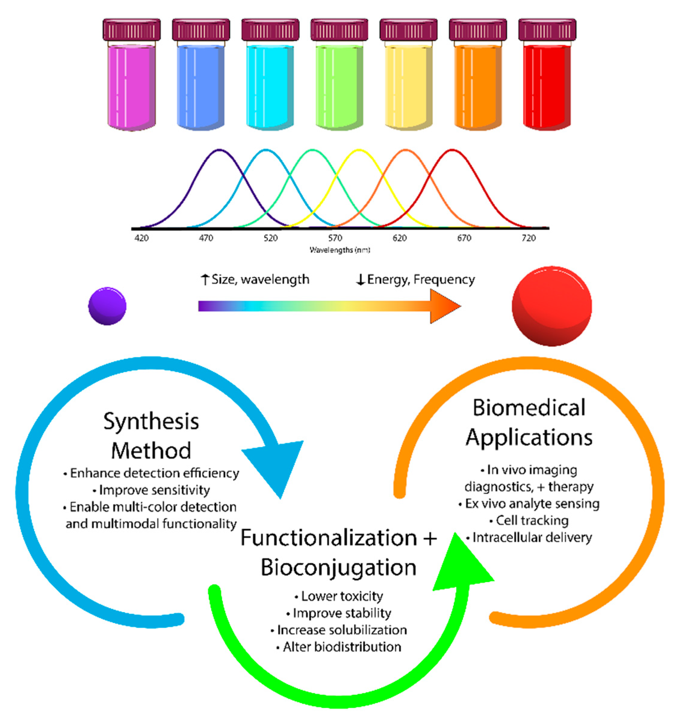
Figure 2.
Utilization of Quantum Dots in Various Biological Imaging Techniques [46].
Figure 2.
Utilization of Quantum Dots in Various Biological Imaging Techniques [46].
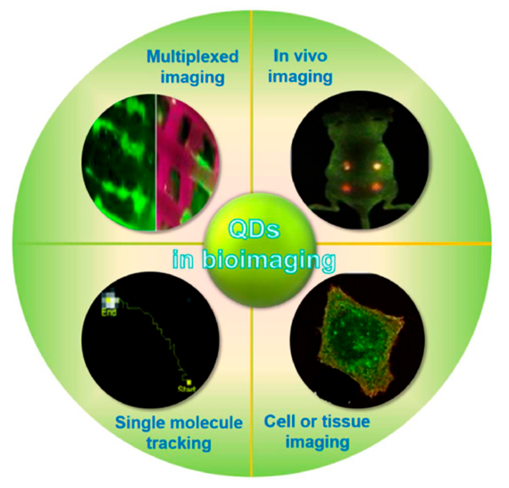
Figure 3.
Methodology for In Vivo Imaging Using Quantum Dots [46].
Figure 3.
Methodology for In Vivo Imaging Using Quantum Dots [46].
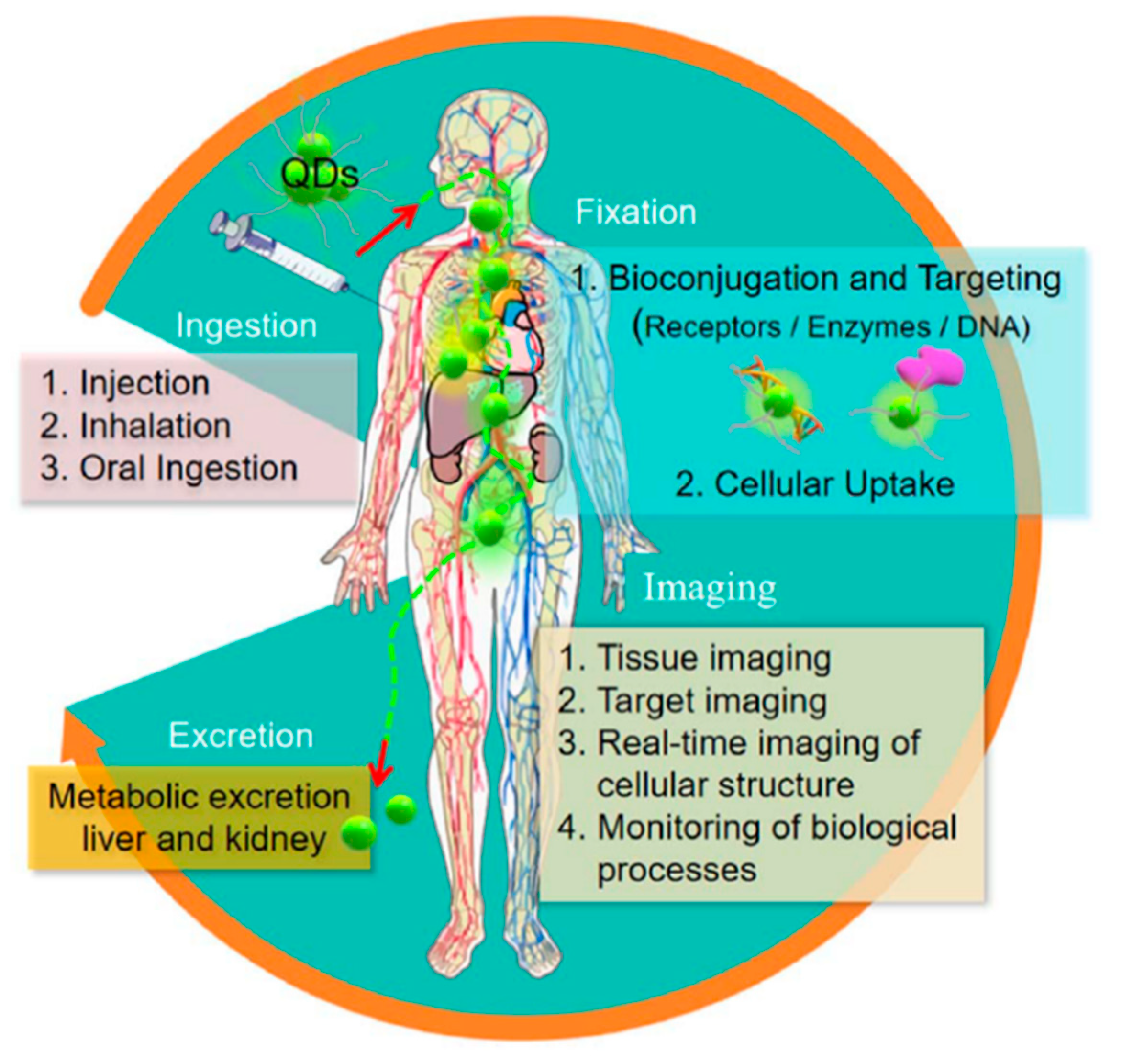
Figure 4.
Depiction of Quantum Dots' Dual Roles in Biomedical Sensing and In Vivo Optical Imaging for Healthcare and Medical Purposes [49].
Figure 4.
Depiction of Quantum Dots' Dual Roles in Biomedical Sensing and In Vivo Optical Imaging for Healthcare and Medical Purposes [49].
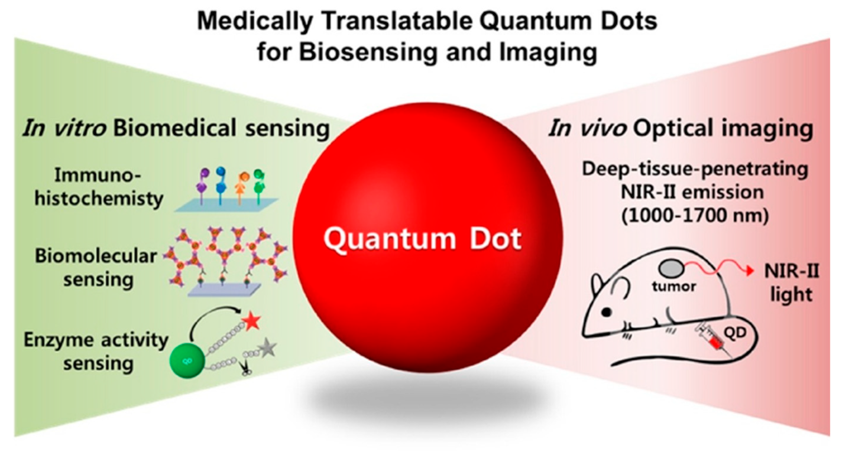
Figure 5.
Range of Applications of Quantum Dots in Bioimaging and Diagnostic Procedures [77].
Figure 5.
Range of Applications of Quantum Dots in Bioimaging and Diagnostic Procedures [77].
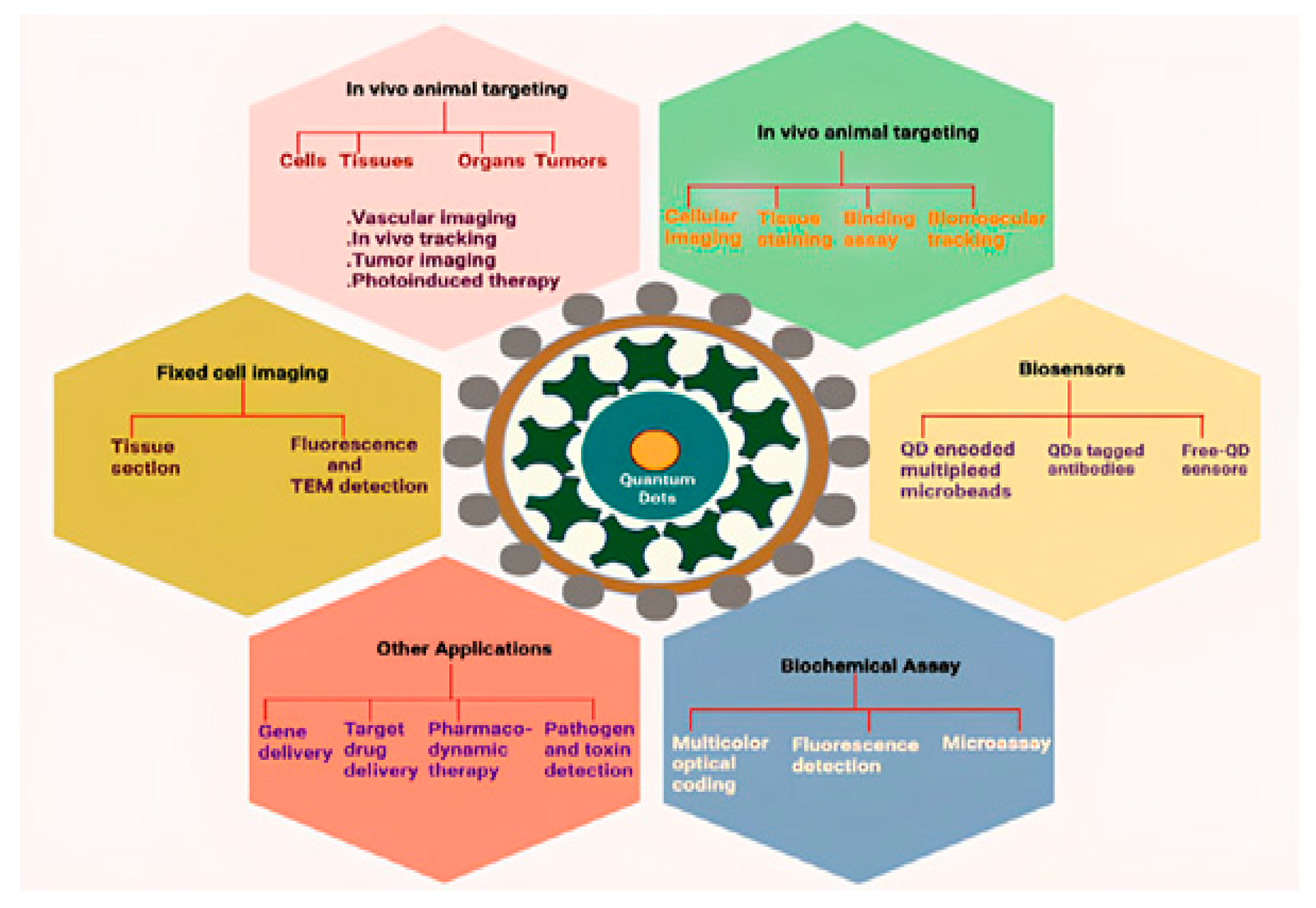
Figure 6.
(a) Schematic of the Synthesis Pathway for Highly Luminescent InP/ZnSe/ZnS Quantum Dots Incorporating ZnF₂. (b) Diagram of Quantum Light-Emitting Diodes (QLEDs) Alongside a Cross-Sectional TEM Image Showing Approximately Four Monolayers of Quantum Dots within the QLED. (c) Graph Displaying the Relationship Between External Quantum Efficiency (EQE) and Luminance [79].
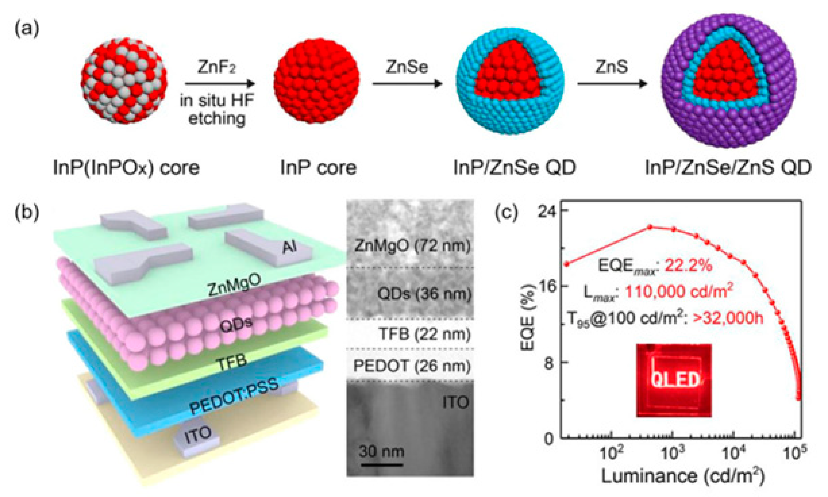
Figure 7.
(a) Enhanced Structural Design of Quantum Light-Emitting Diodes (QLEDs). (b) Performance Characteristics of QLEDs Illustrating EQE in Relation to Luminance and Current Efficiency [79].
Figure 7.
(a) Enhanced Structural Design of Quantum Light-Emitting Diodes (QLEDs). (b) Performance Characteristics of QLEDs Illustrating EQE in Relation to Luminance and Current Efficiency [79].
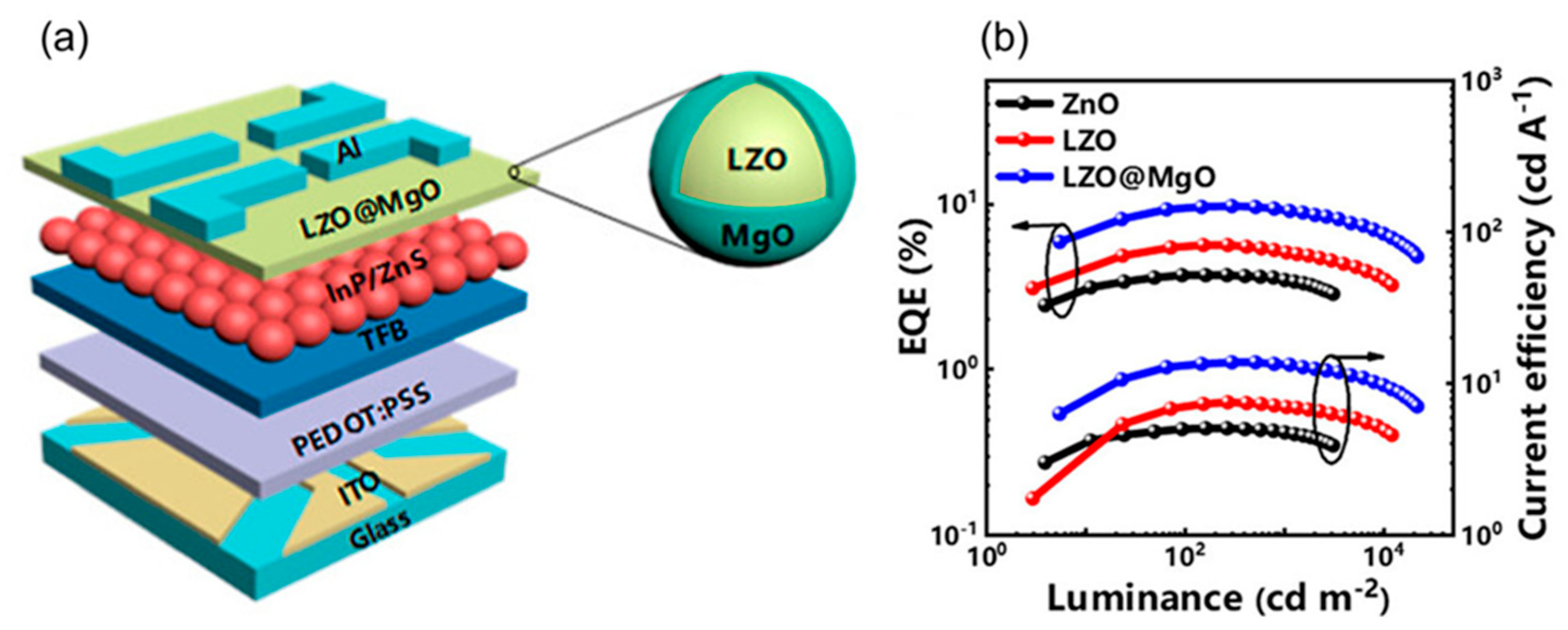
Figure 8.
Illustration Depicting a Nanoparticle Encircled by Organic Surface Ligands and Layered with Strongly Bound Proteins (Hard Corona) alongside Weakly Bound Proteins in Equilibrium with Soluble Proteins (Soft Corona) [48].
Figure 8.
Illustration Depicting a Nanoparticle Encircled by Organic Surface Ligands and Layered with Strongly Bound Proteins (Hard Corona) alongside Weakly Bound Proteins in Equilibrium with Soluble Proteins (Soft Corona) [48].

Figure 9.
Visual Representation of Surface Modification Techniques, Including Small Ligand Replacement, Polymer or Lipid Encapsulation, and Biomolecule Attachment to Quantum Dots [49].
Figure 9.
Visual Representation of Surface Modification Techniques, Including Small Ligand Replacement, Polymer or Lipid Encapsulation, and Biomolecule Attachment to Quantum Dots [49].
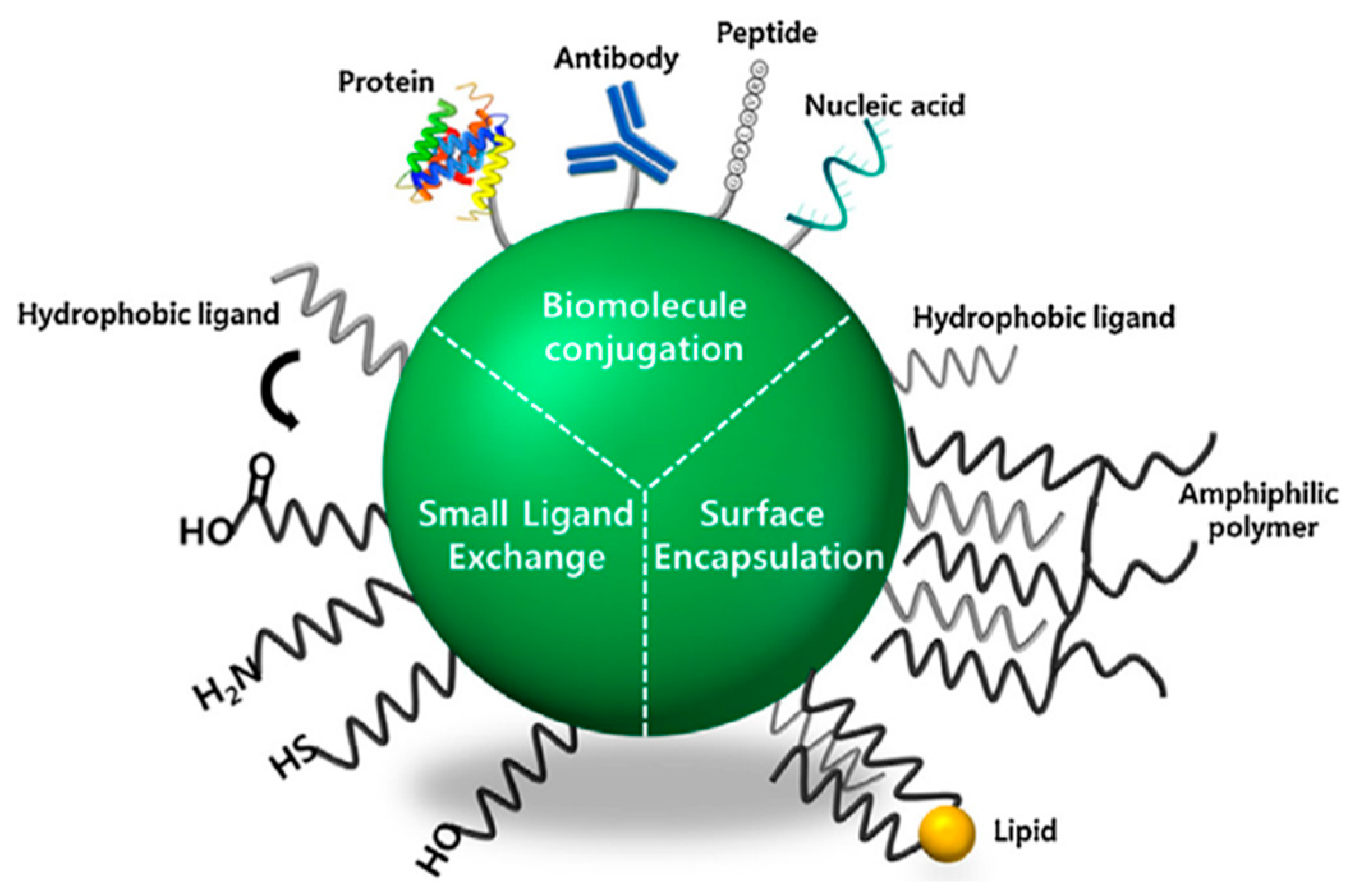
Figure 10.
Approaches for Tumor Targeting. (a) Active Biomolecular Targeting Utilizing Antibodies, Peptides such as RGD, or Small Molecules like Folic Acid. (b) Targeting the Tumor Microenvironment (TME) by Exploiting Acidic pH with Protonatable Surface Groups, Overexpressed Enzymes such as MMPs, or GSH-Mediated Cleavage of Surface Ligands [48].
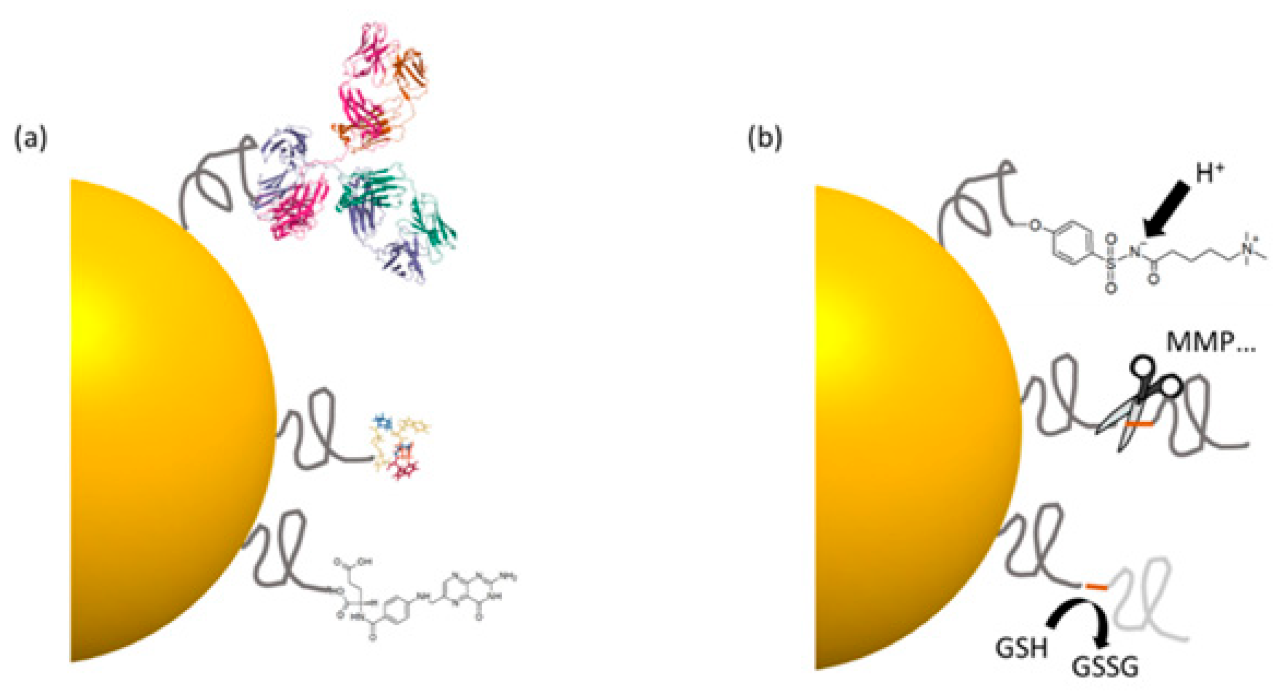
Figure 11.
Illustration of Receptor-Mediated Endocytosis of Ligand-Conjugated Graphene Quantum Dots (GQDs) Carrying an Anticancer Drug, Followed by the Drug's Release Inside a Tumor Cell [179].
Figure 11.
Illustration of Receptor-Mediated Endocytosis of Ligand-Conjugated Graphene Quantum Dots (GQDs) Carrying an Anticancer Drug, Followed by the Drug's Release Inside a Tumor Cell [179].
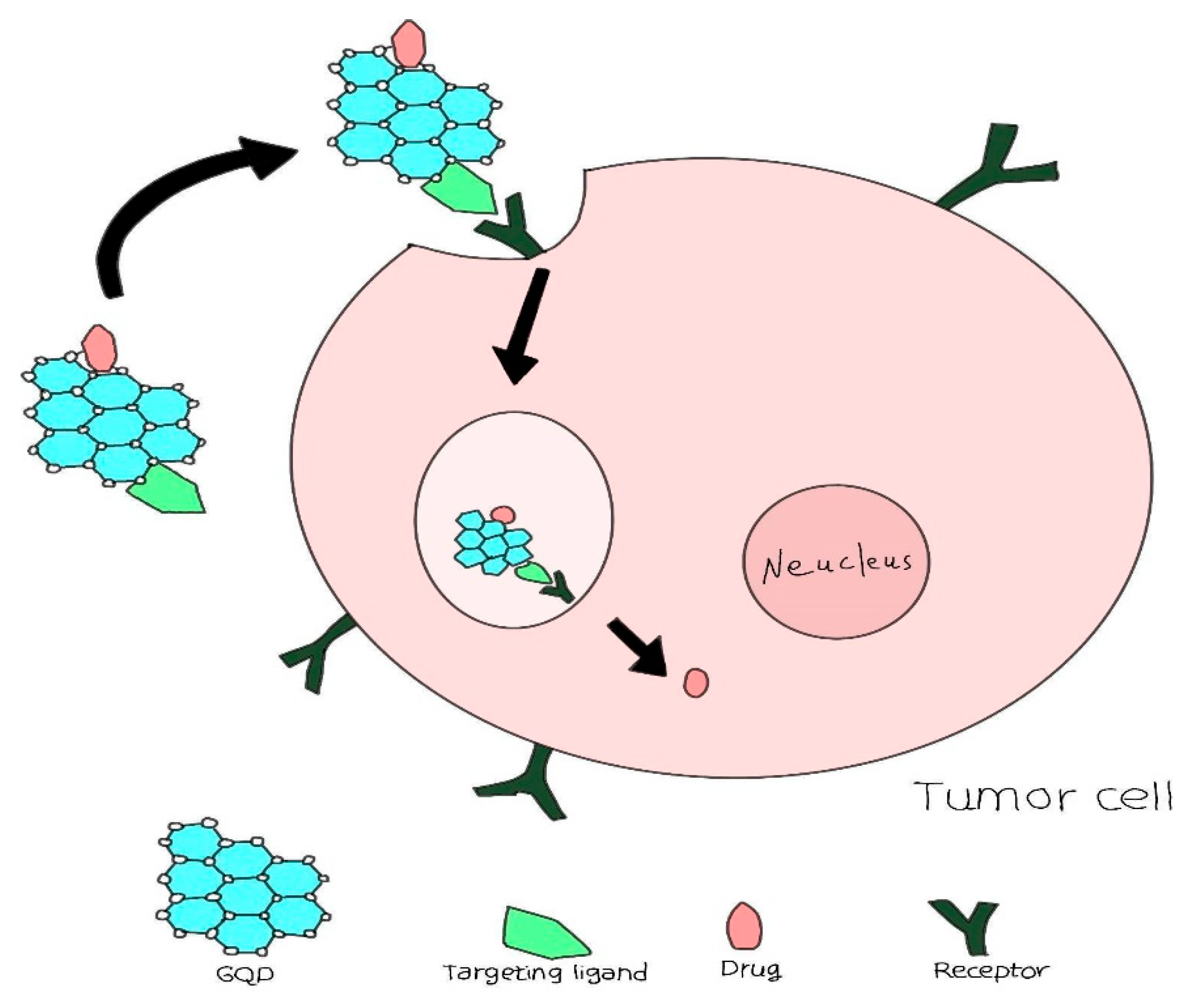
Figure 12.
Quantum Dot Delivery Strategies Include Utilizing Their Fundamental Properties such as Size, Charge, and Surface Coating. Additionally, QDs Can Be Functionalized with Ligands (e.g., Antibodies, Peptides) for Active Targeting. Another Method Involves Encapsulating QDs Within Larger Nanoparticles (e.g., Micelles, Liposomes) to Enhance Biodistribution and Targeting Efficiency [180].
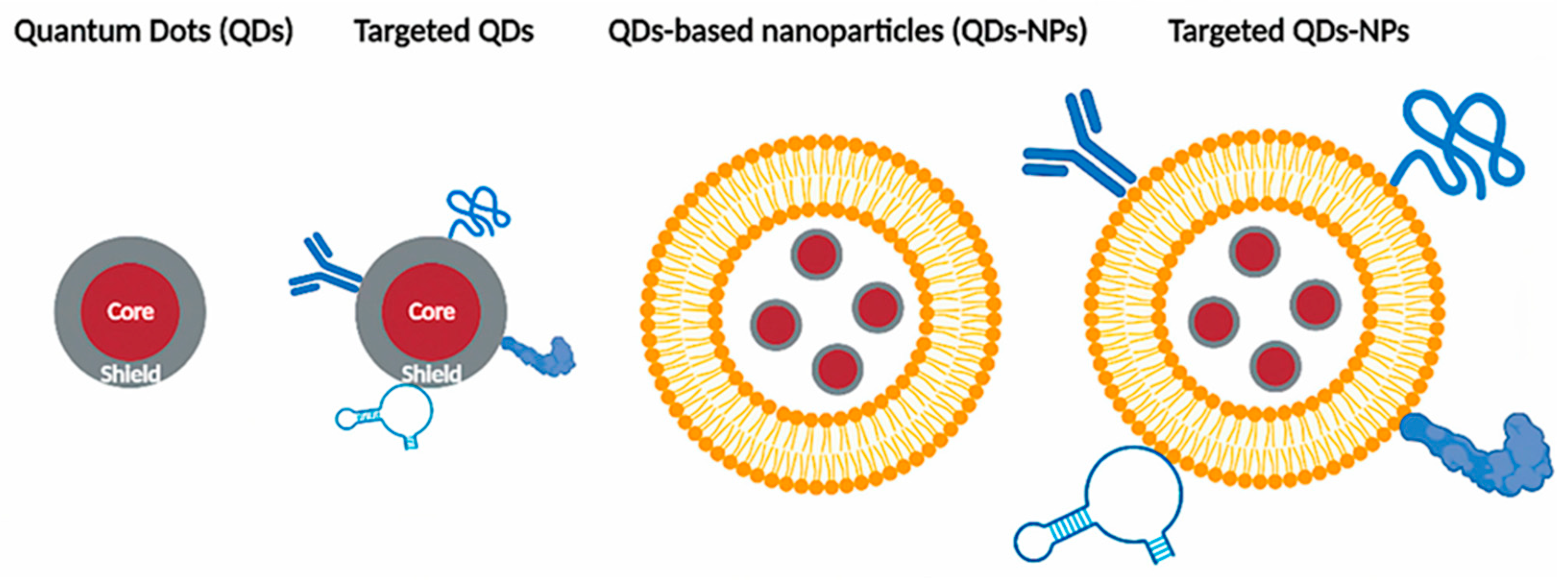
Figure 13.
Roles of Quantum Dots in the Biomedical Sector [209].
Figure 13.
Roles of Quantum Dots in the Biomedical Sector [209].
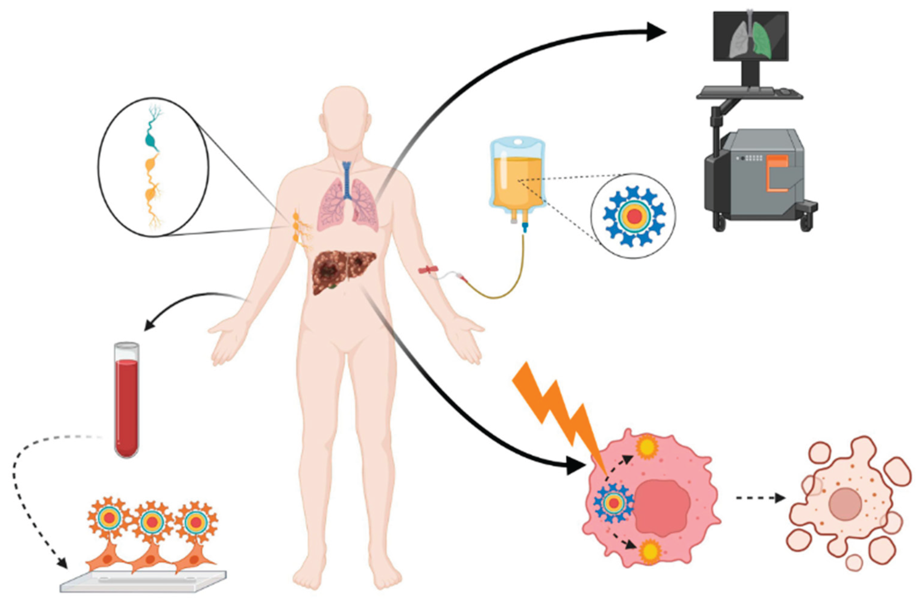

Table 1.
Classification of Various Chemical Compositions Used to Categorize Quantum Dots [6].
Table 1.
Classification of Various Chemical Compositions Used to Categorize Quantum Dots [6].
| Type | Examples |
|---|---|
| I B-VI A | Cu2S |
| I B-VII A | AgBr |
| II B-VI A | ZnSe, ZnS, ZnO, CdS, CdSe, CdTe, HgS |
| III A-V A | AlSb, AlAs, AlP, GaSb, GaAs, InAs, InP |
| IV A-VI A | PbS, PbSe, PbTe |
| IV A | C, Si, Graphene |
| V A | Black Phosphorus |
| I B-III A-VI A | CuInS2, CuInSe2, AgInS |
| P dots | NIR800 |
| TMDCs | TiSe2, TaS2, MoSe2 |
| MXenea | Nb2C, Ti3C2 |
| Perovskiteb | CsPbI3 |
Table 2.
Summary of Different Covalent Bonding Methods for Linking Biomolecules to Quantum Dots.
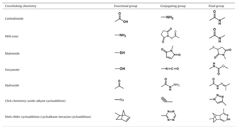 |
Disclaimer/Publisher’s Note: The statements, opinions and data contained in all publications are solely those of the individual author(s) and contributor(s) and not of MDPI and/or the editor(s). MDPI and/or the editor(s) disclaim responsibility for any injury to people or property resulting from any ideas, methods, instructions or products referred to in the content. |
© 2025 by the authors. Licensee MDPI, Basel, Switzerland. This article is an open access article distributed under the terms and conditions of the Creative Commons Attribution (CC BY) license (http://creativecommons.org/licenses/by/4.0/).
Copyright: This open access article is published under a Creative Commons CC BY 4.0 license, which permit the free download, distribution, and reuse, provided that the author and preprint are cited in any reuse.


