Submitted:
08 July 2025
Posted:
09 July 2025
You are already at the latest version
Abstract
Background/Objectives: Aneurysmal subarachnoid hemorrhage (aSAH) is frequently complicated by cerebral vasospasm, a major contributor to delayed cerebral ischemia and poor neurological outcomes. Early prediction remains challenging, and there is a critical need for reliable biomarkers. MicroRNAs (miRNAs) in cerebrospinal fluid (CSF) have emerged as promising indicators of acute neuropathological changes. This study aimed to evaluate CSF miRNA expression profiles in patients with aSAH to identify early predictors of vasospasm and improve clinical risk stratification. Methods: We conducted a prospective observational study involving 48 patients with aSAH (20 who developed vasospasm, 20 who did not) and eight healthy controls. A panel of 20 candidate miRNAs was analyzed in CSF samples collected on days 1 and 5 post-hemorrhage using quantitative real-time PCR. Expression differences between groups were assessed, and receiver operating characteristic (ROC) curves were used to evaluate diagnostic performance. Results: Several miRNAs were differentially expressed in patients who developed vasospasm. On day 1, miR-221-3p and miR-183-5p were significantly upregulated (p = 0.014, p = 0.009), while miR-126, miR-29a, and miR-27b-3p were downregulated (p = 0.006, 0.021, 0.028) compared to controls. MiR-126 remained suppressed on day 5 (p = 0.002). These early changes showed high predictive accuracy (e.g., day-1 AUC for miR-221-3p ≈ 0.98, 95% CI 0.83–1.00). Compared to non-vasospasm patients, miR-221-3p was lower (0.12-fold), while miR-9-3p and miR-183-5p were higher (13.4-fold and 2.7-fold; all p < 0.01). MiR-24 and miR-21-5p correlated with more severe grades and poorer outcomes (p < 0.05). Conclusions: Specific CSF miRNAs—particularly miR-221-3p, miR-9-3p, and miR-183-5p—may serve as early biomarkers for vasospasm, warranting further validation in larger cohorts.
Keywords:
microRNA
; cerebrospinal fluid
; vasospasm
; subarachnoid hemorrhage
; biomarker
; prognosis
1. Introduction
Aneurysmal subarachnoid hemorrhage (aSAH) is a life-threatening form of hemorrhagic stroke that, while accounting for only about 5% of all strokes, disproportionately affects relatively young patients (over half are younger than 60 years) [1]. It remains highly lethal: approximately 30% of aSAH patients die during the initial hospitalization, and many survivors suffer permanent neurological disabilities [2]. Poor outcomes result not only from the initial bleeding event but also from various acute and delayed complications such as cerebral ischemia (often due to vasospasm), hydrocephalus, and re-bleeding [3]. Among these, delayed cerebral vasospasm (DCV) is one of the most dreaded complications. DCV has long been recognized as a major contributor to morbidity after aSAH, occurring in roughly one-third of patients within 3–10 days of the aneurysm rupture [4,5]. This prolonged constriction of cerebral arteries can lead to delayed cerebral ischemia (DCI), which is strongly associated with neurologic deterioration and poor long-term outcome [5]. The pathophysiology underlying vasospasm is complex and not fully understood; proposed mechanisms include calcium-mediated vasoconstriction, oxidative stress, endothelial dysfunction, inflammation, apoptosis, and other pathways of secondary brain injury [6]. Clinically, predicting which aSAH patients will develop vasospasm or DCI remains a significant challenge. Currently, management relies on vigilant neurological monitoring and prophylactic measures (such as hemodynamic augmentation and calcium-channel blockade) applied broadly to at-risk patients [5]. To date, no reliable biomarker or scoring system exists to identify those at highest risk for vasospasm before clinical symptoms manifest. This gap underscores a critical need for early predictors of vasospasm that could facilitate proactive interventions and enhance outcomes in aneurysmal subarachnoid hemorrhage (aSAH).
In recent years, microRNAs (miRNAs) have emerged as promising candidates for such iomarkers in neurological diseases. MiRNAs are small (~22-nucleotide) non-coding RNAs that regulate gene expression at the post-transcriptional level [7,8]. They play critical roles in virtually all biological processes, and dysregulation of miRNAs has been implicated in numerous CNS disorders, including stroke and traumatic brain injury [9]. Notably, mounting evidence suggests that miRNAs are involved in the pathophysiology of aSAH and its complications. Several studies have documented changes in miRNA expression in cerebral tissues and blood vessels after aSAH, particularly in the context of vasospasm [9,10]. A key feature that makes miRNAs attractive as clinical biomarkers is their stability in biofluids. MiRNAs circulate in extracellular fluids such as plasma and cerebrospinal fluid (CSF) packaged in exosomes or protein complexes, which protect them from RNase degradation [11,12]. These characteristics – disease association, biological relevance, and stability in CSF/blood – position miRNAs as potential early indicators of vasospasm risk in aSAH.
Recent evidence strongly suggests that cerebrospinal fluid (CSF) microRNAs (miRNAs) are promising candidates for predicting vasospasm following aneurysmal subarachnoid hemorrhage (aSAH). Few studies have also described miRNA expression in CSF, showing different patterns related to vasospasm and DCI on different days after aSAH [10,13,14,15], which can reflect ongoing acute pathophysiological changes in the brain The sort of pathways in inflammation, endothelial integrity, and cerebrovascular tone that these miRNAs seem to modify are biologically as well as clinically significant. Given the tissue-specific expression patterns of many miRNAs, aSAH-induced changes in brain-specific miRNAs may provide insight into disease mechanisms. Evaluating miRNAs as biomarkers in neurological conditions, particularly by analyzing their levels in CSF, is therefore a promising approach to clarify aSAH pathophysiology and its complications. Studies examining CSF miRNA profiles in aSAH are still minimal; to the best of our knowledge, only a few investigations have focused on miRNA changes in CSF after aSAH.
Nevertheless, the data currently available remain limited by small sample sizes, heterogeneous results, and no single miRNA panel has been established for clinical practice.
To fill this gap, the present study aimed to characterize the expression profiles of a panel of 20 candidate miRNAs in the CSF of aSAH patients, and to determine their association with the development of vasospasm and with patient outcomes. By generating initial data on these potential biomarkers, we sought to address a gap in the literature and lay the groundwork for improved prognostic and therapeutic strategies in aSAH.
2. Materials and Methods
All procedures were conducted by the Declaration of Helsinki and approved by the Clinical Research Ethics Committee of Eskisehir Osmangazi University. Written informed consent was obtained from all participants. At the same time, well-established methods can be briefly described and appropriately cited.
2.1. Study Design and Participants
This prospective study was conducted at the neurosurgery clinic of our university hospital. All aSAH patients were managed by an experienced neurovascular team, and all molecular analyses were performed in a blinded fashion at the medical genetics department. Clinical data were not revealed to the genetics researchers until after miRNA expression results were obtained. The study cohort consisted of 48 individuals: 40 patients with aSAH (20 who developed vasospasm and 20 who did not) and eight non-SAH control subjects. For each aSAH patient, CSF samples were collected at two time points (on day 1 and day 5 after hemorrhage), yielding a total of 80 patient CSF samples. An additional single CSF sample was obtained from each control, bringing the total number of CSF samples analyzed to 88. In subsequent descriptions, patients with aSAH who developed vasospasm are referred to as the vasospasm-positive group (VSP+), and those who did not develop vasospasm are referred to as the vasospasm-negative group (VSP-).
2.2. Inclusion and Exclusion Criteria
The inclusion and exclusion criteria for patient and control selection are outlined below:
2.2.1. Patient Inclusion Criteria
- Age 18–80 years with acute aSAH confirmed by sudden onset of symptoms (e.g., severe headache, nausea/vomiting, altered consciousness, or focal neurological deficits) and evidence of subarachnoid hemorrhage on cranial computed tomography.
- Identification of a ruptured saccular intracranial aneurysm as the bleeding source by digital subtraction angiography or CT angiography.
- Definitive treatment of the ruptured aneurysm (endovascular coiling or microsurgical clipping) within 48 hours of diagnosis.
- Radiological evidence of cerebral vasospasm on CT angiography during the post-hemorrhage course.
- In patients with clinical signs of vasospasm, confirmation of vasospasm by diagnostic cerebral angiography and administration of intra-arterial vasospasm therapy
2.2.2. Patient Exclusion Criteria
- Treatment for an unruptured aneurysm (i.e., cases without acute hemorrhage).
- History of previous aSAH.
- Family history of intracranial aneurysm or aSAH in first-degree relatives
- Known hereditary conditions predisposing to aneurysm (e.g., polycystic kidney disease or connective tissue disorders).
- Traumatic, mycotic, or dissecting aneurysms.
- Presence of multiple aneurysms.
- Presentation >72 hours after the ictus (delayed admission beyond the acute phase).
- Comorbid conditions (e.g., active malignancy, chemotherapy, recent trauma) that could disrupt the blood–brain barrier or confound neurological assessment.
2.2.3. Control Group
Clinically healthy volunteers without a family history of IA/aSAH or neurological and chronic/systemic diseases were included as controls. Individuals with neurological diseases, such as hydrocephalus, were excluded from the control CSF sample collection due to the potential to affect miRNA expression profiles. Instead, cerebrospinal fluid taken from individuals who underwent spinal surgery under spinal anesthesia constituted the control group. Healthy controls did not undergo neuroimaging. Informed consent was obtained from all participants, and 3-4 cc CSF samples were taken and analyzed.
2.3. Clinical Management
The management of subarachnoid hemorrhage patients in our 24-hour accredited stroke center clinic was performed by a neurovascular team consisting of specialists in neurosurgery, interventional neurology, and neurological intensive care. The same team also performed postoperative intensive care of all patients. All patients consulted from the emergency department underwent computerized brain tomography at the time of initial assessment, followed by emergency brain tomography angiography or diagnostic cerebral angiography, depending on the patient's clinical condition. Mannitol, furosemide, hyperosmolar treatment, hypertensive agents, nimodipine, and antiepileptic agents were administered when necessary. External ventricular drainage (EVD) was performed based on the patient's clinical status and the severity of the bleeding. Lumbar drainage (LD) was placed in all surgical patients, and in those treated endovascularly, if EVD or LD was not placed, CSF samples were obtained by puncture. All patients underwent aneurysm closure as soon as possible, within 48 hours at the latest. Tomography angiography was performed on the fifth day for both aneurysm control and vasospasm detection. In patients with suspected vasospasm, diagnostic angiography was performed to confirm the diagnosis. Clinical and radiological data were recorded using the Fischer Score, Hunt-Hess score (HH), Glasgow Coma Scale (GCS), World Federation of Neurosurgical Societies (WFNS), and Modified Rankin Scale (mRS).
2.4. Selection of Target miRNAs
Preliminary research was conducted to select candidate circulating miRNAs reported as potentially indicative of vasospasm risk in aneurysmal subarachnoid hemorrhage (aSAH) cases. This review was conducted by examining relevant articles and databases, including PubMed, Embase, Ovid, Google Scholar, and miRBase. Based on this research, the expression profiles of 20 miRNAs (miR-29a, miR-126, miR-200a-3p, miR-451a, miR-146a-5p, miR-1297, miR-27b-3p, miR-502-5p, miR-143-3p, miR-145-5p, miR-17-5p, miR-221-3p, miR-21-5p, miR-15a, miR-9-3p, miR-630, miR-24, miR-148b-3p, miR-497, and miR-183-5p) were determined in the CSF samples of the aSAH/vasospasm+ and aSAH/vasospasm- groups and compared with control CSF samples.
2.5. RNA Extraction from CSF Samples
The CSF samples from aSAH cases and controls were centrifuged for 5 minutes at 2000 g at +4°C to remove contaminated blood cells. CSF material was carefully pipetted from the tube without disturbing the blood pellet, and aliquots were stored at −80°C until RNA isolation and further analysis. Total RNA was extracted using the miRVANA™ PARIS™ Kit (Applied Biosystems, CA) according to the manufacturer's instructions
2.6. Quantitative Real-Time Polymerase Chain Reaction (qRT-PCR)
Following the manufacturer's guidelines, complementary DNA (cDNA) was synthesized using the miRNA ALL-In-One cDNA Synthesis Kit (ABM, CA). Each reaction mixture contained 2X miRNA cDNA Synthesis SuperMix (10 µL), 2 µL Enzyme Mix, 2 µL total RNA, and 8 µL nuclease-free water, resulting in a total reaction volume of 20 µL. The reverse transcription process involved the following cycle profile: 37°C for 30 minutes, 50°C for 15 minutes, 85°C for 5 minutes, and a hold at 4°C.
Quantitative real-time PCR (qRT-PCR) reactions were performed on a CFX-96 Real-Time PCR Detection System (BIO-RAD, C1000 Touch Thermal Cycler) using BrightGreen miRNA qPCR Master Mix (5 μl), forward primer (0.35 μl), reverse primer (0.35 μl), cDNA (2 μl), and nuclease-free water (2.3 μl) to make a final volume of 10 μl. The amplification reaction conditions consisted of an initial incubation at 95°C for 10 minutes, followed by 40 cycles of denaturation at 95°C for 10 seconds, annealing at 60°C for 20 seconds, and extension at 72°C for 30 seconds. All samples were analyzed in duplicates.
The housekeeping gene U6 was used as a reference gene for normalizing miRNA expression.
(The primer catalogue numbers for U6 (control) CAT:MPH00001, hsa-miR-126-3p CAT:MPH01082, hsa-miR-200a-3p CAT:MPH02296, hsa-miR-451a CAT:MPH02756, hsa-miR-27b-3p CAT:MPH02385, hsa-miR-146a-5p CAT:MPH02204, hsa-miR-502-5p CAT:MPH01742, hsa-miR-1297 CAT:MPH01130, hsa-miR-29a-3p CAT:MPH01310, hsa-miR-15a CAT:MPH01189, hsa-miR-148b-3p CAT:MPH02208, hsa-miR-17-5p CAT:MPH02236, hsa-miR-21-5p CAT:MPH02337, hsa-miR-221-3p CAT:MPH02350, hsa-miR-9-3p CAT:MPH03965, hsa-miR-183-5p CAT:MPH02250, hsa-miR-497 CAT:MPH01733, hsa-miR-24 CAT:MPH01294, hsa-miR-143-3p CAT:MPH02196, hsa-miR-630 CAT:MPH01931, hsa-miR-145-5p CAT:MPH02201 (all applied Biological Materials, ABM).
The cycle threshold (Ct) values of the reference and target miRNA were determined in each sample, and the relative miRNA level was calculated using the 2-ΔΔCt method.
Statistical Analysis
The statistical analysis was conducted using SPSS version 21.0 (IBM Corporation, NY, USA), while GraphPad Prism v.9.5.1 (GraphPad Software) was used to generate the graphs. The Shapiro-Wilk test was employed to evaluate the distribution of the variables. For standard variation variables, comparisons were made using the Student t-test, while the Mann-Whitney U test was used to determine differences between the two groups. The diagnostic potential of candidate miRNAs as a biomarker was assessed by constructing Receiver operating characteristic (ROC) curves for miRNAs with a significant difference in levels and calculating the area under the ROC curve (AUC), specificity, and sensitivity with a 95% confidence interval (95% CI). A value of P < 0.05 was considered statistically significant.
3. Results
3.1. Demographic Characteristics
A total of 40 patients with aSAH (19 females and 21 males) were included in the analysis, along with eight control subjects (4 females and four males). The mean age was similar across the three groups (57.8 ± 14.2 years in the VSP+ group, 57.9 ± 12.0 in the VSP– group, and 57.1 ± 9.7 years in controls; p = 0.988). At presentation, the aSAH patients had a range of severity: in the VSP+ group, 70% of patients were classified as WFNS grades 1–2 and 30% as grades 3–5, while 60% were Hunt–Hess grades 1–2 and 40% were grades 3–5. According to the CT Fisher scale, 45% of the VSP+ patients had Fisher grade 1–3 hemorrhage, and 55% had grade 4. The 3-month functional outcomes (Modified Rankin Scale, mRS) for all aSAH patients showed that 35% achieved good recovery (mRS 1–2) and 65% had significant disability or worse (mRS 3–6). There were no significant differences between the VSP+ and VSP– patient groups in terms of baseline characteristics such as age, sex, history of hypertension, or initial hemorrhage severity scores (Fisher grade, Hunt–Hess grade, WFNS grade, and mRS at presentation were comparable; p > 0.05 for all; see Table 1).
3.2. miRNA Expression Profiles: Vasospasm-Positive vs. Control
The results revealed statistically significant differences in the expression of five miRNAs in the VSP+ patients on the first day after SAH compared to the control group. We observed a marked increase in the expression of miR-221-3p (p = 0.014) and miR-183-5p (p = 0.009). In contrast, miR-126 (p = 0.006), miR-29a (p = 0.021), and miR-27b-3p (p = 0.028) were significantly downregulated. Moreover, the expression levels of miR-126 remained significantly downregulated on the fifth day after SAH (p = 0.002) (Table 2).
ROC analyses were performed to evaluate the diagnostic performance of these 20 miRNAs in distinguishing between VSP+ patients and controls. The ROC analysis indicated that miR-221-3p, miR-126, and miR-27b-3p exhibited high sensitivity and specificity, with AUC values of 0.975, 0.975, and 0.963, respectively (Table 3). Notably, the expression of miR-126 continued to decrease on the fifth day after SAH and demonstrated a significantly high AUC value of 0.981 with 100% sensitivity.
3.3. miRNA Expression Profiles: Vasospasm-Negative vs. Control
When comparing the miRNA expression levels on the first day after SAH between VSP (vasospasm-negative) patients and control groups, miR-221-3p was significantly upregulated (31.91-fold). At the same time, miR-9-3p (0.44-fold) and miR-29a (0.26-fold) were downregulated considerably (p < 0.001 and p = 0.021, respectively). Additionally, a continued downregulation of miR-29a (0.78-fold) was detected on the fifth day after SAH in VSP samples compared to controls (Table 4).
The ROC analysis between these groups indicated that miR-221-3p, with high sensitivity and specificity, exhibited strong discriminative potential, reflected by a high AUC (0.994) value.
3.4. miRNA Expression Profiles: Vasospasm vs. No Vasospasm (VSP+ vs. VSP–)
A key question was whether CSF miRNA levels before the onset of vasospasm could distinguish those aSAH patients who would develop vasospasm (VSP+) from those who would not (VSP–). We directly compared the VSP+ and VSP– groups at each time point. In the Day 1 CSF samples, three miRNAs showed significant differences between the two patient groups (Table 5). MiR-221-3p levels were lower in VSP+ patients on Day 1 compared to VSP– patients (a 0.12-fold relative expression, implying an ~8.3-fold decrease; p < 0.001). By contrast, miR-9-3p and miR-183-5p were higher in the VSP+ group on Day 1: miR-9-3p was elevated ~13.41-fold (p < 0.001) and miR-183-5p ~2.66-fold (p = 0.005) in VSP+ vs. VSP–. These early-post-SAH differences suggest that by 24 hours, patients fated to develop vasospasm already exhibit a distinct CSF miRNA signature (lower miR-221-3p, higher miR-9-3p/miR-183-5p). In the Day 5 samples, no statistically significant differences in miRNA expression were observed between the VSP+ and VSP– groups (after vasospasm had clinically been declared), indicating that the expression differences are most pronounced in the acute phase, before or around the time vasospasm develops. ROC analysis for discriminating VSP+ vs. VSP– on Day 1 showed that these three miRNAs had good diagnostic performance. MiR-9-3p was particularly strong, with an AUC of 0.915 (80% sensitivity, 85% specificity; p < 0.0001). MiR-221-3p also showed high accuracy (AUC = 0.812, 95% CI 0.658–0.918; 95% sensitivity, 75% specificity; p < 0.0001). MiR-183-5p had an AUC of 0.756 (85% sensitivity, 65% specificity; p = 0.001). Optimal cut-off values were identified for each (e.g., >–0.89 ΔCt for miR-221-3p; ≤–2.05 ΔCt for miR-9-3p, etc.; see Table 5). These results demonstrate that a panel of miRNAs measured one day after SAH can effectively distinguish patients who will develop vasospasm from those who will not, well before clinical vasospasm manifests. In practical terms, circulating miRNAs, such as miR-221-3p, miR-9-3p, and miR-183-5p, show strong potential as early biomarkers for the risk of vasospasm.
In Table 5 Day 1 CSF miRNAs distinguishing patients with vs. without vasospasm (VSP+ vs. VSP–). Cut-off values are given as ΔCt thresholds.miR-221-3p: 95.0% sensitivity, 75.0% specificity, AUC = 0.812 (95% CI 0.658–0.918), cut-off ΔCt > –0.89, p < 0.0001.miR-9-3p: 80.0% sensitivity, 85.0% specificity, AUC = 0.915 (95% CI 0.783–0.980), cut-off ΔCt ≤ –2.05, p < 0.0001.miR-183-5p: 85.0% sensitivity, 65.0% specificity, AUC = 0.756 (95% CI 0.595–0.878), cut-off ΔCt ≤ 0.86, p = 0.001. One day after SAH, these three miRNAs achieved high AUC values and could serve as a biomarker panel to predict vasospasm development with reasonable accuracy. By five days post-SAH, no miRNA showed a significant difference between VSP+ and VSP– groups, suggesting the window for early prediction is within the first few days.
3.5. miRNA Expression and Clinical Severity in Vasospasm Patients
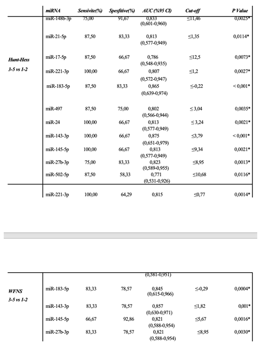
We further investigated whether miRNA expression in vasospasm patients (VSP+ group) was associated with clinical severity measures, including the Fisher grade (hemorrhage amount on CT), Hunt–Hess and WFNS grades (neurological status), and mRS outcomes. For these analyses, within the VSP+ cohort, we stratified patients by clinical severity and compared miRNA levels between subgroups. By hemorrhage amount (Fisher grade): Within the VSP+ cases, patients with a high Fisher grade (Grade 4, indicating diffuse thick SAH) were compared to those with lower Fisher grades (1–3). In the Day 5 CSF samples of VSP+ patients, four miRNAs were significantly elevated in the Fisher grade 4 group (severe hemorrhage) compared to grades 1–3: miR-17-5p (~8.10-fold higher, p = 0.007), miR-21-5p (~5.88-fold, p = 0.020), miR-221-3p (~5.11-fold, p = 0.016), and miR-24 (~2.86-fold, p = 0.046). All four remained significantly upregulated despite the small subgroup sizes. ROC curve analysis for discriminating high vs. low Fisher grades on Day 5 yielded AUCs in the range of ~0.768–0.843 for these miRNAs (e.g., miR-17-5p AUC 0.843, miR-21-5p 0.808, miR-221-3p 0.813, miR-24 0.768; see Table 6). This suggests that in patients who developed vasospasm, those with more extensive hemorrhage burden had distinctly higher levels of specific miRNAs by Day 5, which could relate to more intense secondary brain injury or inflammation. By neurological grade (Hunt–Hess and WFNS): We categorized VSP+ patients as “mild” or “severe” based on admission neurological scales. For Hunt–Hess, grades 1–2 were considered mild, and grades 3–5 were considered severe. On Day 1 post-SAH, miR-148b-3p and miR-21-5p levels were significantly higher in patients with severe Hunt–Hess grades (3–5) compared to those with grades 1–2 (miR-148b-3p 2.80-fold, p = 0.012; miR-21-5p 3.14-fold, p = 0.025). By Day 5, a broader array of miRNAs were elevated in the severe Hunt–Hess group: miR-221-3p (2.37-fold, p = 0.020), miR-183-5p (4.09-fold, p = 0.005), miR-497 (2.85-fold, p = 0.025), miR-24 (2.51-fold, p = 0.020), miR-143-3p (3.93-fold, p = 0.004), miR-27b-3p (2.46-fold, p = 0.039), miR-502-5p (2.72-fold, p = 0.047), and miR-145-5p (2.46-fold, p = 0.020) were all significantly higher in severely graded patients on Day 5. Thus, higher acute clinical severity was associated with greater increases in multiple miRNAs over the first five days. For WFNS grade (another clinical grading system, with grades 1–2 classified as mild and 3–5 as severe), we observed a similar pattern. In the VSP+ group, miR-221-3p on Day 1 was significantly higher in patients with severe WFNS (3–5) vs. mild (1–2) (which is interesting because miR-221-3p was generally lower in VSP+ vs. VSP– overall, but among VSP+ perhaps those with worse clinical state had a rebound increase—this discrepancy might reflect timing or subgroup factors). On Day 5, miR-183-5p, miR-143-3p, miR-145-5p, and miR-27b-3p were significantly higher in the severe WFNS group than in the mild group. The ROC analysis for neurological severity (Table 7) indicated that several of these miRNAs had good predictive value for distinguishing severe vs. mild cases (e.g., miR-148b-3p AUC 0.833, miR-21-5p AUC 0.813 for Hunt–Hess; miR-183-5p AUC 0.845 for WFNS). For example, in Hunt–Hess 3–5 vs 1–2, miR-183-5p achieved 87.5% sensitivity and 83.3% specificity (AUC 0.865, p < 0.001). These data suggest that certain miRNAs (like miR-21-5p, miR-24, miR-143-3p, miR-183-5p, among others) correlate with the severity of the initial hemorrhage and the degree of neurological impairment in vasospasm patients, potentially serving as prognostic biomarkers for outcome.
3.6. Summary of Key miRNA Findings
In summary, our results reveal that aSAH patients who develop vasospasm have a distinct CSF miRNA expression profile in the acute phase, differing from both healthy controls and aSAH patients without vasospasm. The most notable early changes associated with impending vasospasm (within 24 hours post-bleed) were: an increase in miR-221-3p and miR-183-5p (vs. controls) coupled with a decrease in miR-221-3p (vs. non-vasospasm patients) and an increase in miR-9-3p. These specific miRNAs demonstrated strong diagnostic performance for vasospasm prediction (AUC ~0.8–0.92) one day after SAH, well before clinical vasospasm became evident. By day 5, miR-126 emerged as a sustained differentiator (remaining low in vasospasm patients). We also identified miR-24 as a candidate marker. Although not significantly changed on day 1, miR-24 was significantly elevated by day 5 in the vasospasm group. It was strongly associated with poorer clinical status (higher in patients with severe grades and worse outcomes). In addition, miR-21-5p and miR-221-3p levels on day 5 were higher in patients with poor neurological outcomes, suggesting a possible role in prognostication. A panel comprising miR-221-3p, miR-9-3p, miR-183-5p (for early vasospasm prediction) and miR-21-5p, miR-24 (for severity/outcome) could be envisioned as a useful tool, pending further validation.
4. Discussion
Cerebral vasospasm (CVS) is a significant complication of aSAH that contributes heavily to delayed cerebral ischemia (DCI), long-term disability, and mortality [16] The underlying pathological mechanisms of vasospasm are not yet fully understood, but appear to involve a multifactorial cascade including calcium-dependent and -independent vasoconstriction, oxidative stress, endothelial dysfunction, inflammation, apoptosis, autophagy, and altered gene expression [9,17,18]. Early identification of patients at high risk for vasospasm—before clinical symptoms manifest—is crucial for preventing secondary brain injury; however, effective early biomarkers for vasospasm are currently lacking. At present, clinicians have little choice but to monitor and then react once vasospasm or delayed ischemic deficits become evident, underscoring the need for predictive biomarkers [19]. In the era of precision medicine, identifying molecular indicators of vasospasm risk could enable proactive interventions and tailored patient management [16]. Numerous studies have highlighted the potential of miRNAs as biomarkers for complications following aSAH. Most have found that changes in miRNA expression correlate with aSAH occurrence and its acute complications, and that these changes can be detected in extracellular fluids [14]. Nonetheless, no miRNA has yet been validated for clinical use to reliably distinguish which aSAH patients will develop vasospasm (one of the most critical complications) and therefore might benefit from early aggressive therapy, versus those who will not. Given the tissue-specific nature of miRNAs, it is important to focus on miRNAs in cerebrospinal fluid (CSF) – which reflects brain-derived miRNAs – when studying central complications like vasospasm. Indeed, CSF miRNAs are considered potentially better candidates for predicting vasospasm and related outcomes than peripheral miRNAs [9]. To our knowledge, only a limited number of prior studies have examined CSF miRNAs in aSAH in the context of vasospasm or DCI underscoring that this is an emerging area of research. Our study contributes to this area by profiling 20 candidate miRNAs in CSF and identifying those associated with vasospasm development and severity.
In this study, we assessed CSF samples from aSAH patients on days 1 and 5 post-hemorrhage, comparing those who developed vasospasm (VSP+) to those who did not (VSP–), as well as to non-SAH controls. We identified several miRNAs with significant differential expression linked to vasospasm. Notably, on the first day after SAH, miR-221-3p, miR-9-3p, and miR-183-5p showed statistically significant differences between the VSP+ and VSP– groups, even before vasospasm clinically manifested. In VSP+ cases, miR-221-3p was substantially lower (approximately an 8-fold decrease relative to VSP–), whereas miR-9-3p (~13-fold higher) and miR-183-5p (~2.7-fold higher) were elevated. These early alterations were strong enough to distinguish between the two patient groups, with ROC analyses indicating high specificity, sensitivity, and AUC values (miR-9-3p AUC ~0.92; miR-221-3p AUC ~0.81; miR-183-5p AUC ~0.76). Thus, just one day after SAH, these circulating miRNAs demonstrated strong potential as biomarkers for predicting which patients will develop vasospasm.
Most importantly at 5 days post-SAH (approximately the typical vasospasm window) there were no significant differences between the groups in the given miRNAs indicating that the discriminatory window is present in the very early stage. In contrast, focusing on the comparison of VSP+ patients to non-SAH controls we observed a larger number of miRNAs (5 on day 1) that were different, indicative of the overall injury response to SAH with vasospasm. MiR-221-3p and miR-183-5p were dramatically upregulated in VSP+ vs. controls on day 1, and miR-126, miR-29a-3p and miR-27b-3p were downregulated with persistence of markedly low values particularly for miR-126 on day 5. These results are also partially consistent with the previous findings that reported alterations of these miRNAs in different brain injuries. For instance, miR-126 is abundant in endothelial cells and involved in vascular homeostasis, its under-expression after SAH could indicate endothelial impairment and blood-brain barrier breakdown that mediate vasospasm physiopathology [20]. The sustained suppression of miR-126 in our vasospasm patients aligns with its proposed role in maintaining cerebrovascular stability. Our study also found that several miRNAs correlated with clinical severity and outcomes among vasospasm patients. Notably, miR-17-5p, miR-183-5p, and miR-126 showed a significant positive correlation in expression from day 1 to day 5 in VSP+ patients (i.e., their levels rose or remained low in tandem over time), hinting at persistent dysregulation through the vasospasm period. By day 5, patients with poor neurological status (higher Hunt–Hess, higher WFNS, and worse mRS outcomes) had significantly higher CSF levels of miR-183-5p (across all three severity scales) and miR-17-5p (in Fisher grade and mRS comparisons) compared to those with milder presentations. In particular, miR-183-5p stood out: its expression was significantly higher on day 5 in VSP+ patients who had poor outcomes (mRS 3–6) compared to those with good recovery (mRS 1–2). This suggests miR-183-5p might be associated not only with vasospasm occurrence but also with the extent of resulting brain injury and recovery. Furthermore, miR-21-5p, miR-221-3p, and miR-24 were all significantly upregulated in VSP+ patients with more severe clinical presentations and poorer prognoses, which is particularly noteworthy. These miRNAs have been implicated in vascular inflammation and remodeling (as discussed below), and their elevated levels in more severe cases underscore their potential relevance as prognostic markers.
Some of the miRNAs identified in this study have been linked in the literature to SAH complications. For instance, Su et al. (2015) reported that miR-132-3p levels differed in blood samples of patients with delayed cerebral infarction (DCI) versus those without [21]. Bache et al. (2017) found significantly elevated miR-21-5p and miR-221-3p in the CSF of patients who developed DCI and noted involvement of miR-221-3p, miR-132-3p, and miR-19b-3p in those who had vasospasm [14]. Stylli et al. (2017) observed that CSF levels of miR-27a-3p differed between aSAH patients with and without delayed cerebral vasospasm [10]. In a blood-based study, Lopes et al. (2018) – using next-generation sequencing – found significant differences in miRNA profiles between aSAH patients and controls, but not between those with vs. without vasospasm, possibly due to later sampling times (days 7 and 10 in that study) when differences had evened out [22]. More recently, Wang et al. (2021) examined 47 miRNAs in CSF and plasma on post-bleed days 3 and 7, and reported that certain miRNAs had significantly different patterns in patients with DCI, as early as day 3 [9]. They suggested these could predict DCI onset before clinical signs. Similarly, Bache et al. (2020) analyzed CSF miRNA profiles and found that increased miR-9-3p in CSF was associated with worse 3-month functional outcomes (mRS 3–6) [23]. This is intriguing, as our study also found miR-9-3p to be elevated in vasospasm patients (vs. non-vasospasm) and it was one of the strongest early markers; however, in our cohort miR-9-3p itself did not correlate with long-term outcome, perhaps because all our VSP+ patients had significant brain injury. In general, our findings align with these prior studies in highlighting specific miRNAs (e.g., miR-21, miR-24, miR-132, miR-221, miR-9) as relevant in SAH and its complications. Differences between studies likely stem from variations in patient cohorts, sample type (CSF vs. blood), timing of sample collection, and technical methodologies. These factors make direct comparisons challenging, emphasizing the need for more standardized research in this field.
Among the miRNAs we studied, miR-24 emerged as particularly interesting. Recent reports indicate that miR-24 is involved in various diseases through effects on inflammatory signaling pathways. Several studies suggest that upregulation of miR-24 can contribute to SAH-induced cerebrovascular complications by inhibiting endothelial nitric oxide synthase (NOS3, i.e., eNOS) [24,25]. eNOS continuously produces nitric oxide, which is essential for maintaining basal vascular tone; after SAH, reductions in eNOS activity can upset cerebral vascular tone and promote vasospasm [24]. Additionally, miR-24 can target HMOX1 (heme oxygenase-1). Following SAH, the degradation of heme from extravasated blood by heme oxygenase enzymes (especially inducible HMOX1) is neuroprotective; loss of HMOX1 exacerbates inflammation and oxidative injury. Notably, miR-24 has been shown to negatively regulate both NOS3 and HMOX1 expression. In a rat SAH model, increased miR-24 was associated with decreased NOS3/HMOX1 levels and worse vasospasm, whereas experimentally inhibiting miR-24 led to increased HMOX1 and a partial recovery from vasospasm-induced brain injury [26]. In our study, we observed a significant increase in miR-24 expression in VSP+ patients, particularly by day 5 after SAH. This timing coincides with the typical peak of vasospasm and secondary injury, and our data showed that miR-24 levels were higher in patients with severe clinical grades (Hunt–Hess 3–5) and poor outcomes (mRS 3–6) compared to those with milder presentations. These observations support the notion that miR-24 is a candidate molecular marker for vasospasm development and a prognostic indicator: higher miR-24 may identify patients at greater risk of vasospasm-related deficits, who might benefit from more aggressive monitoring or experimental therapies. It is noteworthy that miR-24 levels have been found to be elevated in aSAH patients with angiographic vasospasm compared to those without [25], nd that downregulating miR-24 can mitigate vasospasm injury in experimental models [26]. To our knowledge, our study is the first to demonstrate an association between increased CSF miR-24 and poor patient outcomes after aSAH with vasospasm, highlighting miR-24’s potential as a prognostic biomarker.
Turning to miR-221-3p, this miRNA has been implicated by multiple studies in cerebral vasospasm after SAH. MiR-221-3p’s functions involve modulating vascular smooth muscle cell (VSMC) behavior, inflammatory responses, and vascular remodeling. It is known to promote VSMC proliferation and shift VSMCs to a less contractile, more proliferative phenotype by targeting cell cycle regulators like p27^Kip1 and p57^Kip2. This leads to neointimal hyperplasia and narrowed vessel lumen, which can contribute to vasospasm. MiR-221 (and the closely related miR-222) has also been shown to increase PDGF-induced VSMC migration and phenotypic modulation [27]. MiR-221-3p can also affect endothelial function and inflammation; elevated miR-221-3p may impair angiogenesis and promote an inflammatory microenvironment by influencing endothelial cell proliferation, migration, and possibly cytokine production and leukocyte adhesion [28]. Clinically, CSF levels of miR-221-3p have been reported to be higher in SAH patients with angiographic vasospasm [14], recent meta-analysis by Li et al. (2023) identified miR-221-3p as a potential biomarker for predicting neurological outcomes after SAH: patients with good outcomes had significantly lower miR-221-3p levels than those with poor outcomes [29]. Our findings are in line with these reports. We found that high miR-221-3p expression was associated with vasospasm risk (it was elevated in VSP+ vs. controls and was a strong predictor in ROC analysis on Day 1). Yet paradoxically, when comparing VSP+ vs. VSP– patients directly, we observed that miR-221-3p was lower in the VSP+ group on Day 1. This apparent contradiction might be explained by timing and the dual role of miR-221-3p: it rises after SAH in general, but perhaps an early relative deficiency or delayed rise in the VSP+ patients could signal a dysregulated response that predisposes to vasospasm. By Day 5, we observed that higher miR-221-3p levels were indeed associated with worse clinical grades among VSP+ patients, and importantly, the highest miR-221-3p levels were found in VSP+ patients with poor outcomes across all severity scales (Hunt–Hess, WFNS, mRS). This aligns with the notion that a sustained elevation of miR-221-3p is detrimental [29]. In short, our study confirms miR-221-3p as both a predictive and prognostic biomarker in aSAH: its early aberrant levels herald vasospasm, and persistently high levels are associated with worse neurological outcomes.
The role of miR-183-5p in vasospasm is less established, but our data suggest it may be significant. MiR-183-5p has been studied in other vascular and neurological contexts. In vascular diseases like atherosclerosis, miR-183-5p promotes VSMC proliferation and migration, contributing to vascular remodeling and stenosis [30]. This mechanism could be relevant to vasospasm, where VSMC contraction and proliferation play a part in vessel narrowing. Additionally, miR-183-5p is involved in inflammatory responses and oxidative stress. In intracerebral hemorrhage (ICH) models, miR-183-5p was found to be decreased, and its overexpression alleviated early brain injury by reducing oxidative stress and neuroinflammation – partly via inhibiting HO-1 [31]. Direct evidence of miR-183-5p in vasospasm per se has been lacking; our study provides new insight by showing miR-183-5p was significantly elevated in vasospasm patients vs. non-vasospasm early on, and that a greater rise in miR-183-5p over time was associated with poorer neurological outcomes. We did not find prior literature linking miR-183-5p to vasospasm prognosis, but our data suggest that miR-183-5p could contribute to vasospasm pathophysiology through its effects on VSMCs, inflammation, and angiogenesis. Notably, its upregulation may not be a protective change—rather, higher miR-183-5p in CSF might reflect more severe vascular injury or a maladaptive response. Given the limited prior evidence, further research is needed to clarify miR-183-5p’s role. Nonetheless, our findings indicate it has value in differentiating vasospasm risk early and is worth investigating as part of a biomarker panel. It is conceivable that miR-183-5p, alongside miR-221-3p and miR-9-3p, forms a signature of acute vascular injury from SAH that predisposes to vasospasm
MiR-21 is one of the most extensively studied miRNAs in vascular biology and has been implicated in vasospasm after SAH. MiR-21’s functions are multifaceted, including regulation of inflammation, VSMC proliferation, apoptosis inhibition, and endothelial protection. It is generally upregulated after SAH and other brain injuries [14]. MiR-21 targets multiple pro-apoptotic and pro-inflammatory genes (e.g., PDCD4, PTEN, FasL, IL6R), thereby attenuating cell death and inflammation [32]. In the context of vasospasm, miR-21 may act as a double-edged sword. On one hand, it can promote VSMC proliferation and phenotypic modulation (potentially contributing to vessel wall thickening and vasoconstriction). On the other hand, it has protective effects on endothelial cells and neurons, mitigating apoptosis and blood–brain barrier breakdown [27]. In our study, miR-21-5p was not among the earliest differentiators of vasospasm vs. no vasospasm on day 1 (its levels did not significantly differ then), but by day 5 we found that miR-21-5p was significantly higher in VSP+ patients with severe grades and poor outcomes, compared to those with better clinical status. This suggests miR-21-5p elevation is more a marker of ongoing injury severity rather than a cause of vasospasm onset. Still, it showed discriminative ability between VSP+ and VSP– on day 5 (ROC AUC ~0.81 for certain severity comparisons). Elevated CSF miR-21-5p has been noted in previous studies to associate with poorer outcomes or more severe SAH complications. For instance, a relative increase in miR-21 was observed in the CSF of patients who went on to develop late cerebral ischemia (versus those who did not) [33]. Our results concur: miR-21-5p levels were higher in patients with worse neurological outcomes at 3 months, versus those with good outcomes. This miRNA might be useful as part of a prognostic biomarker panel.
Finally, it is essential to recognize the limitations to our study. The sample size, although moderate for a specialized study, can be considered small – especially if stratifying patients according to clinical grades – which compromises statistical power and external validity. Although our results are encouraging, validation in larger independent cohorts is needed. The observational design can demonstrate association, not causation. We did not confront this lack of biological support with hard, mechanistic studies testing direct targets of these miRNAs in humans (for example, measuring NOS3 or HMOX1 protein levels in parallel with the changes in miR-24 expression), which would have strongly reinforced the biological connection. Second, although we obtained samples at two time points (day 1 and day 5), vasospasm can occur at different time points; some patients may develop vasospasm earlier or later than our sampling window. A shorter sampling interval could detect more dynamic changes. We were also limited in our studies to the expression of a pre-determined panel of 20 miRNAs, and so we cannot exclude the potential involvement of other unexamined miRNAs in vasospasm. Further studies using unbiased high-throughput sequencing or array will probably identify more what peculiar genes. However, despite these limitations, our study has yielded innovative preliminary evidence suggesting that CSF miRNAs may represent early markers of vasospasm and a significant measure of disease severity. It paves the way for more comprehensive studies with particular miRNAs identified as worthy of further investigation.
5. Conclusions
To conclude, this study identified distinct CSF miRNA expression alterations that precede and accompany vasospasm after aSAH. In particular, miR-221-3p, miR-9-3p, and miR-183-5p showed a strong discriminatory capacity for predicting vasospasm development before clinical onset (as early as one day post-hemorrhage). Elevated miR-24 levels in CSF were associated with the development of vasospasm and with worse neurological deficits following SAH. Consistent with previous research, miR-21-5p and miR-221-3p in our study also demonstrated potential as prognostic biomarkers for vasospasm-related outcomes, as their CSF levels correlated with markers of inflammatory activity and clinical severity. These miRNAs, especially in combination, are valuable candidates for an early diagnostic and risk stratification panel for vasospasm. Nonetheless, larger-scale clinical studies are necessary to confirm their efficacy and to establish standardized cutoff thresholds for clinical use. It is noteworthy that we found significant changes in miR-183-5p expression, which may help differentiate vasospasm risk early on, and its continued rise in patients with poor outcomes underscores the need to evaluate this miRNA in relation to long-term neurological recovery. In summary, our findings suggest that CSF miRNA biomarkers could serve as early warning signals of vasospasm after aSAH, offering a window of opportunity for preventive interventions. They may also provide insight into the underlying pathophysiological processes, linking molecular pathways (like endothelial dysfunction and inflammation) to clinical vasospasm. With further validation, incorporating miRNA analysis into post-SAH management could improve prognostication and help personalize therapeutic strategies. We emphasize that while our results are encouraging, they should be considered preliminary. The development of a reliable miRNA-based biomarker panel for vasospasm will require extensive further research, including validation in larger cohorts and correlation with patient outcomes. If successful, such biomarkers hold promise for improving the diagnosis, risk stratification, and potentially the treatment of vasospasm in aSAH – ultimately aiming to reduce the high morbidity and mortality associated with this condition.
Author Contributions
Conceptualization, E.O. and Y.Y.; methodology, X.X.; software, X.X.; validation, Y.Y. and Z.Z.; formal analysis, X.X.; investigation, X.X.; resources, X.X.; data curation, X.X.; writing—original draft preparation, E.O., O.A., E.E.G.; writing—review and editing, N.D.C.; visualization, N.D.C.; supervision, S.A.; project administration, X.X.; funding acquisition, Y.Y. All authors have read and agreed to the published version of the manuscript.
Funding
This work was supported by the Scientific Research Projects Fund of Eskisehir Osmangazi University (Project No: TCD-2021-1710).
Institutional Review Board Statement
The study was conducted in accordance with the Declaration of Helsinki, and approved by the Institutional Review Board of Eskisehir Osmangazi University (05.Jan.2017 -80558721/11).
Informed Consent Statement
Written informed consent was obtained from all subjects involved in the study.
Data Availability Statement
The data of this study will be shared upon request due to ethical restrictions.
Acknowledgments
reviewed and edited the output and take full responsibility for the content of this publication.
Conflicts of Interest
The authors declare no conflicts of interest. The authors declare that the research was conducted in the absence of any commercial or financial relationships that could be construed as a potential conflict of interest.
Abbreviations
The following abbreviations are used in this manuscript:
| aSAH | Aneurysmal subarachnoid hemorrhage |
| CSF | cerebrospinal fluid |
| miRNAs | Micro ribonucleic acids |
| PCR | polymerase chain reaction |
| VSP+ | vasospasm positive |
| VSP- | vasospasm negative |
| WFNS | World Federation of Neurosurgical Societies |
| HH | Hunt-Hess score |
| GCS | Glasgow Coma Scale |
| mRS | Modified Rankin Scale |
| ROC | Receiver operating characteristic |
| AUC | area under curve |
| CI | confidential interval |
References
- Tawk, R.G.; Hasan, T.F.; D'Souza, C.E.; Peel, J.B.; Freeman, W.D. Diagnosis and Treatment of Unruptured Intracranial Aneurysms and Aneurysmal Subarachnoid Hemorrhage. Mayo Clin Proc 2021, 96, 1970-2000. [CrossRef]
- Seule, M.; Oswald, D.; Muroi, C.; Brandi, G.; Keller, E. Outcome, Return to Work and Health-Related Costs After Aneurysmal Subarachnoid Hemorrhage. Neurocrit Care 2020, 33, 49-57. [CrossRef]
- Chung, D.Y.; Abdalkader, M.; Nguyen, T.N. Aneurysmal Subarachnoid Hemorrhage. Neurol Clin 2021, 39, 419-442. [CrossRef]
- Janjua, N.; Mayer, S.A. Cerebral vasospasm after subarachnoid hemorrhage. Curr Opin Crit Care 2003, 9, 113-119. [CrossRef]
- Muraoka, S.; Izumi, T.; Nishihori, M.; Goto, S.; Takeuchi, I.; Saito, R. Emerging Advances in the Management of Delayed Cerebral Ischemia After Aneurysmal Subarachnoid Hemorrhage: A Narrative Review. J Clin Med 2025, 14. [CrossRef]
- Dodd, W.S.; Laurent, D.; Dumont, A.S.; Hasan, D.M.; Jabbour, P.M.; Starke, R.M.; Hosaka, K.; Polifka, A.J.; Hoh, B.L.; Chalouhi, N. Pathophysiology of Delayed Cerebral Ischemia After Subarachnoid Hemorrhage: A Review. J Am Heart Assoc 2021, 10, e021845. [CrossRef]
- Huntzinger, E.; Izaurralde, E. Gene silencing by microRNAs: contributions of translational repression and mRNA decay. Nat Rev Genet 2011, 12, 99-110. [CrossRef]
- Iwakawa, H.O.; Tomari, Y. The Functions of MicroRNAs: mRNA Decay and Translational Repression. Trends Cell Biol 2015, 25, 651-665. [CrossRef]
- Wang, W.X.; Springer, J.E.; Xie, K.; Fardo, D.W.; Hatton, K.W. A Highly Predictive MicroRNA Panel for Determining Delayed Cerebral Vasospasm Risk Following Aneurysmal Subarachnoid Hemorrhage. Front Mol Biosci 2021, 8, 657258. [CrossRef]
- Stylli, S.S.; Adamides, A.A.; Koldej, R.M.; Luwor, R.B.; Ritchie, D.S.; Ziogas, J.; Kaye, A.H. miRNA expression profiling of cerebrospinal fluid in patients with aneurysmal subarachnoid hemorrhage. J Neurosurg 2017, 126, 1131-1139. [CrossRef]
- Mraz, M.; Malinova, K.; Mayer, J.; Pospisilova, S. MicroRNA isolation and stability in stored RNA samples. Biochem Biophys Res Commun 2009, 390, 1-4. [CrossRef]
- Etheridge, A.; Lee, I.; Hood, L.; Galas, D.; Wang, K. Extracellular microRNA: a new source of biomarkers. Mutat Res 2011, 717, 85-90. [CrossRef]
- Segherlou, Z.H.; Saldarriaga, L.; Azizi, E.; Vo, K.A.; Reddy, R.; Siyanaki, M.R.H.; Lucke-Wold, B. MicroRNAs' Role in Diagnosis and Treatment of Subarachnoid Hemorrhage. Diseases 2023, 11. [CrossRef]
- Bache, S.; Rasmussen, R.; Rossing, M.; Laigaard, F.P.; Nielsen, F.C.; Møller, K. MicroRNA Changes in Cerebrospinal Fluid After Subarachnoid Hemorrhage. Stroke 2017, 48, 2391-2398. [CrossRef]
- Wang, L.; Luo, Y.C.; Wang, H.; Zou, Y.B.; Yao, H.Q.; Ullah, S.; Li, Z.Q. Azure-winged magpies fail to understand the principle of mirror imaging. Behavioural Processes 2020, 177, 8. [CrossRef]
- Przybycien-Szymanska, M.M.; Ashley, W.W. Biomarker Discovery in Cerebral Vasospasm after Aneurysmal Subarachnoid Hemorrhage. J Stroke Cerebrovasc Dis 2015, 24, 1453-1464. [CrossRef]
- Chou, S.H. Inflammation, Cerebral Vasospasm, and Brain Injury in Subarachnoid Hemorrhage-A Shifting Paradigm and a New Beginning. Crit Care Med 2018, 46, 1883-1885. [CrossRef]
- Carr, K.R.; Zuckerman, S.L.; Mocco, J. Inflammation, cerebral vasospasm, and evolving theories of delayed cerebral ischemia. Neurol Res Int 2013, 2013, 506584. [CrossRef]
- Mocco, J.; Zacharia, B.E.; Komotar, R.J.; Connolly, E.S. A review of current and future medical therapies for cerebral vasospasm following aneurysmal subarachnoid hemorrhage. Neurosurg Focus 2006, 21, E9. [CrossRef]
- Fu, X.; Niu, T.; Li, X. MicroRNA-126-3p Attenuates Intracerebral Hemorrhage-Induced Blood-Brain Barrier Disruption by Regulating VCAM-1 Expression. Front Neurosci 2019, 13, 866. [CrossRef]
- Su, X.W.; Chan, A.H.; Lu, G.; Lin, M.; Sze, J.; Zhou, J.Y.; Poon, W.S.; Liu, Q.; Zheng, V.Z.; Wong, G.K. Circulating microRNA 132-3p and 324-3p Profiles in Patients after Acute Aneurysmal Subarachnoid Hemorrhage. PLoS One 2015, 10, e0144724. [CrossRef]
- Lopes, K.P.; Vinasco-Sandoval, T.; Vialle, R.A.; Paschoal, F.M.; Bastos, V.A.P.A.; Bor-Seng-Shu, E.; Teixeira, M.J.; Yamada, E.S.; Pinto, P.; Vidal, A.F.; et al. Global miRNA expression profile reveals novel molecular players in aneurysmal subarachnoid haemorrhage. Sci Rep 2018, 8, 8786. [CrossRef]
- Bache, S.; Rasmussen, R.; Wolcott, Z.; Rossing, M.; Møgelvang, R.; Tolnai, D.; Hassager, C.; Forman, J.L.; Køber, L.; Nielsen, F.C.; et al. Elevated miR-9 in Cerebrospinal Fluid Is Associated with Poor Functional Outcome After Subarachnoid Hemorrhage. Transl Stroke Res 2020, 11, 1243-1252. [CrossRef]
- Li, H.T.; Wang, J.; Li, S.F.; Cheng, L.; Tang, W.Z.; Feng, Y.G. Upregulation of microRNA-24 causes vasospasm following subarachnoid hemorrhage by suppressing the expression of endothelial nitric oxide synthase. Mol Med Rep 2018, 18, 1181-1187. [CrossRef]
- Pulcrano-Nicolas, A.S.; Proust, C.; Clarençon, F.; Jacquens, A.; Perret, C.; Roux, M.; Shotar, E.; Thibord, F.; Puybasset, L.; Garnier, S.; et al. Whole-Blood miRNA Sequencing Profiling for Vasospasm in Patients With Aneurysmal Subarachnoid Hemorrhage. Stroke 2018, 49, 2220-2223. [CrossRef]
- Deng, X.; Liang, C.; Qian, L.; Zhang, Q. miR-24 targets HMOX1 to regulate inflammation and neurofunction in rats with cerebral vasospasm after subarachnoid hemorrhage. Am J Transl Res 2021, 13, 1064-1074.
- Yu, X.; Li, Z. MicroRNAs regulate vascular smooth muscle cell functions in atherosclerosis (Review). . International Journal of Molecular Medicine 2014, 923-933. [CrossRef]
- Li, Y.; Yan, C.; Fan, J.; Hou, Z.; Han, Y. MiR-221-3p targets Hif-1α to inhibit angiogenesis in heart failure. Lab Invest 2021, 101, 104-115. [CrossRef]
- Ten Brinck, M.F.M.; Shimanskaya, V.E.; Aquarius, R.; Bartels, R.H.M.A.; Meijer, F.J.A.; Koopmans, P.C.; de Jong, G.; Wakhloo, A.K.; de Vries, J.; Boogaarts, H.D. Outcomes after Flow Diverter Treatment in Subarachnoid Hemorrhage: A Meta-Analysis and Development of a Clinical Prediction Model (OUTFLOW). Brain Sci 2022, 12. [CrossRef]
- Lv, D.; Guo, Y.; Zhang, L.; Li, X.; Li, G. Circulating miR-183-5p levels are positively associated with the presence and severity of coronary artery disease. Front Cardiovasc Med 2023, 10, 1196348. [CrossRef]
- Zhang, J.; Li, A.; Gu, R.; Tong, Y.; Cheng, J. Role and regulatory mechanism of microRNA mediated neuroinflammation in neuronal system diseases. Front Immunol 2023, 14, 1238930. [CrossRef]
- Kousar, K.; Ahmad, T.; Abduh, M.S.; Kanwal, B.; Shah, S.S.; Naseer, F.; Anjum, S. miRNAs in Regulation of Tumor Microenvironment, Chemotherapy Resistance, Immunotherapy Modulation and miRNA Therapeutics in Cancer. Int J Mol Sci 2022, 23. [CrossRef]
- Makowska, M.; Smolarz, B.; Romanowicz, H. microRNAs in Subarachnoid Hemorrhage (Review of Literature). J Clin Med 2022, 11. [CrossRef]

Table 1.
Demographic characteristics of the study population (high clinical grades are included in the table).
Table 1.
Demographic characteristics of the study population (high clinical grades are included in the table).
|
Control (n=8) |
Vasospasm + (n=20) |
Vasospasm - (n=20) |
p-value | |
|---|---|---|---|---|
| Age | 57.75±14.17 | 57.95±12.02 | 57.12±9.73 | 0.988 |
| Sex (female) | 4 (%50) | 9 (%45) | 10 (%50) | 0.943 |
| Hypertension | NDA | 7 (%35) | 6 (%30) | 0.736 |
| Fisher grade (4) | - | 11 (%55) | 12 (%60) | 0.749 |
| Hunt-Hess grade (3-5) | - | 8 (%40) | 6 (%30) | 0.507 |
| WFNS grade (3-5) | - | 6 (%30) | 4 (%20) | 0.465 |
| Modified Rankin Scale (3-6) | - | 13 (%65) | 8 (%40) | 0.113 |
*<0.05 p-value is statistically significant, NDA.; no data available.
Table 2.
Circulating miRNA levels examined in CSF samples of vasospasm positive patients and control group.
Table 2.
Circulating miRNA levels examined in CSF samples of vasospasm positive patients and control group.
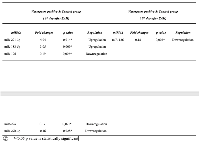
Table 3.
ROC and AUC values of circulating miRNAs examined in CSF samples of vasospasm-positive patients & control group.
Table 3.
ROC and AUC values of circulating miRNAs examined in CSF samples of vasospasm-positive patients & control group.
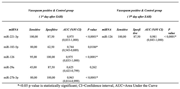
Table 4.
Circulating miRNA levels examined in CSF samples of vasospasm-negative patients &control group.
Table 4.
Circulating miRNA levels examined in CSF samples of vasospasm-negative patients &control group.
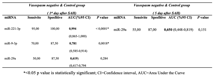
Table 5.
Comparison of miRNA expression profiles in samples of patients with and without vasospasm.
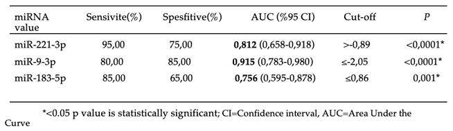
Table 6.
ROC and AUC values of circulating miRNAs analysed between Fisher grade groups in SAH day 5 samples of cases with vasospasm.
Table 6.
ROC and AUC values of circulating miRNAs analysed between Fisher grade groups in SAH day 5 samples of cases with vasospasm.
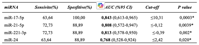
Table 7.
ROC and AUC values of circulating miRNAs analysed among neurologic clinical features in samples of cases with vasospasm.
Table 7.
ROC and AUC values of circulating miRNAs analysed among neurologic clinical features in samples of cases with vasospasm.
Disclaimer/Publisher’s Note: The statements, opinions and data contained in all publications are solely those of the individual author(s) and contributor(s) and not of MDPI and/or the editor(s). MDPI and/or the editor(s) disclaim responsibility for any injury to people or property resulting from any ideas, methods, instructions or products referred to in the content. |
© 2025 by the authors. Licensee MDPI, Basel, Switzerland. This article is an open access article distributed under the terms and conditions of the Creative Commons Attribution (CC BY) license (http://creativecommons.org/licenses/by/4.0/).
Copyright: This open access article is published under a Creative Commons CC BY 4.0 license, which permit the free download, distribution, and reuse, provided that the author and preprint are cited in any reuse.


