
Submitted:
03 July 2025
Posted:
04 July 2025
You are already at the latest version
Abstract
Pancreatic ductal adenocarcinoma (PDAC) is a highly aggressive type in pancreatic cancer. PDAC is difficult to diagnose due to lack of symptoms in early stages, resulting in survival of less than 10%. Moreover, often cancerous tissues cannot be surgically resected due to their deep abdomen location. Therefore, early detection is the essential strategy enabling effective PDAC treatment. Over the past few years, the development of nanomaterials for MRI has expanded and improved imaging quality and diagnostic accuracy. Nanomaterials can be currently designed, manufactured and synthesized with other structures to provide improved diagnosis and advanced therapy. Although MRI equipped with the innovative nanomaterials became a powerful tool for diagnosis and treatment of patients with various cancers, detection of PDAC remains challenging. In this review, we discuss the current applications and properties of iron oxide nanoparticles applied for the detection of pancreatic cancer with MRI in clinical environment.
Keywords:
pancreatic cancer
; iron oxide nanoparticles
; MRI
; contrast agents
; theranostics
1. Introduction
Pancreatic ductal adenocarcinoma (PDAC) is the major type in pancreatic cancer. PDAC is the 4th leading cause of cancer death in Europe and in the US and the 7th leading cause of cancer-related mortality worldwide [1]. The overall 5-year survival of PDAC is less than 10% [1]. This poor survival is associated with late diagnosis due to the lack of specific symptoms and low sensitivity and specificity of clinical imaging in the detection of small pancreatic lesions [2,3]. Therefore, patients with PDAC are being usually diagnosed when cancer cells already metastasized and surgery is inefficient. Furthermore, PDAC cells are very difficult to be surgically resected because of their location in deep abdomen. Due to these impediments, currently PDAC can be surgically removed in only 20% of patients [4]. The only choice remains combination of chemotherapy with various drugs and radiotherapy, yet it still provides limited survival due to high resistance to chemo- and radiotherapy of pancreatic cancers, in particular in advanced stages [2,3]. Recent attempts of applying immunotherapy using immune checkpoint blockade, has shown promise in treating several cancer types, but PDAC patients showed a poor response to this type of treatment [5]. Carbohydrate antigen 19-9 (CA19-9) is the only clinically applicable biomarker, yet its efficacy is limited due to insufficient sensitivity in early stages of cancer [6].
Current research shows that the pancreatic tumor microenvironment comprises several barriers preventing the delivery of sufficient amount of drugs into cancerous cells due to the enriched tumor stromal component (fibroblasts and mesenchymal stromal cells) and disorganized vasculature of pancreatic cancer tissues [7,8,9].
Considering these challenges, new approaches are needed allowing detection of the tumor at very early stages, ideally before metastasis, that may allow more effective treatment. As stated by Borrebaeck, early detection of PDAC could allow the surgical resection in more patients improving survival [8].
Nanomaterials (NMs) and nanotechnologies have provided new opportunities and strategies to deliver contrast agents and therapeutics to targeted tumors. In recent years, precise diagnosis and targeted therapy in PDAC patients have become a widespread research field [10,11,12,13,14,15,16]. Of particular interest is the application of NMs as contrast agents in magnetic resonance imaging (MRI), a non-invasive and safe imaging technique, providing targeted molecular imaging. Biomarkers used in MRI, comprising both contrast and therapeutic agents allow high specificity and sensitivity MRI of cancer. The ideal MRI contrast agent has proper size, narrow size distribution, excellent magnetic properties (in particular high relaxivity), low toxicity, good biodegradability and biocompatibility [17]. Safety and toxicity of materials used in humans are the most concerning factors in their clinical applications.
While gadolinium-based contrast agents have proven to provide good visualization of small lesions, their toxicity brought the need for further research of new contrast agents, such as iron oxide nanoparticles to ensure patient safety.
Although there are many contrast agents that can meet the needs of the MRI technique (mostly based on gadolinium), few of them can also ensure safety and low toxicity. Among paramagnetic materials, iron oxide nanoparticles (IONPs) are well-known and currently used contrast agents in MRI diagnosis. Recently, IONPs became a focus of interest due to their low cytotoxicity, simple synthesis and labelling, biocompatibility and low cost of mass production [18]. The possibility of applying high concentration of nanoparticles (NPs) without side effects, allows the increase their intracellular concentration in pancreatic cancer cells and tumor environment enabling improved contrast in MRI diagnosis, disease monitoring, drug delivery and therapeutic response [18,19].
Herein, we discuss the recent advancement in the design of nanomaterial-based platforms to PDAC diagnosis using the MRI technique. Our aim was to provide an overview of the current role and recent developments of iron oxide based NPs as MRI and theranostic agents for pancreatic cancer.
For a visualization of the topic of the current mini-review, an example of the application of iron-oxide for enhanced detection of PDAC in the animal model is provided in Figure 1 [13]. The authors constructed a ENO1-targeted superparamagnetic (SPIO) nanoparticles (diameter 5-10nm) for improved pancreatic cancer detection with T2-weighted MRI.
2. Iron Oxide Nanoparticles (NPs) as Theranostic Agents for MR Imaging (MRI) and Pancreatic Ductal Carcinoma (PDAC) Treatment
Theranostics agents consisting of superparamagnetic nanoparticles and cancer drugs are currently a feasible application for both diagnosis and treatment of PDAC. These complexes are passing through most biological barriers, are stable and allow selective delivery to the specific sites [20]. Standard, non-targeted NPs accumulate passively in tumors due to their increased blood supply and the enhanced permeability and retention (EPR) effect [21]. Enriching the magnetic surface of the NPs with specific ligands allows increasing selectivity and specificity of NPs. It also reduces side effects of the drug thanks to its selective release in the tumor environment.
The EPR efficiency of NPs depends on their composition, size, shape and surface charge. The EPR effect is observed in most cancers, thus NPs can effectively treat cancers even without specific targeting. However, the EPR effect is limited in PDAC because of the collapse of blood vessels and the presence of a dense desmoplastic stroma [22]. Several nanomedicine-based strategies have been proposed to overcome these problems and their comprehensive description is provided in review papers, for example [23,24]. Selected approaches are provided below.
The PDAC stromal treatment includes application of enzymes, pharmacological suppression, tumor vasculature modification, stromal targeting peptides, etc. (Meng and Nel, 2018). These approaches include application of stromal-directed agents that obliterate the dense stromal microenvironment and improve drug delivery [25]. In particular, PEGylated hyaluronidase (PEGPH20) showed a degradation of hyaluronan, reducing PDAC tumor stroma [24]. Other approach is the reduction of stromal volume, using FDA approved drug Abraxane®. [26], liposomal irinotecan (nal-IRI) plus 5-fluorouracil and leucovorin (5-FU/LV) [27] and nanoliposomal encapsulated irinotecan (nal-IRI) [28]. Other researchers applied vascular modification to improve drug delivery using for example, targeting the TGF-β pathway [29] or monoclonal antibodies that showed enhance vascular access and nanocarriers access to the PDAC tumor site [30].
Other approach is to apply stromal targeting therapy. For example, it was shown that cyclical iRGD peptides can increase PDAC vascular access by a nutrient supply pathway [31]. An interesting and promising approach was proposed by [32] who applied hexagonal supramolecular nanostructures formed by squalenoylation of an anticancer nucleoside analogue. They found these nanoassemblies exhibit strong anticancer activity, following intravenous administration. Wang et al. proposed a photothermal therapy guided with MRI for pancreatic cancer [33]. They used amorphous iron oxide nanoparticles synthesized with a photothermal indocyanine green agent and immunoadjuvant imiquimod. This theranostic agent can be used as a contrast agent for MRI enabling precise photothermal targeting. Furthermore, MRI can be used to monitor temperature of tumor and surrounding tissues during treatment as relaxation times are temperature dependent. In addition, properly selected size of nanoparticles may allow penetration of the dense stroma in PDAC. Other authors radiolabeled iron oxide nanoparticles used for MRI with 68Ga and 177Lu as well as 64Cu for positron emission tomography for diagnosis (MRI, PET) and cancer therapy (radionuclides) [34,35]. Wang et al. used Enolase 1 targeted iron oxide nanoparticles that can increase MRI efficiency for detecting PDAC and facilitate early and accurate detection of PDAC, that could be combined with drugs for cancer therapy [13]. These NPs can be further synthesised, for example with growth factor 1 (IGF1) for targeted delivery and doxorubicin for treatment [10].
Recently, high accumulation, active targeting, efficient drug load became reality for effective diagnosis and treatment of PDAC using iron oxide- poly(lactide-co-glycolide) NPs. These NPs have showed to increase local drug concentration in tumor site and to provide the higher therapeutic efficiency than commonly used NPs. At the same time, the side effects of anti-cancer drugs were reduced when used with the NPs. To achieve active targeting and precise diagnosis, nanomaterials’ surfaces are required to be conjugated with specific ligands such as antibody, aptamer, protein, DNA, peptide or other biomolecules to actively recognize cancer cells. Figure 2 shows the strategies used for the application of biomolecule-modified NPs to target and MR image PDAC using specific biomarkers. In previous reports, plectin-1 [36], carbohydrate antigen-19-9 (CA19.9) [37], urokinase plasminogen activator [38], claudin-4 [39], insulin-like growth factor-1 receptor (IGF-1R) [40], galectin-1 [41], integrin αvβ6 [42], integrin αvβ3 [43] and surviving [44] have been investigated in pancreatic cancer diagnosis using various imaging techniques. Plectin-1 is present in the membrane of both murine and human PDAC cells; 93% of PDAC patients are plectin-1 positive, and the specificity and sensitivity of plectin-1 in distinguishing malignant from benign lesions are close to 83~84% [36,45]. When specific ligand-conjugated NPs successfully target the cancer cells, drugs carried by NPs also can be released and accumulate in cancer cells and kill them. Compared to passive accumulation of NPs (EPR effect), the diagnosis and therapeutic efficiencies of active targeting and accumulation are superior.
Until now, various NPs and nanocomposites have been developed and showed a great potential to treat PDAC. However, the suitable NPs for PDAC treatment are limited in clinical applications considering the requirements and safety. The ideal theranostic (diagnosis and treatment) agents are required to have low cytotoxicity, excellent biodegradation and biocompatibility, as well as good stability in saline. Among the NPs, only gold and iron oxide nanoparticles are approved by the U.S. FDA to use in humans but only iron oxide NPs could be used as a MRI contrast agent due to their paramagnetic properties required for MRI contrast. Among NPs, iron oxide NPs have received attention in MRI-related cancer theranostics. Magnetite (Fe3O4) NPs are the most commonly used iron oxide NPs for MRI because they exhibit very strong T2 shortening hence image contrasting properties. The Fe3O4 based NPs have been also applied to various other biomedicial applications [19,46].
Multiple in vivo and in vitro studies have shown much lower iron oxide toxicity when compared to other contrast agents. However, it was shown iron oxide NPs generated reactive radicals OH*. Wu et al. [47] reported iron oxide NPs with a diameter below 5 nm showed some toxicity to several organs at the dosage of iron oxide NPs exceeding 100 mg/kg. No apparent toxicity was observed when the size of iron oxide NPs was larger than 5 nm. Notably, iron oxide NPs are used in vivo in much lower doses than 100 mg/kg. Furthermore, toxicity of iron oxide NPs depends on various factors, including synthesis method, surface area, particle size, surface coating composition and charge [48]. Toxicity could be further reduced by a surface modification [49]. Furthermore, capping agents frequently used on iron oxide NPs surface to avoid aggregation and provide stability also reduce toxicity. Overall, iron oxide NPs are considered a safe nanomaterial for in vivo and in vitro studies.
Table 1 shows the recently developed iron-based MRI contrast agents for diagnosis of pancreatic cancer.
Kelly et al. synthesized plectin-1 targeted peptides (PTP)-conjugated magnetofluorescent NPs to create an MRI contrast agent to detect PDAC in mouse models [36]. This study showed about 3.13%-injected dose of nanomaterial to be present in the tumors and the MRI sensitivity to be 20-fold higher than the detection threshold. The similar results were also showed in Chen’s study [14], where SPION were conjugated with plectin-1 antibody to create the multi-functional targeted nanoparticle (Plectin-SPION). MR images showed that the Plectin-SPION accumulated mostly in MIAPaCa2 and XPA-1 carcinoma cells but not in non-carcinoma MIN6 cells.
Montet et al. developed a bombesin (BN) peptide-nanoparticle conjugate (BN-CLIO) to distinguish normal pancreas and PDAC [50]. The authors used a model and showed that BN-CLIO accumulation in the pancreas was depended on the receptor presence. BN-CLIO decreased the T2 relaxation time of tumor improving MRI diagnosis in a pancreatic cancer model.
Zhu el al. developed targeting peptide (CKAAKN)-functionalized polymeric magnetic nanoparticles to improve pancreatic tumor-targeting delivery and MRI contrast. CKAAKN can specifically bind to pancreatic cancer cell membrane receptors via Wnt protein-mediated endocytosis hence provides more effective diagnostic accuracy of pancreatic cancer [11]. The similar results were observed in IONPs-PEG-scAbMUC4-scAbCEACAM6-scFvCD44v6 (IONPs-PEG-MCC triple scAbs) nanocomposites [51]. MUC4, CEACAM6 and CD44v6 are potential biomarkers for targeting PDAC. The transverse relaxivity (R2=1/T2) of IONPs-PEG-MCC triple scAbs is 104.2 mM−1s−1 at 3.0T. The cell uptake efficiency of IONPs-PEG-MCC triple scAbs is 5 times higher than the standard IONPs-PEG. The main reason of this very high uptake is that triple scAbs can specifically target pancreas cancer cells. Ren et al. [12] developed emodin (EMO)-loaded, Cy7-functionalized, PEG-coated Fe3O4 (Fe3O4-PEG-Cy7-EMO) for MRI of pancreatic cancer. EMO has been reported to sensitize human pancreatic cancer cells to EGFR inhibitor through suppressing Stat3 signaling pathway [13]. Wang et al. [52] developed ENO1-targeted superparamagnetic iron oxide nanoparticles for detecting pancreatic cancer. ENO1 overexpression could be found in pancreatic cancer patients and ENO1 located on cell membrane [53]. Yang et al. [38] reported that urokinase-type plasminogen activator receptor (uPAR)-targeted SPIO accumulates specifically within tumor of an orthotopic human pancreatic cancer xenograft model in nude mice.
It has also been shown that over 86% of pancreatic cancer tissues have high levels of uPAR expression in tumor cells, its endothelial cells, and its stromal fibroblasts and macrophages [54]. In similar studies, Lee et al [55] used the uPAR-targeted nanocomposites (ATF-IONP-Gem) constructed by conjugating IONPs with ATF peptide of the receptor-binding domain of uPA. ATF-IONP-Gem provided contrast enhancement in MRI of pancreatic cancer following receptor-mediated endocytosis of ATF-IONP-Gem into cancer cells. Zhang et al. [16] developed a nanocomposite (PCN-Fe(III)-PTX NPs) composed of an Fe(III)-complexed porous coordination network (PCN) and PTX to treat pancreatic cancer. PCN-Fe(III)-PTX NPs were found to be suitable for use as a T1 contrast agent and to be viable in monitoring therapeutic efficacy. Besides the above biomarkers, anti-phagocytosis signal, CD47 receptor, has also been found to have meaningful expression in pancreatic cancer cells. The CD47 receptor was observed on pancreatic cancer cells, but not on normal pancreas cells. Trabulo et al. [56] developed anti-CD47 antibody-modified iron oxide magnetic NPs for treating pancreatic cancer. They observed its in vitro effectiveness following administration in CD47-positive pancreatic cancer cells. Zhou et al. [10] studied IONPs conjugated to human insulin-like growth factor1 (IGF1) that selectively binds to IGF1-receptors in pancreatic cancer cells. IGF1-IONPs showed efficient tumor penetration in an orthotopic human-derived tumor model.
3. Conclusions and Future Directions
Currently, early detection is an important key point in decreasing the mortality rate of pancreatic cancers, including PDAC. The tumor-accumulation and targeting ability of NPs could enhance PDAC detection at early stages, considerably improving survival and clearly showing the region of surgical resection. However, the stromal barriers of PDAC may reduce deeper penetration and intratumoral distribution of NPs. To overcome this problem, many specific ligands have been applied on the surface of NPs increasing their penetration into PDAC cells. Nevertheless, the accumulated amount of NPs in PDAC region is still very limited. In other words, the increase of the accumulation of NPs in PDAC still can and should be improved. Compared to detection of early stage of PDAC, inefficient detection still hinders treatment and surgical resection, particularly in late cancer stage in patient with PDAC. Therefore, NPs carrying drugs, combined with chemotherapy or/and radiotherapy may provide improvement to treatment of PDAC at any stage. While using NPs in cancer therapy is very promising, nano-toxicity requires attention, especially using new NPs.
MRI offers increasingly reliable visualization of pancreatic cancers and provides information about staging and monitoring treatment response. Recently, a hybrid imaging technology, combining positron emission tomography (PET) and MRI (PET/MRI), has received increased attention. The simultaneous PET and MRI is a unique clinical tool providing diagnostic improvement in comparison with using each technique alone. MRI anatomical data combined with PET sensitivity provides additional, new diagnostic tool for early detection and specific diagnosis of PDAC.
Overall, NPs have been developed to overcome detection and therapeutic limitations in cancer identification, and the development of active NPs represents an exciting opportunity to improve pancreatic cancer diagnosis and outcome.
Author Contributions
F-Y. Ch, B.T, B.B were responsible for reviewing the literature and preparing the manuscript. F-Y. Ch: Conceptualization, writing—review and editing, writing—original draft preparation, visualization; B.T: Conceptualization validation, writing—review and editing, funding acquisition, B.B: Conceptualization validation, writing—review and editing. All authors have read and agreed to the published version of the manuscript.
Funding
Studies partially financed by National Science Centre (Poland) (project Harmonia 2018/30/M/NZ5/00844).
Conflicts of Interest
The authors declare no conflict of interest.
References
- Siegel, R.L., Miller, K.D., Jemal, A. Cancer statistics. CA Cancer J. Clin. 2018, 68, 7–30. [Google Scholar] [CrossRef]
- Casolino, R., Braconi, C., Malleo, G., Paiella, S., Bassi, C., Milella, M., Dreyer, S.B., Froeling, F.E.M., Chang, D.K., Biankin, A.V., Golan, T. Reshaping preoperative treatment of pancreatic cancer in the era of precision medicine. Annals of Oncology. 2021, 32(2), 183–196. [Google Scholar] [CrossRef]
- Ryan, D.P., Hong, T.S., Bardeesy, N. Pancreatic adenocarcinoma. The New England Journal of Medicine 2014, 371(11), 1039–1049. [Google Scholar] [CrossRef]
- Yeo, C.J., Cameron, J.L., Maher, M.M., Sauter, P.K., Zahurak, M.L. et al. A prospective randomized trial of pancreaticogastrostomy versus pancreatic ojejunostomy after pancreaticoduodenectomy. Ann Surg. 1995, 222, 580–592. [Google Scholar] [CrossRef]
- Henriksen, A.; et al. Checkpoint inhibitors in pancreatic cancer. CancerTreat Rev. 2019, 78, 17–30. [Google Scholar] [CrossRef]
- Yang, J., Xu, R., Wang, C., Qiu, J., Ren, B., You, L. Early screening and diagnosis strategies of pancreatic cancer: a comprehensive review. Cancer Communications. 2021, 41, 1249–1434. [Google Scholar] [CrossRef]
- Kalluri, R., Zeisberg, M. Fibroblasts in cancer. Nat Rev Cancer. 2006, 6, 392–401. [Google Scholar] [CrossRef]
- Borrebaeck, C.A. Precision diagnostics: moving towards protein biomarker signatures of clinical utility in cancer. Nat. Rev. Cancer. 2017, 17, 199–204. [Google Scholar] [CrossRef]
- Hwang, R.F., Moore, T., Arumugam, T., Ramachandran, V., Amos, K.D., Rivera, A. et al. Cancer-Associated Stromal Fibroblasts Promote Pancreatic Tumor Progression. Cancer Res. 2008, 68, 918–926. [Google Scholar] [CrossRef]
- Zhou, H., Qian, W., Uckun, F.M., Wang, L., Wang, Y.A., Chen, H., Kooby, D., Yu, Q., Lipowska, M., Staley, C.A., et al. IGF1 Receptor Targeted Theranostic Nanoparticles for Targeted and Image-Guided Therapy of Pancreatic Cancer. ACS Nano. 2015, 9, 7976–7991. [Google Scholar] [CrossRef]
- Zhu, X., Lu, N., Zhou, Y., Xuan, S., Zhang, J., Giampieri, F. et al. Targeting pancreatic cancer cells with peptide-functionalized polymeric magnetic nanoparticles. Int J Mol Sci. 2019, 20, 2988. [Google Scholar] [CrossRef]
- Ren, S., Song, L., Tian, Y., Zhu, L., Guo, K., Zhang, H. et al. Emodin-Conjugated PEGylation of Fe3O4 Nanoparticles for FI/MRI Dual-Modal Imaging and Therapy in Pancreatic Cancer. Int J Nanomedicine. 2021, 16, 7463–7478. [Google Scholar] [CrossRef]
- Wang, L., Yin, H., Bi, R., Gao, G., Li, K., Liu, H.L. ENO1-targeted superparamagnetic iron oxide nanoparticles for detecting pancreatic cancer by magnetic resonance imaging. J Cell Mol Med. 2020, 24(10), 5751–5757. [Google Scholar] [CrossRef]
- Chen, X., Zhou, H., Li, X., Duan, N., Hu, S., Liu, Y. et al. Plectin-1 targeted dual-modality nanoparticles for pancreatic cancer imaging. EBioMedicine 2018, 30, 129–137. [Google Scholar] [CrossRef]
- Affram, K., Smith, T., Helsper, S., Rosenberg, J.T., Han, B., Trevino, J. et al. Comparative study on contrast enhancement of Magnevist and Magnevist-loaded nanoparticles in pancreatic cancer PDX model monitored by MRI. Cancer Nanotechnology. 2020, 11, 5. [Google Scholar] [CrossRef]
- Zhang, T., Jiang, Z., Chen, L., Pan, C., Sun, S., Liu, C., et al. PCN-Fe (III)-PTX nanoparticles for MRI guided high efficiency chemo-photodynamic therapy in pancreatic cancer through alleviating tumor hypoxia. Nano Res. 2020, 13, 273–281. [Google Scholar] [CrossRef]
- Laurent, S., Forge, D., Port, M., Roch, A., Robic, C., Vander Elst, L. et al. Magnetic iron oxide nanoparticles: synthesis, stabilization, vectorization, physicochemical characterizations, and biological applications. Chem. Rev. 2008, 108, 2064–2110. [Google Scholar] [CrossRef]
- Moore, A., Medarova, Z., Potthast, A., Dai, G. In Vivo Targeting of Underglycosylated MUC-1 Tumor Antigen Using a Multimodal Imaging Probe. Cancer Res. 2004, 64, 1821–1827. [Google Scholar] [CrossRef]
- Barrow, M., Taylor, A., Murray, P., Rosseinsky, M.J., Adams, D. J. Design considerations for the synthesis of polymer coated iron oxide nanoparticles for stem cell labelling and tracking using MRI. Chem. Soc. Rev. 2015, 44, 6733–6748. [Google Scholar] [CrossRef]
- El-Zahaby, S.A., Elnaggar, Y.S.R., Abdallah, O.Y. Reviewing two decades of nanomedicine implementations in targeted treatment and diagnosis of pancreatic cancer: An emphasis on state of art. J. Control. Release. 2019, 293, 21–35. [Google Scholar] [CrossRef]
- Maeda, H. The enhanced permeability and retention (EPR) effect in tumor vasculature: the key role of tumor-selective macromolecular drug targeting. Adv. Enzym. Regul. 2001, 41, 189–207. [Google Scholar] [CrossRef]
- Tanaka, H.Y., Kano, M.R. Stromal barriers to nanomedicine penetration in the pancreatic tumor microenvironment. Cancer Sci. 2018, 109, 2085–2092. [Google Scholar] [CrossRef]
- Adiseshaiah, P.P., Crist, R.M., Hook, S.S., McNeil, S.E. Nanomedicine strategies to overcome the pathophysiological barriers of pancreatic cancer. Nat. Rev. Clin. Oncol. 2016, 13, 750–765. [Google Scholar] [CrossRef]
- Meng, H., Nel, A.E. Use of nano engineered approaches to overcome the stromal barrier in pancreatic cancer. Adv. Drug Deliv. Rev. 2018, 130, 50–57. [Google Scholar] [CrossRef]
- Provenzano, P.P., Cuevas, C., Chang, A.E., Goel, V.K., Von Hoff, D.D., Hingorani, S.R. Enzymatic Targeting of the Stroma Ablates Physical Barriers to Treatment of Pancreatic Ductal Adenocarcinoma. Cancer Cell. 2012, 21, 418–429. [Google Scholar] [CrossRef]
- Von Hoff, D.D., Ervin T., Arena, F.P., Chiorean, E.G., Infante, J., Moore, M., Seay, T., Tjulandin, S.A., Ma, W.W., Saleh, M.N., Harris, M., Reni, M., Dowden, S., Laheru. D., Bahary, N., Ramanathan, R.K., Tabernero, J., Hidalgo, M., Goldstein, D., Van Cutsem, E., Wei, X., Iglesias, J., Renschler, M.F. Increased Survival in Pancreatic Cancer with nab-Paclitaxel plus Gemcitabine. New England Journal of Medicine. 2013, 369, 1691–1703. [Google Scholar] [CrossRef]
- Wang-Gillam, A., Hubner, R.A., Siveke, J.T., Von Hoff, D.D., Belanger, B., de Jong F.A. NAPOLI-1 phase 3 study of liposomal irinotecan in metastatic pancreatic cancer: final overall survival analysis and characteristics of long-term survivors. Eur. J. Cancer. 2019, 108, 78–87. [Google Scholar] [CrossRef]
- Woo, W., Carey, E. T., Choi, M. Spotlight on liposomal irinotecan for metastatic pancreatic cancer: patient selection and perspectives. Oncol. Targets Ther. 2019, 12, 1455–1463. [Google Scholar] [CrossRef]
- ten Dijke, P, Arthur, H.M. Extracellular control of TGF[beta] signalling in vascular development and disease. Nat Rev Mol Cell Biol. 2007, 8, 857–869. [Google Scholar] [CrossRef]
- Kano, M.R., Bae, Y., Iwata, C., Morishita, Y., Yashiro, M., Oka, M., Fujii, T., Komuro, A., Kiyono, K., Kaminishi, M., Hirakawa, K., Ouchi, Y., Nishiyama, N., Kataoka, K., Miyazono, K. Improvement of cancer-targeting therapy, using nanocarriers for intractable solid tumors by inhibition of TGF-β signaling. Proceedings of the National Academy of Sciences. 2007, 104, 3460–3465. [Google Scholar] [CrossRef]
- Sugahara, K.N., Teesalu, T., Karmali, P.P., Kotamraju, V.R., Agemy, L., Greenwald, D.R., Ruoslahti, E. Coadministration of a Tumor-Penetrating Peptide Enhances the Efficacy of Cancer Drugs. Science 2010, 328, 1031–1035. [Google Scholar] [CrossRef]
- Couvreur, P., Reddy, L.H., Mangenot, S., Poupaert, J.H., Desmaële, D., Lepêtre-Mouelhi, S. Discovery of new hexagonal supramolecular nanostructures formed by squalenoylation of an anticancer nucleoside analogue. Small 2008, 4, 247–253. [Google Scholar] [CrossRef]
- Wang, M., Li, Y., Wang, M., Liu, K., Hoover, A.R., Li, M., Towner, R.A., Mukherjee, P., Zhou, F., Qu, J. et al. Synergistic interventional photothermal therapy and immunotherapy using an iron oxide nanoplatform for the treatment of pancreatic cancer. Acta Biomater. 2022, 138, 453–462. [Google Scholar] [CrossRef]
- Salvanou, E.A., Kolokithas-Ntoukas, A., Liolios, C., Xanthopoulos, S., Paravatou-Petsotas, M., Tsoukalas, C., Avgoustakis, K., Bouziotis, P. Preliminary Evaluation of Iron Oxide Nanoparticles Radiolabeled with 68Ga and 177Lu as Potential Theranostic Agents. Nanomaterials. 2022, 12, 2490. [Google Scholar] [CrossRef]
- Jang, H.M., Jung, M.H., Lee, J.S., Lee, J.S., Lim, I.C., Im, H., Kim, S.W., Kang, S.A., Cho, W.J., Park, J.K. Chelator-Free Copper-64-Incorporated Iron Oxide Nanoparticles for PET/MR Imaging: Improved Radiocopper Stability and Cell Viability. Nanomaterials 2022, 12, 2791. [Google Scholar] [CrossRef]
- Kelly, K.A., Bardeesy, N., Anbazhagan, R., Gurumurthy, S., Berger, J., Alencar, H. et al. Targeted Nanoparticles for Imaging Incipient Pancreatic Ductal Adenocarcinoma. PLoS Med. 2008, 15, e85. [Google Scholar] [CrossRef]
- Houghton, J.L., Zeglis, B.M., Abdel-Atti, D., Aggeler, R., Sawada, R., Agnew, B.J. et al. Site-specifically labeled CA19.9-targeted immunoconjugates for the PET, NIRF, and multimodal PET/NIRF imaging of pancreatic cancer. Proc. Natl. Acad. Sci. U. S. A. 2015, 112, 15850–15855. [Google Scholar] [CrossRef]
- Yang, L., Mao, H., Cao, Z., Wang, Y.A., Peng, X., Wang, X., et al. Molecular imaging of pancreatic cancer in an animal model using targeted multifunctional nanoparticles. Gastroenterology. 2009, 136, 1514–1525. [Google Scholar] [CrossRef]
- Neesse, A., Hahnenkamp, A., Griesmann, H., Buchholz, M., Hahn, S.A., Maghnouj, A. et al. Claudin-4-targeted optical imaging detects pancreatic cancer and its precursor lesions. Gut 2013, 62, 1034–1043. [Google Scholar] [CrossRef]
- England, C.G., Kamkaew, A., Im, H.J., Valdovinos, H.F., Sun, H., Hernandez, R. et al. ImmunoPET imaging of insulin-like growth factor 1 receptor in a subcutaneous mouse model of pancreatic cancer. Mol. Pharm. 2016, 13, 1958–1966. [Google Scholar] [CrossRef]
- Rosenberger, I., Strauss, A., Dobiasch, S.,Weis, C., Szanyi, S., Gil-Iceta, L. et al. Targeted diagnostic magnetic nanoparticles for medical imaging of pancreatic cancer. J. Control. Release 2015, 214, 76–84. [Google Scholar] [CrossRef]
- Liu, Z., Liu, H., Ma, T., Sun, X., Shi, J., Jia, B. et al. Integrin alphavbeta(6)-targeted SPECT imaging for pancreatic cancer detection. J. Nucl. Med. 2014, 55, 989–994. [Google Scholar] [CrossRef]
- Trajkovic-Arsic, M., Mohajerani, P., Sarantopoulos, A., Kalideris, E., Steiger, K., Esposito, I. et al. Multimodal molecular imaging of integrin alphavbeta3 for in vivo detection of pancreatic cancer. J. Nucl. Med. 2014, 55, 446–451. [Google Scholar] [CrossRef]
- Tong, M., Xiong, F., Shi, Y. In vitro study of SPIO-labeled human pancreatic cancer cell line BxPC-3. Contrast Media Mol. 2013, 8, 101–107. [Google Scholar] [CrossRef]
- Bausch, D., Thomas, S., Mino-Kenudson, M., Fernández-del, C.C., Bauer, T.W., Williams, M. et al. Plectin-1 as a novel biomarker for pancreatic cancer. Clin. Cancer Res. 2011, 17, 302–309. [Google Scholar] [CrossRef]
- Fernández-Barahona, I., Muñoz-Hernando, M., Ruiz-Cabello, J., Herranz, F., Pellico, J. Iron Oxide Nanoparticles: An Alternative for Positive Contrast in Magnetic Resonance Imaging. Inorganics 2020, 8, 28. [Google Scholar] [CrossRef]
- Wu, L.; Wen, W.; Wang, X.; Huang, D.; Cao, J.; Qi, X.; Shen, S. Ultrasmall Iron Oxide Nanoparticles Cause Significant Toxicity by Specifically Inducing Acute Oxidative Stress to Multiple Organs. Part. Fibre Toxicol. 2022, 19, 24. [Google Scholar] [CrossRef]
- Khalil, I.; Yehye, W.A.; Etxeberria, A.E.; Alhadi, A.A.; Dezfooli, S.M.; Julkapli, N.B.M.; Basirun, W.J.; Seyfoddin, A. Nanoantioxidants: Recent Trends in Antioxidant Delivery Applications. Antioxidants 2019, 9, 24. [Google Scholar] [CrossRef]
- Mesárošová, M.; Kozics, K.; Bábelová, A.; Regendová, E.; Pastorek, M.; Vnuková, D.; Buliaková, B.; Rázga, F.; Gábelová, A. The Role of Reactive Oxygen Species in the Genotoxicity of Surface-Modified Magnetite Nanoparticles. Toxicol. Lett. 2014, 226, 303–313. [Google Scholar] [CrossRef]
- Montet, X., Weissleder, R., Josephson, L. Imaging pancreatic cancer with a peptide− nanoparticle conjugate targeted to normal pancreas. Bioconjugate Chem. 2006, 17, 905–911. [Google Scholar] [CrossRef]
- Zou, J., Chen, S., Li, Y., Zeng, L., Lian, G., Li, J. et al. Nanoparticles modified by triple single chain antibodies for MRI examination and targeted therapy in pancreatic cancer. Nanoscale 2020, 12, 4473. [Google Scholar] [CrossRef]
- Wang, Z., Chen, H., Chen, J. Emodin sensitizes human pancreatic cancer cells to egfr inhibitor through suppressing stat3 signaling pathway. Cancer Manag Res. 2019, 11, 8463–8473. [Google Scholar] [CrossRef]
- Yin, H., Wang, L., Liu, H. L. ENO1 overexpression in pancreatic cancer patients and its clinical and diagnostic Significance. Gastroenterol Res Pract. 2018, 3842198. [Google Scholar] [CrossRef]
- Chen, Y., Zheng, B., Robbins, D.H., Lewin, D.N., Mikhitarian, K., Graham, A. et al. Accurate Discrimination of Pancreatic Ductal Adenocarcinoma and Chronic Pancreatitis Using Multimarker Expression Data and Samples Obtained by Minimally Invasive Fine Needle Aspiration. Int. J. 2007, Cancer. 120, 1511–1517. [Google Scholar] [CrossRef]
- Lee, G.Y., Qian, W.P., Wang, L., Wang, Y.A., Staley, C.A., Satpathy, M. et al. Theranostic Nanoparticles with Controlled Release of Gemcitabine for Targeted Therapy and MRI of Pancreatic Cancer. ACS Nano. 2013, 7, 2078–2089. [Google Scholar] [CrossRef]
- Trabulo, S., Aires, A., Aicher, A., Heeschen, C., Cortajarena, A. L. Multifunctionalized Iron Oxide Nanoparticles for Selective Targeting of Pancreatic Cancer Cells. Biochim. Biophys. Acta (BBA) Gen. Subj. 2017, 1861, 1597–1605. [Google Scholar] [CrossRef]
Figure 1.
Detection of pancreatic tumor by in vivo MRI of ENO1-Dex-g-PCL/SPIO nanoparticles. A, MRI of ENO1-Dex-g-PCL/SPIO nanoparticles in a pancreatic cancer xenograft model. B, Compared with PBS control group and SPIO group, the T2 signal intensity of tumor tissue significantly decreased, and the tumor gradually darkened over time, in which the peak of enhancement was at 24 h in ENO1-SPIO group. C, IHC staining (40×) of the pancreatic tumor tissues 24 h after injection with ENO1-SPIO nanoparticles. D, Prussian blue staining (40×) of the pancreatic tumor tissues 24 h after injection with ENO1-SPIO or SPIO. More positive iron particles were found in ENO1-SPIO group. Results were achieved from representative experiments in triplicate and were shown as mean ± standard deviation (SD). *P < .05, **P < .01 [13].
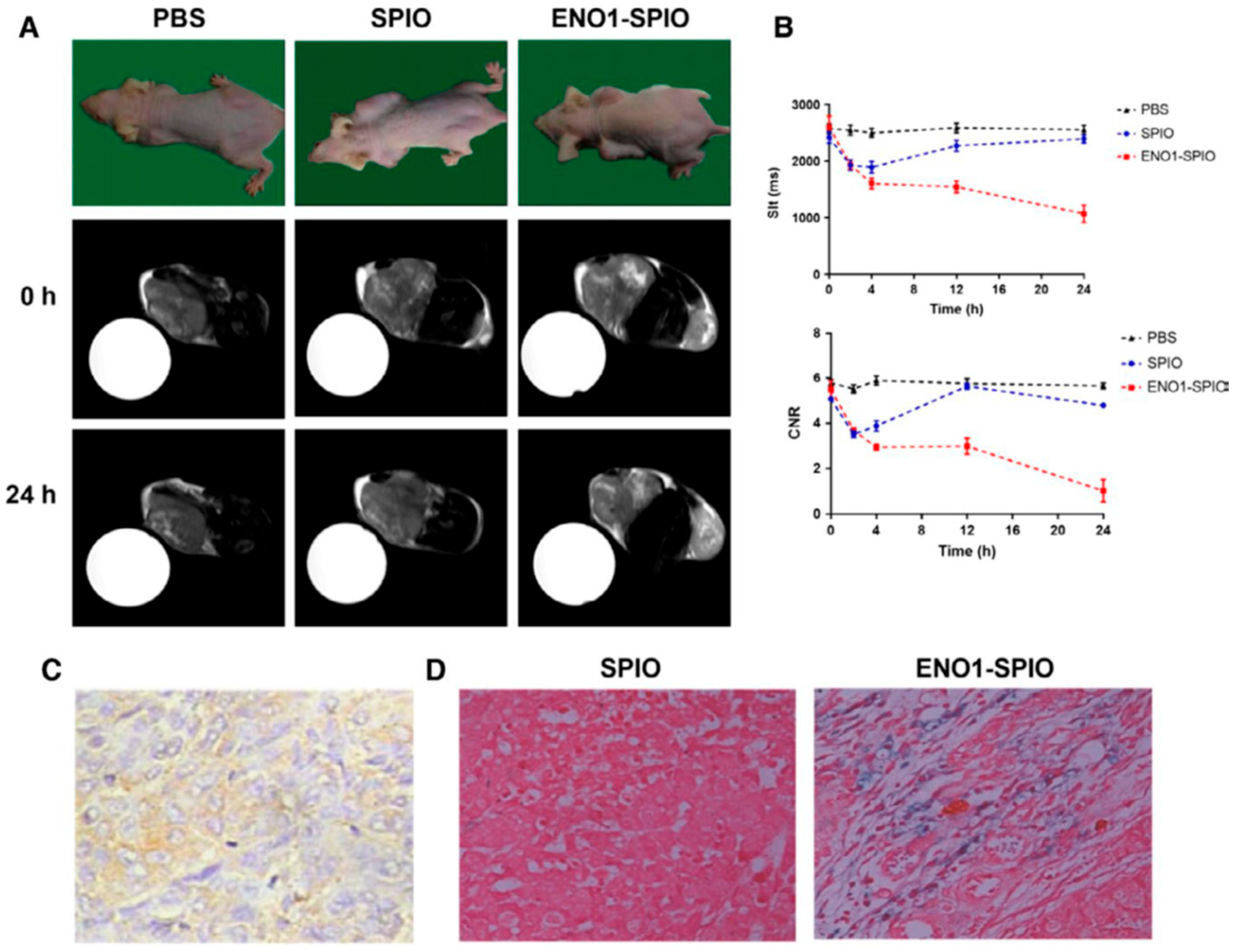
Figure 2.
Scheme of the use of biomarker-modified NPs to target and image PDAC.
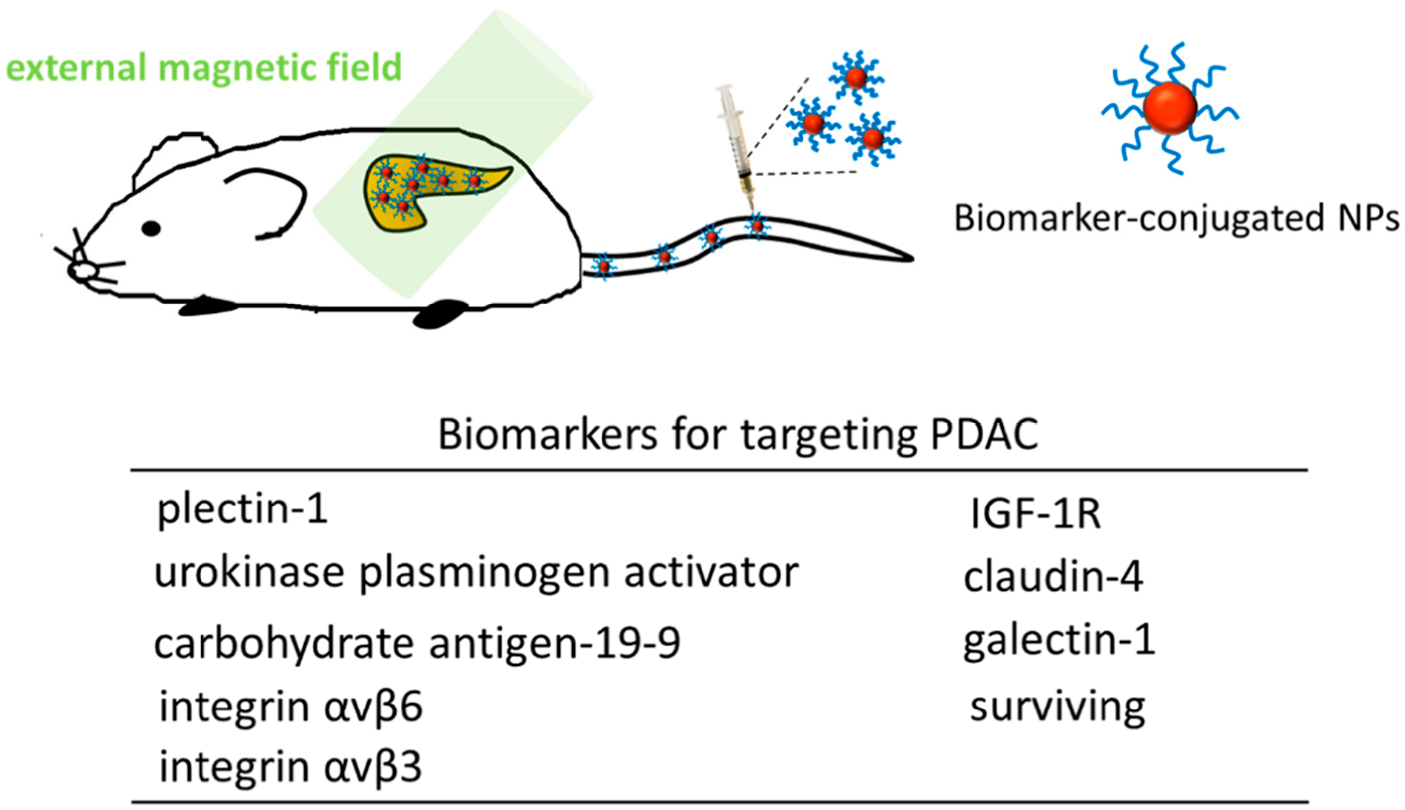

Table 1.
Iron-based MRI contrast agents for detection of pancreatic cancer.
| Type of NPs | Size | Strategy | Reference |
|---|---|---|---|
| Magnetofluorescent nanoparticles | ~39 nm | plectin-1 targeted peptides (PTP)-conjugated magnetofluorescent nanoparticles to detect PDAC | [11] |
| Superparamagnetic iron oxide nanoparticles (SPION) | SPION: 9-15 nm; Plectin-SPION-Cy7: 29 nm |
plectin-1 antibody-conjugated SPION to detect pancreatic cancer | [14] |
| Iron oxide nanoparticles (IONP) | BN-CLIO: 35 nm | BN peptide-nanoparticle conjugate (BN-CLIO) to target normal pancreas for imaging PDAC | [50] |
| Ultra-small superparamagnetic iron oxide (USPIO) | CKAAKN-USPIO: 96 nm | pancreatic cancer targeting peptide (CKAAKN)-functionalized USPIO to target pancreatic cancer cells | [11] |
| Iron oxide nanoparticles | IONPs-PEG-MCC triple scAbs: 24 nm | triple scAbs-conjugated IONPs to target pancreatic cancer | [51] |
| Fe3O4 nanoparticles | Fe3O4-PEG-Cy7-EMO: 27 nm | Fe3O4-PEG-Cy7-EMO to target pancreatic cancer | [12] |
| SPION | SPION: 5-10 nm; ENO1-targeted SPION: 30 nm |
ENO1-targeted SPION for detecting pancreatic cancer | [13] |
| SPIO | SPIO: 10 nm | uPAR-targeted SPIO to target pancreatic cancer | [38] |
| IONP | IONP: 10 nm; ATF-IONP-Gem: 66 nm | uPAR-targeted nanocomposites (ATF-IONP-Gem) to target pancreatic cancer | [7] |
| Fe(III) ions | PCN-Fe(III)-PTX NPs: 317 nm | PCN-Fe(III)-PTX NPs o target pancreatic cancer | [55] |
| IONP | anti-CD47 antibody-modified IONP: 107 nm | anti-CD47 antibody-modified IONP to target pancreatic cancer | [56] |
| IONP | IONP: 10 nm IGF-1-IONP: 17 nm |
human insulin-like growth factor1 (IGF1)-conjugated IONP to target pancreatic cancer | [10] |
Disclaimer/Publisher’s Note: The statements, opinions and data contained in all publications are solely those of the individual author(s) and contributor(s) and not of MDPI and/or the editor(s). MDPI and/or the editor(s) disclaim responsibility for any injury to people or property resulting from any ideas, methods, instructions or products referred to in the content. |
© 2025 by the authors. Licensee MDPI, Basel, Switzerland. This article is an open access article distributed under the terms and conditions of the Creative Commons Attribution (CC BY) license (http://creativecommons.org/licenses/by/4.0/).
Copyright: This open access article is published under a Creative Commons CC BY 4.0 license, which permit the free download, distribution, and reuse, provided that the author and preprint are cited in any reuse.


