
Submitted:
10 June 2025
Posted:
11 June 2025
You are already at the latest version
Abstract
This study investigates the influence of cardiac tissue uniformity on the accuracy of the Body Surface Potential Map (BSPM) generated from human heart simulations. A realistically shaped human torso was modeled using CT scan data, and the heart was reconstructed both as a volume matrix and a boundary surface. Two excitation propagation models were employed: the original Monodomain Reaction-Diffusion Equation (MD-RDE), assuming uniform conductivity, and a modified version accounting for non-uniform material properties. Comparative analysis of activation isochrones and BSPMs revealed slight differences, particularly during the QRS complex, although the overall impact of uniformity on BSPM accuracy was found to be minimal. These results suggest that while local differences exist, uniform and non-uniform models produce largely comparable BSPM results for many clinical and research purposes.
Keywords:
cardiac electrophysiology
; Body Surface Potential Map (BSPM)
; Monodomain Reaction-Diffusion Equation (MD-RDE)
; cardiac excitation
1. Introduction
Within the body, the heart is seen as an anisotropic volume source and an inhomogeneous volume conductor [1,2,3,4,5,6,7,8]. The electrical field produced by that volume source can be quantified on the body’s surface using the Body Surface Potential Map (BSPM). Numerous studies have examined the modeling of the cardiac activation BSPM [1,2,3,4,5,6,7,8], and it has been noted that the fidelity of the BSPM is significantly influenced by the accuracy and structure of the ventricular conduction network [9,10]. The impact of cardiac material uniformity on the computed BSPM is covered in this work.
2. Methods
2.1. The Human Torso and the Human Heart Modeling
Several organs have been modelled in a realistic human torso based on CT scans [11]. To describe the excitation propagation, the heart has been reconstructed in two ways: first, as a 3D volume matrix, and second, as a boundary surface for BSPM computations [12]. The Diffusion Volume scalar index was used to extract the human heart’s conduction network (Figure 1), which has been shown through a variety of methods to be reasonably close to the true conduction network [13,14].
2.2. Activation Isochrones Modeling
The Monodomain Reaction-Diffusion Equation (MD-RDE) and its modified version [15,16,17] are used to predict the excitation propagation of the heart. The heart was regarded as a non-uniform material in the modified version of the equation, while it was regarded as a uniform material in the original (Figure 2 and Figure 3).
According to the difference between the two activations [17], the non-uniform conductivity activation lags in the right ventricle free wall and leads in the left ventricle free wall and IV Septum. Since the right ventricle’s free wall is activated roughly 25 mSec later than the left ventricle in Durrer et al.’s activation isochrones [18], this is far more realistic than the uniform model.
3. Results
A comparison between the two models has been completed and shown as the Correlation-Coefficient (CC) and Relative-Error (RE). The Body Surface Potential Map (BSPM) has been constructed for both models [19], where the body is regarded an inhomogeneous volume conductor in both cases. The BSPM that is produced with the Original MD-RDE is regarded as the reference.
The CC and RE values were computed for 23 distinct time periods, all of which were separated from one another by roughly 5 mSec. Since the first and last time frames reflected the initial and final states of the depolarization, and because their values were equal to zero, they were eliminated. The peak value of the QRS complex is represented by frames 10 through 12. Prior to these frames (10 to 12), the wave is growing up, and following them, it is declining (as shown in Lead II, for instance, although in certain other leads, as Lead aVF, the center frame value is nearly equal to zero). The CC and RE between the non-uniform and uniform heart’s material are shown in Table 1, and Figure 4 demonstrates that the two created BSPMs differ slightly, particularly during the QRS complex’s building and decay. Therefore, it can be said that the modeled BSPM is not much impacted by the material consistency of the heart.
4. Conclusions
The comparison of uniform and non-uniform heart conductivity models reveals that although local differences in activation timing—particularly within the QRS complex—are evident, the overall BSPM output remains relatively stable. The findings indicate that assuming a uniform material model for the heart introduces only minor discrepancies in BSPM simulation, making it a viable simplification for many applications. However, for high-fidelity simulations or cases requiring detailed spatial accuracy, especially in the right and left ventricular regions, incorporating non-uniform conductivity may be beneficial.
References
- D.S. Farina, O. Skipa, C. Kaltwasser, O. Dossel and W.R. Bauer “Personalized Model of Cardiac Electrophysiology of a Patient” IJBEM (2005);7(1): 303-306.
- M. Seger “Modeling the Electrical Function of the Human Heart”, Ph.D. Thesis, Institute of Biomedical Engineering, University for Health Sciences, Medical Informatics and Technology, Austria (2006).
- M. Lorange, and R. M. Gulrajani “A computer Heart Model Incorporating Anisotropic Propagation” Journal of Electrocardiology, (1993);26(4):245-261.
- C. Hintermuller “Development of a Multi-Lead ECG Array for Noninvasive Imaging of the Cardiac Electrophysiology”, Ph.D. Thesis, Institute of Biomedical Engineering, University for Health Sciences, Medical Informatics and Technology,Austria, (2006).
- L. Cheng “Non-Invasive Electrical Imaging of the Heart”, Ph.D. Thesis, The University of Auckland,New Zealand (2001).
- B. He, and D. Wu “Imaging and Visualization of 3-D Cardiac Electric Activity” IEEE Tran. Inf Tech. Biomed. 2001; 5(3): 181-186. [CrossRef]
- Z. Liu, C. Liu, and B. He “Noninvasive Reconstruction of Three-Dimensional Ventricular Activation Sequence From the Inverse Solution of Distributed Equivalent Current Density” IEEE Trans. Med. Imag. (2006); 25(10): 1307-1318. [CrossRef]
- B. He, C. Liu ,and Y. Zhang “Three-Dimensional Cardiac Electrical Imaging From Intracavity Recordings” IEEE Trans. Biomed. Eng. (2007); 54(8): 1454-1460. [CrossRef]
- Elaff “Effect of the Conduction Network Structure of the Heart on Modeling of the Body Surface Potential Map and the ECG”. Preprints 2025, 2025060714. [CrossRef]
- R.L. Winslow, D.F. Scollan, J.L. Greenstein, C.K. Yung, W. Baumgartner, G. Bhanot, D.L. Gresh and B.E. Rogowitz “Mapping, modeling,and visual exploration of structure-function relationships in the heart” IBM Sys J.,(2001); 40(2):342-359. [CrossRef]
- ELAFF “Modeling of 3D Inhomogeneous Human Body from Medical Images”, World Journal of Advanced Engineering Technology and Sciences. 2025, 15(02): 2010-2017. [CrossRef]
- ELAFF “Modeling of the Human Heart in 3D Using DTI Images”, World Journal of Advanced Engineering Technology and Sciences, 2025, 15(02), 2450-2459. [CrossRef]
- IAI El-Aff “Extraction of human heart conduction network from diffusion tensor MRI” The 7th IASTED International Conference on Biomedical Engineering, 217-22.
- ELAFF “Modeling the Human Heart Conduction Network in 3D using DTI Images”, World Journal of Advanced Engineering Technology and Sciences, 2025, 15(02), 2565–2575. [CrossRef]
- ELAFF “Modeling of realistic heart electrical excitation based on DTI scans and modified reaction diffusion equation” Turkish Journal of Electrical Engineering and Computer Sciences: 2018, 26(3): Article 2. [CrossRef]
- ELAFF “Modeling of The Excitation Propagation of The Human Heart”, World Journal of Biology Pharmacy and Health Sciences, 2025, 22(02): 512–519. [CrossRef]
- ELAFF “Effect of the material properties on modeling of the excitation propagation of the human heart”, World Journal of Biology Pharmacy and Health Sciences, 2025, 22(3): 088–094. [CrossRef]
- D. Durrer, R.TH. D. Durrer, R.TH. Van Dam, G.E. Freud, M.J. Janse, F.L. Meijler and R.C. Arzbaecher “Total Excitation of the Isolated Human Heart” Circulation, (1970); 41(6):899-912.
- ELAFF “Modeling of the Body Surface Potential Map for Anisotropic Human Heart Activation”, Research Square, 2025.
Figure 1.
Conduction network of the human heart [14].
Figure 1.
Conduction network of the human heart [14].
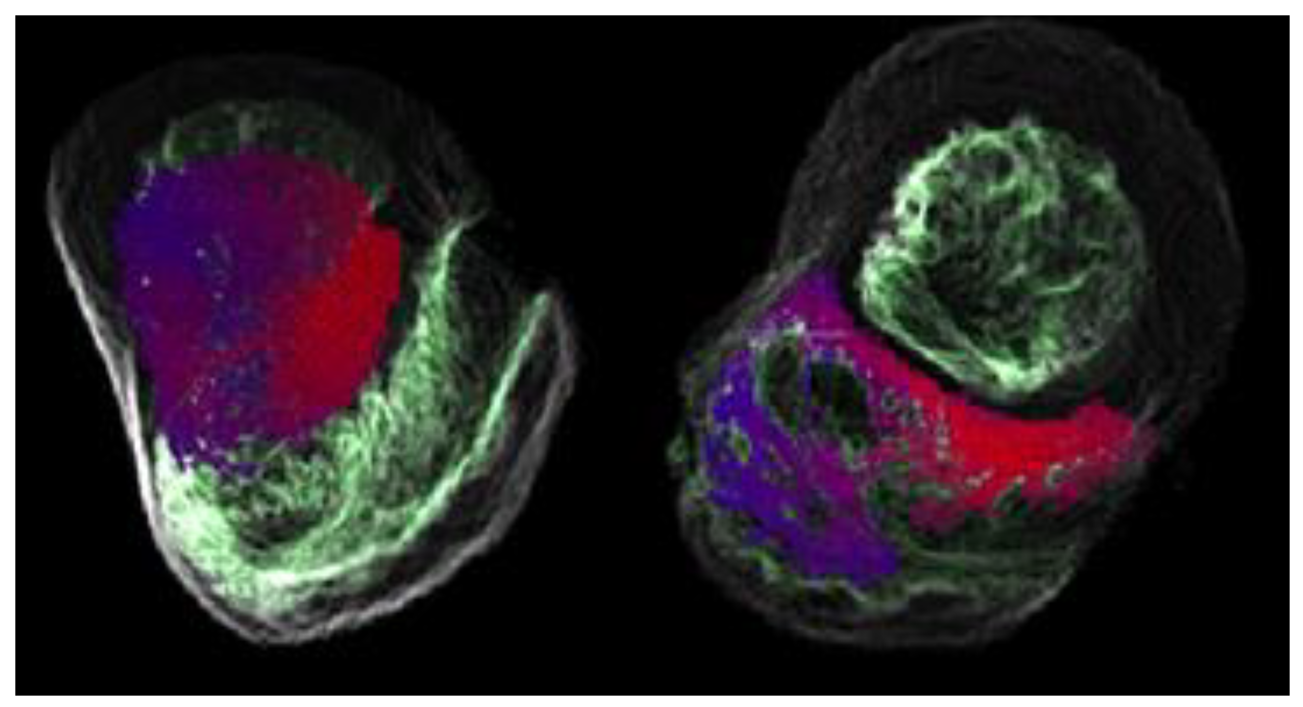
Figure 2.
Excitation Isochrones of Normal Activation using Original MD-RDE [17].
Figure 2.
Excitation Isochrones of Normal Activation using Original MD-RDE [17].
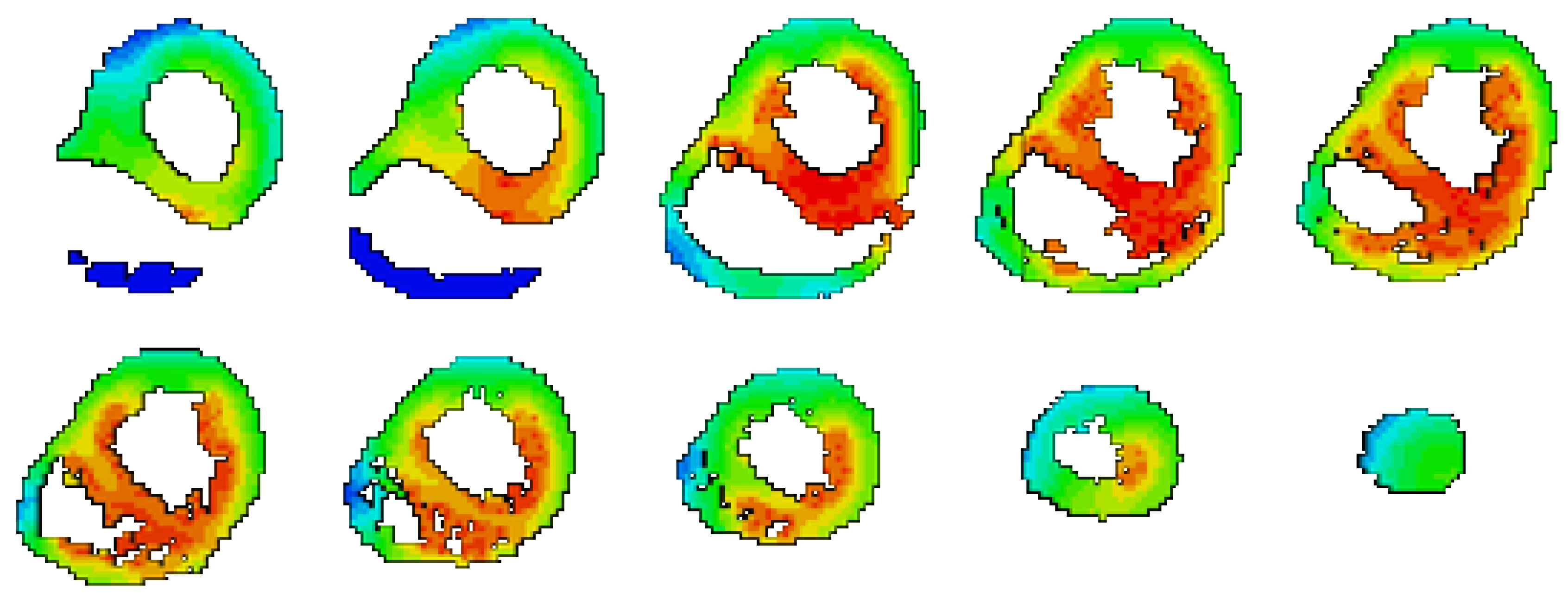
Figure 3.
Excitation Isochrones of Normal Activation using Modified MD-RDE [17].
Figure 3.
Excitation Isochrones of Normal Activation using Modified MD-RDE [17].
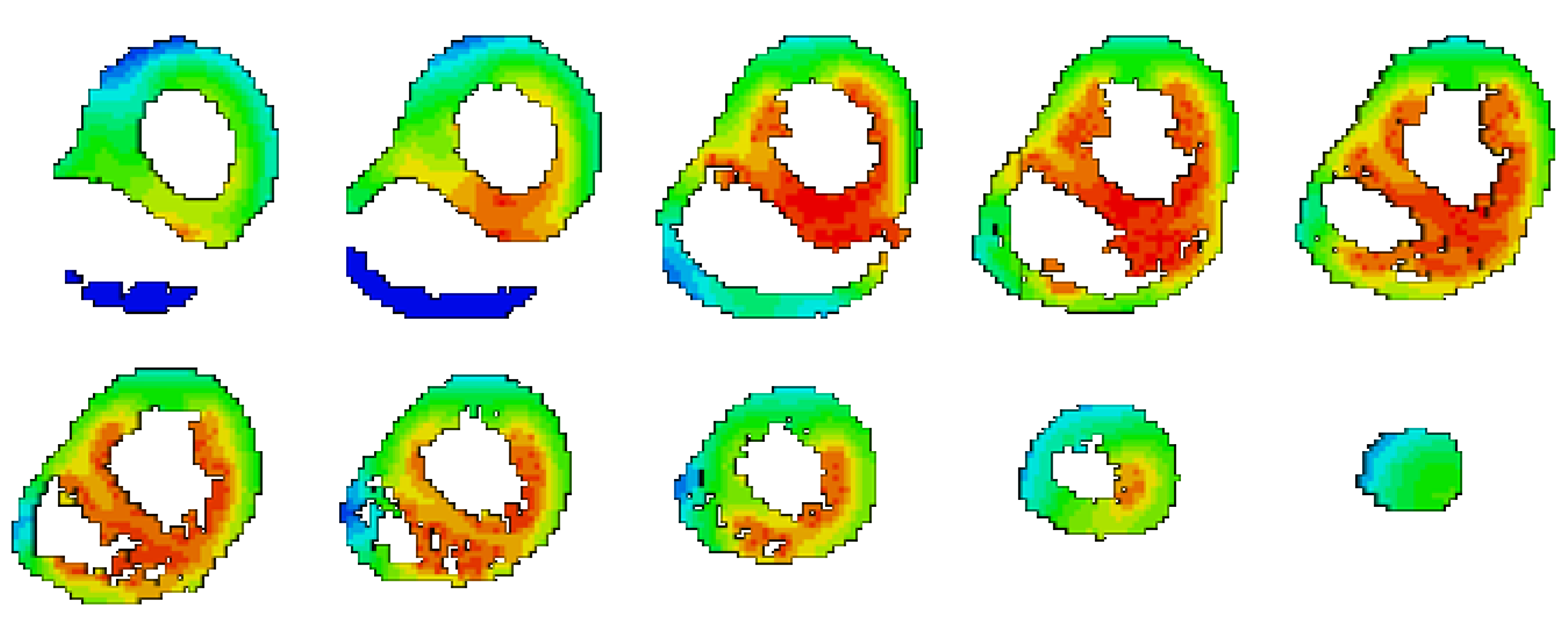
Figure 4.
CC and RE between the BSPMs which have been produced using the Non-Uniform (reference) and the Uniform heart’s material.
Figure 4.
CC and RE between the BSPMs which have been produced using the Non-Uniform (reference) and the Uniform heart’s material.
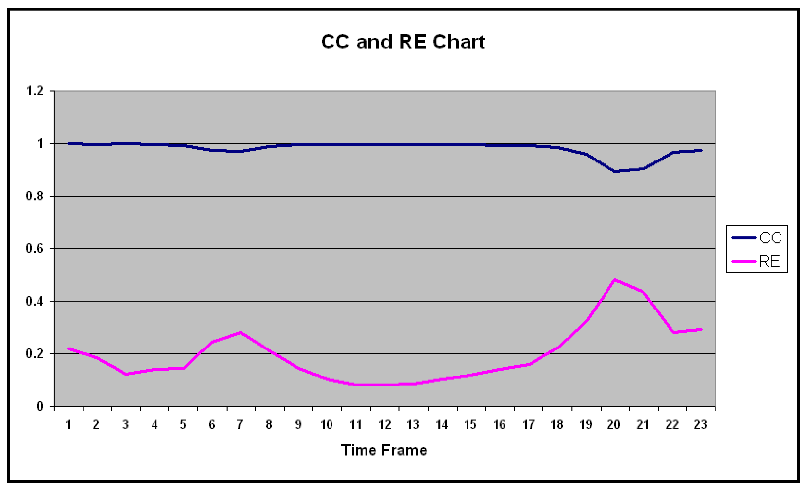

Table 1.
CC and RE between the Non-Uniform and the Uniform heart’s material.
| ID | CC | RE |
| 1 | 0.999 | 0.217 |
| 2 | 0.997 | 0.185 |
| 3 | 0.999 | 0.121 |
| 4 | 0.995 | 0.141 |
| 5 | 0.991 | 0.144 |
| 6 | 0.972 | 0.242 |
| 7 | 0.970 | 0.283 |
| 8 | 0.987 | 0.209 |
| 9 | 0.995 | 0.145 |
| 10 | 0.996 | 0.103 |
| 11 | 0.997 | 0.081 |
| 12 | 0.997 | 0.080 |
| 13 | 0.996 | 0.084 |
| 14 | 0.995 | 0.102 |
| 15 | 0.994 | 0.118 |
| 16 | 0.994 | 0.140 |
| 17 | 0.992 | 0.159 |
| 18 | 0.986 | 0.223 |
| 19 | 0.960 | 0.321 |
| 20 | 0.891 | 0.481 |
| 21 | 0.904 | 0.434 |
| 22 | 0.965 | 0.281 |
| 23 | 0.975 | 0.291 |
| Mean | 0.980 | 0.199 |
| SD | 0.027 | 0.106 |
Disclaimer/Publisher’s Note: The statements, opinions and data contained in all publications are solely those of the individual author(s) and contributor(s) and not of MDPI and/or the editor(s). MDPI and/or the editor(s) disclaim responsibility for any injury to people or property resulting from any ideas, methods, instructions or products referred to in the content. |
© 2025 by the authors. Licensee MDPI, Basel, Switzerland. This article is an open access article distributed under the terms and conditions of the Creative Commons Attribution (CC BY) license (http://creativecommons.org/licenses/by/4.0/).
Copyright: This open access article is published under a Creative Commons CC BY 4.0 license, which permit the free download, distribution, and reuse, provided that the author and preprint are cited in any reuse.


